Embed Size (px)
Citation preview

EXTRACORPOREAL LIFE SUPPORT ORGANIZATION29th ANNUAL ELSO CONFERENCE ABSTRACTS
SCOTTSDALE, AZSeptember 13-16, 2018
Author Index .................................................................................................... Pages 87–92
Published in cooperation with the ASAIO Journal and available online at www.asaiojournal.com.

2018 ELSO Meeting Abstracts Table of Contents
Outcomes of Pediatric Extracorporeal Life Support Patients with Bronchopulmonary Dysplasia and Associated Pulmonary Hypertension ...1
Outcomes After Long Veno-Venous ECMO Runs: A Case Series .................2
Traumatic Brain Injury is not a Contraindication To Veno-Venous Extracorporeal Membrane Oxygenation ....................................................2
V-V ECMO for Chlorine Inhalation Injury ....................................................3
CASE REPORT: Successful V-A-V ECMO Treatment in a patient with ARDS and Cardiogenic shock caused by Streptococcus pyogenes .......................3
Geriatric Extracorporeal Life Support has Acceptable Mortality and Discharge Outcomes ................................................................................... 4
A Retrospective Review of the Use of a Dual Oxygenator Configuration for VV and VA ECMO .................................................................................. 4
Distal Perfusion Monitoring in the Femorally Cannulated VA ECMO Patient ........................................................................................ 5
Veno-Venous Extracorporeal Life Support To Facilitate Airway Foreign Body Removal In A Child With Severe Respiratory Failure .........................6
Incidence and risk factors of serious neurologic injury in pediatric survivors of extracorporeal life support .....................................................7
Fixed and Dilated Pupils: Is ECMO a consideration? ...................................7
The learning curve of multidisciplinary team setups for venovenous extracorporeal membrane oxygenation in acute respiratory failure ..........8
Outcomes of Veno-Venous Extra-Corporeal Membrane Oxygenation when Stratified by Age – Is There a Point of No Return? ...........................8
Quality of life and functional status of patients treated with Venovenous Extracorporeal Membrane Oxygenation at 6 Months ................................. 9
Novel 6 Months Survival Prognostication Tool for Venovenous Extracorporeal Membrane Oxygenation ....................................................9
Long Term VV ECMO Support and Repeat Peripheral Cannulation of a Neonate with Necrotizing MRSA Pneumonia and Complicated Empyema .................................................................................................. 10
Correlation of Anti-Xa Levels and Thromboelastography R Times in Patients on Extracorporeal Life Support ...................................................11
Case Report. First-Presentation Status Asthmaticus Requiring Extracorporeal Cardio Pulmonary Resuscitation: Severe unresponsive asthma or delayed ECLS? .......................................................................... 12
Echocardiography as a Key Component in the Treatment of Adult Veno-Venous Extra-Corporeal Membrane Oxygenation Patients: A Review of Two Vanderbilt University Medical Center Adult Patients. ...13
Outcome Analysis of Percutaneous Veno-Arterial Extracoroporeal Memebrane Oxygenation as Bridge to Durable Left Ventricular Assist Device ....................................................................................................... 13
The Journey to ECMO could start with a single VAPE: A case of severe hypersensitivity pneumonitis in a pediatric patient .................................14
Admistering Inhaled Alteplase (Tpa) as an Airway Clearance Adjunct After Pulmonary Hemorrage Causing Bronchial Blood Clots With Mucus Plugging in Pediatric Patient During Ecmo ...............................................14
Fungus Among Us: A case series of two pediatric patients diagnosed with invasive fungal infections after Extracorporeal Membrane Oxygenation. ............................................................................................ 15
Multidisciplinary Team Approach for Early Initiation of Extracorporeal Life Support for a Patient with Status Asthmaticus, Critical Airway, and Mucopolysaccharidosis. ........................................................................... 15
Complex Management of Airway Bleeding on Veno-venous Extracorporeal Membrane Oxygenation ..................................................16
Veno-Venous Extracorporeal Membrane Oxygenation for Acute Respiratory Distress Syndrome Secondary to Choriamnionitis ................16
The Use of Venovenous Extracorporeal Membrane Oxygenation in Patients with Pulmonary Embolism..........................................................17
Implementation of a Comprehensive ECMO Management Protocol Improves Survival among Patients with Severe ARDS ..............................17
Early nutritional support is not associated with survival in adult patients receiving veno-venous extracorporeal membranous oxygenation ..........18
Malnutrition is associated with veno-venous extracorporeal membrane oxygenation duration and survival to discharge .......................................18
Direct Measurement of Blood Gases in Tubing with Resonance Raman Spectroscopy ............................................................................................ 19
A Multi-Discipline Approach to Early Extubation and Mobility for Patients on Veno-Venous Extracorporeal Membrane Oxygenation: A Case Series ............................................................................................ 20
Veno-Venous Extracorporeal Membrane Oxygenation in a Jehovah’s Witness patient with acute respiratory distress syndrome secondary to pneumonia and pulmonary embolism .....................................................20
VV ECMO, Rapid Extubation, and Pulse Steroids for Near Fatal Asthma ............................................................................................. 21
Congenital alveolar capillary dysplasia - a rare cause of persistent pulmonary hypertension of the newborn ................................................21
Development of a Comprehensive Intramural ECLS Specialist Training Program: No Place like home ...................................................................22
Comparison of bivalirudin versus heparin-based anticoagulation for extracorporeal membrane oxygenation ...................................................22
A free standing ECMO department: The benefits?...................................23
Combination of ECMO and cytokine adsorption therapy for patients with pneumogenic sepsis and severe respiratory failure .........................24
Cytokine removal using the CytoSorb® system in combination with extra corporeal membrane oxygenation: a review of the literature. .......24
Efficacy of postoperative inspiratory muscle training during ECMO therapy in patients with advanced pulmonary emphysema after lung volume reduction surgery ........................................................................ 25
Successful Treatment of Severe Respiratory Failure HIV patients with Veno-Venous Extracorporeal Membrane Oxygenation: not a myth anymore ................................................................................................... 26
Size Matters: Impact of Circuit Size on Coagulation and Hemolysis Complications in Pediatric ECMO Patients ...............................................27
Evaluation of Serotonin Release Assay and ELISA Optical Density Tests in Patients Receiving Extracorporeal Membrane Oxygenation .......28
Mechanisms of platelet loss during pediatric extracorporeal life support (ECLS) .......................................................................................... 28
Pediatric ECMO cannula complications: An under-reported source of ECMO morbidity ....................................................................................... 29
Neuromonitoring in Neonatal Extracorporeal Membrane Oxygenation Patients ..................................................................................................... 29
Outcomes of ECMO During CPR in Adult In-Hospital Cardiac Arrest ........30
Survival in Adult Venovenous and Venoarterial Extracorporeal Membrane Oxygenation Patients Compared to International Survival Prediction Scores: A Single Center Retrospective Review ........................30
Factors Associated With Survival Following Extracorporeal Cardiopulmonary Resuscitation In Children .............................................31

2018 ELSO Meeting Abstracts Table of Contents
Implementation of a Pediatric ECPR Program: Overcoming Challenges and Recognition of Safety Threats .........................................31
Trends in Cardiogenic Shock Treatment in an Institution New to Mechanical Circulatory Support ...............................................................32
Development of a mobile adult ECMO program ......................................32
ECMO for trauma patients: the experience at a Level 1 trauma center ....................................................................................................... 33
Beating the Odds: A Case report of long term ECMO support as a bridge to successful heart transplant in a 2-year-old ventilator dependent child with Glenn physiology .............................................................................. 33
Assessment of Cardiac Recovery Using a Formal Stepwise Weaning Protocol in Patients Supported with Venoarterial Extracorporeal Membrane Oxygenation ........................................................................... 34
Clinical Management after Carotid Artery Repair – an Opportunity for Improvement? .......................................................................................... 34
Outcomes of ECLS for Postcardiotomy Syndrome in the Modern Era ......35
Venoarterial Extracorporeal Membrane Oxygenation Via Carotid Arteryf “Jump Graft”: Friend or Foe? A Case Series .................................36
Cardiac Catheterization Snare Technique To Correct A Malpositioned Avalon Elite® Cannula Without Interruption of Veno-Venous Extracorporeal Life Support In Patient With Unilateral Pulmonary Agenesis ................................................................................................... 37
Different Transfusion Targets for VA-ECMO Patients and Survival: Outcomes at University Hospital Programs - One in U.S. and One in China ..................................................................................................... 38
The Minnesota Resuscitation Consortium’s Advanced Perfusion and Reperfusion Cardiac Life Support Strategy for Out-of-Hospital Refractory Ventricular Fibrillation. ...........................................................39
Surviving Severe Hypothermia with ECLS - 150 Minutes of CPR before ECLS - A Case Review ................................................................................ 39
Extremity Perfusion on ECMO ..................................................................40
LAVA-ECMO for Acute Myocardial Infarction with Profound Cardiogenic Shock .................................................................................... 41
Percutaneous Tibial Access for Distal Perfusion During VA ECMO: Protection Without Bleeding .................................................................... 42
Totally Percutaneous Cannulation and Decannulation for Femoral VA ECMO........................................................................................................ 42
Case report: ECMO, percutaneous thrombectomy, and ultrasound-accelerated catheter-directed thrombolysis use for persistent right heart failure after massive pulmonary embolism ....................................43
A Case Series of Arterial Cannula Thrombosis in Pediatric Extracorporeal Life Support ...................................................................... 43
Swan-Ganz catheter guided VA ECMO wean leads to successful decannulation ........................................................................................... 44
Extracorporeal Membrane Oxygenation Education and Simulation Training in The Cardiothoracic Intensive Care Unit ..................................45
Mechanical Ventilation in Children on V-V ECMO ....................................45
ECMO GO: Implementing a Multidisciplinary Bedside Cannulation Process ..................................................................................................... 46
How to Care for an ECMO Patient- Bedside Nurse Perspective For Better, for Worse- Recognizing Pivotal Clinical Changes in the ECMO Patient .....46
Survival in Heart Transplant Recipients Requiring Extracorporeal Membrane Oxygenation for Primary Graft Dysfunction ...........................47
Comparison of Survival Indicators between Myocardial Infarction Patients and Septic Patients Who Received Veno-Arterial Extracorporeal Membrane Oxygenation Treatment .........................................................47
Management Strategies during a Challenging VA ECMO Run in a Neonate with Septic Shock and Hypoxic Ischemic Encephalopathy: A Case Report ........................................................................................... 48
A Challenging VA ECMO Wean in an Infant with Severe Ebstein’s Anomaly: Finding the Right Balance for Preload- and Afterload- Dependent Pulmonary Blood Flow ............................................................................. 49
Clinical Outcomes Associated with Sedation and Analgesia Management in Patients Supported with Venoarterial Extracorporeal Membrane Oxygenation ............................................................................................. 50
A Call for a Standardized Multi-disciplinary Follow Up Framework for Pediatric Extracorporeal Life Support (ECLS) Survivors; longitudinal follow up to 17 years ................................................................................ 51
Cardiogenic Shock – An Indian Scenario – A Single Centre Experience .................................................................................... 52
Correlation amongst hemolysis biomarkers in patients undergoing extra-corporeal membrane oxygenation (ECMO) ....................................53
Case Report: Extracorporeal Membrane Oxygenation (ECMO) and Emergent Laparotomy used to salvage Massive Pulmonary Embolism patient with cardiac arrest and CPR-induced liver laceration ..................53
Brain natriuretic peptide for predicting mortality in patients with cardiogenic shock who successfully weaned extracorporeal membrane oxygenation ........................................................................... 54
Evaluation of severity of illness scores in the pediatric ECMO population ..................................................................................... 54
State of the Art SHOCK Team: Feasibility and Effectiveness .....................55
Single Center Experience with Impella as LV Venting in VA ECMO ...........55
Protek Duo in VA ECMO; No Need for Venting .........................................56
Does age-adjusted Charlson comorbidity index influence hospital survival and short-term outcome of patients with extracorporeal cardiopulmonary resuscitation? ...............................................................57
A Place to Start: Making Care More Affordable for ECLS Patients ............57
Antimicrobial Management in Extracorporeal Membrane Oxygenation: The AMMO study ..................................................................................... 58
Poster Abstract: Gentamicin dosing in paediatric ECMO: should we be using higher dosages in this population? ...........................................58
Caring for Caregivers: Impact of Supportive Resources on Critical Care ECMO Nurses ............................................................................................ 59
Simulating Nothing: Recreating the Mundane to Improve ECMO Competency ............................................................................................. 60
The Impact of a Structured ECMO Program in a Low Volume ECMO Center ............................................................................................ 61
Oxidative Stress in Extracorporeal Membrane Oxygenation Circuits .......62
Outcomes of children <10kg supported with Extracorporeal Membrane Oxygenation using centrifugal versus roller blood pumps: A Review of the ELSO Registry ................................................................................. 62
ECMO Specialist led Run-Review – an adjunct to Morbidity and Mortality Conference ............................................................................... 63
Institutional Experience Using The Cardiohelp® System for Neonatal and Pediatric Ecmo ...................................................................64
Massive Air De-priming: Simulation Training ...........................................65

2018 ELSO Meeting Abstracts Table of Contents
The Metabolic Demands of Extracorporeal Membrane Oxygenation in Infants and Children ................................................................................. 77
Incidence of Heparin-Induced Thrombocytopenia in Patients with Temporary Mechanical Circulatory Support Devices ................................78
Implementing a Multidisciplinary and Multimodal Approach to an ECLS Specialist Program ............................................................................ 78
Managing severe accidental hypothermia with and without ECLS: Patient outcomes over eleven years from a single center ........................79
Red Book Based Ecmo Education for Bedside Icu Nurses .........................79
Does Pre-Procedural pH Affect Outcomes in Veno-Arterial Extracorporeal Membrane Oxygenation Patients with Myocardial Infarction? ................80
[Poster] Clinical Validation of Simulated Nirs Sro2 Values During Veno-Arterial Ecls with Peripheral Cannulation........................................80
A Practice Change Based on Necessity: A Single Center’s Five-Year Experience Using Plasmalyte Pre-Primed Circuits up to Sixty Days Old ......81
Implementation and Evaluation: The Use of Rapid Cycle Deliberate Practice Simulation Techniques ................................................................81
Standardizing the Delivery of ECMO across a Large, Academic Medical Center ....................................................................................................... 82
Successful Use of Extracorporeal Membrane Oxygenation in the Reversal of Cardiorespiratory Failure Induced by Fulminant Myocarditis: Case Reports ........................................................................ 82
DNP Project: Reducing Adverse Events During Extracorporeal Membrane Oxygenation with In-Hospital Advanced Extracorporeal Life Support Specialists ............................................................................. 83
ECMO and Controlled Hypothermia in Neonates with Hypoxic Ischemic Encephalopathy and Persistent Pulmonary Hypertension: A Single-Center Case Series ...................................................................... 83
A model for a multidisciplinary approach to initiating an ECMO program in a small facility ........................................................................ 84
Institutional experience of long-term VA ECMO support in children..................................................................................................... 84
We train with dummies so you don’t have to – Using simulation as the foundation for a rescue ECLS program .....................................................85
Developing a Rescue ECLS program at a Non-Cardiac Surgical Children’s Hospital .................................................................................... 85
Relative Hyperoxia in Cyanotic Congenital Heart Disease Patients on Veno-Arterial Extracorporeal Life Support is Associated with Decreased Survival to Hospital Discharge ................................................86
Occipital Offloading in the ECMO patient. ...............................................65
Rejuvesol Ex Vivo ECMO Study: Evaluation of Hemolysis, p50, and Red Cell Fragility in ex vivo Extracorporeal Membrane Oxygenation Circuit with Rejuvesol Incubated Red Blood Cells ....................................66
Intraocular Bleeding on ECMO .................................................................67
Does Selective Use of Inhaled Nitric Oxide Affect The Rate of ECMO In Higher Severity CDH? ............................................................................... 68
Successful use of bivalirudin in pediatric and neonatal ECMO .................68
The use of Extracorporeal Membrane Oxygenation (ECMO) in Refractory Pediatric Cardiac Arrest ..........................................................69
Prospective Observational Experience with Bivalirudin Anticoagulation in Pediatric Extracorporeal Membrane Oxygenation ...................................69
A collaborative approach to a challenging ECMO case .............................70
Development of an ECMO Patient Acuity Tool .........................................71
Risk Factors for Acute Kidney Injury in Hantavirus patients treated with ECMO ............................................................................................... 71
Impact of Bleeding and Thrombosis Events on ECLS Mortality: An ELSO Analysis ...................................................................................... 71
The Effect of Oxychlorosene Foley Irrigation on Catheter Associated Urinary Tract Infections (CAUTIs) in ECMO Patients .................................72
Flowcytometric evaluation of immune cells in experimental ECMO model ....72
Pilot Study to Evaluate a Non-Titrating, Weight Based Anticoagulation Scheme for Patients on Veno-Venous Extracorporeal Membrane Oxygenation ............................................................................................. 73
Anticoagulation management guided by Anti-Xa in neonatal and pediatric ECMO ........................................................................................ 73
ECMO survivors’ quality of life and needs after discharge: A descriptive cross-sectional pilot study ........................................................................ 74
Relationship between ECMO center volume and ECMO-related blood product costs ............................................................................................ 74
Anticoagulation review in patients less than 1 year old requiring extracorporeal support............................................................................. 75
Anticoagulation Management during ECMO in Children with Massive Bleeding .................................................................................................... 75
Development of Validated Checklists to Evaluate Clinical Specialists in Pediatric ECMO Emergencies Using Delphi Methods ...............................76
Predicting Oxygenator Failure: A Novel Oxygenator Performance Index ..... 76
Extracorporeal Cardiopulmonary Resuscitation in the Catheterization Laboratory is Associated with Superior Results ........................................77
1
2018 ELSO MEETING ABSTRACTS
Outcomes of Pediatric Extracorporeal Life Support Patients with Bron-chopulmonary Dysplasia and Associated Pulmonary HypertensionAlejandra Pena1, Nicholas Carr2, Veronica Armijo-Garcia1, Donald McCurnin1 1University of Texas Health Science Center San Antonio Joe and Teresa Lozano Long School of Medicine, San Antonio, Texas; 2Brooke Army Medical Center, San Antonio, Texas
Introduction: Evidence based practice is limited by a paucity of existing resources regarding the eligibility of patients with a history of prematurity and bronchopulmonary dysplasia (BPD) for extracorporeal life support (ECLS) as a rescue therapy for respiratory failure. A retrospective review published in 2001 reported 76 cases of infants less than 1 year old that were treated with ECLS and had a pre-existing diagnosis of BPD (1). The survival to discharge of these infants was reported to be 78%, compa-rable to or better than other respiratory ECLS outcomes at that time (1)(2). BPD definition and management strategies have changed over time making prediction of outcomes a challenge. However, it is estimated that co-existing pulmonary hypertension (PH) occurs in 17% of BPD cases (3) and patients with concomitant BPD and PH (BPD+PH) have substantially higher morbidity and mortality compared to patients with BPD alone, with mortality reported as high as 50% (4).Aim: The purpose of this study is to examine if a prior diagnosis of BPD+PH is associated with decreased survival of pediatric patients receiv-ing ECLS when compared to BPD alone in respiratory ECMO cases. Sec-ondary outcomes evaluated mortality from a contemporary cohort from 2012 to 2016, ECLS duration, mode of ECMO, and ECLS complications.Methods: A retrospective review of the ELSO registry was performed from January 1982 through June 2016 for pediatric patients (2 months to 18 years) with a prior diagnosis of BPD (ICD-9 code 770.7) receiving ECLS support for non-cardiac indications. Standardized data from the contributing ELSO centers included patient demographics, diagnoses, ECLS technique, complications, and outcomes. The ICD-9 code 416.8 was used to identify the patients with secondary PH. Patients with a prior diagnosis of congenital heart disease, congenital diaphragmatic hernia, congenital anomalies of airway or lungs (excluding tracheoesophageal fistula), without a valid primary diagnosis available, or required additional ECMO run(s) were excluded. Survival was categorized both by transition off ECLS support and survival to hospital discharge. Descriptive statistics were used for patient characteristics. Fisher’s exact test was used for the primary outcome and for all nominal data. Student-t test was used to ana-lyze continuous data. A multiple logistic regression analysis was used to identify a correlation of survival with PH and other categorical variables. A p value of <0.05 was considered significant.Results: 260 pediatric patients with BPD were identified from the ELSO registry meeting inclusion criteria and 13 (5%) of them had associated PH. Of the 247 patients in the BPD alone group 49.4% were males and 48.2% were females, there was no data on 6 patients. Of the BPD+PH group 46.2% were males and 53.8% were females. 11 (84.6%) of the BPD+PH patients received ECLS after 2012 and there were no reported cases of BPD+PH prior to 2004. The age of patients with BPD was 369
days ± 612 days (mean ± SD) and patients with BPD+PH 298 days ± 127 days (mean ± SD). Pre-ECMO oxygenation index (OI) for BPD patients was 48.5 ± 32.3 (mean ± SD) and BPD+PH was 54.7 + 14.5 (mean ± SD). The overall survival rate was 69.6% in all BPD patients and 76.9% in BPD+PH patients (p=0.759). Accounting for a contemporary review of patients from 2012 to 2016, 63 patients with BPD were treated with ECLS, there was no difference in survival to discharge (BPD 73.0% vs. BPD+PH 72.7%), and no difference in duration of ECLS support (BPD 247.2 hours ± 163 hours mean ± SD vs. BPH+PH 232.8 hours ± 186.9 hours mean ± SD). A multivariate logistic regression also failed to show a correlation between survival and PH, weight, gender, duration of ECMO, or mode of ECMO support. Mode of ECMO was reported as a similar use of VA and VV ECMO for BPD patients (51.9% and 42.3% respectively) and predomi-nant use of VV ECMO for patients with BPD+PH (63.6%). Only 3 patients with the BPD group were crossed from VV to VA ECMO and none in the BPD+PH group. Most common ECLS related complications were hyperten-sion requiring vasodilators (BPD 38.5%, BPD+PH 27.3%), inotrope use on ECLS (BPD 34.6%, BPD+PH 45.5%), and hemofiltration requirement (BPD 21.1%, BPD+PH 36.4 %). Neurologic and pulmonary complications were uncommon in both groups presenting in <10% of the cases.Conclusions: Data from the ELSO registry demonstrates reasonable survival in either patients with BPD alone or BPD+PH. Limited retrospec-tive data suggests that patients with BPD+PH have similar OI prior to rescue ECLS, as well as similar morbidity and mortality as BPD alone cases. Patients with BPD+PH may benefit from ECLS management as a recue therapy for respiratory failure. Prospective evaluation of patients with BPD and associated severe morbidities like PH is needed to better characterize outcomes and complications.
References:1. Hibbs A, Evans JR, Gerdes M, Hunter JV, Cullen JA. Outcome
of Infants with Bronchopulmonary Dysplasia who Receive Extracorporeal Membrane Oxygenation Therapy. J Pediatr Surg. 2001 Oct;36(10):1479–84.
2. ECMO Registry of the Extracorporeal Life Support Organization (ELSO), Ann Arbor, Michigan, July, 2017.
3. An HS et al. Pulmonary Hypertension in Preterm Infants with Bronchopulmonary Dysplasia. Korean Circ J. 2010 Mar;40(3):131–136.
4. Bui CB et al. Pulmonary Hypertension Associated with Bronchopulmonary Dysplasia in Preterm Infants. Journal of Reproductive Immunology. 2017 Nov, volume 124. Pages 21–29, ISSN 0165-0378.
Disclosure: The view(s) expressed herein are those of the author(s) and do not reflect the official policy or position of Brooke Army Medical Center, the U.S. Army Medical Department, the U.S. Army Office of the Surgeon General, the Department of the Air Force, Army, Navy, Depart-ment of Defense or the U.S. Government.Alejandra Pena, [email protected]

2018 ELSO MEETING ABSTRACTS
2
Outcomes After Long Veno-Venous ECMO Runs: A Case SeriesAndrew Vasyluk, MD, Narra S. Reddy, MD, Anthony A. Iacco, MD, Felicia A. Ivascu, MD Oakland University William Beaumont, Department of General Surgery. Royal Oak, MI
The average duration for veno-venous extracorporeal membrane oxygen (ECMO) support in the United States is 12 days (294 hours). The data is unsettled as to whether support longer than 14 days is associated with inferior outcomes. There is little outcomes data for patients requiring support greater than 50 days of VV-ECMO. We report our institutional experience with 3 patients who required veno-venous ECMO support for greater than 50 days. The mean age of our cohort was 60 years. Etiolo-gies of respiratory failure included ARDS due to legionella pneumonia, Influenza A, and one case of ARDS of unknown etiology despite extensive workup. The mean pre-ECMO PaO2/FiO2 ratio was 54 mm Hg. All subjects were cannulated with a right internal jugular 31 French dual-lumen Avalon cannula (Getinge). Two patients were anti-coagulated with arg-atroban infusions and one with a heparin infusion. The mean duration of VV-ECMO was 54 days. All three patients required a circuit change after a mean of 43 days of ECMO due to failure of the oxygenator. Bleeding complications occurred in 2 patients (66%), including a large intra-paren-chymal pulmonary hemorrhage and severe intra-bronchial bleeding. All three (100%) subjects survived to hospital discharge, which occurred at a mean of 85 days of hospitalization. All patients are off of supplemental oxygen and functioning at their pre-hospital baseline at a mean duration of 14 months follow up. This series appears to support available data that suggest ECMO support of 50 days or more can be associated with compa-rable survival rates to shorter runs with reasonable long term outcomes.Corresponding author: Andrew Vasylukemail: Andrew.vasyluk @beaumont.org
TRAUMATIC BRAIN INJURY IS NOT A CONTRAINDICATION TO VENO-VENOUS EXTRACORPOREAL MEMBRANE OXYGENATIONBrandon M. Parker DO1, Jay Menaker MD1, Cherisse D. Berry MD2, Ronald B Tesoriero MD1, James V O’Connor MD1, Deborah M. Stein MD MPH1, Thomas M. Scalea MD1 1R Adams Cowley Shock Trauma Center, University of Maryland Medical Center, Baltimore, Maryland; 2Department of Surgery, New York University Langone Health, New York, New York
Introduction: Veno-venous extracorporeal membrane oxygenation (VV ECMO) may improve survival in trauma patients with respiratory failure. However, many consider traumatic brain injury (TBI) an absolute contraindication for VV ECMO as systemic anticoagulation could worsen intracranial injury. We evaluated outcomes and complications in patients with TBI treated with VV ECMO.Methods: We retrospectively reviewed TBI patients ≥ 18 years of age admitted between January 1st 2007 and December 31st 2017 treated with VV ECMO. Demographics, injury specific data, pre-ECMO and ECMO data were collected. The primary outcome was survival to discharge. Secondary outcomes included progression of intracranial hemorrhage, bleeding complications, and episodes of oxygenator thrombosis requir-ing exchange. Medians and interquartile range were reported where appropriate.Results: 13 patients with TBI received VV ECMO support during the study period. The median age was 28 years old [IQR 25–37.5] and 85% were male. Median admission GCS was 5 [IQR 3–13.5]. Median injury severity score (ISS) was 48 [IQR 33.5–66]. Median head AIS was 4 [IQR 3.5–4.5] with median head AIS 3 [IQR 3–4] in survivors. Median pre-ECMO PaO2:FiO2 ratio was 58 [IQR 47–74.5]. Median time from injury to VV ECMO cannulation was 5 days [IQR 0.75–13]. Median time on ECMO was 192 hours [IQR 48–384]. Five (38.4%) patients survived to hospi-tal discharge. Cause of death included: multisystem organ failure in 4, removal of life sustaining therapy in 3 and death by neurologic criteria in 1. 6 patients (46%) received systemic anticoagulation (A/C). No patient had worsening of intracranial hemorrhage on CT. 1 patient was diagnosed with TBI after initiation of VV ECMO. There were two minor bleeding complications in patients on A/C, neither was related to TBI. 4 patients required oxygenator change; 2 in patients on A/C.Conclusion: VV ECMO is safe in patients with TBI and can be used without A/C in high risk TBI patients without increased oxygenator thrombosis. TBI is not a contraindication to the use of VV ECMO in severe respiratory failure.Brandon Masi Parker DO. [email protected]
2018 ELSO MEETING ABSTRACTS
3
V-V ECMO for Chlorine Inhalation InjuryChantal Branco1 1Lehigh Valley Health Network, Allentown, Pennsylvania
Purpose: To examine and highlight challenges and complications related to chlorine inhalation injury and explore the use of V-V ECMO as a rescuer therapy.Description: This poster reviews the use of extracorporeal membrane oxygenation (ECMO) for the management of adults with severe acute respiratory failure. A brief overview of traditional indications for veno-venous ECMO are discussed, as well as non-traditional indications includ-ing asthma, severe septic shock, and inhalation injury. Included is a case review of a 65 year old male with acute respiratory distress syndrome (ARDS) and multi-system organ failure secondary to a chlorine gas inhala-tion. This case review is presented to highlight the complications of a chlorine gas inhalation injury and the use of V-V ECMO as a non-tradi-tional therapy for this condition. Other highlighted areas include clinical management of this patient as well as mobility tactics used to prepare patient for acute rehab post-hospitalization.Evaluation and Outcomes: V-V ECMO is a rescue therapy used to manage complex cases of severe acute respiratory failure. Traditionally, V-V ECMO in the adult populations is used for the management of ARDS secondary to severe influenza and various types of pneumonia, most commonly aspiration pneumonia. However, in recent years, the list of clinical indica-tions for the use of V-V ECMO has expanded to include profound septic shock of multiple etiologies, status asthmaticus, and gas inhalation injury. Acute chlorine gas exposure greater than 15 minutes with concentrations as low as 40–60 ppm (parts per million) may result in toxic pneumonitis and/or acute pulmonary edema. Concentrations of 400 ppm are generally fatal over 30 minutes. A case examination of a 65 year old male with an approximate chlorine gas exposure time of 25 minutes revealed early rec-ognition and imitation of V-V ECMO therapy to be life-saving. The patient experienced many challenges including acute renal failure and temporary blindness during his 36-day hospitalization. Aggressive and timely inter-ventions provided by the multi-disciplinary healthcare team allowed for successful ventilator weaning and hallway ambulation prior to discharge to an acute rehab facility. The collaborative care delivered by the clinical staff provided a great outcome for this patient.Chantal [email protected]
CASE REPORT: Successful V-A-V ECMO Treatment in a patient with ARDS and Cardiogenic shock caused by Streptococcus pyogenesThang Nguyen Quang1,*, Tuan Nguyen Dang2, Binh Le Van2, Quang Nguyen Ngoc2 1Head Department of Intensive Care Unit - Vinmec International Hospital Times City, Ha Noi, Viet Nam. 2Department of Intensive Care Unit - Vinmec International Hospital Times City, Ha Noi, Viet Nam. *Corresponding author
Background: Recently, veno-arterio venous configuration has been reported in patients with concomitant Lungs and heart failure. In this type of cannulation the arteria outflow is divided, with one part towards the aorta and one part towards the right atrium. In this it combines the advantages and special features of veno-venous and venoarterial ECMO, providing potent respiratory and circulatory support at the same time. Thus it appears very attractive in selected cases with combined heart and lung failure, such as severe left ventricular failure with secondary pneu-monia or right heart decompensation during ARDS.Case presentation: We performed VV ECMO for a 68-year-old man who had ARDS caused by Streptococcus pyogenes. After 2 days of treatment he had acute cardiogenic shock with refractory ventrical tachycardia. Echocardiography showed severe ventricular hypokinesis with an ejection fraction of 30 %. We planned transition from VV ECMO to VAV ECMO to support cardiopulmonary function. An additional return cannula (15 Fr) was inserted from the left Femoral Artery, which was connected to the circuit branch from the original returning cannula. Then We titrated the arterial and venous reinfusion flow by applying a venous clamp on the venous return limb to keep the femoral artery flow/right internal jugular vein flow is ½. He was successfully switched from VV to VAV ECMO showed on ABG and hemodynamic stable. After 10 days of treatment, he was stopped ECMO supporting and discharged hospital after 47 daysConclusions: To our knowledge, this is the first report of successful clini-cal management of ARDS with cardiogenic shock using V-A-V ECMO. We suggest that veno-arterial-venous ECMO could be choiced for severe cardiopulmonary dysfunction treatment.Keywords: Extracorporeal membrane oxygenation, Venoarterial, Venove-nous, Myocarditis, Streptococcus pyogenes. ARDS.
Correspondence toDr Thang Nguyen Quang, [email protected] Dr Quang Nguyen Ngoc, [email protected]

2018 ELSO MEETING ABSTRACTS
4
Geriatric Extracorporeal Life Support has Acceptable Mortality and Discharge OutcomesYas Sanaiha,1 Nancy Satou,1 Cristina Rimicci,1 Kim De la Cruz,1
Jessica Samson 1, Vadim Gudzenko,2 Richard Shemin,1 Peyman Benharash1 1Division of Cardiac Surgery, David Geffen School of Medicine, University of California Los Angeles, CA 2Department of Anesthesia Critical Care, David Geffen School of Medicine, University of California Los Angeles, CA
Introduction: Advanced age has been associated with increased morbid-ity and mortality in various cardiac surgical interventions. Recommenda-tions on relative and absolute age criteria for extracorporeal membrane oxygenation (ECMO) utilization are not well established. The present study aimed to evaluate outcomes of elderly patients requiring extracor-poreal life support (ECLS).Methods: This was a single institutional retrospective review of patients 60 years and older who received extracorporeal life support January 2014- June 2018. Patients were categorized into two separate age groups, 60-65 years old (AGE60-65) and age greater than 65 years (AGE>65). The primary outcomes of the study were ECMO wean, in-hospital mortality, and discharge disposition.Results: Over the study period, 235 patients received ECLS support. Of the present patient population, 34.8% were older than 60, with 64.4% of these patients over the age of 65. The proportion of ECLS patients in the AGE>65 group has significantly increased from 22.6% to 35.9% (P=0.04) of ECLS runs. Though AGE>65 group had a higher proportion of female patients (29.6% vs 16.1%, P=0.2), this difference was not statistically sig-nificant. For both groups, venoarterial mode was the predominant ECLS strategy utilized (83.8 vs 83.3% of all ECLS, P=0.69). For the AGE60-65 group, respiratory failure was the most common primary indication for ECLS (22.6%) while postcardiotomy failure (21.6%) followed by cardiac arrest (19.6%) comprised the most common indications for ECLS in the AGE>65 cohort. Univariate analyses demonstrated a trend towards higher ICU (70.4 vs 56.5%, P=0.25) and ECMO mortality (47.9 vs 40.0%, P=0.52) for the age greater than 65 group though statistical significance was not achieved. Of patients who survived ECMO, AGE>65 group had a trend towards higher rates of non-home discharge (42.4 vs 19.1%, P=0.08).Conclusions: The use of ECLS in geriatric patients has increased in the recent era, with nearly 36% of all ECLS patients over the age of 65. Patients on ECLS with advanced age had acceptable outcomes. Thus, further investigations of the age threshold at which ECLS risks outweigh therapeutic benefits are warranted to help guide patient selection and family counseling.Contact Person: Dr. Peyman Benharash, [email protected]
A Retrospective Review of the Use of a Dual Oxygenator Configuration for VV and VA ECMOErica I Bak BSN1, Robert March MD1, Lauren Michalak PA1, Doshaine Williams NP1, Michael Skreko CCP1, David Durdov CCP1, Nikola Dobrilovic MD1 1Rush University Medical Center, Chicago, IL
Introduction: ECMO provides prolonged cardiopulmonary support for critically ill patients in the ICU. In some situations dual oxygenators may be deployed as a means to improve gas exchange.Methods: We performed a retrospective review of a regional ECMO referral center over a 5 year period. Patients requiring a dual oxygenator circuit at any point during their ECMO course were included in this study. No patients fitting this single criterion were excluded. Indications and outcomes are reported.Results: 167 patients (128 adults, 39 neonatal /pediatric) required ECMO support during this time period. Of the 128 adult cases (72male, 56 female; median age of 48 years) 6 cases were identified in which 2 oxy-genators were required. The mean age of these 6 patients was 36 years (5 male, 1 female)
Modes of ECMO support utilized were VV ECMO with a Fem/Fem configu-ration (n=1), VV ECMO with a RIJV BiCaval configuration (n=1), VV ECMO with a RIJV RVAD configuration (n=3) and VA ECMO with a carotid graft and femoral venous configuration (n=1).
4 patients (67%) were successfully decannulated and 2 patients (33%) expired on ECMO. Of the 2 mortalities one was due to the patient’s self-request to withdraw support.
The oxygenators were arranged in series for one patient and in parallel for 5. We favor the parallel configuration due to the ease of oxygen-ator change out without necessitating temporary removal from ECMO support.
Reasons for dual oxygenator use included CO2 retention despite high sweep gas rate (n=4) and high sweep requirement coupled with poor post oxygenator PaO2 (n=2). The most common indication was CO2 retention not responsive to increased sweep changes. Malfunction of the single oxygenator was ruled out as the cause of reduced efficiency by changing the existing oxygenator and assessing the patient response prior to the addition of a second oxygenator.Conclusions: The use of 2 oxygenators may be useful as an adjunct strategy in certain scenarios to improve CO2 removal and to improve oxygen uptake after ruling out functional issues of the single oxygenator. In all cases the CO2 levels were improved following the change to a dual oxygenator.

2018 ELSO MEETING ABSTRACTS
5
Distal Perfusion Monitoring in the Femorally Cannulated VA ECMO PatientMichael Gaber, RN & Jordan Weingarten, MD Seton Medical Center Austin
AbstractBackground: Despite efforts to prevent it, limb ischemia, including ampu-tation, remains a devastating complication of peripheral VA ECMO can-nulation. Standard recommendations to prevent this include placing some form of distal perfusion catheter, either routinely, or if signs of ischemia develop. However, distal perfusion catheters have complications of their own, and signs of ischemia may be missed until too late. We postulated that we could monitor lower extremity (LE) tissue perfusion using near infrared spectroscopy (NIRS) and restrict distal perfusion catheters to those patients exhibiting evidence of compromised perfusion.Protocol: Whenever possible, we place NIRS sensors on both LEs of all patients prior to peripheral ECMO cannulation. We routinely use 17 Fr Bio-Medicus bullet tip cannulas in an attempt to decrease the degree of arterial compromise from the cannula. If a significant decrease in tissue perfusion occurs, we then place an Arrow wire-reinforced 6-10 Fr distal perfusion sheath in the SFA. Routine nursing care includes hourly Doppler interrogation of DP and PT signals with immediate notification of surgery if there is a loss of a signal.Illustrative Case: A 70-yo man with a past medical history significant for CAD with CABG and ischemic cardiomyopathy was admitted to our facility with new atrial fibrillation and worsening dyspnea and nausea. Following cardioversion he developed cardiogenic shock with lactic acidosis and
hypotension refractory to vasopressors. A decision was made to place him on VA ECMO. NIRS sensors were placed on both lower extremities and the patient was percutaneously cannulated at the bedside with a left 17 Fr arterial Bio-Medicus bullet tip cannula and a right 21 Fr venous Bio-Med-icus multistage cannula. Prior to cannulation LE NIRS were both 40. Fol-lowing arterial cannulation the left leg NIRS fell to 15 indicating markedly decreased tissue perfusion. Following institution of VA ECMO the right leg NIRS rose to 49, indicative of improved tissue perfusion. However, the left leg did not improve. An 8 Fr Arrow wire-reinforced catheter was placed into the left SFA; following connection to the ECMO circuit NIRS rapidly improved; after approximately 30 minutes both legs had NIRS in the low 50s. 5 days later he underwent placement of a HeartMate II LVAD.Recent Results: We began this policy in 2017. In 2 of our last 5 VA ECMO patients in whom we have been able to initiate this protocol and place NIRS sensors prior to cannulation, we have been able to avoid placing a distal perfusion catheter. We have not had ischemia requiring interven-tion (fasciotomy, subsequent placement of SFA perfusion catheter, or amputation) during 57 days of cannulation and ECMO support. We feel that NIRS tissue monitoring and selective distal perfusion catheter place-ment can be an important tool in minimizing or avoiding LE ischemic complications.C/o Frances SimpsonDirector of Critical CareSeton Medical Center Austin1201 West 38th StreetAustin, Texas [email protected]

2018 ELSO MEETING ABSTRACTS
6
Veno-Venous Extracorporeal Life Support To Facilitate Airway Foreign Body Removal In A Child With Severe Respiratory FailureHarry J. Kallas1, Rachelle Wareham2, Carl Owada3, Malcolm MacDonald3,4 1Pediatric Critical Care Medicine, 2ENT Surgery, 3Cardiology and Cardiothoracic Surgery, Valley Children’s Hospital, Madera, CA; 4Cardiothoracic Surgery, Stanford University School of Medicine, Palo Alto, CA
Introduction: Airway foreign body (FB) is a relatively common cause of potentially life-threatening respiratory failure. In the vast majority of patients, various bronchoscopic techniques can successfully remove the FB. However, there is a small subset of patients who may be too unstable to safely tolerate rigid bronchoscopy and constitute a high-risk cohort. We report a case of a child with airway FB and severe respiratory failure where rigid bronchoscopy was safely accomplished without any direct ventilatory support using VVDL ECMO.Case Description: A previously healthy 3-year-old, 15.5-kg boy was found at the bottom of a home swimming pool after a brief lapse in supervision. He was noted to be pulseless and apneic; the mother (a physician) started bystander cardiopulmonary resuscitation (CPR). After approximately 2 min, he had return of spontaneous circulation, agonal respirations and emesis. EMTs performed direct laryngoscopy, removing a piece of hotdog from his larynx. He was endotracheally intubated for hypoxemia and altered mental status. Initial ABG had pH 7.05, PaCO2 51, and BE of -16.After ICU admission, he continued to have progressively severe respiratory fail-ure with hypoxia and hypercapnia. Expiratory airflow obstruction was noted. Chest x-ray had marked bilateral pulmonary infiltrates. Pulmonary aspiration was suspected. Neurologic exam was limited by sedatives, but he was noted to have reactive pupils and likely purposeful movement to painful stimulation.He failed high settings on conventional mechanical ventilation (CMV) and was transitioned to high-frequency ventilation, but still had poor gas exchange (O2 sats 75-90%, PaCO2 70-96) on high settings (VDR4 with FiO2 100%, oscillatory PEEP 17 cmH2O, pulsatile flow rate with peak pressure of 40 cmH2O, convective rate 40 min-1, pulse frequency 450 Hz). He had acceptable hemodynamics without inotropic support.Airway FB was suspected. On hospital day #1 (HD #1), a quick bedside flexible bronchoscopy demonstrated near-occlusive airway FB occupy-ing the mid-trachea. He had significant O2 desaturation during the brief procedure and further bronchoscopy was deemed unsafe.Intervention: A decision was made to place patient on VVDL ECMO to facili-tate rigid bronchoscopy. He had percutaneous placement of a 19 Fr Avalon Elite® cannula via the right internal jugular vein. He was given 50-unit/kg heparin just prior to cannula insertion and heparin infusion was used to ini-tially keep ACT 160-180. Patient was transitioned to CMV on “rest” settings.Once considered “stable” on ECMO, the endotracheal tube (ETT) was removed. Neuromuscular blockade was maintained throughout the sub-sequent laryngoscopy and rigid bronchoscopy. The entire procedure was performed in the ICU. In coordination with the ENT surgeon, the ECMO team controlled the head position to assure cannula safety and adequate ECMO parameters while also optimizing airway access.
Direct laryngoscopy facilitated removal of the first hotdog piece from the posterior oropharynx. Rigid bronchoscopy was then done using a 4.0 ven-tilating bronchoscope. Mucoid partially dissolved material was suctioned from the trachea; then, a hotdog piece was removed from the mid tra-chea using endoscopic grasping forceps. More mucoid partially dissolved material blocking the left mainstem bronchus was removed with a wire basket stone extractor. Bilateral mainstem bronchi were thoroughly suc-tioned and cleared of mucoid material using a 5 Fr catheter through the scope; then, another hotdog piece was visualized in the left inferior lobar bronchus and removed using long endoscopic grasping forceps. Last-look bronchoscopy visualized no more foreign material. At procedure’s end, he was intubated and placed on “rest” CMV settings.During the entire 52 min laryngobronchoscopic procedure, the patient was stable without any direct ventilatory support and on room air; ECMO facilitated adequate gas exchange while providing excellent conditions for airway FB removal in this critically ill child.Follow Up: Patient’s pulmonary compliance markedly improved after bronchoscopic airway clearance. ECMO sweep gas was progressively weaned off as patient was transitioned back to supportive CMV. He was decannulated on HD #2 after being on ECMO for a total of 29 hours.Pulmonary status continued to improve. On HD #3, repeat flexible bronchoscopy revealed no residual FB in visible airways; he was then extubated to high flow nasal cannula. He was weaned to room air by HD #5 and discharged home on HD #7 in excellent condition without appar-ent neurologic morbidity. Neurodevelopmental evaluation 9 months after discharge was deemed to be normal in all parameters.Conclusions: There are scant reports of patients with complicated airway FB where extracorporeal support is employed to facilitate bronchoscopic FB removal (either VA or VV ECMO, or open-chest direct cardiac cannulation for cardiopulmonary bypass). Our case adds to the literature and demonstrates that VVDL ECMO may be a valuable modality in such critically ill patients creating ideal conditions to facilitate bronchoscopic removal of an obstructing airway FB while maintaining patient stability.
Contact:Harry J. Kallas, [email protected]

2018 ELSO MEETING ABSTRACTS
7
Incidence and risk factors of serious neurologic injury in pediatric survivors of extracorporeal life supportHeather K. Viamonte, MD, MPH1, Laura Loftis, MD2 1Children’s Healthcare of Atlanta, Emory University, Atlanta, Georgia; 2Texas Children’s Hospital, Baylor College of Medicine, Houston, Texas
Objectives: To describe the incidence of and the clinical characteristics associated with serious neurologic injury in pediatric survivors of extra-corporeal life support (ECLS)Design: Retrospective cohort using previously collected registry dataPatients: All pediatrics patients (<18 years) who received ECLS at tertiary children’s hospitals part of the PEDECOR collaborativeMethods: All patient records entered into the Pediatric ECMO Outcomes Registry (PEDECOR) since January 2014 were analyzed. Serious neurologic injury included the presence of intracranial hemorrhage, ischemic stroke, or new onset seizures. Survivors with new serious neurologic injury dur-ing their ECLS course were compared to those without serious neurologic injury.Results: Out of a cohort of 433 patients, 57.5% survived, leaving 249 patients for comparative analysis. In the sub-group of survivors, 22% sustained new serious neurologic injury. The majority of patients in both groups (those with and without serious neurologic injury) were <5 years of age. Univariate comparisons of both groups revealed that those who sustained neurologic injury were more likely to be female, have a higher initial Pediatric Cerebral Performance Category (PCPC) score, and a concomitant malignancy. Interestingly, 8% of survivors had a previous history of a neurologic disorder but were not more likely to sustain a new neurologic injury. Severity of illness, reason for ECLS, cannulation type, and a bleeding complication during ECLS were not associated with new serious neurologic injury. An in-hospital CPR event (which occurred in 22.7% of survivors) was also not associated with new neurologic injury. Blood product administration did not differ between the 2 groups except for FFP administration—which was higher in those with new neurologic injury (p=0.058). An evaluation of outcomes revealed that survivors with new serious neurologic injury had longer ICU stay, longer hospital stay, and were more likely to need a tracheostomy.Conclusions: New serious neurologic injury is a frequent, perhaps under-diagnosed, complication of ECLS. Potentially modifiable risk factors for the development of neurologic injury in survivors may include the admin-istration of FFP. The burden of new neurologic injury in survivors is also notable, contributing to longer ICU stays and the need for a tracheostomy. Ongoing collaborative research is needed in order to identify interven-tions and goal-directed strategies that will both decrease the incidence of serious neurologic injury during ECLS and address its contribution to post-ICU morbidity.Name: Heather K. Viamonte, MD, MPHEmail: [email protected]
Fixed and Dilated Pupils: Is ECMO a consideration?Mehul Desai MD(1), Erik Osborn MD(1), Dan Dinescu MD(1), Asma Zakaria MD(1), Linda Bogar MD(2), Chuck Murphy MD(1), Keith Lamb RRT, Heidi Dalton MD(3), Depts of Medicine(1), CV Surgery(2) and Pediatrics(3), INOVA Fairfax Medical Center, Falls Church VA
Intro: ECMO has evolved into new patient groups, including those with underlying neurologic abnormalities. We present 2 cases where fixed and dilated pupils were present in patients with severe cardiogenic shock, but in collaboration with neuro critical care specialists, potential for good neurologic recovery was deemed possible despite initial neurologic exam.Case 1: 35 year old female with suicide attempt by medication overdose and seizures. Patient developed ventricular tachycardia requiring CPR for 4 cycles, subsequent cardioversion and bedside TTE n oted global hypoki-nesis with rapidly escalating vasopressor needs. Patient was emergently placed on femoral VA ECMO and an Impella CP added for LV decompres-sion. Post arrest clinical exam demonstrated bilateral fixed dilated pupils, absent corneal response, absent cough with no withdrawal from pain in all extremities. EEG demonstrated severe generalized dysfunction. ECMO support was continued, with EEG improving after 24 hours. After 48 hours patient had recovery of pupils and progressive improvement in neuro-logic exam. She was decannulated from ECMO on day 9 following cardiac recovery. She was following commands by day 11 and was transferred to rehab neurologically intact. Also of note, despite distal perfusion cannula in arterial cannula access site, patient required fasciotomy for compart-ment syndrome and ultimately required BKA several weeks after ECMO.Case 2: A 29 year old pregnant (10 weeks) female presented with head-ache, nausea, vomiting and fevers with LP demonstrating meningitis. Progressive neurologic deterioration led to intubation. Examination was positive for rigid extremities, extensor posturing along with bilateral mydriasis (and unresponsive pupils) and eyelid twitching. Initial EEG revealed intermittent seizure like activity. ICP monitor with PbtO2 was placed, with initial ICP 23mmhg. Despite burst suppression patient noted to have persistent elevation in ICP (max ICP’s in the 50’s). ICP control was attempted with hyperventilation, diuresis, hyperosmolar therapy, ketamine and ultimately hypothermia. CPP goal maintained 90 -100, with PbtO2 remaining greater then 30mm Hg. CT head demonstrated diffuse cerebral edema. Patient developed worsening cardiogenic shock on day 2 and decision made to emergently place patient on VA ECMO despite unreactive pupils, given that meningitis is recoverable condition. ICP maintained at less than or equal to 20, and CPP maintained above 60mmHg. Spontaneous abortion occurred during ECMO without compli-cation. Cardiac function recovered rapidly, allowing for ECMO decan-nulation after three days. Patient demonstrated progressive neurologic recovery allowing for discharge to acute care rehab for further treatment of her critical illness myopathy.Discussion: Potentially reversible conditions for fixed pupils will be discussed as will etiology of cardiogenic shock in such patients, the role of VA ECMO in support and how team collaboration across service lines can assist in decision [email protected] for correspondence

2018 ELSO MEETING ABSTRACTS
8
The learning curve of multidisciplinary team setups for venovenous extracorporeal membrane oxygenation in acute respiratory failureHye Ju Yeo, MD1, PhD, Woo Hyun Cho, MD1, PhD, Dohyung Kim, MD2 1Division of Pulmonary, Allergy and Critical Care Medicine, Department of Internal Medicine, Pusan National University Yangsan Hospital, Yangsan, Korea; 2Department of Thoracic and Cardiovascular Surgery, Pusan National University Yangsan Hospital, Yangsan, Korea
Background: Extracorporeal membrane oxygenation (ECMO) has become a promising rescue therapy for acute respiratory failure. The quality control is essential for successful ECMO program. We investigated the learning curve in regard to outcome improvement, and focused on the factors to reach a steady state on the learning curve.Method: From August 2011 to May 2017,150 patients were supported with venovenous (VV) ECMO for acute respiratory failure. We have chron-ologically divided the patients into 3 groupswith 50 consecutive patients each, and retrospectively compared by period. The learning curve was analyzed using cumulative sum (CUSUM) analysis. We defined success of ECMO treatment as successful ICU discharge, acceptable failure rate as 40%, and unacceptable failure rate as 60%. The Type I (false-positive) error and Type II (false-negative) error were set at 10% respectively.Results: Overall, weaning and survival to discharge rates were 72% and 56%. There were significant differences of respiratory ECMO survival prediction score (-0.7 ± 3.7 vs 1.9 ± 3.2 vs 1.7 ± 3.0, p < 0.001), dialysis prior to ECMO (64% vs 56% vs 38%, p=0.029), and lactate level prior to ECMO (5.6 ± 3.3 vs 4.6 ± 2.4 vs 3.9 ± 2.3, p=0.007) between three groups. Clinical outcomes were improvedby period: weaning rate (58% vs 78% vs 80%, p=0.025); ICU discharge rate (42% vs 58% vs 78%, p=0.001); survival to discharge rate (40% vs56% vs 72%, p=0.006); 1 year survival rate(40%vs54% vs 72%, p=0.005). CUSUM analysis indicated that a team’s performance begins to improve at case number 42 and cumulative failure chart suggested the ECMO team had achieved below unacceptable per-formance after 89 cases and crossed acceptable level after 107 cases.Conclusions: More than 100 cases of ECMO experiences were necessary for acceptable performance and stabilization of outcomes.Contact person: Hye Ju Yeo e-mail: [email protected]
Outcomes of Veno-Venous Extra-Corporeal Membrane Oxygenation when Stratified by Age – Is There a Point of No Return?Deatrick KB,1 Galvagno SM Jr,1,3 Tesoriero RB,1,3 Mazzeffi MA,1,3 Kaczoroswki DJ,1 Herr DL,1,3 Dolly K,2 Rabinowitz RP,1,3 Scalea TM,1,3 Menaker J1,3 1University of Maryland School of Medicine, Baltimore, Maryland; 2University of Maryland Medical Center, Baltimore Maryland; 3R Adams Cowley Shock Trauma Center, Baltimore, Maryland.
Objective: The use of veno-venous extra-corporeal membrane oxygen-ation (VV ECMO) for respiratory failure continues to increase. Many scoring systems predict lower survival with increasing age; however, no absolute cutoff exists. The purpose of this study was to evaluate survival to hospital discharge and patient outcomes for patients on VV ECMO when stratified by age.Methods: All patients, older than 17 years of age, on VV ECMO admitted to a specialized intensive care unit for the management of VV ECMO between August 2014 and May 2018 were included in the study. Trauma and bridge to lung transplant patients were excluded for this analysis. Demographics, pre-ECMO and ECMO data were collected. Primary outcome was survival to hospital discharge when stratified by age. Secondary outcomes included time on VV ECMO and hospital length of stay (HLOS). Parametric and nonparametric statistics were calculated to describe the cohort and Kaplan-Meir curves were analyzed to compare cumulative survival between differ-ent age strata. Lowess and cubic spline curves were generated to determine inflection points associated with increased mortality.Results: 182 patients on VV ECMO were enrolled in the study. Mean age was 43.5 (±14) years. 108 (59%) were male. Median P/F ratio at time of cannulation was 69 [56-85], RESP score was 3 [1-5]. Median time on ECMO was 319 [180-573] hours. Median hospital length of stay (HLOS) was 30 [18-51] days. Overall survival to hospital discharge was 75.8%. Lowess and cubic spline curves demonstrated an inflection point associated with increased mortality at age > 45 years. Kaplan-Meir analysis demonstrated significantly greater survival in patients < 45 years of age (P =0.029). Survival to hospital discharge for those < age 45 years was 84.6%. Comparatively, survival to hospital discharge for those ≥ 45 years was significantly lower (67.0%, P = 0.009), as was survival for those ≥ 55 years (57%, P=0.001) and patients ≥ age 65 years (16.7%, P= 0.003). Amongst survivors to hospital discharge, there was no difference in median time on ECMO when stratified by age ≥ 45 years (284 vs. 354 hrs, P=0.51) or ≥ 55 years (330 vs. 330 hrs, P=0.88). Median HLOS was not significantly different for patients ≥ 45 years old (34 days vs 40 days, P = 0.11), or ≥ 55 years (35 days vs 48 days, P=0.23).
Figure. Cumulative survival for patients age over and under 45.
Conclusion: We have demonstrated a higher than predicted survival in patients on VV ECMO for respiratory failure in our specialized inten-sive care unit. However, beginning at age 45 years, survival to hospital discharge decreases incrementally. Furthermore patients over the age of 65 years have very low rates of survival to discharge and thus VV ECMO therapy should be limited in this patient population.Contact person: Jay Menaker MDEmail: [email protected]

2018 ELSO MEETING ABSTRACTS
9
Quality of life and functional status of patients treated with Venovenous Extracorporeal Membrane Oxygenation at 6 MonthsKanji HD(1), Chouldechova A(2), Harris S(3), Ronco JJ(1), O’dea E(3), Harvey C(3), Peek G(4) 1Division of Critical Care Medicine, Department of Medicine, University of British Columbia, Vancouver, British Columbia, Canada 2Department of Statistics and Public Policy, Heinz College, Carnegie Mellon University, Pittsburgh, Pennsylvania 3Department of Cardiothoracic Surgery, Heartlink ECMO Centre, Glenfield Hospital, Leicester, United Kingdom 4Division of Pediatric Cardiothoracic Surgery, Department of Cardiothoracic and Vascular Surgery, Children’s Hospital at Montefiore, Bronx, New York, New York
Objectives: The utility of Extracorporeal membrane oxygenation (ECMO) to treat severe respiratory failure continues to increase. In conjunction, associated mortality continues to improve. Despite increased utility and survival, there is a lack of reported data on quality of life, specifically as it relates to the post H1N1 era. We sought to study and report quality of life and functional outcome indicators in patients treated with VV ECMO.Methods: We evaluated 77 survivors of respiratory failure treated with VV ECMO, 43 of whom completed the full follow up at 6 months after discharge from the intensive care unit. At each visit, patients were inter-viewed and assessed by a nurse with specialized training and experience in performing assessments. Questionnaires administered included the EuroQual (EQ-5D), hospital anxiety and depression scale (HADS) and Post-traumatic stress syndrome-14 (PTSS-14). In addition, patients underwent a physical examination, pulmonary-function testing and chest radiograph. Prognostic factors related to impaired QoL were assessed.Results: Patients who survived to 6 months were young (median age, 43 years, IQR 36-53) and high acuity of illness (median Acute Physiology, Age, and Chronic Health Evaluation score, 16 (IQR = 13-18). Lung volume and spirometric measurements were normal by 6 months FEV1 86.0 (75 - 104.5; median, IQR) FVC 91.4 (79-105), as were the majority (68%) of the chest radiographs. Patients had on average low amounts of anxiety and depression [HADS-A 7 (IQR 4-11), HADS-D 5.0 (IQR 1-8)]. Overall func-tional outcome was only mildly impaired [EQ5D 70 (IQR 55-85].Discussion: Survivors of the acute respiratory failure treated with VV ECMO syndrome have relatively intact functional status, recovered pul-monary function, and comparable reported anxiety and depression when compared to general ARDS and ICU populations.
Novel 6 Months Survival Prognostication Tool for Venovenous Extracorporeal Membrane OxygenationKanji HD(1), Chouldechova(2), Finlayson G(1), O’dea E(3), Harvey C(3), Peek G(3)
1Division of Critical Care Medicine, Department of Medicine, University of British Columbia, Vancouver, British Columbia, Canada 2Department of Statistics and Public Policy, Heinz College, Carnegie Mellon University, Pittsburgh, Pennsylvania 3Department of Cardiothoracic Surgery, Heartlink ECMO Centre, Glenfield Hospital, Leicester, United Kingdom 4Division of Pediatric Cardiothoracic Surgery, Department of Cardiothoracic and Vascular Surgery, Children’s Hospital at Montefiore, Bronx, New York, New York
Objectives: The utility of Venovenous Extracorporeal Membrane Oxy-genation (VVECMO) continues to increase. In addition, the indications to which it is applied continue to evolve and grow. Though scores have been derived to prognostic outcomes, they include mostly patients from the H1N1 era, and have many limitations, including only in hospital mortality and limited physiologic and laboratory variables. We sought to identify pre-ECMO prognostic markers that predict 6-month survival, using a robust statistical method from a non-registry, contemporary, prospective and detailed set of data points that are reflective of patients currently considered and treated with ECMO.Methods: We included consecutive patients treated with VVECMO for acute and severe respiratory failure. Data was collected prospectively and included past medical history, demographics, physiologic and laboratory values prior to cannulation. Machine learning approaches (including regularized logistic regression and random forests) were used to derive a prognostic score tool. The final tool was selected using cross-validation; reported results for the tool reflect out-of-sample estimates of performance.Results: We included 170 patients in the study. At 6 months 72% of patients were alive. Patients were a median age of 43 (IQR = 32 - 52.75) and patients had high severity of illness (median SOFA 7, IQR = 5 - 9). The score included the following as important (and dynamic) prognostic variables: age, body mass index (BMI), diastolic blood pressure, serum bicarbonate level, highest lactate, platelets, SOFA, and use of steroids. The tool performed well to predict 6-month survival (Area under Receiver Operator Curve (AUROC) 0.74). This scoring tool when compared to the RESP(AUROC 0.58), PRESERVE(AUROC 0.68), and ECMOnet(AUROC 0.53) better predicted survival at 6 months.Discussion: Including a larger variety of patients as well as including important physiologic and laboratory data, this novel prognostic scoring tool is able to better predict 6-month survival in comparison to available scoring tools. This score needs to be tested at other centers to ensure external validity.

2018 ELSO MEETING ABSTRACTS
10
Long Term VV ECMO Support and Repeat Peripheral Cannulation of a Neonate with Necrotizing MRSA Pneumonia and Complicated EmpyemaLaura Hollinger, MD1, Robert Cina, MD1, Clarice Clemmens, MD2, Elizabeth Emrath, MD3, Whitney Marvin, MD3, Monika Cardona, MSN, RN4, Joel Cochran, DO3 1Department of Surgery, Division of Pediatric Surgery, Medical University of South Carolina, Charleston, SC 2Department of Otolaryngology, Division of Pediatric Ear, Nose and Throat, Medical University of South Carolina, Charleston, SC 3Department of Pediatrics, Division of Pediatric Critical Care, Medical University of South Carolina, Charleston, SC 4Medical University of South Carolina Children’s Health ECMO Department, Charleston, SC
Introduction: Advances in extracorporeal support have allowed for increased utilization of veno-venous (VV) ECMO support in neonates. Here we report a neonate with necrotizing methicillin-resistant staphylo-coccus aureus (MRSA) pneumonia, empyema, and bronchopleural fistula who survived 33 days of VV ECMO support with minimal sequelae and achieved pulmonary recovery. Ultimately the patient succumbed to a second episode of septic shock one month later and despite a success-ful repeat peripheral ECMO cannulation, the family elected to withdraw life-sustaining therapy.Abstract: A term, 3.4kg healthy newborn developed respiratory distress and lethargy on the 12th day of life. Upon triage, he was intubated for hypoxemia and chest radiograph revealed multifocal pneumonia with left lower lobe consolidation and a left pleural effusion. He was transferred to our Children’s Hospital Pediatric Intensive Care Unit in septic shock on pressors and required high frequency oscillatory ventilation (HFOV) to maintain saturations greater than 85-90%. Despite drainage of the left exudative pleural effusion, he required escalating ventilator settings, developed oliguria, and demonstrated signs of decreased perfusion there-fore he was placed on VV-ECMO support via a 13Fr right internal jugular (IJ) OriGen cannula and a Sorin Rollerhead pump. He was ultimately identified to have respiratory syncytial virus and growth of methicillin resistant staphylococcus aureus (MRSA) in his sputum and pleural fluid cultures. After two weeks of VV-ECMO support, he had minimal improve-ment in pulmonary aeration and bloody secretions from his endotracheal tube. Thus, he underwent serial diagnostic and therapeutic bronchosco-pies with extubation, suctioning and delivery of dornase alpha. He then developed a left lower lobe air-fluid loculation and computed tomography imaging demonstrated a necrotizing left lower lobe MRSA pneumonia with large empyema and bronchopleural fistula. We allowed for complete
lung rest for another week and then administered alteplase through the existing left thoracostomy tube for 3 days to help decorticate and evacu-ate the empyema. On VV ECMO day #27, we began gentle recruitment maneuvers and serial therapeutic bronchoscopies to open and clear the airways. Aeration was finally achieved and ultimately, recovery of lung function. After 33 days of VV ECMO support with one oxygenator and one circuit change, he was decannulated and maintained on conventional ventilation. Though CRRT was employed during the ECMO run as per institutional preference, it was ultimately weaned within a week after decannulation and the patient regained full renal function. Over the fol-lowing weeks, ventilator support was weaned and he was extubated 27 days after ECMO decannulation with no apparent neurological sequelae. Unfortunately, the patient developed recurrent MRSA septic shock 2 days after extubation and rapidly decompensated with escalating need for HFOV and pressor support. Multidisciplinary discussion concluded that the patient would be a candidate for a second ECMO support run if able to be re-cannulated peripherally. Right neck cannulation for veno-arterial (VA)-ECMO support was successfully achieved via an open technique. The right IJ vein was recannulated through a series of fogarty balloon-assisted angioplasty of the stenotic vein with stepwise dilation to accommodate an 8Fr Biomedicus venous cannula. The naïve right carotid artery was cannulated with an 8Fr Biomedicus arterial cannula and flow rates of 120-150cc/kg/min were achieved. The patient developed status epilepticus attributed to hypoxic brain injury prompting the family to withdraw life-sustaining therapy, and the patient expired after ECMO cessation.Conclusion: Though ultimately fatal, our report highlights successful long term VV ECMO support of a neonate with necrotizing MRSA pneumonia complicated by empyema and bronchopulmonary fistula who avoided invasive operative intervention and achieved pulmonary recovery. Additionally, we achieved successful repeat same-site peripheral cannula-tion and ECMO support one month after decannulation. Fewer than 10 neonates in the ELSO registry have been supported with VV or VA ECMO for MRSA pneumonia or sepsis. We believe this highlights the thoughtful collaboration of our critical care, surgical, and ECMO teams toward the care of a complex patient with high predicted mortality.Contact:Laura Hollinger, [email protected] Jonathan Lucas StreetMSC 613/CSB 417Charleston, SC 29425
2018 ELSO MEETING ABSTRACTS
11
Correlation of Anti-Xa Levels and Thromboelastography R Times in Patients on Extracorporeal Life SupportEmily Hargrave1,2, Robert Rosenberg MD, PhD1, Melinda Gregory RN, MSN-Ed1, Mark Molitor MD1, Frank Nizzi DO1, Leon Su MD1 1Phoenix Children’s Hospital, Phoenix, AZ; 2University of Arizona College of Medicine, Tucson, AZ;
Background: Current practice for monitoring unfractionated heparin at Phoenix Children’s Hospital rely primarily on anti-Xa measurements, with a target goal of 0.3-0.7 U/mL in the absence of bleeding. In situations where anti-Xa results may not be reliable due to interfering substances, (e.g. elevated plasma free hemoglobin > 300 mg/dL, total bilirubin > 20 mg/dL, triglycerides > 800 mg/dL) other modalities for monitoring anticoagulation are needed. Thromboelastography (TEG) is a point of care device that can display and measure the viscoelastic changes that occur during clot formation, stabilization and dissolution, and in the setting of ECLS, can be used as an alternative to monitor heparin effect. In the presence of heparin, thrombin inhibition on a TEG tracing is primarily detected by the prolongation of the r time, which measures the time from clot initiation to the initial formation of fibrin. The challenge of using TEG to monitor heparin in ECLS is that extraneous factors such as factor deficiencies also affect the r time and therefore prolongation of r time may not accurately reflect the degree of heparin function. To date, there is very little published experience on the ideal TEG r times to target to ensure adequate anticoagulation in ECLS. One potential approach to identifying a target r time range is to determine if there is a linear correla-tion that can be used to predict TEG r times from anti-Xa levels between 0.3-0.7 U/mL.
Methods: From January 2017 to April 2018, 37 ECLS patients managed with unfractionated heparin were monitored with both anti-Xa and TEGs (with and without heparinase). Anti-Xa results drawn at the same time as TEG r times were recorded. Results associated with interfering substances or an INR > 1.2 were excluded to reduce the effect of factor and fibrinogen deficiencies as a confounding variable. The increase in r time ratio, defined as the r time of TEG without heparinase / r time of TEG with heparinase, was calculated and used to correlate with anti-Xa levels. A scatter plot was created using TEG r time ratio with anti-Xa levels between 0.3-0.7 U/mL and Pearson’s correlation coefficient performed to identify the strength and direction of relationship between these two variables. Statistical analysis was performed using Microsoft Excel.Results: 65 sets of data that met the above criteria were evaluated. In nine data sets (14%), a TEG tracing did not form despite normal coagula-tion parameters and an anti-Xa level between 0.3-0.7 U/mL, thus leaving 56 sets of data for evaluation. Mean anti-Xa level was 0.44 U/mL. R time ratio ranged from 2.2-10, with a mean of 4.92. Pearson correlation coef-ficient yielded a value of 0.42, suggesting that a direct and positive cor-relation between R-time ratio and anti-Xa levels exists, but the strength of correlation is weak.Conclusion: Correction of factor levels and using a TEG r time ratio to monitor heparin does not result in a strong linear relationship with anti-Xa levels. As a result, the therapeutic range for TEG r time ratio cannot be reliably extrapolated from a pre-established anti-Xa range. Other means of identifying adequate TEG r-times for monitoring anticoagulation in ECLS should be investigated.Contact Person: Leon Su, [email protected]

2018 ELSO MEETING ABSTRACTS
12
Case Report. First-Presentation Status Asthmaticus Requiring Extracor-poreal Cardio Pulmonary Resuscitation: Severe unresponsive asthma or delayed ECLS?Odion Kalaci MD,1 Farah Joseph BA,2 Renee Mintak RN3, Linda Edwards MD3 1University of British Columbia, Vancouver, BC, Canada; 2Saint James School of Medicine, Kingston, Saint Vincent and the Grenadines; 3British Columbia Children’s Hospital, Vancouver, BC, Canada.
Asthma is the most common chronic respiratory disease in Canada with lifetime prevalence of 12% in Canadian children [1-4]. It accounts for 3 to 7% of emergency department (ED) visits, with nearly 10% requiring hospi-tal admission, and a mortality rate of 2.6 per 1,000,000 children [5-7].
We describe a previously healthy 7-year-old boy with first-presentation status asthmaticus leading rapidly to respiratory failure, cardiac arrest and initiation of extracorporeal cardiopulmonary resuscitation (ECPR).
Despite no prior asthma symptoms nor risk factors for life-threatening asthma, he presented to our ED with wheezing and respiratory distress. He improved rapidly with nebulised salbutamol and dexamethasone and was discharged home following a period of observation. Later, he returned to the ED with worsening respiratory distress requiring treat-ment for severe asthma as per Canadian Paediatric Society guidelines [1]. Despite aggressive medical management with continuous nebulised adrenergic agents, corticosteroids, magnesium sulphate, methylxanthines and intubation, he deteriorated rapidly and suffered a cardiac arrest. The ECLS team was mobilised and during venoarterial cannulation he regained spontaneous circulation after 18 minutes.
Venoarterial ECLS support was maintained via femoral-femoral cannula-tion and mechanical support was uneventful. Influenza A identified in his nasopharyngeal samples was treated with Oseltamivir. The patient was separated easily from ECLS after four days, extubated to BiPAP the fol-lowing day, and transferred to the General Pediatrics ward on day 6 post ECLS. He was discharged home from the ward two days later. Follow up MRI brain after cardiac arrest showed microhemorrhages clustered along the corpus callosum but he made significant clinical improvement with return to neurological baseline.
Children with asthma are at risk for life-threatening events and near-fatal asthma can occur even in low risk patients. In a study of fatal and near-fatal asthma across the USA, 13% of patients had no previous asthma, 63% had no prior asthma hospital admission in the preceding year and 86% had no prior PICU admissions [8]. ECLS for severe asthma has been reported to have good outcomes but reports during cardiopulmonary
resuscitation are limited [9]. Although ECLS was established in 1999 and ECPR since 2010 at our institution, this was the first case of status asthmaticus ECPR. Whilst ECLS is routinely considered for progressive respiratory failure despite maximal conventional therapy, the risk of ECLS is balanced finely for each patient considered. Following this case report, the institution will review all our patients supported with ECLS for respiratory failure and the factors and timing that lead to commencing mechanical support to better understand the decision-making process and the appropriate time to act.
References1. Ortiz-Alvarez O; Mikrogianakis A. Canadian Paediatric Society, Acute
Care Committee. Managing the paediatric patient with an acute asthma exacerbation. Posted May 1, 2012. Reaffirmed January 30, 2017.
2. Garner R, Kohen D. Changes in the prevalence of asthma among Canadian children. Health Rep 2008;19(2):45-50.
3. To T, Dell S, Tassoudji M, Wang C. Health outcomes in low-income children with current asthma in Canada. Chronic Dis Can 2009;29(2):49-55.
4. Public Health Agency of Canada. Fast Facts about Asthma: Data compiled from the 2011 Survey on Living with Chronic Diseases in Canada. Available: http://www.phac-aspc.gc.ca/cd-mc/crd-mrc/asthma_fs_asthme-eng.php. Modified 2015-07-23. Accessed Febru-ary 6, 2018.
5. Guttmann A, Zagorski B, Austin PC, et al. Effectiveness of emergency department asthma management strategies on return visits in chil-dren: A population-based study. Pediatrics 2007;120(6):e1402-10.
6. Rosychuk RJ, Voaklander DC, Klassen TP, Senthilselvan A, Marrie TJ, Rowe BH. Asthma presentations by children to emergency depart-ments in a Canadian province: A population-based study. Pediatr Pulmonol 2010;45(10):985-92.
7. Akinbami L. The state of childhood asthma, United States, 1980–2005: Advance data from vital and health statistics; no 381. Hyattsville: National Center for Health Statistics; 2006.
8. Newth C, Meert KL, Clark AE, et al. Fatal and Near-Fatal Asthma in Children: the Critical Care Perspective. J Pediatr. 2012 August; 161(2): 214–221.
9. Hebbar KB, Petrillo-Albarano T, Coto-Puckett W, et al. Experience with use of extracorporeal life support for severe refractory status asthmaticus in children. Crit Care. 2009;13(2):R29.
Contact Person: Linda Edwards MD. [email protected]

2018 ELSO MEETING ABSTRACTS
13
Echocardiography as a Key Component in the Treatment of Adult Veno-Venous Extra-Corporeal Membrane Oxygenation Patients: A Review of Two Vanderbilt University Medical Center Adult Patients.Lynne Craig, RN, Patrick Henson, DO, Jeremy Nygren, RN, Andrea Orr, RN, Kimberly A Moore, MSN, Adam King, MD
Abstract:Since 2009, there has been a surge of interest in adult venovenous extracorporeal membrane oxygenation (VV ECMO) secondary to the H1N1 influenza pandemic. This treatment modality targets patients with respiratory failure refractory to maximal ventilator support, prone posi-tioning, neuromuscular blockade, and inhaled pulmonary vasodilators. The benefits of VV ECMO are contingent upon preserved cardiac function to deliver oxygenated blood from the ECMO pump to the systemic circula-tion. Assessment data captured from echocardiography in the adult VV ECMO population has been shown to help identify patients who need additional right ventricular support, either inotropic therapy or mechani-cal, as well as patients who require conversion to venoarterial (VA) ECMO for previously unrecognized left sided heart failure. Here we discuss two patients who presented at outside institutions with signs and symptoms of pulmonary insufficiency, who were ultimately referred to Vanderbilt University Medical Center (VUMC) for VV ECMO support. Both patients had refractory respiratory acidosis and hypoxemia despite maximum ven-tilator support algorithms available from the referring centers (including prone ventilation, deep sedation, and neuromuscular blockade). When echocardiography was performed by the VUMC ECMO team, it was deter-mined the cause of the respiratory failure was secondary to mitral valve regurgitation, rather than primary lung injury. Subsequently, one patient was converted from VV to VA ECMO, and initial cannulation strategy for the other was changed to VA ECMO instead of VV. A case review of VUMC adult ECMO data has identified echocardiography as a key component in the treatment plan of VV ECMO patients, despite their admitting diagno-sis thought to be from pulmonary etiology.
OUTCOME ANALYSIS OF PERCUTANEOUS VENO-ARTERIAL EXTRACO-ROPOREAL MEMEBRANE OXYGENATION AS BRIDGE TO DURABLE LEFT VENTRICULAR ASSIST DEVICEMarian Urban, Aleem Siddique, John Um, HelenMari Merritt-Genore, Timothy Ryan, Ann Clark, Michael Moulton Department of Cardiothoracic Surgery, University of Nebraska Medical Center, Omaha
Objective: To examine the outcome of patients in cardiogenic shock who required percutaneous veno-arterial extracorporeal membrane oxygen-ation (VA-ECMO) support and were subsequently transitioned to durable left ventricular assist device (LVAD).Methods: We retrospectively investigated the results of all adult (>19y/o) patients implanted with durable LVAD who were bridged with VA-ECMO at University of Nebraska Medical Center (UNMC) between Jan 2015 – Dec 2017. Relevant patient demographic, clinical, hemodynamic, laboratory and follow-up outcome data were collected. Continuous characteristics are presented as median with inter-quartile range (IQR). For categorical characteristics, data are presented as frequency (percent-age). The Kaplan – Meier estimates were calculated for mortality and freedom from the first readmission (combined major event of first infec-tion, gastro-intestinal (GI) bleeding, neurologic dysfunction and device malfunction). A P-value of <0.05 was considered significant. The statistical analyses were performed with IBM SPSS 18 (SPSS, Inc., Chicago, IL, USA).Results: During the three-year study period, 109 patients required per-cutaneous VA-ECMO. Outcomes of this population consisted of 49 (45%) patients expiring; 44 (40%) were successfully weaned; and 16 (15%) were transitioned to a durable LVAD after 4 (2-9) days of ECMO support. Out of the 16 patients, 10 received HeartMate II (Abbott Laboratories, Chicago, IL, USA) and 6 received Heartware HVAD (Medtronic, Minneapolis, MN, USA). At time of LVAD implantation, 14 (87.5%) required temporary right ventricular assist device (RVAD) support and twelve (75%) experienced significant postoperative bleeding with 7 (44%) requiring mediastinal
exploration. Of note, 4 patients (25%) were diagnosed with heparin-induced thrombocytopenia and all had associated thromboembolic complications. Four patients (25%) died before discharge. Comparison of baseline variables between hospital survivors and non-survivors is presented in Table 1.
VariableHospital
survivors (N = 12)
Hospital non-survivors
(N = 4) P - value
Age, years 54.5 (40.8 – 65.4) 58.5 (46.5 – 64.5) 0.855*BMI 28 (27 – 33) 31.5 (28.8 – 33) 0.311*Lactate, drawn at
ECMO initiation2.8 (1.35 – 6.4) 11.9 (5.5 – 12.2) 0.03*
Bilirubin, mg/dL (at ECMO initiation)
1.5 (0.85 – 2.18) 2.1 (1.2 – 2.2) 0.667*
AST, u/L (at ECMO initiation)
119.5 (54.3 – 932.8) 1321 (612 – 5944) 0.061*
Creatinine, mg/dL (at ECMO initiation)
1.55 (1.112 – 2.37) 1.6 (1.46 – 3.05) 0.065*
Female gender 1 (8.3%) 2 (50%) 0.064**Ischemic etiology 5 (42%) 3 (75%) 0.248**eCPR during ECMO
implantation1 (8.3%) 2 (50%) 0.064**
*Mann-Whitney U-test, **Fisher’s exact test
The median ICU and hospital stay in patients successfully discharged following LVAD implantation was 11.5 (7.25 – 25.5) and 25 (19.5 – 36.25) days respectively. The median length of LVAD support was 216.5 (40.25 – 368.3) days. Five patients underwent cardiac transplantation, four recovered cardiac function and the LVAD was defunctionalized, one died of intracranial hemorrhage after 378 days of support and two patients currently remain on support. The overall survival and freedom from the first readmission at one year was 54 ± 18% (Figure 1) and 33 ± 15% (Figure 2).
Conclusions: We demonstrated that percutaneous VA-ECMO could be utilized as a bridge to long-term durable LVAD in selected patients. Postoperative complications include high rate of bleeding and heparin-induced thrombocytopenia, likely as a result of prolonged exposure to heparin and ECMO related platelet dysfunction. High initial serum lactate, female gender and ECMO initiated during cardio-pulmonary resuscitation portend poor clinical prognosis after LVAD implantation.

2018 ELSO MEETING ABSTRACTS
14
The Journey to ECMO could start with a single VAPE: A case of severe hypersensitivity pneumonitis in a pediatric patientMarissa Attis, MD1, Jennifer King, MD1, Daphne Hardison, MSN1, Brian Bridges, MD1 1Monroe Carell Junior Children’s Hospital, Vanderbilt University School of Medicine, Nashville, Tennessee
Introduction: While the AAP recommends against the use of Electronic Nicotine Delivery Systems (ENDS), such as E-cigarettes, vaping, and hoo-kah, the use of such devices has been on the rise in the adolescent popu-lation. ENDS are becoming mainstream for many teenagers and they are popularly toted on social media as a better alternative to smoking. How-ever, the dangers related to the use of such products are not well known. Recent studies have suggested that the use of ENDS may lead adolescents to have a higher likelihood of smoking cigarettes and they may also be more likely to use marijuana. Furthermore, there is an increasing number of reports of poisonings related to the use of ENDS. Hypersensitivity pneumonitis is a known complication associated with ENDS and patients can present with a broad range of symptoms. We would like to present the first case of an adolescent who had such a severe hypersensitivity pneumonitis secondary to vaping that she required cannulation on to ECMO.Case Presentation: The patient is a 16-year-old female with a history of obesity, scoliosis, and anxiety, who initially presented to an outside hospital with lower back pain, a headache, and shortness of breath. She is a known half-pack per day smoker and had recently started vaping. On presentation, her chief complaint was a pain in her lower back, which was associated with mild respiratory distress. Given her respiratory distress and back pain, an X-ray and CT scan of her chest were obtained, which demonstrated bilateral ground-glass opacities in her lung bases. She was also noted to have an elevated WBC of 23,000 with a neutrophilic predominance. She was admitted to the outside hospital for further man-agement. Infectious disease was consulted, and a full infectious workup was started to determine the underlying cause of her ARDS. She received a dose of Ceftriaxone. Her toxicology workup was negative. Over the next 48 hours, despite antibiotic therapy, her respiratory status quickly decom-pensated. She became more hypoxemic, which led to her intubation. In the setting of an increasing oxygenation index (OI) and respiratory failure, she was life-flighted to our facility due to the potential need for ECMO.On arrival, she required inotropic support for hypotension. She was initially placed on the oscillator and started on nitric oxide for 48 hours. Further imaging of her chest revealed a large right-sided pleural effu-sion, and a chest tube was placed. Given her need for high mean airway pressure and 100% Fi02, in the setting of an oxygenation index of 65, she was cannulated on to VV ECMO. Once on ECMO, she had a bronchos-copy, which showed blood clots and exceptionally thick secretions in her airway. She was started on methylprednisolone, given the high suspicion of hypersensitivity pneumonitis in the setting of a negative infectious dis-ease workup and her recent history of vaping. A repeat bronchoscopy was done 48 hours after the initial one, and her BAL showed a predominance of macrophages. Her airways looked much improved after 48 hours of steroids, but her secretions remained copious and thick. Ninety-six hours after being cannulated on to VV ECMO, we were able to wean her flows and her sweep. At this time the circuit was capped off and she tolerated this well. She was decannulated the following day. While, she continues to be mechanically ventilated at this time, she does have improved lung compliance. We are awaiting a sample of the vape, as we hope to send it for a toxicological analysis, in the setting of other case reports document-ing toxicity from the vape liquid containing diacetyl.Conclusion: As social media touts ENDS as a better alternative to smok-ing, and children are swayed by the influences of social media and the influence of their peers, it is imperative that pediatricians provide appropriate guidance and education surrounding the dangers of vaping. Given the increasing use of ENDS in the adolescent population there is a growing evidence that it might be time for better legislation, advocacy, FDA regulation and awareness to prevent the potential of life-threatening events.Corresponding author: Marissa Attis, [email protected]
ADMISTERING INHALED ALTEPLASE (TPA) AS AN AIRWAY CLEARANCE ADJUNCT AFTER PULMONARY HEMORRAGE CAUSING BRONCHIAL BLOOD CLOTS WITH MUCUS PLUGGING IN PEDIATRIC PATIENT DURING ECMOJoy Jaramillo, RRT1; Linda Budd, RRT1; Melinda Gregory, RN MSN-ED1; Mark Molitor, MD1; Sophia Williams, MD1 1Phoenix Children’s Hospital, Phoenix, Arizona
Objective: To report the use of inhaled Alteplase (TPA) after a pulmonary hemorrhage causing bronchial blood clots with mucus plugging as an adjunct to standard airway clearance techniques in a pediatric patient during extracorporeal membrane oxygenation (ECMO).Design: Case ReportInterventions and Results: Our patient is 6 years old with a history of chromosome 3 deletion, cystic lung lesions with chronic lung disease, global developmental delay, feeding aversion with TPN and G tube dependence. He presented with central line associated blood stream infection in the setting of a recent Broviac repair. He was admitted to the ICU for staph aureus septic shock and progressed to acute cardiopulmo-nary failure requiring prolonged resuscitation and initiation of VA ECMO. His ECMO course was complicated by acute pulmonary hemorrhage, managed by standard measures of increase of ventilator peep and the use of the institutional ECMO active bleeding anticoagulation proto-col. Chest x-ray showed a worsening right lung opacity which, in time, developed into a bilateral opacification. Inhaled Pulmozyme SVN’s were initiated Q12 with interpercussive ventilation (IPV) treatments Q6. Once the patient was stable on ECMO, ENT and Pulmonary Services performed repeated bronchoscopy to remove clot and improve the patency of central airways. This was done with some success, but the lower airways remained occluded. Inhaled TPA SVN was initiated at 1mg/kg Q6 to try to lyse the clots in the airways. The IPV was discontinued, but Xopenex, 3% hypertonic saline and mucomyst SVN’s were also initiated Q6. Daily bronchoscopies were performed and continued removal of clot from the distal airways proceeded rapidly. The patient’s lung compliance improved and recruitment of the formerly opacified lung was facilitated. Patient was successfully decannulated 4 days after administration of inhaled TPA.Conclusions: Though difficult to ascertain, the addition of inhaled TPA appeared beneficial as a method of clot lysis and aided in the eventual lung recruitment and subsequent decannulation from ECMO. Further studies would be of benefit to determine the efficacy of inhaled TPA for bronchial clot lysis in the setting of pulmonary hemorrhage on ECMO.Melinda GregoryPhoenix Children’s Hospital1919 E Thomas RoadPhoenix, Arizona [email protected]

2018 ELSO MEETING ABSTRACTS
15
Fungus Among Us: A case series of two pediatric patients diagnosed with invasive fungal infections after Extracorporeal Membrane Oxygenation.Michelle Gombas, DO, Katherine Clement, MD University of North Carolina Childrens Hospital, Chapel Hill, North Carolina
BACKGROUND: Infections acquired during Extracorporeal Membrane Oxygenation (ECMO) can have significant consequences. The most com-monly encountered pathogens during ECMO support include Coagulase-negative staphylococci, Candida species, Pseudomonas aeruginosa, and Enterobacteriaceae. There are currently no reported cases of Mucormy-cosis associated with ECMO.CASE PRESENTATION: We present two cases of pediatric ECMO patients who were diagnosed with invasive Mucormycosis. The first was a previ-ously healthy 17-year-old female who presented in extremis secondary to Staphylococcal toxic shock syndrome. She was cannulated for VA ECMO secondary to cardiopulmonary collapse and septic shock. She had an uneventful course and was decannulated and extubated twelve days later. She had a persistent left lower lobe effusion that caused her respiratory status to decline and necessitated re-intubation. Effusion and radiographic abnormalities in adjacent abdominal structures prompted abdominal imaging that demonstrated extensive left-sided abdominal infarction. Diagnostic laparotomy revealed extensive necrosis throughout the abdomen involving the peritoneal cavity, and multiple intra-operative cultures grew Rhizopus confirming the diagnosis of Mucormycosis. Our patient was transitioned to comfort care due to her poor prognosis. Autopsy demonstrated disseminated Mucormycosis with multi-organ infarction and no evidence of underlying malignancy or immunodefi-ciency. The second patient was a 3-year-old male with a recent diagnosis of B-cell acute lymphoblastic leukemia in induction chemotherapy who presented with febrile neutropenia and emesis. He rapidly deteriorated and was intubated and cannulated for VA ECMO for presumed septic shock. He was treated with broad-spectrum antibiotics until his blood culture grew Pseudomonas aeruginosa at which time the antimicrobi-als were narrowed appropriately. His hemodynamics did not improve after cannulation, his abdomen became distended and firm, and he had a bedside laparotomy for abdominal compartment syndrome. The patient’s blood culture cleared within 24 hours, and he seemed to briefly improve. He underwent four re-explorations of his abdomen, and while the initial bowel was only noted to be edematous, the bowel from the last exploration was noted to be frankly necrotic in two areas. Within 48 hours, he began to clinically decline and failed a trial off ECMO. As further workup was discussed, the pathology department notified the team that angioinvasive fungus was identified in two of the most recent surgical specimens, confirmed to be Rhizopus spp. The surgery team opened the abdomen one final time and it was noted that the patient’s entire bowel, partial stomach and partial liver was necrotic and non-viable. Diagnosis of disseminated Mucormycosis was confirmed and support was withdrawn.CONCLUSION: Disseminated Mucormycosis is a rare but deadly diagnosis and has been identified in patients with malignancy and immunodefi-ciency, but there is no reported association between ECMO exposure and this pathogen. These two cases have influenced our practice to improve screening for patients at high risk for invasive fungal infections. Further investigation into ECMO as a potential risk factor for disseminated Mucor-mycosis may be warranted.Name: Michelle GombasEmail: [email protected]
Multidisciplinary Team Approach for Early Initiation of Extracorporeal Life Support for a Patient with Status Asthmaticus, Critical Airway, and Mucopolysaccharidosis.Mousumee Shah, MD1; Katherine Clement, MD2 1University of North Carolina Children’s Hospital, Division of Pediatric Critical Care, Chapel Hill, North Carolina 2University of North Carolina Children’s Hospital, Division of Pediatric Critical Care, Chapel Hill, North Carolina
Introduction: Extracorporeal Life Support (ECLS) is a known rescue option for patients in status asthmaticus, whether to improve oxygenation and ventilation or to support those who have additional hemodynamic collapse following high-risk intubation. Patients with critical airways and other comorbidities make intubation and resuscitation even more challenging and complex and may require multidisciplinary teamwork to ensure seamless care.Case: We present a 19-year-old high-functioning male with Mucopoly-saccharidosis type II (MPS II), with a known critical airway and tracheo-bronchomalacia who was admitted to the Pediatric Intensive Care Unit in status asthmaticus secondary to influenza A. He was treated with standard therapies including continuous albuterol, methylprednisolone, intravenous magnesium, terbutaline infusion, ketamine infusion, and non-invasive bi-level positive airway pressure. Despite these interventions, the patient did not improve and requested intubation secondary to his inability to breathe. It was already known that this patient required fiber-optic intubation, and he was at high risk for cardiac arrest with induction secondary to status asthmaticus. The specialists performing these proce-dures are not always readily available in-house overnight. After extensive multi-disciplinary discussion, the decision was made to perform fiberoptic nasal intubation in anticipation of impending respiratory failure with rapid right internal jugular and right femoral vein cordis placement in a controlled setting in the operating room. Cordis placement was achieved first with local anesthesia only. ECLS team was on standby should he suffer cardiopulmonary collapse during the intubation. He was intubated awake by pediatric pulmonology with pediatric ENT available for possible emergent tracheostomy. Intubation was successful, and he returned to the PICU for further medical management. He soon developed worsening ventilation with air trapping and breath-stacking. Anesthesia was con-sulted for inhaled sevoflurane, but with initiation, oxygenation worsened significantly so it was discontinued. Due to continued poor ventilation with hemodynamic instability requiring pressor support, the Surgery team was contacted for emergent veno-venous ECLS cannulation. Each existing cordis was rapidly re-wired for an ECMO cannula. He was managed on VV ECMO for 9 days while his airways recovered and successfully decannu-lated. There were no adverse events while on VV ECMO and he remained neurologically intact. Interestingly, he required increased respiratory support for severe tracheomalacia with failed tracheostomy due to his anatomy. He eventually required transfer to an outside facility for medias-tinal tracheostomy with bronchial stent placement. He is now doing well with extensive rehabilitation and is working on returning to college.Discussion: This patient had severe status asthmaticus not responding to conventional therapies who had impending respiratory collapse. Early planning and coordination was necessary knowing he had a critical airway and could have circulatory collapse with intubation. Our multidisciplinary teamwork allowed for seamless escalation in care in a controlled setting. This case highlights the importance of considering anticipated difficulties with patient anatomy and resources available at a hospital during the day versus overnight. It also reflects an impressive coordinated organized effort between multiple surgical, medical, pediatric, and adult subspecial-ists to provide excellent care for this young man.Name: Mousumee Shah, MD. Pediatric Critical Care Fellow PGY6.Email: [email protected]

2018 ELSO MEETING ABSTRACTS
16
Complex Management of Airway Bleeding on Veno-venous Extracorporeal Membrane OxygenationNarra Reddy, Anthony A. Iacco, Jimmi Mangla, Felicia A. Ivascu Oakland University William Beaumont, Department of Surgery, Royal Oak, Michigan
We present a case of complex airway management in a patient on veno-venous extracorporeal membrane oxygenation (VV ECMO) who experi-enced multiple episodes of significant airway bleeding (on anticoagulation for ECMO) resulting in complete occlusion of the tracheobronchial tree. This is a 65 year old male with Acute Respiratory Distress Syndrome of uncertain etiology who required VV ECMO for hypoxia.While on ECMO (53 day run), his pulmonary compliance and aeration improved however he experienced significant airway diffuse mucosal bleeding. Assessment of his tracheostomy site showed no obvious source of bleeding. Pulmonary and thoracic angiograms were negative. He had several bronchoscopies for therapeutic and diagnostic purposes. Local bronchoscopic instillation of tranexamic acid and epinephrine were used to treat persisting oozing from multiple bronchial segments. Anticoagula-tion was held on multiple occasions, however he continued to bleed and form clot throughout his entire tracheobronchial tree. Given the clot bur-den, we tried bronchoscopy guided (via laryngeal mask airway) removal of clot using ring forceps through his tracheotomy with some success. We also attempted clot removal using a gastroscope and Roth Net via his tra-cheotomy, which was somewhat successful, but time and labor intensive.With minimal sustained results from previous maneuvers, we tried a novel technique where we passed a ring reinforced endotracheal tube (ETT) over a bronchoscope. We maintained the ETT at the location of the clot then applied wall suction to our ETT via a Y connector and removed very large casted clots from the bronchial tree. After this intervention we noted a marked improvement in lung aeration, tidal volumes and oxygen-ation to the point that he was weaned from ECMO 8 days later. He was subsequently weaned from the ventilator and discharged following reha-bilitation. This is an interesting case of persistent and significant airway bleeding where multiple maneuvers were used to evacuate clot from the bronchial tree to improve lung aeration leading to successful liberation from ECMO. This report may aid in developing algorithms for treating the relatively common issue of airway bleeding in ECMO patients.Narra Santosh Reddy M.D. [email protected]
Veno-Venous Extracorporeal Membrane Oxygenation for Acute Respiratory Distress Syndrome Secondary to ChoriamnionitisNarra S. Reddy, Anthony A. Iacco, Felicia A. Ivascu Oakland University William Beaumont, Department of Surgery, Royal Oak, Michigan
We describe the treatment of Acute Respiratory Distress Syndrome (ARDS) in a 34 year old female after the development of chorioamnionitis. This is a patient who presented with pre-term cervical dilation for which an amniocentesis was performed in addition to the placement of a rescue cerclage. Shortly after these interventions, she was diagnosed with intra-uterine fetal demise. The patient subsequently began to show signs of sepsis requiring transfer to the ICU. Over the next few hours she required large volume fluid infusions in addition to vasopressor support. She was placed on broad spectrum antibiotics and met criteria for disseminate intravascular coagulation. She became increasingly difficult to oxygenate unresponsive to aggressive ventilatory maneuvers. An exploratory decom-pressive laparotomy was performed for abdominal hypertension. Despite decompression she had no improvement in her oxygenation and ventila-tion. The decision was made to perform an exploratory laparotomy for abdominal hypertension and place the patient on veno-venous Extracor-poreal Membranous Oxygen support (VV ECMO) for her developing ARDS within 12 hours of mechanical ventilation.Cultures pre-ECMO showed E. coli bacteremia. During her ECMO run she had a persistent leukocytosis for which she was taken to the operating room for suction dilation and curettage to assess for infected retained products of conception. She was able come off ECMO support after 9 days on circuit. She was transferred to inpatient rehab where she continues to recover. Her initial pathology for her delivered placenta showed severe acute chorioamnionitis. There are few descriptions of ARDS secondary to chorioamnionitis and no reports of ECMO used for this indication.Narra Santosh Reddy, [email protected]

2018 ELSO MEETING ABSTRACTS
17
The Use of Venovenous Extracorporeal Membrane Oxygenation in Patients with Pulmonary EmbolismNarra S. Reddy, Andrew Vasyluk, Anthony A. Iacco, Felicia A. Ivascu William Beaumont Hospital, Department of Surgery, Royal Oak, Michigan
Pulmonary embolism (PE) is associated with significant morbidity and mortality. Mortality rates range from 3% to 25%. Massive pulmonary embolism (MPE) is defined as acute PE causing sustained hypotension for at least 15 minutes, pulselessness, or persistent profound bradycar-dia, or requiring inotropic support. Submassive PE is defined as acute PE without systemic hypotension but with RV dysfunction or myocardial necrosis. Mortality from these two entities ranges from 18%-65% and 10%-30%, respectively. Multiple adjuncts including mechanical ventila-tion, anti-coagulation, surgical embolectomy, lytic therapy and venoarte-rial extracorporeal membrane oxygenation (ECMO) have been used to assist in certain subsets of hypoxemic patients with PE. We describe the use of venovenous ECMO in five patients at our institution with massive or submassive pulmonary embolism.The mean age for our five patients (massive and submassive combined) was 56 years. The mean pre-ECMO PaO2 was 54.4 mmHg. The mean pre-ECMO PaCO2 was 57.8 mmHg. The average number of days of mechani-cal ventilation prior to cannulation for ECMO was 0.8 days. The average length of time on ECMO was 6.4 days. The average hospital length of stay was 23.8 days. All 5 patients survived to decannulation; four survived to discharge (80%). Four underwent lytic therapy in addition to anti-coag-ulation prior to ECMO cannulation. In-hospital complications included: requirement for renal replacement therapy (4), arterial occlusion (1), bacteremia (1) and stroke (1).In the three patients with MPE, the average age was 59 years; mean pre-ECMO PaO2 was 50 mmHg; mean pre-ECMO PaCO2 was 62.3 mmHg; and the mean number of pre-ECMO days of mechanical ventilation was 0.3 days. The average length of time on ECMO was 3.3 days with an average hospital length of stay of 17.7 days. The average time until discontinua-tion of vasopressor support after institution of VV ECMO in MPE patients was 48.75 hours. All 3 MPE patients survived to decannulation; 2 of 3 survived to discharge. One patient’s care was withdrawn due to a subse-quent hemorrhagic stroke.In conclusion, this experience suggests that venovenous ECMO is a rea-sonable management option for patients with massive and submassive PE with refractory hypoxemia or hemodynamic compromise.Narra Santosh Reddy M.D. [email protected]
Implementation of a Comprehensive ECMO Management Protocol Improves Survival among Patients with Severe ARDSNeelima Navuluri, MD, MPH,1 Desiree K Bonadonna, MPS,1 Sarah Peskoe, PhD,1 Craig R Rackley, MD1 1Duke University Medical Center, Durham, NC
OBJECTIVE: Extracorporeal membrane oxygenation (ECMO) is an increas-ingly common salvage therapy for patients with severe acute respiratory distress syndrome (ARDS). Currently, there are no accepted guidelines or protocols for management of these patients, and there is wide variability in management practices between institutions. The use of ECMO in this population is often associated with fluid overload, high blood product utilization, as well as thrombotic and bleeding complications, all of which can negatively impact survival. We implemented a comprehensive ECMO management protocol that sought to manage patients with conservative fluid, anticoagulation and transfusion parameters. It also provided ventila-tor and ECMO management strategies aimed at liberating patients from ECMO earlier.METHODS: A retrospective chart review of all patients initiated on veno-venous ECMO for ARDS between Jan 1, 2009 and December 15, 2017 at our institution was conducted. Protocol implementation occurred in August 2016. Detailed demographic, clinical, laboratory, radiologic, fluid balance, transfusion information, and outcomes were obtained. Differ-ences across groups were calculated using a Wilcoxon Rank sum test for means and a Chi-Square test for proportions.RESULTS: 103 patients were initiated on ECMO during this time frame, of which 72 (69.9%) were prior to protocol implementation. After initia-tion of the protocol we saw an increase in survival to hospital discharge (55.6% vs 77.4%, p=0.04). There was no significant difference between pre- and post-protocol groups in regards to age, gender, BMI, SOFA score, RESP Score, lactate, days on vasopressors, or need for hemodialysis. Com-pared to the pre-protocol group, the post-protocol group had a decreased median days on ECMO (11, range 7-17 vs 7, range 3-13), p=0.04), received significantly fewer units of red blood cell transfusions (9.49 vs 4.61, p=0.001), and trended towards lower average daily fluid balance (2.19 L vs 1.43 L, p=0.06). There was no between group difference in thrombotic or bleeding complications despite a lower average PTT in the post-proto-col group during ECMO (54 vs 47, p=0.04).CONCLUSIONS: Implementation of a comprehensive ECMO manage-ment protocol was associated with improved overall survival to hospital discharge and fewer days on ECMO in patient with severe ARDS. This was seen in the setting of reduced anticoagulation, blood transfusions, and fluid administration. Our findings suggest that these management strate-gies should be further assessed as potential ways of improving outcomes among patients on ECMO.Contact: Neelima Navuluri; [email protected]

2018 ELSO MEETING ABSTRACTS
18
Early nutritional support is not associated with survival in adult patients receiving veno-venous extracorporeal membranous oxygenationMiranda Gruenhaupt1, Omar Hernandez1, Janna Nelinson1, Rakin Choudhury1, Kristen M. Tecson2, Andy Lee1, Dan M Meyer1, Nathan Vaughan1, Gary Schwartz1 1Baylor Scott and White Hospital, Dallas, TX 2Baylor Heart and Vascular Institute, Dallas, TX
Introduction: Although the American Society for Parenteral and Enteral Nutrition (ASPEN) guidelines recommend initiating nutrition therapies within 24-48 hours in the critically ill, evidence is sparse regarding adults requiring veno-venous extracorporeal membranous oxygenation (VV-ECMO) therapy, and the practice varies by center.Hypothesis: Nutritional support within 2 days of VV-ECMO is associated with better outcomes.Methods: We performed a retrospective review of consecutive patients receiving VV-ECMO between 2014 and 2017 at a quaternary medical center in a major metropolitan area. We defined the timing of nutritional support as either early (2 days or less) or late (more than 2 days).Results: Out of 114 patients, 99 (86.8%) received early feeding. There were no significant differences in sex, age, or type of nutritional support received between those who were fed early and those who were not. The average body mass index was significantly lower for those who received late nutritional support. Although the lengths of stay in the intensive care unit and hospital were shorter for those who received early nutritional support, there were no differences in survival rates.Conclusion: Although our center emphasizes early nutritional therapy in VV-ECMO patients, these results show that early feeding may not have a substantial effect on clinical outcomes. More studies are needed to determine the overall benefit regarding the type and timing of nutritional support on adult patients requiring VV-ECMO therapy.
Table 1. Patient profile and outcomes by feeding time
VariableNot fed within 2 days (n=15)
Fed within 2 days (n=99) P-value
Sex (Male) 8 (53.3%) 64 (64.7%) 0.397Age 49.0 [40.0, 62.0] 46.0 [33.0, 58.0] 0.270Body mass index at
admission25.2 [23.2, 28.5] 32.0 [26.5, 36.3] 0.006
Apache3 69% [42.0%, 82.0%] 65.0 [51.0%, 79.0%]
0.605
Apache4 35.9% [10.2%, 42.6%] 28.5% [18.0%, 44.7%]
0.760
Respiratory Score 0 [-3, 4] 1 [-1, 4] 0.555P/F Ratio 68 [42, 87] 69 [55, 93] 0.423Enteral nutrition 14 (93.3%) 94 (95.0%) 0.580Oral feeds 0 (0.0%) 3 (3.0%) 1.000Parenteral nutrition 4 (26.7%) 5 (5.1%) 0.017Mechanical ventilation before ECMO 0.357 <48 hours 10 (66.7%) 47 (47.5%) 48 hours – 7 days 4 (26.7%) 31 (31.3%) 7+ days 1 (6.7%) 21 (21.2%) Intensive care unit
LOS (days)25.0 [21.0, 28.0] 19.0 [14.0, 27.0] 0.035
Hospital LOS (days) 31.0 [21.0, 39.0] 22.0 [15.0, 33.0] 0.020ECMO duration
(days)7.0 [5.0, 11.0] 9.0 [6.0, 15.0] 0.198
Survived ECMO 13 (86.7%) 72 (72.7%) 0.348Alive at discharge 9 (60.0%) 60 (60.6%) 0.964Alive 30 days
post-discharge8 (57.1%) 54 (57.5%) 0.983
Alive 60 days post-discharge
8 (57.1%) 51 (54.8%) 0.872
APACHE=acute physiological assessment and chronic health evaluation; P/F=PaO2/FiO2; LOS= length of stay; ECMO= extracorporeal membrane oxygenation
ContactOmar Hernandez, [email protected]
Malnutrition is associated with veno-venous extracorporeal membrane oxygenation duration and survival to dischargeMiranda Gruenhaupt1, Omar Hernandez1, Janna Nelinson1, Rakin Choudhury1, Kristen M. Tecson2, Andy Lee1, Dan M Meyer1, Nathan Vaughan1, Gary Schwartz1 1Baylor Scott and White Hospital, Dallas, TX 2Baylor Heart and Vascular Institute, Dallas, TX
Introduction: Although veno-venous extracorporeal membrane oxygen-ation (VV-ECMO) has previously been shown to be beneficial for patients with respiratory syndromes refractory to conventional mechanical ventila-tion, mortality following this intervention remains high. Further, the rela-tion between VV-ECMO outcomes and nutritional status is unknown.Hypothesis: Malnutrition is detrimental to outcomes in the VV-ECMO population.Methods: We conducted a retrospective review of consecutive patients receiving VV-ECMO therapy between 2014 and 2017 at a quaternary medical center in a major metropolitan area. We categorized the patients’ nutritional statuses based on the American Society for Parenteral and Enteral Nutrition (ASPEN) consensus recommendations for the clinical diagnosis of malnutrition. Differences in patient characteristics and out-comes by nutrition status were assessed using Chi-Square, Fisher’s Exact, two-sample t, or Wilcoxon Rank Sum tests, as appropriate.Results: Out of 114 patients, 24 (21.1%) met the ASPEN definition of malnutrition. Malnourished patients were significantly younger without a significant difference in body mass index, APACHE 3 or APACHE 4 score. While there was no difference in the hospital or intensive care unit length of stay, malnourished patients had longer ECMO duration and worse survival to discharge compared to those who were not malnourished.Conclusion: Malnutrition may have a role in patient survival among those receiving VV-ECMO therapy. Further studies are necessary to determine optimal interventions for malnutrition.
Table 1. Patient profile and outcomes by nutrition status
VariableNot Malnourished
(n=90)Malnourished
(n=24) P-value
Sex (Male) 57 (63.3%) 15 (62.5%) 0.940Age 47.5 [36.0, 58.0] 40.5 [28.5, 58.0] 0.028Body mass index at
admission30.5 [26.4, 34.9] 32.0 [24.1, 46.2] 0.860
Apache3 65.5% [51.0%, 80.0%]
59.5% [47.0%, 79.5%]
0.713
Apache4 30.0% [18.5%, 44.7%]
24.1% [11.8%, 38.6%]
0.554
Respiratory Score 1 [-2, 4] 1 [-1.5, 4] 0.951P/F Ratio 69 [56, 90] 60 [49, 92.5] 0.341Fed within 2 days 81 (90.0%) 18 (75.0%) 0.084Mechanical ventilation
before ECMO 0.915
<48 hours 45 (50.0%) 12 (50.0%) 48 hours – 7 days 27 (30.0%) 8 (33.3%) 7+ days 18 (20.0%) 4 (16.7%) Intensive care unit
LOS (days)20.0 [15.0, 28.0] 21.0 [16.0, 27.0] 0.808
Hospital LOS (days) 22.5 [16.0, 33.0] 21.0 [16.5, 34.5] 0.697ECMO duration (days) 8.5 [6.0, 13.0] 13.5 [7.0, 19.0] 0.013Survived ECMO 68 (75.6%) 17 (70.8%) 0.637Alive at discharge 60 (66.7%) 9 (37.5%) 0.009Alive 30 days
post-discharge55 (64.0%) 7 (31.8%) 0.007
Alive 60 days post-discharge
52 (61.2%) 7 (31.8%) 0.014
APACHE=acute physiological assessment and chronic health evaluation; P/F=PaO2/FiO2; LOS= length of stay; ECMO= extracorporeal membrane oxygenation
ContactOmar Hernandez, [email protected]

2018 ELSO MEETING ABSTRACTS
19
Direct Measurement of Blood Gases in Tubing with Resonance Raman SpectroscopySenay Tewolde, PhD1; CDR Jacob Glaser, MD1; Justin Bequette, RN1; Padraic Romfh, BS, MBA2 1Combat Casualty Care Directorate, Naval Medical Research Unit San Antonio, Fort Sam Houston, TX; 2Pendar Technologies, Cambridge, MA
Background: Despite advances in hemorrhage control strategies and therapies, including early resuscitation and restrictive volume replace-ment, hemorrhagic shock continues to be a persistent threat to our warfighter and the leading cause of deaths on the battlefield. Shock triggers a cascade of events that alters the constituents of blood, leading to acidosis, hypothermia, and coagulopathy, which are collectively known as the ‘triad of death’. Currently, blood gas and chemistry analysis is used as the ‘gold standard’ to guide the treatment decisions. However, currently available systems don’t offer the ability to monitor the blood gases continuously, and the accuracy of blood sample analysis can be influenced by the presence of air bubbles, clotting, or processing delays. Additionally, the delicacy of the instrumentation, and the need for con-sumable reagents limits the use of the blood gas and chemistry analysis in austere field and military environments. A technological gap persists for an automated, forward capable, and continuous blood analyzer that can be easily integrated ‘in-line’ with extracorporeal membrane oxygenation (ECMO) systems, allowing a blood sample to be analyzed rapidly without altering or destroying the sample. There is a need for a blood analyzer that is lightweight, rugged, modular, and user-friendly for implementa-tion in both the field and for transport during fixed or rotary wing medical evacuation.Objective: The objective of this study is to integrate Resonance Raman Spectroscopy (RRS) with ECMO tubing for a direct measurement of hemoglobin-bound blood gases.Methodology: Teams from Pendar Technologies and Naval Medical Research Unit San Antonio (NAMRU-SA) are currently developing a nondestructive, continuous, ECMO-based in-line blood analyzer that will serve as a tool guiding treatment decisions for hemorrhagic shock related injuries. In the first phase of the study, an RRS Oximeter (Pendar Technol-ogies, Cambridge, MA), a high-resolution Resonance Raman Spectrometer
with 405nm laser excitation source, was configured to measure the spec-tral signatures of hemoglobin-bound blood gases through ECMO tubing (Cardiohelp System, Maquet Medical Systems). The oximeter probe head was placed directly on the surface of the ECMO tubing (1/4” ID, 1/16” wall thickness) and covered with black tape to reduce contamination from room light. Prior to adding blood, a background spectrum was recorded from empty ECMO tubing with a 600s integration time to maximize signal-to-noise. Whole blood (previously frozen) at room temperature was added to the tubing and an additional spectrum recorded with a 180s integration time. The recorded spectrum was analyzed with the oximeter software using a regression analysis against a library that includes the ECMO tubing, oxy- and deoxy-hemoglobin, and methemoglobin.Result: Result of the background spectrum showed that clear Raman peaks were peaked in the regions around 1300, 1430, and 1600 cm-1, which are within the region of analysis (1300 – 1400 cm-1), but slightly away from the key oxy/deoxy markers for hemoglobin. The tubing spec-trum was stored in the device library as part of the background noise. The spectrum recorded after blood was added into the ECMO tube illustrated an additional strong peak in the region near 1376 cm-1, which is the μ4 band characteristic of hemoglobin. An initial regression analysis was performed with the consideration of ECMO tubing, oxy-hemoglobin, and deoxy-hemoglobin, which indicated a highly saturated sample (>99%) but left an unexplained residual in the μ4 region. The methemoglobin library spectrum was then included in the regression analysis, which explained the measured spectrum with high confidence and low residual approach-ing the readout noise of the instrument. The final result indicated primar-ily oxy-hemoglobin and methemoglobin in the sample, which is consistent with the storage and handling of the frozen/thawed blood.Conclusion: The study on ex-vivo blood has shown that RRS quantifica-tion of saturated oxy-hemoglobin is practical directly through the existing ECMO tubing with no modification. Additional tests with oxy- and deoxy-hemoglobin blood will be conducted to validate the saturation results at various stages of blood oxygenation. Results support further work to cre-ate RRS capable systems to allow for field blood analysis, and incorpora-tion into military ECMO systems.Corresponding Author: Padraic Romfh, [email protected]

2018 ELSO MEETING ABSTRACTS
20
A Multi-Discipline Approach to Early Extubation and Mobility for Patients on Veno-Venous Extracorporeal Membrane Oxygenation: A Case SeriesDonnie Goodwin*, Valerie Knupp*, Chelsea Ebbert*, Linda Kuntupis*, Paul McCarthy*, Jeremiah Hayanga* *West Virginia University Heart and Vascular Institute, Morgantown, West Virginia
Introduction: Emphasis on less sedation and increased activity in critically ill patients is highlighted by the Awakening and Breathing, Coordina-tion, Delirium Monitoring, and Management and Early Mobility (ABCDE) Bundle and the Clinical Practice Guidelines for the Management of Pain, Agitation, and Delirium. Practices such as proning or paralysis for acute respiratory distress syndrome (ARDS) or limiting mobility for patients with femoral catheters are common and contrary to these recommendations.Advanced centers managing patients with extracorporeal membrane oxygenation (ECMO) often do so with early extubation, minimal sedation and early mobility. Our newly established program has implemented this approach with most patients, including those with femoral cannulation.Methods: This is a review of three recent patients on VV ECMO for ARDS. The age range was 21 – 58 years. Two patients were male and all were managed with prone positioning and heavy sedation prior to cannulation for ECMO. Two patients developed acute kidney injury that required continuous renal replacement therapy. All three patients were on vasopressors prior to ECMO cannulation and all had a femoral can-nula. On presentation, all patients were a minus 4 or 5 on the Richmond Agitation-Sedation Scale (RASS). After cannulation, the average RASS was 0 by ECMO day 2, all were extubated by or before ECMO day 5 and were ambulating by day 6 or before. All were walked over 50 feet by decannulation.
All patients were managed with minimal sedation, benzodiazepines avoidance and a RASS target of 0. Spontaneous breathing trials were done twice daily until extubation and the Johns Hopkins Highest Level of Mobility (JH-HLM) Scale was used daily to assess mobility. Family and staff participated in a mobility plan developed by the physical therapist and multi-discipline mobility rounds occurred weekly.
Results: This case series highlights a minimal sedation, early extubation and mobility approach to patient care in a developing ECMO program. Patients went from an average RASS of minus 4 to 0 by day 2 and an aver-age JH-HLM score of 1 to 7 by day 7. All patients had femoral cannulas and no adverse events occurred due to mobility with femoral cannulas.Conclusions:
• Limiting sedation, extubating and early mobility is feasible for a new ECMO program
• Using the JH-HLM score helps set goals and monitor mobility progress
• Multi-discipline mobility rounds along with family and staff participa-tion the mobility plan has been making a positive impact
• We have not had adverse events related to the mobilization of patients with femoral cannulas
Contact: Paul McCarthy [email protected]
Veno-Venous Extracorporeal Membrane Oxygenation in a Jehovah’s Witness patient with acute respiratory distress syndrome secondary to pneumonia and pulmonary embolismCandice Y. Lee, MD1, Robert J. Moraca, MD1, Stephen H. Bailey, MD1, Subbarao Elapavaluru, MD1 1Department of Thoracic and Cardiovascular Surgery, Allegheny General Hospital, Pittsburgh, PA, USA
AbstractConcern regarding need for anticoagulation and feasibility of blood con-servation has traditionally led to hesitancy when considering Jehovah’s Witness (JW) patients for ECMO. We present a case of a 58-year-old female with severe ARDS secondary to bilateral pneumonia and pulmo-nary embolism with right heart strain who was supported with emergent veno-venous (VV) ECMO. Our bloodless medicine protocol was instituted from the start, including clarification with family regarding acceptable blood adjuncts and devices (e.g. hemodialysis, cell saver, cardiopulmo-nary bypass), administration of Epogen and iron supplements, minimiza-tion of blood draws, and use of pediatric blood tubes and point-of-care labs when necessary. She received systemic alteplase therapy prior to initiation of VV ECMO, so systemic heparin was withheld given initial coagulopathic state (INR 6, aPTT 194 sec). Pulmonary function recovered well over the first 7 days and both ventilator and VV ECMO support were weaned in anticipation of decannulation (Vent FiO2 60% and PEEP 15 cm H2O, ECMO FiO2 21% and sweep 2 lpm). However, patient expired on ECMO day 11 due to massive epistaxis refractory to nasal packing, cau-terization and IR embolization resulting in severe blood loss anemia (Hb < 4.5), distributive shock and lactic acidosis. We surmise that if it were not for refractory epistaxis, the patient might have survived to success-ful decannulation and eventual hospital discharge, as she was effectively weaned from ECMO support with evidence of lung recovery over the first week. Importantly, these goals were achieved with minimal laboratory evaluation, without transfusions, and without systemic heparin therapy despite massive PE burden. Furthermore, there were not complications of oxygenator thrombosis or other circuit-related clotting despite withhold-ing systemic anticoagulation. We feel that VV ECMO in the JW population is feasible and JW faith should not be considered an absolute contraindi-cation to ECMO support.Contact person: Candice Y. Lee, MD; [email protected]

2018 ELSO MEETING ABSTRACTS
21
VV ECMO, Rapid Extubation, and Pulse Steroids for Near Fatal AsthmaP. Keith, G. Marler, O. Gilbert, D. Smull, E. Tatum, T. Exline, M. Gilley, R. Lee Novant Health Forsyth Medical Center, Winston Salem NC
Introduction: Near fatal asthma presents a major treatment challenge with high morbidity and significant mortality despite advances in asthma maintenance and critical care management. The pathologic airway obstruction with air trapping and dynamic hyperinflation is often exacer-bated by positive pressure ventilation leading to frequent complications including death. Extracorporeal membrane oxygenation (ECMO) offers immediate oxygenation, ventilation, and extreme lung protection in this patient population whose morbidity and mortality are driven heavily by iatrogenic complications.Case Description: A 22-year-old female with long-standing asthma pre-sented to her local ED with three days of worsening cough, dyspnea, and wheezing despite frequent bronchodilators at home. She was diagnosed with influenza A but declined hospital admission and was discharged home on steroids, bronchodilators, and oseltamivir. She returned to the ED the following day with markedly worsening symptoms. A portable chest x-ray showed no acute pathology but she appeared acutely ill with diffuse wheezing bilaterally. She was treated with continuous nebulized bronchodilators, high dose IV steroids, IV magnesium, and heliox. CTA of the chest revealed pneumomediastinum tracking into the neck without evidence of pneumothorax or acute lung pathology. She was placed on non-invasive positive pressure ventilation but continued to worsen and required endotracheal intubation. Bronchoscopy was performed and revealed no airway injury. Over the next two days her condition worsened with progressive hypercapnia, hypoxemia, dynamic hyperinflation, hypo-tension, “silent chest,” and inability to effectively oxygenate or ventilate despite lung protective ventilation, IV steroids, frequent bronchodila-tors, ketamine and cisatracurium infusions. Given the continued decline despite maximal medical therapy she was placed on VV ECMO via right IJ dual lumen cannula. Upon admission to the ICU after commencement of ECMO she continued to demonstrate a “silent chest” due to extreme auto-PEEP and hyperinflation. She was disconnected from the ventila-tor for 30 seconds and her ventilator rate was set at 4 breaths/minutes. A one-hour continuous nebulized bronchodilator was administered and “pulse” steroids were started. She showed a rapid improvement in her measured exhaled tidal volumes and three hours after cannulation she was extubated to nasal cannula and continued on full ECMO support. She showed dramatic clinical improvement and was decannulated 44 hours after cannulation. Over the ensuing days she was transitioned to oral steroids, weaned off oxygen, and discharged home on hospital day eight (six days after initiating ECMO).Discussion: This case illustrates the difficulty managing near fatal asthma with traditional medical therapies and mechanical ventilation strategies as well as the potential benefit of ECMO and early extubation in this patient population. We postulate that early implementation of this strat-egy can improve survival while decreasing morbidity and hospital length of stay by providing extreme lung protection.Philip Keith. [email protected]
Congenital alveolar capillary dysplasia - a rare cause of persistent pulmonary hypertension of the newbornRitesh Korumilli1, Omar Alibrahim1 1Jacobs School of medicine and biomedical sciences. Department of Pediatrics. Division of Pediatric Critical Care medicine. Buffalo, NY
Introduction: Congenital alveolar capillary (ACD) is a rare, fatal develop-mental interstitial lung disease of neonates and infants. Profound hypox-emia, persistent pulmonary hypertension, severe metabolic acidosis and right ventricular (RV) failure are common presenting findings. Definitive diagnosis can only be made by lung biopsy that usually occurs postmor-tem in most cases.Case description: We are describing a 2.69 Kg female neonate who was born at full term to a 32-year-old G3P1011 multigravida with gestational DM, chronic hypertension on metformin and labetalol with APGAR scores of 9/9 at 1 min and 5 min. At 10 hrs of life, baby was found to be hypothermic and apneic with a suspected aspiration event. Shortly after admission to the NICU, she progressively deteriorated with worsening hypoxia, pulmonary hypertension and metabolic acidosis despite maxi-mum cardio-respiratory support (HFOV, FiO2 100%, pulmonary vasodila-tors with inhaled nitric oxide (iNO), milrinone and sildenafil). Doppler echocardiogram at that time revealed tricuspid regurgitation with supra systemic RV estimated pressures, with right to left shunt through the PDA and reduced RV function. Patient was then transferred to the PICU for VA ECMO cannulation. While on VA ECMO, patient was placed on rest lung settings via conventional ventilation. ECMO flows were titrated, pulmonary vasodilator therapy was continued and maximized using iNO, milrinone and Sildenafil. Epoprostenol IV infusion was then added to optimize therapy. Multiple low flow and clamp off trials were attempted using different ventilator strategies. Those trials were and continued to be unsuccessful with severe hypoxia and hypercapnia. Echo during the trials revealed a right to left shunt through the PDA with right ventricular pressures initially being 3/4th systemic pressures that quickly progress to supra systemic pressures within few minutes. There was no response to surfactant administration. A lung biopsy via thoracotomy was performed at the bedside on Day#27 to rule out ACD due to persistent refractory pulmonary hypertension. While awaiting the pathology results, hemotho-rax and constant bleeding from the chest tube became more challenging despite escalating doses of tranexamic acid and optimizing anticoagula-tion management. Pathology report confirmed the diagnosis of alveolar capillary dysplasia and care was withdrawn on Day#31 with the acknowl-edgement of the poor prognosis and medical futility.Conclusion: ACD commonly presents as refractory pulmonary hyperten-sion in neonatal period. Premortem diagnosis by lung biopsy has been established in only 15 out of 85 cases reported in a single institutional review by Inwald et.al. ACD is fatal with mortality rate approximating 100%, so early diagnosis via a biopsy could prevent invasive, costly and futile therapies. ECMO is not a contraindication to open lung biopsy, though is associated with high risk of complications. The use of immuno-histochemical markers may facilitate the diagnosis.Contact person is Ritesh Korumilli: [email protected]

2018 ELSO MEETING ABSTRACTS
22
Development of a Comprehensive Intramural ECLS Specialist Training Program: No Place like homeClaire Jara, CCP; Michael Robich, MD; Robert Groom, CCP; Walter DeNino, MD; Michael McGrath, MD; David Seder, MD; Esther Shao, MD; and Doug Sawyer, MD Cardiovascular Institute at Maine Medical Center
Direct Correspondence to: Robert C Groom- [email protected]
The rapid growth of ECLS cases and duration of ECLS support over has sharply increased the demand for trained ECLS specialists. While ELSO has published training guidelines, there is currently no formalized certifica-tion of ECLS specialists or ECLS training programs. A growing number of ECLS courses are offered however there is variation in the equipment and practices at local centers and likely some variation in extramural course curricula. We developed an intramural course that comprises 46 hours of didactic and clinical education that combines the use of; online/web-based courses, intramural lecture, bed-side training and high fidelity simulation to train specialists.The course curriculum includes; history of ECLS, local program gover-nance and structure, physiology of ECLS, technology of ECLS, ECLS man-agement, safety, registry participation and quality improvement.An Intramural ECLS Training program offers the opportunity to teach a curriculum that is consistent with the local practice. It accommodates flexible scheduling and reduces the costs associated with travel, tuition and lodging. Intramural ECLS training provides the opportunity for special-ist to experience learning of the local practice as a team in the local care setting.
Comparison of bivalirudin versus heparin-based anticoagulation for extracorporeal membrane oxygenationRyan M Rivosecchi1, Melissa Grayson2, Penny L Sappington3 1Department of Pharmacy, University of Pittsburgh Medical Center, Pittsburgh, Pennsylvania; 2Procira Perfusion Services, University of Pittsburgh Medical Center, Pittsburgh, Pennsylvania; 3Department of Critical Care Medicine, University of Pittsburgh Medical Center, Pittsburgh, Pennsylvania
Introduction: Bleeding and thrombotic complications are common during extracorporeal membrane oxygenation (ECMO) which requires careful use of systemic anticoagulation. Unfractionated heparin (UFH) has historically been the anticoagulant of choice, but it is not without limitations such as heparin-induced thrombocytopenia. Bivalirudin, a direct-thrombin inhibi-tor, mitigates many of the shortcomings of UFH including a significantly shorter half-life. The purpose of this evaluation was to compare the use of UFH and bivalirudin-based anticoagulation in adult ECMO patients.Methods: This study was a retrospective review of adult patients requir-ing ECMO in a large, academic, high-volume ECMO medical center. Patients were evaluated for inclusion between January 2016 and May 2018. To be considered for inclusion, patients needed to have received at least 24 hours of ECMO support with systematic anticoagulation with either UFH or bivalirudin. Patients receiving a combination of UFH and bivalirudin were excluded; however, a single dose of UFH at the time of cannulation was allowed. Evaluable anticoagulation time was defined as systemic anticoagulation with less than 24 hours of interruption. Unfractionated heparin was titrated per institutional protocols to achieve goal anti-Xa while bivalirudin was titrated per protocol to goal aPTT. The time to first therapeutic anti-Xa or aPTT was defined as the time of two consecutive therapeutic values. ECMO oxygenator and circuit exchanges were determined through review of the bedside perfusion records.Results: A total of 241 patients were evaluated for inclusion with 56 and 41 patients meeting including in the bivalirudin and UFH groups, respectively. The median age of the cohort was 50 (IQR: 37 to 60) and was predominately male (63.9%), there were not statistically significant differences in either between the two groups. There was no difference in the total duration of ECMO support with a median of 170 hours (IQR: 134 to 226) in the bivalirudin group and 163 hours (IQR: 96 to 328) in the heparin group, p = 0.88. There was also no difference in evaluable time at 124 hours (IQR: 90 to 174) and 110 hours (IQR: 60 to 240) between the bivalirudin and heparin groups respectively, p = 0.56. Eleven of the 56 patients (19.6%) in the bivalirudin group required either an oxygenator or circuit exchange compared to 9 of 41 patients (22%) in the UFH group. A statistically higher proportion of patients in the bivalirudin group achieved two consecutive therapeutic anticoagulation values during ECMO sup-port, 84% compared to 67% in the UFH group, p = 0.04. In the patients that achieved therapeutic anticoagulation, the bivalirudin group did so in significantly less time (30 vs. 48 hours, p = 0.03) and with a trend towards less dose titrations (3.5 vs. 5.5, p = 0.18). Lastly, a significantly higher percentage of aPTT values were in goal in the bivalirudin group (56%) compared to anti-Xa in the UFH group (37%), p = 0.01.Conclusion: Based on the results of this study, bivalirudin appears to be an acceptable alternative to UFH for anticoagulation in adult ECMO patients. Bivalirudin demonstrated both a faster time to therapeutic level of anticoagulation and maintained therapeutic levels more frequently. Further evaluations should explore bleeding and non-ECMO circuit related thrombotic complications.Ryan Rivosecchi PharmD, [email protected]

2018 ELSO MEETING ABSTRACTS
23
A free standing ECMO department: The benefits?O ‘Day, S1 1ECMO Coordinator for Atrium Health and Levine Children, Charlotte, NC
Introduction: Over the years, the demand for ECMO support has grown rapidly. Organizations have felt the strain of being able to provide support for the growth. Although it is known, expanding ECMO availability is a must, financial concerns of today’s healthcare system is a barrier in being able to provide these services. Therefore, organization search for creative staffing models that will be able to support ECMO growth while institut-ing a lean program for the healthcare system.department. Across the United States, many staffing models have been utilized. Some institutions train ICU nurses and respiratory therapists as ECMO specialists and pull them to ECMO when needed, while others have a free-standing department. Although there are pros and cons to every staffing model, many organizations desire a ECMO department that is separate from a nursing unit or respiratory care department.Methods: Atrium Health has been a free-standing department for ten years that serves seven ICUs and all patient populations, (neonatal, pedi-atric, and adult). ECMO is provided where the patient resides, however in busy times the patient may be transferred to another ICU for co-horting. The staffing model is a 1:1 ratio per patient pump with an additional spe-cialist to prime, aid with emergencies, and relieve for breaks. The team consists of a coordinator, and senior and junior specialists that are either nurses and respiratory therapist.
The benefits of having a free-standing department include, reducing staff overtime, cost reduction in staff training and education, higher pump hours increase specialist comfort, and increase in employee satisfaction through work life balance. Additionally, ICU nurses and respiratory thera-pist struggle to staff their own department and pulling staff can cause a strain for the unit or department.
Each specialist is responsible for maintaining BLS, ACLS, PALS, NRP, wet drills, quarterly simulation lab, transport safety training, and hospital annual education. In addition, specialists are responsible for attending 70% of the monthly meetings which last two hours and consist of some type of education component. The extra expense for education and staff training is reduced by the enhanced pump time each specialist achieves being in a free-standing department. Furthermore, drilling, simulation, and other education is often achieved in times of low cen-sus which aids in a reduction of education expenses by having specialist
train while on regular scheduled time and not on extra clocked in time. In return, enhances the employee’s work life balance and overall job satisfaction.
Results: The target worked hour is based on 64.2 hours per ECMO pump per day. The actual worked hour to date was 47.24 hours making the department’s productivity index 135.91%. This is above recommended bench marks of 95-105% productive. Our healthcare system’s goal is to reduce employee overtime below 6.46%. Although the average has been below 3.5% since April, the overall fiscal year is above the desired system average at 8.52%. This is mainly due to four open positions and increase in patient volume during January through April.
The department consist of 14 full-time and 3 per-diem (PRN) employees. The annual pump time for the full-time specialist ranging from 800-1100 hours and the PRN specialist from 250-450. Over the years, the depart-ment has seen a reduction in complications associated with specialist errors reported by both Care Events and ELSO registry. On average the ECMO department spends 2.4 hours a month towards education for each employee. Nevertheless, education is completed without incurring overtime with each full-time employee working 36 hours a week there is 4 hours allotted towards education.
Conclusion: Although there are times having a free-standing department is challenging, overall the advantages out weight the disadvantages. The major benefits for the hospital system is the reduction in overtime and cost of employee education and training. Moreover, an increase in ECMO pump hours aids in specialist to handle emergent situations, prevent errors, and to critically think. In addition, when ICU nurses and respiratory therapists can staff their own department, a staffing shortage is not being created by pulling staff from other departments. In the end, a boost in employee satisfaction is created throughout the healthcare system.Shellie O’ Day [email protected]

2018 ELSO MEETING ABSTRACTS
24
Combination of ECMO and cytokine adsorption therapy for patients with pneumogenic sepsis and severe respiratory failureStefan Fischer1, Ali Akil1, Stephan Ziegeler2, Stephanie Rehers2, Lars Richter1, Michael Semik1 1Department of Thoracic Surgery and Lung Support, Ibbenbueren General Hospital, Ibbenbueren, Germany; 2Department of Anesthesiology and Intensive Care Medicine, Ibbenbueren General Hospital, Ibbenbueren, Germany
Objective: Severe sepsis with systemic inflammation in intensive care patients is associated with high mortality and morbidity. Often severe respiratory failure develops, necessitating the application of extracorpo-real respiratory support (ECMO). Here we report the use of a cytokine adsorber system in combination with ECMO for patients with respiratory failure due to severe pneumogenic sepsis.Methods: Between January and April 2018, n = 23 patients with severe sepsis were supported with a cytokine absorber (CytoSorb®) for cytokine removal. The data were prospectively documented and retrospectively analyzed.Results: N = 10 patients were identified with acute respiratory failure due to pneumonia with pneumogenic sepsis and underwent combined ECMO and CytoSorb® therapy. All patients developed severe acute respiratory distress syndrome (ARDS) and were treated empirically for severe sepsis. Subsequent analysis of sputum from direct bronchoscopy revealed the presence of influenza pneumonia in n = 3 patients, fungal pneumonia in n = 1 patient and bacterial-related pneumonia in n = 6 patients. The indication for ECMO therapy was the clinical deterioration due to acute respiratory failure and sepsis-related hemodynamic instability with high dose infusions of catecholamine (norepinephrine). Peripheral veno-venous (v-v) ECMO was implemented in combination with a Cytosorb® immunomodulation filter, which was either connected to the ECMO circuit or added to a hemofiltration circuit. Immediately after CytoSorb® therapy and also during the further course, a significant reduction of elevated infection parameters (CRP and leucocytes) was recorded. Most notably, the initial very high doses of catecholamine could be weaned off within 12 h and all patients required catecholamine support 24 h after CytoSorb® initiation. Mean length of postoperative ECMO support was 8 days (7–10 days). The immunomodulation therapy was terminated after three treatment sessions in all patients after 3 days. There were no adverse events related to the treatment. All patients were discharged for further rehabilitation.Conclusion: The combination of ECMO with an immunomodulatory, cytokine-adsorbing filter system in patients with respiratory failure due to pneumogenic sepsis is an effective treatment to prevent escalation of sepsis. It supports the reversal of septic shock, the rapid weaning off of high-dose catecholamine infusions as well as the quick resolution of leucopenia and reduction of CRP levels. Further Studies are needed with regards to the optimal timing of immunomodulatory therapy and its impact on ECMO-related inflammation.Contact person:Stefan Fischer, MD. MSc. PhDDirector, Department of Thoracic Surgery and Lung Support, Klinikum Ibbenbueren,Grosse Strasse 41, 49477 Ibbenbueren, Germany,Telephone number: 0049-5451-52-2011;Fax number: 0049-5451-52-5059;Email: [email protected] prefer to present an oral presentation and a poster.
Cytokine removal using the CytoSorb® system in combination with extra corporeal membrane oxygenation: a review of the literature.Stefan Fischer1, Ali Akil1, S. Ziegeler2, S. Rehers2, L. Richter1, Michael Semik1. 1Department of Thoracic Surgery and Lung Support, Ibbenbueren General Hospital, Ibbenbueren, Germany; 2Deprtament of Anesthesia and Intensive Care Medicine, Ibbenbueren General Hospital, Ibbenbueren, Germany
Objective: Extracorporeal membrane oxygenation (ECMO) is an increas-ingly applied concept for mechanical support of respiratory and cardio-circulatory failure with promising results. Excessive systemic inflammatory response is observed during sepsis and may lead to multiple organ failure worsening the outcome. Therefore, controlling excessively increased cytokines and the pre-inflammatory response by hemoadsorption may have a positive impact on overall mortality. The aim of the present work is to provide a summary and critical evaluation of the existing literature.Methods: A Medline search was performed (publication period 1980 – 2018). All publication formats were considered. The following keywords were used: Cytosorb, hemoadsorbtion, sepsis, thoracic surgery, extra corporeal membrane oxygenation.Results: 47 articles were found about CytoSorb® application in several modalities and medical fields. Only one multi-center study was presented with data available from 20 centers from four countries provided data for a total of 198 patients. A single-center prospective study was identi-fied, in which n=20 consecutive patients with refractory septic shock were included. In n=6 case series the efficacy of cytokine adsorption was demonstrated for treatment of patients with severe sepsis and septic shock. Other single cases (n=20) reported the benefits of the cytokine adsorption therapy, among which n= 7 cases reported the successful combination of ECMO with cytokine removal therapy in patients with severe sepsis. N= 3 Studies were found evaluating the effect of adsorp-tion of cytokines using CytoSorb® in combination with cardiopulmonary bypass (CPB) during cardiac surgery. N=3 case series and n= 5 case reports were identified concerning hemoadsorption treatment of patients during surgery with CPB.Conclusion: Hemoadsorption therapy using CytoSorb® seems to offer a promising novel option for the treatment of patients with overwhelming inflammatory response leading to faster hemodynamic and metabolic stabilization finally resulting in preserved organ function. Despite the ben-eficial effects, the results must be interpreted with caution, since patient numbers are still small; however the disease severity is remarkably high and suggests that cytokine modulation by adsorption might be consid-ered as an ultimate treatment in such life-threatening situations.Contact person:Stefan Fischer, MD. MSc. PhDDirector, Department of Thoracic Surgery and Lung Support, Klinikum Ibbenbueren,Grosse Strasse 41, 49477 Ibbenbueren, Germany,Telephone number: 0049-5451-52-2011;Fax number: 0049-5451-52-5059;Email: [email protected] prefer to present an oral presentation and a poster.

2018 ELSO MEETING ABSTRACTS
25
Efficacy of postoperative inspiratory muscle training during ECMO therapy in patients with advanced pulmonary emphysema after lung volume reduction surgeryLars Richter, Ali Akil, J. Reichelt, Stefan Fischer Department of Thoracic Surgery and Lung Support, Ibbenbueren General Hospital, Ibbenbueren, Germany
Background: Veno-venous ECMO (v-vECMO) in patients with severe hyper-capnia undergoing lung volume reduction surgery (LVRS) represents an important safety support tool, thereby avoiding postoperative mechanical ventilation and supporting respiratory weaning. The aim of this study was to investigate the effects of inspiratory muscle training (IMT) during ECMO therapy in patients with advanced lung emphysema after LVRS.Materials and Methods: Between November 2017 and February 2018, n=20 patients with end-stage lung emphysema and severe hypercapnia due to acute failure of the breathing pump and which were bridged to LVRS using low-flow ECLS were included in this study. N=10 patients were prospectively randomized to perform postoperative IMT during ECMO sup-port (Group 1) and n=10 patients without postoperative IMT (Group 2). IMT was performed using the Powerbreath concept. All data were prospectively recorded and retrospectively analyzed.Results: In all cases v-v-ECMO was applied intraoperatively and contin-ued postoperatively. Mean length of postoperative ECLS support was 5
days (3–14 days). Pulmonary function parameters showed a significant improvement in group 1 compared to group 2, postoperatively. An improvement in 6-MWT-distance in group 1 was documented postopera-tively compared to group 2. Regarding dyspnea and quality of life, there was a significant improvement in group 1, too. The mean ICU stay was 10 days (6–14 days) and the postoperative hospital stay was 15 days (10–22 days). Mortality was 0%.Conclusion: IMT during ECMO-support accompanied by intensified respiratory therapy is an effective tool supporting respiratory weaning in patients with pulmonary emphysema following LVRS. This can consecu-tively help to improve lung function, exercise capacity, dyspnea and quality of life.
Contact person:Stefan Fischer, MD. MSc. PhDDirector, Department of Thoracic Surgery and Lung Support, Klinikum Ibbenbueren,Grosse Strasse 41, 49477 Ibbenbueren, Germany,Telephone number: 0049-5451-52-2011;Fax number: 0049-5451-52-5059;Email: [email protected] prefer to present an oral presentation and a poster.

2018 ELSO MEETING ABSTRACTS
26
Successful Treatment of Severe Respiratory Failure HIV patients with Veno-Venous Extracorporeal Membrane Oxygenation: not a myth anymoreBurcin Halacli1, Huiying Xu2, Brijesh Patel3, Stephane Ledot3 1Ankara Atatürk Training and Research Hospital, Intensive Care Unit, Ankara, Turkey 2Tan Tock Seng Hospital, Division of Pulmonology and Intensive Care, Novena, Singapore 3Adult Intensive Care Unit, Royal Brompton and Harefield NHS Foundation Trust, London, UK
Objective: Veno-venous extracorporeal membrane oxygenation (VV-ECMO) is a rescue therapy for severe acute respiratory distress syndrome patients refractory to conventional management with 67% overall survival according to ELSO registry data. However, the use of ECMO for immuno-compromised patients with acute respiratory failure has been avoided until recently. Traditionally this group of patients were not considered to benefit from this advanced life support method due to bad outcome related to their underlying condition and the common occurrence of severe complications whilst supported on ECMO. Respiratory failure is one of the leading causes for intensive care admissions in patients with Human Immunodeficiency Virus (HIV) and has been associated with high mortality. However, with the use of new generation of anti-retroviral drugs (highly active antiretroviral therapy or HAART), outcomes have dramatically improved in the last decade. Evidence about the use of VV-ECMO for HIV patients is still scarce and limited only to case reports.
We would like to report our case series of HIV patients supported on VV ECMO for respiratory failure in our tertiary referral ECMO centre.Methods: We retrospectively interrogated our database of severe acute respiratory failure (SARF) patients who were referred to the Royal Brompton Hospital SARF service between 1st January 2010-30th June 2018. Patients with either known or newly diagnosed HIV who received or were considered for VV-ECMO were included for further analysis.Results: During this period, 791 SARF patients were referred, 265 of them were accepted to our centre. Overall hospital survival was 73.9%. Of referred SARF patients, a total of 22 patients had a diagnosis of HIV. Eight of them were assessed and retrieved to our tertiary centre (7 of them on mobile ECMO). Median age was 41.5 (IQR: 38.75–53). Five patients were female (62.5%), 5 (62.5%) had previously known diagnosis of HIV and were on HAART treatment. In half of them, Pneumocystis Jirovecii pneumonia (PJP) was the cause of their acute respiratory failure. One patient was managed awake on ECMO. Median ECMO hours was 278.5 (IQR: 178–442) Hospital mortality was 37.5% (Table).Conclusion: Our comparable positive results with ELSO and our centre survival ratios revealed that ECMO could be considered in HIV patients with refractory SARF. Future research should focus on the early identifica-tion and selection of subgroups of HIV patients who could benefit from this technology.Contact person and presenting author: Stephane Ledot, MD. E-mail address: [email protected]
Table: Patients characteristic of HIV cases with VV-ECMO
1 2 3 4 5 6 7 8
Age, years 63 41 41 50 33 38 54 42Gender F M F M F F F MCCI 9 7 7 7 8 6 8 6APACHE II score 31 26 30 35 30 33 26 12Admission SOFA score 16 13 9 16 14 17 7 8HIV diagnosis/Year Old/2008 Old/2011 Old/2006 Old/2001 Old/2005 New/2015 New/2016 New/2018On HAART + + + + + - - -HAART complience No Yes Yes Yes No Yes Yes YesCD4 count, cells/uL 166 520 570 436 534 9 21 24Viral load Undetectable <20 <20 Undetectable ? ? 1.7 million 649ECMO indication Staph. pneumonia H1N1
pneumoniaPJP Staph.
pneumoniaOther PJP PJP PJP
Retrieved on ECMO No Yes Yes Yes Yes Yes Yes YesCircuit configuration Avalon Avalon Fem-Fem Avalon Avalon Avalon Fem-Fem Fem-FemAwake ECMO No No No No No No Yes NoECMO duration, hours 355 351 181 106 177 206 738 471Hospital outcome Alive Alive Dead Dead Alive Alive Alive Dead
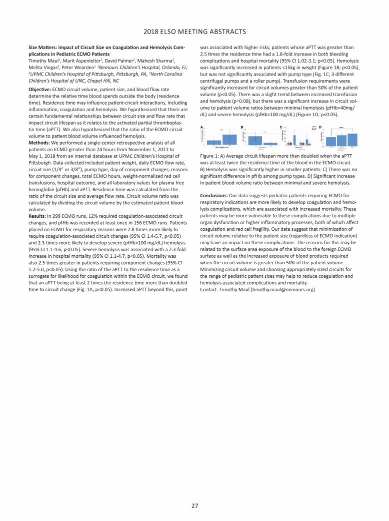
2018 ELSO MEETING ABSTRACTS
27
Size Matters: Impact of Circuit Size on Coagulation and Hemolysis Com-plications in Pediatric ECMO PatientsTimothy Maul1, Marit Aspenleiter2, David Palmer2, Mahesh Sharma3, Melita Viegas2, Peter Wearden1 1Nemours Children’s Hospital, Orlando, FL; 2UPMC Children’s Hospital of Pittsburgh, Pittsburgh, PA, 3North Carolina Children’s Hospital of UNC, Chapel Hill, NC
Objective: ECMO circuit volume, patient size, and blood flow rate determine the relative time blood spends outside the body (residence time). Residence time may influence patient-circuit interactions, including inflammation, coagulation and hemolysis. We hypothesized that there are certain fundamental relationships between circuit size and flow rate that impact circuit lifespan as it relates to the activated partial thromboplas-tin time (aPTT). We also hypothesized that the ratio of the ECMO circuit volume to patient blood volume influenced hemolysis.Methods: We performed a single-center retrospective analysis of all patients on ECMO greater than 24 hours from November 1, 2011 to May 1, 2018 from an internal database at UPMC Children’s Hospital of Pittsburgh. Data collected included patient weight, daily ECMO flow rate, circuit size (1/4” or 3/8”), pump type, day of component changes, reasons for component changes, total ECMO hours, weight-normalized red cell transfusions, hospital outcome, and all laboratory values for plasma free hemoglobin (pfHb) and aPTT. Residence time was calculated from the ratio of the circuit size and average flow rate. Circuit volume ratio was calculated by dividing the circuit volume by the estimated patient blood volume.Results: In 299 ECMO runs, 12% required coagulation-associated circuit changes, and pfHb was recorded at least once in 156 ECMO runs. Patients placed on ECMO for respiratory reasons were 2.8 times more likely to require coagulation-associated circuit changes (95% CI 1.4-5.7, p<0.05) and 2.3 times more likely to develop severe (pfHb>100 mg/dL) hemolysis (95% CI 1.1-4.6, p<0.05). Severe hemolysis was associated with a 2.3-fold increase in hospital mortality (95% CI 1.1-4.7, p<0.05). Mortality was also 2.5 times greater in patients requiring component changes (95% CI 1.2-5.0, p<0.05). Using the ratio of the aPTT to the residence time as a surrogate for likelihood for coagulation within the ECMO circuit, we found that an aPTT being at least 2 times the residence time more than doubled time to circuit change (Fig. 1A; p<0.05). Increased aPTT beyond this, point
was associated with higher risks; patients whose aPTT was greater than 2.5 times the residence time had a 1.8-fold increase in both bleeding complications and hospital mortality (95% CI 1.02-3.1; p<0.05). Hemolysis was significantly increased in patients <15kg in weight (Figure 1B; p<0.05), but was not significantly associated with pump type (Fig. 1C; 3 different centrifugal pumps and a roller pump). Transfusion requirements were significantly increased for circuit volumes greater than 50% of the patient volume (p<0.05). There was a slight trend between increased transfusion and hemolysis (p=0.08), but there was a significant increase in circuit vol-ume to patient volume ratios between minimal hemolysis (pfHb<40mg/dL) and severe hemolysis (pfHb>100 mg/dL) (Figure 1D; p<0.05).
Figure 1: A) Average circuit lifespan more than doubled when the aPTT was at least twice the residence time of the blood in the ECMO circuit. B) Hemolysis was significantly higher in smaller patients. C) There was no significant difference in pfHb among pump types. D) Significant increase in patient blood volume ratio between minimal and severe hemolysis.
Conclusions: Our data suggests pediatric patients requiring ECMO for respiratory indications are more likely to develop coagulation and hemo-lysis complications, which are associated with increased mortality. These patients may be more vulnerable to these complications due to multiple organ dysfunction or higher inflammatory processes, both of which affect coagulation and red cell fragility. Our data suggest that minimization of circuit volume relative to the patient size (regardless of ECMO indication) may have an impact on these complications. The reasons for this may be related to the surface area exposure of the blood to the foreign ECMO surface as well as the increased exposure of blood products required when the circuit volume is greater than 50% of the patient volume. Minimizing circuit volume and choosing appropriately sized circuits for the range of pediatric patient sizes may help to reduce coagulation and hemolysis associated complications and mortality.Contact: Timothy Maul ([email protected])

2018 ELSO MEETING ABSTRACTS
28
Evaluation of Serotonin Release Assay and ELISA Optical Density Tests in Patients Receiving Extracorporeal Membrane OxygenationVivek Kataria1, Leanne Moore1, Sarah Harrison1, Omar Hernandez1, Nathan Vaughan1, Gary Schwartz1, Hoa Nguyen1 1Baylor University Medical Center, Dallas, TX
Purpose: The purpose of this study was to evaluate the application of enzyme-linked immunosorbent assay (ELISA) optical density (OD) thresh-olds and the serotonin release assay (SRA) in patients receiving extracor-poreal membrane oxygenation (ECMO) and aid clinicians in objectively ruling out heparin-induced thrombocytopenia (HIT).Methods: A retrospective study was conducted to evaluate patients admitted to the cardio-thoracic ICU from July 1, 2012 to August 18, 2017 who were placed on ECMO and ordered a SRA. The primary objective of this study was to perform a sensitivity and specificity analysis, which designated patients as HIT negative (OD < 1.0) or HIT positive (OD ≥ 1.0). Secondary endpoints included the proportion of patients switched to an alternative anticoagulant and the incidence of venous thromboembolism (VTE) and bleeding events. Outcome data was also collected such as mor-tality and disposition. Descriptive statistics were used to analyze all data points. P-values less than 0.05 were considered statistically significant.Results: A total of 49 patients were included in this study. Participants that met inclusion criteria were predominately males (61%) with a mean age of 54 years. Average duration of ECMO was 10.7 days, with an ICU length of stay of 26 days. CRRT was concomitantly used in 63% of patients for an average of 7.8 days. Furthermore, platelet counts were dramatically reduced from baseline, with a platelet decrease of 79.5%. All patients except two had an ELISA OD test performed at the first clinical suspicion of HIT; followed by a confirmatory SRA. Utilizing the pre-specified OD thresholds, a specificity and negative predictive value (NPV) of 89.5% and 94.4% were achieved, respectively. Alternative anticoagulation, predominately argatroban, was initiated in 61% of patients for an average duration of 4.1 and 2.1 days, respectively.Conclusion: We report a retrospective observational study in which ELISA OD thresholds were identified in patients on ECMO support. Increasing the OD threshold to > 1.0 resulted in a high specificity and NPV. Further-more, this study demonstrated that a profound drop in platelet count can be anticipated in patients on ECMO.Contact Person:Vivek Kataria, [email protected]
Mechanisms of platelet loss during pediatric extracorporeal life support (ECLS)Wayne L. Chandler1, Afton H. Widdershins1, 1Seattle Children’s Hospital, Department of Laboratory Medicine, University of Washington, Seattle, Washington
Background: Most pediatric patients show a decline in platelet counts after starting ECLS and require continued platelet transfusion to prevent bleeding. The purpose of this study was threefold: 1) develop a method for quantitating the rate of platelet loss during ECLS, 2) measure platelet microvesicle (PMV) levels as a measure of platelet activation, and 3) evaluate other risk factors for increased platelet loss.Methods: We collected data and samples on 65 pediatric patients on ECLS (age 1d to 20Y, 25 VV, 40 VA). The rate of platelet loss expressed as 109 platelets lost/L/hour (bp/L/h) was calculated continuously from estimated blood volume+circuit volume, changes in platelet count and transfused platelets. PMV released through platelet activation were counted using flow cytometry (CD41 and Annexin-V positive). Based on formation of ~11 PMV per platelet activated and a PMV half life of 6 hours, the PMV/plate-let ratio (PMV/Plt) was used to estimate platelet loss due to activation.Results: The initial drop in platelet count after starting ECLS due to dilu-tion by the circuit priming solution (predicted from estimated patient blood volume and circuit volume) was 43%±19%. The actual dilution based on pre/post ECLS platelet counts was higher 52%±21% indicating a rapid loss of platelets (range 0% to 41%) soon after starting ECLS. Platelet activation was highest immediately after going on ECLS (+15 minutes) with platelet activation rates 15 to 57-fold higher than normal suggesting platelet loss in the first hours of ECLS is due to platelet activation. Average platelet loss on ECLS was 3.3 ± 2.9 bp/L/h, with 5% of patients showing no loss, 40% showing higher losses during first 24 hours (6.6 ± 5.3 vs 2.2 ± 2.5 BP/l/H), 33% similar losses early vs late (3.3 ± 1.9 vs 3.2 ± 1.5 bp/L/h) and 22% showing rising platelet losses after 24 hours (1.2 ± 1.3 vs 7.1 ± 7.2 bp/L/h). Average platelet loss during ECLS due to platelet activa-tion was 1.3 ± 0.7 bp/L/h, equivalent to median 54% of total platelet loss (range 7% to ~100%). Average platelet loss rates were higher (2.5 ± 2.5 vs 5.3 ± 2.9 bp/L/h, p = 0.0001) in patients with pre-existing consumptive coagulopathy (20 of 65 patients with low platelets, low fibrinogen, long PT). Platelet loss was also higher (2.9 ± 2.4 vs 5.7 ± 4.0, p = 0.0026) in 11 patients with spontaneous pulmonary, intracranial or GI hemorrhage during ECLS. Platelet loss was not higher in patients with hemolysis or procedural bleeding during ECLS (cannula, chest tube, post operative). Diagnostic phlebotomy platelet loss was 0.14 ± 0.08 bp/L/h, equivalent to 10%±17% of total platelet loss.Conclusions: On average platelet activation due to blood exposure to the circuit accounted for the majority of platelet loss and transfusion during ECLS. Platelet loss rates were higher in patients with pre-existing consumptive coagulopathy and spontaneous (non-procedural) hemor-rhage during ECLS. Diagnostic phlebotomy accounted for a lower but still important cause of platelet loss.Wayne L. Chandler, MD, Seattle Children’s Hospital, Dept of Laboratories, OC.8.734, 4800 Sandpoint Way, NE, Seattle, WA 98105, 1-206-987-2314, [email protected]

2018 ELSO MEETING ABSTRACTS
29
Pediatric ECMO cannula complications: An under-reported source of ECMO morbidityZachary Holowell1; Brian Gray, MD2; Gail Hocutt RNC3 1Indiana University School of Medicine, Indianapolis, IN; 2Indiana University School of Medicine, Division of Pediatric Surgery, Riley Hospital for Children at Indiana University Health, Indianapolis, IN; 3ECMO Program Manager, Riley Hospital for Children at Indiana University Health, Indianapolis, IN
Background/Purpose: Extracorporeal membrane oxygenation (ECMO) cannula complications (CC) are a rare event that can lead to severe complications and death. There is a dearth of literature surrounding can-nula complications and their outcomes, and the ELSO registry does not subcategorize specific complications. The purpose of this study was to review the CC incidence and outcomes at a single tertiary pediatric ECMO center. Our hypothesis is that some ECMO patients may be at a higher risk of CC, and CC may be associated with higher mortality.Methods: We performed a review of all patients supported by ECMO at a Pediatric ECMO center from June of 1987 through July of 2018. All patients with a CC were reviewed further to determine the patient demographics, type of injury, change in care based on the CC, and overall in-hospital mortality. The CC patients were analyzed based on ECMO patient type (neonatal respiratory, pediatric respiratory, and pediatric cardiac), VA/VV support, and type of CC (cardiac injury, venous injury, arterial injury, cannula kinking, malpositioned cannula, obstruction, and pressure/flow issues). The patients with a CC were compared to those without. We performed chi-squared, odds ratio (OR) and relative risk (RR) analyses where indicated. Significance was defined as p<0.05.Results: We found 52/969 ECMO patients who had a CC (5.4%). The neonatal respiratory population did not have an increased risk of CC (RR=1.2; 95% CI: 0.7-2.1; p=0.5), while pediatric respiratory patients had a significantly increased risk of CC (RR=2.9; CI: 1.5-5.7; p=0.002). Cardiac patients had a protective risk of CC when compared to other groups (RR=0.5; CI: 0.25-0.9; p=.02). 48.1% (25/52) of the patients with CC died, while the mortality rate of non-CC patients was significantly less at 31% (284/917; p=0.003). The OR of mortality for a CC patient versus non-CC patient was 2.1 (CI: 1.2-3.6; p=0.01). Neonatal respiratory patients with CC had an increased risk of mortality compared to neonatal respiratory patients without (OR=3.2; CI: 1.5-6.6; p=0.002). The pediatric respiratory and the cardiac patients with CC did not have increased mortality when compared with other patients in their peer groups (OR=0.9; CI: 0.2-3.8; p=0.9 and OR=2.1; CI: 0.6-7.2; p=0.3, respectively). The incidence of VV ECMO patients having a CC was 11.3% (17/151), while incidence in VA patients was 4.6% (35/766; p=0.01). This resulted in an increased risk of CC in VV cannulation (RR=2.3; CI: 1.3-4.0; p=0.003). However, there was an increased odds of mortality for those on VA ECMO who suffered a CC (OR=2.7; CI: 1.4-5.4; p=0.005). The incidence of each injury category was as follows: cardiac injury 1.9% (1/52), venous injury 9.6% (5/52), arterial injury 5.8% (3/52), cannula kinking 7.7% (4/52), malpositioned cannula 55.8% (29/52), obstruction 7.7% (4/52), and pressure/flow issues 11.5% (6/52). In 73.1% (38/52) of patients with CC, the complication resulted in an altered course of ECMO or a change in the patient’s outcome. 7.7% of CC patients (4/52) and 0.4% of all patients (4/969) died directly as a result of a CC.Conclusion: Pediatric ECMO patients suffering from cannula complica-tions had significantly higher mortality than patients who did not suffer CC. The most common CC was a cannula that became malpositioned leading to a repositioning procedure. Neonatal respiratory patients did not have an increased risk of CC but those who did have a CC had higher mortality. Pediatric respiratory patients had a significantly increased risk of suffering a CC but did not have increased mortality. Cardiac patients had a lower risk of suffering a CC and no increased mortality. Addition-ally, VV ECMO was associated with higher risk of CC. However, there was not a significant increase in mortality for those with CC on VV ECMO. Conversely, those on VA ECMO who suffered a CC were more likely to die. Future studies should perform a risk adjusted analysis of these groups, corroborate this data with that from other institutions, and prospectively examine methods to prevent CC.Zachary Holowell; [email protected]
Neuromonitoring in Neonatal Extracorporeal Membrane Oxygenation PatientsZeenia C Billimoria,1 Rachel Chapman,2 Natalie Rintoul,3 Robert DiGeronimo1 for the CHNC ECMO Special Interest Group 1Department of Pediatrics, University of Washington, Seattle, WA 2Department of Pediatrics, USC Keck School of Medicine, Los Angeles, CA 3Department of Pediatrics, University of Pennsylvania School of Medicine, Philadelphia, PA
Background: Extracorporeal membrane oxygenation (ECMO) is utilized in neonates with severe reversible cardiorespiratory failure. These neonates are at increased risk for short and long term adverse neurologic events and poor neurodevelopmental outcomes. As a result, there is increasing use of neuromonitoring modalities in this population. However, data are lacking on the variation in type, timing, indication and duration of neuro-monitoring modalities in neonatal ECMO patients.Objective: Our objective was to determine the practice variation in the utilization of neuromonitoring modalities in neonatal ECMO patients across Level IV neonatal intensive care units (NICUs).Methods: We conducted a survey of all participating hospitals of the Chil-dren’s Hospitals Neonatal Consortium (CHNC). The CHNC is a collabora-tive of 34 Children’s Hospitals with Level IV NICUs in the United States and Canada dedicated to developing quality and research initiatives across participating institutions. 27 of these perform ECMO in the NICU. The survey was created using a web-based application (REDCap). The survey was disseminated to CHNC site sponsors via email, and responses were collected in July 2018. We excluded incomplete responses and multiple responses from the same institutions (used most recent response). We conducted exploratory data analysis and reported proportions of respondents.Results: We had a total of 20 survey respondents, which represents a 63% response rate from ECMO institutions. 85% of the respondents perform ECMO in their NICU with 60% performing >10 cases/year (Figure 1). 55% of respondents had a dedicated neuro-NICU, of which only 36% cared for all ECMO patients. Neurology was not routinely consulted on all ECMO neonates unless on the neuro-NICU service. Routine monitoring for sei-zures was performed in 75% of participating centers. Type and duration of monitoring varied, with the most common modality being continu-ous EEG for the first 24-72 hours of the ECMO run. 50% of respondents used cerebral near infrared spectroscopy (NIRS) with most keeping on for the duration of the ECMO run (70%). All institutions performed head ultrasound (HUS), but timing and duration of imaging varied significantly (Figure 2). In the other category (40%), HUS was performed for first 3 or 5 days followed by 3 times a week, or every other day, or as needed. 50% additionally use head CT to clarify HUS findings or for acute clinical concerns. Following ECMO, head MRI was obtained by the majority of centers (70%) including diffusion weighted imaging (93%). Routine neu-rodevelopmental follow-up after discharge occurs in 95% of participating institutions. However, timing, frequency, type of sub-specialty follow-up (neurology, developmental pediatrics etc.), ancillary services (OT, PT etc.) varied across institutions.
Conclusion: There is significant variation in utilization of neuromonitor-ing modalities in our study cohort of Level IV NICUs. Lack of evidence and clear outcome benefits has resulted in practice variation across institutions. The goal of the ECMO special interest group is to standardize practice and study outcomes in the future, to better aid the utilization of these neuromonitoring modalities.Contact personZeenia C Billimoria — [email protected]

2018 ELSO MEETING ABSTRACTS
30
Outcomes of ECMO During CPR in Adult In-Hospital Cardiac ArrestAmy E. Hackmann, MD1, John Anderson-Dam, MD2, Armin Kiankhooy, MD1 1Division of Cardiothoracic Surgery, Department of Surgery, University of Southern California; 2Departments of Anesthesia and Surgery, University of Southern California
Objectives: As ECMO use increases nationwide for a growing variety of indications, the application of mechanical circulatory support during CPR (ECPR) has increased as well. In 2015, the ACLS guidelines by the Ameri-can Heart Association note that ECMO is a possible alternative in refrac-tory cardiac arrest thought to be from a reversible etiology. The ELSO database contains nearly 5,000 cases of adult ECPR, with 38% survival off ECMO support and 29% survival to discharge. While many of these patients experience out of hospital cardiac arrest, we sought to assess our patient population for in-hospital cardiac arrests where ECMO initiation allowed return of vital signs.Methods: Retrospective review of our ECMO database was performed for the previous 2 years. Demographic data was collected about the patient, as well as duration of chest compressions, if known, location and type of cannulation, length of ECMO support, and outcome. Patients who were peri-arrest but were not receiving chest compressions at the time of can-nulation were excluded.Results: Of 191 ECMO patients at our center during that time, 17 (9% of patients) were cannulated while chest compressions were on-going. Loca-tions where cannulation was performed included the intensive care unit, the operating room, and the cardiac catheterization lab. All but one can-nulation was performed by a cardiac surgeon. Twelve of 17 cannulations were percutaneous femoral access, and 3 were central aortic cannulations in post-sternotomy patients. Overall, 8/17 patients were male, and survi-vors to discharge were 2 men and 3 women. The average age of survivors was 44.8 years (31-58), while non-survivors tended to be older (57.5 years (23-78)). Chest compressions were performed for an average of 38.25 minutes (30-45) on survivors and 66.2 minutes (29-90) on non-survivors. This data was not available for one patient in each group. Difficulties with access for cannulation was noted in 2 survivors and 4 non-survivors. The average time on ECMO support for survivors was 6 days (1-11), while for non-survivors it was 10 days (1-40). 29.4% (5/17) of ECPR patients at our institution were discharged home. One patient weaned from ECMO but died of multi-system organ failure prior to discharge. The majority of patients who died (8/12) had radiographic evidence of devastating neu-rological injury. The remainder failed to recover from multi-system organ failure and ECMO support was discontinued.Conclusions: Although a relatively small sample size, our outcomes mirror those reported in the ELSO database. Given the prolonged time of chest compressions in non-survivors, delays in recognizing appropriate patients in whom to consider ECMO as rescue therapy are a significant barrier to more successes. In addition, technical difficulties during cannulation may contribute to delayed initiation of ECMO and worse outcomes. Standard-ization of when during a cardiac arrest to consider ECMO support as well as immediate access to appropriate equipment are critical to more suc-cessful rescues of the prolonged cardiac arrest patient.Corresponding Author: Amy Hackmann [email protected]
Survival in Adult Venovenous and Venoarterial Extracorporeal Membrane Oxygenation Patients Compared to International Survival Prediction Scores: A Single Center Retrospective ReviewAshley Pigg,1 Andi Orr,1 Adam Aycock,1 Kim Moore,1 William Costello,1 Adam King,1 Stuart McGrane,1 1Vanderbilt University Medical Center, Nashville, TN
AbstractBackground: Extracorporeal Membrane Oxygenation (ECMO) is an advanced form of mechanical life support utilized for organ system sup-port of patients experiencing cardiac or respiratory failure. Selection of patients for ECMO therapy often requires swift decision making based on an abbreviated clinical assessment and a critical timeline in which to establish ECMO support before patient clinical status declines further. The challenges of patient selection relative to survival have been studied by the international consortium of ECMO institutions, Extracorporeal Life Support Organization (ELSO), to develop a scoring system which assists in prediction of survival based on pre-cannulation values. These are known as the Survival After Veno-Arterial ECMO (SAVE) and Respiratory ECMO Survival Prediction (RESP) predictive score models, respectively.Methods: A retrospective analysis was performed of adult patients under-going ECMO at Vanderbilt University Medical Center from January 2017 through March 2018. A review of patient charts was done to correlate Vanderbilt University’s ECMO transport patients and in-house cannula-tion patient outcomes with those predicted by the SAVE and RESP scoring models. These scores were then respectively compared against the international standard outcomes of patients assessed with these scoring models.Results: A total of 85 patients were identified. There were 45 VA ECMO patients and 40 VV ECMO patients. Of these, 29 patients were trans-ported to Vanderbilt from an outside hospital facility. When compared to the reported expected mortality scores for these predictive scoring models, the Vanderbilt adult ECMO program performed at or above the average for both VV and VA ECMO survival outcomes including outside hospital transports.Conclusions: Patients transported on ECMO from outside hospitals have comparable outcomes to patients cannulated within the admission hospital. Based on the findings of this review, further investigation should be dedicated to comparison of differences in outcome among groups of patients transported on ECMO and referral patients for whom ECMO is not a utilized treatment therapy.Author Contact Information:Poster presentationAshley Pigg, RN, BSN, CCRN, ECMO SpecialistVanderbilt University Medical Center1211 Medical Center DriveNashville, TN [email protected]

2018 ELSO MEETING ABSTRACTS
31
Factors Associated With Survival Following Extracorporeal Cardiopulmonary Resuscitation In ChildrenJoel Davis1, Nicholas Melvan1, Michael Heard1, Michael Wolf1, Kirk R. Kanter1, Shriprasad Deshpande1, Bahaaldin Alsoufi1 1Emory University School of Medicine, Atlanta, GA, United States.
ABSTRACT: Objectives: Survival of children having cardiac arrest refractory to con-ventional cardiopulmonary resuscitation (CPR) is very poor. We aimed to examine current era outcomes of extracorporeal CPR (ECPR) support for refractory arrest in children with various pathologies.Methods: We entered demographic, morphologic, clinical, surgical and support details of 182 children who underwent ECPR performed during active chest compression at our institution (2002–17) into a multivariable logistic regression models to determine factors associated with survival.Results: 182 children, median age 54 days (IQR 11–272), required ECPR. Among those, 155 (85%) had cardiac disease including 133 (73%) with congenital heart disease (73 of those with single ventricle). 117 patients (64%) had postoperative ECPR. The remaining 27 (15%) had respira-tory arrest with cardiac disease. Median CPR duration was 27 min (IQR 18–40) min, while median support duration was 3.0 (IQR 1.6–5.3) days. 73% of patients survived > 24 following support discontinuation and 47% survived to hospital discharge. Survival was 49% in cardiac patients (vs. 37% in non-cardiac patients, p=0.046). Mean CPR durations was 29 and 31 min in survivors and non-non-survivors (p=0.87) while mean support duration was 3.9 and 5.6 days, respectively (p=0.054). On regression analysis, factors associated with mortality included pathology other than cardiomyopathy, single ventricle, lower pre-support PH, renal failure, and neurologic complications. CPR duration, age, gender, weight, cannulation site, interval following surgery were not found to be significant factors in our series. Conclusions: ECPR plays a valuable role in children with refractory cardiac arrest with nearly half of the patients surviving to hospital discharge. Survival can be expected even with relatively longer CPR durations. ECPR as a bridge for other device support is valuable in patients with cardiomy-opathy awaiting heart transplantation.Contact author: Bahaaldin Alsoufi, email: [email protected]
Implementation of a Pediatric ECPR Program: Overcoming Challenges and Recognition of Safety ThreatsBrian Gray1, Gail Hocutt RNC2, Kamal Abulebda3, Elizabeth Wetzel4, Michael J Hobson3 1Indiana University School of Medicine, Division of Pediatric Surgery, Riley Hospital for Children at Indiana University Health, Indianapolis, IN; 2ECMO Program Manager, Riley Hospital for Children at Indiana University Health, Indianapolis, IN; 3Indiana University School of Medicine, Division of Critical Care Medicine, Riley Hospital for Children at Indiana University Health, Indianapolis, IN; 4Indiana University School of Medicine, Division of Neonatal-Perinatal Medicine, Riley Hospital for Children at Indiana University Health, Indianapolis, IN.
Objectives: Extracorporeal cardiopulmonary resuscitation (ECPR) is an invaluable care modality to re-establish circulation in witnessed in-hospital pediatric cardiac arrest that is unresponsive to conventional CPR. There is a growing volume of literature supporting the ability of well-structured ECPR systems to improve patient survival and neurologic outcomes by efficiently decreasing the time from initiation of CPR to restoration of perfusion provided by the ECMO circuit. Our primary aim was to develop and implement a structured ECPR system in a tertiary children’s hospital to improve time to ECMO initiation in pediatric cardiac arrest. Our secondary aim was identification of latent safety threats (LSTs) and challenges to system development using in situ high fidelity simulation.Methods: A multidisciplinary team collaborated to develop our ECPR system. We began by constructing a flow diagram identifying key person-nel involved in initiating ECPR and their specific tasks in the system. Multiple discussions and education sessions were conducted with all involved groups to refine and rehearse their specific roles in the process. Additionally, we developed a specific ECPR emergency paging system and created job aids that were given to key personnel and posted at strategic locations in the ICU. To expedite cannulation, we organized a specific sterile instrument set for ICU cannulation and placed it along with other equipment in a centralized storage room in the ICU. To test and refine our newly developed system, we started conducting multidisciplinary in situ simulation sessions of cardiac arrest that necessitated cannulation and initiation of ECMO support.Results: We encountered many obstacles in this process, including obtain-ing buy-in from the various teams involved in initiating ECPR, multiple challenges developing an efficient and reliable paging system, and logisti-cal hurdles orchestrating large multidisciplinary simulations in the midst of a busy ICU. We identified LSTs related to availability of necessary equip-ment, appropriate medication use, adherence to institutional guidelines, team communication, and technical performance. Mean ECPR initiation time prior to this process was 51 minutes in actual activations, but after identifying and mitigating these LSTs, we have been able to reduce our simulated initiation of ECPR to under 40 minutes.Conclusions: Through a concerted multidisciplinary effort, we have devel-oped a structured ECPR system to care for pediatric cardiac arrest. We will continue to refine this system through repeated simulations and debrief-ing after actual ECPR activations. The ultimate goal is to improve survival and functional outcomes through decreased ECPR initiation times, safer more efficient system processes, and enhanced team performance.Contact: Brian Gray, MD, [email protected]

2018 ELSO MEETING ABSTRACTS
32
Trends in Cardiogenic Shock Treatment in an Institution New to Mechanical Circulatory SupportBridget Toy1, Amy Emmarco2, Zachary Kon3, Alex Reyentovich4, Nader Moazami5, Deane Smith6 1Transplant Institute, NYU Langone Health, New York, NY; 2Transplant Institute, NYU Langone Health, New York, NY; 3Department of Cardiothoracic Surgery, NYU Langone Health, New York, NY; 4Department of Medicine, NYU Langone Health, New York, NY; 5Department of Cardiothoracic Surgery, NYU Langone Health, New York, NY; 6Department of Cardiothoracic Surgery, NYU Langone Health, New York, NY.
Introduction: Cardiogenic shock is associated with high in-hospital mor-tality rates regardless of etiology. Recently, there has been an increase in utilization of acute mechanical circulatory support (MCS) in patients with cardiogenic shock. Although there is no consensus as to the most appropriate device for any particular patient presentation, acute MCS devices have been widely adopted in institutions that favor an aggressive approach to cardiogenic shock. Establishment of an Adult ECMO Program at our institution has led to experience with additional acute MCS devices. The complexity of these clinical situations led to the creation of a multidisciplinary team, including a combination of cardiologists, surgeons, interventionalists, and intensivists, committed to treating these patients. As an early ECMO Program and Cardiogenic Shock Team, we sought to review our early experience with acute MCS for cardiogenic shock to define our baseline strategies and outcomes in this patient population.Materials and Methods: We completed a retrospective review of cardiogenic shock patients who were treated with acute MCS by Fiscal Year (September 1 to August 31) beginning with Fiscal Year (FY) 2014. We reviewed implant volume by device, including temporary surgical ventricular assist device (VAD), percutaneous and surgical Impella®, percutaneous RVADs, and veno-arterial Extracorporeal Membrane Oxy-genation (VA-ECMO). Data collected included etiology of shock, indication for device implantation, type of support offered, length of support, and outcome of support. For each category of device, we also reviewed our outcomes by mirroring the established ELSO benchmarks of “Survival from ECMO” and “Survival to Discharge.”Results: We found a steady increase in the use of acute MCS for treat-ment of cardiogenic shock from FY 2014 to FY 2018. From FY 2014 to 2015 there was an increase from zero to 18 implants. The following year (FY 2016) revealed a 100% increase, with a total of 36 implants. The volume increased more slowly (16%/year) the following two years (FY 2017, FY 2018) with 43 and 48 implants, respectively. Overall outcome assessment revealed that 68% of patients survived acute MCS and 53% of patients survived to hospital discharge with an increased survival to discharge in the last fiscal year to 62%.Conclusion: Creation of an Adult ECMO Program and a multidisciplinary Cardiogenic Shock Team correlated with an increase in the number of patients supported with acute MCS. As the team gains experience with these patients and the devices available to support them, we expect the outcomes to continue to improve. Patient selection, surgical approaches and techniques, standardizing medical therapy, and managing and reduc-ing complications will give these patients the best opportunity to survive.Contact Person: Bridget Toy, BSN, RN ([email protected])
Development of a mobile adult ECMO programC. Patrick Henson D.O.(1) Adam King M.D.(1) Will Costello M.D.(1) Clayne Benson M.D. (1)Antonio Hernandez M.D.(1) Stuart McGrane M.D.(1) (1)Department of Anesthesiology, Division of Anesthesiology Critical Care Vanderbilt University Medical Center, Nashville, TN
As the adult ECMO program at Vanderbilt University Medical Center (VUMC) has grown, we have been able to offer mobile ECMO services to outlying communities, resulting in a robust referral and treatment system. Since January 2017, over 40% of our referrals for ECMO have come from outside hospitals. Increasing expansion of our service has led to development of an outreach ECMO service which is increasingly meeting the needs of our referral base. We have developed a consultation and transport system for these patients with the goal of safely minimizing delay and optimizing patient.Challenges often encountered in the process of delivering this service include the communication with the referreing physician, decision-mak-ing, transport of the critically ill patient independent of ECMO, remote cannulation at the referring hospital, and mobile medical care with or without ECMO support. VUMC-directed community outreach services have been developed to support enhanced education and encourage early consultation by our referring hospitals. Herein we describe the infrastructure to include the process for consultation, referral, and trans-portation focused on the involvement of physicians and perfusionists. We present strategies for management of mobile ECMO services and issues that are likely to arise in the near [email protected]

2018 ELSO MEETING ABSTRACTS
33
ECMO for trauma patients: the experience at a Level 1 trauma centerC. Patrick Henson, D.O.(1) Bradley Dennis M.D.(2) Adam King M.D.(1) Will Costello M.D.(1) Clayne Benson M.D.(1) Antonio Hernandez M.D.(1) Stuart McGrane M.D.(1) (1)Department of Anesthesiology, Division of Anesthesiology Critical Care Vanderbilt University Medical Center, Nashville, TN(2) Department of Surgery, Division of Trauma and Surgical Critical Care Vanderbilt University Medical Center, Nashville, TN
At a Level 1 trauma center with a large capture radius, we are con-fronted with several types of patient injury. Both blunt and penetrating trauma can be associated with lung injury, either through direct injury or indirectly, secondary to fluid resuscitation or aspiration, as examples. The adult ECMO service at Vanderbilt University Medical Center (VUMC) has developed a relationship with the division of trauma surgery, which allows for 24 hour consultation, rapid assessment and cannulation of trauma patients who require ECMO. With that consult service in place, an ECMO intensivist is available to be at the bedside and provide con-sultancy, and is also able to provide optimization services, which have reduced unnecessary and inappropriate application of ECMO services in this patient population.Trauma patients commonly have coexisting comorbidities leading to chal-lenges in the management of pulmonary failure, independent of ECMO support. Decisions, including cannulation strategy, anticoagulation, and patient mobilization, will require patient-specific considerations. Many of these are related to acute injuries, and as such, may also have dynamic courses. The VUMC ECMO service received 1-2 consultations per month for the past 18 months, during which time, 8 patients received VV ECMO with 87.5% survival. Here we describe our experience with an ECMO consultation service at a Level 1 trauma center, to include the medical optimization algorithm and presentation of patient [email protected]
Beating the Odds: A Case report of long term ECMO support as a bridge to successful heart transplant in a 2-year-old ventilator dependent child with Glenn physiologyCalsey J. Mischel MD1, Deborah J. Kozik DO2, Deanna R. Todd Tzanetos MD, MSCI1, Katherine E. Potter MD, MEd1 1Pediatric Critical Care, Department of Pediatrics, University of Louisville School of Medicine, Louisville, Kentucky; 2Department of Cardiothoracic Surgery, University of Louisville School of Medicine, Louisville, Kentucky
Case Description: A 2 year 8 month old boy (height 0.84 m, weight 13.6 kg) with a past medical history significant for hypoplastic left heart syndrome (HLHS) status post Norwood procedure with Blalock-Taussig shunt and subsequent bidirectional Glenn procedure, tracheostomy secondary to left vocal cord paralysis and tracheobronchomalacia was referred to our center for cardiac transplant evaluation. He presented with heart failure (moderate tricuspid regurgitation, aortic insufficiency and severely dilated right ventricle with end diastolic pressure of 15 mmHg that was inotrope and ventilator dependent. Complications prior to transfer to our facility included bradycardic arrest secondary to airway dislodgement, atrial flutter, and pleurodesis for persistent effusion.After transfer to our center, heart failure symptoms continued to worsen requiring escalation in inotrope support; he also developed cardio-renal syndrome requiring continuous diuretics. It was determined that his degree of debilitation would require mechanical support for rehabilitation in order to facilitate successful transplantation. His size and Glenn physiol-ogy made continuous flow mechanical support the best initial option with a plan for possible conversion to pulsatile flow. Tunneled Berlin cannulas and a Rota Flow centrifugal pump were utilized and a tricuspid valvec-tomy was required to allow for unobstructed drainage from apical can-nula. Due to his preexisting renal injury and lack of pulsatile flow to the kidneys after VAD placement, he developed anuric renal failure requiring the use of continuous renal replacement therapy. Initially, heparin was utilized for anticoagulation, but due to difficulty achieving therapeutic unfractionated heparin levels secondary to antithrombin III deficiency, he was subsequently transitioned to bivalirudin, clopidogrel, and aspirin. Anticoagulation was monitored utilizing activated partial thromboplastin time to titrate the bivalirudin and thromboelastography with plate-let mapping to titrate aspirin and clopidogrel. After the transition to bivalirudin, the filter on the Prismaflex clotted repeatedly. A continuous low dose infusion of bivalirudin was subsequently infused directly to the Prismaflex circuit which solved the filter clotting issues. With the support of the oxygenator, the patient tolerated trach collar trials allowing him to participate in multiple therapy modalities and rehabilitation. Nutrition was optimized with a combination of enteral and parenteral support.He was successfully transplanted after 121 days on the Rota-flow centrifugal pump. In the immediate post-operative period, he required ECMO support, but tolerated decannulation after three days. He required dialysis for approximately 2 months after his transplant as his kidneys recovered. He was able to eventually be weaned off mechanical ventila-tion and be discharged home on trach collar.Discussion: Stabilizing a patient with mechanical support allows teams to wait for more optimal donor offers. We were able to minimize the number of circuit changes by utilizing bivalrudin, aspirin, and clopidogrel. Studies show the size of available devices has continued to be a limiting factor, but that patients who are bridged with mechanical support have better post-transplant survival with VAD being superior to ECMO. This patient was not only of small size but also ventilator dependent and Glenn physiology. In him, mechanical support with an oxygenator allowed ventilator weaning which facilitated increased mobility and better pre-transplant rehabilitation, all of which have been shown to improve outcomes post-transplant.Calsey J. Mischel [email protected]

2018 ELSO MEETING ABSTRACTS
34
Assessment of Cardiac Recovery Using a Formal Stepwise Weaning Protocol in Patients Supported with Venoarterial Extracorporeal Membrane OxygenationRavi Vuthoori1, Cassandra Heaney1, Karl Bocchieri2, Brian Lima2, Harold Fernandez2, David Majure1, Rajiv Jauhar1, AND Simon Maybaum1 1Donald and Barbara Zucker School of Medicine at Hofstra/Northwell, Hempstead, New York; 2Department of Cardiovascular & Thoracic Surgery at North Shore University Hospital
Introduction: As improving technology has led to increased use of veno-arterial extracorporeal membrane oxygenation (VA ECMO) in cardio-genic shock (CS), protocols for weaning and assessment of recovery are required. We describe a stepwise weaning protocol to assess cardiopul-monary recovery during VA ECMO.Methods: All patients on VA ECMO for CS were considered for a bedside weaning study. Exclusion criteria included sepsis, lung injury, high pres-sor requirement & uncontrolled arrhythmia. Pulmonary toilet and fluid balance were optimized 24 hrs prior to weaning. Ventilator support with FiO2 ≥ 60% and dobutamine 3-5µg/kg/min were initiated prior to weaning. All patients had invasive hemodynamic monitoring (arterial line, Swan-Ganz catheter), echocardiography, cerebral near-infrared spectroscopy, and blood gas analysis. Heparin was used to achieve aPTT > 50s prior to wean and ACT > 280s when ECMO flow < 3 LPM. Flow was decreased in 1 LPM intervals and data was acquired after five minutes at each stage. At ECMO flow of 2 LPM, Impella/IABP support was minimized. After 1 LPM, the inflow line was clamped (with 15s flush every five min-utes) for 30 minutes. After 30 minutes of clamping, final measures were taken. If PaO2/FiO2 < 200 despite adequate hemodynamics, transition to VV ECMO was considered.Results: Over five months, 16 patients (59 ± 8 yrs) had VA ECMO for post-MI (8), post-cardiotomy (3), and other CS (5). Eight were not studied due to neurological injury or multi organ failure, seven were studied at 4-12 days on ECMO, and one is currently on ECMO support. Six out of the seven tolerated clamping for 30 minutes and were successfully decan-nulated (Table 1). The remaining patient was hypotensive at 1 LPM and was transitioned to BiVAD support. At 30 days, four of the six were free of all support, one required an inotrope (and subsequent LVAD) and one passed away due to sepsis. No adverse events occurred during the wean-ing studies.Conclusion: We describe a safe, stepwise weaning protocol to assess cardiopulmonary function during ECMO support. All patients tolerating 30 minutes of ECMO clamping could be successfully decannulated. Further study may elicit criteria for ECMO removal and predictors of recovery.Cassandra Heaney – [email protected]
Clinical Management after Carotid Artery Repair – an Opportunity for Improvement?Christa CJ Kirk, PharmD1, Charles Ho, PharmD1, Larissa Yalon, RN, BSN, CCRN1, Kimberly J Riehle, MD1, Thomas Brogan, MD1, AND Robert DiGeronimo, MD1 1Seattle Children’s Hospital, Seattle, Washington
Background: VA-ECLS is commonly used in neonates and infants with cardiorespiratory failure to preserve heart function and cardiac perfu-sion. Though cannulation for ECLS is a standard procedure, there remains debate about carotid artery repair (CAR) as well as post-decannulation management and follow-up. The existing literature comparing long-term outcomes of CAR versus ligation in patients following decannulation is sparse. Additionally, there are no consensus-based recommenda-tions for anticoagulation or imaging to help guide subsequent clinical management.Objective: To evaluate our institutional experience with CAR, including frequency of the procedure, as well as follow-up diagnostic imaging and anticoagulation after VA-ECLSMethods: We conducted a retrospective, single-center review of all surviving patients who required VA-ECLS at less than 1 year of age who underwent CAR from January 2010 through March 2018 listed in our insti-tutional ECLS registry. Data collected from the Seattle Children’s Hospital electronic database included patient age, type of ECLS support (VV vs. VA), underlying diagnosis, length of ECLS run, type of and timing of vessel repair, anticoagulant used, any diagnostic vessel imaging done post ECLS, and imaging findings when available. During the study time period, we did not have specific criteria for CAR or standardized protocols for antico-agulation management, diagnostic imaging, or defined clinical follow up.Results: From January 2010 – March 2018, 55 patients meeting our study inclusion criteria required VA-ECLS and 34 (62%) underwent CAR at the time of ECLS decannulation. Post repair imaging and anticoagulation were at the discretion of the ECLS team and surgeon. Only 4 (12%) patients had carotid arterial imaging after repair. Of those, 3 patients had evidence of thrombus and 1 patient had no evidence of thrombus. The 3 patients who developed thromboses received aspirin (n=2) or no anticoagulation (n=1). Of patients who underwent CAR, 14 (42%) received anticoagulation post repair: 7 (50%) therapeutic heparin, 3 (22%) prophylactic heparin, 2 (14%) enoxaparin, and 2 (14%) aspirin. In this study population, 20 patients did not receive any anticoagulation. All patients on non-aspirin anticoagula-tion were being treated for other indications.Conclusion: Our data highlight the variability in practice for patients who undergo CAR after decannulation from VA-ECLS. Similar to prior studies, the decision to obtain imaging and initiate anticoagulation was at the discretion of the ECLS team. Specifically, follow up imaging was not consistent across our study population, and often was obtained only in response to clinical suspicion of thrombosis. We did note that all of the clots found in our study population were in patients who were on aspirin or no anticoagulation. However, given the small number of treated patients and limited imaging during follow up, therapeutic effectiveness of different anticoagulation agents could not be determined. Further study is needed to determine the utility, safety, and long-term neuro-logical outcomes in neonates and infants undergoing CAR. For our own institution, this existing knowledge gap has created an impetus to develop and study the impact of standardized guidelines for imaging and antico-agulation management post CAR.Contact: Christa [email protected]

2018 ELSO MEETING ABSTRACTS
35
Outcomes of ECLS for Postcardiotomy Syndrome in the Modern EraYas Sanaiha,1 Nancy Satou,1 Cristina Rimicci,1 Kim De la Cruz,1 Vadim Gudzenko,2Richard Shemin,1 Peyman Benharash1 1Division of Cardiac Surgery, David Geffen School of Medicine, University of California Los Angeles, CA 2Department of Anesthesia Critical Care, David Geffen School of Medicine, University of California Los Angeles, CA
Introduction: Postcardiotomy cardiogenic shock (PCCS) is a rare but catastrophic syndrome which can occur during separation from cardiopul-monary bypass and in the immediate postoperative period. The manage-ment of PCCS is variable and survival has historically been reported to be near 30%. It was our objective to examine modern, institutional outcomes of ECLS for postcardiotomy syndrome.Methods: This was a single institutional retrospective review of all patients who received extracorporeal life support due to postcardiotomy failure from January 2015- June 2018. Postcardiotomy failure was defined as inability to wean from cardiopulmonary bypass and/or suboptimal hemodynamics on maximal inotropic support. The primary outcomes of the study were hospital mortality,discharge location. Baseline and lab characteristics were utilized to develop a predictorResults: Of the 1,940 patients who underwent cardiac surgery at our institution during the study period, 2.3% (N=45) required ECLS for
postcardiotomy sydnrome. ECLS for postcardiotomy syndrome comprised 23% of all VA-ECMO runs over the study period with no significant change in the annual proportion of all ECLS cases (20 to 28%, P=0.4). Patients who required ECLS for postcardiotomy syndrome were similar in mean age (44.9 vs 45.0, P=0.98) and gender distribution (Female 35.6 vs 36.3%, P=0.9) to all other ECLS patients. Of all postcardiotomy patients who received ECLS, 65.8%, compared to 63.8% survival for all VA-ECMO, with only 4.3% requiring left ventricular assist device placement or heart-transplantation. The majority of post-ECLS cannulation is performed via central cannulation with no significant change over the study period (90% to 81.2%, P=0.7). Amongst all patients who undergo VA-ECMO for postcardiotromy syndrome, 27.8% are ultimately discharged home while 38.9% are discharged to referral facility or intermediate care.Conclusions: ECLS for management of postcardiotomy syndrome remains a significant proportion of all ECLS volume at our institution. Survival compared to other indications for ECLS is comparable. Though a mod-est increase in chest closure with percutaneous cannulation has been performed, central cannulation remains the predominant technique for ECLS support for postcardiotomy failure. Further investigation of patient selection criteria and prognostic factors is warranted.Contact Person: Dr. Peyman Benharash, [email protected]

2018 ELSO MEETING ABSTRACTS
36
VENOARTERIAL EXTRACORPOREAL MEMBRANE OXYGENATION VIA CAROTID ARTERY “JUMP GRAFT”: FRIEND OR FOE? A CASE SERIESHannah K. Bauer MD,1 Matthew P. Malone MD,1 1Department of Pediatrics, Division of Critical Care Medicine, University of Arkansas for Medical Sciences, Little Rock, Arkansas
BACKGROUND: In patients requiring venoarterial extracorporeal mem-brane oxygenation (V-A ECMO support), arterial sites for cannulation most often include the carotid or femoral artery, with newer techniques utilizing the subclavian or axillary arteries. Site of cannulation is often a multidisciplinary discussion between the critical care team and the can-nulating surgeon, taking into consideration the age, size, and pathology of the patient. This series reflects some of the first ECMO cannulations performed utilizing carotid artery “jump grafts” at our institution. Data on outcomes and complications prior to utilizing this technique was therefore unavailable. Multiple authors cite the use of novel techniques including subclavian and axillary artery cannulation for V-A ECMO support in adults utilizing a graft placed in an end-to-side fashion as a means to potentially avoid complications associated with traditional cannulation techniques, including sacrifice of the cannulated vessels. Chikotka and colleagues recently published their experience of adults on V-A ECMO utilizing an internal jugular vein drainage cannula with arterial return cannula inserted into a synthetic graft anastomosed to the axillary or innominate artery. This approach facilitated early patient mobilization, as well as reduction in differential oxygenation or Harlequin effect that can be seen in femoral artery cannulation. In a single center experience, Chamogeorgakis, et al found that in 81 adult patients cannulated with a side-graft sewn to the axillary artery, hyperperfusion syndrome to the ipsilateral extremity was the most common complication (n=20). A retrospective analysis of data from the Extracorporeal Life Support Orga-nization registry found pediatric patients aged 18 years or younger had a statistically significant increased odds of neurologic injury with carotid artery cannulation for V-A ECMO. Prior to this case series, there existed no published description of this particular cannulation technique, and no publications of arterial grafts in pediatric populations.METHODS: An electronic medical record review of our institution’s ECMO patients and an in-depth review of the literature was performed. Nine patients underwent arterial cannulation of the carotid artery utilizing a Gore-Tex® “jump graft” in an end-to-side fashion at our institution from
2012 to 2017. Each patient’s hospital course was reviewed with particular consideration given to disease process, site of cannulation, neurologic examination abnormalities noted during ECMO, computed tomography or magnetic resonance imaging (MRI) evidence of intracranial hemorrhage, and outcomes.RESULTS: Of the nine cases reviewed, ages ranged from 5 to 20 years-old with weights ranging from 16 to 85 kg. Three patients were male and 6 were female. All patients received cranial imaging due to a change in neurologic examination during the ECMO course. One patient had petechial hemorrhage on MRI after ECMO, but no neurologic sequelae. Four patients survived to discharge or transfer out of the ICU. Four patients died after withdrawal of ECMO support secondary to complica-tions of their underlying condition or due to medical futility. One patient, a 13-year-old female with pre-B-cell acute lymphoblastic leukemia and Legionnaire’s disease, experienced hyperemia with increased flow to the head, neck, and right arm, as evidenced by significantly elevated cerebral near infrared spectroscopy (NIRS). Interestingly, her systemic mixed venous oxygen saturation and renal NIRS were low. Two days after cannulation, she sustained a large right intraparenchymal hemorrhage with sulcal effacement and midline shift after loss of pupillary and corneal reflexes were noted on examination. This necessitated decannulation from ECMO after which life-sustaining measures were subsequently withdrawn.CONCLUSION AND SIGNIFICANCE: In this review of the use of carotid arterial grafting in V-A ECMO cannulation at our institution, eight of nine patients were successfully cannulated utilizing this technique without neurologic complication. All nine of these patients were cannulated “elec-tively,” not while receiving cardiopulmonary resuscitation. This technique would not be technically feasible during such an emergent situation. In reflecting on our patient with complications of streaming and hyperemia to the head, neck, and right arm following the use of a carotid artery graft, along with subsequent catastrophic neurologic sequelae, one must weigh the risk of neurologic complications with preferential high blood flow rates to the cerebral arterial circulation. However, the prevention of carotid artery sacrifice by utilizing the “jump graft” and later carotid arte-rial repair should be considered. Further work is needed to better define risk factors for neurologic injury and hemorrhage in pediatric ECMO.Contact Information: Hannah K. Bauer, MD - [email protected]

2018 ELSO MEETING ABSTRACTS
37
Cardiac Catheterization Snare Technique To Correct A Malpositioned Avalon Elite® Cannula Without Interruption of Veno-Venous Extracorporeal Life Support In Patient With Unilateral Pulmonary AgenesisHarry J. Kallas1, Paolo Aquino2, Edwin Petrossian2,3 1Pediatric Critical Care Medicine, 2Cardiology and Cardiothoracic Surgery, Valley Children’s Hospital, Madera, CA; 3Cardiothoracic Surgery, Stanford University School of Medicine, Palo Alto, CA
Introduction: Unilateral pulmonary agenesis is a rare condition that can be associated with severe post-natal respiratory distress and pulmo-nary hypertension. We report a newborn with left pulmonary agenesis successfully treated with VVDL ECMO who survived to discharge and describe a potentially dangerous cannula malposition during the case that was corrected without coming off ECMO using a cardiac catheterization snare technique. To our knowledge, repositioning of a VVDL ECMO can-nula using such technique has not been previously reported.Case Description: Patient was a term, 2.98 kg newborn girl with left pulmonary agenesis. Immediately after birth, she had severe respiratory failure and pulmonary hypertension. She failed high settings on conven-tional mechanical ventilation and high-frequency ventilation with HFOV and VDR-4, including use of inhaled nitric oxide and nebulized epopros-tenol, and continued to have severe hypoxemia and hypercapnia. She also needed high-dose dopamine and epinephrine infusions. By echocardiog-raphy, she had cardiac levoposition with normal intracardiac anatomy, absence of the left pulmonary artery, good ventricular function, and a patent ductus arteriosus with predominantly right-to-left shunting.She was cannulated for VVDL ECMO on day of life #1 (DOL #1) using a 13 Fr. Avalon Elite® cannula via the right internal jugular vein with good placement confirmed by echocardiography and chest x-ray (CXR) showing the tip of the cannula in the intrahepatic inferior vena cava (IVC) and the arterial return jet oriented towards the tricuspid valve. She was transi-tioned to “rest” ventilator settings and other support was weaned after institution of ECMO. She developed severe capillary leak syndrome and marked total body edema. Right lung field became completely airless by CXR with essentially no air movement during mechanical ventilation.On DOL #5, CXR and echocardiography indicated displacement of the can-nula with the tip in the right atrium (RA) deforming the atrial wall. Due to potential risk of cardiac perforation, the cannula was initially pulled back. ECMO blood flow and recirculation was relatively worse with the malposi-tioned cannula. Attempts to guide the cannula tip back into the IVC under echocardiographic guidance while still on ECMO were not successful, in part due to the patient’s anatomic deformation. It was determined that
patient would not tolerate coming off ECMO to employ other techniques for cannula repositioning or replacement.Intervention: The patient was taken to the cardiac catheterization lab where a 4 Fr vascular access sheath was placed in the left common femoral vein. Through the sheath, a 4 Fr Glide catheter was advanced to the RA-IVC junction. Under fluoroscopic guidance, a 10 mm snare was advanced through the Glide catheter and the tip of the Avalon® can-nula was secured. As the surgeon advanced the ECMO cannula from the neck, the cannula tip was guided with the snare into the intrahepatic IVC with confirmation by fluoroscopy and echocardiography. The surgeon then secured the ECMO cannula. The snare was then released from the cannula, and the snare and Glide catheter removed; the sheath was left in place. The procedure was well tolerated and the patient was kept on stable ECMO support throughout the procedure.Follow Up: Patient’s lung function and pulmonary hypertension progres-sively improved, and she was weaned off ECMO on DOL #12; the cordis was also removed at that time. Follow up vascular ultrasounds showed a non-occlusive thrombus in the left common femoral and iliac veins. She was extubated on DOL #15, but was not able to tolerate being off non-invasive positive pressure respiratory support over the next two months. She eventually required tracheostomy, and was sent home with mechani-cal ventilatory support on DOL #99.Conclusions: Unilateral pulmonary agenesis is a rare condition that can be associated with clinical decompensation and death. We report a case where VVDL ECMO successfully supported a neonate with post-natal severe respiratory failure and pulmonary hypertension, who subsequently survived to hospital discharge. We also report a potentially dangerous occurrence of Avalon® VVDL cannula displacement during ECMO in this patient with successful cannula repositioning using a cardiac catheteriza-tion snare technique without need to come off ECMO support.
Contact:Harry J. Kallas, [email protected]

2018 ELSO MEETING ABSTRACTS
38
Different Transfusion Targets for VA-ECMO Patients and Survival: Outcomes at University Hospital Programs - One in U.S. and One in ChinaP Li1, G Tomlin2, Y Zhao1, NG Dong1, AND I Wang3 1Department of Cardiovascular Surgery, Wuhan Union Hospital, Huazhong University of Science & Technology, Wuhan, China 2Department of Biomedical Engineering, Purdue University, West Lafayette, Indiana 3Division of Thoracic Transplant, Indiana University Health Methodist Hospital, Indianapolis, Indiana
Background: Patient hemoglobin target is a key factor in deciding blood transfusion in Extracorporeal Membrane Oxygenation (ECMO) patients [1]. Maintaining an “adequate” hemoglobin (Hgb) level is one of the important tenets to successful perfusion on ECMO support [2]. Blood transfusion is not without its potential risks. In the U.S., the transfu-sion goals are often tolerant of anemia with Hgb targets of 7.0 -7.5gm/dL. In China, the practice is typically to transfuse patients to maintain a Hgb level of 10gm/dL. In this international collaboration, the transfusion requirements and outcomes of VA-ECMO patients in a major university hospital in U.S. and China are compared.Methods: A retrospective chart review was conducted of all V-A ECMO patients admitted to IU Health Methodist Hospital (IUH) and Wuhan Union Hospital (WUH) from 2009-2016. Patients were excluded if age was less than 18 or if transfusion records were not available. Primary endpoints were survival off ECMO and survival to discharge. Secondary outcomes included blood products used and length of stay (LOS). χ2 and Mann-Whitney U tests were used to determine any statistical significance of the main endpoints.Results: The charts of all adult (age 18) V-A ECMO patients at IU Health Methodist Hospital (IUH, n=116) and Wuhan Union Hospital (WUH, n= 20) were reviewed. 3 IUH patients were excluded for age while 6 WUH patients were excluded (3 age, 3 incomplete records). While the gender distribution of ECMO patients at both institutions was similar, WUH patients were younger (median 40 years) than IUH (56.5years). Transfusion requirements at each institution are shown below (median ± standard deviation):
PRBC FFP Platelet Cryo
IUH 12.5 ± 17.33 4 ± 8.25 4 ± 4.32 0 ± 2.23WUH 29.5 ± 18.93 7 ± 8.05 10.5 ± 7.06 19 ± 13.53Stat. Significance P < .001 P < .003 P < .001 P < .001
Median duration of ECMO support was 5.2 days at WUH and 6.0 days at IUH. The median hospital length of stay (LOS) was shorter at IUH 18 days versus 62 days at WUH (p < .001). At IUH, 58 patients survived to weaning from ECMO (50%) while 18 were weaned at WUH (90%), statistically not significant (p < .10) On the other hand, survival to discharge was 33% (n= 38) at IUH and 85% at WUH (n= 17), which was significant (p < .01). Of note, the median LOS of survivors to discharge at IUH was longer than the IUH group as a whole (31 vs 18d) and remains shorter than WUH survivors (31 vs 58d).Discussion: IUH and WUH are major university hospitals in their respective countries offering cardiovascular and transplant surgical services with WUH at higher volumes: 2016 cardiopulmonary bypass cases 3000 at WUH vs 900 at IU, and heart transplant WUH 93 vs IUH 23. The rate of VA ECMO support is different between the 2 hospitals. While the transfusion targets and there-fore transfused blood products are different at IUH and WUH, this may not be the determinant of survival differences. One key difference is the indication for VA-ECMO support: 6 of the IUH patients are heart transplant patients, all 20 WUH were heart transplant patients. Furthermore, patient management post-ECMO decannulation was often transitioned to other teams at IUH while WUH patients were continued under the same team. Transfusion target remains a consideration in the management of VA-ECMO patients, but the determinants of optimal outcomes remains complex. Additional international collaborative clinical research may produce further insights.
[1] J. Carson and S. Kleinman, “UpToDate”, Uptodate.com, 2018. [Online]. Available: https://www.uptodate.com/contents/indica-tions-and-hemoglobin-thresholds-for-red-blood-cell-transfusion-in-the-adult. [Accessed: 12- Jul- 2018].
[2] G. Makdisi and I. Wang, “Extra Corporeal Membrane Oxygenation (ECMO) review of a lifesaving technology”, Journal of Thoracic Disease, vol. 7, no. 7, pp. 1-5, 2015.

2018 ELSO MEETING ABSTRACTS
39
The Minnesota Resuscitation Consortium’s Advanced Perfusion and Reperfusion Cardiac Life Support Strategy for Out-of-Hospital Refractory Ventricular Fibrillation.Jason A. Bartos MD, PhD1, Ganesh Raveendran MD, MS1, Ranjit John MD, PhD2, Marc Conterato MD3, RJ Frascone MD4, Alexander Trembley NREMT-P3, Kevin Sipprell MD5, Tom P. Aufderheide MD6, AND Demetris Yannopoulos MD1 1Department of Medicine-Cardiovascular Division, University of Minnesota; 2Division of Cardiothoracic Surgery, University of Minnesota; 3Department of Emergency Medicine, North Memorial Medical Ctr, Minneapolis, MN; 4Department of Emergency Medicine, Regions Hospital, St. Paul, MN; 5Department of Emergency Medicine, Ridgeview Medical Center, Waconia, MN; 6Department of Emergency Medicine, Medical College of Wisconsin, Milwaukee, WI.
Background: In 2015, the Minnesota Resuscitation Consortium imple-mented an advanced perfusion and reperfusion life support strategy designed to improve outcome for patients with out-of-hospital refractory VF/VT. We report the outcomes of the initial 2 years of operations.Methods: Three emergency medical services systems serving the Minne-apolis-St. Paul metro area participated in the protocol. Inclusion transport criteria included refractory VF/VT arrest, age 18-75 years, body habitus accommodating automated LUCAS CPR, and estimated transfer time from the scene to the cardiac catheterization laboratory of ≤30 minutes. Exclu-sion transport criteria included known terminal illness, DNR/DNI status, traumatic arrest, or significant bleeding. Refractory VF/VT arrest was defined as failure to achieve sustained ROSC after treatment with 3 direct current (DC) shocks and administration of 300mg of intravenous/intra-osseous amiodarone. Patients were transported to the University of Min-nesota and taken directly to the cardiac catheterization laboratory where they were immediately assessed for effective resuscitation. Any patient with ≥1 of the following criteria was declared dead: end-tidal CO2 < 10 mmHg, PaO2 < 50 mmHg, or lactic acid > 18 mmol/L. All patients who did not meet these criteria received emergent advanced perfusion strate-gies including extra-corporeal membrane oxygenation (ECMO), coronary angiography was performed, PCI was performed when appropriate, and therapeutic hypothermia was initiated.Results: Over the first 2 years of the protocol, 100 patients were transported with on-going mechanical CPR. Of those, 92 patients (92%) received the full resuscitation treatment. 8 patients were excluded due to failure of the resuscitation criteria. The average age was 56 ± 1.3 years, 77% were men and 81% were white patients. The mean time from 911-call to CCL arrival was 57 ± 1.8 minutes. ECMO was placed in 77/92 patients within 8 ± 1 minutes from arrival to the CCL. 84% of patients had severe coronary artery disease requiring PCI. 46/92 (50%) survived to hospital discharge and 40/46 (87%) survivors were discharged neurologi-cally intact.Conclusions: The MRC refractory VF/VT protocol is feasible and led to a high functionally favorable survival rate with few complications. It repre-sents the first organized protocol of early mobilization for this patient population in the USA.Contact: Jason BartosE-mail: [email protected]
Surviving Severe Hypothermia with ECLS - 150 Minutes of CPR before ECLS - A Case ReviewJessica Hamilton,1 Sam Mandell,1 Eileen Bulger,1 1Harborview Medical Center, Seattle, WA;
Introduction: Harborview Medical Center in Seattle, Washington is the only designated Level I adult and pediatric trauma and burn center in the WAMI region which includes Washington, Alaska, Montana and Idaho. Harborview’s affiliation with the University of Washington Medicine Health System allows the hospital to focus on its mission which is to pro-vide specialized comprehensive emergency services to patients through-out the region. Harborview’s ECMO program started in April of 2016 with the primary goal of bringing ECMO to our trauma patients with ARDS, PE, etc. The program has grown as have our indications. We now offer Veno-Arterial and Veno-Venous ECMO for a variety of indications and we have a rapidly growing E-CPR program. Sitting on the Puget Sound, Seattle is a city surrounded by water. In January of 2017, a 21 year old woman made a distress call to 911 just after 0200. She reported that winds had sent her small boat off course and she was now lost and cold and taking on water. Collaboration with the emergency response system, the Coast Guard and the Seattle Fire Department allowed for this woman to reach Harborview where she was able to be placed on ECMO after two and a half hours of high quality CPR.Case Description: Guided by the information the patient provided to 911, the Coast Guard found and pulled her from approximately 45◦ Celsius water just over an hour after the phone call was unexpectedly cut off. They found her floating face down in the water just before 0500. She was cold to the touch and had no pulse. The Coast Guard started CPR and continued until they were able to get her to paramedics who were waiting at a nearby ferry dock. They brought her to the Emergency Department after establishing an airway and delivering traditional ACLS. Manual CPR was stopped and mechanical CPR initiated. Her core temp was 25◦ Celsius, her pupils fixed and dilated and she was in asystole. He arterial blood gas upon arrival showed a pH less than 6.8, pACO2 of 128, pAO2 of 29 and a lactate of 18. She was taken quickly from the ED to the operating room where she was peripherally cannulated and placed on VA ECMO for rapid rewarming and cardiovascular support. A core tempera-ture of 33◦ Celsius was achieved 2 hours and 37 minutes after CPR was initiated. At that time, mechanical CPR was stopped. Fine VF was seen on the monitor. Conversion to sinus rhythm was achieved after one attempt at defibrillation. Within the hour, her pupils became reactive. She was slowly rewarmed using targeted temperature management while seda-tion was weaned. After 24 hours at 33◦ Celsius, her arterial blood gas had improved to a pH of 7.43, pACO2 of 32, pAO2 of 96 and a lactate of 2.5. On the second hospital day she was following commands and, with the guidance of echocardiography, was able to be completely weaned from ECMO. On hospital day three she was extubated and demonstrated a normal neurological and cognitive exam. On the seventh hospital day she was discharged home with no neurological deficits.Discussion: In the setting of cold water drowning, hypothermia second-ary to exposure, high quality CPR, and urgent initiation of VA ECMO, this patient was able to survive the incident without neurological deficit. This case brings forward several questions around how and when to apply ECMO. Hypothermia is known to aid in preservation of neurological function, but how long is too long for ongoing CPR without ROSC in these patients? This case also presented an opportunity for Harborview Medical Center to better develop the infrastructure and response to emergent ECMO activations so that patients may have decreased time between incident and cannulation. We now have a program that can be activated from the field and we have developed inclusion/exclusion criteria that help all providers identify patients that are most likely to benefit from ECMO. Reaching out to transferring facilities, education and training for EMS and Emergency Department staff and modification to ECMO special-ist activation procedures are making it possible for Seattle to be prepared next time one of our community members calls on us for help.Contact: Jessica Hamilton, [email protected]

2018 ELSO MEETING ABSTRACTS
40
Extremity Perfusion on ECMOOur ECMO team at University of Wisconsin and American Family Chil-dren’s Hospital has recognized the need for improvement in the assess-ment and monitoring of lower extremities in patient placed on ECMO with femoral artery cannulation sites. If not done in a timely manner, the need for surgical intervention is likely. By implementing guidelines, protocols and introducing the use of near infrared spectroscopy can assist in earlier recognition of the first signs and symptoms to warrant further attention, notification and intervention. These measures will improve the quality of care and outcomes of critically ill patients on ECMO.
Jillian Koch, RRTECMO Coordinator
University of Wisconsin & American Family Children’s Hospital 600 High-land Ave, Madison, WI 53792Cell: 608-213-9062Office: 608-720-2551Pager [email protected]

2018 ELSO MEETING ABSTRACTS
41
LAVA-ECMO for Acute Myocardial Infarction with Profound Cardiogenic ShockBennet George1, Kenneth Dulnuan1, Maya Guglin1, Khaled Ziada1, Andrew Kolodziej1, Rajasekhar Malyala2, Julia Akhtarekhavari2, John Gurley1,2 From the Divisions of 1Cardiovascular Medicine and 2Cardiothoracic Surgery, University of Kentucky, Lexington, Kentucky
Introduction: Acute myocardial infarction (AMI) complicated by profound cardiogenic shock poses special problems for mechanical circulatory support. Percutaneous axial-flow pumps can unload the left ventricle (LV), but these devices are susceptible to hemolysis, instability and limb ischemia. They often fail to deliver sustained, adequate organ perfusion and are contraindicated in patients with suspected LV thrombus (a com-mon occurrence in AMI). VA ECMO provides excellent organ perfusion but markedly increases LV afterload. This leads to myocardial ischemia, LV non-ejection, chamber dilation and thrombosis, pulmonary edema, and white lung syndrome. Combining VA ECMO (for organ perfusion) with an axial-flow pump (for LV unloading) is costly and increases the risk of access site complications.Methods: LAVA-ECMO (Left Atrial Veno-Arterial Extracorporeal Mem-brane Oxygenation) is a novel approach to circulatory support in patients with AMI and profound cardiogenic shock. LAVA-ECMO utilizes a single 24 Fr bi-atrial venous cannula placed percutaneously via the right common femoral vein to the left superior pulmonary vein. Inflow holes, extending from the pulmonary vein to the inferior vena cava, drain both atria simul-taneously. The LAVA-ECMO circuit is completed by a centrifugal pump, an oxygenator, and an 18-20 Fr femoral artery cannula. This report describes our initial experience with LAVA-ECMO as the sole means of support in 5 patients with acute MI and profound cardiogenic shock.Cannulation Technique: Totally percutaneous cannulation and decan-nulation was utilized in all patients. Access was ultrasound-guided, with micropuncture technique and distal perfusion catheters (antegrade femoral or retrograde tibial). Anticoagulation was achieved with unfrac-tionated heparin or bivalirudin. Transseptal puncture was performed under intracardiac echo (ICE) guidance. A 6 Fr multipurpose catheter was directed to the left superior pulmonary vein and exchanged over a stiff .035-inch guidewire. The atrial septum and the femoral puncture site were dilated with an 8 X 40 mm balloon. The bi-atrial drainage can-nula was fabricated at tableside by heat shaping a 24 Fr X 68 cm venous
cannula (model VFEM024, Edwards Lifesciences LLC) as shown in Figure 1. The cannula was advanced under fluoroscopy to the left superior pulmo-nary vein. After initiating support, ICE imaging was utilized to confirm LV ejection, good aortic valve excursion and absence of complications. Full therapeutic anticoagulation was continued, with intravenous bivalirudin the preferred anticoagulant.
Figure 1. Custom bi-atrial drainage cannula after heat shaping (left). Inser-tion over guidewire after balloon dilatation of atrial septum.
Results: LAVA-ECMO was successfully implemented in all 5 patients. Four patients were cannulated at initial presentation, while 1 was cannulated for salvage indications on post-MI day 4. Average pump flow was 4.6 0.55 L/min. (range, 4.0 to 5.1). This flow was sustained over a duration of 3 to 10 days with no adverse events. LV ejection preserved in all cases. All 5 patients presented with cardiogenic pulmonary edema; all 5 had radiographic clearing after LAVA-ECMO. While on pump, the pulmonary wedge and right atrial pressures became equalized (average 12 mmHg), indicating balanced bi-atrial drainage. Three patients (60%) survived to hospital discharge. There were no instances of bleeding, thrombosis, or limb ischemia. There were no procedure related complications. At decan-nulation, no attempt was made to close the atrial septostomy. Spontane-ous closure was expected (follow-up data not available at this time).Conclusions: LAVA-ECMO is safe and effective for AMI with cardiogenic shock. LAVA-ECMO provides simultaneous right and left atrial drainage via a single venous cannula and utilizes a conventional pump-oxygenator circuit. It provides full flow, oxygenation, left heart decompression, and myocardial rest. It maintains LV preload, contractility and ejection because left atrial and right atrial pressures are self-balancing. Pulmonary edema resolves on LAVA-ECMO.Address for correspondence: [email protected]; [email protected]; [email protected]

2018 ELSO MEETING ABSTRACTS
42
Percutaneous Tibial Access for Distal Perfusion During VA ECMO: Protection Without BleedingBennet George1, Khaled Ziada1, Thomas Tribble2, Rajasekhar Malyala2, Alexis Shafii2, Julia Akhtarekhavari2, Charles McLendon2, John Gurley1,2 From the Divisions of 1Cardiovascular Medicine and 2Cardiothoracic Surgery, University of Kentucky, Lexington, Kentucky
Introduction: In meta-analysis, limb ischemia (compartment syndrome, fasciotomy and amputation) occurs in up to fourth of patients on femoral VA ECMO. While distal perfusion catheters (DPCs) can reduce ischemia, percutaneous antegrade access can be challenging in patients who have already been cannulated. Ultrasound windows are compromised by large cannulas, and unsuccessful access attempts can lead to major groin bleeding. Antegrade femoral DPCs may not be effective in patients with peripheral vascular disease.Methods: Percutaneous posterior tibial DPC placement was attempted in 16 patients (12 prophylactically and 4 to manage limb ischemia). Tibial access was obtained prior to placement of the ECMO cannula, as the preferred DPC site, whenever the common femoral anatomy was undesir-able for antegrade cannulation (e.g., high bifurcation or atherosclerosis). Combined retrograde tibial + antegrade femoral DPCs were placed in one patient with chronic, total occlusion of the superficial femoral artery.Technique: The posterior tibial artery, veins and nerve were visualized by duplex ultrasound behind the medial malleolus at the ankle (Figure 1, A and B). The artery was accessed under ultrasound guidance utilizing an .021-inch micropuncture needle and a .018-inch guidewire (C). A 4 Fr micropuncture sheath was placed (D) and then exchanged over a .035-inch guidewire for a 6 Fr wire-reinforced sheath (E). The sheath was connected to a Luer adapter spliced into the arterial limb of the ECMO cir-cuit (F). At the time of decannulation, the tibial sheath was removed, and hemostasis was obtained by manual compression.
Figure 1.Ultrasound-guided posterior tibial artery access for distal perfu-sion during femoral VA ECMO.
Results: Posterior tibial cannulation was successful in 14 of 16 attempts (88%). Failure was due to chronic atherosclerotic occlusion of the poste-rior tibial artery (n = 1), and inability to advance the guidewire in a patient with severe vasoconstriction (n = 1). Tibial access was more challenging for patients already on ECMO, due to a lack of arterial pulsations and color Doppler signal. In these cases, the artery was identified by its thick walls and characteristic location relative to veins and nerve. For success-fully cannulated patients, there were no cases of ischemia, hematoma, bleeding, thrombosis or infection. All tibial DPCs remained patent for the duration of ECMO support.Conclusions: Percutaneous posterior tibial access for distal perfusion is safe and effective. Tibial DPCs provide ischemic protection without the added risk of bleeding. While tibial access requires ultrasound and micro-puncture skills, the approach is suitable for most patients on VA ECMO.Address for correspondence: [email protected]; [email protected]; [email protected]
Totally Percutaneous Cannulation and Decannulation for Femoral VA ECMOBennet George1, Navin Rajagopalan1, Khaled Ziada1, Thomas Tribble2, Rajasekhar Malyala2, Alexis Shafii2, Michael Sekela2, John Gurley1,2 From the Divisions of 1Cardiovascular Medicine and 2Cardiothoracic Surgery, University of Kentucky, Lexington, Kentucky
Introduction: Open surgical cannulation for femoral VA ECMO is often complicated by bleeding, infection and seroma formation. Surgical decan-nulation exposes unstable patients to the risks of anesthesia.Methods: Between January 2016 and July 2018, 45 patients were placed on VA ECMO with a totally percutaneous cannulation and decannula-tion strategy as our preferred approach. Semi-stable patients received a single pre-closure suture (Perclose ProGlide, Abbott Vascular) at the time of cannulation. Highly unstable ECPR patients were cannulated without pre-closure. All patients received a percutaneous antegrade femoral or retrograde tibial distal perfusion catheter (DPC). All decannulations were performed percutaneously and utilized the Perclose ProGlide system for arterial closure (regardless of whether or not the artery had been pre-closed).Cannulation Technique: Arterial access was obtained with ultrasound-guided, 4 Fr micropuncture technique. When antegrade DPCs were utilized, the retrograde puncture was located below the inguinal ligament, and the antegrade puncture was located above the femoral bifurcation (approximately 10 mm apart). A single Perclose suture was placed at the retrograde puncture site, aligned longitudinally. This was followed by serial dilation and insertion of an 18 or 20 Fr arterial cannula. Only one femoral artery site was needed in cases where retrograde tibial DPCs were utilized. The pre-closure suture was retained in a protective tube alongside the cannula until decannulation (Left panel).Decannulation Technique: Anticoagulation was not reversed for decan-nulation. The ECMO circuit was discontinued, blood volume was returned to the patient, and the cannulas were flushed with saline. The tubing was clamped and cut, leaving a short segment of tubing attached to the cannulas. The tubing of the arterial cannula was then punctured with an 18 Ga vascular access needle and a .035-inch guidewire was inserted. The arterial cannula was removed over the guidewire and the pre-closure suture was tightened. When necessary, a second Perclose ProGlide device was placed over the guidewire, aligned parallel to the artery and the first suture. The guidewire was retained until successful arterial closure was confirmed by Doppler or imaging ultrasound. Antegrade DPCs were removed over a guidewire and the puncture sites were closed with a single Perclose suture. Retrograde tibial DPC sites were managed with manual compression. After arterial closure, the subcutaneous tissue tracts were irrigated with iodine solution and the sutures were cut. After confirming arterial hemostasis, the venous cannula was removed, and hemostasis was achieved with a temporary purse string suture.
Results: Twenty-four of 45 patients (53%) placed on VA ECMO recovered and were decannulated. All 24 patients were successfully decannulated by the percutaneous approach. There were no instances of arterial bleed-ing, hematoma, ischemia, residual arterial stenosis, surgical rescue, or infection. There were no instances of seroma formation.Conclusions: A totally percutaneous approach to VA ECMO cannulation and decannulation is safe and efficient. The percutaneous approach avoids common hazards of surgical cannulation and decannulation, nota-bly bleeding, infection and seroma formation.Address for correspondence: [email protected]; [email protected]; [email protected]

2018 ELSO MEETING ABSTRACTS
43
Case report: ECMO, percutaneous thrombectomy, and ultrasound-accelerated catheter-directed thrombolysis use for persistent right heart failure after massive pulmonary embolismJ Marinaro1, T Dettmer1, S Guliani,2 1Department of Emergency Medicine, University of New Mexico, Albuquerque, New Mexico 2Department of Surgery, University of New Mexico, Albuquerque, New Mexico
Introduction: Massive pulmonary embolism (PE) has an estimated mortality of 40-60% even with the use of systemic thrombolytics. Recent studies have demonstrated that veno-arterial (VA) ECMO use may reduce mortality to as low as 5%. In some case series up to 40% of patients require no more intervention than therapeutic heparin and ECMO sup-port for clot resolution and improvement in right heart function. However some patients have persistent right heart failure despite ECMO therapy and require further interventions.Presentation: We describe our use of VA ECMO in a 70 year old female with a family history of massive PEs and group 3 pulmonary hypertension who subsequently required percutaneous thrombectomy and ultrasonic thrombolysis catheter placement while on VA ECMO to reverse refrac-tory right heart failure. The patient was on 15 mcg/min of levophed when the adult ECMO service was consulted. Our practice is to place all massive PEs on VA ECMO and to, when able, cannulate the patient under local anesthesia. Percutaneous Proglide sutures were placed and then the patient was then cannulated using a 15 Fr arterial and 25 Fr venous cannulas. She was kept awake and spontaneously breathing during and post procedure. While awake, she had no evidence of left foot ischemia on neuro-motor exam. She was ambulated while on VA ECMO on hospital day 2. On hospital day 3 per our standard we rechecked echocardiogram to assess return of right heart function. The patient was found to have continued severe right heart failure. Repeat CT scan showed worsening of pulmonary clot burden with complete right pulmonary artery occlusion. An attempt of clamping of the circuit resulted in the patient re-developing profound hypotension and becoming tachypneic. On day 6 the patient went to our hybrid room and under local analgesia with conscious seda-tion had a pulmonary artery percutaneous thrombectomy and catheter thrombolysis performed. This was accomplished using the contralateral groin site for venous access. A 6-french sheath was placed, with a pigtail catheter then advanced to the main pulmonary artery. The circuit was then briefly clamped with pulmonary arteriography performed demon-strating a right-sided pulmonary artery occlusion. An 8-french sheath was then advanced to the right pulmonary artery where an Indigo Penumbra percutaneous thrombectomy device was used to remove large right sided proximal clot. An EKOS catheter was then placed to facilitate catheter thrombolysis for distal clot. A dose of 0.5mg/hour of TPA for the first 12 hours was administered with no evidence of bleeding complications and a fibrinogen greater than 150 mg/dl. The TPA was then increased to 1mg/hour for an additional 20 hours to facilitate greater clot dissolution. On day 8 a repeat echocardiogram showed significantly improved right heart function. The patient then had bedside removal of the EKOS catheters with termination of thrombolysis. In the intensive care unit later that day, the patient was percutaneously decannulated under local analgesia with deployment of the Proglide sutures resulting in complete hemostasis. A portable C-arm was used in the intensive care unit to place a retrievable inferior vena cava during the bedside procedure.Conclusion: This case represents the confluence of all ECMO best practice processes. This patient was cannulated for massive PE while awake and never required intubation or general anesthesia for any subsequent hospital procedures. She was awake for interventions needed for dissolu-tion of her pulmonary clot burden, and then had bedside decannulation and inferior vena cava filter placement in the intensive care unit using local analgesia only. During the course of treatment the patient was ambulating during VA ECMO and maintained on a normal diet. We believe utilizing advanced technologies and techniques as adjuncts to ECMO for pulmonary embolism patients will improve outcomes.Contact: Jon [email protected]
A Case Series of Arterial Cannula Thrombosis in Pediatric Extracorporeal Life SupportKatie Clouthier DO1 and Katherine Clement MD1 1University of North Carolina Division of Pediatric Critical Care Medicine, Chapel Hill, NC
Introduction: Patients on extracorporeal life support (ECLS) require anti-coagulation to prevent thrombosis within the circuit. Thrombotic events on ECLS can be catastrophic. Children have a weaker capacity to generate thrombin, yet in clinical practice infants and young children often require higher doses of heparin to remain anticoagulated while on ECLS. This physiology may make achievement of optimal anticoagulation a chal-lenge. In addition to anticoagulation, maintaining adequate circuit flow is imperative for avoiding thrombotic complications, as low flow states can lead to rapid fibrin accumulation, particularly at low shear zones. We report 3 cases of arterial cannula thrombosis requiring emergent inter-vention in infants cannulated for venoarterial ECLS.Case Presentation1) A neonate with pulmonary hypertension and respiratory failure sec-ondary to congenital diaphragmatic hernia was cannulated on day of life 3. Heparin infusion was delayed for several hours secondary to elevated ACT. Approximately 14 hours after cannulation he developed a short run of tachyarrhythmia, resulting in the cessation of flow to the ECLS circuit with a clot soon discovered in the arterial cannula, which had to be replaced.2) A neonate with postnatally diagnosed congenital heart disease and hypertrophic cardiomyopathy was cannulated for ECLS due to profound hypoxemia. He had multiple failed trials off support in attempt to decan-nulate prior to palliative cardiac surgery. He was maintained in a low flow state for a few hours each day, with progressive fibrin accumulation to the arterial cannula, resulting in emergent transition to the OR for next stage cardiac repair.3) Five month old with Trisomy 21, chronic lung disease and unrepaired atrioventricular canal defect, cannulated for ECLS for gram negative rod sepsis. She was moved during routine care that lead to complete loss of flow and required CPR. During circuit rescue, a large occlusive throm-bus was noted in the arterial cannula, which was able to be manually evacuated.Discussion: These cases demonstrate how quickly thrombosis can occur during periods of low circuit flow. Small clots are common and typically lead to no harm to the patient or circuit, however in infants with small cannulas an average sized clot can have devastating effects. Well estab-lished flow in combination with adequate anticoagulation are essential for avoiding thrombosis in ECLS patients, especially in pediatric patients as the cannula sizes and flow rates are much less than that in adults. This series of thrombotic events has prompted an evaluation in practice to direct immediate attention to cannula patency during periods of no to low flow. Our institution has performed a thorough investigation of anticoagulation practices and management leading to the initiation of heparin infusions at a higher rate to help achieve therapeutic anticoagula-tion in a shorter time frame since ECLS initiation.Person of contact: Katie Clouthier, [email protected]
2018 ELSO MEETING ABSTRACTS
44
Swan-Ganz catheter guided VA ECMO wean leads to successful decannulationMonique North1, Kelly Wilson, RN, CCRC1, Katarzyna Hryniewicz, MD2 1Minneapolis Heart Institute Foundation, Minneapolis, MN, 2Minneapolis Heart Institute, Abbott Northwestern Hospital, Minneapolis, MN
Introduction: Veno-arterial extracorporeal membrane oxygenation (VA ECMO) is an accepted modality in the treatment of refractory cardiogenic shock. Weaning protocols for VA ECMO are center specific and no widely accepted guidelines are available at this time. Decision regarding decan-nulation is based on weaning parameters and, if not accurate, may lead to serious consequences related to premature removal of ECMO support with associated increases in morbidity and mortality. Use of invasive hemodynamics with Swan-Ganz catheters during VA ECMO may provide objective information regarding potential recovery of the heart and help with the timing of decannulation. Our study evaluated the efficacy of Swan-Ganz catheter guided VA ECMO wean in successful decannulation.Methods: We reviewed electronic medical records of all adult patients supported with VA ECMO at our institution between January 2012 and March 2017. Demographics, ECMO support time, vasoactive medication use, and laboratory data were collected. Hemodynamic and echocardio-graphic data were recorded during weaning trials and following decannu-lation. ECMO weans were performed by a perfusionist and a cardiologist with 3000U IV Heparin bolus given prior to decreasing circuit flow. Flow was decreased incrementally by 1000cc every 1-2 minutes. Patients were evaluated after 10 minutes of 0.5 -1.5L/min of support. Successful wean was defined by the following parameters: MAP > 60mmHg; cardiac index (CI, modified Fick method) > 2.2; CVP ≤ 16mmHg; and EF ≥ 20% on low doses of inotropes or/and pressors. Inotrope score was defined ∑(dopamine x1 + dobutamine x1 + epinephrine x1 + norepinephrine x100, isoproterenol x100 + levosimendan x15 (all in µg/kg/min)), and was calculated for all patients prior to decannulation.Results: A total of 60 patients were evaluated; mean age 58 ± 14, 70 % males. Forty-two patients (70%) were successfully weaned from VA ECMO support. The remaining 18 patients were implanted with durable left ventricular assist device (LVAD) or expired. All patients that weaned successfully were decannulated, with survival to discharge of 88%. Thirty-nine of 42 (93%) patients were successfully decannulated during
the first attempt. The three patients that did not decannulate on the first attempt decompensated as follows: one patient developed complica-tions during decannulation that included external iliac artery dissection with hemorrhagic shock and was placed on central ECMO, the other two patients became hypotensive and hemodynamically unstable and were emergently placed back on VA ECMO. All three patients were eventually successfully decannulated and survived to discharge. Overall, 70% of all patients survived to decannulation without further mechanical support and 18% survived transition to LVAD. Of the surviving patients, two LVAD and five weaned patients expired from other complications. Ultimately, 77% survived to discharge.The patients that were successfully weaned off of VA ECMO demon-strated significantly higher pulmonary artery (PA) systolic pressures than the patients that did not wean (38mmHg vs 22mmHg, p = .045; α = .05). Neither CI (3.3 L/min/m2 vs 3.8 L/min/m2, p= .3) nor PA saturation (64% vs 62%, p=.4) were significantly different in weaned patients. Both the CI and PA saturation values varied widely in patients that did not wean, which may have had an effect on their insignificant difference compared to those that did wean. Inotrope score trended towards lower in patients who successfully weaned (2.9 vs 4.7, p = .11; α = .1).The average number of echo- guided wean attempts for patients that successfully decannulated was 2 ± 1, the average for all who did not wean is 1 ± 1. There was a 5% rate of minor complications attributed to the use of Swan-Ganz catheters; two cases of notable site bleeding, one case of central line infection (CLABSI).Conclusions: Our study demonstrates that successful Swan-Ganz catheter guided wean from VA ECMO is associated with high rate of successful decannulation and excellent survival to discharge with very low rate of Swan-Ganz catheter associated complications. Lower values of PA systolic pressures in patients that did not wean were most likely related to higher ECMO flows at time of evaluation, hence more hemodynamic instability. Lack of significance in CI values may be secondary to large variability of PA saturations, but also differences in inotrope score, which was higher in patients who did not wean. This is because CI may have been “artificially” higher due to increased inotrope dosages, which did not translate into overall hemodynamic stability. More routine use of invasive hemodynam-ics could be considered in the management of patients supported with VA ECMO.Kelly Wilson RN, CCRC; [email protected]

2018 ELSO MEETING ABSTRACTS
45
Extracorporeal Membrane Oxygenation Education and Simulation Training in The Cardiothoracic Intensive Care UnitLidia Hernandez,1 Yvonne Tumbali,2 Margaret Faut-Callahan3 1College of Nursing, Rush University, Chicago, Illinois; 2College of Nursing, Rush University, Chicago, Illinois; 3Health Science Division, Loyola University, Chicago, Illinois.
Objectives: Extracorporeal membrane oxygenation (ECMO) is a type of cardiopulmonary life support for patients in life-threatening conditions. It is a high risk and complex treatment modality in an intensive care unit. The cardiothoracic intensive care unit located at a midwest university medical center has seen a rising number of ECMO patients. Nurses are expected to provide quality patient care, but the current care provided does not meet evidence-based standards set by the Extracorporeal Life Support Organization (ELSO). The purpose of this project is to improve the competence and knowledge of the nursing staff in the cardiothoracic intensive care unit through the implementation of a standardized ECMO education and simulation training that meets ELSO standards.Methods: A group of twenty nurses from the cardiothoracic intensive care unit participated in an ECMO education and simulation training. Prior to training, nurses completed a twenty-question exam on ECMO physiol-ogy. Nurses also completed a self-efficacy survey, allowing nurses to share how confident they were in their ability to provide effective care to ECMO patients with their current training. The training was composed of two-hour didactic lectures, one-hour hands on training, and two-hour simula-tion training. The mannequin was a full body connected to a ventilator, ECMO circuit, bladder reservoir, and a crash cart simulating an intensive care unit room. The mannequin was connected to a monitor that allowed participants to visualize hemodynamic values such as heart rhythm, arte-rial blood pressure, pulmonary artery pressures, central venous pressures, and oxygen saturation in real time. Participants were expected to provide care as deemed appropriate by the scenarios. Each scenario lasted 15-20 minutes. Small debriefings were conducted after each scenario. After the simulation training, nurses took a post knowledge, self-efficacy survey, and a questionnaire to obtain feedback on the training overall. The post assessment tools were the same as the pre-assessment tools.Results: Twenty nurses (100%) completed the trainings and surveys. Knowledge (p<0.05) and self-efficacy (p<0.05) both showed statistical significance. Pre-training scores on the knowledge exam averaged 70%. Post training knowledge exam scores averaged 85%. Self-efficacy scores improved on each item of the survey post training. The oxygenator failure vs. lung injury scenario was the most challenging for the nurses to troubleshoot in the simulation lab. At the end of the training 100% of nurses who participated recommended the course for future ECMO education on the unit.Conclusions: A standardized ECMO education and simulation training based on evidence-based guidelines demonstrated to improve the self-efficacy and knowledge base of cardiothoracic nurses managing patients on ECMO.Lidia Hernandez, [email protected]
Mechanical Ventilation in Children on V-V ECMOMatthew L Friedman1 and Brian D Benneyworth1 1Divisions of Pediatric Critical Care, Riley Hospital for Children and Indiana University School of Medicine
Introduction: Acute respiratory failure (ARF) is a leading cause of morbid-ity and mortality in pediatric critical care. Most children with ARF are supported with mechanical ventilation (MV), however, there are some children who fail MV and are placed on veno-venous extracorporeal membrane oxygenation (V-V ECMO). V-V ECMO relieves the lungs from their usual functions of oxygenation and ventilation. MV settings can be reduced, potentially limiting ventilator induced lung injury, while on V-V ECMO.The goal of this study is to evaluate for any associations between MV set-tings on V-V ECMO and clinical outcomes.
Methods: Data was obtained from the Extracorporeal Life Support Organization (ELSO) registry. All children aged 14 days to 18 years on V-V ECMO from 2012-2016 were included. Patients were excluded if they had a lung transplant, a diagnosis of cystic fibrosis, asthma listed as the primary diagnosis, or no ventilator data was available at 24 hours after ECMO initiation. For patients with multiple ECMO courses, only the first course was included.Lung injurious settings were defined as peak inspiratory pressure (PIP) >28 cmH2O OR Fraction inspired O2 (FiO2)>/=0.6.Continuous variables that were grouped by survival were analyzed with a t-test. Spearman correlation was used to assess for associations between continuous variables. Chi-squared test was used to evaluate two nominal variables. Multivariable analysis was done with binary logistic regression using all ventilator settings associated with mortality, defined as p<0.05, and pre-ECMO oxygenation index (OI).The statistical software used for analysis was SPSS version 24 (IBM, Armonk, NY).Results: After exclusion criteria were applied, there were 1161 runs eligible for analysis. The median age was 3.4 years (IQR 0.87-12 years) Median length of ECMO was 197 hours (IQR 116 - 346 hours). Survival to discharge was 68.3%.On ECMO, conventional mechanical ventilation (CMV) was used for 88% of patients. High frequency oscillatory ventilation was used for 9% patients. Other high frequency ventilation modes were used in 2.5% of patients. Mode of ventilation was not associated with mortality.Non-survivors had higher ventilator FiO2 on ECMO (0.52 vs 0.46, p <0.001) and higher mean airway pressure (MAP) on ECMO (16.4 cmH2O vs. 15 cmH2O, p=0.005). Isolating only the patients on CMV on ECMO, non-survivors had higher rate (14.5 vs 13.8 p=0.03), higher FiO2 (0.52 vs 0.46 p<0.001), higher PIP (24.5 cmH2O vs 23.2 cmH2O p=0.001) and higher pos-itive end expiratory pressure (PEEP)(10.2 cmH2O vs. 9.5 cmH2O p=0.003).A multivariable analysis for all patients was done and included pre-ECMO OI, FiO2 and MAP. Pre-ECMO OI and FiO2 were associated with mortality (p<0.05), while MAP was not. The multivariable analysis for the patients on CMV included pre-ECMO OI, PIP, PEEP, rate and FiO2. In this model FiO2 and pre-ECMO OI were predictive of mortality, (p<0.05) however, rate, PIP and PEEP were not.Ventilator FiO2 was >/=0.6 in 27% of patients, with 7% of patients on an FiO2 of 1. PIP was >28 cmH2O in 17% of patients. There was 34% of patients that had either an FiO2>/0.6 or a PIP>28 cmH2O, defined as lung injurious settings. Amongst those patients with lung injurious settings sur-vival was 61% compared to 72% for those without lung injurious settings (p<0.001). The relative risk of mortality for patients with lung injurious settings on ECMO was 1.41 (1.19-1.67).Conclusions: There are differences between survivors and non-survivors in the ventilator management of children on V-V ECMO. Higher ventilator settings are associated with mortality. It was unclear if this was due to ventilator induced lung injury from the higher settings or if this is biased by severity of lung disease. Therefore, multivariable analyses were done for all patients and for just those on CMV using pre-ECMO OI to adjust for severity of lung disease. In both of these analyses FiO2 and pre-ECMO OI were the only variables found to be associated with mortality. This implies that the association between ventilator setting, with the exception FiO2, and mortality is biased by the severity of lung disease and is not likely to be causative. However, in both multivariable analyses FiO2 remained significantly associated with survival after adjustment for severity of lung disease. This suggests a causative connection between ventilator FiO2 on V-V ECMO and mortality. FiO2 is a modifiable ventilator setting that may be contributing to mortality. The judicious use of FiO2 and acceptance of low oxygen saturation, unless there are clinical signs of poor O2 delivery, may reduce mortality. This needs to be prospectively studied to make definitive conclusions.High PIP and high FiO2 are linked to poor outcomes and VILI, however 34% of patients in this study had a lung injurious FiO2 or PIP. These patients had a 40% higher risk of mortality. These high settings should be avoided.Contact: Matthew L. Friedman, MD [email protected]

2018 ELSO MEETING ABSTRACTS
46
ECMO GO: Implementing a Multidisciplinary Bedside Cannulation ProcessPlourde, M.; Park, J.; Perfette, B.; Olivas, F.; Regan-Baggs, J.; Kritek, P.; Riordan, B.; University of Washington Medical Center (UWMC), Seattle WA
Purpose: To standardize the process by which a multidisciplinary team arrives and executes a bedside ExtraCorporeal Life Support (ECLS) cannulation.Background: Since 2012, UWMC has had a process for cannulating ECLS patients at the bedside. These cannulations are often urgent, requir-ing quick activation. As the frequency of bedside cannulations have increased, the medical center looked for opportunities to streamline and standardize the process.Methods: Based on staff feedback and patient outcomes, the ECLS Steer-ing committee chose to re-evaluate bedside cannulations. 1) Evaluated current process, checklists, and call trees needed to activate and initiate bedside ECLS. 2) Compared our system to similar in-house processes and other centers. 3) Developed a draft protocol and presented it to a multi-disciplinary team of cardiologists, cardiac surgeons, nurses, ECMO special-ists, charge nurses and respiratory therapists. 4) Created an updated protocol. 5) Tested the new protocol (“ECMO GO”) in the simulation lab. 6) Re-evaluated required supplies and supply deliver.Evaluation/Outcomes: Baseline survey found the CTICU Charge nurse made 10 to 15 calls to activate the system; now only 1-4 calls are required by using teleservices. Based on multidisciplinary team feedback, a standardized team was identified for 24-hour ECMO GO activation. Used simulation to fine-tune/revise our process and train staff on the new process. We decreased supplies from five carts to one cart and one ECMO console. Data on “ECMO GO” cannulations is being collected and pre-sented monthly. We have had two ECMO GO patient activations: Patient #1 Activated 23:24 – On Pump 00:48; Patient #2: Activated 10:20 – On pump 12:07.Conclusions: In the 6 months since program initiation, we have done three drills and had five patient activations. This is a process of continued learning and we are creating goals from baseline data to further improve this life-saving process.
How to Care for an ECMO Patient- Bedside Nurse Perspective For Better, for Worse- Recognizing Pivotal Clinical Changes in the ECMO PatientMonika Tukacs1 1Cardio-Thoracic Intensive Care Unit, Columbia University Irving Medical Center- New York Presbyterian, New York, New York
As ECMO therapy delivered in intensive care transformed from an intra-operative bypass, the team caring for the ECMO patient has grown to involve disciplines other than perfusion and surgery. It includes nurses, who are at the bedside around the clock. High competency in ECMO is inevitable for nurses to help in timely recognition of crucial changes within the ever-changing hemodynamic state during ECMO. Among these changes, lower extremity ischemia, differential hypoxemia, hyperperfu-sion syndrome and recovering heart syndrome are pivotal.Lower extremity ischemia occurs in femoral VA ECMO, which provides retrograde blood flow. The anatomy of the artery and the size of the ECMO cannula that sits in it can impede ipsilateral peripheral perfusion. Unrecognized, this change can lead to ischemia, compartment syndrome and amputation. Placing an antegrade limb perfusion cannula can solve the problem. Fibrin or clot formation in the reperfusion cannula can rein-state the danger of leg ischemia. Nursing assessment of the reperfusion cannula and hourly vascular/pedal pulse checks are key.Differential hypoxemia occurs in femoral VA ECMO with concomitant respiratory failure when the LVEF >20% and the ECMO flow provides only partial cardiac support. The brain and the coronaries receive hypoxemic blood from the failed lungs and heart, as the mixing point of the blood ejected by the heart and the ECMO blood is in the aorta past its junc-tion with the innominate artery. Unrecognized, this change can cause hypoxemia and brain and myocardial hypoxia. Maximizing ECMO flows or reconfiguring ECMO alleviates this setback. Nursing identification of worsening lung function, understanding the location and effect of the mixing point of the dual circulation and monitoring of right radial arterial blood gases are necessary.Hyperperfusion syndrome of the ipsilateral upper extremity develops in VA ECMO with reinfusion cannula in the axillary or innominate artery. It is related to the angle of the reinfusion graft and oversupply of blood to the cannulated arm. Unrecognized, this condition can lead to compart-ment syndrome. Reconfiguring ECMO or banding of the cannulated artery distal to the graft is used to treat this issue. Continuous blood pressure monitoring of bilateral upper extremity after ECMO initiation, identifying other variables (e.g. improper transducing of blood pressure, effect of vasopressors and sedation) are focal nursing interventions.Recovering heart syndrome occurs in femoral VA ECMO with unrecog-nized improving cardiac function and insufficiently weaned ECMO flows. By improving (bi)ventricular function, more blood is ejected by the heart and less diverted into the ECMO circuit, causing increase in blood pressure and decrease in ECMO flow. Unnoticed, it can lead to increased cardiac workload and oxygen demand, and redundant increase in ECMO pump speed and use of medical therapy. Appropriate nursing assessment of the correlation between vital signs, ECMO settings, medical and other therapies (e.g. CRRT) helps timely weaning from femoral ECMO.Demystifying timely identification of changes in the ECMO patient provides nurses with an additional skill, and ultimately improves the efficiency of the team as a whole caring for the ECMO patient. Moreover, it helps prevent further complications and improve outcomes.Monika Tukacs, BSN, RN, CCRNEmail [email protected]

2018 ELSO MEETING ABSTRACTS
47
Survival in Heart Transplant Recipients Requiring Extracorporeal Membrane Oxygenation for Primary Graft DysfunctionNavin Rajagopalan, Thomas Tribble, Julia Akhtarekhavari, Michael E. Sekela Gill Heart and Vascular Institute, University of Kentucky, Lexington, Kentucky
Objective: Primary graft dysfunction is a complication following heart transplantation that may require extracorporeal membrane oxygenation (ECMO) to maintain end-organ perfusion while cardiac function recovers. Poor post-transplant survival has been reported for transplant recipients who require ECMO. Our purpose was to assess clinical outcomes follow-ing the utilization of ECMO after heart transplantation at our institution.Methods: We retrospectively identified 14 patients who required ECMO after heart transplantation for primary graft dysfunction from January 2012 through April 2017 (10% of total heart transplant volume). We reviewed clinical variables of the donor and recipient at the time of trans-plantation as well as 1-year survival. The ECMO group was compared to the remaining heart transplant recipients (non-ECMO). Major complica-tions were also reported.Results: Table shows comparison between the 2 groups (* indicates p < 0.05 ECMO vs. non-ECMO groups). ECMO patients were more likely to have mechanical circulatory support device at the time of transplant (3 patients had a Syncardia total artificial heart and 6 with left ventricular assist device). Ischemic time was greater in the ECMO group, but this was not statistically significant. All patients in the ECMO group were status 1A at the time of transplant, a significantly higher percentage of 1A patients than the non-ECMO group. ECMO was deployed in the operating room at the time of heart transplantation in 6 patients and in the remaining patients within 72 hours of transplant. Six patients had central can-nulation, whereas the remainder had peripheral femoral cannulation. Twelve of the 14 ECMO patients survived to hospital discharge – in these patients, average duration of ECMO support was 79 ± 39 hours (range 46 - 156 hours). Both deaths were due to multisystem organ failure. All patients who survived to hospital discharge were alive at 1-year post-transplant resulting in 86% 1-year survival (compared to 96% in the non-ECMO group). Two patients in ECMO group developed end-stage renal failure requiring ongoing hemodialysis at 1-year. Another patient suffered cerebrovascular accident likely from ECMO which resulted in mild impairment.
ECMO (n=14)
non-ECMO (n=126)
Mean age (yrs) 52 ± 12 52 ± 13Male gender 86% 71%Pre-Transplant MCS 64% 44%Status 1A 100%* 67%Previous sternotomy 86% 66%Donor Age (yrs) 37 ± 13 34 ± 12Donor/Recipient weight ratio 0.9 ± 0.1 1.1 ± 0.3Ischemic time (min) 239 ± 58 208 ± 62One-year survival 86% 96%
Conclusion: Prompt institution of ECMO for primary graft dysfunction fol-lowing heart transplantation can result in acceptable 1-year survival.Contact: Navin Rajagopalan, MDEmail: [email protected]
Comparison of Survival Indicators between Myocardial Infarction Patients and Septic Patients Who Received Veno-Arterial Extracorporeal Membrane Oxygenation TreatmentNiharika Boinpally, Kennedy Gallagher, Michael Healy, James K. Wu, MD Division of Cardiothoracic Surgery, Lehigh Valley Health Network, Allentown, PA, United States.
Objectives: This was a comparative retrospective study between patients with myocardial infarction (MI) and patients with sepsis who all required veno-arterial extracorporeal membrane oxygenation (VA-ECMO) support.This study sought to determine the survival indicators for those patients who presented with an MI and those who presented with sepsis.Methods: This analysis was conducted on all myocardial infarction and septic patients who underwent VA-ECMO from 2013 to 2018 at the Lehigh Valley Health Network. A total of 54 patients were included in this study: 34 MI patients with a mean age of 57.8 years [20,84] and a gender distribution of 76% males to 24% females and 20 septic patients with a mean age of 56 years [30,76] and a gender distribution of 90% males to 10% females. Demographic information, related variables, and outcomes were recorded.Results: Septic patients who exhibited any of the following demonstrated a higher mortality rate: elevated lactate levels, elevated activated partial thromboplastin time (APTT), and/or elevated prothrombin time (PT). In comparison, lower pH levels, elevated PT levels, and/or administration of extracorporeal cardiopulmonary resuscitation (ECPR) demonstrated higher mortality rates in MI patients. Other variables including troponin levels, ejection fraction, and fibrinogen levels were not consistent indica-tors of patient survival.Conclusions: PT served as a reliable indicator for determining outcomes in both septic and MI patients. Lactate levels and APTT served as indica-tors only in septic patients while pH levels and the need for resuscitation were indicative of survival of MI patients.Contact Person: Niharika [email protected]

2018 ELSO MEETING ABSTRACTS
48
Management Strategies during a Challenging VA ECMO Run in a Neonate with Septic Shock and Hypoxic Ischemic Encephalopathy: A Case ReportVilmaris Quinones Cardona, MD1,3, Daniel Conway, MD2,3, Nadji Gilliam, RNC-NIC3, Tracy Anne Low, BSN, RN3, Ogechukwu Menkiti, MD1,3 1 Division of Neonatology, Department of Pediatrics, St. Christopher’s Hospital for Children; 2Division of Immunology, Department of Pediatrics, St. Christopher’s Hospital for Children; 3ECMO Program, Department of Pediatrics, St. Christopher’s Hospital for Children
Introduction: Advances in neonatal care for hypoxic respiratory failure, with high frequency ventilation and inhaled nitric oxide (iNO), have led to a decreased need for extracorporeal membranous oxygenation (ECMO), however, neonates resistant to such therapies are more complex and at higher risk of mortality. One such population includes those with hypoxic ischemic encephalopathy (HIE) undergoing controlled hypothermia (CH). While CH has become the mainstay for HIE management due to its ben-efits in mitigating reperfusion injury, the effects of hypothermia on other organ systems can be problematic. We present a challenging case of a term neonate with E Coli septic shock, hypoxic respiratory failure and HIE requiring CH and VA ECMO.Case Presentation: Full term female infant with perinatal depression con-cerning for HIE was transferred to our institution for CH and management of hypoxic respiratory failure. Over the course of 18 hours after initiation of CH, patient developed worsening pulmonary hypertension, systemic hypotension with severe neutropenia and coagulopathy (INR 7.45) consis-tent with sepsis, later confirmed as E Coli. Treatment included significant fluid resuscitation with crystalloids and blood products, multiple ino-tropes, high-dose steroids, broad-spectrum antibiotics and iNO without improvements. Failure of medical management led to the consideration of ECMO rescue therapy for this profoundly coagulopathic infant. Despite the high-risks, detailed discussion with the family provided clarity on their wishes to proceed with ECMO.Infant was successfully placed on VA ECMO with a modified heparin dose of 50 units/kg while on maximum inotrope support and CH. She
required ECMO pump flows of 180-190ml/kg/min in addition to ongoing fluid resuscitation and inotrope therapy, sweep gas flow 0.7-0.85L/min and FiO2 of 100% for the first 10 days of the run. Despite ECMO support, malignant pulmonary hypertension ensued secondary to E Coli pneumo-nia with bilateral lung collapse despite recruitment maneuvers. Patient had severe thrombocytopenia requiring numerous transfusions and wors-ening capillary leak due to sepsis prompting the use of continuous veno-venous hemofiltration (CVVH) with an AN69 filter to aid in cytokine and fluid removal. Impending right ventricular (RV) failure with RV pressures of 117+ right atrial pressures negated initiation of prostaglandin therapy to re-open the ductus arteriousus and relieve RV strain. Subsequent bron-choscopy, pulmozyme therapy, targeted antibiotic treatment resulted in modest improvement in lung recruitment. However, pulmonary recovery lagged behind heart recovery forcing undesired discussions around risks and benefits of conversion to VV ECMO versus VV-A ECMO. This was deemed too risky and infant was continued on VA ECMO while weaning blood flows with eventual decannulation and hospital discharge.Discussion: ELSO database reports a 58% survival rate for neonatal pneu-monia with sepsis due to the combination of respiratory failure, shock, persistent pulmonary hypertension, cardiac failure and profound coagu-lopathy. Although these conditions can be worsened by CH, making ECMO a risky proposition as in our patient, it is important to recall that death was almost certain with inaction. Our case demonstrates that clearly defined goals with expectations for family and ECMO team intermingled with proactive subspecialists eager to assist is critical for success. We also illustrate the successful and timely deployment of strategic medical inter-ventions while on VA ECMO to aid in the survival of an extremely high-risk infant. The management intracranial hemorrhage remains a concern, as it warrants discontinuation of ECMO support, therefore the importance of close monitoring and prompt management of coagulopathy cannot be overstated.Contact:Ogechukwu Menkiti, [email protected]

2018 ELSO MEETING ABSTRACTS
49
A Challenging VA ECMO Wean in an Infant with Severe Ebstein’s Anomaly: Finding the Right Balance for Preload- and Afterload- Dependent Pulmonary Blood FlowOthman Aljohani1, Harjot Bassi1, Justin Yeh1, David Werho1 1University of California San Diego, Rady Children’s Hospital, San Diego, CA
Study: We present an infant with severe Ebstein’s anomaly with complex conformational anatomy resulting in a challenging VA ECMO weanMethods: This is a case report.Results: An 8-month-old male with single-ventricle physiology presented with acute respiratory failure due to rhinovirus resulting in acute respira-tory distress syndrome (ARDS) requiring intubation and mechanical ventilation. His cardiac anatomy consisted of severe Ebstein’s anomaly, an unrestrictive atrial septal defect with right to left (R-L) shunting, hypo-plastic right ventricle (RV) with a small-moderate ventricular septal defect (VSD) with L-R shunting, and pulmonary valve stenosis. Earlier in life, his pulmonary blood flow (PBF) was ductal dependent, but after battling severe congestive heart failure, his ductus arteriosus was test-occluded and eventually closed in the catheterization lab after demonstrating that he had adequate PBF from his left ventricle (LV) shunting through his VSD. During this admission, due to his ARDS, he required very high mean airway pressures (MAP) for adequate gas exchange. However, as his MAP increased, his PBF became more preload-dependent, and despite maximized respiratory and hemodynamic support, he continued to be hypoxemic; therefore, he was placed on VA ECMO. Initially, he was supported with 45% ECMO flow (on 30% FiO2 through the circuit) with SpO2 in the 80s% and significant intermittent desaturations. Due to his inability to oxygenate consistently, he underwent a diagnostic cardiac catheterization while on ECMO support which revealed a large LV to right
atrium (RA) shunt created by the flow through the VSD via a regurgitant Ebstein’s valve. This lesion created the equivalent of severe systemic AV valve regurgitation and limited native cardiac output. Mean pulmonary artery pressure was ~20 mmHg while on ECMO support and during ECMO circuit clamping. Angiograms revealed significant displacement of the Ebstein’s valve into the right ventricle outflow tract (RVOT) causing RVOT obstruction and a small VSD with most of the PBF coming from the restrictive VSD (not antegrade through the RV). The VSD and RVOT were balloon dilated. Weaning ECMO support was challenging as his PBF was both preload- and afterload- dependent. Decreased preload resulted in reduced PBF and hypoxemia due to a conformational change in the triscupid valve tissue which further occluded PBF, while increased preload resulted in improved PBF and progressive pulmonary edema. Similarly, low systemic vascular resistance (SVR) resulted in decreased L-R shunting through the VSD and decreased PBF causing hypoxemia, while high SVR resulted in increased PBF and limited systemic output due to the LV-RA shunt. He was managed with active titration of both volume/diuretics and his SVR was titrated with vasopressin and epinephrine to achieve an adequate balance of PBF. He was successfully decannulated after 7 days of VA ECMO support. Once his respiratory illness resolved, he underwent VSD baffle closure, pulmonary artery banding, and resection of tricuspid leaflet (sub-pulmonary stenosis) and was discharged home on POD 20.Conclusion: This case highlights the utility of VA ECMO supporting a patient with complex congenital heart disease and respiratory failure despite the challenges faced due to complex conformational anatomy. Finding the right preload and afterload balance resulted in a favorable outcome while on VA ECMO support.Corresponding Author: Othman A. Aljohani Email: [email protected]
2018 ELSO MEETING ABSTRACTS
50
Clinical Outcomes Associated with Sedation and Analgesia Manage-ment in Patients Supported with Venoarterial Extracorporeal Membrane OxygenationPaige Skelton, PharmD1, Matthew Lillyblad, PharmD2, Kelly Wilson, RN, CCRC3, Katarzyna Hryniewicz, MD2 1Department of Pharmacy, Abbott Northwestern Hospital, Minneapolis, MN; 2Minneapolis Heart Institute, Abbott Northwestern Hospital, Minneapolis, MN; 3Minneapolis Heart Institute Foundation, Minneapolis, MN
Introduction: Veno-arterial extracorporeal membrane oxygenation (VA ECMO) use is rapidly increasing in adult patients with refractory cardiogenic shock. Supported patients often receive multiple medications including sedatives and analgesics, therefore it is essential that clinicians gain a better understanding of how the pharmacokinetic (PK) effects of the ECMO circuit influence the therapeutic effect and patient outcomes. Sedatives and analgesics are highly lipophilic (logP >1-2) and protein bound (>50-70%) resulting in increased volume of distribution (Vd) from drug sequestration into the ECMO circuit. Alterations in Vd influence serum levels and may affect achievement of optimal sedation and analge-sia. The aim of this study was to assess sedative, analgesic, and antipsy-chotic practices in VA ECMO patients before and after decannulation at our institution and to determine if drug selection and dosing impacts delirium, time to extubation, need for tracheostomy, length of stay (LOS), and in-hospital mortality.Methods: All patients supported with VA ECMO at Abbott Northwest-ern Hospital between January 1, 2016, and December 31, 2017 were analyzed, with 53 meeting criteria for study inclusion. Our institu-tional guidelines for sedative and analgesic selection are available and encouraged to follow, however choice of medications are ultimately based on provider preference with initial dosing following the Society of Critical Care Medicine (SCCM) guidelines. Goal Richmond Agitation and Sedation Scale (RASS) scores are determined by the provider but it is recommended that initial RASS goals during the first 24 to 48 hours after cannulation aim for moderate sedation (RASS -3), with the intent of lightening sedation to a RASS goal of -1 to -2 after 48 hours on VA ECMO support. Baseline demographics and total daily doses of sedatives, analgesics and antipsychotics were collected for each day while on VA ECMO support, as well as for 48 hours after VA ECMO decannulation. A 50% sedation reduction was defined as a 50% or more reduction in total daily benzodiazepine dose within the first 48 hours after decannula-tion compared to the 24 hours immediately preceding decannulation. Delirium was defined as an intensive care delirium screening checklist (ICDSC) score ≥4 per the SCCM guidelines and was collected for 48 hours after decannulation. Patients were divided into three groups: sedation reduction of less than 50% within 48 hours after ECMO decannulation (group one); sedation reduction greater than or equal to 50% within 48 hours after ECMO decannulation (group two); and patients who did not
receive any sedatives (benzodiazepines) within 24 hours prior to ECMO decannulation (group three). The primary endpoint of the study was the incidence of delirium within 48 hours after VA ECMO decannulation in patients who received an empiric ≥50% sedation reduction compared to those who did not. Secondary endpoints included time to extubation from mechanical ventilation after decannulation, need for tracheostomy after decannulation, intensive care unit (ICU) LOS after decannulation, total hospital LOS from VA ECMO cannulation to hospital discharge, and in-hospital mortality.Results: Fifty-three adult patients supported with VA ECMO were included in the analysis. The average age was 59 years with 70% males. The overall incidence of delirium within 48 hours after decannula-tion occurred in 47% of patients and was the same in patients who received an empiric sedation reduction greater than or equal to 50% at decannulation and those who did not (50% vs 50%, p=NS). Second-ary endpoints including time to extubation from mechanical ventilation after decannulation (6 days vs 7 days, p=0.9) and total hospital LOS from ECMO cannulation to discharge (31 days vs 23 days, p=0.6) were not different between groups, however there was a trend towards shorter ICU LOS after decannulation (11 days vs 14 days, p=0.9), less need for tracheostomy after decannulation (36% vs 60%, p=0.4), and in-hospital mortality (5% vs 10%, p=0.5) in patients who received an empiric reduc-tion in sedation of 50% or more (group two) compared to those who did not (group one). In addition, there was no statistically significant difference in the incidence of delirium compared to no delirium after decannulation in patients who received greater than 150 mcg/hr of fen-tanyl (52% vs 46%, p=0.8) or 5 mg/hr of midazolam (52% vs 68%, p=0.3) while supported with VA ECMO. Interestingly, there was a trend towards no delirium compared to delirium after decannulation in patients who received ketamine (12% vs 0%, p=0.2) or antipsychotics (16% vs 8%, p=0.7) during ECMO support, but no significant difference was seen in patients treated with dexmedetomidine. We did not observe a benzodi-azepine sparing effect in patients treated with ketamine.Conclusions: An empiric reduction in sedation of 50% or more after VA ECMO decannulation did not significantly improve patient outcomes in our patient population although there was a trend toward shorter dura-tion of mechanical ventilation and ICU LOS. While PK data suggests that a significant amount of sedatives and analgesics sequester to the VA ECMO circuit, the optimal percent of dose reduction at the time of decannula-tion remains unclear. Adjunct treatment with ketamine and antipsychotics during VA ECMO support may reduce delirium after decannulation but large studies are needed to support this practice. Prospective, random-ized trials with a larger sample size are still needed to determine the most appropriate approach to sedation and analgesia management in patients supported with VA ECMO.Paige Skelton, PharmD; [email protected]

2018 ELSO MEETING ABSTRACTS
51
A Call for a Standardized Multi-disciplinary Follow Up Framework for Pediatric Extracorporeal Life Support (ECLS) Survivors; longitudinal follow up to 17 yearsPhilip Knight, Renee Mintak, Magalie Caudron, Linda Edwards, Anne Synnes, University of British Columbia; Pediatric Critical Care, British Columbia Children’s Hospital, Vancouver, Canada
Objectives: We describe the framework for follow up of ECLS patients at our institution and report their outcomes. Our aim is to use this informa-tion to suggest a standardized follow up for neonatal and pediatric ECLS patients in addition to improving service provision.Introduction: Extracorporeal Life Support (ECLS) is associated with signifi-cant mortality and morbidity (1). However, due to the significant hetero-geneity of pathologies, outcomes, and ages at which these outcomes are measured, describing the neurological outcomes of this population using a meta-analysis framework is challenging (2). We describe the results of our 18 year follow up with recommendations for standardized outcome measures and ages at follow up.Methods: All patients (0-17years) that required ECLS between 1999 and 2018 in British Columbia were eligible to attend up to 9 standardized longitudinal multi-disciplinary follow-up visits between 4 months and 17 years. Mortality, patient baseline characteristics, motor, cognitive,
language, visual and hearing outcomes were assessed and collated. Sec-ondary analysis was performed by ECLS indication and by the presence or absence of chromosomal anomalies.Results: 159 ECLS patients survived to hospital discharge. We have captured 408 follow up visits. Over 69% (18/26) of patients reviewed at 11years old had mild or no disability. 39 (25%) patients of the original cohort were lost to follow up.Conclusion: Long-term ECLS follow up to adulthood is achievable and recommended. We advocate for standardised outcomes of ECLS survivors and describe our yield for long term outcome beyond pre-school years.
References1. https://www.elso.org/Registry/Statistics/InternationalSummary.
aspx2. Neurologic Outcomes After Extracorporeal Membrane Oxygen-
ation—A Systematic Review Boyle, Katharine, MD1; Felling, Ryan, MD, PhD2; Yiu, Alvin, BS1; Battarjee, Wejdan, MD1; Schwartz, Jamie McElrath, MD1,3; Salorio, Cynthia, PhD4; Bembea, Melania M., MD, MPH, PhD1,3 Pediatric Critical Care Medicine: June 11, 2018
Corresponding Author: Philip Knight, [email protected]

2018 ELSO MEETING ABSTRACTS
52
CARDIOGENIC SHOCK – AN INDIAN SCENARIO – A SINGLE CENTRE EXPERIENCEVenkat Goyal1, Pranay Oza2, 1 & 2Riddhivinayak Critical care & Cardiac centre, Mumbai 4000064, Maharashtra, India
Background: The incidences of cardiogenic shock due to varied etiology are increasing in both developed & developing countries. According to shock registry, overall incidence of shock complicating acute MI is 5 to 7%. The main cause of cardiogenic shock in acute myocardial infarction is LV failure (79%), severe MR (7%) & VSD (4%). The in – hospital mortality of cardiogenic shock is around 60%. As per Shock trial, there was no signifi-cant difference in 30 days mortality (46.7% vs 56%, p=0.11) between the cardiogenic shock patient who has been managed with immediate revas-cularization & managed on medical therapy only. However, there was significant decrease in all-cause mortality at 6 months (50.3% vs. 63.1% p=0.027). Shock II trial on cardiogenic shock & IABP, fails to show 30 days mortality benefits of using IABP in cardiogenic shock. At 30 days 119 patients in the IABP group (39.7%) and 123 patients in the control group (41.3%) had died (P = 0.69)(2). So for the patient of cardiogenic shock sec-ondary to Myocardial Infarction or any other causes we don’t have much to offer. Mechanical circulatory support is emerging as better alternative with almost survival rate around 40%. Amongst all Mechanical circulatory supports ECMO remains the most economic & easy to initiate.Methods: It is a Retrospective analysis of data collected of patients with cardiogenic shock from January 2014 to January 2017. Patient who were
more sicker unable to be managed on high ionotropes with or without IABP & who can afford ECMO are put on ECMO. Data has been analyzed with Ischemic & non Ischemic cardiogenic shock & outcome, Outcome with & without ECMO, outcome with or without cardiac intervention.Results: The total 380 cases were recruited in the studies out of which 67 patient were managed on ECMO & 313 cases managed without ECMO. Detailed analysis of causes & outcome of cardiogenic shock is given in table 2 & 3. Even our experience of 67 patients on cardiac ECMO in last 5 years has survival of 39%. In our study, we also found lactate trend & inotropic score trend remained as good prognostic marker. Decrease in Inotropic score & lactates level by 75% in first 24 hrs of initiation of ECMO has good survival chance.Conclusions: Prevalence of cardiogenic shock in Indian scenario is signifi-cant, almost a 8 cases in month in single centre. Non Ischemic cardiogenic shock has better survival than ischemic cardiogenic shock both with & without ECMO. Only 21.4% of the patient was put on ECMO (who were more sicker nearly 90% mortality with conventional management) & out of that 39% was the survival. ECMO remains as a mainstay of therapy in cardiogenic shock of varied aetiology. It’s safe & has reasonably good out-come. The usual duration of ECMO in cardiac ICU is 5 – 7 days (± 3 days). ECMO needs to be used more frequently & at early stage in cardiogenic shock for better outcome.Keywords: ECMO in cardiogenic shock, cardiac ECMO, ECMO in India,Corresponding Author E-mail [email protected] or [email protected]
Table 1. Cardiogenic Shock & ECMO outcome
No Title InvestigatorYear of
publication Journal Type Sample sizeOutcome survival %
1 ELSO registry from 2013 ELSO Jan 2018 ELSO website Cardiogenic shock
2869 43%
2 ELSO registry – overall ELSO Jan 2018 ELSO website Cardiac 15201 41%3 Clinical outcome of MCS for refractory CS in
current eraHiroo Takayama,
Lauren truby, etalJan 2013 J of Heart &
Lung transMixed 46/90 49%
4 Outcome & long term quality of life of patient supported by ECMO for refractory CS
Alain Combes, Pascal Leprince, etal
2008 Criticare medicine
Mixed 81 42%
5 Role of ECMO in Aluminium phosphide poisoning induced reversible myocardial Dysfunction: A Novel Therapeutic Modality
Bishav Mohan, Vivek Gupta, etal
Nov 2015 J of Emergency medicine
Aluminium phosphide poisoning
7 70%
6 ECMO in post-cardiotomy cardiogenic shock (PCCS) in adults: a systematic review
Alireza Kashani, Con Manganas
2015 Heart, Lung & circulation
Mixed 1328 31.48
Table 2. Ischemic & Non Ischemc cardiogenic Shock - outcome
Ischemic Non Ischemic
Overall With ECMO Without ECMO Overall With ECMO Without ECMO
Survival 121 (39%) 7 (28%) 114 37 (51%) 19 (45%) 18 (60%)Mortality 187 18 169 35 23 12Total 308 25 283 72 42 30
Table 3. Cardiogenic shock dignosis & outcome
Ischemic Poisoning Valvular Myocarditis Cardiomyopathy Pul embolism Miscellaneous
Survival 121 13 10 8 1 4 1Mortality 187 8 9 5 5 5 2Total 308 21 19 13 6 9 3

2018 ELSO MEETING ABSTRACTS
53
Correlation amongst hemolysis biomarkers in patients undergoing extra-corporeal membrane oxygenation (ECMO)Stephanie Ghaleb MD1, Jonathan Byrnes MD1 1The Heart Institute, Cincinnati Children’s Medical Center, Cincinnati, OH.
Purpose: Hemolysis is a common complication associated with mortal-ity on ECMO. Screening hemolysis is not well standardized. Plasma free hemoglobin (PFH) is the commonly accepted biomarker for hemolysis on ECMO, although urinary hemoglobin (U-Hb) would be expected to be utilized as an acceptable noninvasive biomarker. The purpose of this study was to study the correlation between U-Hb and serum biomarkers of hemolysis and the correlation amongst the serum hemolysis biomarkers.Methods: Biomarkers (U-Hb, PFH, lactate dehydrogenase (LDH), aspartate aminotransferase (AST), gross hemolysis and red cell distribution width (RDW) were collected retrospectively in 118 consecutive patients less than 21 years old placed on ECMO in a quaternary children’s hospital between January 2013 and December 2016. Descriptive statistics and repeated bivariate analyses were determined in SPSS 22.0.Results: The median age on day 0 of ECMO was 0.08 years (0.005; 0.875). The median BMI was 14.95 m2 (11.87; 16.4) and the median total duration of the ECMO run was 10.48 days (4.25; 14) with 82% of all the patients being on VA ECMO. There was no correlation between hematu-ria on urinalysis and the level of PFH (p=0.338). There was a statistically significant positive correlation between PFH and the following respective biomarkers: gross hemolysis on the routine chemistry studies (p < 0.01, rho =0.439), AST (p < 0.01, rho =0.439), RDW (p < 0.01, rho=0.190), LDH (p < 0.01, rho = 0.584) and AST (when associated elevated alanine trans-aminase (ALT) levels were censored) (p < 0.01, rho = 0.552).Conclusion: The results of this study suggest that given that U-Hb did not correlate with PFH, the diagnosis of hemolysis in patients on ECMO will be based on the standard serum biomarker PFH; although AST, in the absence of ALT elevation; and LDH can be useful surrogates.Limitations: Since this was retrospective study and specimens were obtained for clinical care, the urinary hemoglobin was obtained incon-sistently. The study did not account for pre-analytic factors such as phlebotomy technique and time to run samples which could have skewed hemolysis markers.Contact Person: Stephanie Ghaleb ([email protected])
Case Report: Extracorporeal Membrane Oxygenation (ECMO) and Emergent Laparotomy used to salvage Massive Pulmonary Embolism patient with cardiac arrest and CPR-induced liver lacerationS Guliani,1 J Marinaro,2 1Department of Surgery, University of New Mexico, Albuquerque, New Mexico 2Department of Emergency Medicine, University of New Mexico, Albuquerque, New Mexico
Introduction: Massive pulmonary embolism is a common cause of death. ECMO use in patients presenting with hypotension from pulmonary embolism has shown promising results. Complications of prolonged cardiopulmonary support (CPR) and subsequent ECMO cannulation for massive pulmonary embolism have not been widely reported however.Presentation: We report a case involving a 61-year old patient with an iat-rogenic left lobe liver injury and hemorrhagic shock who was cannulated for ECMO after developing cardiac arrest from a pulmonary embolism. The patient presented to our emergency room awake and talking but with undetectable oxygen saturations and blood pressure. She was noted to have a recent history of international travel. Shortly after arrival she became unresponsive and pulseless. Cardiopulmonary resuscitation (CPR) was begun using an automated compression device. Given the present-ing history, the patient was emergently cannulated for veno-arterial ECMO for a presumed diagnosis of massive pulmonary embolism. This was accomplished after approximately 23 minutes of CPR. She was then emergently taken for computed topography (CT) imaging to establish a diagnosis. A large clot burden was noted within the pulmonary vascula-ture with a right ventricle to left ventricle ratio of greater than 2.0. Intra-abdominal fluid of unclear etiology was also noted on imaging. She was then taken to the intensive care unit (ICU) where a reperfusion cannula was initially placed for an ischemic appearing extremity. Over the next four hours the patient’s hematocrit was noted to decrease from 41 to 20 with an increase in abdominal distention. The patient was then taken back for a repeat CT scan demonstrating an increase in abdominal free fluid. In the CT scanner the patient’s flows abruptly decreased to less than 1 Liter. She was then taken for emergent laparotomy where greater than 4 Liters of blood was evacuated. The patient was found to have a large left lobe liver laceration, presumably from prolonged CPR. A left lobe wedge resection was performed and the liver packed, with the patient being taken back to the ICU with a temporarily abdominal closure device. Her flows were then elevated to 4-5 Liters as her lactate had peaked at 17 after her operation. Over the next several hours the patient had large volume of bleeding from her temporarily abdominal closure drains requiring multiple transfusions. She then received a bedside repacking of the abdomen in the intensive care unit, improving hemostasis. Over the next 12 hours the patient’s lactate progressively cleared with subsequent improvement in her hemodynamics and organ function. The following morning the patient begun following commands on the ventilator. By hos-pital day 2 the patient had improvement in right heart function on serial echocardiography. On hospital day 3 she was subsequently decannulated and her abdomen closed, with an inferior vena cava filter placed. Hospital day 4 the patient awoke with left sided weakness. Subsequent magnetic resonance imaging workup revealing evidence of right sided embolic stroke. Echocardiography bubble-study obtained demonstrated a patient foramen ovale, thought to be the etiology of the stroke. Over the next several days the patient’s exam improved and she was subsequently extu-bated. She was eventually discharged on hospital day 20 with complete recovery of her left sided strength and a normal neurologic exam.Conclusion: Our case demonstrates that ECMO can be used to salvage patients for massive pulmonary embolism even in the presence of hemor-rhagic shock, and that emergency surgery should in this patient popula-tion should not be withheld when it is needed. Practitioners should also be alert to hemorrhagic complications from prolonged CPR in patients who are then placed on ECMO.Contact:Sundeep [email protected]

2018 ELSO MEETING ABSTRACTS
54
Brain natriuretic peptide for predicting mortality in patients with cardiogenic shock who successfully weaned extracorporeal membrane oxygenationSunghoon Park,1 Hyoung Soo Kim,2 Sang Ook Ha,3 AND Sun Hee Lee2 1Department of Pulmonary, Allergy and Critical Care Medicine; 2Department of Cardiothoracic surgery; 3Department of Emergency Medicine, Hallym University Sacred Heart Hospital, Anyang, South Korea
Background: Role of brain natriuretic peptide (BNP) in patients who received extracorporeal membrane oxygenation (ECMO) for cardiogenic shock are not well known.Methods: Data on patients with cardiogenic shock due to ischemia and who successfully weaned ECMO were collected between January 2015 and December 2017. BNP and troponin I (TnI) levels were collected at six time points (on ECMO day1 [pre- and post-ECMO initiation], ECMO day2, ECMO day3, and on ECMO weaning day [prior to weaning], and on the day after ECMO weaning)Results: A total of 26 patients, who had received and successfully weaned ECMO, were included (cardiogenic shock, n = 7; in-hospital cardiac arrest, n = 13; out-of-hospital cardiac arrest, n = 6). Median age was 60.5 years old, and five were female. Fifteen patients received ECMO during cardiopulmonary resuscitation, and all patients received primary coronary intervention before or during ECMO treatment. Of all patients, six (23.1%) died after successful ECMO weaning. TnI levels were not differ-ent between hospital survivors and non-survivors at all six time points. Regarding BNP levels, the levels during the early ECMO period were not different between the two groups. However, BNP levels before (467.6 pg/mL [86.0 – 984.9 pg/mL] vs. 1119.0 pg/mL [613.4 – 4950.5 pg/mL]) and after ECMO weaning (870.7 pg/mL [240.3 – 1321.8 pg/mL] vs. 2095.5 pg/mL [1297.3 – 4092.9 pg/mL]) were significantly different between survivors and non-survivors.Conclusions: In patients with cardiogenic shock who successfully weaned ECMO, BNP levels before and after ECMO weaning may be associated with patients’ outcomes.Name: Sunghoon Park,E-mail address: [email protected]
Evaluation of severity of illness scores in the pediatric ECMO populationVenessa Pinto MBBS,1 Laura Loftis MD,1 Danielle Guffey MS,1 Sheila Hanson MD2 1Baylor College of Medicine, Houston, TX; 2Medical College of Wisconsin, Milwaukee, WI
Background: Commonly used severity of illness scores, based on the first 24 hours of ICU admission, have not been validated exclusively in the ECMO population. Patients requiring ECLS are sicker and have a higher morbidity and mortality both related to their underlying disease process as well as complications of therapy. Additionally, most severity of illness scores are validated for clinical variables at the time of or shortly after ICU admission whereas ECMO cannulation is often remote from admission with patients having undergone multiple procedures and therapies that can alter outcomes in the interim, with variation in clinical course and management from the time of admission. P-PREP and Ped-RESCUERS are predictive scores at the time of ECMO initiation for pediatric respiratory failure, with a model ROC of 0.69.Objective: To determine the predictive value of Pediatric Index of Mortal-ity 2 (PIM2), Pediatric Risk of Mortality Score III (PRISM III) and Pediatric Logistic Organ Dysfunction (PELOD) scores on ICU admission with mortal-ity in pediatric patients on ECMO.Methods: We included all children from birth up to 18 years of age at the time of ICU admission, entered into the PEDECOR (Pediatric ECMO Outcomes Registry) database. Logistic regression and Receiver Operating Characteristics (ROC) curves were used to calculate the area under the curve (AUC) for predicting mortality with PIM2, PRISM III, and PELOD. The ROC curves and AUC’s were compared to each other.Results: There were 438 total patients who were treated by ECMO, 192 (43.8%) patients died. Age at ECMO, gender, race, PIM 2, PELOD, and PRISM III scores do not vary by mortality. The odds of mortality is not associated with PIM 2; the AUC for PIM 2 predicting mortality is 0.5017. The odds of mortality is not associated with PELOD; the AUC for PELOD predicting mortality is 0.5326. The odds of mortality is not associated with PRISM III; the AUC for PRISM III predicting mortality is 0.5019.Conclusion: PIM2, PRISM III and PELOD scores on ICU admission are not associated with mortality for pediatric patients on ECMO. For research purposes, these scores may not discriminate illness severity between pediatric ECMO patient cohorts. We need better scoring systems to pre-dict outcomes for all critically ill children treated with ECMO.Venessa Lynn [email protected]

2018 ELSO MEETING ABSTRACTS
55
State of the Art SHOCK Team: Feasibility and EffectivenessViktoriya Kagan MSN, APN-BC1, Sara Humes BSN1, Randy McGregor1, Amy Zhou2, Arif Jivan, MD2, Duc Thin Pham, MD1, Esther Vorovich, MD2. 1Section of Cardiac Surgery; 2Section of Cardiology, Northwestern Memorial Hospital, Chicago IL
Introduction: Utilization of acute mechanical circulatory (AMCS) devices as a treatment for refractory cardiogenic shock (CS) is increasing. Despite increasing use of AMCS and medical advancements, patients with CS continue to exhibit unacceptably high mortality rates approaching 50%. Delayed recognition and interventions are likely significant contributors to poor outcomes and multiple barriers to the mobilization of multidis-ciplinary interventions have been identified. In response, our center has developed a multidisciplinary SHOCK team that consists of representa-tives from cardiac surgery, advanced heart failure cardiology, interven-tional cardiology and perfusion specialists. Activation of the SHOCK team results in an immediate multidisciplinary conference call allowing for rapid assessment and deployment of appropriate advanced therapies. Further, we created a separate extracorporeal cardiopulmonary resuscita-tion (eCPR) team that allows for immediate deployment of veno arterial extracorporeal membrane oxygenation (VA ECMO) to the bedside for patients suffering from cardiac arrest as eCPR has been shown to improve survival and neurological outcomes.Aim: To provide an overview of outcomes after implementation of SHOCK and eCPR teams at Northwestern Memorial Hospital.Method: A retrospective chart review was performed on the SHOCK and eCPR calls made from November 2017 to June 2018 at our institution. Percentages, means, and standard deviations were calculated using stan-dard statistical methods.Results: A total of 92 activations occurred during the referenced time period: 60 (65%) SHOCK activations and 32 (35%) eCPR activations. Of the Shock activations, 9 patients received an intra-aortic balloon pump (IABP), 9 patients received an Impella device, 17 patients were placed on VA ECMO, and 21 patients were treated with medical management. Two patients had more than one shock call placed. Survival was 67% (6/9) in patients treated with IABP, 67% (6/9) in patients supported with an Impella, 76% (13/17) in patients supported with VA ECMO who were not undergoing eCPR, and 43% (9/21) of the patient managed medically. In the eCPR cohort, 50% (16/32) patients were successfully placed on VA ECMO while 16 did not receive an intervention due to contraindications or death. Survival for patients placed on VA ECMO as eCPR was 25%. The mean time from call to intervention was 4.8 hours with a SD 9.8 hours. Mean amount of time on support was 109.6 hours.Conclusion: Creation of a multi-disciplinary SHOCK team at a tertiary academic medical center is feasible and effective. Survival rates for shock and eCPR patients after SHOCK team implementation numerically exceeds survival rates published in contemporary series. Further research is neces-sary to identify predictors of appropriate response to AMCS in order to effectively allocate resources and improve clinical care of CS patients.Contact:Viktoriya KaganEmail: [email protected]
Single Center Experience with Impella as LV Venting in VA ECMOViktoriya Kagan MSN, APN-BC1, Sara Humes MSN1, Randy McGregor1, Amy Zhou, BA2, Arif Jivan, MD2, Esther Vorovich, MD2, Duc Thin Pham, MD1. 1Section of Cardiac Surgery; 2Section of Cardiology, Northwestern Memorial Hospital, Chicago IL
Background: The use of venoarterial extracorporeal membrane oxy-genation (VA ECMO) in supporting patients in acute cardiogenic shock is increasing. A known limitation can be left ventricular (LV) distention due to high afterload necessitating an LV vent. The Impella (Abiomed, Danvers, MA) can been used to facilitate LV decompression to prevent thrombus formation, pulmonary edema and help improve myocardial recovery.Aim: Describe a single center’s experience in utilizing the Impella as an LV vent in patients on VA ECMO.Methods: A retrospective chart review was preformed from June 2017 to June 2018 of patients placed on VA ECMO with Impella as an LV vent at Northwestern Memorial Hospital. Statistical analysis was performed using SAS. Continuous variables were expressed as mean ± SD and analyzed using Wilcoxon signed-rank test.Results: An Impella was used in eight patients on VA ECMO as an LV vent. 7 patients received an Impella CP and 1patient received an Impella 5.0. Mean age of patients was 62 ± 10.0. Average days of support on Impella with VA ECMO was 4.48 ± 7.8 days. Mean diastolic pulmonary artery (PA) pressure was 13 (n=5) and mean central venous pressure was 8 (n=7). Based on subjective echo findings, left ventricular distention was minimal. 1 patient proceeded to receive durable mechanical circulatory support, 7 patients did not survive due to severity of disease.Conclusion: The impella device may be an alternative way to decompress the left ventricle in patients who are supported on VA ECMO. More hemo-dynamic data and echo findings are needed to determine level of support and decompression. Earlier intervention for patients is key and additional studies are needed to identify time for support with impella and ECMO.Contact:Viktoriya KaganEmail: [email protected]

2018 ELSO MEETING ABSTRACTS
56
Protek Duo in VA ECMO; No Need for VentingViktoriya Kagan MSN, APN-BC1, Sara Humes MSN1, Randy McGregor1, Amy Zhou, BA2, Arif Jivan, MD2, Sandeep Bharadwaj, Esther Vorovich, MD2, Duc Thin Pham, MD1. 1Section of Cardiac Surgery; 2Section of Cardiology, Northwestern Memorial Hospital, Chicago IL
Introduction: Venoarterial extracorpeal membrane oxygenation (VA ECMO) is being used as acute mechanical circulatory support (AMCS) to stabilize critically ill patients and bridge them to transplantation, durable MCS, or recovery. A known challenge with VA ECMO is increased afterload and lack of left ventricular (LV) decompression necessitating LV venting. Percutaneous venting strategies have included the use of intra-aortic bal-loon pump (IABP), Impella and other cannula configurations. These strate-gies can add vascular complications and can be cumbersome as patients are often already peripherally vasoconstricted. TandemLife Protek Duo (TPD) is a dual lumen percutaneous right ventricular assist device (RVAD) typically placed in the right internal jugular with the inflow cannula in the right atrium (RA) and outflow cannula in the pulmonary artery (PA). This offers an alternative to arterial access. Our center has used the TPD in VA ECMO in a dual drainage configuration using both the RA and PA cannulas as inflow cannulas in order to decompress the right side of the heart by minimizing forward flow through the pulmonary vasculature. The purpose of this study was to evaluate the efficacy of the TPD as a LV vent in VA ECMO patients.Aim: Describe a single center’s experience in utilizing the Protek Duo as a dual drainage configuration for the purpose of LV venting in VA ECMO patients.
Method: A retrospective chart review was performed of patients on VA ECMO at Northwestern Memorial Hospital with the use of a TPD as a form of LV venting from December 2016 to June 2018. Statistical analysis was performed using SAS. Continuous variables were expressed as mean ± SD and analyzed using Wilcoxon signed-rank test. Categorical variables were expressed as number or percentage. A probability value p<0.05 was considered statistically significant.Results: TPD with dual drainage was used as the LV venting strategy in 9 patients on VA ECMO. Mean age was 44 ± 19 years, and 5 were male. Dura-tion of support was 9.9 ± 7.8 days. LV end diastolic dimension was 4.56 cm. Hemodynamics were not available in all patients; however, PA diastolic pres-sure was 9 mmHg (n=5). No other venting strategies were required in any of the patients. Assessment of lung compliance was also evaluated using the FiO2 requirement on mechanical ventilation post PTD configuration. There is a statistically significant difference between pre- and post-FiO2 of TPD implant (p=0.004). 87.5% of patients (n=9) had mild mitral valve regurgita-tion or less. 2 patients survived to discharge, 1 patient was transferred to an OSH, and 1 patient is still on ongoing support.Conclusion: The Tandem Protek Duo cannula is an effective way to vent the LV in VA ECMO patients when utilized as dual drainage system. This technique can increase the chance of myocardial recovery and ultimately removal of VA ECMO support. Other advantages include increase patient mobility and flexibility to transition to other cannulation techniques including isolated RVAD support or VV ECMO.Contact:Viktoriya KaganEmail: [email protected]

2018 ELSO MEETING ABSTRACTS
57
Does age-adjusted Charlson comorbidity index influence hospital survival and short-term outcome of patients with extracorporeal cardiopulmonary resuscitation?Li-Jung Tseng*1,2, Hsi-yu Yu*2, Chih-Hsein Wang MD, PhD2, Nai-Hsin Chi MD, PhD2, Shu-Chien Huang MD, PhD2, Hsin-Chin Shin1, Yih-Sharng Chen MD, PhD2, 1Institute of Emergency and Critical Care Medicine, National Yang-Mingh University Hospital, Taipei, Taiwan 2Department of Surgery, National Taiwan University Hospital, Taipei, Taiwan *these authors contributed equally
Background: Extracorporeal cardiopulmonary resuscitation (ECPR) has gradually come to be regarded as an effective therapy, but the hospital mortality rate after ECPR is still high and unpredictable. The present study tested whether age-adjusted Charlson comorbidity index (ACCI) can be used as an objective selection criterion to ensure the most efficient utili-zation of medical resources.Methods: Adult patients (age ≥ 18 years) receiving ECPR at our institu-tion between 2006 and 2015 were included. Data regarding ECPR events and ACCI characteristics were collected immediately after extracorporeal membrane oxygenation (ECMO) setup. Adverse events during hospitaliza-tion were also prospectively collected. The primary endpoint was survival to hospital discharge. The second endpoint was short-term (2-year) follow-up outcome.Results: A total of 461 patients included in the study were grouped into low ACCI (ACCI 0–3) (240, 52.1%) and high ACCI (ACCI 4–13) (221, 47.9%) groups. The median ACCI was 2 (IQR: 1–3) and 5 (IQR: 4–7) for the low and high ACCI groups, respectively (P < .001). Cardiopulmonary resus-citation (CPR)-to-ECMO duration was comparable between the groups (42.1 ± 25.6 and 41.3 ± 20.7 min in the low and high ACCI groups, respec-tively; P = .754). Regarding hospital survival rate, 256 patients (55.5%) died on ECMO support. A total of 205 patients (44.5%) were successfully weaned off ECMO, but only 138 patients (29.9%) survived to hospital discharge. Multivariate logistic regression analysis revealed CPR-to-ECMO duration and CPR cause of septic shock as significant risk factors for hospi-tal survival after ECPR (P = .043 and .014, respectively), whereas age and ACCI were not (P = .334 and .164, respectively). The 2-year survival rate after hospital discharge for the 138 hospital survivors was 96% and 74% in the low and high ACCI groups, respectively (P = .002.)Conclusions: High ACCI before ECPR does not predict poor outcome of hospital survival. Therefore, ECPR should not be rejected solely due to high ACCI. However, high ACCI in hospital survivors is associated with a higher 2-year mortality rate than low ACCI, and patients with high ACCI should be closely followed up.Contact:Yih-Sharng Chen, MD, PhD, No.7, Chung-Shan South Road, Department of Surgery, National Taiwan University Hospital, Taipei, Taiwan, 100, e-mail: [email protected]
A Place to Start: Making Care More Affordable for ECLS PatientsLawrence, A; O’ Brien, C; Smith, R; Frischer, J Cincinnati Children’s Hospital Medical Center, Cincinnati, Ohio USA
INTRODUCTION: In our institution we were asked to find ways to decrease cost for patients and families while still continuing to provide safe and quality care. As a group we found that extracorporeal life sup-port (ECLS) patients require numerous labs each day for monitoring and to evaluate recovery status. Not all labs in our ECLS order set were found to add valuable data to the patients care.METHODS: We posted a question to the ECLS community regarding prac-tices around monitoring membrane gas exchange. Specifically, the use of pre and post membrane gases. We reviewed our current lab battery and identified the use and need for each of the labs. Lastly we evaluated our ECLS anticoagulation guidelines and compared them with other ECLS centers.RESULTS: The results from the ECLS community showed us that other ECLS centers only draw pre-membrane blood gases as needed to assess membrane gas exchange. Our previous lab battery included obtaining a co-oximetry lab sample every 12 hours to calibrate our continuous SVO2 monitor. However we were only calibrating the continuous SVO2 monitor once a day per manufacture guidelines. Also we were obtaining co-oxim-etry lab samples on patients without appropriate access to obtain true venous sample. We found that as an institution we were not aligned with other ECLS centers and were collecting more ACT labs than necessary.CONCLUSIONS: We presented our results to ECLS leadership at our institution regarding the over collection of some labs. It was decided that we could continue to provide safe care while only collecting pre-mem-brane blood gases as needed instead of every 6 hours. In addition, we decreased our ACT collection to every 6 hours on ECLS day two. We also determined that it was not necessary to collect a co-oximetry samples if we were unable to correctly obtain the sample to calibrate the continu-ous SVO2 monitor. These changes allowed us to continue to provide safe and quality care while decreasing the average daily cost for patients by $2,314.88. We continue to evaluate how we can decrease the cost of ECLS for patients and the institution.Adam [email protected]

2018 ELSO MEETING ABSTRACTS
58
Antimicrobial Management in Extracorporeal Membrane Oxygenation: The AMMO studyShah, Aditya(1); Dhungana, Prabij(2); Sampathkumar, Priya(1,2); Bohman, J. Kyle(2); OHoro, John(1,2) (1)Department of Infectious Diseases, Mayo Clinic, Rochester, Minnesota. (2)Department of Critical Care Medicine, Mayo Clinic, Rochester, Minnesota.
Background: The use of extracorporeal membrane oxygenation (ECMO) in critically ill adults is increasing with no guidelines for antimicrobial prophylaxis. Patients on ECMO are at a high risk for infections, with 6.1% of neonates and 20.5% of adults acquiring infections while on ECMO. An Extracorporeal Life Support Organization (ELSO) Infectious Disease Task Force statement concluded that no additional antibiotic coverage is need-ed for patients on ECMO though it also noted the limited data available to base any recommendation. We proposed an antimicrobial prophylaxis protocol for patients on ECMO and evaluated its impact.Methods: Our initial antimicrobial prophylaxis protocol included recom-mendations to use cefazolin and fluconazole for prophylaxis if the patient had cannulation in the groin and if ECMO extended beyond 8 days. Alternatively we also proposed starting this regimen at ECMO initiation if done in an emergent situation. Recommendations included substituting vancomycin for cefazolin in patients who had a history of MRSA infection or colonization. Finally, we proposed use of broad spectrum antimicrobial prophylaxis with vancomycin, cefepime and fluconazole in patients with prosthetic valves or cardiovascular implantable devices. Substitution was permitted to echinocandins from fluconazole if the patient had a prolonged QT interval. We then conducted a retrospective review of 294 patients on ECMO between 7/1/2011 and 7/1/2017. The ECMO antimi-crobial prophylaxis protocol went live 7/1/2014. We had a cohort of 133 patients before and 161 patients after the implementation of protocol. We evaluated primary outcomes of gross days of antimicrobial use, antibiotic-free days and days of individual antimicrobial use, adjusted for APACHE scores and ECMO duration.Results: Total days of antimicrobial use after the protocol decreased to 2186 from 2508 (p=0.01) with statistically significant reduction of individual antimicrobials; vancomycin (407 vs 266 days, p<0.03), cefepime (196 vs 165 days, p<0.06), along with reduced days of anidulafungin, caspofungin, fluconazole, meropenem, daptomycin. However when adjusted for mean days on ECMO 7 (414) before as compared to 5 (39.5) after (p<0.012), antimicrobial free days actually reduced after implemen-tation of the protocol. Early trends of improved stewardship were off set when time frame and number of patients were increased. No difference was seen in rate of nosocomial infections including Clostridium diffi-cile. Standardizing antibiotic usage in ECMO patients, led to regression towards the mean both for the highest and lowest users.
Conclusion: Protocolization and standardization of antimicrobial pro-phylaxis recommendations for patients on ECMO resulted in increased use of antimicrobials (reduced antimicrobial free days), but no change in the rate of nosocomial infections. These data are presently being used to design and implement a more restrictive protocol based on lessons learnt from this research. Specifically, this suggests that there is no need for echinocandin substitution just on the basis of QTc monitoring, and an insignificant difference in infection risk with higher versus lower levels of antimicrobial prophylaxis to justify enacting a more restrictive protocol.Aditya Shah MB [email protected]
Poster Abstract: Gentamicin dosing in paediatric ECMO: should we be using higher dosages in this population?J. Lilie1, A. Lim1 1Paediatric Intensive Care (PICU), Evelina London Children’s Hospital, London, UK
Background: Gentamicin is a hydrophilic, renally excreted aminoglycoside antibiotic. There is insufficient data in the literature to inform optimal therapeutic drug monitoring (TDM) dosing for gentamicin in the paediat-ric patient population on extracorporeal membrane oxygenation (ECMO). It is generally accepted that ECMO increases the volume of distribution of gentamicin and may decrease the clearance and therefore prolong elimination half-life.Methods: A retrospective case review was carried out of ECMO patients on PICU at Evelina London Children’s Hospital in 2017 and 2018 to identify patients with either peak or trough levels on gentamicin therapy whilst on ECMO. Additional data pertaining to renal function (creatinine level) and whether they had received Renal Replacement Therapy (RRT) was also collected. Our unit policy initial dosing regime is for 8mg/kg every 24 hours, with subsequent TDM to guide dosing aiming for a peak level of 8-20mg/L at 1-hour post infusion and/or a trough level of <2mg/L. We analysed the frequency and magnitude of the peak levels to ascertain the proportion of therapeutic doses at the 8mg/kg dose level.Results: A total of 23 children were identified as having received ECMO on PICU between January 2017 and May 2018. 19 children had genta-micin therapy whilst on ECMO and had gentamicin levels available and were included in the analysis. 58% were male and age at onset of ECMO ranged from term neonate (birth) to 13 years. Weight ranged from 2.6kg to 80kg with a mean of 10.3kg. 68% of patients had a cardiac diagnosis and the rest had sepsis. 91 discrete levels were recorded, of which 30 (33%) were peak gentamicin levels. 18/30 (60%) of peak gentamicin levels were deemed therapeutic as per our unit policy. 3 (10%) were >20mg/L and therefore in toxicity range. The remaining 9 results (30%) were sub-therapeutic despite an 8mg/kg dose. The lowest peak level was 5.81mg/L.Conclusion: This review shows an 8mg/kg dosing strategy using peak level TDM is an appropriate approach to gentamicin therapy in our paediatric ECMO population. Only a small proportion of levels fell into toxicity range and there was a significant proportion (30%) which did not reach thera-peutic levels despite 8mg/kg dosing, raising the point that perhaps we should be considering even higher doses of patients on ECMO.Contact: Angela Lim, [email protected]

2018 ELSO MEETING ABSTRACTS
59
Caring for Caregivers: Impact of Supportive Resources on Critical Care ECMO NursesBridget Toy1, Mary Saputo2, Amy Emmarco3, Deane Smith4 1Transplant Institute, NYU Langone Health, New York, NY; 2Department of Cardiothoracic Surgery, NYU Langone Health, New York, NY; 3Transplant Institute, NYU Langone Health, New York, NY; 4Department of Cardiothoracic Surgery, NYU Langone Health, New York, NY.
Introduction/Aim: Caring for critically ill patients is labor intensive and challenging for bedside nurses. Utilization of extracorporeal life support (ECLS) for these patients adds an additional layer of complexity. This requires supplemental education and increased diligence and compassion at the bedside to provide excellent patient care. As our institution’s Adult ECMO Program grew, we developed a program to formalize the resources available to support the nursing staff caring for this patient population.Material and Methods: Our institution’s first adult ECMO patient was in December 2014. In early 2016 with the addition of an ECMO Coordinator, we began to formalize the Adult ECMO Program. Our first initiative was a formal education program in June 2016. A comprehensive initial ECMO course, geared towards the critical care nurse’s role and responsibility in caring for an ECMO patient was developed. Programmatic processes, clinical practice guidelines, and protocols were created and made easily accessible to the nursing staff. The second initiative was our ECMO Team’s collaboration with Palliative Care Services in October 2016. The collabora-tion established Palliative Care’s role in ECMO, including an automatic consult to their service upon every ECMO initiation. Our third initiative was development of unit-based multidisciplinary ECMO debriefs. The debriefs were held within a few days of each patient coming off of ECMO support and were led by the ECMO Surgical Director, ECMO Coordinator, and ICU Nursing Leadership. The first debrief was in May 2017. Our last initiative included collaboration with Integrative Health Services. From July 2017 to October 2017, their service provided biweekly wellness ses-sions for the nurses. While the service provided support to the nurses, it was also utilized to support their patients and family members. A mixed method evaluation of these initiatives supporting critical care nurses caring for ECMO patients was conducted. Quantitative data was collected to assess ECMO nursing competency and Palliative Care’s role in ECMO. For ECMO competency, we evaluated the percentage of nurses who were
ECMO educated pre- and post-implementation of a structured education plan. For the ECMO Team’s collaboration with Palliative Care Services, pre- and post-initiative data was collected upon retrospective chart review. Qualitative data collection was conducted through focus groups with the Cardiovascular Surgical Intensive Care Unit (CVICU) nursing staff. Each focus group included a semi-structured interview with a series of open-ended questions related to implemented initiatives and resources. Themes were identified for participation in ECMO debriefs and utilization of Integrative Health Services.Results: Quantitative data showed an increase in ECMO competent nurses and an increase in the use of Palliative Care Services. Pre-education initiative, 5 out of 26 (19%) CVICU nurses were educated on ECMO. Post-initiative, 26 out of 26 (100%) CVICU nurses were educated with the first roll out of a structured ECMO course. Since then, the CVICU nursing staff remains 100% compliant in both initial and annual ECMO educational requirements, allowing our ECMO Program to transition to a Perfusion-rounding model. For the ECMO Team’s collaboration with Palliative Care Services, pre- and post-initiative data was collected by retrospective chart review. Both groups totaled 13 patients. The pre-initiative group (10/2015 – 10/2016) showed 4 out of 13 patients (31%) had consults within the first 48 hours of ECMO initiation, while the post-initiative (10/2016 – 10/2017) group showed 10 out of 13 (77%) patients had consults within this timeframe. The timeliness of consults in the post-initiative group also showed an average time of < 24 hours to consult placement. Qualitative assessment of the focus group evaluation of ECMO debriefs identified two themes of “teamwork” and “reassurance.” Per the focus groups, the ECMO debriefs proved to be beneficial, allowing for acknowledgement of the teamwork needed to care for ECMO patients and reassurance that the team provided optimum care to the patient regardless of the outcome. The theme “self-care” was identified for Integrative Health Services. The nurses emphasized that they utilized the service for themselves, their patients, and the family members and found their services useful in decreasing stress and anxiety.Conclusions: Establishment of structured education, consistent support-ive services, and recurring multidisciplinary team discussions promote self-confidence and continuing education in the critical care nursing staff caring for adult ECMO patients.Contact Person: Bridget Toy, BSN, RN ([email protected])

2018 ELSO MEETING ABSTRACTS
60
Simulating Nothing: Recreating the Mundane to Improve ECMO CompetencyBridget Toy1, Heda Dapul2, Jason Fisher3, Erin Cicalese4, Sourabh Verma5, Arun Chopra6 1Transplant Institute, NYU Langone Health, New York, NY; 2Department of Pediatrics, NYU Langone Health, New York, NY; 3Department of Surgery, NYU Langone Health, New York, NY; 4Department of Pediatrics, NYU Langone Health, New York, NY; 5Department of Pediatrics, NYU Langone Health, New York, NY; 6Department of Pediatrics, NYU Langone Health, New York, NY.
Introduction/Aim: NYU Langone Health’s first neonatal ECMO patient was in March 2015, marking the start of the Pediatric and Neonatal ECMO Program within the institution. Since then, our program averages 5 ECMO patients per fiscal year (September 1 to August 31). A core ECMO Team, consisting of a Medical Director, a Surgical Director, an ECMO coordina-tor, 2 Chiefs of Perfusion, and 3 ECMO Intensivists, was identified to establish a reservoir of ECMO expertise within our new, low volume ECMO program. When a patient requires ECMO support, the core ECMO Team collaborates with the multidisciplinary ICU team to optimize both patient and circuit management. The teams provide concurrent care with the ECMO Team overseeing all ECMO-related decision making. Despite having the core ECMO Team as a resource during each ECMO case, a low volume of ECMO patients per year augments slower institutional learning and highlights the need for more frequent educational opportunities. The core ECMO Team worked together to create a recurring multidisciplinary Pediatric ECMO In-situ Simulation to bridge the educational gap in a new, low volume ECMO center.Material and Methods: The goal of establishing Pediatric ECMO In-Situ Simulation was to have either a real life patient on ECMO support or have a simulated ECMO patient once a month to establish routine ECMO exposure and promote multidisciplinary learning and competency. The first simulation session took place in September 2017. For 9 consecu-tive months, we achieved this goal with 4 real life ECMO patients and 5 simulated ECMO patients. Each simulation session took place over 4 hours and included a complete critical care team, consisting of an ICU Attending Physician, an Advanced Practice Provider, a Resident, 2 Critical
Care Nurses, a Perfusionist, and a Respiratory Therapist. Pre- and post-simulation, participants completed self-assessments and knowledge tests, which were then, analyzed using the Wilcoxon Signed-rank test.Simulation logistics included a high fidelity simulation mannequin that was connected to a running ECMO circuit, as well as IV infusions and a mechanical ventilator. Simulation medications, fluids, blood products, and bedside supplies were readily available for the participants. Contact infor-mation to simulated ancillary departments, such as Inpatient STAT Lab, Blood Bank and Radiology, was distributed. We also collaborated with Hospital Informatics to create a virtual medical record for the simulated patient, which allowed the participants to view the ECMO order set, lab values, imaging results, vital signs, etc. The participants could also place orders in real time and document in the “patient’s” medical record. The primary learning objective of the simulation was to improve competency in the daily management of an ECMO patient with less emphasis on ECMO circuit troubleshooting and emergency management. Scenarios included routine ECMO practices, such as conducting multidisciplinary ECMO rounds, adhering to programmatic processes, completing hourly patient assessments and documentation requirements, and monitoring patient fluid volume status.Results: 27 participants took pre- and post-course tests to assess their ECMO knowledge. They also filled out pre- and post-course self-assessments to determine their level of self-confidence in caring for an ECMO patient. One participant was excluded from the data analysis due to incomplete test scores and survey responses. Using the Wilcoxon Signed-rank test, we found a statistically significant improvement in the self-assessment scores (p=0.00001284). There was also a trend towards improvement in the knowledge scores (p=0.09).Conclusions: High fidelity in-situ simulation targeting various learner groups is effective with improvement in self-confidence and written knowledge. Recurring simulation opportunities in a new, low volume ECMO Center promotes continued familiarity and experience in caring for ECMO patients. Next steps include conducting multiple simulation ses-sions throughout a longer time span, such as over a 12 to 24 hour period.Contact Person: Bridget Toy, BSN, RN ([email protected])

2018 ELSO MEETING ABSTRACTS
61
The Impact of a Structured ECMO Program in a Low Volume ECMO CenterBridget Toy1, Sourabh Verma2, Jason Fisher3, Erin Cicalese4, Heda Dapul5, Arun Chopra6 1Transplant Institute, NYU Langone Health, New York, NY; 2Department of Pediatrics, NYU Langone Health, New York, NY; 3Department of Surgery, NYU Langone Health, New York, NY; 4Department of Pediatrics, NYU Langone Health, New York, NY; 5Department of Pediatrics, NYU Langone Health, New York, NY; 6Department of Pediatrics, NYU Langone Health, New York, NY.
Introduction/Aim: Our Pediatric ECMO Program was launched in March of 2015. Based on historical volumes and projections we anticipated a low volume center. Per ELSO Guidelines, ECMO centers require institutional structure and organization for effective use of ECMO therapy. Also, litera-ture review of pediatric ECMO patient outcomes imply that high volume ECMO centers are associated with improved survival rates in pediatric ECMO patients. Our goal was to implement a formal Pediatric ECMO Program, along with a structured educational course, to demonstrate that successful patient survival rates are obtainable in a low volume ECMO center.Material and Methods: A comprehensive review was completed to identify gaps and areas for improvement within the current state of ECMO at the institution with the goal of establishing an ECMO Program, per ELSO Guidelines, that ensures safe use of ECMO, improved patient outcomes, and optimized programmatic processes. Interventions imple-mented to formalize the ECMO Program included establishment of clinical practice guidelines and protocols, creation of an ECMO credentialing process, standardization of intrahospital processes related to ECMO, and organization of patient data collection. To assure reasonable use of this resource all cannulation and decannulation decisions required agreement of both the Medical and Surgical ECMO Directors. Formalization of the interprofessional collaboration with the critical care teams and ancil-lary departments was established with structured morning and evening rounds. Collaboration continued through implementation of formal multidisciplinary team meetings, including patient case reviews and qual-ity assurance meetings. Last, a primary intervention in formalization of an
ECMO Program was the establishment of an ECMO education course that was required for all providers who would be involved in the care of ECMO patients. The course was geared towards frontline critical care physicians and advanced practice providers caring for pediatric ECMO patients. The course included didactic learning and simulation training with a high fidelity simulation mannequin and a running ECMO circuit. Pre- and post-course, participants completed self-assessments and knowledge tests.Results: Since March 2015, our Pediatric ECMO Program averages 5 ECMO patients per fiscal year (September 1 to August 31). In the first fiscal year (FY) 2015, our survival from ECMO rate was 33% with 0% of our patients surviving to discharge. Our hemorrhagic and neurologic complication rates were 67%. The formalization of our ECMO Program and implementation of ECMO education occurred in April 2016, mid FY 2016. Our survival from ECMO rate for FY 2016 improved from 33% to 86%, showing a 160.6% increase. Subsequently, our survival from ECMO rates continued to improve with FY 2017 having 100% survival rate and FY 2018 having 80% survival rate. We also observed a notable decrease in hemorrhagic and neurologic complications per FY. These complications decreased by 40.3% and 70.1%, respectively. Along with tracking our ECMO patient outcomes and complications, our program closely monitors our ECMO consults. Since March 2015, we average 23 ECMO consults per FY with an average of 5 patients (20%) placed on ECMO and 18 patients (80%) denied from ECMO. Out of the patients who were denied ECMO support, 30% of patients expired and 70% of patients recovered and survived to discharge, emphasizing the importance of strict patient selec-tion criteria and centralized ECMO decision-making. For our ECMO course results, our team has held seven courses since August 2016. Among the first time participants (n=82), 84% showed improvement, 10% did worse, and 6% had incomplete data in their post-test written knowledge test.Conclusions: Despite a low institutional case volume of ECMO patients, creation of a structured ECMO Program with a recurring comprehensive ECMO course, promotes both self-confidence and clinical abilities of the multidisciplinary critical care team, as well as improves patient outcomes. Ongoing data collection and quality improvement will be essential to maintain this high level of performance.Contact Person: Bridget Toy, BSN, RN ([email protected])

2018 ELSO MEETING ABSTRACTS
62
Oxidative Stress in Extracorporeal Membrane Oxygenation CircuitsCA Carroll,1 JR Grunwell, 1 ML Heard,2 M Wetzel,1 TW Gauthier,1 LA Brown,1 AND ML Paden1 1Emory University, Atlanta, GA; 2Children’s Healthcare of Atlanta, Atlanta, GA
Background: Oxidative reactions and the generation of reactive oxygen species (ROS) play a critical role in cellular signaling and immune function in humans.1 When the generation of ROS exceeds the body’s ability to compensate by endogenous antioxidant production, this causes a state of oxidative stress (OS). This redox “balance”, or the capacity of the system to prevent formation of ROS, can be measured by the major cellular redox systems of the thiol/disulfide pairs: reduced glutathione/oxidized glutathione (GSH/GSSG) and cysteine/cystine (Cys/CySS). Since there is a substantial amount of data available on the detrimental effects of oxida-tive stress in the organs of critically ill patients2, it stands to reason that inducing oxidative stress during initiation of extracorporeal membrane oxygenation (ECMO) may have adverse effects on multiple organ systems. Therefore, we investigated the ECMO circuit and prime fluid’s effects on oxidative stress.Methods: We compared the redox metabolites and potentials in the packed red blood cell (pRBC) units before being incorporated into the ECMO prime with those of the ECMO prime fluid after circulating on the ECMO circuit for a minimum of 15 minutes. Fifteen ECMO circuits from clinical practice at a quaternary care children’s hospital were studied. Samples of blood were taken from each of the 2–4 units used to prime each ECMO circuit and one sample from the circulating pump with a standardized ECMO blood prime solution. The ECMO prime solution contains pRBCs, heparin, albumin, bicarbonate, calcium gluconate, and gas components of carbogen (standard prime) or 100% oxygen (extra-corporeal cardiopulmonary resuscitation prime). Samples were tested by high-performance liquid chromatography (HPLC) for Cys, CySS, Total Cys, Cys:CySS, EhCySS, GSH, GSSG, GSH:GSSG, Total GSH, EhGSSG, and mixed disulfides (GSH-Cys). Biomarker levels were compared between the pRBC units and the blood primed circuit to understand the effect of the ECMO circuit and prime components on oxidative stress.Results: Compared to the pRBC units used to create the prime fluid, the ECMO circuit prime fluid has less Cys (17.97 vs. 15.34 µM, p = 0.001), more mixed disulfides (0.088 vs. 0.22 µM, p = 0.001), a higher percent-age of CySS (91.1% vs. 91.7%, p = 0.02), and more oxidized Eh CySS (-77 vs. -74 mV, p = 0.001). The thiol/disulfide pool of GSH/GSSG and its redox potential remained unchanged.Conclusions: This analysis of ECMO circuit prime fluid demonstrates that after pRBCs are exposed to the mechanical components of the ECMO circuit and drugs are added to create the ECMO prime fluid, there is imbalance in the redox potential of the prime fluid. Cysteine, which is the major antioxi-dant in human blood, is significantly reduced in ECMO prime blood. Addi-tionally, an increase in the percent of the oxidized form (CySS), an increase in the mixed disulfides (GSH-Cys), and a more oxidized CySS pool indicate multiple impairments of the systems that reduce oxidative stress. Based on these findings, concern exists that current ECMO priming strategies may worsen a patient’s oxidative stress overall and contribute to organ system dysfunction or delay in organ recovery. Further investigation is needed to elucidate the differences in redox metabolites and potentials in novel ECMO circuit prime fluids (i.e. different additives and circulation times), and ultimately the clinical effects on patients requiring ECMO support.
References:1. Goodyear-Bruch C, Pierce JD.Oxidative stress in critically ill patients.
Am J Crit Care200211543-553.2. Hirthler M, Simoni J, Dickson M.Elevated levels of endotoxin,
oxygen-derived free radicals, and cytokines during extracorporeal membrane oxygenation.J Ped Surg1992271199-1202.
Christin A. Carroll, MD, MS, FAAP1405 Clifton Road NEAtlanta, Georgia 30322Ph: 404-785-6397, E-mail: [email protected] as either poster or oral.
Outcomes of children <10kg supported with Extracorporeal Membrane Oxygenation using centrifugal versus roller blood pumps: A Review of the ELSO RegistryConor O’Halloran1, Ravi R Thiagarjan2, Vamsi Yarlagadda3, Ryan Barbaro4, Marc Anders5, Peter Rycus6, Peta MA Alexander2 1Department of Pediatrics, Boston Children’s Hospital, Boson, MA, 2Department of Cardiology, Boston Children’s Hospital and Department of Pediatrics, Harvard Medical School, Boston, MA, 3Division of Pediatric Cardiology, Department of Pediatrics, Lucile Packard Children’s Hospital, Stanford University, Stanford, CA, 4Divisions of Pediatric Critical Care & Cardiology, Department of Pediatrics, C.S. Mott Children’s Hospital, University of Michigan, Ann Arbor, MI, 5Baylor College of Medicine and Texas Children’s Hospital, Houston, TX, 6Extracorporeal Life Support Organization, Ann Arbor, MI.
Background: Extracorporeal membrane oxygenation (ECMO) circuits utilize either roller or centrifugal pumps to drive blood though circuit and augment cardiac output. We aim to determine if outcomes differ by pump type utilized in children weighing less than 10kg.Methods: Children weighing less than 10kg in the Extracorporeal Life Support Organization (ELSO) registry from 2011-2016 were propensity matched based on conditional probability of centrifugal pump recipient using variables age, weight, race, gender, indication for ECMO, central vs peripheral cannulation, and selected pre-ECMO supports. Nearest neigh-bor matching with a centrifugal to roller pump ratio of 1 was utilized. Outcomes were compared between the propensity matched groups.Results: There were 12,890 ECMO runs during the study period. Use of centrifugal pumps surpassed use of roller pumps in infants <10kg during the study period. After removing 1,429 runs for missing data, 11,461 runs were propensity matched into a cohort of 8,668 (4334 in each pump type group). Venoarterial (VA) and Veno-venous (VV) ECMO runs were propensity matched separately (AUC = 0.62 and 0.68 respectively). The propensity matched cohorts had similar baseline characteristics, with the exception of earlier year of ECMO (p<0.001) and younger age (median 6 vs 7 days, p<0.001) in the roller pump group. Within the propensity matched cohort, mortality was higher in the Centrifugal Pump group (40.3 vs 43.5%, Odds Ratio = 1.14, p=0.002). The rates of hemolytic, infectious, limb, mechanical, metabolic, neurologic, pulmonary, and renal complications were higher in the centrifugal pump group (p≤0.001). Hemorrhagic complications were similar between groups. Using logistic regression, controlling hemolytic or renal complications eliminated the association between pump type and mortality, while controlling for the other complication categories did not eliminate the association.Conclusions: Use of centrifugal pumps in the children weighing <10kg is increasing. In this propensity-score matched cohort study of 8,668 pediat-ric ECMO recipients weighing <10kg, centrifugal pumps had an increased rate of death. This difference may be mediated via the complications of hemolysis or renal dysfunction.Contact: Conor O’Halloran, [email protected]

2018 ELSO MEETING ABSTRACTS
63
ECMO Specialist led Run-Review – an adjunct to Morbidity and Mortality ConferenceCora Emelander BSN, RN1, Mandy Winton BSN, RN1, Curtis Shelley MEd RRT1, Matthew T. Harting, MD, MS1,2 1Children’s Memorial Hermann Hospital, Houston, Texas 2Department of Pediatric Surgery, McGovern Medical School at UTHealth, Houston, Texas
Objective: The extracorporeal membrane oxygenation (ECMO) multidis-ciplinary team environment and inter-professional dynamics are critical for optimizing team functioning and patient care. Our objective was to demonstrate the need for an ECMO specialist driven Run Review (RR), modeled after the medical mortality and morbidity conference (M&M), to improve ECMO team member satisfaction through greater involvement in patient case review, and to enhance and improve patient safety and quality of care at the bedside. We hypothesize that team dynamics, team member empowerment, and team member knowledge base are good but could be improved.Methods: ECMO specialists completed an online survey to measure the sense of empowerment as a member of the ECMO team, as well as the work environment prior to implementation of RR. A debriefing tool was created to be filled out by participating members of any “incident” that occurred at the bedside during an ECMO run. An incident was defined as any procedure that took place while the patient was on ECMO, to include: initiation, decannulation, component exchange, bedside surgery, in-house transport, or any other procedure deemed appropriate by a member of the ECMO team. Data are collected from the debriefing tools and are reviewed and discussed at the monthly RR meetings. Significant findings from RR were shared with the medical team during the physician-led M&M.
Results: The initial 10 question survey was sent to all members of the full-time and part-time ECMO team, which was a potential sample of 21. Results were obtained from 16 team members (76%). 81.3% of team members either strongly agreed or agreed that they were involved in making decisions that affected their work, while 87.5% felt they could openly speak with team leaders regarding concerns. 81.3% felt that they could openly discuss mistakes they made during an ECMO run. Alterna-tively, only 43.8% strongly agreed or agreed that their voices were heard during M&M. Team members self-assessed their knowledge base as a 6.7 and their confidence at the bedside as a 7.2 (both out of 10). Finally, team dynamics, were scored 3.8 out of 5.Conclusions: We found significant opportunities for improvement in team empowerment, particularly with the opportunity to be heard at ECMO M&M. Information obtained throughout the RR process will be utilized to implement process changes, guideline and policy changes, and any other changes to patient care that may be identified to improve safety and qual-ity of care provided by the ECMO team. We plan to survey the team after 3 months and 12 months of RR and we will utilize the findings to optimize team protocols, processes, and dynamics. We hypothesize that the addi-tion of a specialist-driven immediate debriefing and a RR will improve team dynamics, team member empowerment, and team member knowledge. By providing a safe, open setting where specialists are able to participate in a transparent discussion regarding the previous months ECMO cases, and any errors, mistakes, or good catches that occurred, specialists are provided with an opportunity to speak on difficult patient care situations in a non-punitive learning environment.Cora Emelander BSN, RN: [email protected]

2018 ELSO MEETING ABSTRACTS
64
INSTITUTIONAL EXPERIENCE USING THE CARDIOHELP® SYSTEM FOR NEONATAL AND PEDIATRIC ECMONisha Agasthya1,2, Michael Golecki2, Daniel Dirnberger2, Marisa Meyer1,2, Kendra Froehlich2, Christopher Beaty2, Sharon McCants2, Timothy Maul3, Mark Ogino2, AND Curt Froehlich2 1Sidney Kimmel Medical College at Thomas Jefferson University, Philadelphia, PA; 2Nemours A.I. duPont Hospital for Children, Wilmington, DE; 3Nemours Children’s Hospital, Orlando FL.
Background: Extracorporeal membrane oxygenation (ECMO) systems have continued to evolve and improve since their first use in the 1970s. Smaller, more portable ECMO systems were introduced in 2008. Cardio-help® portable life support system (Maquet Cardiovascular, Bridgewater, NJ) is a compact self-contained ECMO system used widely in adults and for ECMO transport. Reports of its use in neonatal and pediatric centers remain limited. The aim of this study was to review our institution’s use of the Cardiohelp®, describing flow rates, complications, and outcomes.Methods: We present our experience with the Cardiohelp® system in 8 neonatal and pediatric patients. Retrospective analysis of variables was completed on patients admitted January 2018 through May 2018.Results: The median patient age was 3.6 years (N=8, Newborn-10 years). Fifty percent of patients were on VA ECMO (n=4), with 38% on VV ECMO (n=3) and one patient on VVA ECMO. All VV and VVA runs (n=4, 50 %) were accom-plished with bi-caval dual lumen cannulas. Median duration on ECLS was 174 hours (48-266 hours). All patients were anticoagulated with heparin, utilizing our anti-Xa based protocol with a usual anti-Xa goal range of 0.3 to 0.6, unless
bleeding. Mean anti-Xa for patients was 0.31 (range 0.01 to 1.26), an average heparin dose of 20.4 units/kg/hr (range 2.5-33 units/kg/hr). The mean PTT for all patients was 79 seconds (range 16- >250), and mean ACT was 205 seconds (range 87- >400). No circuits were changed due to coagulopathy or consumption. Seventy-five percent of patients utilized an intra-circuit shunt (bridge) to ensure adequate blood flow through the oxygenator while limiting patient blood flow. Two patients (25%) experienced bleeding from the cannu-lation site, for which anticoagulation was stopped. One of the patients with bleeding also had a congenital factor deficiency that contributed to bleeding. There was no circuit malfunction with any patient. One patient survived 48 hours in our center and was successfully transported to another institution on the Cardiohelp for surgical referral. Of the remaining seven patients, 100% survived ECMO. Five of six ECMO survivors (83%) survived to discharge, with one ECMO survivor remaining hospitalized.
Conclusion: In our single institution experience, the Cardiohelp® has pro-vided safe and effective support of neonatal and pediatric ECMO patients for all respiratory and cardiac indications. Further studies are warranted to compare complications, outcomes and overall cost with other institu-tions and other existing ECMO systems.Corresponding author:Curt Froehlich, MDAlfred. I. duPont Hospital for Children1600 Rockland Road,Wilmington, DE [email protected]
ModeWeight
(kg)Primary Disease
Total ECMO Hours
Minimum flow
(mL/min)
Maximum flow
(mL/min)
Average flow
(mL/min)Shunt in circuit
Average RPM
Survived ECMO
Survived to DC
1. VV 7.2 Viral Bronchiolitis 209 360 720 652 Yes 1958 Yes Yes2. VVA 55 ARDS 266 2700 5060 3949 Yes 3005 Yes No3. VV 10 Viral bronchiolitis 230 510 750 684 No 2385 Yes Yes4.. VA 23 Myocarditis 139 1100 2714 2171 No 2994 Yes Yes5. VV 25.4 Air leak syndrome 236 762 1778 1697 Yes 2647 Yes Inpatient6. VA 3.2 PPHN 48 160 320 234 Yes 2880 Yes Yes7. VA 14 Cardiogenic shock 48 980 1680 1526 Yes 3086 Yes Transfer8. VA 3.3 PPHN 50 165 370 247 Yes 2172 Yes Yes

2018 ELSO MEETING ABSTRACTS
65
Massive Air De-priming: Simulation TrainingDaphne Hardison RN MSN, Barbara Wood RN MSN, John Pietsch, MD, Melissa Danko, MD Monroe Carell Jr. Children’s Hospital at Vanderbilt, Nashville, TN
ECMO specialists are trained to maximize patient safety and ensure qual-ity of care. Most critical ECMO events are uncommon but life threaten-ing. Assuring that the ECMO specialist can effectively troubleshoot and manage these emergencies is often best accomplished using simulation training. Recently four patients experienced a massive air de-priming of the circuit during an operative procedure. In two cases a circuit change was required while in the others it took approximately eight minutes to re-prime the circuit.
In reviewing our standard policies and simulation training topics, re-priming strategies were not included. We, therefore, have updated our quarterly scheduled simulation sessions to include dealing with massive air de-priming. In addition to clamping the arterial, venous lines and call-ing for assistance, correcting the situation depends on the type of circuit utilized.
1. For patients that have just had a circuit primed, the connections to the priming bag are now being left intact until the patient is on ECMO and the procedure completed. This now allows for a rapid re-prime (two minutes or less).
2. For our infants, the circuit consists of a rollerhead pump and hemofilter. In these patients clamping the hemofilter hinders and prolongs de-aeration.
3. For older children we utilize a centrifugal pump without a hemofil-ter. In these cases re-priming must include tilting the cone to allow air to move retrograde out of the cone, followed by repositioning the pump with the cone in an upward direction while re-priming is completed.
4. For patients who are currently on ECMO and are undergoing a procedure we have developed a custom rapid prime kit (see illustra-tion) that is kept at the bedside.
We have also made a video and photographs for the specialists to review. All specialists are required to complete the quarterly timed simulations sessions.
Occipital Offloading in the ECMO patient.Debora Bassy, MSN, RN, CCRN and Kathy Swartz, BSN, RN
Introduction/Purpose: Immobility-related pressure injuries are defined as localized areas of tissue destruction that develop when soft tissue is compressed between a bony prominence and an external surface for a prolonged time (Manning, 2015). For the patient, pressure injuries result in excessive pain, suffering, and increased cost of care with extended days of hospitalization. Because of the impact of the new legislation, reimbursement changes, and the negative effects of skin breakdown on patient care, efforts were put into place in our hospital system to evaluate products and techniques that would be beneficial for patients in protect-ing their skin from harm. One vulnerable patient population that acquires immobility- related pressure injuries is the Extracorporeal Membrane Oxygenation (ECMO) patient. ECMO is the use of mechanical devices to support patients whose lungs and/or heart are failing and require maxi-mal support where traditional treatments have not been effective. The patients who progress to ECMO generally have a mortality rate of 80-90% without ECMO treatment (Menaker, 2017). During the first six months of 2017, half of our ECMO patients developed occipital pressure injuries due to the inability to turn from being hemodynamically unstable.Methodology: An experimental design was used for this study. Due to the increasing number of immobility-related pressure injuries specifically occipital pressure injuries noted in our ECMO patient population it was decided that our unit would trial a fluid positioner pillow. We trialed a fluid positioner pillow for six months from July of 2017 through January of 2018. All ECMO bedside nurses were initially trained, followed by the entire staff on 3 East Surgical Intensive Care Unit. Fluid positioner pillows have the unique ability to form and support the patient’s head in a comfortable therapeutic position until time to reposition again. Fluid positioner pillows are three-dimensional and can be molded into any particular shape, they are adaptable to anatomic sites, and are able to accommodate medical devices and are latex free. Our goal was to trial at least 10 patients over a s6-month period.Findings/Results: Prior to using the positioner pillow we noted 50% of our ECMO patients developed occipital pressure injuries. During our 6-month trial, 20 ECMO patients received a fluid positioner pillow. After utilizing the pillow .06% of these patients developed occipital pressure injuries. ECMO patients were on circuit from 1 to 63 days, with a length of stay in the Surgical Intensive Care Unit ranging from 5 days to 3 months.Discussion/Nursing Implications: Implementing the use of a fluid posi-tioner pillow for our ECMO patient population has substantially decreased immobility-related occipital pressure injuries. Early identification and recognition of a patient’s risk factors allow nurses across the continuum of care to implement preventative strategies at the earliest possible time to help prevent and decrease the development of pressure injuries.
ReferencesBrennan, M., Laconti, D., Gilchrist, R., (2013). Using conformational positioning to reduce hospital-acquired pressure ulcer. Journal of Nursing Care Quality. 1-6.
Manning, M., Gauvreau, K., Curly, M., (2015). Factors associated with occipital pressure ulcers in hospitalized infants and children. AJCC. 24(4), 342-349.
Menaker, J., Folly, K., Rector, R., etal. (2017). The lung resave unit-Does dedicated intensive care unit for venovenous extracorporeal membrane oxygenation improve survival to discharge? Journal of Trauma Acute Care Surgery. 83 (3), 438-442.
Molnlycke, Fluidized Positioner, 5550 Peachtree Parkway, Suite 500, Nor-cross, GA 300092, www.molnlycke.us
Contact information: Debora Bassy- [email protected]

2018 ELSO MEETING ABSTRACTS
66
Rejuvesol Ex Vivo ECMO Study: Evaluation of Hemolysis, p50, and Red Cell Fragility in ex vivo Extracorporeal Membrane Oxygenation Circuit with Rejuvesol Incubated Red Blood CellsDesiree Bonadonna, MPS, CCP, FPP1; Sebastian Diaz, MS, CCP1; Amudan J. “Jay” Srinivasan, BS2; Kevin Watt, MD1; Ira Cheifetz, MD1; Ian Welsby, MD1; 1Duke ECLS Program, Duke University Hospital, Durham, North Carolina, USA; 2Duke University School of Medicine, Durham, North Carolina, USA *Disclosure: This study was funded by Zimmer Biomet.
BACKGROUND: Hemolysis is a known potential consequence of extracor-poreal membrane oxygenation (ECMO).1 Plasma hemoglobin, a marker of hemolysis, can rise 10 to 25 fold during ECMO.2 Hemolysis on ECMO is associated with increased inotropic support and acute kidney injury.4 Acute kidney injury more than doubles the mortality seen on ECMO.7 Factors believed to contribute to ECMO-induced hemolysis include shear stress generated by flow through the circuit and oxygenator and blood exposure to foreign surface area and potentially the fragility of banked red blood cells (RBCs).3,4,5,6 Rejuvesol is an FDA approved rejuvenation solution restoring 2,3-diphosphoglycerate (2,3-DPG) and adenosine triphosphate (ATP) of liquid stored RBC concentrate to fresh levels. Reju-vesol has been shown to restore ATP and 2,3-DPG levels in RBCs stored up to 120 days.8 We hypothesized utilizing blood rejuvenated with Rejuvesol would decrease cell fragility and therefore decrease hemolysis rates in ECMO.METHODS: In the laboratory setting, circuits were configured with a cen-trifugal pump (Sorin, Denver, CO), a Quadrox D Adult oxygenator (surface area of 1.8 m2) (Maquet, Germany), a reservoir bag replicated patient blood volume and compliance. Hoffman clamps were used to replicated clinical pressure drops across cannulae, as a result of patient afterload, and circuit components. The circuit prime included pRBCs, 25% albumin, and sodium bicarbonate. Paired like-type units of RBCs were mixed together to create a homogenous sample across circuits, then split into sufficient for one control and one rejuvenated circuit prime. One cohort of 3 day old and one cohort of 18 day old blood was tested to represent the average age of blood transfused to our institution’s pediatric cardiac population, and the later an approximation of the average storage age of blood in the United States. The standard circuits were primed per current standard of care as described above (N=3 paired circuits of 3 day old blood and N=3 of 18 day old blood). The test circuits were primed as above following RBC rejuvenation and washing with Rejuvesol® Solution (Biomet, Warsaw, IN) and Fresenius CATS cell washing system (Terumo, Ann Arbor, MI). Blood was circulated for24 hours at 1L/min. Samples were collected at time point 0, 4, 8, 12, and 24 hours from each paired circuit to test plasma free hemoglobin levels. RBC deformability was measured using the LORRCA (Mechatronics, The Netherlands) and p50 (Hemox ana-lyzer, TCS Corporation, New Hope, PA) prior to priming and/or rejuvena-tion, at time point 0 and 24 hours.. Paired t-tests were used to compare data and p<0.05 was considered statistically significant.RESULTS: The p50 was higher in all rejuvenated units. Even by Day 3 of storage, the p50 had fallen from normal (25-28mmHg) to a range of 18.3-23.5 and 15.6-17.9 mmHg in the Day 18 blood; rejuvenated blood had a p50 of 35-36.7 and 31.5-33.7 mmHg respectively and these increases were significant (p<0.01). No significant change in deformability was measured. Rejuvenated circuits exhibited higher plasma free hemoglobin
levels with Day 3 blood but similar levels with Day 18 blood; while none of these values were significantly different, the 24 hours timepoints for all circuits were significantly higher than at time 0. Of note, all values of hemolysis were less than the AABB/FDA mandated limit of acceptability of 0.8% for a unit of RBC. .CONCLUSION: Our results indicate that rejuvenation of RBCs stored for 3 days (pediatric model) and 18 days (adult model) does not reduce the amount of hemolysis seen during 24 hours of circulation in a realistic ECMO model. As expected, the p50 was significantly increased after rejuvenation but, had already fallen unexpectedly below normal even after only 3 days of storage. While small numbers, these data suggest that there is inter-donor variability in the rate of p50 decline during stor-age, with major implications for the expected benefit of RBC transfusion of “fresh” versus “older” units, which merits further study. While it is surprising for fresher units to demonstrate more hemolysis, the Day 3 and Day 18 units were from different donors and this observation is entirely consistent with expected inter-donor variability.
REFERENCES1. Steinhorn RH, Isham-Schopf B, Smith C, et al. Hemolysis dur-
ing long term extracorporeal membrane oxygenation. J Pediatr. 1989;115:625–630
2. Skogby M. Mellgren K, Adrian K, Friberg LG, Chevalier JY, Mellgren G. Induced cell trauma during in vitro perfusion: a comparison between two different perfusion systems. Artif Organs 1998; 22:1045-1051
3. Byrnes J, McKamie W, Swearingen C, Prodhan P, Bhutta A, Jaquiss R, Imamura M, Fiser R. Hemolysis during cardiac extracorporeal membrane oxygenation: a case-control comparison of roller pumps and centrifugal pumps in a pediatric population. ASAIO J. 2011 Sep-Oct;57(5):456-61
4. Barrett CS, Jaggers JJ, Cook EF, Graham DA, Yarlagadda VV, Teele SA, Almond CS, Bratton SL, Seeger JD, Dalton HJ, Rycus PT, Laussen PC, Thiagarajan RR. Pediatric ECMO Outcomes: Comparison of Centrifu-gal Versus Roller Blood Pumps Using Propensity Score Matching. ASAIO J. 2013 Mar;59(2):145-151.
5. Bearss MG. The relationship between membrane oxygenator blood path pressure drop and hemolysis: an in-vitro evaluation. J Extracor-por Technol 1993; 25:87–92
6. Gbadegesin R, Zhao S, Charpie J, Brophy PD, Smoyer WE, Lin JJ. Sig-nificance of hemolysis on extracorporeal life support after cardiac surgery in children. Pediatr Nephrol. 2009 Mar;24(3):589-95.
7. Williams DC, Turi JL, Hornik CP, Bonadonna DK, Williford WL, Walczak RJ, Watt KM, Cheifetz IM. Circuit Oxygenator Contributes to Extracorporeal Membrane Oxygenation-Induced Hemolysis. ASAIO J. 2014 Nov 21. Transfusion. 2011 Jul;51(7):1574-9. doi: 10.1111/j.1537-2995.2010.03021.x. Epub 2011 Jan 20.
8. Meyer EK, Dumont DF, Baker S, Dumont LJ. Rejuvenation capac-ity of red blood cells in additive solutions over long-term storage. Transfusion. 2011 Jul;51(7):1574-9. doi: 10.1111/j.1537-2995.2010.03021.x. Epub 2011 Jan 20.
Contact: Desiree Bonadonna [email protected]

2018 ELSO MEETING ABSTRACTS
67
Intraocular Bleeding on ECMODesiree Machado1, Mohamad Ebraheem1, Timothy Bantle3, Casey Beal4, Mark Bleiweis1,2, Karl Reyes1,2, Joseph Philip1 1Congenital Heart Center, University of Florida; 2Department of Cardio-Thoracic Surgery, University of Florida; 3ECMO Specialist, UF Health Shands Hospital, University of Florida; 4Department of Ophtalmology, University of Florida.
Introduction: The use of ECMO is a well stablished strategy for critically ill patients requiring cardiorespiratory support when conventional therapies fail. Balancing inflamatory response and optimal anticoagulation can be challenging and complications such as bleeding and thrombosis are frequent. We report a case of severe hyphema and its management in a pediatric patient supported on veno-arterial ECMO.Methods: Case report, retrospective chart review.Case report: A previously healthy 6-year-old female patient was admitted with a history of 1-week upper respiratory viral illness, unspecific abdominal pain, and signs of congestive heart failure (HF). Despite optimal medical management, she developed worsening HF signs and declining mentation six days after admission. She was intubated and progressed to cardiogenic shock and hypoxemic respiratory distress and was placed on VA ECMO through neck cannulation. Cardiac catheterization obtained-biopsy was posi-tive for lymphocytic infiltrate consistent with myocarditis although negative cultures. Following a failed wean attempt, she was transferred to our facility for management of HF and consideration for ventricular assist device. Her baseline echocardiogram on epinephrine 0.05mcg/kg/min and milrinone 0.5mcg/kg/min demonstrated left atrial dilation, severely diminished systolic function (SFv15%, estimated EF 33%). “Resting heart” strategy was adopted, inotropes were weaned off and patient was taken again to catheterization for balloon atrial sepstostomy and left atrial decompression. Patient was severely fluid overloaded requiring aggressive diuresis. She was noted to have pending edema to left side of her face and conjunctival edema as head was kept tilted to the left due to right side neck cannulation. On Day 5 of ECMO she was noted to have a spontaneous hyphema on left eye involving ~90% of anterior chamber, with corneal blood staining (Picture 1). The intraocular pressure (IOP) of her left eye (OS) at this time ranged from 32 to 38mmHg. Her right eye (OD) presented normal IOP. She was started on topical treatment to lower intraocular hypertension (IOH), placed on a rigid shield over OS, head of the bed was kept elevated and anticoagulation was adjusted targeting lower ACT levels (180-200s) and normalized coagulation levels to prevent worsening bleeding.Her biventricular function improved and she was weaned from ECMO after total of 7 days of support. She developed worsening IOH with non-reabsorbed hyphema (subsequent IOP measurements OS was 46mmHg). Ten-days post ECMO decannulation she underwent surgical anterior
chamber washout OS under general anesthesia. Ophthalmic exam also revealed corneal stromal scars inferiorly and vitreous core hemorrhage. The patient continued to recover well, IOP OS decreased to 7 and 8mmHg on post-operative days 1 and 3 respectively. Patient was discharged from hospital on post-operative day 5 of hyphema surgery for a total of 20 days of hospital stay. Her hyphema improved, she was weaned off all ophthal-mic solutions and although visual acuity continues to improve she still has diffuse vitreous hemorrhage with flat retina. She continues to be followed by ophthalmology with periodically monitoring for glaucoma.Conclusion: Ocular complications on ECMO are rare and can pose signifi-cant morbidity and impact on quality of life of patients. Rising IOP associ-ated with a hyphema is a sight-threatening condition since optic nerve damage can occur within days or hours of the hyphema onset, leading to permanent visual loss. Early recongnition, early management and frequent monitoring of ocular injuries on patients on ECMO especially if sedated and fluid overloaded with ocular tissue edema are mandatory.
Picture 1.
Contact information: Desiree Machado, [email protected]

2018 ELSO MEETING ABSTRACTS
68
Does Selective Use of Inhaled Nitric Oxide Affect The Rate of ECMO In Higher Severity CDH?Fouad D, Stone S, Thatch K, Parker J, Pelaez R, Kays D. 1Department of Surgery, Johns Hopkins University, Johns Hopkins All Children’s Hospital, St Petersburg, FL. 2Department of Pediatrics, Johns Hopkins All Children’s Hospital, St. Petersburg, FL.
Background: Despite a lack of objective data showing outcome benefit associated with the use of inhaled nitric oxide (iNO) in Congenital Dia-phragmatic Hernia (CDH), iNO continues to be used frequently in these patients. We sought to evaluate if selective use of iNO reduces the rate of ECMO in a more severe CDH population.Methods: We reviewed all CDH patients born from January 1st 2016 to January 1st 2018, evaluating the frequency of iNO use and relation to end points including need for ECMO and survival to discharge. We restricted the use of iNO to patients more than 6 hours old and initiated treatment only for declining clinical status approaching need for ECMO, including pre-ductal saturations <85%, post-ductal saturations <60%, or a falling post-ductal PaO2 <35mmHg. These clinical triggers correlated with an oxygenation index of 35-40.Results: Of 48 consecutive CDH patients, 43 were prenatally diagnosed and inborn, and 5 were outborn. Anatomically, 25 (52%) were left liver-up, 12 (25%) were left liver-down, 10 (21%) were right sided, and 1 (2%) was bilateral. Median MRI observed/expected total fetal lung volume (TFLV) was 28% (11% - 65%).28 of 48 (58%) patients declined sufficiently to be treated with iNO, and of these, 24 (86%) subsequently declined further and received escalating support with ECMO. 3 patients went onto ECMO at less than 6 hours of life and did not receive iNO. Overall 27 CDH patients (56%) were treated with ECMO.Median time to commencing iNO was 22 hours. Of 24 treated with iNO and subsequently requiring ECMO, median o/e TFLV was 22% (11 – 42.7%) while of those receiving iNO and avoiding ECMO (2 LLU and 2 R), o/e TFLV was higher at 38.8% (37.5 – 44.7%), p=0.03.Overall survival to discharge was 47 of 48 (98%). The patient with bilateral CDH supported with ECMO did not survive, while survival in unilateral CDH was 47/47 (100%).Conclusion: Selective use of iNO in CDH patients approaching ECMO criteria was associated with a low but clinically important rate of ECMO avoidance. Patients successfully rescued with iNO had larger lungs than those not rescued, suggesting that more severe degrees of pulmonary hypoplasia in CDH are associated with a less clinically important response to attempts at pulmonary vasodilation.Corresponding Author:Dina Fouad: [email protected]
Successful use of bivalirudin in pediatric and neonatal ECMODina Fouad,1 Stacey Stone,1 Jason Parker,1 Rachael Pelaez,1 Joy Perkins,1 Raquel Gonzalez,1 David Kays1 1Johns Hopkins All Children’s Hospital, St Petersburg, Florida
Objective: To evaluate the outcomes when bivalirudin was primarily used for ECMO anticoagulation in our neonatal and pediatric patients.Methods: Review of prospectively collected data from January 1st 2016 to January 1st 2018 was completed. All pediatric or neonatal patients who underwent ECMO with bivalirudin were included. Complications including mechanical, thrombotic, hemorrhagic and neurologic were reported.Results: 30 patients (M:F 15:15) were identified (25 CDH neonates, 5 pediatric: 1 PPHN (aged 12 days), 1 influenza (aged 11 years), 1 leukemia (aged 17 months) and 2 respiratory failure (aged 2 days and 9 months). 28 patients had VA ECMO and 2 VV. Survival was 90% (27/30): 2 severe bilateral CDH patients and 1 patient with leukemia died.100% (30/30) of patients had no evidence of hemorrhage or thrombi on final brain ultrasound (n=28) or CT (n=2), compared to ELSO reported national of 13.6%. 25 patients had a final brain MRI: 84% (n=21) were normal and 16% (n=4) abnormal (1 evidence of previous IVH, 1 mild peri-ventricular petechial hemorrhage, 1 punctate hemorrhage, 1 evidence of subacute blood products in supra-tentorial region within subarachnoid space). 23% (7/30) of patients had bleeding (3 pulmonary hemorrhage, 1 GI bleeding, 1 cannula site bleeding, 1 chest tube bleeding and 1 pulmo-nary hemorrhage + chest tube bleed). Thrombotic complications occurred in 7% (2/30): 1 IVC thrombus (day 7 of ECMO) and 1 right atrial thrombus (day 32 of ECMO). 43% (13/30) of patients had circuit clots, all of which were removed.37% (n=11/30) had aortic thrombi however these findings were unrelated to ECMO. All CDH patients in our unit routinely have UAC catheters in place beyond 7 days and have an aortic ultrasound following removal. All aortic/IVC thrombi were identified following cessation of ECMO (median 15 days, range 9 – 42). 55% (n=6) of these thrombi had resolved on follow up imaging.Conclusion: Bivalirudin can be considered a safe anticoagulation option in neonatal and pediatric ECMO. Our cohort of ECMO patients treated with bivalirudin had an equivalent rate of circuit component clots (43% vs 44%), lower rate of cannula site bleeding (3% vs 11.6%) and CNS hemor-rhage (0% vs 13.6%) when compared to the ELSO national reports.

2018 ELSO MEETING ABSTRACTS
69
The use of Extracorporeal Membrane Oxygenation (ECMO) in Refractory Pediatric Cardiac ArrestDominick M. Carella RN MSN MBA,1 Shaheen Timmapuri MD,2 Daniel L. Marino RN,3 David Sorrentino MD4 The Bristol-Myers Squibb Children’s Hospital at Robert Wood Johnson University Hospital, One Robert Wood Johnson Place, New Brunswick, NJ, USA
Primary Objective: To evaluate the contemporary data regarding treat-ment of refractory pediatric cardiac arrest (RPCA) with regard to in-hospi-tal cardiac arrest (IHCA) versus out-of-hospital cardiac arrest (OHCA) and the application of extracorporeal cardiopulmonary resuscitation (ECPR) to OHCA patients admitted to the Pediatric Emergency Department.Background: Classically, the absence of the return of spontaneous circula-tion (ROSC) after 30 minutes in pediatric advanced life support (PALS) is considered to be refractory and treatment options are limited. At present; Veno-arterial (VA) ECMO use may be considered in patients with acute fulminant myocarditis who are at high risk of imminent cardiac arrest (Class IIb, LOE C-EO; Benefit ≥ Risk). The institutional requirements for the establishment of an ECMO program including ECPR have been well-described and established by the Extracorporeal Life Support Organiza-tion (ELSO). Optimal outcomes from ECMO are achieved in settings with existing ECMO protocols, expertise, and equipment. The feasibility of using ECPR in the Pediatric Emergency Room in addition to the chain of survival which focuses on bystander intervention, high-quality CPR, and aggressive post-resuscitation care (2015 AHA Guidelines for CPR and ECC) in an Out-of-Hospital (OHCA) refractory pediatric cardiac arrest (RPCA) warrants the identical system algorithms to be safely implemented within a health care system.Methods: Retrospective review of the ELSO registry of pediatric ECPR car-diac arrest patients; In-Hospital versus Out-of-Hospital arrests (OHCA) and retrospective review of refractory pediatric cardiac arrest (RPCA) within our own system with critical attention to the intervals of a resuscitation schema.Results: Ongoing. The optimization of ECPR and its use involving univari-ate to multiple regression analysis in order to create algorithms for systems to compare processes and outcomes between current resuscita-tive practices and efficacy of ECPR for OHCA refractory pediatric cardiac arrest.Conclusions: In cases OHCA, the utility of ECPR has not been demon-strated in randomized trials (prospective review); although ECPR alone has shown to be effective in ECPR resuscitation compared to standard resuscitation without ECPR. Again, to date; Veno-arterial (VA) ECMO use may be considered in patients with acute fulminant myocarditis who are at high risk of imminent cardiac arrest (Class IIb, LOE C-EO; Benefit ≥ Risk) albeit in established hospital systems with ECPR.
Prospective Observational Experience with Bivalirudin Anticoagulation in Pediatric Extracorporeal Membrane OxygenationKelsey R. Balutis1, Mary E. Bauman1, Lee-Ann R. Nelson2, Donald A. Granoski3,4, M. Patricia Massicotte1, Laurance L. Lequier4, Lindsay M. Ryerson3 1Department of Pediatrics, University of Alberta, Edmonton, AB, Canada; 2University of Northern British Columbia, Prince George, BC, Canada; 3Pediatric Cardiac Intensive Care Unit, Stollery Children’s Hospital, Edmonton, AB, Canada; 4Pediatric Intensive Care Unit, Stollery Children’s Hospital, Edmonton, AB, Canada
Background: Pediatric patients on ECMO most commonly receive unfrac-tionated heparin (UFH) anticoagulation. Patients who “fail” UFH (heparin resistance, heparin induced thrombocytopenia or significant thrombosis) necessitate alternative anticoagulants. Bivalirudin (BV), a direct thrombin inhibitor with a short half-life, is an alternative anticoagulant with little evidence to support its benefit. The objective of this prospective obser-vational study was to examine traditional measures of anticoagulation (activated partial thromboplastin time (PTT), international normalized ratio (INR) and activated clotting time (ACT)) for BV in pediatric ECMO patients. We hypothesized that BV would be associated with more stable anticoagulation and less circuit interventions than UFH.Methods: Prospective observational study of pediatric ECMO patients receiving BV. BV dose was monitored by PTT. Data were analyzed with STATA 14 IC. Data were not normal; Spearman ranked correlations (á=0.05) were performed with the Bonferroni multiple comparison cor-rection on BV dose and monitoring parameters (PTT, ACT, platelets, INR, fibrinogen, plasma free hemoglobin and antithrombin (AT)). The time required to reach target PTT of 60-90s and time in target range were cal-culated. A paired two-tailed t-test was completed for circuit intervention rate for the 16 runs on UFH prior to BV. Dose and monitoring parameters were examined prior to circuit intervention to discern whether a trend was apparent.Results: Twenty ECMO runs (18 patients) used BV; 90% were venoarte-rial ECMO. Median age was 5 months (IQR 1.6-35). Fifteen patients (83%) had an underlying cardiac diagnosis. Of the 20 runs using BV, 16 (80%) were initially started on UFH and transitioned to BV due to ongoing circuit thrombosis with UFH >40 U/kg/hr (n=4), ongoing circuit thrombosis despite therapeutic anti-Xa levels (n=11) or in one case due to an absence of increase in ACT after a bolus of 100U/kg of UFH and escalation of UFH infusion. No patients were transitioned to BV for heparin-induced throm-bocytopenia. The remaining four runs were initiated on BV immediately post-cannulation. Initial BV dose ranged from 0.3mg/kg/hr to 0.5mg/kg/hr; no bolus doses were used. Mean (SD) BV dose was 0.9 mg/kg/hr (0.3) and median (range) BV dose was 0.9 mg/kg/hr (0.3-1.6). Average (SD) time on ECMO was 362 hours (406) and on BV was 235 hours (370). Median (IQR) time to target PTT was 4.0 hours (2-24) with patients within goal PTT range 68% (SD 27) of time. BV dose was significantly correlated to ECLS flow (rs = 0.2866, p = 0.0341), ACT (rs = 0.6326, p<0.00001), INR (rs=0.6726, p < 0.00001), and AT (rs = 0.3583, p= 0.0010), but not to PTT (rs=0.26, p=0.11).Prior to circuit intervention, monitoring variables were within target range and with no apparent trends in BV dose or monitoring variables to suggest need for circuit intervention. The rate of circuit intervention was significantly lower in patients on BV (0.12 interventions/day; SD = 0.16) vs. (0.23 interventions/day; SD = 0.17; p = 0.009) for BV and UFH respec-tively. Four patients had a severe bleeding complication on BV. Survival to hospital discharge was 56% in this patient cohort.Conclusions: BV is a viable option for systemic anticoagulation in pediatric ECMO patients who have failed UFH, but questions remain, namely its optimal monitoring strategy. This pilot study supports the need for larger prospective studies of BV use in pediatric ECMO, particularly focusing on meaningful monitoring parameters.Dr. Kelsey [email protected]

2018 ELSO MEETING ABSTRACTS
70
A collaborative approach to a challenging ECMO caseElizabeth Emrath, MD1, Laura Hollinger, MD2, Jason Buckley, MD3, Minoo Kavarana, MD4, Monika Cardona, MSN, RN5 1Medical University of South Carolina, Department of Pediatrics, Division of Pediatric Critical Care 2Medical University of South Carolina, Department of Surgery, Division of Pediatric Surgery 3Medical University of South Carolina, Department of Pediatrics, Division of Pediatric Cardiology 4Medical University of South Carolina, Department of Surgery, Division of Cardiothoracic Surgery 5Medical University of South Carolina Children’s Health ECMO Department
Introduction: As advances in extracorporeal technology are made and this form of support is becoming increasingly utilized, the indications and contraindications for its use are constantly evolving. We report a complicated pediatric case of successful extracorporeal support despite multiple challenges.Case: A 16-year-old previously healthy female presented to an outside hospital with chest pain and cough after a recent diagnosis of Influenza A. She decompensated and was emergently intubated prior to being transferred to the local pediatric intensive care unit (PICU). In the PICU she continued to require aggressive resuscitation for hypotension and echocardiogram revealed an ejection fraction (EF) of 25% concerning for viral induced myocarditis. Cardiac function continued to decline despite maximal medical therapy with a follow-up echocardiogram showing an EF down to 10%. She also required one round of cardiopulmonary resuscitation for bradycardia. After a multidisciplinary discussion, it was determined that she would benefit from venoarterial extracorporeal membrane oxygenation (VA ECMO) cannulation but was too unstable to transfer to our institution, which is the regional ECMO center. She was taken to the operating room (OR) by the facility’s adult cardiothoracic surgeon and placed on VA ECMO prior to being transferred to their cardiovascular intensive care unit. Cannulation was successful centrally with a 23 French cannula in the right atrium and a 21 French cannula in the aorta. The day following cannulation she developed hypotension and bleeding and returned to the OR for chest exploration, although no significant bleeding was identified. Coordination of the patient’s transfer, which was on VA ECMO day number two, occurred without issue. The patient was successfully transported by the referring institution with ECMO support delivered using the CARDIOHELP centrifugal system. Tran-sition to the centrifugal pump utilized by our institution (Sorin Revolution ECMO System) was completed without complication upon admission to the pediatric cardiovascular intensive care unit (PCICU). Over the next 10 days, she continued on VA ECMO, rest ventilator settings, and other
supportive care including continuous renal replacement therapy (CRRT) as the team waited for recovery of cardiac function. Serial echocardio-grams demonstrated concern for right atrial thrombus. On VA ECMO day number 12, she was taken to the OR for a planned thrombectomy. Right atrial exploration revealed damaged trabeculae likely related to the ECMO cannula in addition to an appendage thrombus that was removed. Bronchoscopy was effective at secretion clearance, and the patient was able to successfully separate from cardiopulmonary bypass, so she was decannulated and returned to the PCICU off ECMO. While initially requiring moderate ventilator settings, inhaled nitric oxide, vasoactive medications for hemodynamic support, and CRRT, she weaned on her support and extubated about two weeks after decannulation. Respiratory support after extubation was high-flow nasal cannula. However, the fol-lowing day, the patient developed worsening hypoxia requiring escalation to bilevel positive airway pressure ventilation. A few hours later, she suf-fered emesis and severe aspiration resulting in emergent intubation. She remained hypoxic despite aggressive respiratory support including high frequency oscillatory ventilation, so she was cannulated onto venovenous (VV) ECMO using a 31 French bicaval dual-lumen cannula in the right internal jugular vein and the Sorin Revolution ECMO System. Rest ventila-tor settings were initiated again to allow for lung recovery. She initially required increased vasoactive support for worsening cardiac function, but did not require conversion to VA ECMO and cardiac function improved during the course of her ECMO run. She was decannulated on VV ECMO day number seven when respiratory status had improved. She continued to require mechanical ventilation and CRRT over the next week, and received a tracheostomy eight days after decannulation from ECMO and prior to being transferred to the PICU for further management. Over the next four weeks, she had complete recovery of all organ systems and was successfully decannulated from her tracheostomy. She discharged to a rehabilitation unit without neurologic deficits, oxygen requirements, or cardiac medications. Additionally, she was tolerating a regular diet.Conclusion: This is a case of successful extracorporeal support in a teenager despite multiple challenges that may have been viewed as con-traindications to its use. This case highlights the importance of a careful, collaborative, multidisciplinary approach to the provision of ECMO.Contact:Elizabeth EmrathMedical University of South Carolina135 Rutledge Ave, MSC 561Charleston, SC [email protected]

2018 ELSO MEETING ABSTRACTS
71
Risk Factors for Acute Kidney Injury in Hantavirus patients treated with ECMOKathryn Welch, MSIV1, Fatumata Saho, MSIV1, AND Charles A. Dietl, MD2. 1School of Medicine,University of New Mexico, Albuquerque, New Mexico; 2Department of Surgery, Division of Cardiothoracic Surgery. University of New Mexico Health Sciences Center, Albuquerque, New Mexico, USA.
AbstractBackground: Renal complications have been frequently associated with patients who suffer from hemorrhagic fever renal syndrome, caused by a Hantavirus commonly found in Asia and Europe. Previous studies have suggested that renal sequelae in patients with Hantavirus cardiopulmo-nary syndrome may also occur. The aim of this study was to evaluate the risk factors for acute kidney injury (AKI), and the effects of AKI on mortal-ity in patients with Hantavirus cardiopulmonary syndrome who required extracorporeal membrane oxygenation (ECMO).Methods: A retrospective chart review was done of all patients with severe Hantavirus cardiopulmonary syndrome who were treated with ECMO between April 1994 and December 2014 at the University of New Mexico Health Sciences Center.Results: The records of 56 patients were reviewed. Twenty-nine patients (51.8%) developed AKI, of whom 17 (58.6%) died. There was no mortality among patients without AKI (p<0.0001). Risk factors for AKI included: preoperative lactate level >10 mmol/L (10/15, OR=66.7%) vs <10 mmol/L (15/36, OR=41.7%) (p=0.044); preoperative weight >80 kg (16/20, OR=80.0%) vs <80 kg (13/36, OR=36.1%) (p=0.01); duration of ECMO >120 hours (12/17, OR=70.6%) vs <120 hours (16/38, OR=42.1%) (p=0.033); and receiving more than 20 units (u) of packed red cells (PRBC) (14/18, OR=77.8%) vs <20 u PRBC (15/33, OR=45.5%) (p=0.034).Conclusions: Incremental risk factors for AKI in patients with Hantavirus cardiopulmonary syndrome who required ECMO included: preoperative lactate level >10 mmol/L, weight >80 kg, ECMO duration more than 5 days, and more than 20 u PRBC transfused. Patients who developed AKI hadVsignificantly increased mortality.Contact:Fatumata Saho, [email protected]
Development of an ECMO Patient Acuity ToolEmily Beck,1 Hailey Converse,2 Melody Nungaray-Ortiz3 1Banner – University Medical Center, Phoenix, AZ; 2Banner – University Medical Center, Phoenix, AZ; 3Banner – University Medical Center, Phoenix, AZ
Background: Current staffing for our ECMO program includes two ECMO specialists per ECMO patient. We sought to identify if this is the most appro-priate staffing for our ECMO patient population. Upon reviewing the current literature, and data from over 20 comparable ECMO medical centers, it was discovered that there are currently no tools available for ECMO specialists to assess ECMO patient acuity and appropriate staffing ratios based on acuity.Objective: To develop an ECMO patient acuity tool and evaluate the content validity and interrater reliability of the tool.Methods: An ECMO patient acuity tool was drafted and presented to sixteen ECMO content experts who were asked to rate each item. An individual content validity index was computed for each item. Items with a content validity index of less than .78 were revised or discarded. The revised tool was presented to the ECMO specialists for review and feed-back about any additional information they felt needed to be added.Results: Feedback from the ECMO specialists suggests that further revi-sion of the tool, including the addition of more items that may impact the acuity of an ECMO patient, is necessary.Discussion: Once content validity of the ECMO patient acuity tool is estab-lished, we will initiate the next phase of our study which is to establish inter-rater reliability. The development of an ECMO patient acuity tool is important to ensure the most productive, and efficient, use of available resources while also providing the highest quality of care for the ECMO patient population.Contacts: Emily Beck [email protected] Nungaray-Ortiz [email protected]
Impact of Bleeding and Thrombosis Events on ECLS Mortality: An ELSO AnalysisFausto R. Cabezas1, Robb D. Kociol1, Katelyn Rick1, Mandeep R. Mehra2, E. Wilson Grandin1. 1Beth Israel Deaconess Medical Center, Boston, MA. 2Brigham and Women’s Hospital, Boston, MA
Introduction: Bleeding and thrombosis events (BTE) are common compli-cations in patients supported with extracorporeal life support (ECLS). A recent analysis of a pediatric cohort from the ELSO registry demonstrated increased mortality associated with BTE. The association between various BTE and survival on veno-arterial extracorporeal membrane oxygenation (VA-ECMO) has not been explored in a large adult population.Methods: We queried the ELSO Registry for adults supported with VA-ECMO between January 1, 2010 and December 31, 2016. We excluded patients receiving E-CPR, multiple ECLS runs, or with diagnoses of coagu-lopathy, disseminated intravascular coagulopathy (DIC), or hemorrhage prior to initiating VA-ECMO support. We identified bleeding and thrombo-sis adverse events as captured in the ELSO registry. Bleeding events were divided into surgical and non-surgical bleeding and thrombotic events into minor or major events based on a priori severity and association with survival. The primary outcome was in-hospital mortality. Multivariate logistic regression models were generated to examine the association of different BTEs with mortality on ECLS.Results: Of 12,027 patients supported with VA-ECMO, 5,095 (42.4%) had at least one BTE. In the cohort, the mean age was 55 ± 15 years, 67 % were male and 57% white. Prior to ECLS, the mean arterial pressure was 61 ± 22 mmHg and pH 7.28 ± 0.2. The most common diagnoses were acute on chronic heart failure (24%), severe valvular disease (14.8%) and acute myocardial infarction (13.2%). The median time on VA-ECMO was 4.5 ± 5.6 days and in-hospital mortality was 55.7%. The most common BTEs were surgical site bleeding, cannulation site bleeding, and circuit thrombosis (Table 1). Non-surgical bleeding, DIC, and cerebrovascular events were most strongly associated with increased in-hospital mortality (Table 1). Each additional BTE event conferred a significant incremental risk of death prior to discharge.Conclusions: BTEs are common during VA-ECMO support and substantial heterogeneity exists with regard to their impact on survival to discharge. Additional studies are needed to examine patient factors that may be associated with the development of BTE and to determine if tailored anti-coagulation management can reduce the burden of these events.

2018 ELSO MEETING ABSTRACTS
72
The Effect of Oxychlorosene Foley Irrigation on Catheter Associated Urinary Tract Infections (CAUTIs) in ECMO PatientsG Tomlin1, D Gutteridge2, M Wack3, T Layne2, D Roe2, and I Wang4 1Department of Biomedical Engineering, Purdue University, West Lafayette, IN 2Transplant Pulmonology, 3Infectious Disease, and 4Thoracic Transplant, Indiana University Health Methodist Hospital, Indianapolis, IN
Background: Catheter Associated Urinary Tract Infections (CAUTIs) account for 30% of all hospital-acquired infections according to the Cen-ter of Disease Control and Prevention [1]. CAUTIs are a major contributor to the development of bloodstream infections and increased mortality [2]. Patients on ECMO are critically ill, often requiring indwelling Foley catheters for prolonged periods, resulting in heightened risks of develop-ing CAUTI. In March 2014, IU Health Methodist updated their ECMO patient Foley catheter care protocol to include oxychlorosene irrigation, a topical bactericide, to prevent UTI infections. The objective of this study was to determine the efficacy of oxychlorosene bladder irrigation on the prevalence of CAUTIs in ECMO patients.Methods: A single center retrospective chart review was conducted of all ECMO patients from 2009-2017. On March 10, 2014, oxychlorosene foley irrigation protocol was instituted for ECMO patients: 100cc x 2 of 0.2% oxychlorosene placed to indwell in Foley and bladder every 3 days while on ECMO starting Day 3 of ECMO support. Patients were separated into historical control versus oxychlorosene treatment. The primary endpoint was the rate of CAUTI’s per thousand ECMO days of support. Secondary endpoints of duration of ICU and overall length of stay, survival off ECMO and to hospital discharge were also measured. Demographics including gender, age, underlying disease, type and duration of ECMO support were collected. and ICU and overall length of stay were collected. Basic descrip-tive statistics and a χ2 analysis with Yates correction factor were calculated to determine the statistical significance.Results: The charts of all ECMO (n = 421) patients at IU Health Methodist Hospital were reviewed from 2009-2017. This included tranfer patients who were cannulated at referring hospitals. 224 (53%) patients received oxychlorosene irrigation and 197 patients did not. The demographics between control and oxychlorosene groups were not different. No other Foley catheter care changes were made during the study period. The rate of CAUTI (incident per thousand patient-ECMO days) was 0.83 for oxychlorosene versus 6.18 for control (p < 0.017). There were no statisti-cally significant differences in the secondary outcomes. No increase in the incidence of hematuria was noted the oxychlorosene group to suggest chemical cystitis.Conclusions: Oxychlorosene foley irrigation significantly reduced the rate of CAUTI’s reported at our facility to less than 15% of our historic rate. This represents a marked improvement in a select patient popula-tion where indwelling Foley catheters are often required for prolonged periods. The outcomes warrant additional investigation into other select patient populations who may require prolonged Foley catheter support due to their clinical needs.
[1] National Healthcare Safety Network (NHSN) Catheter-associated Urinary Tract Infection (CAUTI) Outcome Measure. National Healthcare Safety Network (NHSN); Centers for Disease Control and Prevention (CDC), 2018, pp. 1-4.
[2] Saint S. Clinical and economic consequences of nosocomial catheter-related bacteriuria. Am J Infect Control. 2000;28:68–75
Flowcytometric evaluation of immune cells in experimental ECMO modelDo Wan Kim1, Hwa Jin Cho2, Muhammadjon Qayumov3, Hwa seon Han3, In Seok Jeong3 1Department of Thoracic and Cardiovascular Surgery, Chonnam National University Hospital and Medical School, Gwangju, Korea 2Department of Pediatrics, Chonnam National University Children’s Hospital and Medical School, Gwangju, Korea 3Chonnam National University Medical School, Postgraduate School, Gwangju, Korea
Introduction: Although a variety of experimental extracorporeal mem-brane oxygenation (ECMO) models have been reported in small animals, there are no studies on the immunologic change by ECMO treatment in the rat model. We established the experimental ECMO model in rat, and investigated the changes of immune cells with flowcytometric analysis.Materials and Methods: 20 Sprague-Dawley rats (500-600g) underwent both Veno-Arterial (VA) mode ECMO (N=10) and Veno-Veno (VV) mode (N=10). ECMO components consisted of perfusion cannula (24-guage catheter to right carotid artery or left jugular vein), drainage cannula (neonatal feeding tube to right jugular vein), small animal membrane oxygenator (polypropylene type), small roller pump and Polyvinyl chloride tube(internal diameter: 2.5mm). ECMO circulation was maintained for 120 minutes with the flow rate of 17ml/min. Blood samples were taken from the animals at the day before ECMO initiation and end of ECMO (before decannulation). With these blood samples, we measured levels of cytokines of IL-1β, IL-6, IL-10 and TNF-α. Flow cytometric analysis were also performed with antibodies of CD3, CD4, CD8, CD43, CD45, CD45R, CD48, CD161 and His 48.Results: All rats survived after decannulation of ECMO. Regardless of ECMO mode, the numbers of granulocyte were significantly increased in the post-ECMO state (p<0.05). On the other hand, those of innate immue cells including Monocyte, NK cell and B cell were significantly decreased after running of ECMO (p<0.05). In the VA mode, the numbers of T cells were also decreased statistically (p<0.05), but in VV mode, it was not observed. The level of IL-1β significantly decreased at the end of experi-ment, compared to pre ECMO rats (p<0.05). The leves of IL-6, IL-10 and TNF-α were significantly increased in post ECMO state (p<0.05).Conclusion: We think that rat ECMO model may be feasible for study of ECMO pathology. Immune function was exacerbated after ECMO initia-tion, especially in the VA ECMO mode. Further investigation of long term evaluation will be needed in next experiment.In Seok Jeong, email: [email protected]

2018 ELSO MEETING ABSTRACTS
73
Pilot Study to Evaluate a Non-Titrating, Weight Based Anticoagulation Scheme for Patients on Veno-Venous Extracorporeal Membrane OxygenationDeatrick KB,1 Galvagno SM Jr,1,3 Mazzeffi MA,1,3 Kaczoroswki DJ,1 Herr DL, 1,3 Rector R,2 Hochberg E,2 Rabinowitz RP,1,3 Scalea TM,1,3 Menaker J1,3
1University of Maryland School of Medicine, Baltimore, Maryland; 2University of Maryland Medical Center, Baltimore Maryland; 3R Adams Cowley Shock Trauma Center, Baltimore, Maryland.
Objective: There is no universally accepted algorithm for anticoagulation in patients on veno-venous extracorporeal membrane oxygenation (VV ECMO). The risk of bleeding complications must be balanced with that of circuit clotting and poor performance. Many centers use a continuous infusion of heparin that is titrated to a specific partial thromboplastin time (PTT) while some use an alternative monitoring or anticoagulating agent. The purpose of this randomized pilot study was to compare the safety and efficacy of a non-titrating weight-based heparin infusion to that of a targeted level of anticoagulation using a titration algorithm.Methods: All patient admitted to the Lung Rescue Unit on VV ECMO between April 2016 and June 2017 were screened for eligibility. Once consented, we performed a prospective randomized non blinded study of patients into 1 of 2 arms. Arm 1 – Current in house standard practice of titrating heparin infusion to PTT goal of 45s-55s. Arm 2 – Maintain a non-titrating weight based (10units/kg) infusion. Demographics, pre-ECMO and ECMO data were collected. Primary outcome was need for oxygenator/circuit changes in each arm. Secondary outcomes included evaluating differences in hemolysis and bleeding episodes. Primary and secondary outcomes were compared using either the Chi-Squared test or Fisher’s Exact Test as appropriate.Results: 12 patients were enrolled in the study with intent to treat. 2 patients were excluded from analysis. 1 did not receiving any anticoagu-lation during ECMO and 1 patient required a higher PTT goal. Thus, 10 patients were included for analysis. Mean age was 44 (±14) years. At the time of cannulation median pH was 7.18 [7.08-7.28], P/F ratio 75 [63-86], peak inspiratory pressure (PIP) 36 cmH2O [34-40], positive end expiratory pressure (PEEP) 15 cmH2O [10-16] and mean airway pressure (MAP) 24 cmH2O [22-28]. Median time on ECMO was 277 hours [170-389]. All 10 patients had a separate drainage and return cannula (23 or 25 Fr. femoral vein, 21 or 23 Fr. internal jugular respectively). Nine patients survived to hospital discharge.6 patients were randomized to Arm 1. 4 patients randomized to Arm 2. There was no difference in age, pH, P/F ratio, PIP, PEEP, or MAP at time of cannulation, time on ECMO or survival to hospital discharge in the 2 arms. (p > 0.05 for all) All survivors had at least 1 cannula-associated deep vein thrombosis following decannulation. Arm 1 had a statistically higher PTT (49s (±9) vs 40s (±4), p < 0.008) and lower LDH (808 units/L [727-1112] vs 940 units/L [809-1137] p=0.02) than ARM 2. There was no difference in plasma hemoglobin (4.3 [2.5-8.7] vs 4.3 [3.0-7.3], p=0.65) between the 2 arms. There was no difference in mean oxygenator/circuit change, transfused packed red blood cell or documented bleeding complications per patient in each arm (p=0.56, 0.43, 0.77 respectively).Conclusion: In this small, randomized, pilot study a non-titrating, weight-based heparin infusion appears as effective in preventing VV ECMO circuit thrombotic complications as compared to a titration algorithm. Addition-ally, there was no difference in bleeding complications or need for red cell transfusions between the two arms. A steady weight-based infusion could eliminate the need for standard anticoagulation monitoring and create an opportunity for significant cost saving. Larger studies are needed to confirm these preliminary findings.Contact person: Jay MenakerEmail: [email protected]
Anticoagulation management guided by Anti-Xa in neonatal and pediatric ECMOKevin Kilgallon,1 Melissa Moore-Clingenpeel,2 Josey Hensley,2 W. Joshua Frazier,3 Jennifer A Muszynski2,3 1Division of Critical Care Medicine, Children’s Hospital Colorado, Denver, CO; 2The Research Institute at Nationwide Children’s Hospital, Columbus, OH; 3Division of Critical Care Medicine, Nationwide Children’s Hospital, Columbus, OH
Background and Objective: Optimum anticoagulation monitoring strategies for pediatric and neonatal ECMO remain uncertain. In 2014, the pediatric intensive care unit (PICU) and neonatal intensive care unit (NICU) ECMO programs at Nationwide Children’s Hospital transitioned from anticoagulation management guided by activated clotting time (ACT) and activated partial thromboplastin time (APTT) to an anti-Xa-targeted strategy. We present here a single center retrospective study evaluating outcomes related to this change in anticoagulation monitoring protocol.Methods: The study protocol was reviewed by the Institutional Review Board at Nationwide Children’s Hospital (NCH) and determined to be an analysis for quality improvement purposes. Subjects were identified by existing registries. All ECMO patients cared for in the PICU or NICU at NCH from January, 2012 through December, 2017 were included. Patients cared for in a separate cardiothoracic intensive care unit (CTICU), which did not initiate the same protocol change, were excluded. Non-index ECMO runs were also excluded. All subjects were anticoagulated with continuous heparin infusion. Per protocol, anti-Xa levels were obtained at least twice daily. Frequency could be increased at the discretion of the treatment team (ie. in the setting of bleeding). Target anti-Xa levels were 0.3 to 0.8 U/ml for non-bleeding patients, 0.3 – 0.6 U/ml for patients with minor bleeding, and 0.3 – 0.4 U/ml for patients with major bleeding (defined as > 10 ml/kg/hour). A hematopathologist with ECMO anticoagulation expertise rounded daily with the primary care teams on all ECMO patients throughout both protocol eras. Our primary outcome measures were life threatening hemor-rhage (LTH) events and thrombotic complications. LTH event was defined as any intracranial hemorrhage or bleeding requiring massive transfusion, use of adjunct hemostatic agent, or temporary cessation of heparin drip. Thrombotic complication was defined as clinically identified arterial or venous clot, circuit change, or circuit component change. Comparisons by era were assessed by Fisher’s exact test or by Wilcoxon rank sum tests for categorical or continuous data, respectively. Time to first LTH or clotting events were compared using log-rank tests.Results: Sixty-five patients were identified and included in analyses (30 pre-protocol era, and 35 post-protocol era). Proportions of neonates were similar in both eras (63% pre vs. 49%, p = 0.3), as were acute diagnoses (p = 0.3 for neonates; p>0.99 for non-neonates). ECMO indications (p=0.66) and proportion of VA vs. VV ECMO runs (p = 0.7) also did not differ by protocol era. Centrifugal pumps were used in all cases. The Cardiohelp system was used in 29% of patients in the post-protocol era and 0 patients in the pre-protocol era (p = 0.001). ECMO duration was similar in both eras (4.5 [3, 9] days pre vs. 6 [3, 13] days post, p=0.2). Blood product transfusion guidelines did not change during the study period, and use of Antithrombin III was not significantly different by protocol era (p=0.9). Overall, there were 27 LTH events in 16 (25%) patients. Among those with a LTH event, median time to LTH was 4 [2, 12] days. Risk of LTH event did not differ based on protocol era (HR 1.47, 95% CI = 0.54, 3.99, p = 0.4). Regarding thrombotic complications, 15 (23%) patients had at least one thrombotic complication, with a median time to clot of 5 [2,10] days pre-protocol and 6 [5,6] days post-protocol (p = 0.047). Risk of thrombotic complication was significantly greater in the pre-protocol vs. post-protocol era (HR 3.99, 95% CI = 1.19, 9.7, p = 0.02).Discussion: While differences in life threatening hemorrhage were not seen, our findings suggest that anticoagulation management guided by anti-Xa may be associated with decreased risk of thrombotic complication in infants and children on ECMO. Our findings are limited by single center study and insufficient sample size to account for potential confounding variables, including ECMO system. Further studies are warranted to expand upon these findings with larger sample sizes, and to prospectively compare outcomes related to anticoagulation monitoring and management.Email Contact: [email protected]

2018 ELSO MEETING ABSTRACTS
74
ECMO survivors’ quality of life and needs after discharge: A descriptive cross-sectional pilot studyJennifer Tiedebohl, BSN, RN, CCRN,1 Michelle DeFabio, DNP, MS, RN, ACNS-BC,2 1WellSpan York Hospital, York, Pennsylvania; 2WellSpan York Hospital, York, Pennsylvania
Introduction: Extracorporeal Membrane Oxygenation (ECMO) is a rescue therapy used when conventional medical supportive measures fail. ECMO provides temporary support to a sick heart, lungs, or both and sustains the functions of the body while the damaged organs rest and heal. ECMO research in the literature has focused on survival outcomes. However, few studies have focused on quality of life. Of those few studies, health-related quality of life (HRQoL) was a common outcomes measure. HRQoL measures consequential cognitive, emotional and/or social function from critical illness and is collected via self-report. Studies indicate that ECMO survivors suffer from a decreased HRQoL and although most studies recommend follow-up after discharge, no prescribed method was identi-fied. The site hospital does not have standard follow-up care or good understanding of the discharge needs and quality of life of this patient population.Purpose: The purpose of this research study is to describe HRQoL and characterize patients’ perception of their needs for follow-up in dis-charged adult patients who have been treated with and survived ECMO.Methods: A descriptive cross-sectional pilot study is being completed. Out of 56 potential adult ECMO participants who were discharged from the hospital from January 1, 2016 - March 31, 2018, there were 48 patients who met pre-call eligibility criteria. Quantitative and qualitative data is being collected via phone interviews. The Short Form 36 (SF-36) is the psychometrically valid and reliable tool being used to measure HRQoL in ECMO survivors. There are eight quality of life domains within the tool and consist of physical functioning, role-functioning physical, bodily pain, general health, vitality, social functioning, role-functioning emotional, and mental health. The eight subscale scores are converted into a 0-100 scale. Higher scores reflect a higher quality of life. ECMO survivors were also asked about post-discharge healthcare services and were encouraged to share their insight into their follow-up needs.Results: The research study will evaluate up to 48 patients who have survived ECMO from January 1, 2016 through March 31, 2018; to date 17 patients have been enrolled in the study and have fully participated in the phone interviews. Results of the phone interviews will be compiled and general demographic information will be presented, such as age, gender, race, health insurance status, time since discharge, and reason for ECMO. HRQoL as measured by the SF-36 will be summarized and presented for each of the eight domains. Participants’ comments regarding their per-ceived needs for follow-up will be summarized.Conclusions/Implications: Conclusions and implications will be expanded upon as results of the study are analyzed. The results will be used by the ECMO Steering Committee to strategically explore ECMO program enhancements to address post-discharge needs and provide comprehen-sive health care to this patient population. A better understanding about the post-discharge impact of ECMO therapy on survivors is imperative to ensuring comprehensive patient management across the continuum of care.Jennifer Tiedebohl, BSN, RN, CCRNWellSpan York Hospital1001 South George StreetYork, PA [email protected]
Relationship between ECMO center volume and ECMO-related blood product costsJessica Daniels MD1, Melissa E. Danko MD2, Brian C. Bridges MD1, and Andrew H. Smith MD MSCI MMHC1,3 1Division of Pediatric Critical Care Medicine, Department of Pediatrics, Vanderbilt University School of Medicine, Nashville, Tennessee, USA 2Department of Pediatric Surgery, Vanderbilt University Medical Center, Nashville, Tennessee, USA 3Thomas P. Graham Jr. Division of Pediatric Cardiology, Department of Pediatrics, Vanderbilt University School of Medicine, Nashville, Tennessee, USA
Background: As the use of extracorporeal membrane oxygenation for refractory cardiopulmonary failure has grown, so have the number of centers providing this resource-intensive service. Previous studies have shown a relationship between higher ECMO center volume and survival, though less is known about center-specific practice variation and poten-tial performance impact. As there is evidence of increased mortality independently associated with transfusion volume while on ECMO, we sought to further characterize this relationship by exploring variation in blood product utilization with respect to ECMO center volume.Methods: Data was retrospectively collected from the Pediatric Health Information System (PHIS) database for all patients who received ECMO charges from January 2007 through December 2015. Centers met inclu-sion criteria if they provided a minimum of one ECMO run annually and reported financial data for at least 75% of their ECMO-supported patients. Patients met inclusion criteria if they had an ICD-9 procedure code consistent with ECMO support (39.65) and had resource utilization data available for analysis. Complex chronic condition (CCC) comorbidities were also noted, an organ system-based, ICD-code driven identification of chronic conditions necessitating subspecialty pediatric care used in PHIS for mortality risk stratification among hospitalized patients. Charges for blood products utilized during days in which ECMO support was billed were captured. Charges were adjusted by the Centers for Medicare and Medicaid Services Wage Index. Estimated blood product costs were derived from these charges by center, year, and service-specific cost to charge ratios, and finally adjusted to 2015 dollars using the Consumer Price Index.Results: From 2007 through 2015, 6,650 cases from 27 centers met inclusion criteria. The median age of patients was 7 days [IQR 0 days – 1.7 years] and duration of ECMO run was 6 days [IQR 4-11 days]. All-cause hospital mortality was 42%. ECMO center volumes were divided into tertiles, with low volume centers reporting less than 300 cases (mean 33 cases annually), moderate volume 300-428 cases, and high volume centers greater than 428 cases. High (4 centers) or moderate (7 centers) volume centers when compared to low volume centers (16 centers) provided support to a larger percentage of cardiac surgical patients (38% v 34%, p=0.004). Median ECMO duration was shorter among low volume centers (7 [4-11] v. 6 [3-10] days, p<0.001). Across all centers, total estimated cost of blood products used per day of ECMO was $729 [IQR $408-$1318], largely comprised of red blood cell utilization ($303 per day of ECMO support [IQR $157-$589]) and platelet use ($299 per day of ECMO support [IQR $131-$566]). Median red blood cell cost ($355 [$207-$614] v. $266 per day ECMO [$130-$573], p<0.001), platelet transfusion costs ($371 [$203-$657] v. $259 per day ECMO [$85-$498], p<0.001), and total blood product costs ($871 [$551-$1448] v. $638 per day ECMO [$332-$1223], p<0.001) were all significantly higher among low volume center patients. Multivariate linear regression analysis demonstrates that independent of covariates including patient age, ECMO duration, history of cardiac surgery, prematurity, and year of discharge, patients supported at low volume ECMO centers incurred $240 more in blood product costs per day of ECMO support (95% CI $177-$305, p<0.001).Conclusions: We report an independent relationship between higher ECMO center volume and lower costs of blood product utilization. Future investigation is needed to identify center-specific transfusion prac-tices that may contribute to volume-related variation in the use of this resource-intensive modality.Contact: Jessica Daniels, MD, [email protected]

2018 ELSO MEETING ABSTRACTS
75
Anticoagulation review in patients less than 1 year old requiring extracorporeal supportJohn R. Priest BSRT, RRT-NPS1, Daniel D. Choi BHS, RRT-NPS1, Sierra Coyle BS, RRT-NPS1, Daniel Gagner RRT-NPS1, 1Boston Children’s Hospital Department of ECMO/Respiratory Care, Boston, MA
Introduction: Anticoagulation strategies during ECMO have included activated clotting time (ACT), anti-factor Xa assay (antiXa), partial throm-boplastin time, and thromboelastography (TEG). This review investigates the use of the activated clotting time as the primary measurement of anticoagulation in patients less than one year old. Institutional practice in the medical-surgical intensive care unit changed in 2016 and reverted back to the ACT from the anti-factor Xa assay.Methods: All children (0-1-year-old) who required either veno-venous (VV) or veno-arterial (VA) ECMO were queried from an institutional database for this retrospective review from 2016-2017. Only patients managed in the medical-surgical intensive care unit were included in this review. Patient characteristics including age, diagnosis, type of ECMO, anticoagulation panel (PT, PTT, INR, Fibrinogen, AntiXa, Antithrombin III, ACT), and circuit interventions were collected. The anticoagulation panel was drawn from the ECMO circuit every six hours. The ACT was drawn from the ECMO circuit hourly. Only the ACT that was drawn with the Q6anticoagulation panel was used in analysis.Results: Nine children met inclusion criteria. All patients had a diagnosis of congenital diaphragmatic hernia (8 left CDH). Eight patients required aminocaproic acid for a mean of 51 hours (range 23-113 hours). Mean time to formation of the first thrombosis noted in the ECMO circuit was 12 hours (range 4-19 hours). Between the nine patients there were 5 circuit changes and 3 membrane changes due to clot formation. Mean ECMO duration was 20 days (range 4-32 days). Both PT (r= -0.330) and INR (r= -0.331) had inverse correlation to the heparin dose. The three most common measures of anticoagulation, PTT, AntiXa, and ACT, had R-values of 0.104, 0.020, and 0.022 respectively.Conclusion: Anticoagulation strategies in this patient population still remains a mystery. Further detailed studies are needed to isolate the optimal anticoagulation strategy or to match the proper strategy to age group or diagnosis. This data gives more evidence to the importance of strong bedside assessment skill and critical thinking as no anticoagulation data shows strong correlation to unfractionated heparin dosing.John Priest: [email protected]
Anticoagulation Management during ECMO in Children with Massive BleedingJose Olarte, MD, Jill Pittman, RN, BSN, Olga Rodriguez, PharmD, Danielle Ransonette, RN, ADN, Tammy Elizondo, RN, BSN, and Roberto Caballero, MD Cook Children’s Medical Center, Fort Worth, TX
Introduction: We have developed a protocol for the management of children with massive bleeding while on ECMO. This protocol includes the replacement of clotting factors, RBCs, and platelets. In addition, we promptly initiate anti-thrombolysis agents with a bolus and continu-ous infusion. Furthermore, the ECMO circuit is treated with a low dose heparin infusion and antithrombin III (AT III). These infusions were “Y-”in together directly into the oxygenator. The dose of heparin varies with the size of circuit, 10-20 units/hr for the infant circuit (¼” tubing circuit with the PediMag pump and the Quadrox iD pediatric oxygenator) and 100 to 200 units/hr for the pediatric/adult circuit (3/8” tubing circuit with the Quadrox iD adult oxygenator and the CentriMag pump). The dose of AT III is about the same for both circuits, around 20 units/hr. Anti Xa levels are monitored and they are regularly non therapeutic (< 0.1). We maintain the protocol until the massive bleeding resolves.Design and Methods: A retrospective chart review of patients with massive bleeding (>4 ml/kg/hr) during ECMO in whom the protocol was implemented from 2015 through 2018.Results: We identified 11 patients with ages ranging from 1 day to 17 years. We used the infant circuit in 5 patients and the pediatric/adult circuit in the other 6 patients. Four patients had central VA cannulation, 2 patients had VV cannulation and the other 5 patients had peripheral VA cannulation. The causes of bleeding included major trauma (1 patient), cardiovascular surgery (6 patients), GI bleeding (2 patients), complications from VV cannulation (1 patient), and pulmonary hemorrhage (1 patient). In 6 patients the massive bleeding resolved spontaneously, in the other 5 patients a surgical intervention was necessary to resolve the massive bleeding. During the protocol implementation none of the patients expe-rienced loss of the ECMO circuit or mayor circuit clotting. The protocol duration ranged from 7 hours to 180 hours with a median of 41.5 hours. The mean bleeding rate decreased from 11 ml/kg/hr to a mean rate of less than 2 ml/kg/hr within the first 12 hours after the protocol initiation. In one patient with massive gastric bleeding from ibuprophen poisoning, the bleeding did not decreased until the gastric ulcers were cauterized. Another patient experienced rebleeding due to dislodgment of the arte-rial cannula.Limitations: A major limitation of this study was to determine the exact amount of blood products transfused during massive bleeding. Another limitation was the inability to compare to controls.Conclusion: Maintaining an ECMO circuit with a low dose heparin infu-sion and antithrombin III while replacing clotting factors and platelets in patients with massive bleeding is feasible and can be implemented safely. The use of this protocol could potentially decrease the amount of blood products in patients with massive bleeding during ECMO.
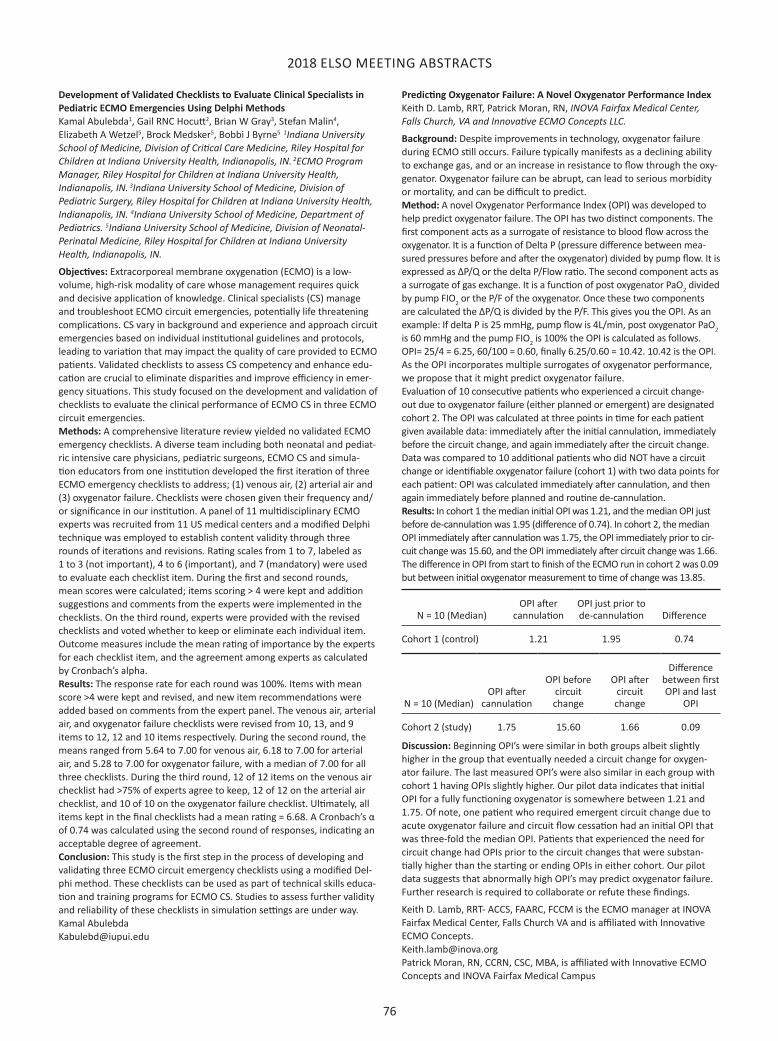
2018 ELSO MEETING ABSTRACTS
76
Development of Validated Checklists to Evaluate Clinical Specialists in Pediatric ECMO Emergencies Using Delphi MethodsKamal Abulebda1, Gail RNC Hocutt2, Brian W Gray3, Stefan Malin4, Elizabeth A Wetzel5, Brock Medsker5, Bobbi J Byrne5 1Indiana University School of Medicine, Division of Critical Care Medicine, Riley Hospital for Children at Indiana University Health, Indianapolis, IN. 2ECMO Program Manager, Riley Hospital for Children at Indiana University Health, Indianapolis, IN. 3Indiana University School of Medicine, Division of Pediatric Surgery, Riley Hospital for Children at Indiana University Health, Indianapolis, IN. 4Indiana University School of Medicine, Department of Pediatrics. 5Indiana University School of Medicine, Division of Neonatal-Perinatal Medicine, Riley Hospital for Children at Indiana University Health, Indianapolis, IN.
Objectives: Extracorporeal membrane oxygenation (ECMO) is a low-volume, high-risk modality of care whose management requires quick and decisive application of knowledge. Clinical specialists (CS) manage and troubleshoot ECMO circuit emergencies, potentially life threatening complications. CS vary in background and experience and approach circuit emergencies based on individual institutional guidelines and protocols, leading to variation that may impact the quality of care provided to ECMO patients. Validated checklists to assess CS competency and enhance edu-cation are crucial to eliminate disparities and improve efficiency in emer-gency situations. This study focused on the development and validation of checklists to evaluate the clinical performance of ECMO CS in three ECMO circuit emergencies.Methods: A comprehensive literature review yielded no validated ECMO emergency checklists. A diverse team including both neonatal and pediat-ric intensive care physicians, pediatric surgeons, ECMO CS and simula-tion educators from one institution developed the first iteration of three ECMO emergency checklists to address; (1) venous air, (2) arterial air and (3) oxygenator failure. Checklists were chosen given their frequency and/or significance in our institution. A panel of 11 multidisciplinary ECMO experts was recruited from 11 US medical centers and a modified Delphi technique was employed to establish content validity through three rounds of iterations and revisions. Rating scales from 1 to 7, labeled as 1 to 3 (not important), 4 to 6 (important), and 7 (mandatory) were used to evaluate each checklist item. During the first and second rounds, mean scores were calculated; items scoring > 4 were kept and addition suggestions and comments from the experts were implemented in the checklists. On the third round, experts were provided with the revised checklists and voted whether to keep or eliminate each individual item. Outcome measures include the mean rating of importance by the experts for each checklist item, and the agreement among experts as calculated by Cronbach’s alpha.Results: The response rate for each round was 100%. Items with mean score >4 were kept and revised, and new item recommendations were added based on comments from the expert panel. The venous air, arterial air, and oxygenator failure checklists were revised from 10, 13, and 9 items to 12, 12 and 10 items respectively. During the second round, the means ranged from 5.64 to 7.00 for venous air, 6.18 to 7.00 for arterial air, and 5.28 to 7.00 for oxygenator failure, with a median of 7.00 for all three checklists. During the third round, 12 of 12 items on the venous air checklist had >75% of experts agree to keep, 12 of 12 on the arterial air checklist, and 10 of 10 on the oxygenator failure checklist. Ultimately, all items kept in the final checklists had a mean rating = 6.68. A Cronbach’s α of 0.74 was calculated using the second round of responses, indicating an acceptable degree of agreement.Conclusion: This study is the first step in the process of developing and validating three ECMO circuit emergency checklists using a modified Del-phi method. These checklists can be used as part of technical skills educa-tion and training programs for ECMO CS. Studies to assess further validity and reliability of these checklists in simulation settings are under way.Kamal [email protected]
Predicting Oxygenator Failure: A Novel Oxygenator Performance IndexKeith D. Lamb, RRT, Patrick Moran, RN, INOVA Fairfax Medical Center, Falls Church, VA and Innovative ECMO Concepts LLC.
Background: Despite improvements in technology, oxygenator failure during ECMO still occurs. Failure typically manifests as a declining ability to exchange gas, and or an increase in resistance to flow through the oxy-genator. Oxygenator failure can be abrupt, can lead to serious morbidity or mortality, and can be difficult to predict.Method: A novel Oxygenator Performance Index (OPI) was developed to help predict oxygenator failure. The OPI has two distinct components. The first component acts as a surrogate of resistance to blood flow across the oxygenator. It is a function of Delta P (pressure difference between mea-sured pressures before and after the oxygenator) divided by pump flow. It is expressed as ∆P/Q or the delta P/Flow ratio. The second component acts as a surrogate of gas exchange. It is a function of post oxygenator PaO2 divided by pump FIO2 or the P/F of the oxygenator. Once these two components are calculated the ∆P/Q is divided by the P/F. This gives you the OPI. As an example: If delta P is 25 mmHg, pump flow is 4L/min, post oxygenator PaO2 is 60 mmHg and the pump FIO2 is 100% the OPI is calculated as follows.OPI= 25/4 = 6.25, 60/100 = 0.60, finally 6.25/0.60 = 10.42. 10.42 is the OPI.As the OPI incorporates multiple surrogates of oxygenator performance, we propose that it might predict oxygenator failure.Evaluation of 10 consecutive patients who experienced a circuit change-out due to oxygenator failure (either planned or emergent) are designated cohort 2. The OPI was calculated at three points in time for each patient given available data: immediately after the initial cannulation, immediately before the circuit change, and again immediately after the circuit change. Data was compared to 10 additional patients who did NOT have a circuit change or identifiable oxygenator failure (cohort 1) with two data points for each patient: OPI was calculated immediately after cannulation, and then again immediately before planned and routine de-cannulation.Results: In cohort 1 the median initial OPI was 1.21, and the median OPI just before de-cannulation was 1.95 (difference of 0.74). In cohort 2, the median OPI immediately after cannulation was 1.75, the OPI immediately prior to cir-cuit change was 15.60, and the OPI immediately after circuit change was 1.66. The difference in OPI from start to finish of the ECMO run in cohort 2 was 0.09 but between initial oxygenator measurement to time of change was 13.85.
N = 10 (Median)OPI after
cannulationOPI just prior to de-cannulation Difference
Cohort 1 (control) 1.21 1.95 0.74
N = 10 (Median)OPI after
cannulation
OPI before circuit change
OPI after circuit change
Difference between first OPI and last
OPI
Cohort 2 (study) 1.75 15.60 1.66 0.09
Discussion: Beginning OPI’s were similar in both groups albeit slightly higher in the group that eventually needed a circuit change for oxygen-ator failure. The last measured OPI’s were also similar in each group with cohort 1 having OPIs slightly higher. Our pilot data indicates that initial OPI for a fully functioning oxygenator is somewhere between 1.21 and 1.75. Of note, one patient who required emergent circuit change due to acute oxygenator failure and circuit flow cessation had an initial OPI that was three-fold the median OPI. Patients that experienced the need for circuit change had OPIs prior to the circuit changes that were substan-tially higher than the starting or ending OPIs in either cohort. Our pilot data suggests that abnormally high OPI’s may predict oxygenator failure. Further research is required to collaborate or refute these findings.
Keith D. Lamb, RRT- ACCS, FAARC, FCCM is the ECMO manager at INOVA Fairfax Medical Center, Falls Church VA and is affiliated with Innovative ECMO [email protected] Moran, RN, CCRN, CSC, MBA, is affiliated with Innovative ECMO Concepts and INOVA Fairfax Medical Campus

2018 ELSO MEETING ABSTRACTS
77
Extracorporeal Cardiopulmonary Resuscitation in the Catheterization Laboratory is Associated with Superior ResultsMichael A. Hart1, Claire Donovan2, David Raile2, Ivan Chavez1, Yale Wang1, Michael Mooney1, Miranda Kunz2, Kelly Wilson2, Katarzyna Hryniewicz1 1Minneapolis Heart Institute at Abbott Northwestern Hospital, Minneapolis, MN 2Minneapolis Heart Institute Foundation, Minneapolis, MN
Background: In recent years, veno-arterial extracorporeal membrane oxygenation (VA-ECMO) has been used in cardiopulmonary resuscitation (eCPR) in selected patients. Few centers have published their experience with eCPR, leading to a paucity of literature on the clinical characteristics of this population and a lack of standardization in care. The aim of our study was to evaluate short and long-term outcomes of adult patients in cardiac arrest placed on VA-ECMO in the catheterization laboratory.Methods: We performed a retrospective analysis of adult patients in refractory cardiac arrest who underwent eCPR at the Minneapolis Heart Institute (MHI) at Abbott Northwestern Hospital from January 2012 to December 2017. Demographics, baseline clinical characteristics, and laboratory variables were obtained from electronic medical records. Activation of the VA-ECMO team to the catheterization laboratory for cannulation mirrored our standardized process for STEMI activations. We evaluated eCPR duration, arrest to ECMO flow time, total ECMO support time, and outcomes.Results: Twenty-seven adult patients underwent eCPR at the study site during the defined time period. Mean age was 59 and 18 (67%) were men. Fourteen (52%) patients had known hypertension, 10 (37%) had known coronary artery disease, and 17 (63%) had a history of tobacco use. Twenty (74%) patients suffered in-hospital arrest compared to 7 (26%) out-of-hospital arrests. All patients underwent manual or mechani-cal CPR with a LUCAS device. Median time from CPR to flow initiation was 46 (22, 71) minutes, and median duration of ECMO support was 103 (66, 145) hours. Nine (33%) patients required continuous renal replacement therapy (CRRT) during index hospitalization, and 3 (11%) patients bridged to LVAD. Both 30-day and 6-month survival was 67%.Discussion: Despite significant advances in temporary cardiopulmonary support, including cannulation techniques and equipment, survival of patients in cardiac arrest undergoing eCPR remains low. Compared to the most recent annual Extracorporeal Life Support Organization (ELSO) report, we achieved a superior rate of survival to discharge despite pre-ECMO resuscitation times similar to other studies. Likely contributions include: rapid initiation of high fidelity CPR with the LUCAS device; skilled and timely VA cannulation in the catheterization laboratory; a multidis-ciplinary approach to patient care, with early complication recognition and management; and longer duration of ECMO support, with reliance on stringent weaning parameters to ensure adequate myocardial recovery time. In addition, based on our data, we encourage early identification of patients who may fail weaning from VA-ECMO, thus prompting swift transition to durable mechanical support. Larger multicenter studies are needed to further evaluate the approach to patients in cardiac arrest who may be candidates for eCPR in an effort to standardize and better streamline care.Kelly [email protected]
The Metabolic Demands of Extracorporeal Membrane Oxygenation in Infants and ChildrenLinette Ewing1,2, Rogelio Ramirez2, JoAnne Starr2, Danny Lam2, Richard Mink1 1Harbor UCLA Medical Center, Torrance, California; 2Children’s Hospital Orange County, Orange, California
Introduction/Hypothesis: Both overfeeding and underfeeding critically ill children is problematic. The intake of excessive calories can lead to prolonged mechanical ventilator support, and insufficient calories can impede healing, leading to a loss of muscle mass. Extracorporeal mem-brane oxygenation (ECMO) provides cardiorespiratory support and may alter metabolic demands. Although studies have shown the metabolic needs of neonates requiring ECMO are similar to those of healthy indi-viduals, similar data are not available for older infants and children. The hypothesis of this study was that infants and children on ECMO have the same caloric needs as age matched healthy infants and children.Methods: Patients ages 30 days to 21 years requiring ECMO were included in this study. Having an uncuffed endotracheal tube, active sei-zures, or a ketogenic diet were exclusion criteria. The indirect calorimeter was used to measure VO2 and VCO2 from the mechanical ventilator and then from the oxygenator. Blood gases were obtained pre- and post-oxygenator and the PO2 and PCO2 was converted to O2 and CO2 content in the blood using the 0149 Physiome model (Dash & Bassingthwaighte, 2015) multiple by ECMO blood flow to get the VO2 and VCO2 from the oxygenator. The VO2 and VCO2 values from the ventilator and oxygen-ator were added together and placed in the Weir equation to calculate the resting energy expenditure (REE) in kcal/day. Measurements were obtained 1 day after ECMO cannulation and 1 day prior to and one day post-decannulation. Data were compared to the predicted REE of children using the Schofield equation.Results: Four patients have been enrolled to date ranging in age from 31 days to 9 years. In 7 of the 8 measurements obtained during ECMO, the REE exceed that predicted for healthy children by 116% to 450%. One patient was declared brain dead on ECMO and had a REE 4% of predicted on the day of pronouncement. All remaining patients post decannulation had a REE lower than predicted at 32%, 39%, and 51% of healthy children.Conclusions: Resting energy expenditure can be successfully determined in infants and children on ECMO when VO2 and VCO2 are measured from the ventilator and oxygenator. REE results while on ECMO were greater than predicted which may be due to high protein catabolism. REE results immediately after ECMO are much lower than predicted most likely due to sedation. For optimal care, individual testing should be performed to match metabolic needs to the number of calories provided.Contact:Linette [email protected]

2018 ELSO MEETING ABSTRACTS
78
Incidence of Heparin-Induced Thrombocytopenia in Patients with Temporary Mechanical Circulatory Support DevicesDana Attar,1 Brittany Lines,1 Melissa McCarty,1 Victor Coba,1 Jona Lekura,1 Zachary Smith,1 Long To1 1Henry Ford Hospital, Detroit, Michigan
Introduction: Temporary mechanical circulatory support (MCS) devices are used for hemodynamic support in patients with cardiogenic shock. MCS devices are prone to thrombosis and require anticoagulation, often with unfractionated heparin. Heparin exposure and device related throm-bocytopenia complicate the diagnosis of heparin-induced thrombocyto-penia (HIT) and may lead to unnecessary treatment with direct thrombin inhibitors (DTI). To provide safe and effective care for patients with newly implanted MCS devices, the incidence of laboratory confirmed HIT needs to be further characterized.Objective: To determine the incidence of HIT in patients with newly-implanted temporary MCS devicesStudy Design: Retrospective, cohort studyMethods: This study included patients admitted to the cardiovascular intensive care unit with a newly-implanted temporary MCS device at Henry Ford Hospital from January 2014 to January 2017. MCS devices included in this study were extra-corporeal membrane oxygenation (ECMO) and Impella®. The primary endpoint was the incidence of labora-tory confirmed HIT. Definite HIT was defined as a positive serotonin release assay (SRA) or heparin-induced platelet antibody assay (HIPA) with an optical density (OD) greater than or equal to 2. Probable HIT was defined as an OD between 1.01 and 1.99 +/- SRA intermediate, while possible HIT was defined as an OD between 0.41 and 1 +/- SRA intermedi-ate. HIT negative was defined as an OD less than 0.4 or a negative SRA. Non-HIT related thrombocytopenia was defined as an absence of HIPA or SRA order. In cases where SRA and OD were both ordered but SRA interpretation conflicted with OD interpretation, HIT category was deter-mined based on SRA due to this assay’s greater sensitivity and specificity compared to HIPA. However, if SRA result was intermediate, then the like-lihood of HIT was determined based on OD instead. Secondary endpoints include use of DTI such as argatroban in patients with unconfirmed HIT, incidence of HIT amongst each of the MCS devices, and utility of the 4T score, HIT Expert Probability (HEP) score, and Lillo-Le Louёt model in pre-dicting the presence of HIT. Categorical variables are presented as n (%). Continuous variables are presented as mean (standard deviation (SD)) or median (interquartile range), as appropriate.Results: A total of 182 cases with newly-implanted temporary MCS devices met inclusion criteria: 122 Impella® (67%) and 60 ECMO (33%). Diagnostic HIT assays were ordered in 43 of 182 cases (23.6%). In this population, 43 HIPA and 16 SRA assays were ordered. The median OD was 0.19 (0.11 – 0.52); two SRAs were positive and 14 were negative. Of the 182 cases, two had definite HIT (1.1%), zero had probable HIT, two had possible HIT (1.1%), and 39 were HIT negative (21.4%). The incidence of definite HIT was 1.7% (1/60) in the ECMO group and 0.82% (1/122) in the Impella® group. For patients with diagnostic HIT assays ordered, the 4T score sensitivity was 0.25 (0.01 – 0.78) and specificity was 0.79 (0.63 – 0.90). By comparison, the sensitivity of the HEP score was 0.50 (0.09 – 0.91) and specificity was 0.62 (0.45 – 0.76). Argatroban was administered to 16 patients with suspected HIT (37.21%) and was continued for an average of 6.25 days (SD 8.25); two of these patients were later deter-mined to be HIT negative.Conclusion: Definite or probable HIT occurs infrequently with ECMO and Impella devices, yet over one-third of patients receive alternative anti-coagulation despite being negative for HIT. Current HIT scoring systems failed to accurately screen for HIT in temporary support MCS devices and a modified MCS HIT scoring model is needed. Further analysis is needed to assess the implications of over-diagnosing HIT and the subsequent initiation of alternative anticoagulants.Address correspondence to Long To, [email protected]
Implementing a Multidisciplinary and Multimodal Approach to an ECLS Specialist ProgramPlourde, M; Kallestad, R. Perfette, B.; Greco, S; Nasenbeny, K; Cheng, A.; von Homeyer, P.; University of Washington Medical Center (UWMC), Seattle WA
Purpose: To use a multidisciplinary approach to implement an Extracor-poreal Life Support (ECLS) Specialist course, using didactic, simulation and online learning to gain confidence and competence to transition to an ECLS Specialist model of care.Background: Traditionally, ECLS is a challenging therapy requiring skilled multidisciplinary management of high acuity critically ill patients. Since 2012, UWMC has used a perfusionist model to manage the ECLS pump. The success of the program prompted a switch from a perfusion model to an ECLS Specialist model to accommodate for growth and to care for patients consistently and cost-effectively.Methods: An ECLS Educator was hired to collaborate with physician, per-fusion, and nursing, to review best practices and develop a 40-hour ECLS Specialist course. Twenty five nurses and respiratory therapists were hired as Specialists. The course incorporated online learning(4 hours), didactic content(16 hours) with simulation-based learning(16 hours) including competency testing(4 hours). Course objectives were established by using ELSO guidelines and physician experts and were validated through course evaluations. Four courses were administered over four months to keep class sizes small. Competency and confidence levels were evaluated using simulation. Knowledge assessment was performed using written exam. After the course, each Specialist spent a minimum of 36 hours completing a skills checklist with Perfusion managing an ECLS patient. Quarterly simu-lation sessions were designed to maintain confidence and competence of each Specialist.Evaluation/Outcomes: Overall course evaluation results showed 95% of course objectives were met. Knowledge assessment scores were required to be >80%. Evaluation of skills competency at the end of the course resulted in 100% of staff deemed competent. Six months post course competency was measured based on ability to simulate skills within a timely fashion. Competency was defined by priming the circuit in under 20 minutes (80% of Specialists achieved this goal) and removing large air in under 15 minutes (90% of Specialists achieved this goal). Confidence was defined at 6 month using a self-reporting survey which measured scores on a scale of 1 to 10 (1 = high confidence, 10 = low confidence). Overall confidence level resulted in a score of 6, with scores for priming the pump (mean=5.7), managing large air situations (mean=6.5) and hand cranking (mean=5.4). Confidence scores were re-evaluated at the end of the six month simulation session resulting in improved confidence scores with a mean score of 4: priming the pump (mean=3.8); managing large air situations (mean=3.6); hand cranking (mean=3.5).Conclusions: An ECLS Specialist model of caring for these patients can be successfully implemented with a multidisciplinary team approach to plan-ning and implementation and multimodal approach to learning achieved improvements in confidence levels and competency levels.

2018 ELSO MEETING ABSTRACTS
79
Managing severe accidental hypothermia with and without ECLS: Patient outcomes over eleven years from a single centerMelissa Touroutoutoudis, DO,1 Jessica Boland, MD,1,5 Parvati Singh, MD,1 Michael Jaeger, MD,1 Dirck Rilla LP, CCP,2 Beth Heather RN,3 Domenico Calcaterra MD, PhD,4 Matthew Prekker, MD, MPH1,5,6 1Department of Medicine, Hennepin County Medical Center, Minneapolis, MN. 2Specialty Care, Minneapolis, MN. 3Critical Care Services, Hennepin County Medical Center, Minneapolis, MN. 4Cardiovascular Surgery, Minneapolis Heart Institute at Abbott Northwestern Hospital, Minneapolis, MN. 5Department of Emergency Medicine, Hennepin County Medical Center, Minneapolis, MN. 6University of Minnesota Medical School, Minneapolis, MN
Background: Extracorporeal rewarming using peripheral veno-arterial (V-A) ECMO, when available, is increasingly favored for the resuscitation of patients with cardiac arrest due to severe hypothermia; this modality can be initiated at the bedside, allows for rapid internal rewarming, and facilitates adequate perfusion and gas exchange. We sought to describe the experience of managing severe accidental hypothermia at an urban referral center in the northern U.S. over an eleven-year period, comparing patients managed with and without ECLS.Materials/Methods: We conducted a retrospective review of 31 adult patients presenting to a single center between March 2007 and April 2018 with severe hypothermia (core body temperature < 28°C). We cap-tured patient and encounter characteristics and clinical outcomes, includ-ing rewarming methodology and survival. The primary outcome was survival to hospital discharge, analyzed descriptively between patients managed with and without ECLS.Results: The initial core temperature was similar, on average, among the 15 patients rewarmed with ECLS (25.1°C) and the 16 patients rewarmed with non-ECLS modalities (25.6°C). As compared to the non-ECLS group, the ECLS group tended to be younger (mean age 47 vs 51 years, respec-tively) and had a higher prevalence of hypothermic cardiac arrest (87% vs 38%, respectively). Non-ECLS rewarming modalities included warmed-fluid lavage of body cavities (bladder [N=10], gastric [3], thoracic [3], and peritoneal [2]) and/or use of an intravascular temperature management catheter (N=15). Despite similar time-to-normothermia in the ECLS and non-ECLS groups (mean 404 vs 427 minutes, respectively- excluding patients who underwent therapeutic hypothermia [N=2] or were declared dead prior to reaching 36°C [N=2]), survival to hospital discharge was higher in the ECLS group (12/15, 80%) than the non-ECLS group (8/16, 50%). The most recent 8 patients with severe hypothermia selected for ECLS rewarming (beginning in December 2016) had V-A ECMO initiated at the bedside in the Emergency Department; 75% survived to hospital discharge neurologically-intact.Conclusions: In this single-center experience over 11 winters in the northern U.S., clinicians utilized both ECLS and non-ECLS rewarming strategies for patients with severe accidental hypothermia. Acknowledg-ing the selection bias inherent in this observational study, we observed higher survival to hospital discharge in the ECLS-rewarmed group, despite a greater burden of cardiac arrest, as compared to the non-ECLS group; patient age and initial core body temperature were similar between the groups.Melissa Touroutoutoudis, email [email protected].
RED BOOK BASED ECMO EDUCATION FOR BEDSIDE ICU NURSESParker MJ, Cardona MF, Gibbs AD, Weckbacher JL 1The ECMO Department, Medical University of South Carolina, Charleston, SC
Introduction: ECMO is the most invasive, demanding and life-saving therapy offered to ICU patients. Despite significant risk and reward, ECMO’s low-volume, high-acuity nature in many hospitals coupled with a historic lack of guidelines for training bedside ICU nurses on ECMO-specific nursing care left many RNs under-educated on how to best care for these critical patients. Integrating the nursing management chapters added to the 5th edition ELSO Red Book, ECMO for Critical Care courses were built to meet the specific educational needs for each ICU’s nurses based on the ECMO modalities and patient populations they care for at the Medical University of South Carolina (MUSC).Background: Data abounds to support the relationship between nurse educa-tion and advancing nursing practice with improved patient outcomes. Despite this, guidelines for how to best educate bedside ICU nurses to care for ECMO patients has not been readily available. Recognizing this educational gap, ELSO Red Book recommendations for nursing management (welcome addi-tions to the 5th edition) were used to build an “ECMO for Critical Care Nurses” course targeting bedside nurses with greater than one year of experience who provide care for ECMO patients. The creation and delivery of this educa-tion was especially timely as the MUSC ECMO Program (already an ELSO Center of Excellence) was expanding ECMO care into additional adult ICUs and pursuing the ELSO Path to Excellence Award for its’ adult program.Course Content: Four specific versions of the “ECMO for Critical Care Nurses” course were designed to teach bedside ICU nurses in each type of unit where ECMO care is delivered - NICU, PICU, PCICU and the adult ICUs – how to best care for their specific patient populations. Each course is four hours long and is approved for four CNEs by the South Carolina Nurses Association. ECMO department structure and roles, VA and VV physiology and special consider-ations, inclusion and exclusion criteria, ECMO pump components, pre-ECMO and ECMO cannulation nursing roles, weaning, trialing off, and decannula-tion and long term outcomes are taught and tailored to each version of the course. Emphasis is placed on the impact of ECMO on each body system and the related assessment, nursing management and interventions according to Red Book recommendations and MUSC standards of practice. Topics unique to patient populations (i.e. ECMO in single ventricles) are presented. Interac-tive course conversation regarding experiences caring for ECMO patients enrichens and exemplify teaching points. Hands-on time deepens under-standing of pump/patient interface and provides opportunity for nurses to practice emergency response in a low-stress setting.Evaluation and Outcomes: Eighty-eight bedside ICU nurses across six ICUs have completed the “ECMO for Critical Care Nurses” course since summer 2017. 100% of participants reported intent to use course information in practice settings. Comments elaborated on appreciation for the educa-tional opportunity, increased confidence in caring for ECMO patients, and ways RNs plan to improve their care of ECMO patients. Pre- and post- Leikart scale reports showed >1.5 point increase in measured categories: understanding ECMO physiology, recognition of ECMO pump components and their purposes, ECMO-specific multisystem assessment and nursing care for their patient population, bedside nurses’ roles in starting and stopping ECMO, and overall confidence in caring for ECMO patients. ECMO knowledge gain scores (as measured by pre- and post- course assessments) increased from 54% to 91% in the Children’s Hospital group and from 23% to 78% for adult ICU nurses.Expanding Education: To integrate this new educational standard into existing nursing education, completion of the “ECMO for Critical Care Nurses” course is a highly prioritized educational requirement prior to caring for ECMO patients in Children’s Hospital ICUs as a part of the MUSC Center of Excellence ECMO program, and will become mandatory by the end of 2018. It is a mandatory training for nurses preparing to care for ECMO patients in those adult ICUs included where ECMO is expanding as a part of the MUSC Path to Excellence Adult ECMO program. Annual competency requirements are in development. Following the success and positive word-of-mouth of the course’s relevance and quality, requests for additional similar education for nurses in the pediatric and adult cath labs, ECHO technicians, physician assistants and nurse prac-titioners, and residents and fellows have been received and similar tailored education is being designed for those care providers.

2018 ELSO MEETING ABSTRACTS
80
Does Pre-Procedural pH Affect Outcomes in Veno-Arterial Extracorporeal Membrane Oxygenation Patients with Myocardial Infarction?Michael Healy, Niharika Boinpally, Kennedy Gallagher, James K. Wu, MD Division of Cardiothoracic Surgery, Lehigh Valley Health Network, Allentown, PA, United States.
Objectives: This was a retrospective study of myocardial infarction (MI) patients who required veno-arterial extracorporeal membrane oxygen-ation (VA-ECMO) support, including those that underwent extracorporeal cardiopulmonary resuscitation (ECPR). VA-ECMO is an effective form of mechanical circulatory support for patients with cardiac failure. This study analyzed the relationship between initial pH levels and outcomes of patients.Methods: The analysis was conducted on all patients who presented with a MI and subsequently required VA-ECMO between 2013 and 2018. This includes a total of 34 patients with a mean age of 57.8 years [20,84] and a gender distribution of 76% males to 24% females. Demographic informa-tion, related variables, and procedure outcomes were recorded.Results: The findings showed that of the 34 patients who underwent ECMO, 35% had an outcome of survival to discharge. Of the 15 ECPR patients, only 6.7% of patients survived while 58% of the 19 Non-ECPR patients survived. This study demonstrated that patients with a pH lower than 7.02 within 24 hours before ECMO cannulation did not survive. On average, patients who survived had a higher initial pH than those who did not survive.Conclusions: Though limited by the number of cases, this study demon-strates that initial pH levels 24 hours before ECMO cannulation could be indicative of survival within this cohort. In addition, overall survival of MI patients who underwent ECPR is very poor.Contact Info: Michael HealyEmail: [email protected]
[POSTER] CLINICAL VALIDATION OF SIMULATED NIRS SrO2 VALUES DURING VENO-ARTERIAL ECLS WITH PERIPHERAL CANNULATIONMichael D. Johnson Swedish Hospital, Seattle, Washington
Objective: Clinicians are using cerebral and somatic near-infrared spec-troscopy (NIRS) regional tissue hemoglobin oxygen saturation (SrO2) to monitor the distribution of oxygenated blood and the adequacy of tissue oxygenation during extra-corporeal life support (ECLS). For educational purposes it is useful to simulate the effects of patient pathology and ECLS circuit intervention on patient NIRS values. A model to simulate NIRS values during V-A ECLS was created and the simulator output values were compared to NIRS values observed during adult ECLS procedures.Method: An evidence-based physiologic mathematical model was created to simulate SrO2 levels during different modes of ECLS. The NIRS SrO2 esti-mating model was integrated into the CALIFIA® (www.biomedsimulation.com) patient simulator. After qualifying for exemption with the institu-tional review board, deidentified clinical data snap shots were collected from five adult femoral-cannulated veno-arterial ECLS patients to set-up scenarios to emulate their ECMO run in the CALIFIA simulator. The NIRS values generated by CALIFIA were statically compared to the observed patient NIRS values. Bland-Altman techniques were employed to compare the simulator predicted SrO2 values to the actual patient SrO2 values under various critical care situations.Results: The CALIFIA estimated NIRS values were not significantly dif-ferent from the actual patient values. The correlation between patient observed NIRS values and simulator generated SrO2 values is significant. The comparison between the simulated SrO2 values and the patient observed SrO2 values passed the Bland-Altman tests.Discussion: High-fidelity simulation of NIRS monitoring during ECLS procedures is useful to orient clinicians to the monitoring device and the expected SrO2 values in the clinical setting. Clinicians can practice using SrO2 values to evaluate the adequacy of patient and ECLS ventilation, and the distribution of ECLS circuit oxygenated blood in the aortic arch during partial V-A ECLS. Clinicians can demonstrate their competency to use and interpret NIRS monitoring to manage ECLS patients by manipulating independent variables such as ECLS blood flow, cardiac output, gas flow, ventilator settings, and cannulation sites.
Reference1. Wong JK, et al. Cerebral and lower limb NIRS in adults on ECMO.
Artificial Organs. 2012;36:659.2. Benni PB, et al. A validation method for NIRS based tissue oximeters
for cerebral and somatic tissue oxygen saturation measurements. J Clin Monit Comput. 2018;32:269.
3. Mariappan R. et al. Cerebrovascular reactivity to carbon dioxide under anesthesia: A qualitative systemic review. J Neurosurg Anes-thesiol. 2015;27:123.
4. Stevens MC, et al. Flow mixing during peripheral veno-arterial ECMO – A simulation study. J Biomechanics. 2017;55:64.
5. Alwardt CM, et al. Regional perfusion during V-A ECMO: A case report and educational modules on the concept of dual circulations. J Extracorpor Technol. 2013;45:187.
6. Lucero AA, Addae G, Lawrence W, et al. Reliability of muscle blood flow and oxygen consumption response from exercise using near-infrared spectroscopy. Exp Physiol. 2018; 103:90.
7. Willie CK, Tzeng Y-C, et al. Integrative regulation of human brain blood flow. J Phyisol. 2014;592:841.
8. Toya T, Fujita T, Fukushima S, Shimahara Y, et al. Efficacy of regional saturation of oxygen using near-infrared spectroscopy for lower limb ischemia during minimally invasive cardiac surgery. J Artificial Organs 2018. https://doi.org/10.1007/s10047-018-1057-y
Contact: Johnson, Michael D [email protected]

2018 ELSO MEETING ABSTRACTS
81
A Practice Change Based on Necessity: A Single Center’s Five-Year Experience Using Plasmalyte Pre-Primed Circuits up to Sixty Days OldML Heard1, JD Davis1, SF Wagoner1, CA Carroll2, ML Paden2, and JD Fortenberry2 1Children’s Healthcare of Atlanta, Atlanta, Georgia; 2Emory University, Atlanta, Georgia
Introduction: Children’s Healthcare of Atlanta began a practice of pre-priming and storing ECMO circuits for 30 days in the 1990’s (1). Years of practical experience demonstrated safety and efficacy of this policy. In 2013, a shortage of the Maquet Adult Quadrox iD (Getinge Group, Wayne, NJ) oxygenators prompted our center to evaluate the need to store cir-cuits longer since no other oxygenators were immediately available. Royal Hospital for Sick Children (Glasgow, Scotland) had completed a study in 2011 documenting the microbiology, plasticizer levels and oxygenator performance of pre-primed circuits up to 60 days (2). We decided to implement this practice immediately to reduce wastage of oxygenators. We report our single center experience with this practice.Methods: In August 2013 we began storing our pre-primed circuits for 60 days. All circuits are assembled under sterile conditions in an isolated room and primed with Plasmalyte A (Baxter International, Inc., Deerfield, Il). Once primed in a standard practice, the circuit is maintained in a non-circulating mode, stored in a limited access room and covered with an equipment cover. The circuit is visually checked every 12 hours by the ECMO primer and integrity of the circuit is verified. A tracking sheet with all lot numbers and types of disposables used is maintained for each pre-primed circuit the entire time it is stored. The date used and the associ-ated patient are documented and maintained by the ECMO department.Results: From August 2013 through June 2018, 390 ECMO circuits were primed and stored for up to 60 days. The average length of time a circuit was stored before patient use was 15.6 days (1-60, median 9 days). Thirty eight circuits were used between 31 and 60 days of storage time. There were no immediate oxygenator failures, other disposable failures, or infections that could be associated with the longer storage time. Thirteen of the 390 circuits (3%) were discarded unused after 60 days. Fifty one total circuits would have been discarded prior to the new 60 day storage policy and resulted in approximately $150,000 savings in circuitry costs over this time period.Conclusion: The implementation of a new practice where pre-primed circuits were maintained for 60 days came about due to a shortage of oxygenators without immediate availability of another product. This review of our experience demonstrates that a 60 day storage time for ECMO circuits in a high volume pediatric center can be accomplished with no apparent detrimental effects to the circuit components. Future studies will investigate patient centered outcomes of infection risk and morbidity associated with storage of Plasmalyte-A primed circuits stored for up to 60 days.
References:1. Kesser, K., Heard, M., Labuz, M., Pettignano, R., Dykes, F. (1998)
Pre-Primed Extracorporeal Circuit Survey. Presented poster at CNMC ECMO Symposium: ECMO & Advanced Therapies for Respiratory Failure. Keystone, CO.
2. Cornejo P, Torrance I, Lucas C, Williams C, Davidson M, Young D, Walker A, Gourlay T, Danton M (2011)
Microbiological and Plasticizer Analysis of “Ready to Use” Plasmalyte Primed ECMO Circuits Over a 60 day Period.
Implementation and Evaluation: The Use of Rapid Cycle Deliberate Practice Simulation TechniquesML Heard1, J Young1, SF Wagoner1, JD Davis1 and KB Hebbar2 1Children’s Healthcare of Atlanta, Atlanta, Georgia; 2Emory University, Atlanta, Georgia
Introduction: Teaching new techniques for managing emergency procedures can be difficult, particularly since many procedures must be performed in a specific order to avoid patient harm. Simulation is commonly employed to practice and reinforce skills for extracorporeal membrane oxygenation, ECMO (1). Rapid Cycle Deliberate Practice (RCDP) is a simulation training methodology that uses repetitive performance of skills until proficiency is achieved (2). This technique is useful in teaching sequential procedures, where one must complete the steps in a specific order, such as an algorithm. We describe the use of RCDP to train a new technical skill to ECMO specialists at Children’s Healthcare of Atlanta.Methods: Our ECMO program recently removed the Bigger Better Blad-derTM from our centrifugal circuits due to concerns over clot migration into the centrifugal pump head. This precipitated a need to change the procedure for removal of accidental air entrainment into the venous line. The procedure was taught at an annual simulation activity. ECMO Specialists were required to sign up for the activity in groups of three. One specialist was assigned as the Primary, one specialist was assigned as a Helper, and the third specialist was assigned to Observe.RCDP technique was used to teach the new algorithm for air removal and involved the creation of four hard stops (key steps that cannot be skipped) and three soft stops. During a ‘hard stop’ learners were coached on proper procedure and then asked to start the simulation from the beginning. This continues until the hard stop is practiced perfectly. ‘Soft stops’ are a pause, used to correct a minor aspect of the algorithm, fol-lowed by resumption of the simulation where it had been stopped. These stops were also used to provide positive feedback. Coaching with positive reinforcement or constructive criticism was delivered by experts in ECMO trained simulation debriefing and facilitation. Crisis Resource Manage-ment (CRM) skills were reinforced during this procedure, as this is an institutional requirement of all staff during Simulation.Results: RCDP simulation debriefing demonstrated each specialist’s confi-dence in their ability to manage venous air entrainment into the centrifu-gal pump head. Three months later, annual water drills were required for all Specialists. At that time, the trainer evaluated each specialist’s ability to complete the procedure. A 21-step evaluation form, inclusive of CRM skills was used to evaluate each individual. The specialists were placed into their original three groups: Observers had the worst retention of the procedure with an average recall of 45% of the steps required (0-90%, median 42%) with four recalling ≤ 3/21 steps. Helpers had slightly better recall with an average of 54% (0-95%, median 66%), with most recalling > 4 /21 steps and only one who didn’t have any memory of the procedure. Finally, the Primary group had the best memory at 80% (52-95%, median 78), with every specialist recalling at least 15/21 steps.Conclusion: RCDP techniques are a superb training methodology for ECMO procedures. Many procedures require knowledge and memoriza-tion of specific steps in sequence, making this technique well suited to teach or reinforce skills. The evaluation of our three groups demonstrates that each individual must have direct feedback with RCDP; observation or a helping role does not afford the same knowledge or retention.
References:1. Tofil, N (2017). Rapid Cycle Deliberate Practice (RCDP).2. Burton, K., Pendergrass, T., Byczkowski, T., Taylor, R., Moyer,
M., Falcone, R. and Geis, G. (2011). Impact of Simulation-Based Extracorporeal Membrane Oxygenation Training in the Simulation Laboratory and Clinical Environment. Simulation in Healthcare: The Journal of the Society for Simulation in Healthcare, 6(5), pp.284-291.
Micheal Heard ~ [email protected]

2018 ELSO MEETING ABSTRACTS
82
Standardizing the Delivery of ECMO across a Large, Academic Medical CenterMonika Cardona MSN, RN1, Alicha Gibbs BSN, RN2, Mical Parker BSN, RN, MA3, Jessica Wechbacker BSN, RN4, Luca Paoletti MD5 1Medical University of South Carolina MUSC Health ECMO Department 2Medical University of South Carolina MUSC Health ECMO Department 3Medical University of South Carolina MUSC Health ECMO Department 4Medical University of South Carolina MUSC Health ECMO Department 5Medical University of South Carolina, Department of Medicine, Division of Pulmonary and Critical Care
Introduction: The provision of extracorporeal life support (ECLS) is a very complex, high risk procedure utilized in the care of critically ill patients in cardiac, pulmonary, or cardiopulmonary failure. Careful, strategic coordination of all aspects of this therapeutic modality optimizes this unique, life-saving therapy. Failure to do so can increase the propensity to negatively affect patient outcomes within this group of patients. If a health care system decides as an organization to offer ECLS to the patients it serves, multiple steps must be taken at the both the administrative and programmatic levels to ensure a safely structured program is in place prior to implementation. We report our center’s experience with the process of ECLS standardization.Experience: The provision and delivery of ECLS support across our aca-demic medical center, inclusive of a children’s and adult hospital, has its origins in 1991. Born out of the neonatal ICU, the original program was established with strategic structure, leadership, and dedicated ECMO Team. However, as ECLS support grew and expanded into adult medicine as a viable treatment option, the adult program became less formalized. Treatment varied across the system as ECMO was delivered apart from the oversight and coordination of the ECMO Department. This maneuver led to distinguishable differences in patient management and outcomes. Therefore in early 2017, a change initiative was identified by the hospital in order to standardize ECMO care utilizing a uniformed ECMO team used in both pediatrics and adults. The first step of the multiphasic process involved having programmatic leadership in the adult program with the assistance of the pediatric program. The second phase involved partnerships with perfusion services and other key physician leaders from the adult side of the medical center. These partnerships facilitated the creation of a decision-making process for management algorithms, order sets, equipment, education, and staffing models. Training sessions for staff and targeted implementation dates were inaugurated. Clear expecta-tions were dispersed to all involved with and affected by the process. This allowed all staff to be involved and aide in the overall success and sustain-ability of the program. The final phase of the process involved solidifying these relationships with time. As we are in this phase eight months later, all identified change agents continue to work collaboratively and in a synergistic manner for the standardization and to optimize patient out-comes. Since the implementation occurred, thirty-one adult patients have been cared for under the newly incepted ECLS care delivery model, with a 51% overall survival rate (6 pulmonary and 10 cardiac patients) with the following breakdown: Pulmonary 67% survival (6 out of 9 patients) and Cardiac 45% survival (10 out of 22 patients) compared to ELSO Interna-tional Outcomes July 2018 of 59% and 42% respectively.Conclusion: The standardization of the care for an ECLS patient ensures consistency and safety across an organization. It fosters predictability of care for the critically ill patient requiring advanced support in unpredict-able circumstances. Establishing a formal leadership hierarchy and crucial partnerships acts to initiate phase 1 of the three-phased proposed stan-dardization approach. From there, educational and staffing models can be incepted that will be strategically paired with selected equipment and disposables. In turn, impacting the propensity to positively affect overall patient outcomes in a positive manner.Monika F. Cardona, MSN, [email protected]
SUCCESSFUL USE OF EXTRACORPOREAL MEMBRANE OXYGENATION IN THE REVERSAL OF CARDIORESPIRATORY FAILURE INDUCED BY FULMINANT MYOCARDITIS: CASE REPORTSNguyen Minh Tien1, Le Vu Phuong Thy1 1City Children’s Hospital, Ho Chi Minh city, Vietnam
Introduction: Fulminant acute myocarditis (FAM) occurs rapidly, causes pump failure or lethal arrhythmias, leading to death by cardiogenic shock.Case Report: Case 1: a fifteen-year-old girl developed rapid onset tachy-cardia, hypotension and respiratory failure following a five-day episode of respiratory tract infection. The patient was treated with inotropes such as dopamine, dobutamine, epinephrine, and endotracheal intubation and ventilation after being diagnosed as fulminant myocarditis with cardio-genic shock. Due to deteriorating condition, patient was put on venous arterial extra corporeal membrane oxygenation (VA ECMO) and sup-ported for 72 hours along with supportive treatment. After improvement in ejection fraction, patient was weaned off ECMO, ventilator. Patient was discharged on 13rd day of admission in a stable condition with an ejction fraction of 55%.Case 2: a nine-year-old boy have had mild fever for two days, then developed rapidly convulsion, admitted in condition of lethargy, car-diopulmoary collapse with severe bradycardia (heart rate 30-35 bpm). The patient was diagnosed as fulminant myocarditis with A-V block type III, cardiogenic shock and treated with inotropes such as dopamine, dobutamine, epinephrine, and endotracheal intubation, ventilation and temporary pacemaker insertion. After that the patient became worsen with ventricular tachycardia, receiving anti-arrhythmic drugs such as lidocain, amiodaron but the rhythm did not revert to sinus rhythm, result-ing in profound shock. Thus, the patient was carried out venous arterial extra corporeal membrane oxygenation (VA ECMO) and supported for 96 hours along with supportive treatment, leading to his clinical betterment, weaning from ECMO and ventilator. Patient was discharged on 23rd day of admission in a stable condition with an ejction fraction of 58%.Discussion: The majority of children with myocarditis present with acute or fulminant disease, causing arrhythmias and hemodynamic compro-mise, requiring intensive and supportive care to maintain hemodynamic stability and adequate systemic perfusion, detection and treatment of arrhythmias. In cases unresponsive to significant supportive care (eg, intravenous inotropic support, mechanical ventilation) ECMO may be needed and used as salvage therapy during the acute illness prior to myocardial recovery.Conclusion: Timely extracorporeal membrane oxygenation (ECMO) sup-port in fulminant acute myocarditis (FAM) with arrythmias and shock due to myocarditis, along with medical treatment could prevent lethal outcomes.Keywords: Extracorporeal membrane oxygenation (ECMO), Fulminant acute myocarditis (FAM)

2018 ELSO MEETING ABSTRACTS
83
DNP Project: Reducing Adverse Events During Extracorporeal Membrane Oxygenation with In-Hospital Advanced Extracorporeal Life Support SpecialistsNicole R. O’Connor,1,2; Danielle K. Sebbens,2 1St. Louis Children’s Hospital, St. Louis MO; 2Rush University School of Nursing, Chicago, IL
Problem: Extracorporeal membrane oxygenation (ECMO) is a form of car-diopulmonary bypass used to support infants, children, and adults with severe pulmonary or cardiopulmonary failure. Despite advancements in technology and training, multiple life-threatening issues and emergen-cies can arise at any time during therapy. The lack of 24 hour in-hospital support for all emergency troubleshooting at St. Louis Children’s Hospital has been identified as a contributing problem in detailed root cause analysis for two separate adverse patient events on ECMO, both resulting in mortalities.Purpose: The purpose of this project is to reduce the risk of adverse patient events and improve the safety of the ECMO program at SLCH by ensuring clinicians with the knowledge and technical skill in ECLS are both regularly involved in the care of patients on ECMO to prevent issues and in-hospital to respond for questions, troubleshooting, and emergency backup.Methods: The output of this project includes the deployment of an advanced ECLS specialist in-hospital 24 hours a day. Trained to trouble-shoot and resolve ECMO issues and emergencies and develop expertise in daily management of patients on ECMO, the goal is to improve the quality of care and overall safety of the program. The deployment of the advanced ECLS specialist at SLCH included three phases: (1) selection and promotion of current ECMO specialists into the advanced ECLS special-ist role to provide 24 hour in-hospital coverage, (2) initial training during a structured four-week orientation, and (3) ongoing mentorship while competency is developed and strengthened.Results: There was a reduction in the percent of time perfusionists support ECMO when in-hospital and when responding by phone when on-call. Depending on the respondent, there were mixed results regard-ing the percent of time the perfusionist on-call responded to ECMO by returning to the hospital. The advanced ECLS specialist role improved both confidence that specialists will be able to troubleshoot and resolve issues and emergencies on ECMO and the ease of obtaining support at the bedside of patients on ECMO when needed. Results regarding any reduction in complications requires additional time to evaluate for effectiveness.Conclusions: Nurses with training and expertise to fill the role of the advanced ECLS specialist as their primary job are capable and effec-tive for improving the delivery of ECMO care and reducing the risk of adverse patient safety events when a perfusionist only staffing model is impractical.Contact: Nicole R. O’Connor: [email protected]
ECMO and Controlled Hypothermia in Neonates with Hypoxic Ischemic Encephalopathy and Persistent Pulmonary Hypertension: A Single-Center Case SeriesSwosti Joshi, MD1, Vilmaris Quinones Cardona, MD1,3, Rivkah Blutstein, MD2, Ogechukwu Menkiti, MD1,3 1Division of Neonatology, Department of Pediatrics, St. Christopher’s Hospital for Children; 2Department of Pediatrics, St. Christopher’s Hospital for Children; 3ECMO Program, Department of Pediatrics, St. Christopher’s Hospital for Children
Introduction: Controlled hypothermia (CH) for patients with hypoxic-ischemic encephalopathy (HIE) is standard of care after several stud-ies demonstrated reduction in adverse outcomes such as death and improved neurodevelopmental outcomes in neonates with moderate to severe HIE at 18 months of age. However, HIE is a multiorgan injury complicated by respiratory failure, persistent pulmonary hypertension of the newborn (PPHN), coagulopathy and systemic hypotension, which can all be aggravated by CH. In asphyxiated neonates with PPHN refractory to medical treatment, concomitant use of CH and ECMO has been shown to reduce mortality, however, the results on safety and associated morbidi-ties of published studies are conflicting.Case presentation: We present a case series and describe the com-mon short-term complications of ten neonates with HIE who required concomitant ECMO and CH therapies in our tertiary academic institution between 01/2011-05/2018. Infants with HIE included met criteria for CH with evidence of perinatal depression, >36 weeks gestational age, >1800 grams, <6 hours old, and 2 of the following: 10-minute Apgar of ≤ 5, cord gas or ABG/VBG with pH<7.0 or base deficit >16 within 1 hour of life, presence of seizures or evidence of encephalopathy. Additionally, infants met criteria for ECMO if they had evidence of cardiopulmonary decom-pensation refractory to medical management. The 10 neonates who required combined ECMO and CH therapies represented approximately 8% of total infants transferred to our institution for CH. All infants who qualified for CH were successfully initiated within 6 hours of life. PPHN was the most common indication for ECMO in this population. While all of the infants presented with severe PPHN, only half had systemic hypoten-sion. Modality of ECMO was evenly split with 50% VV and 50% VA ECMO. Confounding disorders included: culture proven gram negative sepsis in 1 infant (10%), pneumothorax in 2 infants (20%) and meconium aspiration syndrome in 3 infants (30%). Four infants (40%) suffered moderate to severe coagulopathy resulting in 2 cases with intracranial hemorrhage (ICH), 1 with ICH and pulmonary hemorrhage, and 1 had fulminant pulmonary hemorrhage. Length of hospital stay ranged from 24-74 days, excluding the 1 infant that died at 6 days of life. This resulted in a total mortality of 10%. Nine infants (90%) survived to decannulation with subsequent hospital discharge. While none of the infants required home oxygen or tracheostomy prior to discharge, three infants (30%) required gastrostomy tubes for nutritional support.Discussion: This case series illustrates that 90% of the asphyxiated neo-nates in this series with PPHN refractory to medical treatment benefited from combined CH for HIE and ECMO for PPHN with some comorbidi-ties, mainly bleeding and nutritional support. Our population suggests that coagulopathy and severe hemorrhage can be problematic and even devastating in this heparinized group undergoing CH, as in one of our cases which lead to death. Controlled hypothermia can increase the risk of bleeding via platelet dysfunction, alteration in the enzymes of coagula-tion cascade and increased fibrinolytic activity. Furthermore, rewarming could be associated with fluctuation of intracerebral blood flow which can further increase the risk for ICH. Careful monitoring of coagulation profile with gradual rewarming are strategies our institution has deployed to reduce bleeding risks and theoretically improve patient outcomes.Contact:Ogechukwu Menkiti, [email protected]

2018 ELSO MEETING ABSTRACTS
84
A model for a multidisciplinary approach to initiating an ECMO program in a small facilityPam Zinnecker MSNEd, BAN, RN, CCRN-K1 Mary Goldammer, BSN, RN, CCRN2 Amber Hellekson, BSN, RN, CCRN3 1ECMO Program Coordinator, Billings Clinic ICU, Billings, MT 2ECMO Specialist, Billings Clinic ICU, Billings, MT 3ECMO Specialist, Billings Clinic ICU, Billings, MT
Our hospital is a 304-bed hospital with a Level II Emergency and Trauma center, serving the state of Montana, western North Dakota and northern Wyoming. Billings Clinic is a recipient of Magnet Recognition from the American Nurses Credentialing Center and has achieved Beacon Status from the American Association of Critical Care Nurses.In 2016 an ECMO program was started as a backup support for LVAD patients, patients with difficulty coming off surgical bypass, and patients with severe respiratory issues.The ECMO program coordinator was the integral link to establishing and maintaining the ECMO program. Training was obtained at an ELSO course and with the ECMO specialist team to obtain knowledge and competency. The program coordinator collaborated with perfusionists to obtain the necessary equipment to initiate the program.ECMO specialists were trained by bringing in instructors from a consulting company for weeklong training. ECMO specialists remain competent via quarterly education/competency sessions and working with the program coordinator when caring for the ECMO patient.An ECMO trained/certified MD is required to manage a patient on ECMO. Physician training was organized through a private consultant who came to our facility twice to provide and enhance education. A core group of physicians also attended an ELSO ECMO course.In addition to educating the primary team, ancillary team members, including but not limited to respiratory therapists, pharmacists, labora-tory, dietary, physical and occupational therapy were educated to their role regarding ECMO and care of the patient on ECMO.A plan for staffing ECMO, charging for the service, and developing order sets was completed and we were ready to begin our journey.Staffing the ECMO patient is 1:1 or 1:2 depending on the overall acuity of the patient. An ECMO specialist is always part of the staffing matrix, following the orders and guidelines for patient management to optimize ECMO therapy including anticoagulation, sweep, FIOs and volume admin-istration. Non-specialist ICU nurses caring for the patient on ECMO have received education and complete annual competencies.Perfusionists round daily to monitor the circuits and are available as a bedside resource.The consensus of two ECMO certified physicians is required to place a patient on ECMO and the coordinator is notified to ensure there is specialist coverage. Cardiology and/or Cardiovascular surgery physicians place, monitor, and reposition the cannula daily. Intensivists are the primary ECMO MD when caring for VV patients and assist as requested with VA patients.Our facility can support one patient on ECMO at a time and we average one patient per month. Due to limited resources, including nursing and equipment, patients may require transfer to a larger facility after being placed on ECMO at our facility.We have experience greater survival rates for our VV ECMO patients in comparison with the ELSO database. We attribute our success to the multidisciplinary team.Conclusion: A well trained multidisciplinary team is needed to safely care for ECMO patients. Clearly defined roles and collaboration allowed us to initiate and maintain an ECMO program with desirable outcomes. We can provide local and regional support for patients in a large referral area.Contact: Pam Zinnecker; [email protected]
Institutional experience of long-term VA ECMO support in childrenStephanie Larsen1, Ravi Thiagarajan2, Jill Zalieckas3, Francis Fynn-Thompson4, Peta Alexander5 1MBA, RRT-NPS, Department of Respiratory Care/ECMO, Boston Children’s Hospital, Boston, MA 2MBBS, MPH, Department of Cardiology, Boston Children’s Hospital, Professor of Pediatrics, Harvard Medical School, Boston, MA 3MD, MPH, Department of Surgery, Boston Children’s Hospital, Harvard Medical School, Boston, MA 4MD, Department of Cardiac Surgery, Boston Children’s Hospital, Harvard Medical School, Boston, MA 5MD, Department of Cardiology, Boston Children’s Hospital, Instructor of Pediatrics, Harvard Medical School, Boston, MA
Introduction/Aim: ECMO technological advances have extended potential support duration for vulnerable patients. Our program has used long-term VA-ECMO support in selected children since 2012. We reviewed outcomes of VA-ECMO support greater than 30-days.Methods: All children (0-<18 years) supported on VA-ECMO for >30-days were identified from institutional database for this retrospective case series. Patients transitioned from VA-ECMO to other MCS within 30 days were excluded. Patient characteristics including age, diagnosis, ECMO support intention and strategy, and survival to hospital discharge were collected.Results: Eleven children met inclusion criteria (3 neonates, 2 infants, 6 children). The most common diagnostic category was neonatal respiratory [CDH, air leak (3)], followed by cardiac [4, ECPR (1)], pediatric respiratory (4). ECMO duration was median 978 (IQR 742, 1283) hours, longest run 4281 hours. Complications occurred in all patients (84 total), most com-monly circuit thrombosis (28, 33%), hemorrhage (9, 10%) renal dysfunc-tion (5, 5.6%), pneumothorax (4, 4.8%) neurological injury (3, 3.6%). Interventions were common, occurring in all runs (68 total), with circuit/component change the most common (46, 67.6%). Three patients were supported as bridge-to-transplantation, all others targeting end-organ recovery. The majority of patients died after redirection of care or within 24 hours of decannulation (6/12), 2 patients died greater than 24 hours post-decannulation,3 patients survived to hospital discharge, of which one was bridged-to-transplantation.Conclusion: Long-term VA-ECMO support of this vulnerable population is associated with high mortality, complications and circuit interventions. Further studies are required informing patient selection and optimizing outcomes.

2018 ELSO MEETING ABSTRACTS
85
We train with dummies so you don’t have to – Using simulation as the foundation for a rescue ECLS programTanya Spence CNS1, Steve Menzies CPC1, Dr. Jaime Blackwood 1Division of Pediatric Critical Care, Department of Pediatrics, University of Calgary
The Alberta Children’s Hospital (ACH) is a large, freestanding academic tertiary care center in Calgary, Alberta, Canada that serves a population of approximately three million people. Our Pediatric Intensive Care Unit (PICU) is a 15-bed medical surgical unit in a province where pediatric cardiac surgical services are regionalized to the Stollery Children’s Hos-pital approximately 300km away. Our rescue program is one where we cannulate and stabilize patients on VA and VV ECLS and then transport to our partner program at the Stollery Children’s Hospital. ACH is also home to the world-class simulation program KidSIM. We have been able to combine the KidSIM simulation program and our rescue ECLS program to incorporate simulation into all aspects of our ECLS educational cur-riculum. From development of the rescue ECLS program to continued maintenance of skills, high fidelity simulation is a core component in the success of our program.
The development of an ECLS program in a non-cardiac tertiary stand-alone pediatric hospital was a novel concept. The availability of high fidelity simulation allowed us to ensure the appropriate education to all multi-disciplinary team members, including those that had no prior experience or exposure to ECLS. We were able to publish our experience of using simulation to help establish our rescue program in the Journal of Pediatric Surgery “Evaluating the introduction of extracorporeal life support technology to a tertiary-care pediatric institution: Smoothing the learning curve through interprofessional simulation training” May 2015 Volume 50, Issue 5, Pages 798–804. In this study we demonstrated that simulation improved surgical performance in neck cannulation, improved technical and behavioural skills in ECLS specialists, improved team func-tioning, attitudes and confidence and allows a proficient transfer of those skills to the real medical domain.
Being a rescue ECLS program with no pediatric cardiac surgery patients our cannulation rates are low. On average we cannulate 8 patients per year. As such maintenance of skills is of utmost importance. We have developed a robust simulation curriculum that allows each key team member to practice aspects of their role during cannulation. All simula-tions are done in situ to maximize realism and are carried out in various areas of the hospital. These simulations test process and include the full multidisciplinary team. We have developed our own cannulation man-nequin that allows our general surgeons to practice neck cannulation and connection to circuit. We are then able to flow on pump and practice initial stabilization of the patient and troubleshooting skills.
We believe our ECLS simulation curriculum is unique as it is specific to our one of a kind rescue ECLS program. We have lots of experience to share with other centers who would like to incorporate simulation into their education curriculum.
Submission is for oral presentation
Developing a Rescue ECLS program at a Non-Cardiac Surgical Children’s HospitalTanya Spence CNS1, Steve Menzies CPC1, Dr. Jaime Blackwood 1Division of Pediatric Critical Care, Department of Pediatrics, University of Calgary
The Alberta Children’s Hospital (ACH) is a large, freestanding academic tertiary care center in Calgary, Alberta, Canada that serves a population of approximately three million people. Our Pediatric Intensive Care Unit (PICU) is a 15-bed medical surgical unit that sees approximately 1000 admissions annually. As pediatric cardiac surgical services are regionalized in Canada, our hospital does not have a pediatric cardiac surgery pro-gram; our patients are referred to another hospital in Alberta for surgical care approximately 300 km away.
In 2009, we determined a need for a rescue ECLS program for pediatric patients who needed emergent cardiac or respiratory support. We began establishing a program with a multi-disciplinary team that had little to no exposure to ECLS cannulation or the management of patients on ECLS. The process of developing our Rescue ECLS program took place over the next two years and our program became official in 2011.
Key components in the development and success of the program included:
• A robust educational curriculum centered around the use of simula-tion for training and skill maintenance of a large multidisciplinary team including the ability to cannulate and flow on circuit
• Training and maintaining competency of ECLS Specialists made up of PICU RNs and RTs and ensuring 24/7 coverage
• Establishing a rescue ECLS program and building a partnership with our referring pediatric cardiac surgical program for ongoing ECLS management
• Establishing a unique relationship with the adult perfusion program at a different hospital in Calgary to work in partnership with our ECLS Specialists
• A staged implementation approach to our program and its expansion
• A comprehensive Quality Improvement program that reviews all ECLS cases within our own program and in conjunction with the Quality Improvement program at the referral surgical site
• Development of a long term follow-up program for all ECLS patients• Sharing of coordinator responsibilities between a PICU Clinical
Nurse Specialist and Adult Perfusionist
The program was expanded over the course of last 7 years and we now offer ECLS and ECPR, VV and VA cannulations to all inpatient areas of our facility. Every child at ACH now has the same access to this life-saving therapy. Our multi-disciplinary team collaborated to establish this life-saving program and to date we have saved the lives of 32 children with our Rescue ECLS program. To our knowledge, we are the only rescue ECLS program in a non-cardiac center in Canada, and perhaps in North America.Submission is for oral presentation

2018 ELSO MEETING ABSTRACTS
86
Relative Hyperoxia in Cyanotic Congenital Heart Disease Patients on Veno-Arterial Extracorporeal Life Support is Associated with Decreased Survival to Hospital DischargeThomas P. Fogarty III, MD,1 Renee Willett, MD,2 Peter Rycus, MPH,3 Peta M.A. Alexander, MBBS, FCICM, FRACP,2 Marc Anders, MD1 1Pediatric Critical Care Medicine, Texas Children’s Hospital, Baylor College of Medicine, Houston, TX; 2Department of Cardiology, Boston Children’s Hospital, and Department of Pediatrics, Harvard Medical School, Boston, MA; 3Extracorporeal Life Support Organization, Ann Arbor, Michigan
Background: Extracorporeal membrane oxygenation (ECMO) is an estab-lished intervention for respiratory or cardiorespiratory support in children with congenital heart disease (CHD) when all other interventions have failed. Hyperoxia following successful resuscitation has been associated with increased mortality in pediatric and adult studies, including, specifi-cally, hyperoxia during ECMO management. We hypothesized that this effect may be pronounced in patients with lower arterial oxygen satura-tion at baseline, such as those with cyanotic CHD. We aimed to determine if relative hyperoxia in children with cyanotic single ventricle circulation on Veno-Arterial (VA)-ECMO is a risk factor for mortality in a large multi-center registry analysis.Methods: All children (0-18 years) with ICD-9/ICD-10 diagnoses consistent with cyanotic CHD who received VA-ECMO support from 2008 to 2017 reported to the Extracorporeal Life Support Organization (ELSO) registry were included in this analysis. Only ECMO runs with interpretable arterial blood gas results available pre-ECMO initiation and 24 hours post-ECMO were included. Demographic and procedural information, pre-ECMO comorbidities, associated diagnoses, pre-ECMO support, and reason for ECMO initiation were assessed. The primary study outcome was survival to hospital discharge. Statistical analyses were carried out using JMP® (version 13, SAS, Cary, North Carolina, USA). Standard descriptive statisti-cal measures were applied, and logistic multivariable regression was
used to determine independent associations with the study outcome. Statistical significance was defined as a p-value < 0.05. This study was granted exemption by the Institutional Review Board of the Baylor College of Medicine.Results: There were 3,614 patients who met inclusion criteria during the study period. Of these, 1,717 were excluded for missing or non-sensical demographic or arterial blood gas data, and 96 patients were not man-aged primarily on VA-ECMO. Thus 1,801 patients were included in the analysis. The overall survival to hospital discharge rate was 44.8%.Pre-ECMO variables associated with improved survival on univariate analysis (p< 0.05) included first run of ECMO, weight (> 3.27 kg), age (> 28 days), non–Asian race, cardiac rather than E-CPR ECMO support, ventila-tory pressures (peak inspiratory pressure (PIP) < 25 cm H2O, PEEP < 5.2 cm H2O, mean airway pressure (MAP) < 13 cm H2O), pH (> 7.15), no prior to ECMO cardiac arrest (RR 1.22, 95% CI 1.09-1.38), and higher blood pres-sure (systolic blood pressure (SBP) > 55 mmHg, diastolic blood pressure > 34 mmHg, and mean blood pressure > 40 mmHg). Variables recorded 24 hours into the ECMO run that were associated with improved survival on univariate analysis (p< 0.05) included ventilatory pressures (PIP < 24 cm H2O, MAP < 10.8 cm H2O), arterial pH (> 7.39), and PaO2(< 124 mmHg).By multivariable analysis, the pre-ECMO variables SBP (< 54 mmHg), MAP (> 13 mmHg), Asian race, as well as the 24 hour variables PIP (> 25 cm H2O), PEEP (> 5.3 cm H2O), pH (< 7.14), and PaO2 (> 124 mmHg), were all independently associated with decreased survival to hospital discharge with p-values < 0.05.Conclusion: In addition to factors related to severity of critical illness at ECMO cannulation, management of patient ventilation and oxygenation during ECMO, specifically ongoing acidemia and relative hyperoxia were associated with decreased survival to hospital discharge. These findings have implications for the care of this vulnerable population and warrant further investigation.Abstract Contact: Thomas Fogarty – [email protected]

ELSO Author Index
87
Abulebda, Kamal ...................................................................................... 31Abulebda, Kamal ...................................................................................... 76Aditya, Shah.............................................................................................. 58Agasthya, Nisha ........................................................................................ 64Akhtarekhavari, Julia ................................................................................ 41Akhtarekhavari, Julia ................................................................................ 42Akhtarekhavari, Julia ................................................................................ 47Akil, Ali ...................................................................................................... 24Akil, Ali ...................................................................................................... 24Akil, Ali ...................................................................................................... 25Alexander, Peta ......................................................................................... 84Alexander, Peta M.A. ................................................................................ 86Alexander, Peta MA. ................................................................................. 62Alibrahim, Omar ....................................................................................... 21Aljohani, Othman ..................................................................................... 49Alsoufi, Bahaaldin ..................................................................................... 31Anders, Marc ............................................................................................ 62Anders, Marc ............................................................................................ 86Anderson-Dam, John ................................................................................ 30Aquino, Paolo ........................................................................................... 37Armijo-Garcia, Veronica.............................................................................. 1Aspenleiter, Marit ..................................................................................... 27Attar, Dana ................................................................................................ 78Attis, Marissa ............................................................................................ 14Aufderheide, Tom P. ................................................................................. 39Aycock, Adam ........................................................................................... 30Bailey, Stephen H. .................................................................................... 20Bak, Erica I .................................................................................................. 4Balutis, Kelsey R. ...................................................................................... 69Bantle, Timothy ........................................................................................ 67Barbaro, Ryan ........................................................................................... 62Bartos, Jason A. ....................................................................................... 39Bassi, Harjot ............................................................................................. 49Bassy, Debora ........................................................................................... 65Bauer, Hannah K. ..................................................................................... 36Bauman, Mary E. ..................................................................................... 69Beal, Casey ............................................................................................... 67Beaty, Christopher .................................................................................... 64Beck, Emily ............................................................................................... 71Benharash, Peyman .................................................................................. 35Benharash, Peyman .................................................................................... 4Benneyworth, Brian D. ............................................................................ 45Benson, Clayne ........................................................................................ 32Benson, Clayne ........................................................................................ 33Bequette, Justin ........................................................................................ 19Berry, Cherisse D. ...................................................................................... 2Bharadwaj, Sandeep ................................................................................. 56Billimoria, Zeenia C. ................................................................................. 29Blackwood , Jaime .................................................................................... 85Blackwood , Jaime .................................................................................... 85Bleiweis, Mark .......................................................................................... 67Blutstein, Rivkah ....................................................................................... 83Bocchieri, Karl ........................................................................................... 34Bogar, Linda ................................................................................................ 7Bohman, J. Kyle. ....................................................................................... 58Boinpally, Niharika .................................................................................... 47Boinpally, Niharika .................................................................................... 80Boland, Jessica .......................................................................................... 79Bonadonna, Desiree ................................................................................. 66Bonadonna, Desiree K. ............................................................................ 17Branco, Chantal .......................................................................................... 3Bridges, Brian ........................................................................................... 14Bridges, Brian C. ...................................................................................... 74Brogan, Thomas ........................................................................................ 34Brown, LA. ............................................................................................... 62Buckley, Jason ........................................................................................... 70Budd, Linda ............................................................................................... 14
Bulger, Eileen ............................................................................................ 39Byrne, Bobbi J. ......................................................................................... 76Byrnes, Jonathan ...................................................................................... 53Caballero, Roberto .................................................................................... 75Cabezas, Fausto R. ................................................................................... 71Calcaterra, Domenico ............................................................................... 79Cardona, Monika ...................................................................................... 10Cardona, Monika ...................................................................................... 70Cardona, Monika ...................................................................................... 82Cardona, Vilmaris Quinones ..................................................................... 48Cardona, Vilmaris Quinones ..................................................................... 83Carella, Dominick M. ............................................................................... 69Carr, Nicholas .............................................................................................. 1Carroll, CA. ............................................................................................... 62Carroll, CA. ............................................................................................... 81Caudron, Magalie ..................................................................................... 51Chandler, Wayne L. .................................................................................. 28Chapman, Rachel ...................................................................................... 29Chavez, Ivan .............................................................................................. 77Cheifetz, Ira .............................................................................................. 66Chen, Yih-Sharng ...................................................................................... 57Cheng, A ................................................................................................... 78Chi, Nai-Hsin ............................................................................................. 57Cho, Hwa Jin ............................................................................................. 72Cho, Woo Hyun ........................................................................................... 8Choi, Daniel D. ......................................................................................... 75Chopra, Arun ............................................................................................ 60Chopra, Arun ............................................................................................ 61Choudhury, Rakin ..................................................................................... 18Choudhury, Rakin ..................................................................................... 18Chouldechova, A ......................................................................................... 9Cicalese, Erin ............................................................................................ 60Cicalese, Erin ............................................................................................ 61Cina, Robert .............................................................................................. 10Clark, Ann ................................................................................................. 13Clement, Katherine ................................................................................... 15Clement, Katherine ................................................................................... 15Clement, Katherine ................................................................................... 43Clemmens, Clarice .................................................................................... 10Clouthier, Katie ......................................................................................... 43Coba, Victor .............................................................................................. 78Cochran, Joel ............................................................................................ 10Conterato, Marc ....................................................................................... 39Converse, Hailey ....................................................................................... 71Conway, Daniel ......................................................................................... 48Costello, Will ............................................................................................ 32Costello, Will ............................................................................................ 33Costello, William ....................................................................................... 30Coyle, Sierra.............................................................................................. 75Craig, Lynne .............................................................................................. 13Dalton, Heidi ............................................................................................... 7Dang, Tuan Nguyen .................................................................................... 3Daniels, Jessica ......................................................................................... 74Danko, Melissa ......................................................................................... 65Danko, Melissa E. ..................................................................................... 74Dapul, Heda .............................................................................................. 60Dapul, Heda .............................................................................................. 61Davis, JD. .................................................................................................. 81Davis, JD. .................................................................................................. 81Davis, Joel ................................................................................................. 31Deatrick, KB .............................................................................................. 73Deatrick, KB ................................................................................................ 8DeFabio, Michelle ..................................................................................... 74De la Cruz, Kim ......................................................................................... 35De la Cruz, Kim ........................................................................................... 4DeNino, Walter ......................................................................................... 22Dennis, Bradley ......................................................................................... 33

88
ELSO Author Index
Desai, Mehul .............................................................................................. 7Deshpande, Shriprasad............................................................................. 31Dettmer, T. ............................................................................................... 43Diaz, Sebastian ......................................................................................... 66Dietl, Charles A. ....................................................................................... 71DiGeronimo, Robert ................................................................................. 29DiGeronimo, Robert ................................................................................. 34Dinescu, Dan............................................................................................... 7Dirnberger, Daniel .................................................................................... 64Dobrilovic, Nikola ....................................................................................... 4Dolly, K ........................................................................................................ 8Dong, NG. ................................................................................................ 38Donovan, Claire ........................................................................................ 77Dulnuan, Kenneth ..................................................................................... 41Durdov, David ............................................................................................. 4Ebbert, Chelsea ........................................................................................ 20Ebraheem, Mohamad ............................................................................... 67Edwards, Linda ......................................................................................... 12Edwards, Linda ......................................................................................... 51Elapavaluru, Subbarao .............................................................................. 20Elizondo, Tammy ....................................................................................... 75Emelander, Cora ....................................................................................... 63Emmarco, Amy ......................................................................................... 32Emmarco, Amy ......................................................................................... 59Emrath, Elizabeth ..................................................................................... 10Emrath, Elizabeth ..................................................................................... 70Ewing, Linette ........................................................................................... 77Exline, T. .................................................................................................. 21Faut-Callahan, Margaret ........................................................................... 45Fernandez, Harold .................................................................................... 34Finlayson, G ................................................................................................ 9Fischer, Stefan .......................................................................................... 24Fischer, Stefan .......................................................................................... 24Fischer, Stefan .......................................................................................... 25Fisher, Jason ............................................................................................. 60Fisher, Jason ............................................................................................. 61Fogarty, Thomas P. .................................................................................. 86Fortenberry, JD. ....................................................................................... 81Fouad, Dina .............................................................................................. 68Frascone, RJ. ............................................................................................ 39Frazier, W. Joshua ..................................................................................... 73Friedman, Matthew L. ............................................................................. 45Frischer, J .................................................................................................. 57Froehlich, Curt .......................................................................................... 64Froehlich, Kendra ..................................................................................... 64Fynn-Thompson, Francis .......................................................................... 84Gaber, Michael ........................................................................................... 5Gagner, Daniel .......................................................................................... 75Gallagher, Kennedy ................................................................................... 47Gallagher, Kennedy ................................................................................... 80Gauthier, TW. ........................................................................................... 62George, Bennet ........................................................................................ 41George, Bennet ........................................................................................ 42George, Bennet ........................................................................................ 42Ghaleb, Stephanie .................................................................................... 53Gibbs, Alicha ............................................................................................. 82Gilbert, O. ................................................................................................ 21Gilley, M. .................................................................................................. 21Gilliam, Nadji ............................................................................................ 48Glaser, CDR Jacob ..................................................................................... 19Goldammer, Mary .................................................................................... 84Golecki, Michael ....................................................................................... 64Gombas, Michelle ..................................................................................... 15Gonzalez, Raquel ...................................................................................... 68Goodwin, Donnie...................................................................................... 20Goyal, Venkat............................................................................................ 52
Grandin, E. Wilson .................................................................................... 71Granoski, Donald A. ................................................................................. 69Gray, Brian ................................................................................................ 29Gray, Brian ................................................................................................ 31Gray, Brian W ............................................................................................ 76Grayson, Melissa ...................................................................................... 22Greco, S .................................................................................................... 78Gregory, Melinda ...................................................................................... 11Gregory, Melinda ...................................................................................... 14Groom, Robert .......................................................................................... 22Gruenhaupt, Miranda ............................................................................... 18Gruenhaupt, Miranda ............................................................................... 18Grunwell, JR. ............................................................................................ 62Gudzenko, Vadim ...................................................................................... 35Gudzenko, Vadim ........................................................................................ 4Guffey, Danielle ........................................................................................ 54Guglin, Maya ............................................................................................ 41Guliani, S. ................................................................................................. 43Guliani, S. ................................................................................................. 53Gurley, John .............................................................................................. 41Gurley, John .............................................................................................. 42Gurley, John .............................................................................................. 42Gutteridge, D. .......................................................................................... 72Ha, Sang Ook ............................................................................................ 54Hackmann, Amy E. ................................................................................... 30Halacli, Burcin ........................................................................................... 26Hamilton, Jessica ...................................................................................... 39Han, Hwa seon ......................................................................................... 72Hanson, Sheila .......................................................................................... 54Hardison, Daphne ..................................................................................... 14Hardison, Daphne ..................................................................................... 65Hargrave, Emily......................................................................................... 11Harris, S ...................................................................................................... 9Harrison, Sarah ......................................................................................... 28Hart, Michael A. ....................................................................................... 77Harting, Matthew T. ................................................................................. 63Harvey, C ..................................................................................................... 9Harvey, C ..................................................................................................... 9Hayanga, Jeremiah ................................................................................... 20Healy, Michael .......................................................................................... 47Healy, Michael .......................................................................................... 80Heaney, Cassandra ................................................................................... 34Heard, Michael ......................................................................................... 31Heard, ML. ............................................................................................... 62Heard, ML. ............................................................................................... 81Heard, ML. ............................................................................................... 81Heather, Beth ........................................................................................... 79Hebbar, KB. .............................................................................................. 81Hellekson, Amber ..................................................................................... 84Hensley, Josey ........................................................................................... 73Henson, C. Patrick..................................................................................... 32Henson, C. Patrick..................................................................................... 33Henson, Patrick......................................................................................... 13Hernandez, Antonio ................................................................................. 32Hernandez, Antonio................................................................................. 33Hernandez, Lidia ....................................................................................... 45Hernandez, Omar ..................................................................................... 18Hernandez, Omar ..................................................................................... 18Hernandez, Omar ..................................................................................... 28Herr, DL ..................................................................................................... 73Herr, DL ....................................................................................................... 8Ho, Charles ............................................................................................... 34Hobson, Michael J. .................................................................................. 31Hochberg, E .............................................................................................. 73Hocutt, Gail ............................................................................................... 29Hocutt, Gail ............................................................................................... 31

89
ELSO Author Index
Hocutt, Gail RNC ....................................................................................... 76Hollinger, Laura ......................................................................................... 10Hollinger, Laura ......................................................................................... 70Holowell, Zachary ..................................................................................... 29Hryniewicz, Katarzyna .............................................................................. 44Hryniewicz, Katarzyna .............................................................................. 50Hryniewicz, Katarzyna .............................................................................. 77Huang, Shu-Chien ..................................................................................... 57Humes, Sara.............................................................................................. 55Humes, Sara.............................................................................................. 55Humes, Sara.............................................................................................. 56Iacco, Anthony A. ..................................................................................... 16Iacco, Anthony A. ..................................................................................... 16Iacco, Anthony A. ..................................................................................... 17Iacco, Anthony A. ....................................................................................... 2Ivascu, Felicia A. ....................................................................................... 16Ivascu, Felicia A. ....................................................................................... 16Ivascu, Felicia A. ....................................................................................... 17Ivascu, Felicia A. ......................................................................................... 2Jaeger, Michael ......................................................................................... 79Jara, Claire ................................................................................................ 22Jaramillo, Joy ............................................................................................ 14Jauhar, Rajiv .............................................................................................. 34Jeong, In Seok ........................................................................................... 72Jivan, Arif .................................................................................................. 55Jivan, Arif .................................................................................................. 55Jivan, Arif .................................................................................................. 56John, Ranjit ............................................................................................... 39Johnson , Michael D. ................................................................................ 80Joseph, Farah ............................................................................................ 12Joshi, Swosti ............................................................................................. 83Kaczoroswki, DJ ........................................................................................ 73Kaczoroswki, DJ .......................................................................................... 8Kagan, Viktoriya ........................................................................................ 55Kagan, Viktoriya ........................................................................................ 55Kagan , Viktoriya ....................................................................................... 56Kalaci, Odion ............................................................................................. 12Kallas, Harry J. .......................................................................................... 37Kallas, Harry J. ............................................................................................ 6Kallestad, R ............................................................................................... 78Kanji, HD ..................................................................................................... 9Kanji, HD ..................................................................................................... 9Kanter, Kirk R. .......................................................................................... 31Kataria, Vivek ............................................................................................ 28Kavarana, Minoo ...................................................................................... 70Kays, David ............................................................................................... 68Keith, P. .................................................................................................... 21Kiankhooy, Armin ..................................................................................... 30Kilgallon, Kevin ......................................................................................... 73Kim, Dohyung ............................................................................................. 8Kim, Do Wan ............................................................................................. 72Kim, Hyoung Soo ...................................................................................... 54King, Adam ............................................................................................... 13King, Adam ............................................................................................... 30King, Adam ............................................................................................... 32King, Adam .............................................................................................. 33King, Jennifer ............................................................................................ 14Kirk, Christa CJ. ........................................................................................ 34Knight, Philip............................................................................................. 51Knupp, Valerie .......................................................................................... 20Kociol, Robb D. ......................................................................................... 71Kolodziej, Andrew ..................................................................................... 41Kon, Zachary ............................................................................................. 32Korumilli, Ritesh ....................................................................................... 21Kozik, Deborah J. ..................................................................................... 33Kritek, P .................................................................................................... 46
Kuntupis, Linda ......................................................................................... 20Kunz, Miranda .......................................................................................... 77Lam, Danny ............................................................................................... 77Lamb, Keith ................................................................................................. 7Lamb, Keith D. ......................................................................................... 76Larsen, Stephanie ..................................................................................... 84Lawrence, A .............................................................................................. 57Layne, T. ................................................................................................... 72Ledot, Stephane ....................................................................................... 26Lee, Andy .................................................................................................. 18Lee, Andy .................................................................................................. 18Lee, Candice Y. ......................................................................................... 20Lee, R. ...................................................................................................... 21Lee, Sun Hee ............................................................................................. 54Lekura, Jona .............................................................................................. 78Lequier, Laurance L. ................................................................................. 69Le Van, Binh ................................................................................................ 3Li, P. .......................................................................................................... 38Lilie, J. ...................................................................................................... 58Lillyblad, Matthew .................................................................................... 50Lim, A. ...................................................................................................... 58Lima, Brian ................................................................................................ 34Lines, Brittany ........................................................................................... 78Loftis, Laura .............................................................................................. 54Loftis, Laura ................................................................................................ 7Low, Tracy Anne ........................................................................................ 48MacDonald, Malcolm ................................................................................. 6Machado, Desiree .................................................................................... 67Majure, David ........................................................................................... 34Malin, Stefan ............................................................................................ 76Malone, Matthew P. ................................................................................ 36Malyala, Rajasekhar ................................................................................. 41Malyala, Rajasekhar ................................................................................. 42Malyala, Rajasekhar ................................................................................. 42Mandell, Sam............................................................................................ 39Mangla, Jimmi .......................................................................................... 16March, Robert ............................................................................................ 4Marinaro, J................................................................................................ 43Marinaro, J................................................................................................ 53Marino, Daniel L. ..................................................................................... 69Marler, G. ................................................................................................. 21Marvin, Whitney....................................................................................... 10Massicotte, M. Patricia ............................................................................. 69Maul, Timothy .......................................................................................... 27Maul, Timothy .......................................................................................... 64Maybaum, Simon ..................................................................................... 34Mazzeffi, MA............................................................................................. 73Mazzeffi, MA............................................................................................... 8McCants, Sharon ...................................................................................... 64McCarthy, Paul.......................................................................................... 20McCarty, Melissa ...................................................................................... 78McCurnin, Donald ...................................................................................... 1McGrane, Stuart ....................................................................................... 30McGrane, Stuart ...................................................................................... 32McGrane, Stuart ...................................................................................... 33McGrath, Michael ..................................................................................... 22McGregor, Randy ...................................................................................... 55McGregor, Randy ...................................................................................... 55McGregor, Randy ...................................................................................... 56McLendon, Charles ................................................................................... 42Medsker, Brock ......................................................................................... 76Mehra, Mandeep R. ................................................................................. 71Melvan, Nicholas ...................................................................................... 31Menaker, J ................................................................................................ 73Menaker, J .................................................................................................. 8Menaker, Jay ............................................................................................... 2

90
ELSO Author Index
Menkiti, Ogechukwu ................................................................................ 48Menkiti, Ogechukwu ................................................................................ 83Menzies, Steve ......................................................................................... 85Menzies, Steve ......................................................................................... 85Merritt-Genore, HelenMari ...................................................................... 13Meyer, Dan M. ......................................................................................... 18Meyer, Dan M. ......................................................................................... 18Meyer, Marisa ........................................................................................... 64Michalak, Lauren ........................................................................................ 4Mink, Richard ........................................................................................... 77Mintak, Renee .......................................................................................... 12Mintak, Renee .......................................................................................... 51Mischel, Calsey J. ..................................................................................... 33Moazami, Nader ....................................................................................... 32Molitor, Mark ........................................................................................... 11Molitor, Mark ........................................................................................... 14Mooney, Michael ...................................................................................... 77Moore, Kim ............................................................................................... 30Moore, Kimberly A. ................................................................................. 13Moore, Leanne ......................................................................................... 28Moore-Clingenpeel, Melissa ..................................................................... 73Moraca, Robert J. ..................................................................................... 20Moran, Patrick .......................................................................................... 76Moulton, Michael ..................................................................................... 13Murphy, Chuck ........................................................................................... 7Muszynski, Jennifer A ............................................................................... 73Nasenbeny, K ............................................................................................ 78Navuluri, Neelima ..................................................................................... 17Nelinson, Janna ........................................................................................ 18Nelinson, Janna ........................................................................................ 18Nelson, Lee-Ann R. .................................................................................. 69Ngoc, Quang Nguyen .................................................................................. 3Nguyen, Hoa ............................................................................................. 28Nizzi, Frank ............................................................................................... 11North, Monique ........................................................................................ 44Nungaray-Ortiz, Melody ........................................................................... 71Nygren, Jeremy ......................................................................................... 13Ogino, Mark .............................................................................................. 64O’Horo, J. ................................................................................................. 58Olarte, Jose ............................................................................................... 75Olivas, F .................................................................................................... 46Orr, Andi ................................................................................................... 30Orr, Andrea ............................................................................................... 13Osborn, Erik ................................................................................................ 7Owada, Carl ................................................................................................ 6Oza, Pranay ............................................................................................... 52O‘Day, S .................................................................................................... 23O’Brien, C ................................................................................................. 57O’Connor, James V ...................................................................................... 2O’Connor, Nicole R. .................................................................................. 83O’dea, E ...................................................................................................... 9O’dea, E ...................................................................................................... 9O’Halloran, Conor ..................................................................................... 62Paden, ML. ............................................................................................... 62Paden, ML. ............................................................................................... 81Palmer, David ............................................................................................ 27Paoletti, Luca ............................................................................................ 82Park, J ....................................................................................................... 46Park, Sunghoon ........................................................................................ 54Parker, Brandon M. .................................................................................... 2Parker, Jason ............................................................................................. 68Parker, Mical ............................................................................................. 82Patel, Brijesh ............................................................................................. 26Peek, G ....................................................................................................... 9Peek, G ....................................................................................................... 9Pelaez, Rachael ......................................................................................... 68
Pena, Alejandra .......................................................................................... 1Perfette, B ................................................................................................. 46Perfette, B ................................................................................................. 78Perkins, Joy ............................................................................................... 68Peskoe, Sarah ........................................................................................... 17Petrossian, Edwin ..................................................................................... 37Pham, Duc Thin ........................................................................................ 55Pham, Duc Thin ........................................................................................ 55Pham, Duc Thin ........................................................................................ 56Philip, Joseph ............................................................................................ 67Phuong Thy, Le Vu .................................................................................... 82Pietsch, John ............................................................................................. 65Pigg, Ashley .............................................................................................. 30Pinto, Venessa .......................................................................................... 54Pittman, Jill ............................................................................................... 75Plourde, M ................................................................................................ 46Plourde, M ................................................................................................ 78Potter, Katherine E. .................................................................................. 33Prabij, Dhungana ...................................................................................... 58Prekker, Matthew ..................................................................................... 79Priest, John R. .......................................................................................... 75Priya, Sampathkumar ............................................................................... 58Qayumov, Muhammadjon ........................................................................ 72Quang, Thang Nguyen ................................................................................ 3Rabinowitz, RP .......................................................................................... 73Rabinowitz, RP ............................................................................................ 8Rackley, Craig R. ....................................................................................... 17Raile, David ............................................................................................... 77Rajagopalan, Navin ................................................................................... 42Rajagopalan, Navin ................................................................................... 47Ramirez, Rogelio ....................................................................................... 77Ransonette, Danielle ................................................................................ 75Raveendran, Ganesh ................................................................................ 39Rector, R ................................................................................................... 73Reddy, Narra ............................................................................................. 16Reddy, Narra S. ........................................................................................ 16Reddy, Narra S. ........................................................................................ 17Reddy, Narra S. .......................................................................................... 2Regan-Baggs, J .......................................................................................... 46Rehers, S. ................................................................................................. 24Rehers, Stephanie ..................................................................................... 24Reichelt, J. ................................................................................................ 25Reyentovich, Alex ..................................................................................... 32Reyes, Karl ................................................................................................ 67Richter, L. ................................................................................................. 24Richter, Lars .............................................................................................. 24Richter, Lars .............................................................................................. 25Rick, Katelyn ............................................................................................. 71Riehle, Kimberly J. ................................................................................... 34Rilla, Dirck ................................................................................................. 79Rimicci, Cristina ........................................................................................ 35Rimicci, Cristina .......................................................................................... 4Rintoul, Natalie ......................................................................................... 29Riordan, B ................................................................................................. 46Rivosecchi, Ryan M. ................................................................................. 22Robich, Michael ........................................................................................ 22Rodriguez, Olga ........................................................................................ 75Roe, D. ..................................................................................................... 72Romfh, Padraic ......................................................................................... 19Ronco, JJ ..................................................................................................... 9Rosenberg, Robert .................................................................................... 11Ryan, Timothy ........................................................................................... 13Rycus, Peter .............................................................................................. 62Rycus, Peter .............................................................................................. 86Ryerson, Lindsay M. ................................................................................. 69Saho, Fatumata ......................................................................................... 71

91
ELSO Author Index
Samson, Jessica .......................................................................................... 4Sanaiha, Yas .............................................................................................. 35Sanaiha, Yas ................................................................................................ 4Sappington, Penny L. ............................................................................... 22Saputo, Mary ............................................................................................ 59Satou, Nancy............................................................................................. 35Satou, Nancy............................................................................................... 4Sawyer, Doug ............................................................................................ 22Scalea, Thomas M. ..................................................................................... 2Scalea, TM ................................................................................................ 73Scalea, TM .................................................................................................. 8Schwartz, Gary .......................................................................................... 18Schwartz, Gary .......................................................................................... 18Schwartz, Gary .......................................................................................... 28Sebbens, Danielle K. ................................................................................ 83Seder, David .............................................................................................. 22Sekela, Michael ......................................................................................... 42Sekela, Michael E. .................................................................................... 47Semik, Michael ......................................................................................... 24Semik, Michael ......................................................................................... 24Shafii, Alexis .............................................................................................. 42Shafii, Alexis .............................................................................................. 42Shah, Mousumee ..................................................................................... 15Shao, Esther .............................................................................................. 22Sharma, Mahesh ...................................................................................... 27Shelley, Curtis ........................................................................................... 63Shemin, Richard........................................................................................ 35Shemin, Richard.......................................................................................... 4Shin, Hsin-Chin ......................................................................................... 57Siddique, Aleem ....................................................................................... 13Singh, Parvati ............................................................................................ 79Sipprell, Kevin ........................................................................................... 39Skelton, Paige ........................................................................................... 50Skreko, Michael .......................................................................................... 4Smith, Andrew H. ..................................................................................... 74Smith, Deane ............................................................................................ 32Smith, Deane ............................................................................................ 59Smith, R .................................................................................................... 57Smith, Zachary .......................................................................................... 78SM Jr, Galvagno ........................................................................................ 73SM Jr, Galvagno .......................................................................................... 8Smull, D. ................................................................................................... 21Sorrentino, David ...................................................................................... 69Spence, Tanya ........................................................................................... 85Spence, Tanya ........................................................................................... 85Srinivasan, Amudan J. “Jay” ..................................................................... 66Starr, JoAnne ............................................................................................ 77Stein, Deborah M. ...................................................................................... 2Stone, Stacey ............................................................................................ 68Su, Leon .................................................................................................... 11Swartz, Kathy ............................................................................................ 65Synnes, Anne ............................................................................................ 51Tatum, E. .................................................................................................. 21Tecson, Kristen M. ................................................................................... 18Tecson, Kristen M. ................................................................................... 18Tesoriero, RB ............................................................................................... 8Tesoriero, Ronald B ..................................................................................... 2Tewolde, Senay ......................................................................................... 19Thiagarajan, Ravi ...................................................................................... 84Thiagarjan, Ravi R. ................................................................................... 62Tiedebohl, Jennifer ................................................................................... 74Tien, Nguyen Minh ................................................................................... 82Timmapuri, Shaheen ................................................................................ 69To, Long .................................................................................................... 78Todd Tzanetos, Deanna R. ........................................................................ 33Tomlin, G. ................................................................................................. 38
Tomlin, G. ................................................................................................. 72Touroutoutoudis, Melissa ......................................................................... 79Toy, Bridget ............................................................................................... 32Toy, Bridget ............................................................................................... 59Toy, Bridget ............................................................................................... 60Toy, Bridget ............................................................................................... 61Trembley, Alexander ................................................................................. 39Tribble, Thomas ........................................................................................ 42Tribble, Thomas ........................................................................................ 42Tribble, Thomas ........................................................................................ 47Tseng, Li-Jung ........................................................................................... 57Tukacs, Monika ......................................................................................... 46Tumbali, Yvonne ....................................................................................... 45Um, John .................................................................................................. 13Urban, Marian .......................................................................................... 13Vasyluk, Andrew ....................................................................................... 17Vasyluk, Andrew ......................................................................................... 2Vaughan, Nathan ...................................................................................... 18Vaughan, Nathan ...................................................................................... 18Vaughan, Nathan ...................................................................................... 28Verma, Sourabh ........................................................................................ 60Verma, Sourabh ........................................................................................ 61Viamonte, Heather K. ................................................................................ 7Viegas, Melita ........................................................................................... 27von Homeyer, P ........................................................................................ 78Vorovich, Esther ....................................................................................... 55Vorovich, Esther ....................................................................................... 55Vorovich, Esther ....................................................................................... 56Vuthoori, Ravi ........................................................................................... 34Wack, M. .................................................................................................. 72Wagoner, SF. ............................................................................................ 81Wagoner, SF. ............................................................................................ 81Wang, Chih-Hsein ..................................................................................... 57Wang, I. ................................................................................................... 38Wang, I. ................................................................................................... 72Wang, Yale ................................................................................................ 77Wareham, Rachelle .................................................................................... 6Watt, Kevin ............................................................................................... 66Wearden, Peter ........................................................................................ 27Wechbacker, Jessica ................................................................................. 82Weingarten, Jordan .................................................................................... 5Welch, Kathryn ......................................................................................... 71Welsby, Ian ............................................................................................... 66Werho, David ............................................................................................ 49Wetzel, Elizabeth ...................................................................................... 31Wetzel, Elizabeth A. ................................................................................. 76Wetzel, M. ............................................................................................... 62Widdershins, Afton H. ............................................................................. 28Willett, Renee ........................................................................................... 86Williams, Doshaine ..................................................................................... 4Williams, Sophia ....................................................................................... 14Wilson, Kelly ............................................................................................. 44Wilson, Kelly ............................................................................................. 50Wilson, Kelly ............................................................................................. 77Winton, Mandy ........................................................................................ 63Wolf, Michael............................................................................................ 31Wood, Barbara ......................................................................................... 65Wu, James K. ........................................................................................... 47Wu, James K. ........................................................................................... 80Xu, Huiying ............................................................................................... 26Yalon, Larissa ............................................................................................ 34Yannopoulos, Demetris ............................................................................ 39Yarlagadda, Vamsi ..................................................................................... 62Yeh, Justin ................................................................................................. 49Yeo, Hye Ju .................................................................................................. 8Young, J. ................................................................................................... 81

92
ELSO Author Index
Yu, Hsi-yu .................................................................................................. 57Zakaria, Asma ............................................................................................. 7Zalieckas, Jill ............................................................................................. 84Zhao, Y. .................................................................................................... 38Zhou, Amy ................................................................................................ 55Zhou, Amy ................................................................................................ 55Zhou, Amy ................................................................................................ 56
Ziada, Khaled ............................................................................................ 41Ziada, Khaled ............................................................................................ 42Ziada, Khaled ............................................................................................ 42Ziegeler, S. ............................................................................................... 24Ziegeler, Stephan ...................................................................................... 24Zinnecker, Pam ......................................................................................... 84