Embed Size (px)
Citation preview

ARTICLE IN PRESS
Available at www.sciencedirect.com
WAT E R R E S E A R C H 4 2 ( 2 0 0 8 ) 1 6 4 4 – 1 6 5 0
0043-1354/$ - see frodoi:10.1016/j.watres
�Corresponding autE-mail addresses
journal homepage: www.elsevier.com/locate/watres
Extracellular polymeric substances and structural stabilityof aerobic granule
Sunil S. Adava, Duu-Jong Leea,�, Joo-Hwa Tayb
aDepartment of Chemical Engineering, National Taiwan University, Taipei 10617, TaiwanbInstitute of Environmental Science and Engineering, Nanyang Technological University, Singapore
a r t i c l e i n f o
Article history:
Received 28 April 2007
Received in revised form
3 October 2007
Accepted 10 October 2007
Available online 15 October 2007
Keywords:
EPS
Stability
Hydrolysis
b-polysaccharides
nt matter & 2007 Elsevie.2007.10.013
hor. Tel.: +886 2 23625632: [email protected], djlee
a b s t r a c t
The contributions of individual components in extracellular polymeric substances (EPSs)
on structural stability of phenol-fed, aerobic granules were examined. The roles of proteins,
a- and b-polysaccharides, and lipids were studied via their selective hydrolysis using
enzymes, and the structural changes of granule were probed using in situ fluorescent
staining and confocal laser scanning microscopy. Selective enzymatic hydrolysis of
proteins, lipids, and a-polysaccharides had a minimal effect upon the three-dimensional
structural integrity of the granules. Conversely, selective hydrolysis of b-polysaccharides
fragmented the granules. The b-polysaccharides were expected to form the backbone of a
network-like outer layer with embedded proteins, lipids, a-polysaccharides, and cells to
support the mechanical stability of granules.
& 2007 Elsevier Ltd. All rights reserved.
1. Introduction
The aerobic granule process has been extensively investi-
gated (Morgenroth et al., 1997; Beun et al., 1999; Peng et al.,
1999; Tsuneda et al., 2003; Liu and Tay, 2004; de Kreuk et al.,
2005; Chiu et al., 2006). Aerobic granules yield a high biomass
concentration, settle fast under idle conditions (Liu and Tay,
2004), and have a capacity to degrade high-strength waste-
water (Moy et al., 2002), or wastewater with high levels of
toxicity (Jiang et al., 2002, 2004; Tay et al., 2004, 2005; Adav
et al., 2007a, b).
Extracellular polymeric substances (EPSs) and cells form
bioaggregates, such as biofilms and sludge flocs (Nielsen and
Jahn, 1999). Microbial EPSs are biopolymers consisting of
polysaccharides (Costerton et al., 1981), proteins, nucleic
acids (Frolund et al., 1996; Nielsen et al., 1996), and lipids
(Neu, 1996; Takeda et al., 1998). Failed microbial aggregation
due to metabolic blocking of EPS synthesis has been described
(Cammarota and Sant’Anna, 1998; Yang et al., 2004; Wu et al.,
2006; Hwang et al., 2006). Quarmby and Forster (1995)
r Ltd. All rights reserved.
; fax: +886 2 [email protected] (D.-J. L
identified a weak structure of anaerobic granules due to EPS
deficiency.
Aerobic granule stability determines the feasibility of long-
term aerobic granule processes (Liu et al., 2004a, b). Wang
et al. (2005) and McSwain et al. (2005) demonstrated that some
EPSs contribute primarily to the aerobic granule stability.
Specifically, Wang et al. (2005) determined that non-soluble
b-polysaccharide forms the outer shell of aerobic granules,
providing granule strength. Conversely, McSwain et al. (2005)
and Zhang et al. (2007) argued that a non-cellular protein core
in aerobic granules provides stability. Thus, controversy exists
regarding the roles of different components of EPSs in the
structural stability of aerobic granules.
Chen et al. (2007a, b) was the first to describe the distribu-
tions of EPSs (proteins, a- and b-polysaccharides, and lipids)
and cells (total and dead) in aerobic granules using a novel
six-fold staining scheme and confocal laser scanning micro-
scopy (CLSM). This work selectively hydrolyzed lipids,
proteins, and a- and b-polysaccharides in phenol-degrading
granules using specific enzymes. The staining scheme
ee).
ARTICLE IN PRESS
WA T E R R E S E A R C H 4 2 ( 2 0 0 8 ) 1 6 4 4 – 1 6 5 0 1645
developed by Chen et al. (2007a) was employed to demon-
strate qualitatively the distributions of the four components
of EPSs in hydrolyzed granules.
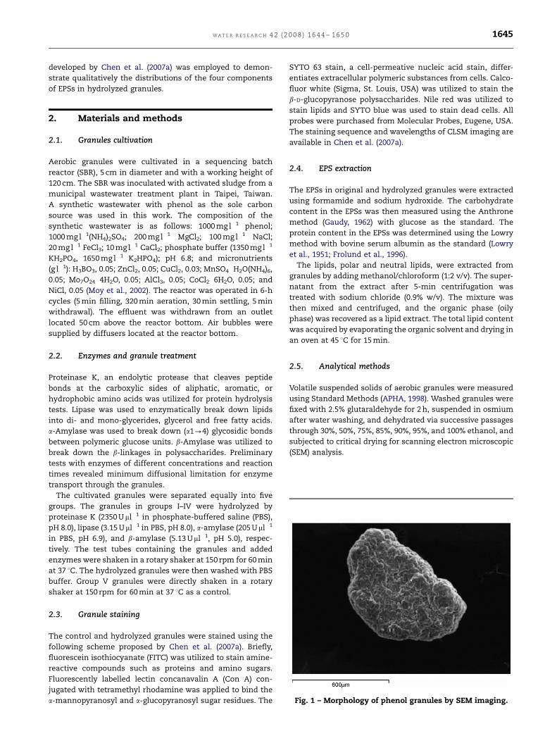
Fig. 1 – Morphology of phenol granules by SEM imaging.
2. Materials and methods
2.1. Granules cultivation
Aerobic granules were cultivated in a sequencing batch
reactor (SBR), 5 cm in diameter and with a working height of
120 cm. The SBR was inoculated with activated sludge from a
municipal wastewater treatment plant in Taipei, Taiwan.
A synthetic wastewater with phenol as the sole carbon
source was used in this work. The composition of the
synthetic wastewater is as follows: 1000 mg l�1 phenol;
1000 mg l�1(NH4)2SO4; 200 mg l�1 MgCl2; 100 mg l�1 NaCl;
20 mg l�1 FeCl3; 10 mg l�1 CaCl2; phosphate buffer (1350 mg l�1
KH2PO4, 1650 mg l�1 K2HPO4); pH 6.8; and micronutrients
(g l�1): H3BO3, 0.05; ZnCl2, 0.05; CuCl2, 0.03; MnSO4 �H2O(NH4)6,
0.05; Mo7O24 � 4H2O, 0.05; AlCl3, 0.05; CoCl2 � 6H2O, 0.05; and
NiCl, 0.05 (Moy et al., 2002). The reactor was operated in 6-h
cycles (5 min filling, 320 min aeration, 30 min settling, 5 min
withdrawal). The effluent was withdrawn from an outlet
located 50 cm above the reactor bottom. Air bubbles were
supplied by diffusers located at the reactor bottom.
2.2. Enzymes and granule treatment
Proteinase K, an endolytic protease that cleaves peptide
bonds at the carboxylic sides of aliphatic, aromatic, or
hydrophobic amino acids was utilized for protein hydrolysis
tests. Lipase was used to enzymatically break down lipids
into di- and mono-glycerides, glycerol and free fatty acids.
a-Amylase was used to break down (a1-4) glycosidic bonds
between polymeric glucose units. b-Amylase was utilized to
break down the b-linkages in polysaccharides. Preliminary
tests with enzymes of different concentrations and reaction
times revealed minimum diffusional limitation for enzyme
transport through the granules.
The cultivated granules were separated equally into five
groups. The granules in groups I–IV were hydrolyzed by
proteinase K (2350 Uml�1 in phosphate-buffered saline (PBS),
pH 8.0), lipase (3.15 Uml�1 in PBS, pH 8.0), a-amylase (205 Uml�1
in PBS, pH 6.9), and b-amylase (5.13 Uml�1, pH 5.0), respec-
tively. The test tubes containing the granules and added
enzymes were shaken in a rotary shaker at 150 rpm for 60 min
at 37 1C. The hydrolyzed granules were then washed with PBS
buffer. Group V granules were directly shaken in a rotary
shaker at 150 rpm for 60 min at 37 1C as a control.
2.3. Granule staining
The control and hydrolyzed granules were stained using the
following scheme proposed by Chen et al. (2007a). Briefly,
fluorescein isothiocyanate (FITC) was utilized to stain amine-
reactive compounds such as proteins and amino sugars.
Fluorescently labelled lectin concanavalin A (Con A) con-
jugated with tetramethyl rhodamine was applied to bind the
a-mannopyranosyl and a-glucopyranosyl sugar residues. The
SYTO 63 stain, a cell-permeative nucleic acid stain, differ-
entiates extracellular polymeric substances from cells. Calco-
fluor white (Sigma, St. Louis, USA) was utilized to stain the
b-D-glucopyranose polysaccharides. Nile red was utilized to
stain lipids and SYTO blue was used to stain dead cells. All
probes were purchased from Molecular Probes, Eugene, USA.
The staining sequence and wavelengths of CLSM imaging are
available in Chen et al. (2007a).
2.4. EPS extraction
The EPSs in original and hydrolyzed granules were extracted
using formamide and sodium hydroxide. The carbohydrate
content in the EPSs was then measured using the Anthrone
method (Gaudy, 1962) with glucose as the standard. The
protein content in the EPSs was determined using the Lowry
method with bovine serum albumin as the standard (Lowry
et al., 1951; Frolund et al., 1996).
The lipids, polar and neutral lipids, were extracted from
granules by adding methanol/chloroform (1:2 v/v). The super-
natant from the extract after 5-min centrifugation was
treated with sodium chloride (0.9% w/v). The mixture was
then mixed and centrifuged, and the organic phase (oily
phase) was recovered as a lipid extract. The total lipid content
was acquired by evaporating the organic solvent and drying in
an oven at 45 1C for 15 min.
2.5. Analytical methods
Volatile suspended solids of aerobic granules were measured
using Standard Methods (APHA, 1998). Washed granules were
fixed with 2.5% glutaraldehyde for 2 h, suspended in osmium
after water washing, and dehydrated via successive passages
through 30%, 50%, 75%, 85%, 90%, 95%, and 100% ethanol, and
subjected to critical drying for scanning electron microscopic
(SEM) analysis.

ARTICLE IN PRESS
WAT E R R E S E A R C H 4 2 ( 2 0 0 8 ) 1 6 4 4 – 1 6 5 01646
3. Results
3.1. Characteristics of phenol-degrading granule
Fig. 1a presents SEM images of cultivated, phenol-fed
granules. The granules had a compact surface and an
equivalent diameter of approximately 900 mm. This batch of
granules exhibited a phenol degradation rate of near
40 mg phenol g VSS�1 h�1 at 1000 mg l�1 phenol without a
phase lag (Adav et al., 2007a).
The proteins, carbohydrates, and lipids in control granules
were 240713, 61.079.4, and 51.177.8mg g�1 VSS, respectively
(Table 1). The protein/carbohydrate ratio was approximately 3.9
for phenol-fed granules. This ratio correlated with that
obtained by McSwain et al. (2005), who indicated that their
granules had a higher protein than polysaccharide content. We
measured the quantities of DNA in formamide and sodium
hydroxide extraction EPS, and noted that the quantities were all
less than 2% of total DNA in the aerobic granules. Hence, the
present work did not induce serious cell lysis in EPS extraction.
Table 1 – Constituents in extracted EPS of the original (control
Samples Protein (mg g�1 VSS)
Control 240713
Hydrolyzed with proteinase K 9.072.8
Hydrolyzed with lipase 263719
Hydrolyzed with a-amylase 24379.1
Hydrolyzed with b-amylase 215741
0
50
100
150 Col 15 vs Col 16
020
40
60
80
100
120Col 17 vs Col 18
20
40
60
80Col 19 vs Col 20
Flu
ore
scen
t in
ten
sit
y
020406080
100120140160
Col 21 vs Col 22
0
50
100
150Col 23 vs Col 24
Distance (μm)
0 50 100 150 200 250 3000
20
40
60
80
100 Col 25 vs Col 26
A
D
Fig. 2 – CLSM images of phenol-degrading granule (control). (A) Y
acid (SYTO 63); (D) light blue: a-polysaccharide (Con A); (E) pink:
white).
Fig. 2 presents the fluorescent staining results, probed at
240mm from the outer surface of the control granule. The
fluorescence intensity plot along the line is indicated in Fig. 2
for distributions of lipids (Nile red), proteins (FITC), cells
(SYTO 63), a-D-glucopyranose polysaccharides (rhodamine–
Con A conjugate), dead cells (SYTO blue), and b-D-glucopyr-
anose polysaccharides (calcofluor white). The protein and
dead cells were principally distributed at the core, whereas
live cells and a-polysaccharides were located at the outer rim
of the granules. The b-polysaccharides were distributed in the
core and at the outer rim regimes of the phenol-fed granules.
Chen et al. (2007a) identified a similar pattern for EPSs and
cell distributions in granules.
3.2. Hydrolyzed granules
Table 1 presents the extracted EPS content of hydrolyzed
granules. Clearly, hydrolysis using specific enzymes generally
removed individual EPS compounds. For example, the gran-
ules hydrolyzed using proteinase K exhibited a protein
) and hydrolyzed phenol-fed granules
Carbohydrate (mg g�1 VSS) Lipid (mg g�1 VSS)
61.079.4 51.177.8
63.5711.1 40.079.2
58.9714.8 7.072.2
27.573.2 39711
24.177.8 41.379.7
C B
FE
300 μm 300 μm 300 μm
300 μm300 μm300 μm
ellow: lipid (Nile red); (B) green: protein (FITC); (C) red: nucleic
dead cells (SYTO blue); (F) blue: b-polysaccharide (calcofluor
ARTICLE IN PRESS
WA T E R R E S E A R C H 4 2 ( 2 0 0 8 ) 1 6 4 4 – 1 6 5 0 1647
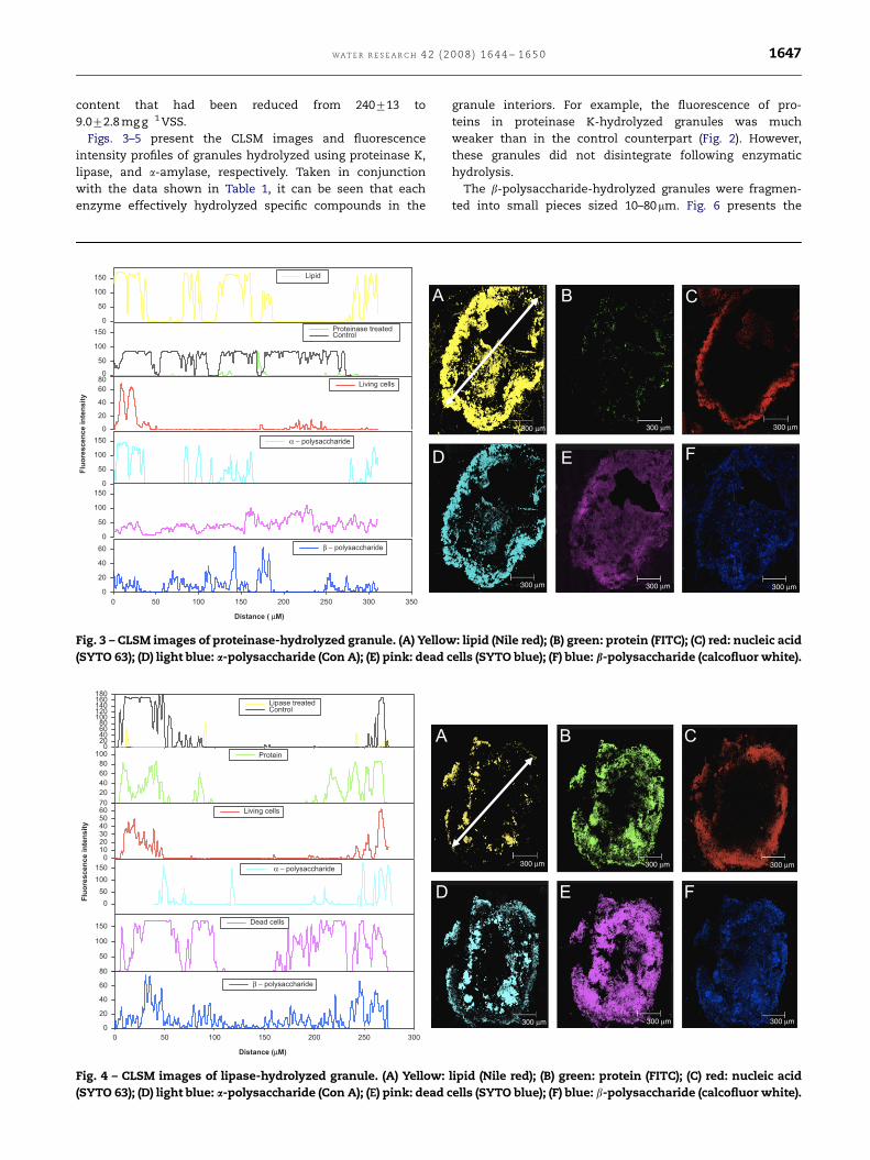
content that had been reduced from 240713 to
9.072.8 mg g�1 VSS.
Figs. 3–5 present the CLSM images and fluorescence
intensity profiles of granules hydrolyzed using proteinase K,
lipase, and a-amylase, respectively. Taken in conjunction
with the data shown in Table 1, it can be seen that each
enzyme effectively hydrolyzed specific compounds in the
0
50
100
150 Lipid
0
50
100
150 Proteinase treated Control
0
20
40
60
80Living cells
Flu
ore
scen
ce i
nte
ns
ity
0
50
100
150 α − polysaccharide
0
50
100
150
De a d c el l s
Distance ( μM)
0 50 100 150 200 250 300 350
0
20
40
60 β − polysaccharide
A
D
Fig. 3 – CLSM images of proteinase-hydrolyzed granule. (A) Yellow
(SYTO 63); (D) light blue: a-polysaccharide (Con A); (E) pink: dead c
020406080
100120140160180
Lipase treatedControl
20
40
60
80
100 Protein
010203040506070
Living cells
Flu
ore
scen
ce in
ten
sit
y
0
50
100
150 α − polysaccharide
50
100
150Dead cells
Distance (μM)
0 50 100 150 200 250 300
0
20
40
60
80
β − polysaccharide
A
D
Fig. 4 – CLSM images of lipase-hydrolyzed granule. (A) Yellow:
(SYTO 63); (D) light blue: a-polysaccharide (Con A); (E) pink: dead c
granule interiors. For example, the fluorescence of pro-
teins in proteinase K-hydrolyzed granules was much
weaker than in the control counterpart (Fig. 2). However,
these granules did not disintegrate following enzymatic
hydrolysis.
The b-polysaccharide-hydrolyzed granules were fragmen-
ted into small pieces sized 10–80mm. Fig. 6 presents the
CB
E F
300 μm 300 μm 300 μm
300 μm300 μm300 μm
: lipid (Nile red); (B) green: protein (FITC); (C) red: nucleic acid
ells (SYTO blue); (F) blue: b-polysaccharide (calcofluor white).
CB
FE
300 μm 300 μm 300 μm
300 μm300 μm300 μm
lipid (Nile red); (B) green: protein (FITC); (C) red: nucleic acid
ells (SYTO blue); (F) blue: b-polysaccharide (calcofluor white).

ARTICLE IN PRESS
WAT E R R E S E A R C H 4 2 ( 2 0 0 8 ) 1 6 4 4 – 1 6 5 01648
CLSM images of collected fragments and corresponding
fluorescence intensity profiles. Staining results indicated that
proteins, lipids, and a-polysaccharides were distributed over
the fragments. Hydrolysis of b-polysaccharides decreased the
structural stability of the granules.
Distance (μM)
0 50 100 150 200 250
0
20
40
60
80
β - polysaccharide
0
50
100
150 Dead cells
0
20
40
60
80
100Living cells
0
20
40
60
80
100
Protein
0
50
100
150 Lipid
Flu
ore
sc
en
ce
in
ten
sit
y
0
50
100
150 α - amylase treatedControl
A
D
Fig. 5 – CLSM images of a-amylase-hydrolyzed granule. (A) Yellow
(SYTO 63); (D) light blue: a-polysaccharide (Con A); (E) pink: dead c
20406080
100120140160180
Lipid
20
40
60
80
100
Protein
20
40
60
80
Living cells
Flu
ore
sc
en
ce
in
ten
sit
y
20
40
60
80
100
120
α − polysaccharide
Y D
ata
20
40
60
80
Dead cells
Distance (μM)
0 20 40 60 80
Y D
ata
0
5
10
15
20
25
30
β − polysaccharide
A
D
Fig. 6 – CLSM images of b-amylase-hydrolyzed granule. (A) Yellow
(SYTO 63); (D) light blue: a-polysaccharide (Con A); (E) pink: dead c
4. Discussions
Based on the redundant protein contents measured (Table 1)
and the CLSM images (Fig. 2) for the original granules, the
B C
E F
300 μm 300 μm 300 μm
300 μm 300 μm 300 μm
: lipid (Nile red); (B) green: protein (FITC); (C) red: nucleic acid
ells (SYTO blue); (F) blue: b-polysaccharide (calcofluor white).
C B
E F
300 μm 300 μm 300 μm
300 μm300 μm300 μm
: lipid (Nile red); (B) green: protein (FITC); (C) red: nucleic acid
ells (SYTO blue); (F) blue: b-polysaccharide (calcofluor white).

ARTICLE IN PRESS
WA T E R R E S E A R C H 4 2 ( 2 0 0 8 ) 1 6 4 4 – 1 6 5 0 1649
non-cellular protein core may be responsible for granule
stability, as suggested by McSwain et al. (2005) and Zhang
et al. (2007). However, hydrolysis using proteinase K emptied
the core regime of the granules (Fig. 3), but did not decompose
the granules. The non-cellular protein core did not contribute
significantly to the structural stability of the present phenol-
fed granules. Based on results in Table 1 and Figs. 4 and 5,
neither lipids nor a-polysaccharides corresponded to granule
stability. These experimental results support the proposal by
Wang et al. (2005) that the b-polysaccharides are the most
important component for granule stability.
The structural paradigm by Wang et al. (2005) assumed that
b-polysaccharide shell formed a continuous and integrated
structure that held the soft core of granules. However, in all
tested phenol granules, no evidence demonstrated that the
b-polysaccharides formed a continuous shell supporting
granule structure (Fig. 2). Conversely, the b-amylase-hydro-
lyzed granules contained some large (sized up to 80mm) and
numerous small-sized (about p10mm) fragments (Fig. 6).
Staining results revealed that these large fragments con-
tained concentrated proteins, lipids, and a-polysaccharides,
compounds resembling those at the outer layers of the
original granules (Fig. 2). Hence, these large fragments may
originate from the outer layers of granules prior to hydrolysis.
The b-polysaccharides likely form the backbone of a network-
like outer layer of embedded proteins, lipids, a-polysacchar-
ides, and cells, whose elasticity supports granule mechanical
stability. An adequate mechanical strength can be acquired
using limited quantities of b-polysaccharides. This observa-
tion shows how granules sustain their structure at a high
protein/polysaccharide ratio.
The b-polysaccharides in the core regime may also con-
tribute to the structural stability of granules. However, the
role of b-polysaccharides in core regime should be less
significant than those located on the outer layer. Moreover,
cations facilitate cross-linking between ECP (Eriksson and
Hardin, 1984; Kielding and Nielsen, 1997), whose role was not
discussed herein.
5. Conclusions
This study evaluated whether a correlation existed between
different EPS constituents and the structural stability of
phenol-fed aerobic granules. The contents of proteins,
carbohydrates, and lipids in the original granules were
240713, 61.079.4, and 51.177.8 mg g�1 VSS, respectively,
yielding a protein/carbohydrate ratio of approximately 3.9.
A non-cellular core which consisted of proteins and some
b-polysaccharides was found in all tested granules, which
may be considered to account for the mechanical strength of
granules. However, the selective hydrolysis of lipids, proteins,
and a- and b-polysaccharides using specific enzymes revealed
that removing proteins, lipids, and a-polysaccharides had
minimal impacts on the structural stability of granules.
Conversely, hydrolysis of b-polysaccharides caused granules
to disintegrate. The granule structure is viewed as a network
with b-polysaccharides as the backbone for embedded
proteins, lipids, a-polysaccharides, and cells supported the
structural integrity of granules.
R E F E R E N C E S
Adav, S.S., Chen, M.Y., Lee, D.J., Ren, N.Q., 2007a. Degradation ofphenol by aerobic granules and isolated yeast Candidatropicalis. Biotechnol. Bioeng. 96, 844–852.
Adav, S.S., Chen, M.Y., Lee, D.J., Ren, N.Q., 2007b. Degradation ofphenol by Acinetobacter strain isolated from aerobic granules.Chemosphere 67, 1566–1572.
APHA, 1998. Standard Methods for the Examination of Water andWastewater, 20th ed. American Public Health Association,Washington, DC.
Beun, J.J., Hendriks, A., Van Loosdrecht, M.C.M., Morgenroth, E.,Wilderer, P.A., Heijnen, J.J., 1999. Aerobic granulation in asequencing batch reactor. Water Res. 33, 2283–2290.
Cammarota, M.C., Sant’Anna, G.L., 1998. Metabolic blocking ofexopolysaccharides synthesis: effects on microbial adhesionand biofilm accumulation. Biotechnol. Lett. 20, 1–4.
Chen, M.Y., Lee, D.J., Tay, J.H., 2007a. Distribution of extracellularpolymeric substances in aerobic granules. Appl. Microbiol.Biotechnol. 73, 1463–1469.
Chen, M.Y., Lee, D.J., Tay, J.H., 2007b. Staining of extracellularpolymeric substances and cells in bio-aggregates. Appl.Microbiol. Biotechnol. 75, 467–474.
Chiu, Z.C., Chen, M.Y., Lee, D.J., Tay, S.T.L., Tay, J.H., Show, K.Y.,2006. Diffusivity of oxygen of aerobic granules. Biotechnol.Bioeng. 94, 505–513.
Costerton, J.W., Irvin, R.T., Cheng, K.J., 1981. The bacterialglycocalyx in nature and disease. Annu. Rev. Microbiol. 35,299–324.
de Kreuk, M.K., Pronk, M., van Loosdrecht, M.C.M., 2005. Forma-tion of aerobic granules and conversion processes in anaerobic granular sludge reactor at moderate and low tem-peratures. Water Res. 39, 4476–4484.
Eriksson, L., Hardin, A.M., 1984. Settling properties of activatedsludge related to floc structure. Water Sci. Technol. 16, 55–68.
Frolund, B., Palmgren, R., Keiding, K., Nielsen, P.H., 1996. Extrac-tion of extracellular polymers from activated sludge using acation exchange resin. Water Res. 30, 1749–1758.
Gaudy, A.F., 1962. Colorimetric determination of protein andcarbohydrate. Ind. Water Wastes 7, 17–22.
Hwang, K.J., You, S.F., Don, T.M., 2006. Disruption kinetics ofbacterial cells during purification of poly-beta-hydroxyalk-anoate using ultrasonication. J. Chin. Inst. Chem. Eng. 37,209–216.
Jiang, H.L., Tay, J.H., Tay, S.T.L., 2002. Aggregation of immobilizedactivated sludge cells into aerobically grown microbial gran-ules for the aerobic biodegradation of phenol. Lett. Appl.Microbiol. 35, 439–445.
Jiang, H.L., Tay, J.H., Tay, S.T.L., 2004. Changes in structure, activityand metabolism of aerobic granules as a microbial responseto high phenol loading. Appl. Microbiol. Biotechnol. 63,602–608.
Kielding, K., Nielsen, P., 1997. Desorption of organic macromole-cules from activated sludge: effect of ionic composition. WaterRes. 31, 1665–1671.
Liu, Y., Tay, J.H., 2004. State of the art of biogranulation technologyfor wastewater treatment. Biotechnol. Adv. 22, 533–563.
Liu, Y., Liu, Q.S., Qin, L., Tay, J.H., 2004a. Comments on ‘‘Effect ofextended idle conditions on structure and activity of granularactivated sludge’’ by Zhu and Wilderer. Water Res. 38,3465–3466.
Liu, Y.Q., Liu, Y., Tay, J.H., 2004b. The effects of extracellularpolymeric substances on the formation and stability ofbiogranules. Appl. Microbiol. Biotechnol. 65, 143–148.
Lowry, O.H., Rosebrough, N., Farr, A.L., Randall, R.J., 1951. Proteinmeasurements using Folin phenol reagent. J. Biol. Chem. 193,265–275.

ARTICLE IN PRESS
WAT E R R E S E A R C H 4 2 ( 2 0 0 8 ) 1 6 4 4 – 1 6 5 01650
McSwain, B.S., Irvine, R.L., Hausner, M., Wilderer, P.A., 2005.Composition and distribution of extracellular polymeric sub-stances in aerobic flocs and granular sludge. Appl. Environ.Microbiol. 71, 1051–1057.
Morgenroth, E., Sherden, T., Van Loosdrecht, M.C.M., Heijnen, J.J.,Wilderer, P.A., 1997. Aerobic granular sludge in a sequencingbatch reactor. Water Res. 31, 3191–3194.
Moy, B.Y.P., Tay, J.H., Toh, S.K., Liu, Y., Tay, S.T.L., 2002. Highorganic loading influences the physical characteristics ofaerobic granules. Lett. Appl. Microbiol. 34, 407–412.
Nielsen, P.H., Jahn, A., 1999. Extraction of EPS. In: Wingender, J.,Neu, T.R., Flemming, H.C. (Eds.), Microbial Extracellular Poly-meric Substances. Spinger, Berlin, pp. 21–47.
Nielsen, P.H., Frolund, B., Keiding, K., 1996. Changes in thecomposition of extracellular polymeric substances in acti-vated sludge during anaerobic storage. Appl. Microbiol.Biotechnol. 44, 823–830.
Neu, T.R., 1996. Significance of bacterial surface-active com-pounds in interaction of bacteria with interfaces. Microbiol.Rev. 60, 151–166.
Peng, D.C., Bernet, N., Delgenes, J.P., Moletta, R., 1999. Aerobicgranular sludge—a case report. Water Res. 33, 890–893.
Quarmby, J., Forster, C.F., 1995. An examination of the structure ofUASB granules. Water Res. 29, 2449–2454.
Takeda, M., Nakano, F., Nagase, T., Iohara, K., Koizumi, J.I., 1998.Isolation and chemical composition of the sheath of Sphaer-otilus natans. Biosci. Biotechnol. Biochem. 62, 1138–1143.
Tay, S.T.L., Moy, B.Y.P., Maszenan, A.M., Tay, J.H., 2005. Comparingactivated sludge and aerobic granules as microbial inocula forphenol biodegradation. Appl. Microbiol. Biotechnol. 67,708–713.
Tsuneda, S., Nagano, T., Hoshino, T., Ejiri, Y., Noda, N., Hirata, A.,2003. Characterization of nitrifying granules produced in anaerobic upflow fludized bed reactor. Water Res. 37, 4965–4973.
Wang, Z.W., Liu, Y., Tay, J.H., 2005. Distribution of EPS and cellsurface hydrophobicity in aerobic granules. Appl. Microbiol.Biotechnol. 69, 469–473.
Wu, S.T., Huang, C.C., Yu, S.T., Too, J.R., 2006. Effects ofnitrogen and phosphorus on poly-beta-hydroxyalkanoateproduction by Ralstonia eutropha. J. Chin. Inst. Chem. Eng. 37,501–508.
Yang, S.F., Tay, J.H., Liu, Y., 2004. Inhibition of free ammonia tothe formation of aerobic granules. Biochem. Eng. J. 17,41–48.
Zhang, L.L., Feng, X.X., Zhu, N.W., Chen, J.M., 2007. Role ofextracellular protein in the formation and stability of aerobicgranules. Enzyme Microb. Tech. Available online doi:10.1016/j.enzmictec.2007.05.001.