Embed Size (px)
Citation preview

The cornea is a unique six-layer avascular tissue of the human eye. Corneal transparency is essential for normal vision. In certain disorders, corneal transparency is lost due to tissue degeneration leading to irreversible stromal opacity with or without neovascularization. Conservative treatment may no longer be able to restore adequate eyesight warranting corneal transplantation. In bullous keratopathy (BK, Figure 1A) and Fuchs’ endothelial dystrophy (FECD, Figure 1B), endothelial pathology is prominent [1,2], whereas in infec-tious diseases, such as herpes simplex virus (HSV), keratitis corneal scarring with or without newly formed stromal vessels is evident.
The stroma is the thickest component of the cornea, full of extracellular matrix (ECM) fibers; therefore, the
stroma is the most significant developmental challenge for transparency. The transparency of the stroma depends on the well-organized microstructure of the main building elements, such as collagen fibrils and different types of proteoglycans [3]. The most important collagens of the stroma are type I and type V fibrillary collagens, while the most character-istic proteoglycans attached to them are decorin, mimecan, lumican, and keratocan [4]. The main significance of these proteoglycans is the regulation of interfibrillar space charac-teristics and collagen fibril diameters [5]. Cells and molecules of the stroma form a unique and strictly organized network of approximately 60 lamellae. Because the rays of visible light are able to pass through, this fascinatingly delicate archi-tecture is the key determinant of corneal transparency. The collagen fibrils taking part in the construction of this network are perpendicular to the corneal surface, are uniform in size (24–25 mm), and have regular spacing between them [6]. For
Molecular Vision 2021; 27:26-36 <http://www.molvis.org/molvis/v27/26>Received 9 September 2020 | Accepted 13 January 2021 | Published 15 January 2021
© 2021 Molecular Vision
26
Extracellular matrix changes in corneal opacification vary depending on etiology
László V. Módis,1,2 Gréta Varkoly,3 János Bencze,1,4 Tibor G. Hortobágyi,5 László Módis Jr,6 Tibor Hortobágyi1,5,7,8
(The first two and last two authors contributed equally to the work)
1ELKH-DE Cerebrovascular and Neurodegenerative Research Group, Department of Neurology, University of Debrecen, Debrecen, Hungary; 2Department of Behavioural Sciences, Faculty of Medicine, University of Debrecen, Debrecen, Hungary; 3Szabolcs-Szatmár-Bereg County Hospitals, Department of Ophthalmology, Nyíregyháza, Hungary; 4Deparment of Medical Imaging, Faculty of Medicine, University of Debrecen, Debrecen, Hungary; 5Institute of Pathology, Faculty of Medicine, University of Szeged, Szeged, Hungary; 6Department of Ophthalmology, Faculty of Medicine, University of Debrecen, Debrecen, Hungary; 7Institute of Psychiatry Psychology and Neuroscience, King’s College London, London, UK; 8Centre for Age-Related Medicine, SESAM, Stavanger University Hospital, Stavanger, Norway
Purpose: The purpose of this study is to examine the expression of tenascin-C and matrilin-2 in three different disorders, which frequently require corneal transplantation. These pathological conditions include bullous keratopathy (BK), Fuchs’ endothelial corneal dystrophy (FECD), and corneal scarring in herpetic keratitis.Methods: Histological sections of corneal buttons removed during keratoplasty were analyzed in BK (n = 20), FECD (n = 9), herpetic keratitis (n = 12), and cadaveric control (n = 10) groups with light microscopy following chromogenic immunohistochemistry. The sections were evaluated by three investigators, and semiquantitative scoring (0 to 3+) was applied according to standardized methods at 400X magnification. Each layer of the cornea was investigated; moreover, the stroma was subdivided into subepithelial, middle, and pre-Descemet’s membrane areas for more detailed analysis.Results: Excessive epithelial and stromal expression of tenascin-C was identified in all investigated conditions; the results were most pronounced in the pre-Descemet’s membrane. Regarding matrilin-2, when examined in BK, there was increased labeling intensity in the epithelium (p<0.001) and stromal layers (p<0.05), and a decrease in the endothelium (p<0.001). In the other investigated conditions, only a low degree of stromal localization (p<0.05) of matrilin-2 was detected.Conclusions: The expression of tenascin-C and matrilin-2 differs when examined in various corneal pathologies result-ing in opacification. Both molecules seem to be involved in regeneration and wound healing of the corneal matrix in these diseases.
Correspondence to: Tibor Hortobágyi, Institute of Pathology, Faculty of Medicine, University of Szeged, P.O. Box: 6701 Szeged, Pf. 427, Szeged, Hungary, Phone: +36 30 687 5983, email: [email protected]

Molecular Vision 2021; 27:26-36 <http://www.molvis.org/molvis/v27/26> © 2021 Molecular Vision
27
light rays to cross through the corneal stroma, the undamaged structure of proteoglycans is also required [7].
Different alterations of the corneal ECM were observed in various conditions of the cornea affecting visual acuity. Ever since the microstructure of the stromal ECM was revealed, researchers have aimed to understand the altera-tions of this structure to unveil the underlying biologic changes leading to corneal opacity. Many molecules, such as epidermal growth factor (EGF), platelet-derived growth factor (PDGF), transforming growth factor (TGF), fibroblast growth factors, different interleukins (ILs), tumor necrosis factor alpha (TNF-α), and different collagen types, have been investigated as possible participants in the emergence of corneal haze [8,9].
However, tenascin-C and matrilin-2 have not yet been thoroughly investigated in corneal diseases. Tenascin-C is a hexameric ECM glycoprotein with immediately increased expression in injury and infection. At the cellular level, tenascin-C is responsible for various dynamic cellular activities, such as cell adhesion, cell–ECM interaction, tissue remodeling, and proliferation [10-15].
Matrilin-2, however, is a member of the von Willebrand factor type A-like module superfamily [16]. The matrilins form a family of four, with each member binding to different types of collagenous and non-collagenous ECM structures, determining tissue stability via various interactions in the ECM [17]. Matrilins mainly mediate interactions between collagen-containing fibrils and other matrix constituents, such as aggrecan. Matrilin-2 has been thoroughly examined only in cartilage, muscle, and nerves, although this molecule is a crucial component of basement membranes throughout
the body, including the corneal epithelial basement membrane [16-18].
The aim of this study was to examine the expression of tenascin-C and matrilin-2 in three different disorders, which frequently require corneal transplantation. Two of these conditions, BK and FECD, are similar in clinical presentation. The third, scar formation after HSV keratitis, is different, enabling us to compare diseases with different pathogenesis.
METHODS
Selection of patients and control corneas: Corneal buttons were obtained from 41 patients who underwent corneal trans-plantation at the Department of Ophthalmology, University of Debrecen (Hungary). Most of the cases were patients with BK (n = 20, mean age 65.75±4.40 years), followed by patients with corneal scarring after HSV infection (n = 12, mean age 53.33±7.70 years), and patients with FECD (n = 9, mean age 67.33±5.10 years). Informed consent was not necessary due to the retrospective anonymized nature of the study on residual material.
Healthy corneas (n = 10, mean age 63.75±7.40 years) collected from cadavers (Department of Pathology, University of Debrecen) served as normal controls. These healthy control corneas were selected from cadavers in which previous ocular inflammation and surgical intervention could be ruled out. According to the referring regulation of ethics in Hungary, cadaveric tissue can be removed by presumed consent. In this case we applied the principle of presumed consent. The study was approved by the Institutional Scientific and Research Ethics Board (4468–2015). The use of tissue samples were in accordance with the Declaration of Helsinki.
Figure 1. Major clinical features of the investigated corneal diseases. In long-standing bullous keratopathy, the cornea becomes edematous and opaque. In the final stage newly formed vessels (arrow) penetrate the corneal tissue (A). In advanced forms of Fuchs' dystrophy the normal hexagonal endothelial cell monolayer on the posterior corneal surface is altered with a decrease in cell number (arrow), concomitant stromal edema and thickening (B). Stromal degeneration (arrow) after HSV infection. Some newly formed peripheral stromal vessels are also present (C).
Molecular Vision 2021; 27:26-36 <http://www.molvis.org/molvis/v27/26> © 2021 Molecular Vision
28
Laboratory procedures:
Preparation of histological slides—After removal, the corneas were immersion-fixed using a formaldehyde solution (pH = 7.4; 10% v/v) changed subsequently to paraformalde-hyde (pH = 7.4; 4% v/v) overnight and then embedded in paraffin wax according to standard laboratory procedures. Seven-micron-thick sections were cut from the paraffin-embedded blocks (Leica RM2245 microtome, Leica Biosys-tems Nussloch Gmbh, Nussloch, Germany), coverslipped with DPX (BDH Laboratory Supplies, Poole, England), and left to dry overnight at room temperature.
Antibodies—For the immunohistochemical (IHC) reac-tion, two types of antibodies were applied. For tenascin-C, mouse monoclonal antibody (DB7; ab86182, 1:50; Abcam, Cambridge, UK) in 1:50 dilution was used. For matrilin-2, rabbit polyclonal antibody (ARP57667_P050, 1:300; Aviva Systems Biology, Corp., San Diego, CA) was used in 1:300 dilution with a standard overnight immunohistochemistry protocol [19].
Chromogenic immunohistochemistry: Chromogenic immuno-histochemistry (CHR-IHC) was carried out as follows. After dewaxing and rehydration according to standard protocols [20], the endogenous peroxidase activity was blocked with Biocare Medical Peroxidazed 1 blocking reagent (Biocare Medical, Pacheco, CA) in Biocare Medical Peroxidazed diluent (Biocare Medical) diluted at 1:3, following the manu-facturer’s instructions. Then heat-induced epitope retrieval was performed in a microwave oven (5 min at 800 W, 2 × 5 min at 250 W) with sodium citrate buffer (pH = 6.0) as buffer solution. The non-specific IHC reaction was blocked with 1% (v/v) bovine serum albumin (BSA; Sigma-Aldrich, St. Louis, MO) for 1 h at room temperature. The antibody dilution was 1:50 for tenascin-C and 1:300 for matrilin-2. Prelimi-nary experiments were performed to establish the optimal antibody concentration and incubation time. As a result, the tenascin-C-treated sections were incubated overnight at 4 °C, while the matrilin-2-treated sections were incubated for 45 min at room temperature. After the primary antibody was washed out with Tris-buffered saline (TBS; Sigma-Aldrich, TRIS-Buffered Saline 20x solution, 20 mmol Tris, pH 7.4), biotin-free secondary antibodies were added (MACH 4 Universal HRP-Polymer, Biocare Medical) for 30 min at room temperature. After washing in TBS, for tenascin-C an immunoperoxidase technique was applied using 3,3′-diami-nobenzidine tetrahydrochloride (DAB; Biocare Medical) as chromogen in humid chambers at room temperature. The matrilin-2-treated sections, however, were developed with alkaline phosphatase streptavidin, and alkaline phosphate substrate was used as chromogen. Then, the specimens were
rinsed in running tap water for 10 min and in distilled water for 1 min.
Subsequently, the sections were treated with Harris’ hematoxylin as a nuclear counterstain. This step was followed by routine dehydration in ascending concentrations of ethyl alcohol solutions. Finally, the corneal buttons were covered with DPX mounting medium (Sigma-Aldrich) and were coverslipped manually.
Evaluation:
Semiquantitative scoring—Semiquantitative image analysis was performed by three authors of this article (MVL, GV, TH). A Nikon Eclipse 80i light microscope (Tokyo, Japan) was used at the magnification of 400X, and analysis was performed according to standardized methods of CHR-IHC assessment as described previously [21]. The semiquantitative scoring system had five scales (0, 0.5+, 1+, 2+, 3+), where 0 means no staining (negative), 0.5+ minimal, 1+ mild, 2+ moderate, and 3+ marked positivity (Figure 2). Every layer of the cornea was analyzed, even the acellular layers. In the epithelium and endothelium, 100 cells were counted in each section, and the arithmetic mean of the scores was calculated. In the other three layers (the Bowman’s membrane, stroma, and Descemet’s membrane), however, the general CHR sign intensity was established throughout the thickness of that particular layer. Because different layers of the stroma have different molecular characteristics and patho-logical alterations [22], for evaluation the stroma was divided into three parts: subepithelial, middle, and pre-Descemet’s membrane.
Statistical analysis—We used SigmaPlot 12.0 software for Windows (Systat Software Inc., San Jose, CA) for statis-tical analysis. The non-parametric data of differences in IHC sign intensity was assessed with the Mann–Whitney U-test; differences of p values of less than 0.05 were considered statistically significant.
RESULTS
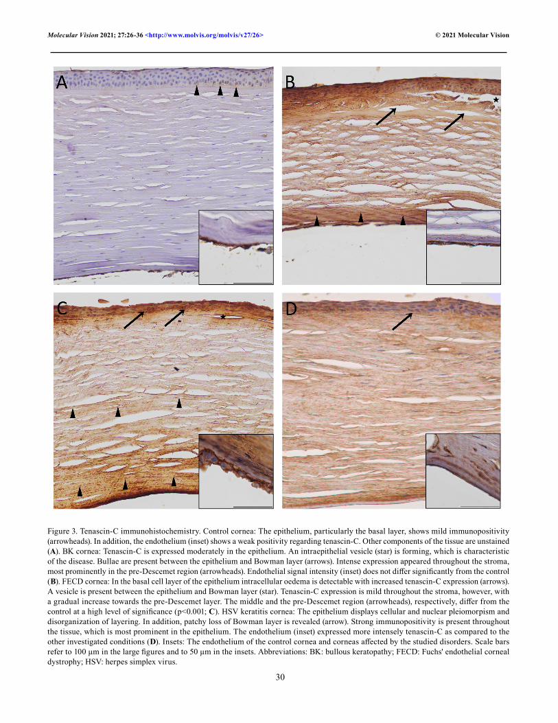
Tenascin-C: We assessed the distribution of tenascin-C immunopositivity (Figure 3, Table 1). Control corneal buttons showed mild immunopositivity for tenascin-C in the corneal epithelium (the weighted arithmetic mean for tenascin-C scores in the intact corneas was 1.15, (Figure 3A). In this layer of the cornea, in BK, statistically significant immunoreactivity was detected (p<0.01) with a mean score of 2.00. The Bowman’s membrane was intensely stained in the control group, whereas the most pronounced posi-tivity was noted in the stroma. In every subdivision of the
Molecular Vision 2021; 27:26-36 <http://www.molvis.org/molvis/v27/26> © 2021 Molecular Vision
29
stroma, immunolabeling was unequivocally present in BK (p<0.001). Close to the Bowman’s membrane, the intensity was 1.50. The lowest value of tenascin-C expression was observed in the middle part of the corneal connective tissue (1.40), becoming more intense toward the pre-Descemet’s membrane region. The intensity peaked in the vicinity of the Descemet’s membrane (1.85). The DAB CHR signal strength of the endothelium was not statistically significantly above the intact corneal signal strength level (Figure 3B). In the corneas with FECD (Figure 3C), the epithelium and the Bowman’s membrane were shown to be statistically signifi-cantly positive at the value of 1.67 (p<0.01). Noteworthy positivity in the stroma also occurred in the corneas with FECD. Although less intense than in the corneas with BK, a gradual elevation of the signal intensity toward the pre-Descemet’s membrane part of the stroma was also character-istic in the corneas with FECD (with semiquantitative scores of 0.88 in the subepithelial part, 1.00 in the middle part, and 1.67 in the pre-Descemet’s membrane region). There were no changes in the expression pattern of tenascin-C in the endothelial layer. In the corneas with HSV keratitis (Figure 3D), the most pronounced immunoreactivity (2.33) ensued in the epithelial layer (p<0.001). The Bowman’s membrane was unstained, and statistically significant labeling was detectable throughout the stroma (p<0.001). In contrast to the previous conditions, the endothelium was positive in the corneas with HSV keratitis (p<0.05).
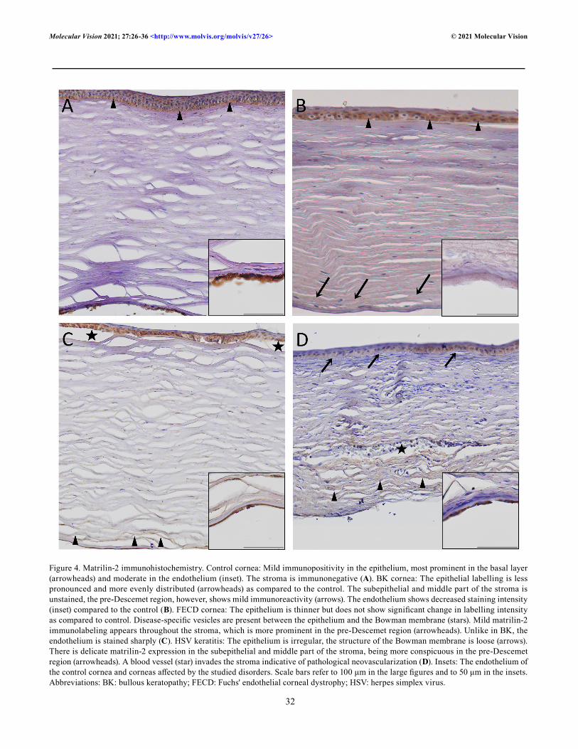
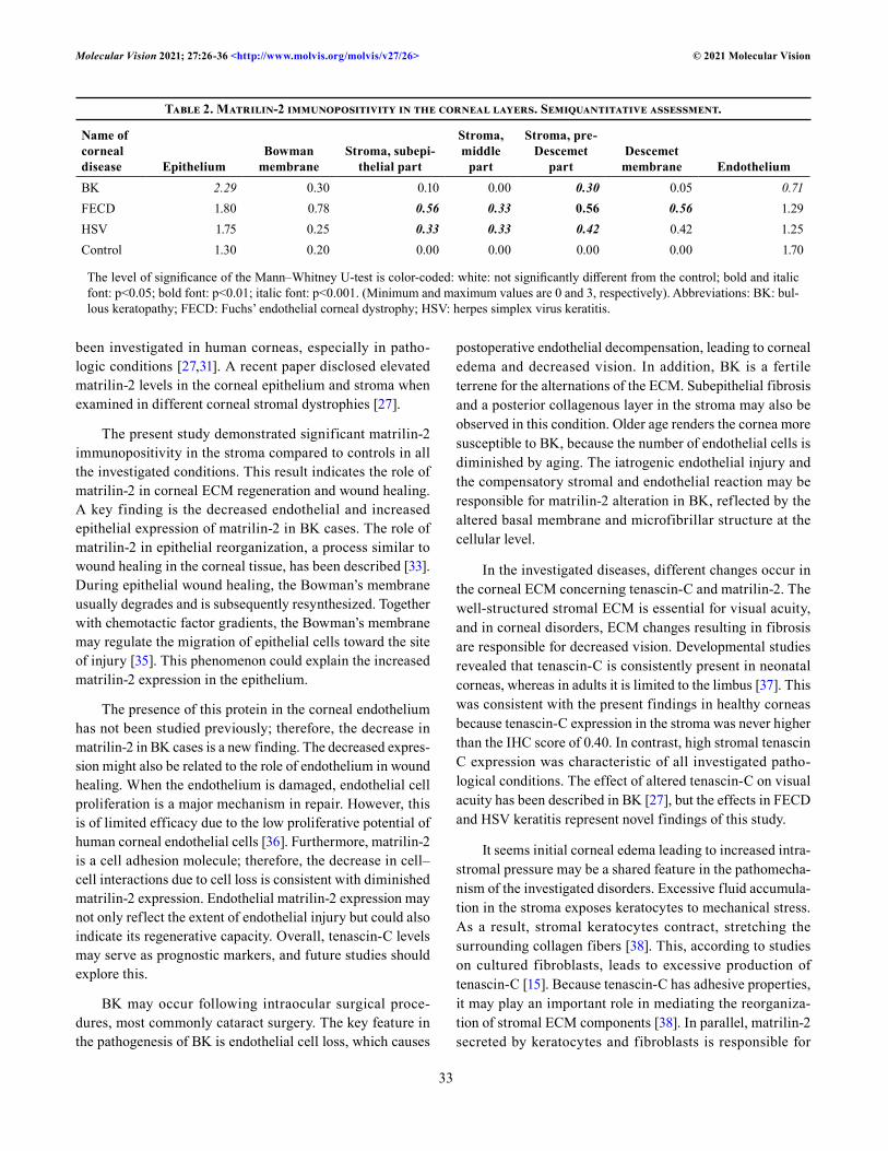
Matrilin-2: We then assessed the distribution of matrilin-2 immunopositivity (Figure 4 and Table 2.). No matrilin-2 immunoreactivity (score 0.00) was evident in the control group in any layer of the corneal stroma; mild positivity was detected in the epithelium (1.30) and the endothelium (1.70, Figure 4A). In the corneas with BK (Figure 4B), intense (2.29) staining was noted in the epithelium (p<0.001) and mild positivity (0.30) in the pre-Descemet’s membrane divi-sion of the stroma (p<0.05), whereas other divisions were unstained. The acellular layers also remained unstained,
while in the endothelium moderate immunostaining was present regarding the control at a score of 0.71 (p<0.001). In the corneas with FECD (Figure 4C), the epithelium did not show immunopositivity, neither did Bowman’s membrane. The subepithelial (0.56) layer and the middle division (0.33) of the stroma stained for matrilin-2 (p<0.05), while the pre-Descemet’s membrane (0.56) showed even more statistically significant positivity (p<0.01). In addition, Descemet’s membrane was stained (0.56) in the corneas with FECD with matrilin-2 (p<0.05). In the corneas with HSV keratitis (Figure 4D), there was no immunostaining in the epithelium and endothelium or in the acellular layers. The only component of the cornea that showed positive reaction with the antigens was the stroma. All three layers exhibited the same level of statistical significance (p<0.05), with a value of 0.33 in the subepithelial, 0.33 in the middle layer, and 0.42 in the pre-Descemet’s membrane.
Summarizing the results, the key finding was the statistically significantly elevated expression of tenascin-C in the epithelium and every layer of the stroma in all three investigated conditions. A statistically significant increase in tenascin-C expression was present in the Bowman’s membrane when examined in the corneas with BK and FECD. In the corneas with FECD, a notable increase was detected in the endothelium. In the corneas with BK, the expression level of matrilin-2 was statistically significantly elevated in the epithelium and the pre-Descemet’s membrane part of the stroma, and diminished in the endothelium. In the middle and pre-Descemet’s membrane stromal layer of the corneas with FECD and HSV keratitis, matrilin-2 showed an excessive presence compared to controls.
Tenascin-C revealed statistically significant differences only in the subepithelial stromal components when compared with the corneas with BK and FECD, whereas all other tissue layers showed similar staining intensity. Matrilin-2 stained differently in the epithelium (p<0.001), the Descemet’s membrane (p<0.05), and the endothelium (p<0.05).
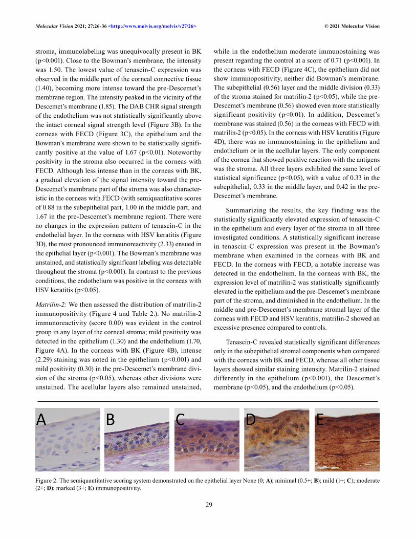
Figure 2. The semiquantitative scoring system demonstrated on the epithelial layer None (0; A); minimal (0.5+; B); mild (1+; C); moderate (2+; D); marked (3+; E) immunopositivity.
Molecular Vision 2021; 27:26-36 <http://www.molvis.org/molvis/v27/26> © 2021 Molecular Vision
30
Figure 3. Tenascin-C immunohistochemistry. Control cornea: The epithelium, particularly the basal layer, shows mild immunopositivity (arrowheads). In addition, the endothelium (inset) shows a weak positivity regarding tenascin-C. Other components of the tissue are unstained (A). BK cornea: Tenascin-C is expressed moderately in the epithelium. An intraepithelial vesicle (star) is forming, which is characteristic of the disease. Bullae are present between the epithelium and Bowman layer (arrows). Intense expression appeared throughout the stroma, most prominently in the pre-Descemet region (arrowheads). Endothelial signal intensity (inset) does not differ significantly from the control (B). FECD cornea: In the basal cell layer of the epithelium intracellular oedema is detectable with increased tenascin-C expression (arrows). A vesicle is present between the epithelium and Bowman layer (star). Tenascin-C expression is mild throughout the stroma, however, with a gradual increase towards the pre-Descemet layer. The middle and the pre-Descemet region (arrowheads), respectively, differ from the control at a high level of significance (p<0.001; C). HSV keratitis cornea: The epithelium displays cellular and nuclear pleiomorpism and disorganization of layering. In addition, patchy loss of Bowman layer is revealed (arrow). Strong immunopositivity is present throughout the tissue, which is most prominent in the epithelium. The endothelium (inset) expressed more intensely tenascin-C as compared to the other investigated conditions (D). Insets: The endothelium of the control cornea and corneas affected by the studied disorders. Scale bars refer to 100 μm in the large figures and to 50 μm in the insets. Abbreviations: BK: bullous keratopathy; FECD: Fuchs' endothelial corneal dystrophy; HSV: herpes simplex virus.

Molecular Vision 2021; 27:26-36 <http://www.molvis.org/molvis/v27/26> © 2021 Molecular Vision
31
DISCUSSION
In the present study, we investigated the expression patterns of tenascin-C and matrilin-2 in three different diseases of high prevalence with corneal opacities: BK, FECD, and corneal scarring after HSV keratitis. The most conspicuous finding of the study was the statistically significantly elevated immunoreactivity for both antibodies in the posterior corneal stroma compared to healthy samples. This result was detected in all disorders. The epithelium also showed higher immu-nopositivity (except tenascin-C in the corneas with FECD). The other striking feature we discovered was the statistically significantly higher matrilin-2 expression in the epithelium and the decreased expression in the endothelium when examined in BK cases compared to controls. In addition, the corneal endothelium disclosed mild immunopositivity in the investigated diseases.
Tenascin-C binds to numerous ECM components, cell membrane elements (syndecan 4, EGFR, contactin, integrin, annexin II), and soluble factors (VWF, different subtypes of FGF, PDGF, VEGF, PGF, BMP2, TGFβ 1 and 2) [10-15]. Through this large number of interactions, tenascin-C contributes to forming barriers, cues, niches, and tracks in the ECM, regulating the mechanical and adhesion interac-tions in cell–ECM and cell–cell relations with an impact on intracellular signal transduction pathways. Due to tenascin-C’s affinity for soluble factors, it may be a reservoir for these molecules [10]. Certain conditions induce persistent tenascin-C production, such as chronic non-healing wounds, autoim-mune diseases, cancer, and fibrotic diseases [11].
Concerning the cornea, higher tenascin-C expression was reported in BK [23], keratoconus [24], inflammation, fibrosis, and neovascularization [25]. Furthermore, the role of tenascin-C has been proven in corneal wound healing [26]. Tenascin-c and matrilin-2 proteins are expressed resolutely in lattice type I and granular stromal dystrophy [27].
Previous researchers documented excessive tenascin-C expression in the epithelium, basement membrane, subepithe-lial layer, and pre-Descemet’s membrane part of the stroma in BK [23,28]. This was consistent with the present findings. Moreover, we found a similar pattern in FECD. Previous studies also disclosed tenascin-C immunopositivity in different corneal pathologies with inflammation [25], scar-ring [25,29], and angiogenesis [30].
The most common characteristic clinicopathological finding in BK and FECD is corneal endothelial cell loss. This may serve as a possible explanation for the mild tenascin-C immunopositivity in this corneal layer. The subsequent stromal edema usually progresses to the epithelium causing intra- and interepithelial edema. Finally, stromal degenera-tion with loss of corneal transparency develops. This may be why the distribution of tenascin-C is similar in these three diseases. In addition, the much slower disease progression in FECD compared to BK is also consistent with the less intense epithelial immunopositivity.
These findings are similar to the processes we observed and demonstrated in the corneas with HSV keratitis. In such cases, the infection first manifests in the epithelium and then progresses deeper in the cornea. This explains why the highest tenascin-C immunopositivity was detected in the epithelium. The significant endothelial immunoposi-tivity compared to controls may represent a strong adhesion between the Descemet’s membrane and endothelium after inflammation, as all surgical procedures were performed on a non-inflamed eye.
Matrilin-2, the non-collagenous basement membrane component, colocalizes with microfibrils and connects to fibrillin-2 [31]. The function of matrilin-2 is highly variable in the human body, including promotion of axonal growth and Schwann cell migration during peripheral nerve regeneration [32], modulation of dermal wound healing [33], and tumor development [34]. However, the role of matrilin-2 has rarely
Table 1. Tenascin-C immunopositivity in the corneal layers. Semiquantitative assessment.
Name of corneal disease Epithelium
Bowman membrane
Stroma, subepi-thelial part
Stroma, middle
part
Stroma, pre-Descemet
partDescemet
membrane EndotheliumBK 2.00 0.80 1.50 1.40 1.85 0.15 1.07FECD 1.67 0.88 0.88 1.00 1.67 0.00 1.11HSV 2.23 0.67 1.58 1.33 1.67 0.00 1.33Control 1.15 0.22 0.30 0.25 0.35 0.15 0.70
The level of significance of the Mann–Whitney U-test is color-coded: white: not significantly different from the control; bold and italics font: p<0.05; bold font: p<0.01; italic font: p<0.001. (Minimum and maximum values are 0 and 3, respectively). Abbreviations: BK: bul-lous keratopathy; FECD: Fuchs’ endothelial corneal dystrophy; HSV: herpes simplex virus keratitis.
Molecular Vision 2021; 27:26-36 <http://www.molvis.org/molvis/v27/26> © 2021 Molecular Vision
32
Figure 4. Matrilin-2 immunohistochemistry. Control cornea: Mild immunopositivity in the epithelium, most prominent in the basal layer (arrowheads) and moderate in the endothelium (inset). The stroma is immunonegative (A). BK cornea: The epithelial labelling is less pronounced and more evenly distributed (arrowheads) as compared to the control. The subepithelial and middle part of the stroma is unstained, the pre-Descemet region, however, shows mild immunoreactivity (arrows). The endothelium shows decreased staining intensity (inset) compared to the control (B). FECD cornea: The epithelium is thinner but does not show significant change in labelling intensity as compared to control. Disease-specific vesicles are present between the epithelium and the Bowman membrane (stars). Mild matrilin-2 immunolabeling appears throughout the stroma, which is more prominent in the pre-Descemet region (arrowheads). Unlike in BK, the endothelium is stained sharply (C). HSV keratitis: The epithelium is irregular, the structure of the Bowman membrane is loose (arrows). There is delicate matrilin-2 expression in the subepithelial and middle part of the stroma, being more conspicuous in the pre-Descemet region (arrowheads). A blood vessel (star) invades the stroma indicative of pathological neovascularization (D). Insets: The endothelium of the control cornea and corneas affected by the studied disorders. Scale bars refer to 100 μm in the large figures and to 50 μm in the insets. Abbreviations: BK: bullous keratopathy; FECD: Fuchs' endothelial corneal dystrophy; HSV: herpes simplex virus.
Molecular Vision 2021; 27:26-36 <http://www.molvis.org/molvis/v27/26> © 2021 Molecular Vision
33
been investigated in human corneas, especially in patho-logic conditions [27,31]. A recent paper disclosed elevated matrilin-2 levels in the corneal epithelium and stroma when examined in different corneal stromal dystrophies [27].
The present study demonstrated significant matrilin-2 immunopositivity in the stroma compared to controls in all the investigated conditions. This result indicates the role of matrilin-2 in corneal ECM regeneration and wound healing. A key finding is the decreased endothelial and increased epithelial expression of matrilin-2 in BK cases. The role of matrilin-2 in epithelial reorganization, a process similar to wound healing in the corneal tissue, has been described [33]. During epithelial wound healing, the Bowman’s membrane usually degrades and is subsequently resynthesized. Together with chemotactic factor gradients, the Bowman’s membrane may regulate the migration of epithelial cells toward the site of injury [35]. This phenomenon could explain the increased matrilin-2 expression in the epithelium.
The presence of this protein in the corneal endothelium has not been studied previously; therefore, the decrease in matrilin-2 in BK cases is a new finding. The decreased expres-sion might also be related to the role of endothelium in wound healing. When the endothelium is damaged, endothelial cell proliferation is a major mechanism in repair. However, this is of limited efficacy due to the low proliferative potential of human corneal endothelial cells [36]. Furthermore, matrilin-2 is a cell adhesion molecule; therefore, the decrease in cell–cell interactions due to cell loss is consistent with diminished matrilin-2 expression. Endothelial matrilin-2 expression may not only reflect the extent of endothelial injury but could also indicate its regenerative capacity. Overall, tenascin-C levels may serve as prognostic markers, and future studies should explore this.
BK may occur following intraocular surgical proce-dures, most commonly cataract surgery. The key feature in the pathogenesis of BK is endothelial cell loss, which causes
postoperative endothelial decompensation, leading to corneal edema and decreased vision. In addition, BK is a fertile terrene for the alternations of the ECM. Subepithelial fibrosis and a posterior collagenous layer in the stroma may also be observed in this condition. Older age renders the cornea more susceptible to BK, because the number of endothelial cells is diminished by aging. The iatrogenic endothelial injury and the compensatory stromal and endothelial reaction may be responsible for matrilin-2 alteration in BK, reflected by the altered basal membrane and microfibrillar structure at the cellular level.
In the investigated diseases, different changes occur in the corneal ECM concerning tenascin-C and matrilin-2. The well-structured stromal ECM is essential for visual acuity, and in corneal disorders, ECM changes resulting in fibrosis are responsible for decreased vision. Developmental studies revealed that tenascin-C is consistently present in neonatal corneas, whereas in adults it is limited to the limbus [37]. This was consistent with the present findings in healthy corneas because tenascin-C expression in the stroma was never higher than the IHC score of 0.40. In contrast, high stromal tenascin C expression was characteristic of all investigated patho-logical conditions. The effect of altered tenascin-C on visual acuity has been described in BK [27], but the effects in FECD and HSV keratitis represent novel findings of this study.
It seems initial corneal edema leading to increased intra-stromal pressure may be a shared feature in the pathomecha-nism of the investigated disorders. Excessive fluid accumula-tion in the stroma exposes keratocytes to mechanical stress. As a result, stromal keratocytes contract, stretching the surrounding collagen fibers [38]. This, according to studies on cultured fibroblasts, leads to excessive production of tenascin-C [15]. Because tenascin-C has adhesive properties, it may play an important role in mediating the reorganiza-tion of stromal ECM components [38]. In parallel, matrilin-2 secreted by keratocytes and fibroblasts is responsible for
Table 2. Matrilin-2 immunopositivity in the corneal layers. Semiquantitative assessment.
Name of corneal disease Epithelium
Bowman membrane
Stroma, subepi-thelial part
Stroma, middle
part
Stroma, pre-Descemet
partDescemet
membrane EndotheliumBK 2.29 0.30 0.10 0.00 0.30 0.05 0.71FECD 1.80 0.78 0.56 0.33 0.56 0.56 1.29HSV 1.75 0.25 0.33 0.33 0.42 0.42 1.25Control 1.30 0.20 0.00 0.00 0.00 0.00 1.70
The level of significance of the Mann–Whitney U-test is color-coded: white: not significantly different from the control; bold and italic font: p<0.05; bold font: p<0.01; italic font: p<0.001. (Minimum and maximum values are 0 and 3, respectively). Abbreviations: BK: bul-lous keratopathy; FECD: Fuchs’ endothelial corneal dystrophy; HSV: herpes simplex virus keratitis.

Molecular Vision 2021; 27:26-36 <http://www.molvis.org/molvis/v27/26> © 2021 Molecular Vision
34
modulating ECM assembly and ECM–cell communication [39].
That both investigated molecules could be identified in the end stage of the most common corneal pathologies which finally require surgical intervention may be of great clinical relevance. These proteins seem to be involved in regeneration and reorganization of the corneal ECM either in degenerative (BK, FECD) or post-inflammatory corneal processes (scar formation after HSV keratitis). Better understanding of expression patterns and interactions of corneal ECM proteins in the context of pathogenesis and prognosis may pave the way for novel conservative therapy or less invasive surgical interventions in the treatment of corneal opacity. This is the main significance and the clinical relevance of the present findings.
Although in diseases with corneal opacification the gold standard treatment is penetrating keratoplasty, posterior lamellar transplantation is becoming more common in BK and FECD. Scraping of the Descemet’s membrane and the endothelium from the recipient before a healthy endothelial cell suspension is injected has shown promising results [40]. Furthermore, blocking extracellular high-mobility group box 1 in mice with corneal wounds successfully decreased the inflammation-mediated haze grade [41]. Tenascin-C and matrilin-2 may also become therapeutic targets for such less invasive procedures to treat corneal opacity.
In conclusion, tenascin-C and matrilin-2 are implicated in the pathogenesis of BK, FECD, and HSV keratitis with corneal scarring. Both molecules appear to be involved in regeneration and wound healing of the corneal matrix in these disease conditions. However, we detected substantial differ-ences in immunolocalization regarding the different corneal layers. More studies are needed to further elucidate the role of these molecules in ECM regeneration and remodeling in relation to corneal opacification.
ACKNOWLEDGMENTS
Special thanks to former PhD students Renáta Nóra Szabó, Balázs Murnyák and Mahan Kouhsari for the vivid discus-sions and joyful company in the laboratory. The authors express their gratitude to Zoltán Mészár for his help in digitalizing our histological sections. Author contributions Conception and design of the study by LMJr and TH. Orig-inal draft preparation by LVM, further drafts by LVM, TGH, TH. Immunohistochemistry was performed by LVM and GV. LMJr and TH diagnosed the cases. Histological assessment by LVM, GV, TH. Data analysis and statistics by LVM, JB, TGH and TH. Digital images by JB and LVM. Manuscript editing by LMJr and TH. Supervision and project administration by
LMJr and TH. All authors have read, commented and agreed to the final version of the manuscript. Funding This study was supported by the ÚNKP-20–4 New National Excel-lence Program of the Ministry of Innovation and Technology from the source of the National Research Development and Innovation Fund; EFOP‐3.6.3‐VEKOP‐16‐2017‐00009 (JB); Hungarian Brain Research Program (NAP) Grant No. KTIA_13_NAP-A-II/7; GINOP-2.3.2–15–2016–00043; SZTE ÁOK-KKA No. 5S 567 (A202); NKFIH_SNN_132999 (TH).
REFERENCES1. Röck T, Bartz-Schmidt K, Röck D. Trends in corneal trans-
plantation at the University Eye Hospital in Tübingen, Germany over the last 12 years: 2004 - 2015. PLoS One 2018; 13:e0198793-[PMID: 29939996].
2. Módis L, Szalai E, Facskó A, Fodor M, Komár T, Berta A. Corneal transplantation in Hungary (1946–2009). Clin Experiment Ophthalmol 2011; 39:520-5. [PMID: 21819505].
3. Michelacci Y. Collagens and proteoglycans of the corneal extracellular matrix. Braz J Med Biol Res 2003; 36:1037-46. [PMID: 12886457].
4. Meek K, Fullwood N. Corneal and scleral collagens–a micros-copist’s perspective. Micron 2001; 32:261-72. [PMID: 11006506].
5. Scott J. Extracellular matrix, supramolecular organisation and shape. J Anat 1995; 187:259-69. [PMID: 7591990].
6. Maurice D. The structure and transparency of the cornea. J Physiol 1957; 136:263-86. [PMID: 13429485].
7. Módis L. Structure of the Corneal Stroma. In Módis L. Orga-nization of the Extracellular Matrix. A Polarization Micro-scopic Approach.: CRC Press, Inc.; 1991. p. 124–130.
8. Meek K, Knupp C. Corneal structure and transparency. Prog Retin Eye Res 2015; 49:1-16. [PMID: 26145225].
9. Mobaraki M, Abbasi R, Vandchali S, Ghaffari M, Moztarzadeh F, Mozafari M. Corneal Repair and Regeneration: Current Concepts and Future Directions. Front Bioeng Biotechnol 2019; 7:135-[PMID: 31245365].
10. Midwood K, Chiquet M, Tucker R, Orend G. Tenascin-C at a glance. J Cell Sci 2016; 129:4321-7. [PMID: 27875272].
11. Udalova I, Ruhmann M, Thomson S, Midwood K. Expression and immune function of tenascin-C. Crit Rev Immunol 2011; 31:115-45. [PMID: 21542790].
12. Orend G, Chiquet-Ehrismann R. Tenascin-C induced signaling in cancer. Cancer Lett 2006; 244:143-63. [PMID: 16632194].
13. Imanaka-Yoshida K, Hiroe M, Yasutomi Y, Toyozaki T, Tsuchiya T, Noda N, Maki T, Nishikawa T, Sakakura T, Yoshida T. Tenascin-C is a useful marker for disease activity in myocarditis. J Pathol 2002; 197:388-94. [PMID: 12115886].
14. Wiemann S, Reinhard J, Faissner A. Immunomodula-tory role of the extracellular matrix protein tenascin-C in

Molecular Vision 2021; 27:26-36 <http://www.molvis.org/molvis/v27/26> © 2021 Molecular Vision
35
neuroinflammation. Biochem Soc Trans 2019; 47:1651-60. [PMID: 31845742].
15. Chiquet-Ehrismann R, Tannheimer M, Koch M, Brunner A, Spring J, Martin D, Baumgartner S, Chiquet M. Tenascin-C expression by fibroblasts is elevated in stressed collagen gels. J Cell Biol 1994; 1272093-101. [PMID: 7528751].
16. Deák F, Piecha D, Bachrati C, Paulsson M, Kiss I. Primary structure and expression of matrilin-2, the closest rela-tive of cartilage matrix protein within the von Willebrand factor type A-like module superfamily. J Biol Chem 1997; 272:9268-74. [PMID: 9083061].
17. Wagener R, Ehlen HW, Ko YP, Kobbe B, Mann HH, Sengle G, Paulsson M. The matrilins–adaptor proteins in the extracellular matrix. FEBS Lett 2005; 579:3323-9. [PMID: 15943978].
18. Wilson S, Torricelli A, Marino G. Corneal epithelial basement membrane: Structure, function and regeneration. Exp Eye Res 2020; •••:194-[PMID: 32179076].
19. Nemeth G, Felszeghy S, Kenyeres A, Szentmary N, Berta A, Suveges I, Módis L. Cell adhesion molecules in stromal corneal dystrophies. Histol Histopathol 2008; 23:945-52. [PMID: 18498069].
20. Bencze J, Szarka M, Bencs V, Szabó RN, Módis LV, Aarsland D, Hortobágyi T. Lemur Tyrosine Kinase 2 (LMTK2) Level Inversely Correlates with Phospho-Tau in Neuropathological Stages of Alzheimer’s Disease. Brain Sci 2020; 10:68-[PMID: 32012723].
21. Bencze J, Szarka M, Bencs V, Szabó RN, Smajda M, Aarsland D, Hortobágyi T. Neuropathological Characterization of Lemur Tyrosine Kinase 2 (LMTK2) in Alzheimer’s Disease and Neocortical Lewy Body Disease. Sci Rep 2019; 9:17222-[PMID: 31748522].
22. Dua H, Faraj L, Said D, Gray T, Lowe J. Human Corneal Anatomy Redefined: A Novel Pre-Descemet’s Layer (Dua’s Layer). Ophthalmology 2013; 120:1778-85. [PMID: 23714320].
23. Ljubimov A, Saghizadeh M, Spirin K, Khin H, Lewin S, Zardi L, Bourdon MA, Kenney MC. Expression of tenascin-C splice variants in normal and bullous keratopathy human corneas. Invest Ophthalmol Vis Sci 1998; 39:1135-42. [PMID: 9620072].
24. Kenney M, Nesburn A, Burgeson R, Butkowski RJ, Ljubimov AV. Abnormalities of the extracellular matrix in keratoconus corneas. Cornea 1997; 16:345-51. [PMID: 9143810].
25. Maseruka H, Bonshek R, Tullo A. Tenascin-C expression in normal, inf lamed, and scarred human corneas. Br J Ophthalmol 1997; 81:677-82. [PMID: 9349157].
26. Sumioka T, Kitano A, Flanders K, Okada Y, Yamanaka O, Fujita N, Iwanishi H, Kao WW, Saika S. Impaired cornea wound healing in a tenascin C-deficient mouse model. Lab Invest 2013; 93:207-17. [PMID: 23207449].
27. Szalai E, Felszeghy S, Hegyi Z, Módis JL, Berta A, Kaarni-ranta K. Fibrillin-2, tenascin-C, matrilin-2, and matrilin-4 are strongly expressed in the epithelium of human granular
and lattice type I corneal dystrophies. Mol Vis 2012; 18:1927-36. [PMID: 22876117].
28. Akhtar S, Bron A, Hawksworth N, Bonshek R, Meek K. Ultrastructural morphology and expression of proteoglycans, betaig-h3, tenascin-C, fibrillin-1, and fibronectin in bullous keratopathy. Br J Ophthalmol 2001; 85:720-31. [PMID: 11371495].
29. Meltendorf C, Burbach G, Bühren J, Bug R, Ohrloff C, Deller T. Corneal femtosecond laser keratotomy results in isolated stromal injury and favorable wound-healing response. Invest Ophthalmol Vis Sci 2007; 48:2068-75. [PMID: 17460262].
30. Sumioka T, Fujita N, Kitano A, Okada Y, Saika S. Impaired angiogenic response in the cornea of mice lacking tenascin C. Invest Ophthalmol Vis Sci 2011; 51:2462-7. [PMID: 21087965].
31. Kabosova A, Azar DT, Bannikov GA, Campbell KP, Durbeej M, Ghohestani RF, Jones JC, Kenney MC, Koch M, Ninomiya Y, Patton BL, Paulsson M, Sado Y, Sage EH, Sasaki T, Sorokin LM, Steiner-Champliaud MF, Sun TT, Sundarraj N, Timpl R, Virtanen I, Ljubimov AV. Composi-tional differences between infant and adult human corneal basement membranes. Invest Ophthalmol Vis Sci 2007; 48:4989-99. [PMID: 17962449].
32. Malin D, Sonnenberg-Riethmacher E, Guseva D, Wagener R, Aszódi A, Irintchev A, Riethmacher D. The extracellular-matrix protein matrilin 2 participates in peripheral nerve regeneration. J Cell Sci 2009; 122:995-1004. [PMID: 19295126].
33. Ichikawa T, Suenaga Y, Koda T, Ozaki T, Nakagawara A. DeltaNp63/BMP-7-dependent expression of matrilin-2 is involved in keratinocyte migration in response to wounding. Biochem Biophys Res Commun 2008; 369994-1000. [PMID: 18328806].
34. Sharma M, Watson M, Lyman M, Perry A, Aldape K, Deák F, Gutmann DH. Matrilin-2 expression distinguishes clinically relevant subsets of pilocytic astrocytoma. Neurology 2006; 66:127-30. [PMID: 16401863].
35. Yu F, Yin J, Xu K, Huang J. Growth factors and corneal epithe-lial wound healing. Brain Res Bull 2010; 81:229-35. [PMID: 19733636].
36. Mimura T, Yamagami S, Amano S. Corneal endothelial regen-eration and tissue engineering. Prog Retin Eye Res 2013; 35:1-17. [PMID: 23353595].
37. Maseruka H, Ridgway A, Tullo A, Bonshek R. Developmental changes in patterns of expression of tenascin-C variants in the human cornea. Invest Ophthalmol Vis Sci 2000; 41:4101-7. [PMID: 11095602].
38. Ljubimov AV, Burgeson RE, Butkowski RJ, Couchman JR, Wu RR, Ninomiya Y, Sado Y, Maguen E, Nesburn AB, Kenney MC. Extracellular matrix alterations in human corneas with bullous keratopathy. Invest Ophthalmol Vis Sci 1996; 37:997-1007. [PMID: 8631643].
39. Korpos É, Deák F, Kiss I. Matrilin-2, an extracellular adaptor protein, is needed for the regeneration of muscle, nerve and

Molecular Vision 2021; 27:26-36 <http://www.molvis.org/molvis/v27/26> © 2021 Molecular Vision
36
other tissues. Neural Regen Res 2015; 10:866-9. [PMID: 26199591].
40. Okumura N, Kagami T, Watanabe K. KS, SM, Koizumi N. Feasibility of a cryopreservation of cultured human corneal endothelial cells. PLoS One 2019; 14:e0218431-[PMID: 31226131].
41. Zhou Y, Wang T, Wang Y, Meng F, Ying M, Han R, Hao P, Wang L, Li X. Blockade of extracellular high-mobility group box 1 attenuates inflammation-mediated damage and haze grade in mice with corneal wounds. Int Immunopharmacol 2020; 83:106468-[PMID: 32279044].
Articles are provided courtesy of Emory University and the Zhongshan Ophthalmic Center, Sun Yat-sen University, P.R. China. The print version of this article was created on 15 January 2021. This reflects all typographical corrections and errata to the article through that date. Details of any changes may be found in the online version of the article.