Embed Size (px)
Citation preview

RESEARCH Open Access
Extracellular acidification-induced CXCL8production through a proton-sensingreceptor OGR1 in human airway smoothmuscle cells: a response inhibited bydexamethasoneMaiko Kadowaki1, Hidenori Yamada2, Koichi Sato3, Hiroko Shigemi1, Yukihiro Umeda1, Miwa Morikawa1,Yuko Waseda1, Masaki Anzai1, Yosuke Kamide2, Haruka Aoki-Saito2, Takeshi Hisada2, Fumikazu Okajima4 andTamotsu Ishizuka1*
Abstract
Background: Human airway smooth muscle cells (ASMCs) contribute to bronchial contraction and airwayhyperresponsiveness in patients with bronchial asthma. They also generate cytokines, chemokines, and matricellularproteins. Ovarian cancer G protein-coupled receptor 1 (OGR1) senses extracellular protons and mediates theproduction of interleukin-6 (IL-6) and connective tissue growth factor (CTGF) in ASMCs.
Methods: ASMCs were stimulated for the indicated time by pH 6.3 or pH 7.4-adjusted Dulbecco’s ModifiedEagle Medium (DMEM) containing 0.1% bovine serum albumin (BSA) (0.1% BSA-DMEM). As a control stimulant,pH 7.4-adjusted 0.1% BSA-DMEM containing 10 ng/mL tumor necrosis factor-α (TNF-α) was used. Interleukin-8/C-X-C motif chemokine ligand 8 (CXCL8) mRNA expression in ASMCs was quantified by RT-PCR using real-timeTaqMan technology. CXCL8 secreted from ASMCs was measured by enzyme-linked immunosorbent assay (ELISA).Phosphorylation at serine 536 of NF-κB p65 and binding of p65 to oligonucleotide containing an NF-κB consensusbinding site were analyzed by Western blotting and an ELISA-based kit.
Results: Acidic pH induced a significant increase of CXCL8 mRNA expression and CXCL8 protein secretion in ASMCs.ASMCs transfected with small interfering RNA (siRNA) targeted for OGR1 produced less CXCL8 compared with thosetransfected with non-targeting siRNA. Protein kinase C (PKC) inhibitor, MEK1/2 inhibitor, and the inhibitor of IκBphosphorylation reduced acidic pH-stimulated CXCL8 production in ASMCs. Dexamethasone also inhibited acidicpH-stimulated CXCL8 production of ASMCs in a dose-dependent manner. Dexamethasone did not affect eitherphosphorylation or binding to the consensus DNA site of NF-κB p65.
Conclusions: CXCL8 released from ASMCs by extracellular acidification may play a pivotal role in airwayaccumulation of neutrophils. Glucocorticoids inhibit acidic pH-stimulated CXCL8 production independent ofserine 536 phosphorylation and the binding to DNA of NF-κB p65, although NF-κB activity is essential forCXCL8 production in ASMCs.
Keywords: OGR1, Airway smooth muscle cell, Proton, CXCL8, NF-κB, Dexamethasone, Bronchial asthma, COPD
* Correspondence: [email protected] Department of Internal Medicine, Faculty of Medical Sciences,University of Fukui, 23-3 Matsuoka-Shimoaizuki, Eiheiji, Fukui 910-1193, JapanFull list of author information is available at the end of the article
© The Author(s). 2019 Open Access This article is distributed under the terms of the Creative Commons Attribution 4.0International License (http://creativecommons.org/licenses/by/4.0/), which permits unrestricted use, distribution, andreproduction in any medium, provided you give appropriate credit to the original author(s) and the source, provide a link tothe Creative Commons license, and indicate if changes were made. The Creative Commons Public Domain Dedication waiver(http://creativecommons.org/publicdomain/zero/1.0/) applies to the data made available in this article, unless otherwise stated.
Kadowaki et al. Journal of Inflammation (2019) 16:4 https://doi.org/10.1186/s12950-019-0207-1

BackgroundBronchial asthma is a disease characterized by chronicairway inflammation, airway hyperresponsiveness, andreversible airway obstruction. Airway smooth muscle cellhyperplasia and hypertrophy are often observed in pa-tients with severe asthma. An increase in airway smoothmuscle mass could contribute to airway obstruction andhyperresponsiveness [1]. Human airway smooth musclecells (ASMCs) may play an important role in airwayinflammation because they can release cytokines suchas IL-6 and CXCL8 in response to environmentalstimuli [2–8]. Extracellular pH is strictly maintainedat 7.35 to 7.45 in the blood. Although the precisemechanism of the acid-base balance in the airway isnot fully understood, airway acidification is a well-rec-ognized feature in asthma and contributes to thepathophysiology of the disease [9, 10]. The degree ofacidification depends on the severity of asthma, andairway pH seems to reach 5.2. It is normalized bycorticosteroid therapy [9, 11].Previous studies have shown that the proton-sensing
capsaicin-sensitive TRPV1 channel and acid-sensing ionchannels (ASICs) in sensory nerves play critical roles inasthmatic symptoms [10, 12, 13]. It has also beendemonstrated that ovarian cancer G protein-coupled re-ceptor 1 (OGR1) family G protein-coupled receptors(GPCRs), including OGR1 (also known as GPR68), Gprotein-coupled receptor 4 (GPR4), and T-cell death-as-sociated gene 8 (TDAG8 or GPR65), sense extracellularprotons and mediate cellular actions induced by alkalineand acidic pHs of 8 to 6 through histidine residues in avariety of cell types [14–17]. We have shown that,among these proton-sensing GPCRs, only OGR1 isexpressed in ASMCs, and that IL-6 and CTGF are se-creted from ASMCs through OGR1-mediated stimula-tion of intracellular signaling pathways in response toextracellular acidification [18, 19]. Interleukin-8 (IL-8/CXCL8) is a member of the CXC chemokine subfamilyof cytokines and a potent neutrophil chemoattractant.Because the most results of clinical trials of anti-CXCL8or C-X-C motif chemokine receptor 2 (CXCR2) antago-nists in asthma or chronic obstructive pulmonary disease(COPD) are disappointing, these bring into question therole of CXCL8-mediated neutrophil recruitment in thepathogenesis of asthma and COPD [20, 21]. However, el-evated concentrations of CXCL8 are found in sputum,bronchoalveolar lavage fluid, and bronchial tissue of pa-tients with respiratory diseases such as severe asthma,occupational asthma, cystic fibrosis, and COPD [22, 23].CXCL8 is secreted by different cell types including al-veolar macrophages [24], airway epithelial cells [25], andASMCs [7, 26–34] following stimulation by viruses, vari-ous cytokines, hypoxia, reactive oxygen species, and bac-terial particles.
In the current study, extracellular acidification wasshown to increase CXCL8 secretion from ASMCsthrough OGR1-mediated stimulation, and this secretionwas inhibited by dexamethasone (DEX) in a dose-dependent manner. The intracellular cell signaling andNF-κB activation involved in acidic pH-induced CXCL8production were explored, and the effects of DEX onserine 536 phosphorylation and DNA binding of NF-κBp65 in ASMCs were examined.
MethodsMaterials and reagentsRecombinant human tumor necrosis-α (TNF-α) was fromPeprotech (Rocky Hill, NJ). U0126, SB-203580, and bisin-dolylmaleimide I were from Cayman Chemical (AnnArbor, MI). BAY 11–7082 was from Focus Biomolecules(Plymouth Meeting, PA). Goat anti-rabbit IgG-HRP wasfrom GE Healthcare Japan Corporation (Tokyo, Japan).Antibodies for phospho-NF-κB p65 (Ser536) (93H1) andglyceraldehyde-3-phosphate dehydrogenase (GAPDH)(14C10) were from Cell Signaling Technology (Danvers,MA). Fatty acid-free bovine serum albumin (BSA) wasfrom EMD chemicals (San Diego, CA). Dexamethasone(DEX), Dulbecco’s Modified Eagle Medium (DMEM), andother chemicals were purchased from Sigma-Aldrich(St. Louis, MO).
Cell cultureHuman bronchial smooth muscle cells (BSMCs, catalogNo. CC-2576) originated from non-diseased individualswere purchased from Lonza (Walkersville, MD). Eight tonine passages of BSMCs were used as human airwaysmooth muscle cells (ASMCs) in this study. Cells weregrown in the complete culture medium (Lonza): smoothmuscle-cell basal medium supplemented with humanepidermal growth factor, insulin, human fibroblastgrowth factor β, 5% fetal bovine serum, gentamicin, andamphotericin B, under a humidified atmosphere of 95%air plus 5% CO2 at 37 °C, as previously reported [19].ASMCs were cultured in 12-well plates for ELISA ofCXCL8, 6-well plates for real-time PCR using TaqManprobes or Western blotting, and 10-cm culture dishesfor extraction of nuclear protein.
Cell stimulation and ELISA of human CXCL8The complete culture medium of ASMCs in 12-wellplates was switched to DMEM containing 0.1% BSA(0.1% BSA-DMEM) before 16 h in each experiment.ASMCs were stimulated by switching the medium topH-adjusted 0.1% BSA-DMEM (pH 6.3, 6.6, 7.0, 7.2 or 7.4)or 10 ng/mL TNF-α (pH 6.3, 7.0, or 7.4) and incubated forthe indicated time. The pH of the DMEM containing 25mM HEPES, 27mM NaHCO3, and 0.1% BSA was adjustedby titration with HCl or NaOH. Inhibitors of cell signaling
Kadowaki et al. Journal of Inflammation (2019) 16:4 Page 2 of 10

molecules and DEX were added 30min before stimulation.Supernatants of cells were collected and stored at − 30 °C.Concentrations of CXCL8 were measure by enzyme-linkedimmunosorbent assay (ELISA) (Duo Set® ELISA develop-ment system, R&D Systems, Minneapolis, MN).
Quantitative RT-PCR using real-time TaqMan technologyTotal RNA was isolated using the RNeasy Plus MiniKit (Qiagen, Hilden, Germany) according to the manu-facturer’s instructions. Up to 2.5 μg of total RNA werereverse-transcribed using random priming and reversetranscriptase (SuperScript™ VILO™ Master Mix, ThermoFisher Scientific, Waltham, MA). To evaluate the expressionlevels of OGR1, CXCL8, and glyceraldehyde-3-phosphatedehydrogenase (GAPDH) mRNA, quantitative RT-PCR wasperformed using real-time TaqMan technology with aStepOne™ real-time PCR system (Thermo Fisher Scientific).TaqMan probes specific for human OGR1 (Hs00268858_s1),CXCL8 (Hs00174103_m1), and GAPDH (Hs02758991_g1)were purchased from Thermo Fisher Scientific. The expres-sion levels of CXCL8 or OGR1 mRNA were normalized tothe relative ratio of the expression of GAPDH mRNA, aspreviously reported [18, 19].
Transfection of small interfering RNASmall interfering RNA (siRNA) targeted for OGR1(OGR1-siRNA) and non-targeting siRNA (NT-siRNA) asa control were purchased from Thermo Fisher Scientific.The ID number was L-005591-00-0005 for OGR1-siRNA and D-001810-10-05 for NT-siRNA. OGR1-siRNA or NT-siRNA was transfected at a final concen-tration of 3 nM into cells using RNAiMAX reagent(Thermo Fisher Scientific) according to the manufac-turer’s instructions. After ASMCs suspended in thecomplete culture medium without gentamicin andamphotericin B were mixed with siRNA and the RNAi-MAX reagent, they were then cultured in 6 or 12-wellplates for 48 h before using them in further experiments.
Western blottingThe incubation was terminated by washing once withice-cold PBS and adding 0.1 ml of the lysis buffer com-posed of 50 mM Tris (pH 8.0), 150 mM NaCl2, 0.5% so-dium deoxycholate, 0.1% sodium dodecyl sulfate, 1%NP-40 substitute, 1.04 mM AEBSF, 0.8 μM aprotinin,0.04 mM bestatin, 14 μM E-64, 20 μM leupeptin, and15 μM pepstatin A. The cells were then harvested fromthe dishes with cell scrapers. The recovered lysate wasincubated for 30 min on ice and centrifuged at 17,800 gfor 15 min. The supernatant was then analyzed by West-ern blotting with specific antibodies for phospho-NF-κBp65 (Ser 536) and GAPDH.
NF-κB p65 transcription factor assayASMCs were incubated with 1 μM DEX or control ve-hicle, 0.1% ethanol (EtOH), for 30 min and stimulated byreplacing the medium to 0.1% BSA-DMEM containing10 ng/mL TNF-α (pH 7.4), 0.1% BSA-DMEM (pH 7.4),or 0.1% BSA-DMEM (pH 6.3). Nuclear protein was ex-tracted at 60 min after each stimulation using a nuclearextract kit (Active Motif, Carlsbad, CA). Activation ofNF-κB p65 was measured using the TransAM® NFκBfamily transcription factor assay kit (Active Motif ) ac-cording to the manufacturer’s instructions. Briefly, nu-clear extracts were applied to a 96-well plate to whichan oligonucleotide containing the NF-κB consensus site(5’-GGGACTTTCC-3′) had been immobilized. The ac-tive form of NF-κB contained in nuclear extracts specif-ically bound to this oligonucleotide was detected andquantified using an anti-p65 specific antibody. DNAbinding of NF-κB p65 was measured as OD 450 nm.
Statistical analysisAll experiments were performed independently at leastthree times. The results of multiple observations areexpressed as means ± SEM. The data were analyzedusing Excel statistics software (SSRI, Tokyo, Japan). Dif-ferences between the mean values of two independentgroups were determined using Student’s t-test. Paired ttest was used to analyze a statistical difference betweentwo conditions. In analyses of more than two groups,ANOVA was used to examine the significance of differ-ences, and post hoc analysis (Bonferroni test) was per-formed when significance was found. P values less than0.05 were considered significant.
ResultsExtracellular acidification increases CXCL8 production ofASMCsWhether extracellular acidification affected CXCL8 mRNAand protein expression was examined first. ASMCs wereserum deprived for 16 h in 0.1% BSA-DMEM and thenstimulated by replacing with pH 6.3-adjusted 0.1% BSA-DMEM or pH 7.4-adjusted 0.1% BSA-DMEM. The cell su-pernatants were obtained at 4, 8, 12, and 24 h after stimula-tion. Acidic pH (pH 6.3) induced significantly moreproduction of CXCL8 protein than pH 7.4. AlthoughCXCL8 production was observed in incubation with pH7.4-adjusted medium, it was increased about 5-fold in incu-bation with pH 6.3-adjusted medium compared with pH7.4 at 24 h (Fig. 1a). We examined the effects of variousextracellular pH on CXCL8 secretion in ASMC. Acidic pH(less than pH 7.0) seemed to increase CXCL8 secretion. Sig-nificant increase of CXCL8 secretion compared with extra-cellular pH 7.4 was observed at pH 6.3 (Fig. 1b). Toexamine the mRNA expression of CXCL8, ASMCs were in-cubated for 2 or 5 h in pH 6.3- or pH 7.4-adjusted medium.
Kadowaki et al. Journal of Inflammation (2019) 16:4 Page 3 of 10

CXCL8 mRNA was significantly increased at 2 or 5 h afterreplacing with pH 6.3-adjusted medium compared with pH7.4-adjusted medium (Fig. 1c). The absolute values ofCXCL8 secreted from ASMSs for 24 h at pH 6.3 and 7.4 in19 independent experiments are shown in Fig. 2a. Themean CXCL8 secreted from ASMSs for 24 h in pH 7.4adjusted medium was 1.0 ng/mL. The mean CXCL8
generated at 24 h after pH 6.3-stimulation was 7.2 ng/mL.We mainly used this lot of ASMCs originated from anon-diseased individual in our study. In order to make surethat the acidic-pH induced CXCL8 secretion is a commonfeature of ASMCs, we investigated acidic pH-stimulatedCXCL8 secretion in other three kinds of ASMCs (lot A, B,and C) originated from non-diseased individuals. Although
Fig. 1 Acidic pH-induced CXCL8 protein secretion and mRNA expression in ASMC. a CXCL8 is produced by extracellular acidification in ASMCs.ASMCs generate CXCL8 protein by extracellular acidification in a time-dependent manner. Data are expressed as percentages of CXCL8 valuesobtained at pH 6.3 for 24 h (mean ± SEM, n = 3; **, P < 0.01; *, P < 0.05 compared with pH 7.4). b CXCL8 secretion is significantly increased at pH6.3 compared with that at pH 7.4. Data are expressed as percentages of CXCL8 values obtained at pH 7.4 for 24 h (mean ± SEM, n = 4; **, P < 0.01).c CXCL8 mRNA expression is increased until 5 h after pH 6.3 stimulation. The expression levels of CXCL8 mRNA are standardized to those of GAPDHmRNA. Data are expressed as percentages of CXCL8 mRNA expression at 5 h after pH 6.3-stimulation (mean ± SEM, n = 3; **, P < 0.01)
Fig. 2 Acidic pH or TNF-α-stimulated CXCL8 secretion in ASMCs a The absolute values of CXCL8 secreted at pH 6.3 and 7.4 in the lot of ASMCsmainly used in this study are shown. In all independent experiments, extracellular pH 6.3 increases CXCL8 secretion at 24 h in ASMCs comparedwith pH 7.4 (n = 11, **; P < 0.01). b Extracellular acidification (pH 6.3) induces significant increase of CXCL8 secretion at 24 h compared with pH 7.4in other three kinds of ASMCs, lot A, B and C, originated from different non-diseased individuals (mean ± SEM, n = 4; **, P < 0.01; *, P < 0.05). c Theamount of CXCL8 generated from ASMCs at 24 h after pH 6.3-stimulation is 23.5% of that generated from TNF-α (10 ng/mL)-stimulated ASMCs.Data are expressed as percentages of CXCL8 values obtained at pH 7.4 without TNF-α for 24 h (mean ± SEM, n = 4; **, P < 0.01; *, P < 0.05)
Kadowaki et al. Journal of Inflammation (2019) 16:4 Page 4 of 10
the amount of CXCL8 secreted at pH 7.4 and pH 6.3 ineach lot of ASMCs was various, acidic pH (pH 6.3) inducedsignificant increases of CXCL8 secretion in all lots ofASMCs (Fig. 2b). The amount of CXCL8 secreted by pH6.3-stimulation was about 20–25% of CXCL8 generatedfrom TNF-α (10 ng/mL)-stimulated ASMCs (Fig. 2c).
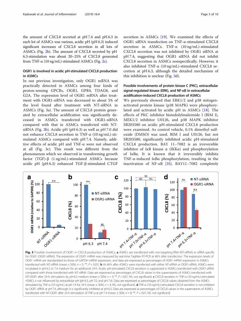
OGR1 is involved in acidic pH-stimulated CXCL8 productionin ASMCsIn our previous investigation, only OGR1 mRNA waspractically detected in ASMCs among four kinds ofproton-sensing GPCRs, OGR1, GPR4, TDAG8, andG2A. The expression level of OGR1 mRNA after treat-ment with OGR1-siRNA was decreased to about 5% ofthe level found after treatment with NT-siRNA inASMCs (Fig. 3a). The amount of CXCL8 protein gener-ated by extracellular acidification was significantly de-ceased in ASMCs transfected with OGR1-siRNAcompared with that in ASMCs transfected with NT-siRNA (Fig. 3b). Acidic pH (pH 6.3) as well as pH 7.0 didnot enhance CXCL8 secretion in TNF-α (10 ng/mL)-sti-mulated ASMCs compared with pH 7.4. Namely, addi-tive effects of acidic pH and TNF-α were not observedat all (Fig. 3c). This result was different from thephenomenon which we observed in transforming growthfactor (TGF)-β (1 ng/mL)-stimulated ASMCs becauseacidic pH (pH 6.3) enhanced TGF-β-stimulated CTGF
secretion in ASMCs [19]. We examined the effects ofOGR1 siRNA transfection on TNF-α-stimulated CXCL8secretion in ASMCs. TNF-α (10 ng/mL)-stimulatedCXCL8 secretion was not inhibited by OGR1 siRNA atpH 7.4, suggesting that OGR1 siRNA did not inhibitCXCL8 secretion in ASMCs nonspecifically. However, italso inhibited TNF-α (10 ng/mL)-stimulated CXCL8 se-cretion at pH 6.3, although the detailed mechanism ofthis inhibition is unclear (Fig. 3d).
Possible involvements of protein kinase C (PKC), extracellularsignal-regulated kinase (ERK), and NF-κB in extracellularacidification-induced CXCL8 production of ASMCsWe previously showed that ERK1/2 and p38 mitogen-activated protein kinase (p38 MAPK) were phosphory-lated and activated by acidic pH in ASMCs [18]. Theeffects of PKC inhibitor bisindolylmaleimide I (BIM I),MEK1/2 inhibitor U0126, and p38 MAPK inhibitorSB203580 on acidic pH-stimulated CXCL8 productionwere examined. As control vehicle, 0.1% dimethyl sulf-oxide (DMSO) was used. BIM I and U0126, but notSB203580, significantly inhibited acidic pH-stimulatedCXCL8 production. BAY 11–7082 is an irreversibleinhibitor of IκB kinase α (IKKα) and phosphorylationof IκBα. It is known that it irreversibly inhibitsTNF-α-induced IκBα phosphorylation, resulting in theinactivation of NF-κB [35]. BAY11–7082 completely
Fig. 3 Possible involvement of OGR1 in CXCL8 production of ASMCs. a ASMCs are transfected with non-targeting RNA (NT-siRNA) or siRNA specificfor OGR1 (OGR1-siRNA). The expression of OGR1 mRNA was measured by real-time TaqMan RT-PCR at 48 h after transfection. The expression levels ofOGR1 mRNA are standardized to those of GAPDH mRNA expression, and data are expressed as percentages of OGR1 mRNA expression in ASMCstransfected with NT-siRNA (mean ± SEM, n = 5; **, P < 0.01). b At 48 h after ASMCs were transfected with either NT-siRNA or OGR1-siRNA, ASMCs wereincubated in pH 6.3 or 7.4 medium for an additional 24 h. Acidic pH-stimulated CXCL8 secretion is suppressed in ASMCs transfected with OGR1-siRNAcompared with those transfected with NT-siRNA. Data are expressed as percentages of CXCL8 values in the supernatants of ASMCs transfected withNT-OGR1 after 24-h stimulation by pH 6.3 medium (mean ± SEM, n= 3; **, P< 0.01; NS, not significant). c CXCL8 secretion in TNF-α (10 ng/mL)-stimulatedASMCs is not influenced by extracellular pH (pH 6.3, pH 7.0, and pH 7.4). Data are expressed as percentages of CXCL8 values obtained from the ASMCsstimulated by TNF-α (10 ng/mL) at pH 7.4 for 24 h (mean ± SEM, n= 4; NS, not significant). d TNF-α (10 ng/mL)-stimulated CXCL8 secretion is not inhibitedby OGR1 siRNA at pH 7.4, although it is significantly inhibited at pH 6.3. Data are expressed as percentages of CXCL8 values in the supernatants of ASMCstransfected with NT-OGR1 after 24 h-stimulation of TNF-α at pH 7.4 (mean ± SEM, n= 8; **, P< 0.01; NS, not significant)
Kadowaki et al. Journal of Inflammation (2019) 16:4 Page 5 of 10

blocked pH 6.3-stimulated CXCL8 production. Theamount of CXCL8 generated from acidic pH-stimulatedASMCs in the presence of BAY11–7082 was signifi-cantly less than that generated from ASMCs withoutacidic pH stimulation (Fig. 4).
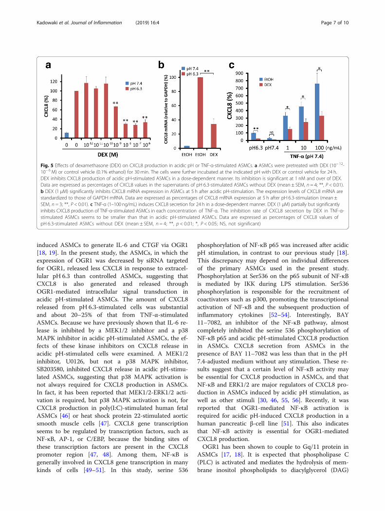
Effects of dexamethasone on CXCL8 production in ASMCsstimulated by extracellular acidificationIt is well known that CXCL8 production induced byTNF-α is inhibited by glucocorticoids in many kinds ofcells [26, 27, 36–38]. The effects of dexamethasone(DEX) (10− 12–10− 6 M) on acidic pH or TNF-α-stimu-lated CXCL8 production were examined in ASMCs.DEX inhibited acidic pH-stimulated CXCL8 productionin ASMCs in a dose-dependent manner. The inhibitionwas significant at 1 nM and over of DEX (Fig. 5a). DEX(1 μM) also significantly inhibited CXCL8 mRNA expres-sion in pH 6.3-stimulated ASMCs (Fig. 5b). We examineddose-dependent effects of TNF-α on CXCL8 secretion inASMCs. TNF-α induced CXCL8 secretion in adose-dependent manner at 1–100 ng/mL. DEX (1 μM)partially but significantly inhibited TNF-α (1–100 ng/mL)-stimulated CXCL8 production in ASMCs. The inhib-ition rate of DEX on TNF-α -stimulated CXCL8 secretionwas about 50% (Fig. 5c).
Effects of DEX on extracellular acidification-inducedphosphorylation of NF-κB p65 and DNA binding of NF-κB p65Serine 536 phosphorylation of NF-κB p65 was investi-gated in ASMCs. TNF-α (10 ng/mL)-stimulated ASMCswere used as a positive control of NF-κB p65 phosphor-ylation. The amount of phosphorylated NF-κB p65 wasmarkedly increased at 30–60min after addition of 10 ng/mL TNF-α. It was also slightly increased at 30–60 min inpH 6.3-stimulated ASMCs, but not in pH 7.4-stimulatedASMCs (Fig. 6a). In the presence of BAY11–7082, phos-phorylation of NF-κB p65 was almost completely inhib-ited with stimulation in all cases, pH 6.3, pH 7.4, andTNF-α (Fig. 6b). Next, whether DEX (1 μM) affectedserine 536 phosphorylation of NF-κB p65 was examinedin ASMCs. Thirty-min preincubation with DEX (1 μM)did not change the amount of phosphorylated p65 in allcases, pH 6.3, pH 7.4, and TNF-α-stimulated ASMCs(Fig. 6c). The binding of NF-κB p65 to its consensusDNA sequence (5’-GGGACTTTCC-3′) was analyzed.The binding of NF-κB p65 to DNA was increased ineither pH 6.3 or TNF-α (10 ng/mL)-stimulated ASMCscompared with that in the cells incubated in pH7.4-adjusted medium (P < 0.01). The increase of DNAbinding was smaller in acidic pH-stimulated ASMCsthan in TNF-α-stimulated cells. DEX (1 μM) did notaffect the binding of NF-κB p65 to its consensusDNA sequence in all cases, pH 6.3, pH 7.4, andTNF-α (10 ng/mL) (Fig. 6d).
DiscussionHypertrophy and hyperplasia of airway smooth musclecells (ASMCs) are characteristic pathological findings ofsevere asthma [1, 39]. ASMCs are increasingly recog-nized as an important source of inflammatory cytokinesand chemokines, as well as the effector cells of broncho-constriction [2–8, 40]. Bronchial thermoplasty has re-cently been introduced as a nonpharmacological therapyfor moderate-to-severe asthma patients who are uncon-trolled despite optimal medical therapy. This treatmentcould reduce exacerbations and the emergency roomvisit rate, resulting in improved quality of life [41–43].Although the mechanism of action is incompletelyunderstood, it has been suggested that bronchial ther-moplasty works by reducing ASMCs [44]. Reduction ofASMCs may decrease the secretion of cytokines andchemokines from ASMCs in patients with severeasthma. Interestingly, it has been demonstrated that in-creased expressions of CXCL8 and eotaxin in ASMCs,as well as an increase of airway smooth muscle area, areseen in patients with severe asthma compared with thosewith moderate asthma [45].CXCL8 is secreted by ASMCs following various stim-
uli, such as TNF-α and cigarette smoke [7, 26–34]. Ourprevious study showed that extracellular acidification
Fig. 4 Possible involvements of PKC, ERK1/2, p38 MAPK, and NF-κBin CXCL8 production of acidic pH-stimulated ASMCs. ASMCs werepretreated with 10 μM bisindolylmaleimide I (BIM I), 10 μMU0126,10 μM SB203580, 30 μM BAY11–7082, or control vehicle (0.1% DMSO)for 30 min. The cells were further incubated at the indicated pH with10 μM BIM I, 10 μMU0126, 10 μM SB203580, 30 μM BAY11–7082, orcontrol vehicle (0.1% DMSO) for an additional 24 h. CXCL8 secretioninduced by extracellular acidification in ASMCs is significantlysuppressed by U0126 and BIM I, but not SB203580. BAY11–7082almost completely inhibits CXCL8 secretion in ASMCs. Data areexpressed as percentages of CXCL8 values in the supernatants of pH6.3-stimulated ASMCs without any inhibitors. The data are expressed asmeans ± SEM (n = 4, **, P < 0.01; *, P < 0.05; NS, not significant)
Kadowaki et al. Journal of Inflammation (2019) 16:4 Page 6 of 10
induced ASMCs to generate IL-6 and CTGF via OGR1[18, 19]. In the present study, the ASMCs, in which theexpression of OGR1 was decreased by siRNA targetedfor OGR1, released less CXCL8 in response to extracel-lular pH 6.3 than controlled ASMCs, suggesting thatCXCL8 is also generated and released throughOGR1-mediated intracellular signal transduction inacidic pH-stimulated ASMCs. The amount of CXCL8released from pH 6.3-stimulated cells was substantialand about 20–25% of that from TNF-α-stimulatedASMCs. Because we have previously shown that IL-6 re-lease is inhibited by a MEK1/2 inhibitor and a p38MAPK inhibitor in acidic pH-stimulated ASMCs, the ef-fects of these kinase inhibitors on CXCL8 release inacidic pH-stimulated cells were examined. A MEK1/2inhibitor, U0126, but not a p38 MAPK inhibitor,SB203580, inhibited CXCL8 release in acidic pH-stimu-lated ASMCs, suggesting that p38 MAPK activation isnot always required for CXCL8 production in ASMCs.In fact, it has been reported that MEK1/2-ERK1/2 acti-vation is required, but p38 MAPK activation is not, forCXCL8 production in poly(I:C)-stimulated human fetalASMCs [46] or heat shock protein 22-stimulated aorticsmooth muscle cells [47]. CXCL8 gene transcriptionseems to be regulated by transcription factors, such asNF-κB, AP-1, or C/EBP, because the binding sites ofthese transcription factors are present in the CXCL8promoter region [47, 48]. Among them, NF-κB isgenerally involved in CXCL8 gene transcription in manykinds of cells [49–51]. In this study, serine 536
phosphorylation of NF-κB p65 was increased after acidicpH stimulation, in contrast to our previous study [18].This discrepancy may depend on individual differencesof the primary ASMCs used in the present study.Phosphorylation at Ser536 on the p65 subunit of NF-κBis mediated by IKK during LPS stimulation. Ser536phosphorylation is responsible for the recruitment ofcoactivators such as p300, promoting the transcriptionalactivation of NF-κB and the subsequent production ofinflammatory cytokines [52–54]. Interestingly, BAY11–7082, an inhibitor of the NF-κB pathway, almostcompletely inhibited the serine 536 phosphorylation ofNF-κB p65 and acidic pH-stimulated CXCL8 productionin ASMCs. CXCL8 secretion from ASMCs in thepresence of BAY 11–7082 was less than that in the pH7.4-adjusted medium without any stimulation. These re-sults suggest that a certain level of NF-κB activity maybe essential for CXCL8 production in ASMCs, and thatNF-κB and ERK1/2 are major regulators of CXCL8 pro-duction in ASMCs induced by acidic pH stimulation, aswell as other stimuli [30, 46, 55, 56]. Recently, it wasreported that OGR1-mediated NF-κB activation isrequired for acidic pH-induced CXCL8 production in ahuman pancreatic β-cell line [51]. This also indicatesthat NF-κB activity is essential for OGR1-mediatedCXCL8 production.OGR1 has been shown to couple to Gq/11 protein in
ASMCs [17, 18]. It is expected that phospholipase C(PLC) is activated and mediates the hydrolysis of mem-brane inositol phospholipids to diacylglycerol (DAG)
Fig. 5 Effects of dexamethasone (DEX) on CXCL8 production in acidic pH or TNF-α-stimulated ASMCs. a ASMCs were pretreated with DEX (10− 12-10− 6 M) or control vehicle (0.1% ethanol) for 30 min. The cells were further incubated at the indicated pH with DEX or control vehicle for 24 h.DEX inhibits CXCL8 production of acidic pH-stimulated ASMCs in a dose-dependent manner. Its inhibition is significant at 1 nM and over of DEX.Data are expressed as percentages of CXCL8 values in the supernatants of pH 6.3-stimulated ASMCs without DEX (mean ± SEM, n = 4; **, P < 0.01).b DEX (1 μM) significantly inhibits CXCL8 mRNA expression in ASMCs at 5 h after acidic pH-stimulation. The expression levels of CXCL8 mRNA arestandardized to those of GAPDH mRNA. Data are expressed as percentages of CXCL8 mRNA expression at 5 h after pH 6.3-stimulation (mean ±SEM, n = 3; **, P < 0.01). c TNF-α (1–100 ng/mL) induces CXCL8 secretion for 24 h in a dose-dependent manner. DEX (1 μM) partially but significantlyinhibits CXCL8 production of TNF-α-stimulated ASMCs in each concentration of TNF-α. The inhibition rate of CXCL8 secretion by DEX in TNF-α-stimulated ASMCs seems to be smaller than that in acidic pH-stimulated ASMCs. Data are expressed as percentages of CXCL8 values ofpH 6.3-stimulated ASMCs without DEX (mean ± SEM, n = 4; **, p < 0.01; *, P < 0.05; NS, not significant)
Kadowaki et al. Journal of Inflammation (2019) 16:4 Page 7 of 10

and inositol 1,4,5-trisphosphate, which in turn triggersthe release of calcium from intracellular stores followingGq/11 activation [14, 57, 58]. Because the PKC inhibitorbisindolylmaleimide I inhibited OGR1-mediated CXCL8secretion in ASMCs, PKC activation following PLC maybe involved in the pathway to CXCL8 gene transcription.In the present study, DEX inhibited CXCL8 secretionfrom acidic pH-stimulated ASMCs in a dose-dependentmanner. CXCL8 mRNA expression was also inhibited byDEX, as well as CXCL8 protein. Although DEX partiallyinhibited CXCL8 release in TNF-α-stimulated ASMCs,the inhibition rate was smaller than that in acidicpH-stimulated cells. Glucocorticoids (GCs) may regulatecytokine gene expression in several ways. The gluco-corticoid receptor (GR) influences gene expression byphysically interacting with other transcription factorswithout contacting DNA itself. This mechanism is calledtransrepression [59]. Activated GRs translocate to the nu-cleus and bind to coactivators to inhibit histone acetyl-transferase activity and recruiting histone deacetylase-2,which reverses histone acetylation, leading to suppression
of targeted genes [60, 61]. Although NF-κB activity wasessential for CXCL8 secretion of ASMCs, DEX affectedneither serine 536 phosphorylation of NF-κB p65 norbinding of NF-κB p65 to its consensus DNA sequence.These results support the possibility that CXCL8 genetranscription is inhibited by DEX at the step after a dimerof p50 and p65 NF-κB proteins binds to a specific NF-κBrecognition site. The detailed mechanism of DEX with re-spect to CXCL8 gene transcription in ASMCs and the rea-son why the inhibition rate by DEX is different betweenacidic pH-stimulated cells and TNF-α-stimulated onesshould be elucidated by further research.As a limitation of this research, our experiments were
done using ASMCs originated from non-diseased indi-viduals. If CXCL8 secreted via OGR1 is involved in neu-trophilic airway inflammation of COPD, the results inthis study might support the usefulness of inhaled corti-costeroids as well as long acting bronchodilators inpharmacotherapy of COPD. In future, it is very import-ant to investigate whether DEX inhibits OGR1-mediatedCXCL8 secretion in ASMCs isolated from patients with
Fig. 6 Effects of DEX on phosphorylation and DNA binding of NF-κB p65. a ASMCs were incubated for the indicated time in pH 6.3 or pH 7.4-adjustedmedium. As a positive control of serine 536 phosphorylation and DNA binding of NF-κB p65, ASMCs were stimulated with 10 ng/ml TNF-α in the pH7.4-adjusted medium. A weak increase of phospho-NF-κB p65 (Ser 536) is observed in ASMCs at 30–60min after pH 6.3-stimulation, but not after pH7.4-stimulation. GAPDH was used as an endogenous control protein. Representative data of 3 independent experiments are shown. b ASMCs werepretreated with 30 μM BAY11–7082 or control vehicle (0.1% DMSO) for 30min. The cells were further incubated for 60min in the pH 6.3-adjustedmedium, pH 7.4-adjusted medium, or pH 7.4-adjusted medium containing 10 ng/ml TNF-α. BAY11–7082 almost completely decreases the expressionsof phospho-NF-κB p65 (Ser536) in all cases, pH 6.3, pH 7.4, and TNF-α (pH 7.4). Representative data of 3 independent experiments are shown. c After30-min preincubation with DEX (1 μM) or control vehicle (0.1% ethanol), ASMCs were stimulated for 60 min by pH 6.3-adjusted medium, pH 7.4-adjusted medium, or pH 7.4-adjusted medium containing 10 ng/ml TNF-α. DEX does not affect serine 536 phosphorylation of NF-κB p65in all cases, pH 6.3, pH 7.4, and TNF-α (pH 7.4). Representative data of 3 independent experiments are shown. d Binding of NF-κB p65 toits consensus DNA sequence is significantly increased at 60min after pH 6.3- or TNF-α-stimulation compared with pH 7.4-stimulation (P < 0.01).DEX (1 μM) does not substantially affect the binding of NF-κB p65 to its consensus DNA sequence in all cases, pH 6.3, pH 7.4, and TNF-α (pH 7.4). Dataare expressed as percentages of absorbance at 450 nm (OD450) of pH 6.3-stimulated ASMCs without DEX (mean ± SEM, n = 5; NS, not significant)
Kadowaki et al. Journal of Inflammation (2019) 16:4 Page 8 of 10

steroid-resistant asthma or COPD, because ASMCs ofpatients with severe asthma seem to be insensitive tocorticosteroids [62, 63].
ConclusionsTaken together, the present data demonstrate for the firsttime that extracellular acidification induces CXCL8 secre-tion of ASMCs through OGR1-mediated cellular activation,and that DEX inhibits OGR1-mediated CXCL8 secretion ofASMCs originated from non-diseased individuals.
AbbreviationsAP-1: Activator protein 1; ASMC: Airway smooth muscle cell; C/EBP: CCAAT/enhancer binding protein; COPD: Chronic obstructive pulmonary disease;CTGF: Connective tissue growth factor; CXCL8: C-X-C motif chemokine ligand8; CXCR2: C-X-C motif chemokine receptor 2; DEX: Dexamethasone;DMSO: Dimethyl sulfoxide; ERK: Extracellular signal-regulated protein kin-ase; EtOH: Ethanol; GC: Glucocorticoid; IKK: IκB kinase; IL-6: Interleukin-6;LPS: Lipopolysaccharide; MAPK: Mitogen-activated protein kinase;MEK: MAP/ERK kinase; NF-κB: Nuclear factor-kappa B; OGR1: Ovariancancer G protein-coupled receptor 1; PKC: Protein kinase C;PLC: Phospholipase C; TNF-α: Tumor necrosis factor-α
AcknowledgmentsThe authors are grateful to Ayumi Suzuki and Emi Takeuchi for technicalassistance and to Kazuyo Takeuchi for manuscript preparation.
FundingThis work was supported by a Grant-in-Aid for scientific research from the JapanSociety for the Promotion of Science (T. Ishizuka and F. Okajima) from the Ministryof Education, Culture, Sports, Science, and Technology of Japan.
Availability of data and materialsThe datasets used and analyzed during the current study are available fromthe corresponding author on reasonable request.
Authors’ contributionsTI and FO provided the research idea. MK, HY, and TI did cell culture andstimulation. MK and HY performed ELISA and RT-PCR. MK did Western blottingand experiments using siRNA and nuclear protein. MK, HY, KS, HS, YU, MM, YW,MA, YK, HA-S, TH, FO, and TI analyzed data and discussed interpretation of data.MK and TI prepared the manuscript. All authors read and approved the finalmanuscript.
Ethics approval and consent to participateNot applicable.
Consent for publicationNot applicable.
Competing interestsThe authors declare that they have no competing interests.
Publisher’s NoteSpringer Nature remains neutral with regard to jurisdictional claims inpublished maps and institutional affiliations.
Author details1Third Department of Internal Medicine, Faculty of Medical Sciences,University of Fukui, 23-3 Matsuoka-Shimoaizuki, Eiheiji, Fukui 910-1193, Japan.2Department of Medicine and Molecular Science, Gunma University GraduateSchool of Medicine, 3-39-15 Showa-machi, Maebeshi 371-8511, Japan.3Laboratory of Signal Transduction, Institute for Molecular and CellularRegulation, Gunma University, 3-39-15 Showa-machi, Maebeshi 371-8511,Japan. 4Laboratory of Signal Transduction, Faculty of PharmaceuticalSciences, Aomori University, 2-3-1 Kobata, Aomori 030-0943, Japan.
Received: 11 September 2018 Accepted: 27 January 2019
References1. Hirst SJ. Airway smooth muscle as a target in asthma. Clin Exp Allergy. 2000;
30(Suppl 1):54–9.2. Damera G, Tliba O, Panettieri RA Jr. Airway smooth muscle as an immunomodulatory
cell. Pulm Pharmacol Ther. 2009;22:353–9.3. Howarth PH, Knox AJ, Amrani Y, Tliba O, Panettieri RA Jr, Johnson M. Synthetic
responses in airway smooth muscle. J Allergy Clin Immunol. 2004;114(2 Suppl):S32–50.
4. Tliba O, Panettieri RA Jr. Noncontractile functions of airway smooth musclecells in asthma. Annu Rev Physiol. 2009;71:509–35.
5. Xia YC, Redhu NS, Moir LM, Koziol-White C, Ammit AJ, Al-Alwan L, Camoretti-Mercado B, Clifford RL. Pro-inflammatory and immunomodulatory functions ofairway smooth muscle: emerging concepts. Pulm Pharmacol Ther. 2013;26:64–74.
6. Oliver BG, Johnston SL, Baraket M, Burgess JK, King NJ, Roth M, Lim S, Black JL.Increased proinflammatory responses from asthmatic human airway smoothmuscle cells in response to rhinovirus infection. Respir Res. 2006;7:71.
7. Gosens R, Rieks D, Meurs H, Ninaber DK, Rabe KF, Nanninga J, Kolahian S,Halayko AJ, Hiemstra PS, Zuyderduyn S. Muscarinic M3 receptor stimulationincreases cigarette smoke-induced CXCL8 secretion by human airway smoothmuscle cells. Eur Respir J. 2009;34:1436–43.
8. Iwata S, Ito S, Iwaki M, Kondo M, Sashio T, Takeda N, Sokabe M, HasegawaY, Kume H. Regulation of endothelin-1-induced interleukin-6 production byCa2+ influx in human airway smooth muscle cells. Eur J Pharmacol. 2009;605:15–22.
9. Kodric M, Shah AN, Fabbri LM, Confalonieri M. An investigation of airwayacidification in asthma using induced sputum: a study of feasibility andcorrelation. Am J Respir Crit Care Med. 2007;175:905–10.
10. Ricciardolo FL, Gaston B, Hunt J. Acid stress in the pathology of asthma.J Allergy Clin Immunol. 2004;113:610–9.
11. Hunt JF, Fang K, Malik R, Snyder A, Malhotra N, Platts-Mills TA, Gaston B.Endogenous airway acidification. Implications for asthma pathophysiologyAm J Respir Crit Care Med. 2000;161(3 Pt 1):694–9.
12. Faisy C, Planquette B, Naline E, Risse PA, Frossard N, Fagon JY, Advenier C,Devillier P. Acid-induced modulation of airway basal tone and contractility:role of acid-sensing ion channels (ASICs) and TRPV1 receptor. Life Sci. 2007;81:1094–102.
13. Lee LY, Gu Q. Role of TRPV1 in inflammation-induced airway hypersensitivity.Curr Opin Pharmacol. 2009;9:243–9.
14. Ludwig MG, Vanek M, Guerini D, Gasser JA, Jones CE, Junker U, Hofstetter H,Wolf RM, Seuwen K. Proton-sensing G-protein-coupled receptors. Nature.2003;425:93–8.
15. Tomura H, Mogi C, Sato K, Okajima F. Proton-sensing and lysolipid-sensitiveG-protein-coupled receptors: a novel type of multi-functional receptors. CellSignal. 2005;17:1466–76.
16. Okajima F. Regulation of inflammation by extracellular acidification andproton-sensing GPCRs. Cell Signal. 2013;25:2263–71.
17. Aoki H, Mogi C, Okajima F. Ionotropic and metabotropic proton-sensingreceptors involved in airway inflammation in allergic asthma. Mediat Inflamm.2014;2014:712962.
18. Ichimonji I, Tomura H, Mogi C, Sato K, Aoki H, Hisada T, Dobashi K, IshizukaT, Mori M, Okajima F. Extracellular acidification stimulates IL-6 productionand Ca2+ mobilization through proton-sensing OGR1 receptors in humanairway smooth muscle cells. Am J Physiol Lung Cell Mol Physiol. 2010;299:L567–77.
19. Matsuzaki S, Ishizuka T, Yamada H, Kamide Y, Hisada T, Ichimonji I, Aoki H,Yatomi M, Komachi M, Tsurumaki H, et al. Extracellular acidification inducesconnective tissue growth factor production through proton-sensing receptorOGR1 in human airway smooth muscle cells. Biochem Biophys Res Commun.2011;413:499–503.
20. O'Byrne PM, Metev H, Puu M, Richter K, Keen C, Uddin M, Larsson B, Cullberg M,Nair P. Efficacy and safety of a CXCR2 antagonist, AZD5069, in patients withuncontrolled persistent asthma: a randomised, double-blind, placebo-controlledtrial. Lancet Respir Med. 2016;4:797–806.
21. Barnes PJ. New anti-inflammatory targets for chronic obstructive pulmonarydisease. Nat Rev. Drug Discov. 2013;12:543–59.
22. Gibson PG, Simpson JL, Saltos N. Heterogeneity of airway inflammation inpersistent asthma : evidence of neutrophilic inflammation and increasedsputum interleukin-8. Chest. 2001;119:1329–36.
Kadowaki et al. Journal of Inflammation (2019) 16:4 Page 9 of 10

23. Yamamoto C, Yoneda T, Yoshikawa M, Fu A, Tokuyama T, Tsukaguchi K,Narita N. Airway inflammation in COPD assessed by sputum levels ofinterleukin-8. Chest. 1997;112:505–10.
24. Pantelidis P, Southcott AM, Black CM, Du Bois RM. Up-regulation of CXCL8secretion by alveolar macrophages from patients with fibrosing alveolitis: asubpopulation analysis. Clin Exp Immunol. 1997;108:95–104.
25. Carmona EM, Lamont JD, Xue A, Wylam M, Limper AH. Pneumocystis cellwall beta-glucan stimulates calcium-dependent signaling of CXCL8 secretionby human airway epithelial cells. Respir Res. 2010;11:95.
26. Pang L, Knox AJ. Synergistic inhibition by β2-agonists and corticosteroids ontumor necrosis factor-alpha-induced interleukin-8 release from cultured humanairway smooth-muscle cells. Am J Respir Cell Mol Biol. 2000;23:79–85.
27. Robins S, Roussel L, Schachter A, Risse PA, Mogas AK, Olivenstein R, MartinJG, Hamid Q, Rousseau S. Steroid-insensitive ERK1/2 activity drives CXCL8synthesis and neutrophilia by airway smooth muscle. Am J Respir Cell MolBiol. 2011;45:984–90.
28. Pang L, Knox AJ. Bradykinin stimulates CXCL8 production in culturedhuman airway smooth muscle cells: role of cyclooxygenase products.J Immunol. 1998;161:2509–15.
29. Patel BS, Rahman MM, Baehring G, Xenaki D, Tang FS, Oliver BG, Ammit AJ.Roflumilast N-oxide in combination with formoterol enhances theantiinflammatory effect of dexamethasone in airway smooth musclecells. Am J Respir Cell Mol Biol. 2017;56:532–8.
30. Wuyts WA, Vanaudenaerde BM, Dupont LJ, Van Raemdonck DE, DemedtsMG, Verleden GM. Interleukin-17-induced interleukin-8 release in humanairway smooth muscle cells: role for mitogen-activated kinases and nuclearfactor-κB. J Heart Lung Transplant. 2005;24:875–81.
31. Pera T, Atmaj C, van der Vegt M, Halayko AJ, Zaagsma J, Meurs H. Role forTAK1 in cigarette smoke-induced proinflammatory signaling and CXCL8release by human airway smooth muscle cells. Am J Physiol Lung Cell MolPhysiol. 2012;303:L272–8.
32. Keglowich L, Roth M, Philippova M, Resink T, Tjin G, Oliver B, Lardinois D,Dessus-Babus S, Gosens R, Hostettler Haack K, et al. Bronchial smoothmuscle cells of asthmatics promote angiogenesis through elevatedsecretion of CXC-chemokines (ENA-78, GRO-alpha, and CXCL8). PLoSOne. 2013;8:e81494.
33. Patel BS, Rahman MM, Rumzhum NN, Oliver BG, Verrills NM, Ammit AJ.Theophylline represses CXCL8 secretion from airway smooth muscle cellsindependently of phosphodiesterase inhibition. Novel role as a proteinphosphatase 2A activator. Am J Respir Cell Mol Biol. 2016;54:792–801.
34. Nakajima M, Kawaguchi M, Matsuyama M, Ota K, Fujita J, Matsukura S,Huang SK, Morishima Y, Ishii Y, Satoh H, et al. Transcription elongationfactor P-TEFb is involved in IL-17F signaling in airway smooth muscle cells.Int Arch Allergy Immunol. 2018;176:83–90.
35. Pierce JW, Schoenleber R, Jesmok G, Best J, Moore SA, Collins T, Gerritsen ME.Novel inhibitors of cytokine-induced IκBα phosphorylation and endothelial celladhesion molecule expression show anti-inflammatory effects in vivo. J BiolChem. 1997;272:21096–103.
36. Zhao Y, Leung PC, Woo KS, Chen GG, Wong YO, Liu SX, van Hasselt CA.Inhibitory effects of budesonide, desloratadine and dexamethasone oncytokine release from human mast cell line (HMC-1). Inflamm Res. 2004;53:664–9.
37. Ek A, Larsson K, Siljerud S, Palmberg L. Fluticasone and budesonide inhibitcytokine release in human lung epithelial cells and alveolar macrophages.Allergy. 1999;54:691–9.
38. Pan NY, Hui WS, Tipoe GL, Taylor GW, Leung RY, Lam WK, Tsang KW, Mak JC.Inhibition of pyocyanin-potentiated CXCL8 release by steroids in bronchialepithelial cells. Respir Med. 2006;100:1614–22.
39. Holgate ST, Peters-Golden M, Panettieri RA, Henderson WR Jr. Roles of cysteinylleukotrienes in airway inflammation, smooth muscle function, and remodeling.J Allergy Clin Immunol. 2003;111(1 Suppl):S18–34 discussion S34–16.
40. Berair R, Hollins F, Brightling C. Airway smooth muscle hypercontractility inasthma. J Allergy (Cairo). 2013;2013:185971.
41. Cox G, Thomson NC, Rubin AS, Niven RM, Corris PA, Siersted HC, OlivensteinR, Pavord ID, McCormack D, Chaudhuri R, et al. Asthma control during theyear after bronchial thermoplasty. N Engl J Med. 2007;356:1327–37.
42. Pavord ID, Cox G, Thomson NC, Rubin AS, Corris PA, Niven RM, Chung KF,Laviolette M. Safety and efficacy of bronchial thermoplasty in symptomatic,severe asthma. Am J Respir Crit Care Med. 2007;176:1185–91.
43. Castro M, Rubin AS, Laviolette M, Fiterman J, De Andrade Lima M, Shah PL,Fiss E, Olivenstein R, Thomson NC, Niven RM, et al. Effectiveness and safety
of bronchial thermoplasty in the treatment of severe asthma: a multicenter,randomized, double-blind, sham-controlled clinical trial. Am J Respir CritCare Med. 2010;181:116–24.
44. Pretolani M, Bergqvist A, Thabut G, Dombret MC, Knapp D, Hamidi F,Alavoine L, Taille C, Chanez P, Erjefalt JS, et al. Effectiveness of bronchialthermoplasty in patients with severe refractory asthma: clinical and histopathologiccorrelations. J Allergy Clin Immunol. 2017;139:1176–85.
45. Pepe C, Foley S, Shannon J, Lemiere C, Olivenstein R, Ernst P, Ludwig MS,Martin JG, Hamid Q. Differences in airway remodeling between subjectswith severe and moderate asthma. J Allergy Clin Immunol. 2005;116:544–9.
46. Faksh A, Britt RD Jr, Vogel ER, Thompson MA, Pandya HC, Martin RJ, PabelickCM, Prakash YS. TLR3 activation increases chemokine expression in humanfetal airway smooth muscle cells. Am J Physiol Lung Cell Mol Physiol. 2016;310:L202–11.
47. Kang SH, Lee JH, Choi KH, Rhim BY, Kim K. Roles of ERK and NF-κB inInterleukin-8 expression in response to heat shock protein 22 in vascularsmooth muscle cells. Korean J Physiol Pharmacol. 2008;12:171–6.
48. Roebuck KA. Regulation of interleukin-8 gene expression. J Interf CytokineRes. 1999;19:429–38.
49. He W, Qu T, Yu Q, Wang Z, Lv H, Zhang J, Zhao X, Wang P. LPS inducesCXCL8 expression through TLR4, MyD88, NF-κB and MAPK pathways inhuman dental pulp stem cells. Int Endod J. 2013;46:128–36.
50. Ahn SH, Park H, Ahn YH, Kim S, Cho MS, Kang JL, Choi YH. Necrotic cellsinfluence migration and invasion of glioblastoma via NF-κB/AP-1-mediatedCXCL8 regulation. Sci Rep. 2016;6:24552.
51. Chandra V, Karamitri A, Richards P, Cormier F, Ramond C, Jockers R,Armanet M, Albagli-Curiel O, Scharfmann R. Extracellular acidificationstimulates GPR68 mediated CXCL8 production in human pancreaticbeta cells. Sci Rep. 2016;6:25765.
52. Hall G, Singh IS, Hester L, Hasday JD, Rogers TB. Inhibitor-kappaB kinase-beta regulates LPS-induced TNF-alpha production in cardiac myocytesthrough modulation of NF-κB p65 subunit phosphorylation. Am J PhysiolHeart Circ Physiol. 2005;289:H2103–11.
53. Oh SM, Lee SH, Lee BJ, Pyo CW, Yoo NK, Lee SY, Kim J, Choi SY. A distinctrole of neutrophil lactoferrin in RelA/p65 phosphorylation on Ser536 byrecruiting TNF receptor-associated factors to IκB kinase signaling complex.J Immunol. 2007;179:5686–92.
54. Buss H, Handschick K, Jurrmann N, Pekkonen P, Beuerlein K, Muller H, WaitR, Saklatvala J, Ojala PM, Schmitz ML, et al. Cyclin-dependent kinase 6phosphorylates NF-κB P65 at serine 536 and contributes to the regulationof inflammatory gene expression. PLoS One. 2012;7:e51847.
55. Li H, Zhu S, He S, Hao L. Anti-inflammatory effects of moxifloxacin on ratairway smooth muscle cells exposed to allergen: inhibition of extracellular-signal-regulated kinase and nuclear factor-κB activation and of interleukin-8and eotaxin synthesis. Respirology. 2012;17:997–1005.
56. Shih CJ, Chiou YL. Zinc sulfate inhibited inflammation of Der p2-inducedairway smooth muscle cells by suppressing ERK1/2 and NF-κB phosphorylation.Inflammation. 2013;36:616–24.
57. Lyon AM, Tesmer JJ. Structural insights into phospholipase C-β function.Mol Pharmacol. 2013;84:488–500.
58. Hu YL, Mi X, Huang C, Wang HF, Song JR, Shu Q, Ni L, Chen JG, Wang F, Hu ZL.Multiple H+ sensors mediate the extracellular acidification-induced [Ca2+]ielevation in cultured rat ventricular cardiomyocytes. Sci Rep. 2017;7:44951.
59. Ratman D, Vanden Berghe W, Dejager L, Libert C, Tavernier J, Beck IM, DeBosscher K. How glucocorticoid receptors modulate the activity of othertranscription factors: a scope beyond tethering. Mol Cell Endocrinol. 2013;380:41–54.
60. Ito K, Barnes PJ, Adcock IM. Glucocorticoid receptor recruitment of histonedeacetylase 2 inhibits interleukin-1beta-induced histone H4 acetylation onlysines 8 and 12. Mol Cell Biol. 2000;20:6891–903.
61. Ito K, Yamamura S, Essilfie-Quaye S, Cosio B, Ito M, Barnes PJ, Adcock IM.Histone deacetylase 2-mediated deacetylation of the glucocorticoid receptorenables NF-κB suppression. J Exp Med. 2006;203:7–13.
62. Chang PJ, Bhavsar PK, Michaeloudes C, Khorasani N, Chung KF. Corticosteroidinsensitivity of chemokine expression in airway smooth muscle of patientswith severe asthma. J Allergy Clin Immunol. 2012;130:877–885 e875.
63. Chang PJ, Michaeloudes C, Zhu J, Shaikh N, Baker J, Chung KF, Bhavsar PK.Impaired nuclear translocation of the glucocorticoid receptor in corticosteroid-insensitive airway smooth muscle in severe asthma. Am J Respir Crit Care Med.2015;191:54–62.
Kadowaki et al. Journal of Inflammation (2019) 16:4 Page 10 of 10


![acidification] ab197244 [Extracellular Glycolysis Assay€¦ · 22/05/2020 · ab197244 Glycolysis Assay [ECA/ECAR] 1 1. Overview Glycolysis Assay [Extracellular Acidification] (ab197244)](https://img.dokumen.tips/doc/110x75/5fcea1323cad0c1f6d536509/acidification-ab197244-extracellular-glycolysis-assay-22052020-ab197244-glycolysis.jpg)


![acidification] ab197244 [Extracellular Glycolysis Assay · Glycolysis Assay [Extracellular Acidification] (ab197244) is an easy mix-and-measure, 96 or 384 well fluorescence plate](https://img.dokumen.tips/doc/110x75/5ec5a381691079698166a0a6/acidification-ab197244-extracellular-glycolysis-assay-glycolysis-assay-extracellular.jpg)


![acidification] ab197244 [Extracellular Glycolysis Assay · ab197244 Glycolysis Assay [ECA/ECAR] 1 1. Overview Glycolysis Assay [Extracellular Acidification] (ab197244) is an easy](https://img.dokumen.tips/doc/110x75/5e162848f38add2f073828c8/acidification-ab197244-extracellular-glycolysis-assay-ab197244-glycolysis-assay.jpg)