Embed Size (px)
Citation preview
Egyptian Journal of Ear, Nose, Throat and Allied Sciences (2012) 13, 1–6
Egyptian Society of Ear, Nose, Throat and Allied Sciences
Egyptian Journal of Ear, Nose, Throat and Allied
Sciences
www.ejentas.com
ORIGINAL ARTICLE
Extended sublabial vestibulotomy (ESV) approach
for inverted papilloma
Samy Elwany, Zeyad Mandour *
Oto-Rhino-Laryngology Head & Neck Surgery Department, Faculty of Medicine, University of Alexandria, Egypt
Received 27 November 2011; accepted 21 January 2012
Available online 21 February 2012
20
Sc
Pe
Th
do
*
E
KEYWORDS
Inverted papilloma;
Sublabial vestibulotomy
(ESV)
90-0740 ª 2012 Egyptian So
iences. Production and hosti
er review under responsibili
roat and Allied Sciences.
i:10.1016/j.ejenta.2012.01.005
Production and h
Corresponding author.
-mail address: ziad301@hot
ciety of
ng by Els
ty of Eg
osting by E
mail.com
Abstract Background: The reported incidence of inverted papilloma of the sinonasal areas is 0.5–
4% of all primary nasal tumours. Aggressive surgical approaches and endoscopic sinus techniques
have been used as the main line of management.
Study design: To evaluate 30 cases of inverted papilloma as regarding their clinical and radiolog-
ical findings, as well as the use of the extended sublabial vestibulotomy (ESV) approach in their
management.
Results: The age average was 56.13 years with male prevalence. The lateral nasal wall and nasal
cavity were involved in all the cases. The individual sinuses were involved to a variable extent. All
patients treated by extended sublabial vestibulotomy with endoscopic assistance whenever indicated
with no significant complications or recurrence.
Conclusion: ESV approach is ideally for extensive benign lesions in the sinonasal areas; as well, it
enables the access to the nasopharynx. It can be considered as a feasible approach.ª 2012 Egyptian Society of Ear, Nose, Throat and Allied Sciences.
Production and hosting by Elsevier B.V. All rights reserved.
Ear, Nose, Throat and Allied
evier B.V. All rights reserved.
yptian Society of Ear, Nose,
lsevier
(Z. Mandour).
1. Introduction
Inverted papilloma of the nose and paranasal sinuses are raretumors, the reported incidence being 0.5–4% of all primary na-sal tumours.1 It is recognized as a neoplastic growth of the epi-
thelium that inverts into the underlying stroma rather thanproliferating outward from the surface.2 The neoplasm is char-acterized by its capacity to destroy the surrounding, tendency
to recur after removal and its association with malignancy.3
The aetiology of the tumour remains unknown. Inflammation,allergy, tobacco and occupation exposures have been dis-
counted as significant factors. Viral aetiology remains incon-clusive and investigations using in situ hybridization (ISH)and PCR have been used to determine its link with HPV.4
The reported prevalence of carcinoma in patients with inverted
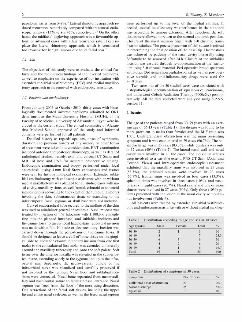
Table 1 Distribution according to age and sex in 30 cases.
Age (years) Male Female Total %
30–39 2 1 3 10
40–49 3 4 7 23.3
50–59 6 3 9 30
60–69 4 2 6 20
70–79 4 1 5 16.7
Total 19 11 30 100
Table 2 Distribution of symptoms in 30 cases.
Symptoms No. of cases %
Unilateral nasal obstruction 29 96.7
Nasal discharge 25 83.3
Epistaxis 12 40
2 S. Elwany, Z. Mandour
papilloma varies from 5–8%.5 Lateral rhinotomy approach re-duced recurrence remarkably compared with transnasal endo-scopic removal (13% versus 45%, respectively).6 On the other
hand, the midfacial degloving approach was a favourable op-tion for advanced cases with a fair recurrence rate. It can re-place the lateral rhinotomy approach, which is considered
too invasive for benign tumour due to its facial scar.7
1.1. Aim
The objectives of this study were to evaluate the clinical fea-tures and the radiological findings of the inverted papilloma,as well to emphasise on the experience of our institution with
extended sublabial vestibulotomy (ESV) and medial maxillec-tomy approach in its removal with endoscopic assistance.
1.2. Patients and methodology
From January 2005 to October 2010, thirty cases with histo-logically documented inverted papilloma admitted to ORLdepartment at the Main University Hospital (MUH), of the
Faculty of Medicine, University of Alexandria, Egypt were in-cluded in the current study. The ethical committee of Alexan-dria Medical School approved of the study and informed
consents were performed for all patients.Detailed history as regards age, sex, onset of symptoms,
duration and previous history of any surgery or other formsof treatment were taken into consideration. ENT examination
included anterior and posterior rhinoscopy, as well as detailedradiological studies, namely, axial and coronal CT Scans andMRI of nose and PNS for accurate preoperative staging.
Endoscopic examination and biopsy performed under localanaesthesia, using 4 mm Karl–Storz endoscopes and tissueswere sent for histopathological examination. Extended subla-
bial vestibulotomy with endoscopic assistance with or withoutmedial maxillectomy was planned for all studied cases with na-sal cavity, maxillary sinus, as well frontal, ethmoid or sphenoid
sinuses lesions according to the extent of the tumour. Tumoursinvolving the skin, subcutaneous tissue or extending to theinfratemporal fossa, zygoma or skull base were not included.
Curved endotracheal tube secured to the midline of the chin
was used to administer general anaesthesia. Nasal mucosa wastreated by injection of 1% lidocaine with 1:100,000 epineph-rine into the planned intranasal and sublabial incisions and
the canine fossa to ensure local haemostasis. Sublabial incisionwas made with a No. 10 blade or electrocautery. Incision wascarried down through the periosteum of the canine fossa. It
should be designed to leave a cuff of loose tissue on the gingi-val side to allow for closure. Standard incision from one firstmolar to the contralateral first molar was extended unilaterally
around the maxillary tuberosity and onto the soft palate. Softtissue over the anterior maxilla was elevated in the subperios-teal plane, extending widely to the zygoma and up to the infra-orbital rim. Superiorly, the neurovascular bundle of the
infraorbital nerve was visualized and carefully preserved ifnot involved by the tumour. Nasal floor and sublabial inci-sions were connected. Nasal bone separated from nasomaxil-
lary and nasofrontal suture to facilitate nasal entrance. Nasalseptum was freed from the floor of the nose using dissection.Full retractions of the facial soft tissues, including the upper
lip and entire nasal skeleton, as well as the freed nasal septum
were performed up to the level of the medial canthus. Ifneeded, medial maxillectomy was performed in the standardway according to tumour extension. After resection, the soft
tissues were allowed to return to the normal anatomic position.Closure of the nasal incision began with 3–0 chromic trans-fixation stitches. The precise placement of this suture is critical
in determining the final position of the nasal tip. Haemostasiswas achieved by packing of the nasal cavity bilaterally usingSofratulle to be removed after 24 h. Closure of the sublabial
incision was assured through re-approximation at the fraenu-lum using 3–0 chromic material. Peri-operative broad-spectrumantibiotics (3rd generation cephalosporin) as well as postoper-ative steroids and anti-inflammatory drugs were used for
7–10 days.Two cases out of the 30 studied cases were associated with
histopathological documentation of squamous cell carcinoma,
and underwent Cobalt Radiation Therapy (6000cGy) postop-eratively. All the data collected were analysed using S.P.S.S.version 11.
2. Results
The age of the patients ranged from 30–79 years with an aver-
age age of 56.13 years (Table 1). The disease was found to bemore prevalent in males than females and the M:F ratio was1.7:1. Unilateral nasal obstruction was the main presenting
symptom and it was encountered in 29 cases (96.7%). The na-sal discharge was in 25 cases (83.3%), while epistaxis was onlyin 12 cases (40%) (Table 2). The lateral nasal wall and nasalcavity were involved in all the cases. The individual sinuses
were involved to a variable extent. PNS CT Scan (Axial andCoronal Views) and intra-operative endoscopic assessmentexhibited that the maxillary sinus was involved in 25 cases
(83.3%), the ethmoid sinuses were involved in 20 cases(66.7%), frontal sinus was involved in four cases (13.3%),sphenoid sinus was involved in three cases (10%), and naso-
pharynx in eight cases (26.7%). Nasal cavity and one or moresinuses were involved in 27 cases (90%). Only three (10%) pa-tients presented with the lesion in the nasal cavity without si-
nus involvement (Table 3).All patients were treated by extended sublabial vestibulot-
omy and endoscopic assistance with or withoutmedial maxillec-

Table 3 Anatomical site and extent of involvement of the
tumour.
Site of involvement No. of cases %
Nasal cavity 30 100
Lateral nasal wall 30 100
Maxillary sinus 25 83.3
Ethmoid sinus 20 66.7
Frontal sinus 4 13.3
Sphenoid sinus 3 10
Nasopharynx 8 26.7
Nasal cavity and one or more sinuses 27 90
Nasal cavity without sinus involvement 3 10
Extended sublabial vestibulotomy (ESV) approach for inverted papilloma 3
tomy whenever indicated. No intra-operative or postoperativemortality occurred in this series. The post-operative complica-tions found in the current study were minimal.
Oedema of the face was significant in all patients; this
immediate postoperative facial oedema subsided within 5–7 days. This was minimized with steroidal and non-steroidalanti-inflammatory drugs. Due to lack of the proper mouth hy-
giene, oral infection was aggravated by the accumulation offood debris. Moderate nasal crusting for 3–4 weeks and infra-orbital hypoesthesia were detected. The crusting was easily
managed with periodic nasal saline irrigation. Hypoesthesia re-solved spontaneously within 1–2 months. No postoperativecomplications such as epistaxis, vestibular stenosis, or aes-
thetic problems of the nose were seen. The cosmetic resultswere excellent and the patients were satisfied. Epiphora wasnot encountered after the procedure as well as there was nopost-operative bleeding or significant pain at the site of opera-
tion. During 6 months postoperatively of nasal endoscopy fol-low up of all cases, no recurrence was observed. (Figs. 1–7).
3. Discussion
Inverting papilloma commonly originates from the lateral na-sal wall or middle meatus and extends to adjacent paranasal
sinuses or other nearby structures.8 Inverted papilloma is anuncommon tumour of the sinonasal cavity. Due to a high rateof recurrence and an association with squamous cell carci-
noma, inverted papillomas are clinically aggressive despitethe pathologic benign nature of this type of tumour.9 Accuratepreoperative evaluation with CT and MR imaging has also
influenced the trend towards the use of less invasive surgical
Figure 1 (1.A–C) CT. CT images of a patient with inverted papilloma
opacification of left nasal cavity until the nasopharynx with extension
sinuses. The mass is homogeneous, has a density like that of the soft
approaches. Aggressive surgical approaches, such as medialmaxillectomy via external incision, have been used to treat in-verted papilloma.9
Since the 1990s, with the accumulated experiences of endo-scopic sinus surgical techniques, many surgeons have used rela-tively less invasive intranasal endoscopic approaches to resect
sinonasal-inverted papilloma.10However, in their technique therewas no full access to themaxillary sinus and as a result, they had ahigh rate of residual tumour of 29% in the maxillary sinus after
first endoscopic resection.11 Other reported techniques sufferfrom the same problem of lack of access to the maxillary sinus,hence citing maxillary sinus involvement as a contraindicationfor endoscopic resection of inverting papilloma.12,13
Although all age groups from 30–79 years in the currentstudy are susceptible, the higher incidence was in the 5th dec-ade with an average of 52.3 years. Our results were similar to
the findings of Lee et al.9 work although other authors14–16 sta-ted higher age incidence.
The disease found to be more prevalent in males than fe-
males with M:F ratio of 1.7:1, which was inconsistent withother authors.4,15,17,18
The presenting symptoms seen in this study were similar
with the findings of many authors who believe that the signsand symptoms of the disease depend on the location and extentof the tumour.3,4,15,17,19
All patients presented with involvement of the nasal cavity
and the lateral nasal wall as assessed from CT scan and nasalendoscopy at the time of surgery. The different sinuses were in-volved to a variable extent; the maxillary sinus was the com-
monest (83.3%) and to lesser extent followed by the ethmoidsinuses (66.7%), the frontal sinus (13.3%), the sphenoid sinus(10%) and the nasopharynx (26.7%). Nasal cavity and one or
more sinuses was involved in 90% of cases, on the other hand,isolated nasal cavity lesion was detected in only 10% of cases.In consistency with our findings, Vrabec3 and Momose et al.20
in their studies stated that the lateral nasal wall is one of themost common sites of bone erosion. Although in certain situ-ation, these tumours can be multi-centric in origin and have apropensity to invert into connective tissue stroma.21
As surgery was the treatment of choice entailing the com-plete tumour removal, all patients were treated by extendedsublabial vestibulotomy and endoscopic assistance with or
without medial maxillectomy depending on the tumour extent.No intra-operative or postoperative mortality occurred in
this series. The post-operative complications found in the
. (A) coronal (B) sagittal and (C) Axial CT images show soft tissue
to the ipsilateral maxillary, ethmoid, frontal and sphenoid sinus
tissue, and may contain calcium.
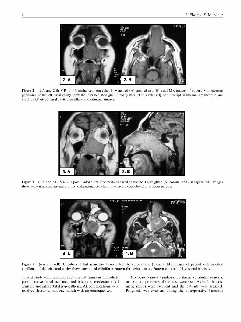
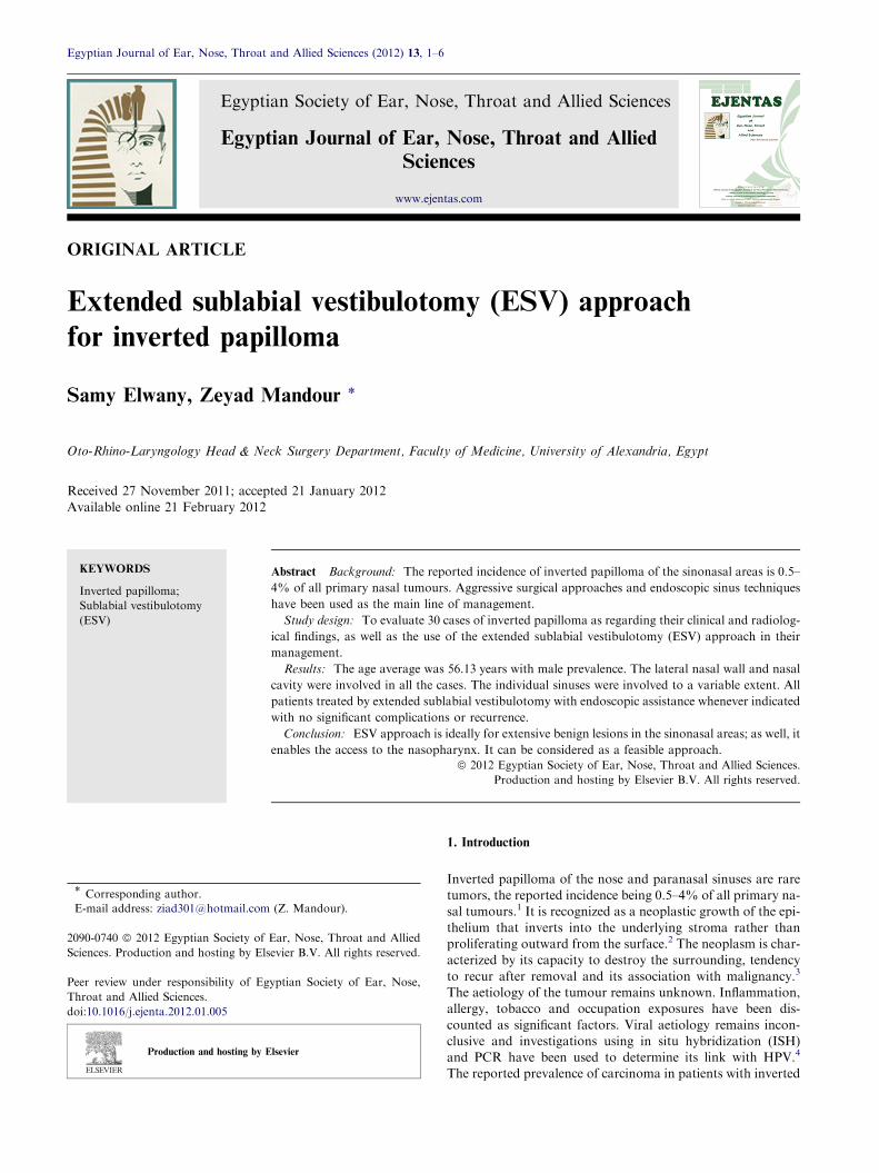
Figure 2 (2.A and 2.B) MRI-T1. Unenhanced spin-echo T1-weighted (A) coronal and (B) axial MR images of patient with inverted
papilloma of the left nasal cavity show the intermediate-signal-intensity mass that is relatively non descript in internal architecture and
involves left-sided nasal cavity, maxillary and ethmoid sinuses.
Figure 3 (3.A and 3.B) MRI-T1 post Gadolinium. Contrast-enhanced spin-echo T1-weighted (A) coronal and (B) sagittal MR images
show well-enhancing stroma and less-enhancing epithelium that create convoluted cribriform pattern.
Figure 4 (4.A and 4.B). Unenhanced fast spin-echo T2-weighted (A) coronal and (B) axial MR images of patient with inverted
papilloma of the left nasal cavity show convoluted cribriform pattern throughout mass. Pattern consists of low signal intensity.
4 S. Elwany, Z. Mandour
current study were minimal and entailed transient immediatepostoperative facial oedema, oral infection, moderate nasalcrusting and infraorbital hypoesthesia. All complications were
resolved shortly within one month with no consequences.
No postoperative epiphora, epistaxis, vestibular stenosis,or aesthetic problems of the nose were seen. As well, the cos-metic results were excellent and the patients were satisfied.
Prognosis was excellent during the postoperative 6 months
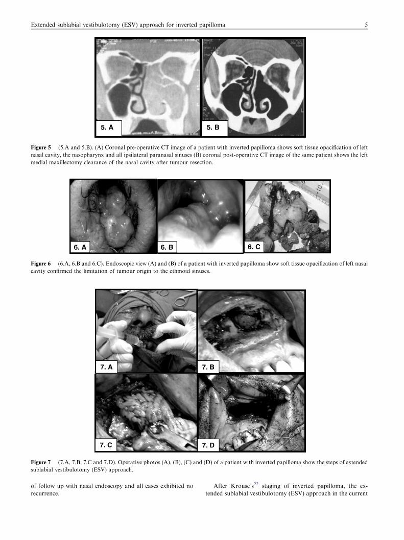
Figure 5 (5.A and 5.B). (A) Coronal pre-operative CT image of a patient with inverted papilloma shows soft tissue opacification of left
nasal cavity, the nasopharynx and all ipsilateral paranasal sinuses (B) coronal post-operative CT image of the same patient shows the left
medial maxillectomy clearance of the nasal cavity after tumour resection.
Figure 6 (6.A, 6.B and 6.C). Endoscopic view (A) and (B) of a patient with inverted papilloma show soft tissue opacification of left nasal
cavity confirmed the limitation of tumour origin to the ethmoid sinuses.
Figure 7 (7.A, 7.B, 7.C and 7.D). Operative photos (A), (B), (C) and (D) of a patient with inverted papilloma show the steps of extended
sublabial vestibulotomy (ESV) approach.
Extended sublabial vestibulotomy (ESV) approach for inverted papilloma 5
of follow up with nasal endoscopy and all cases exhibited norecurrence.
After Krouse’s22 staging of inverted papilloma, the ex-tended sublabial vestibulotomy (ESV) approach in the current

6 S. Elwany, Z. Mandour
study was suitable for the adequate en bloc tumour resectionwith maximum functional tissue integrity preservation in thefirst three stages of this classification. The approach can deal
with the tumour confined to the nasal cavity, extending furtherto involve the ethmoid sinuses or the medial, the lateral, theanterior wall or the superior region of the maxillary sinus, as
well the region close to the roots of teeth. Furthermore, the ap-proach assisted with endoscopic guidance can facilitate theresection achievement of all confined papilloma compared to
those of the nose and paranasal sinuses. On the other hand,ESV seems to be not suitable for papilloma extending beyondthe nose and paranasal sinuses.
One of the major advantages of the mentioned approach is
the ability to obtain excellent exposure of the mid third of theface, such as maxilla, paranasal sinuses, nasal cavity, and naso-pharynx, without skin incisions. It provides aesthetically pleas-
ing outcomes, leaving no visible scars and virtually nofunctional disability, with a low complication rate. It has supe-rior advantages over conventional surgical approaches, such as
Weber–Fergusson or lateral rhinotomy, though easy to per-form yet they result in facial scars and possible epiphora.These methods may be associated with disfigurement of the
face such as upward contracture of the alae and deviation ofthe nose, asymmetry of the upper lip and nasolabial groove,and medial canthal deformity. As well, it is more time consum-ing than the mid-facial degloving approach. The use of this
procedure was limited by the incomplete exposure of the infra-temporal fossa, and therefore was not suitable for thosepatients supposed to have tumour extension to this area.
4. Summary
� Thirty cases of inverted papilloma were evaluated asregarding their clinical, radiological findings and their
management.� The age average was 56.13 years with male prevalence.� The lateral nasal wall and nasal cavity were involved in all
the cases.� The individual sinuses were involved to a variable extent.� Unilateral nasal obstruction was the main presenting symp-
tom followed by nasal discharge and to lesser extent byepistaxis.� All patients were treated by extended sublabial vestibulot-omy and endoscopic assistance with or without medial max-
illectomy whenever indicated.� The post-operative complications found in the currentstudy were minimal.
� Extended sublabial vestibulotomy (ESV) approach was ide-ally suited for extensive benign lesions in the nasal cavity,the maxillary, the ethmoid and the sphenoid sinuses; as
well, it enabled the access to the nasopharynx.� Extended sublabial vestibulotomy (ESV) approach pro-vided an excellent cosmetic outcome and time savingprocedure.
5. Conclusion
Extended sublabial vestibulotomy (ESV) approach was ideallysuitable for extensive benign lesions in the nasal cavity, the
maxillary, the ethmoid and the sphenoid sinuses; as well, it en-abled the access to the nasopharynx. In addition, it can be con-sidered as a feasible procedure with low mortality, excellent
cosmetic outcome and as a time saver in reaching the tumourmass or in wound closure after the resection.
References
1. Som PM, Brandwein M. Tumors and tumorlike conditions:
sinonasal cavities-inflammatory diseases, tumors, fractures, and
postoperative findings. In: Som PM, Cutin HD, eds. Head Neck
Imaging. 2nd ed. Mosby: St. Louis; 1996:185–262.
2. Yousem DM, Fellows DW, Kennedy DW, Bolger WE, Kashima H,
Zinreich SJ. Inverted papilloma: evaluation with MR imaging.
Radiology. 1992;185:501–505.
3. Vrabec PD. The inverted schniderian papilloma: A clinical and
pathological study. Laryngoscope. 1975;85:186–220.
4. Buchwald C, Franzmann MB, Jacobson GK, Lindeberg H. Human
Papilloma Virus (HPV) in sino-nasal papillomas: a study of 78 cases
using in-situ hybridization and Polymerase Chain Reaction.
Laryngoscope. 1995;105:66–71.
5. Lawson W, Le Bengen J, Som P, Bernard PJ, Biller HF. Inverted
papilloma: an analysis of 87 cases.Laryngoscope. 1989;99:1117–1124.
6. Suh KW, Facer GW, Devine KD, Weiland LH, Zujko RD.
Inverting papilloma of the nose and paranasal sinuses. Laryngo-
scope. 1977 Jan;87(1):35–46.
7. Kim WS, Hyun DW, Kim CH, Yoon JH. Treatment outcomes of
sinonasal inverted papillomas according to surgical approaches.
Acta Otolaryngol. 2010;130(4):493–497.
8. Busquets JM, Hwang PH. Endoscopic resection of sinonasal
inverted papilloma: a meta-analysis. Otolaryngol Head Neck Surg.
2006;134:476–482.
9. Lee DK, Chung SK, Dhong H-J, Kim HY, Kim H-J, Bok KH.
Focal hyperostosis on CT of sinonasal inverted papilloma as a
predictor of tumor origin. Am J Neuroradiol. 2007;28:618–621.
10. Phillips PP, Gustafson RO, Facer GW. The clinical behavior of
inverting papilloma of the nose and paranasal sinuses: report of 112
cases and review of the literature. Laryngoscope. 1990;100:463–469.
11. Kamel R. Transnasal endoscopic medial maxillectomy in inverted
papilloma. Laryngoscope. 1995;105:847–853.
12. Stankiewicz JA, Girgis SJ. Endoscopic surgical treatment of nasal
and paranasal sinus inverted papilloma. Otolaryngol Head Neck
Surg. 1993;109:988–995.
13. Lund VJ. Optimum management of inverted papilloma. J Laryn-
gol Otol. 2000;114:194–197.
14. Vrabec PD. The inverted schniderian papilloma: a 25-year study.
Laryngoscope. 1994;104:582–604.
15. Myers EN, Fernau JL, Johnson JT, Tabet JC, Bernes L. Manage-
ment of inverted papilloma. Laryngoscope. 1990;100:481–490.
16. Phillips PP, Gustafson RO, Facer GW. The clinical behaviour of
inverting papilloma of the nose and paranasal sinuses: report of
112 cases and review of literature. Laryngoscope. 1990;100:
463–469.
17. Lawson W, Ho BT, Jacobson A. Inverted papilloma: a report of
112 cases. Laryngoscope. 1995;105:282–288.
18. Kelly JH, Joseph M, Caroll E, et al. Inverted papilloma of the
nasal septum. Arch Otolaryngol. 1980;106:767–771.
19. Dolgin SR, Zaveri VD,CasianoRP,ManigliaAJ.Different options
for treatment of inverted papilloma of the nose and paranasal
sinuses: a report of 41 cases. Laryngoscope. 1992;102:231–236.
20. Momose EN, Weber AL, Goodman M. Papilloma. Radiology.
1980;134:73–79.
21. Ringertz N. Pathology of malignant tumours arising in nasal and
paranasal cavities and maxilla. Acta Otolaryngol. 1938;27:31–42.
22. Krouse JH. Development of a staging system for inverted
papilloma. Laryngoscope. 2000;110(6):965–968.