Embed Size (px)
Citation preview

RESEARCH
Expression Pattern of Id Proteins in Medulloblastoma
Andrew D. Snyder & Ashley N. Dulin-Smith &
Ronald H. Houston & Ashley N. Durban &
Bethany J. Brisbin & Tyler D. Oostra &
Jordan T. Marshall & Basil M. Kahwash &
Christopher R. Pierson
Received: 3 July 2012 /Accepted: 21 December 2012 /Published online: 9 February 2013# Arányi Lajos Foundation 2013
Abstract Inhibitor of DNA binding or inhibitor of differ-entiation (Id) proteins are up regulated in a variety of neo-plasms, particularly in association with high-grade, poorlydifferentiated tumors, while differentiated tissues show littleor no Id expression. The four Id genes are members of thehelix-loop-helix (HLH) family of transcription factors andact as negative regulators of transcription by binding to andsequestering HLH complexes. We tested the hypothesis thatId proteins are overexpressed in medulloblastoma byperforming immunohistochemistry using a medulloblasto-ma tissue microarray with 45 unique medulloblastoma and11 normal control cerebella, and antibodies specific for Id1,Id2, Id3, and Id4. A semi-quantitative staining score thattook staining intensity and the proportion of immunoreac-tive cells into account was used. Id1 was not detected innormal cerebella or in medulloblastoma cells, but 78 % oftumors showed strong Id1 expression in endothelial nucleiof tumor vessels. Id2 expression was scant in normal cere-bella and increased in medulloblastoma (median stainingscore: 4). Id3 expression was noted in some neurons of the
developing cerebellar cortex, but it was markedly up regu-lated in medulloblastoma (median staining score: 12) and intumor endothelial cells. Id4 was not expressed in normalcerebella or in tumor cells. Id2 or Id3 overexpression droveproliferation in medulloblastoma cell lines by altering theexpression of critical cell cycle regulatory proteins in favorof cell proliferation. This study shows that Id1 expression inendothelial cells may contribute to angiogenic processes andthat increased expression of Id2 and Id3 in medulloblastomais potentially involved in tumor cell proliferation andsurvival.
Keywords Medulloblastoma . Id proteins . Id2 . Id3 .
Cerebellum
Introduction
Medulloblastoma is the most common high grade braintumor in children and accounts for about 20 % of all pedi-atric brain tumors [1]. Five-year survival rates are higherthan they have ever been, but many challenges remain; aschildren continue to die of disease well after this 5-yearperiod and survivors are too commonly left with adverselife-long complications due to the effects of radiation ther-apy administered to the developing brain [2]). These com-plications typically manifest as declines in intellectualfunction or psychological issues that impair a survivor’sability to achieve their full potential [2]. There is a pressingneed to learn more about medulloblastoma biology to iden-tify new therapeutic targets.
There are four inhibitor of DNA binding or inhibitor ofdifferentiation (Id) proteins, which are encoded by differentgenes (ID1, ID2, ID3 and ID4). Id proteins are members ofthe helix-loop-helix (HLH) family of transcription factors[3]. Dimerization of HLH transcription factors mediates
A. D. Snyder :A. N. Dulin-Smith :A. N. Durban :B. J. Brisbin :T. D. Oostra : J. T. Marshall : B. M. Kahwash : C. R. PiersonThe Research Institute, Nationwide Children’s Hospital,Columbus, OH, USA
R. H. Houston :C. R. PiersonDepartment of Pathology and Laboratory Medicine,Nationwide Children’s Hospital, Columbus, OH, USA
B. J. Brisbin : T. D. Oostra :C. R. PiersonThe Department of Pathology, The Ohio State University Collegeof Medicine, Columbus, OH, USA
C. R. Pierson (*)Department of Laboratory Medicine, Anatomic Pathology, J0359,Nationwide Children’s Hospital, 700 Children’s Drive,Columbus, OH 43205, USAe-mail: [email protected]
Pathol. Oncol. Res. (2013) 19:437–446DOI 10.1007/s12253-012-9599-4

transcriptional activity, but unlike other HLH proteins, Idproteins lack a DNA binding domain, so when an Id proteinbinds another HLH protein the complex cannot bind DNAand fails to mediate transcription. This makes Id proteins anaturally occurring dominant negative regulator oftranscription.
Much of the early work on Id proteins centered on theirrole in development [4–6], but Id proteins also regulatemany essential aspects of tumor biology such as cell prolif-eration, differentiation, survival, and invasion as well asangiogenesis and have therefore attracted attention as apotential cancer specific target [3, 7, 8]. Increased Id proteinexpression is typically associated with high-grade tumors[7] and may be due to persistent growth factor stimulation,but epigenetic mechanisms may also be involved [3, 8, 9].Studies in models of development and neoplasia indicatethat Id proteins maintain cells in a poorly differentiated,proliferative state, and while the precise mechanism of Idprotein function is not known, Id proteins are consideredpositive-regulators of the cell cycle, and can modulate tran-scription of cyclin-dependent kinase inhibitors [10–12];while Id2, in particular, binds retinoblastoma and may havea regulatory role in G1 progression [3, 13, 14].
Studies of Id protein expression in human cerebellum andmedulloblastoma are limited, so we initiated this study totest the hypothesis that Id protein expression is deregulatedin medulloblastoma. We addressed this hypothesis byimmunostaining a medulloblastoma tissue microarray with45 tumor and 11 control cerebella using antibodies thatspecifically recognize all four Id proteins. We also startedto explore the role of Id proteins in cell cycle regulationusing medulloblastoma cell lines.
Methods
Medulloblastoma Tissue Microarray
Medulloblastoma tissue microarray slides were obtainedfrom the Biopathology Center at the Research Institute atNationwide Children’s Hospital, following a review of theproject by the Institutional Review Board and the Children’sOncology Group. The array includes 45 unique medullo-blastoma cases and 11 normal cerebella as controls and wasprepared using Children’s Oncology Group samples thatunderwent diagnostic workup and expert peer review. De-mographic data were available from 44 of the medulloblas-toma cases consisting of 28 males and 16 females. Theaverage age of medulloblastoma patients was 7±4 years(median: 6.75 years, range: 1 month to 16 years, 9 months).The 11 normal cerebellar controls comprised 9 males and 2females. Five of the controls came from patients within thefirst year of life (23 gestational weeks, 3 days, 7 days,
4 months and 8 months), which is a critical period ofcerebellar development, while the remainder were fromolder patients (5 years, 6 years, 15 years, 16 years, 21 yearsand 26 years). The array contained between 1 and 8 duplicateareas from represented tumors and normal controls that wererandomly spaced throughout the slide.
Immunohistochemistry
The immunohistochemistry procedure was automated on theBondMax IHC system (Leica Microsystems, Bannockburn,IL) using the Id protein specific antibodies described inTable 1. Antigen retrieval was performed as denoted inTable 1 and signal was detected using the Refine Polymer,DAB (Leica Microsystems, Bannockburn, IL). All antibod-ies were validated using multi-tissue blocks, containingbreast, prostate and kidney. Negative controls were obtainedby substituting the primary antibody with a Universal Neg-ative Control Serum (Biocare Medical, Concord, CA)
Staining intensity was scored on a four-tier scale: 0, nostaining; 1, mild staining; 2, moderate staining; and 3, strongstaining. A categorical system was used for the percentage oftumor cells stained: 0, no tumor cells stained; 1, 1 % to 25 %;2, 26 % to 50 %; 3, 51 % to 75 %; and 4, over 76 % of tumorcells stained. Two observers (CRP, BMK) recorded immuno-histochemical findings using a Zeiss light microscope (Thorn-wood, NY) with consensus achieved on difficult cases. Animmunohistochemical staining score was derived as the prod-uct of the overall staining intensity and the percentage oftumor cells stained according to the categorical scale. Manytumors were represented multiple times on the array and thehighest staining score was reported for these cases.
Cell Culture
Medulloblastoma cell lines UW288, UW426, D341 andD283 were gifts from Dr. Corey Raffel at Nationwide Child-ren’s Hospital. All cell lines were cultured in DMEM sup-plemented with 10 % fetal bovine serum and incubated at37°C in 5 % CO2. Stable cell lines over expressing Id2 orId3 or empty expression vectors as controls (OrigeneRC205324, RC200583, PS100007, respectively) were gen-erated using Lipofectamine 2000 per the manufacturer’srecommendations (Life Technologies, Grand Island, NY).
Proliferation Assay
One thousand cells were plated in each well of a 96 well plateand cultured in DMEM supplemented with 10 % fetal bovineserum and incubated at 37°C in 5 % CO2. Ten microlitre ofAlamarBlue (Life Technologies, Grand Island, NY) wasadded to each well and fluorescence was measured at theindicated time points using the SpectraMaxM2E plate reader
438 A.D. Snyder et al.

(Molecular Devices, Sunnyvale, CA). Each experiment wasperformed three times with four replicates for each time point.
Western Blotting
Cells were lysed using Mammalian Protein ExtractionReagent (Pierce Biochemical Rockford, IL) and proteinconcentrations determined using the BCA method (Pierce,Rockford, IL). Protein samples were separated through 4–20 % Tris-Glycine gels (Invitrogen), transferred to PVDFmembranes (Biorad, Hercules, CA), and probed with appro-priate antibodies (Table 2). Immun-Star Western C (Biorad,Hercules, CA) chemiluminescent substrate was applied tothe membranes, which were imaged using a VersaDoc (Bio-rad, Hercules, CA). Densitometric analysis was performedusing contour analysis in ImageLab software v.2.0.1 (Bio-rad, Hercules, CA)). Each immunoblotting experiment wasperformed three times.
Statistics
Id2 and Id3 immunostaining scores in control and tumortissue were compared using the Mann–Whitney two-tailed
nonparametric U-test in SigmaPlot 12.0 (Systat Software,San Jose, CA). Id2, Id3 and Id4 immunostaining scores inpatients less than 7 years of age were compared to patientswho were 7 years and older using the same Mann–Whitneytest. A two-tailed t-test was used to compare cells overexpress-ing either Id2 or Id3 to cells transfected with empty vector atvarious time points. Statistical significance was defined asp<0.05 for all tests. Data was plotted using SigmaPlot 12.0.
Results
Id Proteins are Differentially Expressed in MedulloblastomaTumor Cells and Endothelia
Hematoxylin and eosin (H&E) stained sections of the 45 me-dulloblastoma cases on the tissue microarray showed that allexcept one was of classic histopathology as defined by theWHO; the lone exception belonged to the desmoplastic/nodularsubtype (Fig. 1a–e) [1]. Id1 protein was not detected in tumorcells, but intense stainingwas observed in the endothelial nucleiof 78% (35/45) of the medulloblastoma cases (Fig. 1f–j) (Table3). Id2 protein immunostaining was positive in 89 % (40/45) of
Table 1 Immunohistochemistrymethods and antibodies Antigen Vendor Clone Dilution Antigen retrieval
Id1 Abcam, ab66495 2456C1 1:40 EDTA based, pH 9.0 (ER2 (Leica))
Id2 Abcam, ab52093 rabbit polyclonal 1:25 None
Id3 Abcam, ab41834 rabbit polyclonal 1:150 Citrate based, pH 6.0 (ER1 (Leica))
Id4 Abcam, ab20988 rabbit polyclonal 1:300 Citrate based, pH 6.0 (ER1 (Leica))
Table 2 Antibodies forimmunoblotting Id protein antibodies Dilution Company Product number
Id 1 1:200 Santa Cruz Biotechnology sc-488
Id 2 1:200 Santa Cruz Biotechnology sc-489
Id 3 1:200 Santa Cruz Biotechnology sc-490
Id 4 1:200 Santa Cruz Biotechnology sc-491
Cell cycle antibodies
Cyclin D1 1:1000 Cell Signaling 2926
p27 Kip1 1:1000 Cell Signaling 2552
p15 INK4B 1:1000 Cell Signaling 4822
p16 INK4A 1:1000 Cell Signaling 4824
CDK6 1:1000 Cell Signaling 3136
Cyclin D3 1:1000 Cell Signaling 2936
p21 Waf/Cip1 1:1000 Cell Signaling 2946
CDK4 1:1000 Cell Signaling 2906
Retinoblastoma-underphosphorylated 1:250 BD Biosciences 554164
Retinoblastoma 1:250 BD Biosciences 554136
Cleaved PARP 1:500 Cell Signaling 5625
GAPDH 1:5000 Abcam ab9484
Expression pattern of Id proteins in medulloblastoma 439

Table 3 Demographics and clinical information of medulloblastoma tissue microarray patients and Id protein staining scores
ID Age Sex Id1 Id2 score Id3 score Id4 score
1 03-L6010 8 y 9 m F Endo + 1 4 0
2 04-L2068 7 y 9 m F Endo + 4 16 1
3 09-L6002 3 y 4 m M Endo + 0 6 0
4 11-L1203 2 y 1 m M Endo + 0 8 0
5 12-L4156 7 y 6 m M Endo + 2 12 0
6 02-L4074 15 y7 m
M Endo + 4 16 1
7 03-L4308 7 y 8 m M – 1 0 0
8 04-L4083 5 y 6 m M Endo + 3 16 0
9 11-L4024 5 y 1 m M Endo + 4 16 0
10 12-L6018 2 y 6 m M – 0 2 0
11 02-L1025 5 y 1 m M Endo + 9 16 1
12* 03-L1177 Unk Unk Endo + 0 8 1
13 04-L1024 3 y 6 m F Endo + 1 9 0
14 03-L010 9 y 4 m M Endo + 1 2 0
15 08-L025 4 y 7 m M – 4 0 0
16 10-L036 1 y11 m
F Endo + 6 16 0
17 10-L042 15 y8 m
F Endo + 4 9 1
18 05-L012 10 y4 m
F Endo + 1 9 0
19 05-L027 11 y6 m
M – 9 6 0
20 05-L055 7 y 3 m M Endo + 2 12 0
21 05-L098 9 y 1 m M Endo + 2 12 1
22 07-L048 7 y 3 m M – 4 12 0
23 08-L357 5 y 5 m F Endo + 0 8 0
24 09-L256 6 y10 m
M Endo + 1 16 1
25 03-L115 8 y10 m
F Endo + 2 16 0
26 06-L187 5 y 7 m M Endo + 9 16 0
27 06-L235 9 y 5 m M Endo + 4 16 0
28 07-L049 9 y 2 m F – 4 16 0
29 08-L352 4 y 2 m F Endo + 1 6 1
30 11-L014 16 y9 m
F Endo + 9 16 0
31 01-L141 8 m M Endo + 4 16 0
32 02-L061 10 y7 m
F Endo + 4 16 0
33 02-L113 1 y 8 m M – 9 16 0
34 04-L464 1 y 0 m F – 2 16 0
35 05-L600 2 y 7 m F – 1 6 0
36 10-L299 6 y 8 m M Endo + 4 12 0
37 10-L323 7 y 2 m M Endo + 1 16 0
38 11-L1254 1 y11 m
F Endo + 4 8 0
39 11-L461 5 y 4 m M – 2 6 0
40 12-L025 6 y 8 m M Endo + 2 12 0
41 01-L1137 3 y 8 m M Endo + 12 16 0
42 05-L2010 3 y11 m
F Endo + 12 16 0
440 A.D. Snyder et al.
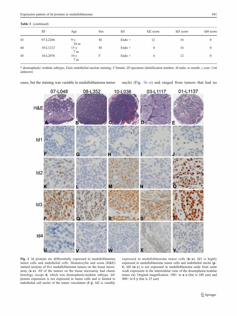
cases, but the staining was variable in medulloblastoma tumor nuclei (Fig. 1k–o) and ranged from tumors that had no
Table 3 (continued)
ID Age Sex Id1 Id2 score Id3 score Id4 score
43 07-L2266 9 y10 m
M Endo + 12 16 0
44 10-L1212 15 y7 m
M Endo + 4 16 0
45 10-L2076 10 y7 m
F Endo + 6 12 0
* desmoplastic/ nodular subtype; Endo endothelial nuclear staining; F female; ID specimen identification number; M male; m month; y year; Unkunknown
Fig. 1 Id proteins are differentially expressed in medulloblastomatumor cells and endothelial cells. Hematoxylin and eosin (H&E)stained sections of five medulloblastoma tumors on the tissue micro-array (a–e). All of the tumors on the tissue microarray had classichistology except d, which was desmoplastic/nodular subtype. Id1protein expression is not expressed in tumor cells and is limited toendothelial cell nuclei of the tumor vasculature (f–j). Id2 is variably
expressed in medulloblastoma tumor cells (k–o). Id3 is highlyexpressed in medulloblastoma tumor cells and endothelial nuclei (p–t). Id4 (u–y) is not expressed in medulloblastoma aside from someweak expression in the internodular zone of the desmoplastic/nodulartumor (x). Original magnification: 100× in a–e (bar is 100 μm) and400× in f–y (bar is 25 μm)
Expression pattern of Id proteins in medulloblastoma 441

immunoreactivity (Fig. 1n) to others with intense immunoreac-tivity (Fig. 1o) (Table 3). Id3 protein expression was moreuniformly increased in medulloblastoma tumor nuclei and intumor endothelial nuclei (Fig. 1p–t) with 98 % (44/45) ofmedulloblastomas demonstrating immunoreactivity (Table3). Id4 is not expressed to a significant degree in medulloblas-toma (Fig. 1u–y) and only 18 % of cases (8/45) showed weakimmunoreactivity (Table 3). The internodular zone of the onlydesmoplastic/nodular medulloblastoma on the tissue micro-array was weakly Id4 immunoreactive, but the nodular areaswere negative (Fig. 1x).
Id2 protein expression is elevated in medulloblastoma(N=45) with a median staining score of 4, while the medianstaining score in normal cerebellar cortex (N=11) is 0(p<0.001, Mann–Whitney U-test) (Fig. 2a). Id3 staining wassignificantly elevated in medulloblastoma tumor cells with amedian score of 12 (N=45), compared to normal cerebellarcortex (N=11), with a median score of 1 (p<0.001, Mann–Whitney U-test) (Fig. 3b). Id proteins are developmentallyregulated so Id2, Id3 and Id4 immunostaining scores werecompared between patients who are less than 7 years old andthose who were 7 years of age or older. Twenty-four patients
Fig. 2 Dot plot of Id2 and Id3 staining scores with a line designatingmedian staining scores. a The median Id2 staining score in medullo-blastoma (N=45) is 4, while the median staining score in normalcerebellar cortex (N=11) is 0 (p<0.001, Mann–Whitney U-test). b
The median Id3 staining score in medulloblastoma is 12 (N=45),which is significantly higher than the median score in normal cerebel-lar cortex (N=11) of 1 (p<0.001, Mann–Whitney U-test)
Fig. 3 Id proteins aredifferentially expressed duringthe development of the normalhuman cerebellar cortex.Normal human cerebella from awide range of ages (3 days,8 months, 5 years and 15 years)were used as controls. Id3 wasthe only Id protein detected at3 days (a–d) and at 8 months ofage (e–h). Id3 expressionappeared decreased at 8 monthsrelative to 3 days of age. At5 days of age (i–l) scatteredinternal granule cells andPurkinje cells expressed Id3(k). No Id protein expressionwas detected in cerebellarcortex at 15 years of age (m–p).Original magnification is 400×;bar is 25 μm
442 A.D. Snyder et al.

were less than 7 years old and 20 were 7 years or older. Onepatient’s age was unknown. There was no significant differ-ence in Id2 (<7 years: 3.9±3.7; ≥7 years: 3.9±3.1; p=0.718),Id3 (<7 years: 11.5±5.2; ≥7 years: 11.7±5.2; p=0.930) or Id4(<7 years: 0.1±0.3, ≥7 years: 0.2±0.4; p=0.515) immunos-taining scores between the two age groups.
Id protein expression is minimal in normal cerebellarcortex (Fig. 3a–p). Id1, Id2 and Id4 are not expressed innormal cerebellar cortex at any of the studied time points.Id3 protein is expressed in external granule neurons, Pur-kinje cells and endothelial cells at 3 days (Fig. 3c) and8 months (Fig. 3g). Purkinje cell and granule neuron Id3expression remains strong at 5 years (Fig. 3k), but at15 years of age Id3 protein expression is minimal and isonly noted in rare, scattered granule neurons (Fig. 3o).
Taken together these data show that Id2 and Id3 proteinsare differentially expressed in medulloblastoma tumor cellsrelative to controls. Id3 expression shows the most signifi-cant up regulation in medulloblastoma and its expression ininfant cerebellar cortex suggests that it could have an im-portant role in development. Id1 is not expressed in endo-thelial cells from normal cerebellar cortex, but the majorityof tumors show substantial Id1 expression in tumor endo-thelial nuclei. Id3 is also expressed in tumor and normalendothelial nuclei.
Id Protein Expression is Altered in HumanMedulloblastoma Cell Lines
Id protein expression was studied in UW288, UW426, D341and D383 medulloblastoma cell lines (Fig. 4). Id1 proteinwas not detected in any of the cell lines studied (data notshown). Abundant Id2 protein expression was detected inUW426 and UW288 and these cell lines also express Id4.Id3 was abundantly expressed in all four of the testedmedulloblastoma cell lines. These data show that medullo-blastoma cell lines model the Id protein expression profile ofhuman tumors. We used UW426 and UW288 cell lines insubsequent experiments as the Id protein expression profile
in these cell lines most accurately modeled Id protein ex-pression in human medulloblastoma.
Id2 and Id3 Overexpression Enhances Cell Proliferationin Medulloblastoma Cell Lines
Id protein expression is increased in proliferating, undiffer-entiated cells. To determine the effects of Id2 and Id3 on
Fig. 5 Id2 and Id3 overexpression drives UW426 and UW288 cellproliferation. Cell proliferation was assessed using the AlamarBlue cellproliferation assay and by quantitating relative fluorescence at 1, 8 and24 h after plating. UW288 cells overexpressing Id2 showed 33 % morerelative fluorescence than control cells expressing empty vector at 8 h(p<0.001), and 31 % more relative fluorescence than empty vectorcontrols at 24 h (p<0.001) (a). UW288 cells overexpressing Id3showed 10 % more relative fluorescence (p<0.01) than controls at8 h and 17 % more relative fluorescence (p<0.001) at 24 h than emptyvector controls (a). UW426 cells overexpressing Id2 showed a 10 %increase in relative fluorescence (p=0.11) at 8 h and an 8 % increase inrelative fluorescence (p=0.04) relative to empty vector controls at 24 h(b). UW426 cells overexpressing Id3 showed a 20 % increase inrelative fluorescence (p<0.001) at 8 h and a 16 % increase in relativefluorescence (p=0.002) relative to empty vector controls at 24 h (b)
Fig. 4 Id proteins are expressed in medulloblastoma cell lines. Id2expression was noted in UW426 and UW288 cells, with minimalexpression in D341 cells and no expression in D283 cells. All fourcell lines expressed Id3 at high levels. Minimal Id4 protein expressionwas noted in UW426 and UW288 lines, but not in D341 or D283. Id1was not detected in any cell line (data not shown)
Expression pattern of Id proteins in medulloblastoma 443

medulloblastoma tumor cells the UW426 and UW288 celllines were stably transfected to drive the expression of Id2and Id3. Western data showed that the Id2 and Id3 proteinexpression levels were doubled relative to empty vectorcontrols (data not shown). AlamarBlue assay was performedto quantitate cell proliferation. Fluorescence was determinedat 1 h, 8 h and 24 h after plating. No significant differencewas noted in relative fluorescence between UW288 cellstransfected with empty vector or cells overexpressing Id2(p=0.79) or Id3 (p=0.4) at 1 h. UW288 cells overexpressingId2 showed 33 % more relative fluorescence (p<0.001) thancontrol cells expressing empty vector at 8 h, whileUW288 cells overexpressing Id3 showed 10 % morerelative fluorescence (p<0.01) than controls at this timepoint (Fig. 5a). At 24 h Id2 overexpressing UW288 cellsshowed 31 % more relative fluorescence (p<0.001) whileId3 overexpressors showed 17 % more relative fluores-cence (p<0.001) than controls (Fig. 5a). No significantdifference was noted in relative fluorescence betweenUW426 cells transfected with empty vector or cells over-expressing Id2 (p=0.47) or Id3 (p=0.34) at 1 h. Id2overexpressing UW426 cells showed 10 % more relativefluorescence (p=0.11) while Id3 overexpressors showed a20 % increase in relative fluorescence (p<0.001) whencompared to empty vector controls at 8 h (Fig. 5b).At 24 h UW426 cells overexpressing Id2 showed an 8 %increase in relative fluorescence (p=0.04) while Id3 overex-pressing UW426 cells showed a 16 % increase in relativefluorescence (p=0.002) compared to empty vector controls.These increases were not due to differences in cell death as
immunoblotting using antibodies specific to cleaved-PARP, amarker of apoptosis, showed no difference amongst Id over-expressing cells or controls in either cell line (data not shown).Taken together, these data show that Id2 and Id3 proteinspotentiate cell proliferation in the UW288 and UW426 me-dulloblastoma cell lines.
Id2 and Id3 Overexpression Deregulates the Expressionof Critical Cell Cycle Regulatory Proteinsin Medulloblastoma Cells
Cell cycle progression is tightly regulated and is dependenton the proper coordination of cyclin dependent kinases(CDK). CyclinD/CDK4/6 complex activity hyperphosphor-ylates retinoblastoma, initiates S-phase, and commits a cellto proliferate by releasing HLH transcription factors. Cip/-Kip protein family members such as p21, p27 and p57 act asCDK inhibitors and block the transition to S-phase. To studythe effects of Id2 and Id3 on cell cycle regulation weperformed immunoblotting of UW288 cells overexpressingId2 or Id3 relative to empty vector controls. Cyclin D1 wasdecreased 36 % in Id2 over expressing cells, but no changewas noted in Id3 overexpressing cells (Fig. 6). Cyclin D3and CDK6 expression was not altered by Id2 or Id3 over-expression in medulloblastoma cells (Fig. 6). No change inp15 was noted and p16 was not detectable in medulloblas-toma cells (data not shown). Independent overexpression ofId2 or Id3 decreased p27 expression by 48 % and 35 %,respectively (Fig. 6). Overexpression of Id2 decreased p21expression 16 %, while Id3 overexpression decreased p21expression 60 % (Fig. 6), while p57 was not detectable (datanot shown). Id2 and Id3 over expression in UW288 cellsincreased phosphorylated retinoblastoma by 39 % and 46 %,respectively (Fig. 6). These data show that the up regulationof Id2 and Id3 expression drives cell proliferation by per-turbing the regulation of cell cycle proteins resulting inincreased retinoblastoma phosphorylation and decreased ex-pression of Cip/Kip family proteins.
Discussion
Most studies of Id protein expression in the developingmammalian nervous system are based on rodent tissues[15, 16], with relatively few studies performed in humantissues [17]. In mice, proliferating, relatively undifferentiat-ed neuroepithelial cells show abundant Id1 and Id3 proteinexpression and then levels decline and become undetectablein the brains of adult mice [6]. Id1 and Id3 are co-expressedduring murine neurogenesis [4, 18, 19] and angiogenesis [4,5]. Id2 is also expressed in the early phases of centralnervous system development; however, in contrast to Id1and Id3, Id2 expression is retained postnatally in mice and
Fig. 6 Id2 and Id3 protein overexpression perturb cell cycle proteinregulation in UW288 cells. Cyclin D1 was decreased 36 % in Id2 overexpressing cells, but no change was noted in Cyclin D3 in medullo-blastoma cells overexpressing Id2 or Id3. There was no change inCDK6 expression. No change was noted in p15 expression and p16was not detectable (data not shown). Id2 or Id3 over expressiondecreased p27 expression 48 % and 35 %, respectively. Over expres-sion of Id2 or Id3 decreased p21 expression 16 % and 60 %, respec-tively. p57 was not detectable (data not shown). Id2 and Id3 overexpression increased phosphorylated retinoblastoma by 39 % and46 %, respectively
444 A.D. Snyder et al.

persists in maturing neurons of the cerebral cortex andPurkinje cells of the cerebellar cortex [5, 6]. Our data showthat Id3 is expressed at substantial levels, while Id2 isminimally expressed in the early postnatal human cerebellarcortex, specifically in external and internal granule cells andPurkinje cells up to 5 years of age. It is possible that Id3could regulate neuronal differentiation or survival and therole of Id3 in human cerebellar development merits furtherstudy.
Increased Id protein expression has been reported inmany different cancers and tends to be associated withhigh-grade, often aggressive tumors (reviewed by Fong etal. [7]), which is in keeping with our findings of increasedId2 and Id3 expression in medulloblastoma tumor cells.Information regarding Id protein expression in brain tumorsis limited, but it has been studied in gliomas where Id1, Id2and Id3 expression are up regulated in high grade tumorsand correlate with tumor proliferation rate [20]. Id proteinsare important in tumor angiogenesis and endothelial expres-sion of Id1, Id2 and Id3 are up regulated in gliomas, but notdetectable in native vessels [20]. In contrast, we detected Id1and Id3, but not Id2 in endothelial cells of medulloblastomatumor vessels. Other brain tumor types should be studied todetermine if up regulating Id protein expression is a generalmechanism in brain tumor biology.
Our data shows that forced Id2 and Id3 overexpressionenhanced medulloblastoma cell proliferation, which wasaccompanied by decreased expression of two key cyclindependent kinase inhibitors: p21 and p27. Id2 and Id3 areknown to modulate cell cycle progression by down regulat-ing p21 and p27 [21, 22]. Id3 in particular, plays a criticalrole in cell cycle progression and survival of neural crestprogenitors by regulating p27 and the Id1 −/− Id3 +/−knockout mouse shows a reduction in proliferating neuro-epithelial cells that is accompanied by an up regulation ofp27 [5]. Cerebellar granule cell proliferation is tightly con-trolled by p27, which suppresses the development of me-dulloblastoma in Patched 1 heterozygous mice [23, 24].Future experiments will further examine the role of Id3and cyclin dependent kinase inhibitors in medulloblastomabiology.
Studies have shown that the expression profile of HLHtranscription factors and Id proteins is important to thebiology of embryonal brain tumors like medulloblastoma[25]. However, little is known regarding the role of Idproteins in human cerebellar granule precursor cells, whichare believed to give rise to a subset of medulloblastomascharacterized by altered sonic hedgehog (SHH) signaling[26]. SHH induces Id2 and N-myc expression, which drivesthe proliferation of cerebellar granule cells in vitro [27].Furthermore, in granule cells differentiation is coupled toId2 degradation, which initiates cell cycle exit and axonalgrowth [28]. Because the modulation of Id protein levels is
critical to granule precursor cell proliferation and differen-tiation, it is possible that perturbations in postnatal Id pro-tein expression in these cells have a key role inmedulloblastoma pathogenesis.
Forced Id2 and Id3 overexpression in medulloblastomacell lines increased levels of phosphorylated retinoblastomaprotein. Id2 directly binds retinoblastoma, preventing for-mation of the retinoblastoma/E2F complex and allowingcell cycle progression from G1 to S phase [29]. However,the biological significance of Id2 and retinoblastoma maybeeven more complex than this as there also appears to be agenetic interaction between retinoblastoma and Id2, since anID2 knockout can partially rescue the lethal phenotype ofthe retinoblastoma knock out mouse, indicating that retino-blastoma may be restraining Id2 activity [29, 30]. Some havespeculated about the possibility of a retinoblastoma-Id2-p16pathway, where retinoblastoma loss, as is common in manycancers, leads to an increase in Id2, which in turn sequestersmore transcription factors, decreasing levels of p16, a media-tor of cellular senescence [8, 31]. We were unable to detectp16 in untransfected control and Id overexpressing medullo-blastoma cells, so this mechanism may not occur in medullo-blastoma. How Id2 contributes to medulloblastoma tumor cellproliferation will be the subject of future studies.
Clinical information was only available from 16 me-dulloblastoma patients whose tumors where representedon the tissue microarray so no survival analysis waspossible and we were unable to correlate Id proteinexpression with patient outcome. This is a limitationof the study; however, since Id2, and especially Id3expression levels, were consistently elevated in the ma-jority of the tumors we studied it seems unlikely that adifferential pattern of Id protein expression could becorrelated with clinical outcome.
Taken together our findings show that Id proteins aredifferentially expressed in medulloblastoma tumor cellsand endothelia, and that Id2 and Id3 overexpression enhan-ces cell proliferation in the former. Future work can addressthe suitability of exploiting Id proteins as a therapeutictarget in medulloblastoma. Id proteins are attractive thera-peutic targets because they are involved in multiple aspectsof tumor biology, allowing many facets of the malignantphenotype to be conveniently targeted using a single ap-proach. Therapies targeting Id proteins could be potent, assupported by studies in preclinical models suggesting thateven a partial reduction of Id levels can show therapeuticbenefit [3, 7, 8, 13]. Furthermore, Id proteins are highlyexpressed in tumor cells and tumor blood vessels, but min-imally expressed in normal, mature tissues; indicating that atherapeutic approach directed against Id proteins would beexpected to have limited toxicity in normal tissues. Thisproperty of Id proteins would be an asset in our search forways to refine therapy, with the goal of ameliorating the life-
Expression pattern of Id proteins in medulloblastoma 445
long complications currently experienced by survivors ofmedulloblastoma.
Acknowledgements The tissue microarray was acquired from theBiopathology Center at the Research Institute at Nationwide Children’sHospital following approval from the Institutional Review Board andthe appropriate Children’s Oncology Group tumor subcommittees.This project was supported by a grant from the Mathew Larson Pedi-atric Brain Tumor Foundation (to CRP). BJB and TDO were supportedby the Samuel J. Roessler Research Scholarship from the Ohio StateUniversity College of Medicine.
References
1. Louis DN OH, Wiestler OD, Cavenee WK (eds) (2007) WHOclassification of tumors of the central nervous system. Lyon
2. Ris MD, Packer R, Goldwein J, Jones-Wallace D, Boyett JM(2001) Intellectual outcome after reduced-dose radiation therapyplus adjuvant chemotherapy for medulloblastoma: a Children’sCancer Group study. J Clin Oncol 19(15):3470–3476
3. Perk J, Iavarone A, Benezra R (2005) Id family of helix-loop-helixproteins in cancer. Nat Rev Cancer 5(8):603–614
4. Jen Y, Manova K, Benezra R (1997) Each member of the Id genefamily exhibits a unique expression pattern in mouse gastrulationand neurogenesis. Dev Dyn 208(1):92–106
5. Lyden D, Young AZ, Zagzag D, Yan W, Gerald W, O’Reilly R et al(1999) Id1 and Id3 are required for neurogenesis, angiogenesis andvascularization of tumour xenografts. Nature 401(6754):670–677
6. Neuman T, Keen A, Zuber MX, Kristjansson GI, Gruss P, NornesHO (1993) Neuronal expression of regulatory helix-loop-helixfactor Id2 gene in mouse. Dev Biol 160(1):186–195
7. Fong S, Debs RJ, Desprez PY (2004) Id genes and proteins aspromising targets in cancer therapy. Trends Mol Med 10(8):387–392
8. Ruzinova MB, Benezra R (2003) Id proteins in development, cellcycle and cancer. Trends Cell Biol 13(8):410–418
9. Bain G, Cravatt CB, Loomans C, Alberola-Ila J, Hedrick SM, MurreC (2001) Regulation of the helix-loop-helix proteins, E2A and Id3,by the Ras-ERK MAPK cascade. Nat Immunol 2(2):165–171
10. Deed RW, Hara E, Atherton GT, Peters G, Norton JD (1997)Regulation of Id3 cell cycle function by Cdk-2-dependent phos-phorylation. Mol Cell Biol 17(12):6815–6821
11. Yokota Y, Mori S (2002) Role of Id family proteins in growthcontrol. J Cell Physiol 190(1):21–28
12. Zebedee Z, Hara E (2001) Id proteins in cell cycle control andcellular senescence. Oncogene 20(58):8317–8325
13. Fong S, Itahana Y, Sumida T, Singh J, Coppe JP, Liu Y et al(2003) Id-1 as a molecular target in therapy for breast cancercell invasion and metastasis. Proc Natl Acad Sci U S A 100(23):13543–13548
14. Loveys DA, Streiff MB, Kato GJ (1996) E2A basic-helix-loop-helix transcription factors are negatively regulated by serum
growth factors and by the Id3 protein. Nucleic Acids Res 24(14):2813–2820
15. Tzeng SF (2003) Inhibitors of DNA binding in neural cell prolif-eration and differentiation. Neurochem Res 28(1):45–52
16. Tzeng SF, de Vellis J (1998) Id1, Id2, and Id3 gene expression inneural cells during development. Glia 24(4):372–381
17. Ellmeier W, Aguzzi A, Kleiner E, Kurzbauer R, Weith A (1992)Mutually exclusive expression of a helix-loop-helix gene and N-myc in human neuroblastomas and in normal development. EMBOJ 11(7):2563–2571
18. Duncan M, DiCicco-Bloom EM, Xiang X, Benezra R, Chada K(1992) The gene for the helix-loop-helix protein, Id, is specificallyexpressed in neural precursors. Dev Biol 154(1):1–10
19. Ellmeier W, Weith A (1995) Expression of the helix-loop-helixgene Id3 during murine embryonic development. Dev Dyn 203(2):163–173
20. Vandeputte DA, Troost D, Leenstra S, Ijlst-Keizers H, RamkemaM, Bosch DA et al (2002) Expression and distribution of idhelix-loop-helix proteins in human astrocytic tumors. Glia 38(4):329–338
21. Kee Y, Bronner-Fraser M (2005) To proliferate or to die: role ofId3 in cell cycle progression and survival of neural crest progen-itors. Genes Dev 19(6):744–755
22. Prabhu S, Ignatova A, Park ST, Sun XH (1997) Regulation of theexpression of cyclin-dependent kinase inhibitor p21 by E2A and Idproteins. Mol Cell Biol 17(10):5888–5896
23. Ayrault O, Zindy F, Rehg J, Sherr CJ, Roussel MF (2009) Twotumor suppressors, p27Kip1 and patched-1, collaborate to preventmedulloblastoma. Mol Cancer Res 7(1):33–40
24. Miyazawa K, Himi T, Garcia V, Yamagishi H, Sato S, Ishizaki Y(2000) A role for p27/Kip1 in the control of cerebellar granule cellprecursor proliferation. J Neurosci 20(15):5756–5763
25. Phi JH, Kim JH, Eun KM,Wang KC, Park KH, Choi SA et al (2010)Upregulation of SOX2, NOTCH1, and ID1 in supratentorial primi-tive neuroectodermal tumors: a distinct differentiation pattern fromthat of medulloblastomas. J Neurosurg Pediatr 5(6):608–614
26. Taylor MD, Northcott PA, Korshunov A, Remke M, Cho YJ,Clifford SC et al (2012) Molecular subgroups of medulloblastoma:the current consensus. Acta Neuropathol 123(4):465–472
27. Oliver TG, Grasfeder LL, Carroll AL, Kaiser C, Gillingham CL,Lin SM et al (2003) Transcriptional profiling of the Sonic hedge-hog response: a critical role for N-myc in proliferation of neuronalprecursors. Proc Natl Acad Sci U S A 100(12):7331–7336
28. Lasorella A, Stegmuller J, Guardavaccaro D, Liu G, Carro MS,Rothschild G et al (2006) Degradation of Id2 by the anaphase-promoting complex couples cell cycle exit and axonal growth.Nature 442(7101):471–474
29. Iavarone A, Garg P, Lasorella A, Hsu J, Israel MA (1994) Thehelix-loop-helix protein Id-2 enhances cell proliferation and bindsto the retinoblastoma protein. Genes Dev 8(11):1270–1284
30. Lasorella A, Noseda M, Beyna M, Yokota Y, Iavarone A (2000)Id2 is a retinoblastoma protein target and mediates signalling byMyc oncoproteins. Nature 407(6804):592–598
31. Ohtani N, Zebedee Z, Huot TJ, Stinson JA, SugimotoM, Ohashi Yetal (2001) Opposing effects of Ets and Id proteins on p16INK4aexpression during cellular senescence. Nature 409(6823):1067–1070
446 A.D. Snyder et al.