Embed Size (px)
Citation preview
Expression of the Cholinergic Signal-Transduction PathwayComponents During Embryonic Rat Heart Development
DIEGO FRANCO, ANTOON F.M. MOORMAN, AND WOUTER H. LAMERSDepartment of Anatomy and Embryology, Academic Medical Center,University of Amsterdam, 1105 AZ Amsterdam, The Netherlands
ABSTRACT Background:Previous studies showed that acetylcholines-terase (AChE) activity is present in the downstream (arterial) part of theembryonic chick and rat heart, but its functional significancewas unclear.To establish whether other components of a cholinergic signal-transduc-tion pathway are present in the embryonic heart, we localised the mRNAsencoding choline acetyltransferase (ChAT), acetylcholinesterase (AChE),and themuscarinic receptor isoforms (mAChRs; m1–m5).Methods:Messenger RNAdetection and localisation by in situ hybridisa-
tion and reverse transcriptase-polymerase chain reaction were employed.Results: Expression of ChAT and AChE mRNAs was observed from 15
embryonic days onward in the neural tissue covering the dorsocranialwall of the atria. Muscarinic receptors (m1, m2, m4) were observed at thesame localisation as AChE and ChAT mRNAs, both during embryogenesisand after birth. In addition, m1 and m4 mAChRs showed a low level ofexpression in the atrial myocardium during the fetal period. No expres-sion of the m3 or the m5 mAChRs was observed in or near the embryonichearts. ChAT,AChE, and mAChRs (m1, m2, m4) mRNAs always colocalisedin the cardiac ganglia. However, none of these mRNAs was found at adetectable level in the outflow tract and/or the ventricular trabeculations.Conclusons: The AChE activity in the arterial part of the embryonic
heart is probably synthesised elsewhere and subserves a function differ-ent from the hydrolysis of locally produced acetylcholine. Anat. Rec.248:110–120, 1997. r 1997 Wiley-Liss, Inc.
Key words: acetylcholinesterase; choline acetyltransferase; muscarinicacetylcholine receptors; in situ hybridisation; heart; rat; devel-opment
Key proteins in the cholinergic signal-transductionpathway are the enzyme choline acetyltransferase(ChAT) that synthesises acetylcholine (ACh), the en-zyme acetylcholinesterase (AChE) that hydrolysesACh,and the acetylcholine receptors. ACh signals throughtwo types of unrelated membrane receptors referred toas nicotinic (nAChR) and muscarinic (mAChR) acetyl-choline receptors (see, e.g., Hosey, 1992). Muscarinicreceptors mediate several actions of ACh in the centraland peripheral nervous systems and in cardiac tissue.Muscarinic receptors are pharmacologically classifiedinto three types designated as M1–M3, although fivedifferent genes (m1–m5) have been identified in rodents(Mei et al., 1989; Hulme et al., 1990). The M1, M2, andM3 types correspond to the m1, m2, and m3 molecularisoforms, respectively. In general, it is accepted thatm2- and m4-encoded gene products are coupled toinhibition of adenylate cyclase, whereas m1-, m3-, andm5-encoded gene products are acting via stimulation ofinositol triphosphate and diacylglycerol production(Hulme et al., 1990). In the adult heart, ACh releasedfrom parasympathetic nerve terminals acts on mAChR
to decrease both the rate and the force of contraction bymediating the inhibition of adenylate cyclase (Tang andGilman, 1992) and the stimulation of inositol triphos-phate and diacylglycerol production (Barnett et al.,1990; Masters et al., 1985).ChAT is the enzyme that catalyses the formation of
the ACh. ChAT localisation is always restricted toneural tissue and, hence, is a more reliable marker toassess cholinergic innervation than is AChE (Ecken-stein and Sofroniew, 1983; Appleyard, 1992). ChATactivity has been demonstrated in the rat heart from 19embryonic days (ED) onward (Marvin et al., 1980), butno information is available concerning its cardiac local-isation.AChE activity has been detected in early embryonic
rat (Lamers et al., 1987; Nakamura et al., 1994) and
Received 15 March 1996; accepted 19 November 1996.Address reprint requests to Wouter H. Lamers, Department of
Anatomy and Embryology,AcademicMedical Center, Meibergdreef 15,1105AZAmsterdam, The Netherlands.Contract grant sponsor: Ministerio de Educacion y Ciencia (Spain);
Contract grant number: EX93-27383269.
THE ANATOMICAL RECORD 248:110–120 (1997)
r 1997 WILEY-LISS, INC.
chicken (Lamers et al., 1990) hearts, where it is ex-pressed in the myocardial layer of the outflow tract andin the ventricular trabeculations. Its expression de-clines in the fetal period and disappears shortly afterbirth. Because these parts of the heart are character-ised by a long duration of the contraction (De Jong etal., 1992; Moorman and Lamers, 1994), an ACh-dependent calcium mobilisation has been suggested toplay an important role in those areas expressing highAChE activity (Lamers et al., 1990). In vitro experi-ments in embryonic chicken cardiac cells showed thatACh stimulation of mAChRs results in intracellularaccumulation of inositol triphosphate (Oettling et al.,1989, 1992; Lohmann et al., 1991). These data suggestthat these mAChRs belong to the m1, m3, or m5subtype. Inositol triphosphate accumulation results inan increased intracellular calcium content that triggerscontraction.In this study we wished to test whether such a
mechanism is effectively present in the embryonic ratheart. For that purpose, we studied the mRNA expres-sion patterns of AChE, ChAT, and the five mAChRsisoforms described so far.
MATERIALS AND METHODSEmbryos
Wistar rat embryos were obtained from timed-pregnant rats (12–20 embryonic days; ED). Neonatalhearts (3, 8, and 10 days old) and adult hearts (3months old) and adult spinal cord were also processed.Samples were fixed for 4 hr in 4% freshly preparedformaldehyde in phosphate-buffered saline (PBS) atroom temperature (in situ hybridisation) or overnightin methanol:acetone:water (2:2:1; immunohistochemis-try). The embryos and tissues were dehydrated in agraded series of ethanol and embedded in paraplast.Serial sections were cut at 7 µm thickness andmountedon RNAse-free 3-aminopropyltriethoxysilane (AAS)-coated slides (in situ hybridisation) or on polylysine-coated slides (immunohistochemistry).
In Situ Hybridisation
Complementary RNA probes of rat ChAT mRNA(Brice et al., 1989); human AChE mRNA (Soreq et al.,1990); rat b-myosin heavy chain (b-MHC) mRNA;sarcoplasmic reticulum calcium ATPase (SERCA2)(Moorman et al., 1995); glutamine synthetase (GS)mRNA (van der Zande, 1989); and human m1, m2, m3,m4, and m5 mAChRs (Peralta et al., 1987) mRNAswere radiolabelled with 35S-UTP by in vitro transcrip-tion according to standard protocols (Melton et al.,1984). ChAT cDNA [EcoRI-NcoI; nucleotides (nt)1–1870] was subcloned into pBluescript (Stratagene).AChE cDNAwas linearised with BstXI (nt 1484–2227),BstEII (nt 822–2227), or Asp718 (full length) to assessthe specificity of the probe. Linearisation of AChEcDNA with BstXI gave the lowest background signaland therefore was eventually used. Human m1 (StuI-SacI; nt 1259–1410), m2 (BamHI-RsaI; nt 995–1204),m3 (AvaI-AvaI; nt 1586–1975), m4 (HindIII-PstI; nt867–1114), and m5 (AccI-TaqI; nt 1038–1308) frag-ments were subcloned into pBluescript (Stratagene) toobtain probes that were specific for each subtype.
The hybridisation conditions were as detailed else-where (Moorman et al., 1995). Briefly, the sections weredeparaffinised, rinsed in absolute ethanol, and dried inan air stream. Pretreatment of the sections was asfollows: 20 min 0.2 N HCl, 5 min bidistilled water, 20min 23 SSC (70°C), 5 min bidistilled water, 2–20 mindigestion in 0.1% pepsin dissolved in 0.01 N HCl, 30 secin 0.2% glycine/PBS, twice for 30 sec each in PBS, 20min of postfixation in 4% freshly prepared formalde-hyde, 5 min in bidistilled water, 5 min in 10 mM EDTA,5 min in 10 mM dithiothreitol (DTT), and finally dryingin an air stream. The prehybridisation mixture con-tained 50% formamide, 10% dextransulphate, 23 SSC,23 Denhardt’s solution, 0.1% Triton X-100, 10 mMDTT, and 200 ng/µl heat-denatured herring spermDNA. The sections were hybridised overnight at 52°Cand washed as follows: a rinse in 13 SSC; 30 min at52°C in 50% formamide dissolved in 13 SSC; 10 min in13 SSC; 30 min in RNAse A (10 µg/ml); 10 min in 13SSC; 10 min in 0.13 SSC; and dehydration in 50%,70%, and 90% ethanol containing 0.3 M ammoniumacetate. The sections were dried and immersed innuclear autoradiographic emulsion (Ilford). The expo-sure time ranged from 10 to 24 days and the develop-ment time from 4 to 8 minutes as indicated. Photo-graphs were taken with a Zeiss Axiophot microscope,usingAgfa 25ASAfilms.
Reverse Transcriptase-Polymerase Chain Reaction(RT-PCR)
Embryos ranging from 13ED to 20ED were used toisolate total RNAaccording to Chomszynski and Sacchi(1987). Embryos were excised from the uterus in ice-cold sterile PBS, and the hearts were removed and theatria and ventricles were dissected separately. TotalRNA from embryonic ventricles at different stages,16ED rat spinal cord (16SP), adult spinal cord (SP),adult ventricles (AV), and adult liver (L), were isolated.13ED and 14ED embryos from two different litterswere pooled. RNA samples were stored (220°C) in TEcontaining 0.01% sodium dodecyl sulfate (SDS). Thesingle-step RT-PCR reaction according toAatsinki et al.(1994) was performed in 50 µl total volume containing 3µg total RNA, 100 pmol of each oligonucleotide, 100 µMdNTPs, 100 mM Tris HCl buffer (pH 8.2), 0.01% bovineserum albumin (BSA), 1.5 mM MgCl2, 10U AMV-RT(Promega), and 2.5 U Taq polymerase (Eurogentec,Berse, Belgium). Briefly, an initial annealing step (15min, 65°C) was performed. Subsequently, the reactionmixture was incubated at 42°C (1 hr, RT step) andheated for 5 min at 95°C (96°C for AChE oligonucleo-tides; RT denaturation step). Thirty-five PCR cycleswere performed as follows: 1 min at 95°C (denaturationstep), 2 min at 52°C (60°C for AChE oligonucleotides;annealing step), 10 min at 72°C (elongation step), witha final elongation step of 5 min at 72°C. Specific 30-meroligonucleotides as designed by Soreq et al. (1994) wereused to distinguish betweenAChE and butyrylcholines-terase mRNA. These sequences were adapted to the ratAChE cDNA (Brice et al., 1989). Specific oligonucleo-tides complementary to AChE and GS mRNAs, respec-tively, were designed to distinguish amplified mRNAproduct from amplified genomic (DNA) product (anintron was always present between the upstream anddownstream oligonucleotides; see Table 1). Amplifica-
111CHOLINERGIC SIGNAL TRANSDUCTION IN PRENATAL HEART
tion of GS mRNA resulted in a fragment 555 nt inlength, whereas the corresponding genomic fragment(gDNA) was 4 kilobases (kb) in length. Amplification ofAChE mRNA resulted in a fragment of 786 nt length,whereas its corresponding genomic fragment was 1186nt. RT-PCR products were loaded and run on a 1%agarose gel and transferred to nylon membranes (Hy-bond N; Amersham, Amersham, U.K.). Southern blotswere performed according to Sambrook et al. (1989) andhybridised to full-length AChE cDNA or an 800 bpEcoRI fragment of the rat GS cDNA (van der Zande,1989). 32P labelling of the cDNAs was by the random-primed method. Unincorporated dNTPs were removedon Sephadex G-50 columns. Probes were quantified in aliquid scintillation counter (1900CATri-Carb; Packard)and hybridised overnight at 52°C. Membranes werewashed in 40 mM sodium phosphate 1% SDS buffer(52°C, 2 3 15 min; RT, 1 3 30 min). A final wash withbuffer only was performed. The membranes were thenexposed in a phosphorimage analyser (Molecular Dy-namics) for 2–3 hr and analysed.
Immunohistochemistry
Specific primary monoclonal antibodies against rat68 kD neurofilament protein (Dako Inc.), Leu-7 (Becton-Dickinson Co.), a-smooth muscle actin (Sigma), andb-myosin heavy chain (Wessels et al., 1991) were usedto visualise the neural tissue and the myocardium.After deparaffination and hydration, the sections werewashed in PBS and treated with hydrogen peroxide (3%in PBS, 30 min) to reduce endogenous peroxidaseactivity. Subsequently, an incubation in TENG-T (10mM Tris, 5 mM EDTA, 150 mM NaCl, 0.25% gelatin,0.05% Tween-20, pH 8.0; 30 min) was performed toavoid nonspecific binding. After this pretreatment, thesections were incubated overnight with the specificprimary antibody. Binding of the first antibody wasdetected using a rabbit antimouse immunoglobulin,followed by a goat antirabbit immunoglobulin andfinally a rabbit peroxidase antiperoxidase (PAP) com-plex. Each incubation lasted 2 hr and was followed bythree washes in PBS (5 min each). All sera were dilutedin PBS. The visualisation of the PAP complex wasperformed by incubation with 0.5 mg/ml 3,38-diamino-benzidine and 0.02% hydrogen peroxide in 30 mMimidazole, 1 mM EDTA (pH 7.0) buffer. Sections weredehydrated and mounted in Entellan (Merck).
RESULTSIn Situ Hybridization
In the present study, adult brain and spinal cord andembryonic spinal cord were used to assess the specific-ity of AChE, ChAT, and mAChRmRNA localisation. GS(neural tissue and myocardium), SERCA2, and b-MHCmRNAs were used as internal controls of embryonicand adult hearts. The expression pattern of the respec-tive mRNAs is first described for control tissues andsubsequently for the heart.
Adult neural tissue
The specificity of the ChAT, AChE, and mAChRsmRNAs was evaluated using adult rat spinal cord and
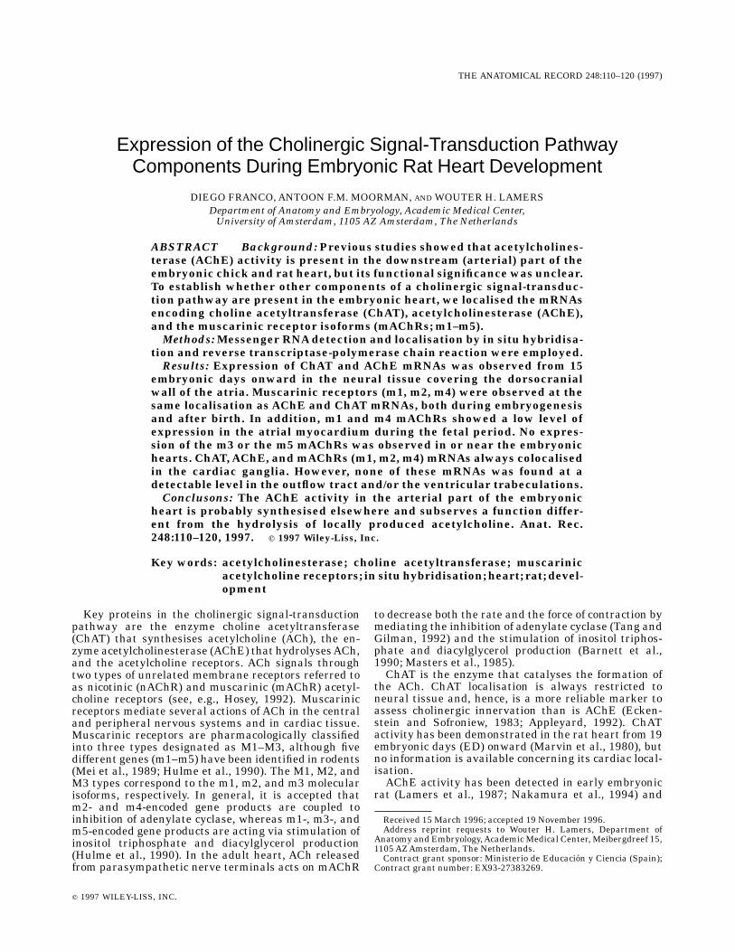
Fig. 1. Adult neural tissue. Expression of AChE (A) and ChAT (B, C)mRNAs in the spinal cord.Aand B are transverse sections through thespinal cord. C shows a high-magnification view of neurons withperinuclear expression of ChAT mRNA (arrow). Scale bars 5 70 µm inA, B, 50 µm in C.
TABLE 1. Oligonucleotides sequences used foramplification of acetylcholinesterase and glutamine
synthetasemRNAsequences after reverse transcription1
rache01 58AAGACTGCCTTTATCTTAATGTGTGGACACC38 (nt 384–414)
rache02 58GCTTCAGCAAAGACAATGAATCTCTCATCAG38 (nt 1127–1157)
gs1 58CCACCTCAGCAAGTTCCC 38 (nt 36–53)gs2 58CAGCACCCCTGGTTTGG 38 (nt 510–526)1Oligonucleotide sequences for the rat acetylcholinesterase (rache01 andrache02) and rat glutamine synthetase (gs1 and gs2). The numbers inparentheses indicate the correspondingmRNAannealing sequence.
112 D. FRANCO ET AL.
brain. Figure 1A,B illustrates the expression patternsofAChE and ChATmRNAs, respectively. ThesemRNAswere found to be specifically localised in the perinucleararea of the motor neurons of the spinal cord (see, e.g.,high magnification of ChAT mRNA in Fig. 1C). Thesefindings are in accordance with previously publisheddata (Oh et al., 1992; Vilaro et al., 1992; Lauterborn etal., 1993; Landwehrmeyer et al., 1993; Hoover et al.,1994), demonstrating that we could reliably visualisethese rare mRNAs.
Neonatal and adult heart
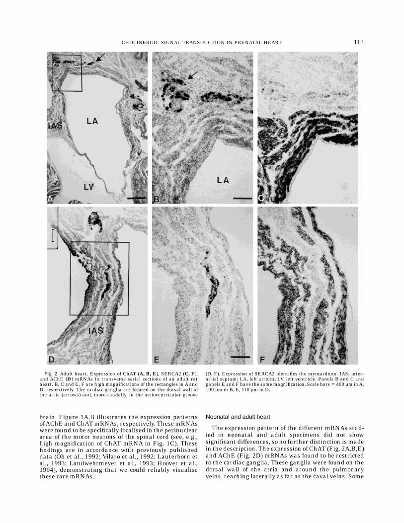
The expression pattern of the different mRNAs stud-ied in neonatal and adult specimens did not showsignificant differences, so no further distinction is madein the description. The expression of ChAT (Fig. 2A,B,E)and AChE (Fig. 2D) mRNAs was found to be restrictedto the cardiac ganglia. These ganglia were found on thedorsal wall of the atria and around the pulmonaryveins, reaching laterally as far as the caval veins. Some
Fig. 2. Adult heart. Expression of ChAT (A, B, E), SERCA2 (C, F),and AChE (D) mRNAs in transverse serial sections of an adult ratheart. B, C and E, F are high magnifications of the rectangles in A andD, respectively. The cardiac ganglia are located on the dorsal wall ofthe atria (arrows) and, more caudally, in the atrioventricular groove
(D, F). Expression of SERCA2 identifies the myocardium. IAS, inter-atrial septum; LA, left atrium; LV, left ventricle. Panels B and C andpanels E and F have the samemagnification. Scale bars5 400 µm inA,100 µm in B, E, 110 µm in D.
113CHOLINERGIC SIGNAL TRANSDUCTION IN PRENATAL HEART
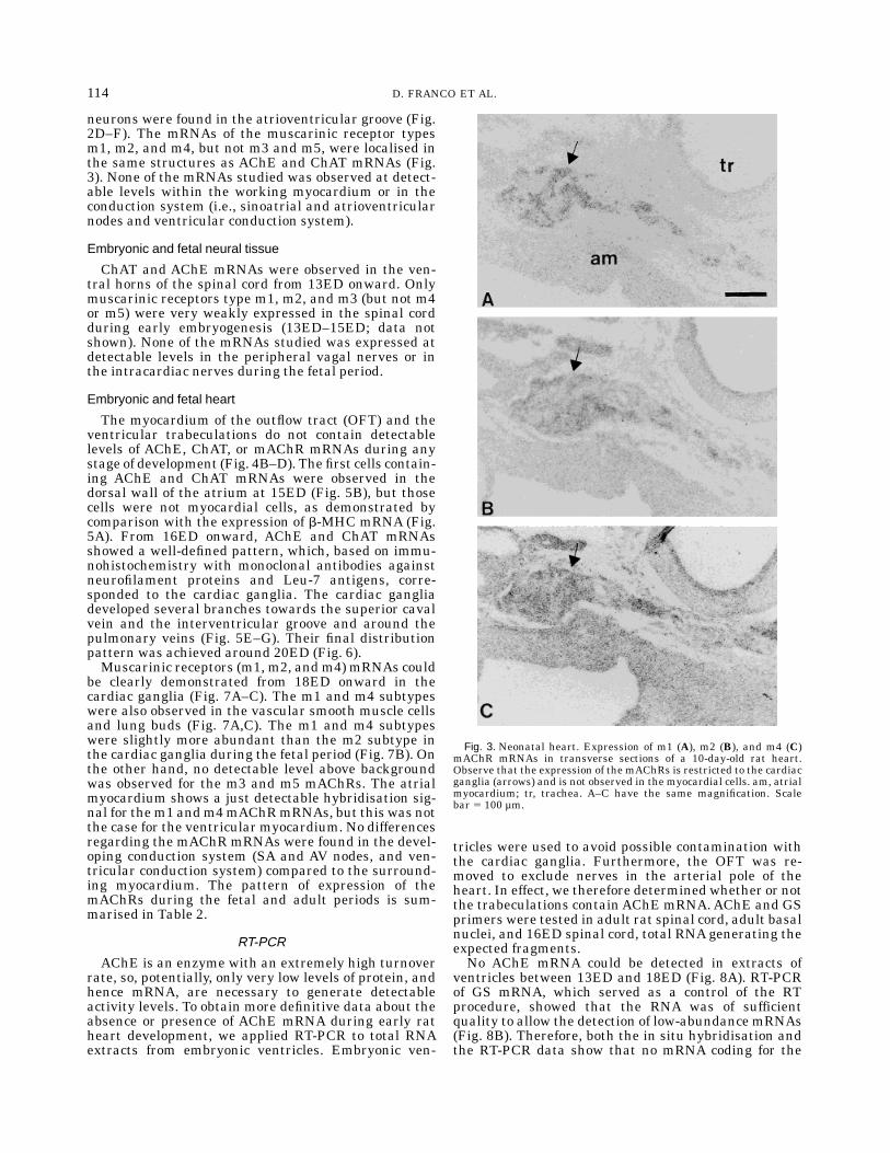
neurons were found in the atrioventricular groove (Fig.2D–F). The mRNAs of the muscarinic receptor typesm1, m2, and m4, but not m3 and m5, were localised inthe same structures as AChE and ChAT mRNAs (Fig.3). None of the mRNAs studied was observed at detect-able levels within the working myocardium or in theconduction system (i.e., sinoatrial and atrioventricularnodes and ventricular conduction system).
Embryonic and fetal neural tissue
ChAT and AChE mRNAs were observed in the ven-tral horns of the spinal cord from 13ED onward. Onlymuscarinic receptors type m1, m2, and m3 (but not m4or m5) were very weakly expressed in the spinal cordduring early embryogenesis (13ED–15ED; data notshown). None of the mRNAs studied was expressed atdetectable levels in the peripheral vagal nerves or inthe intracardiac nerves during the fetal period.
Embryonic and fetal heart
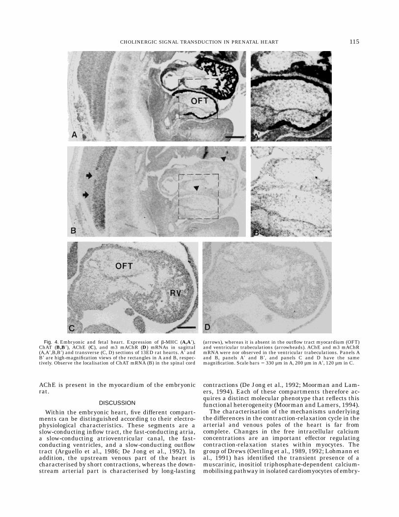
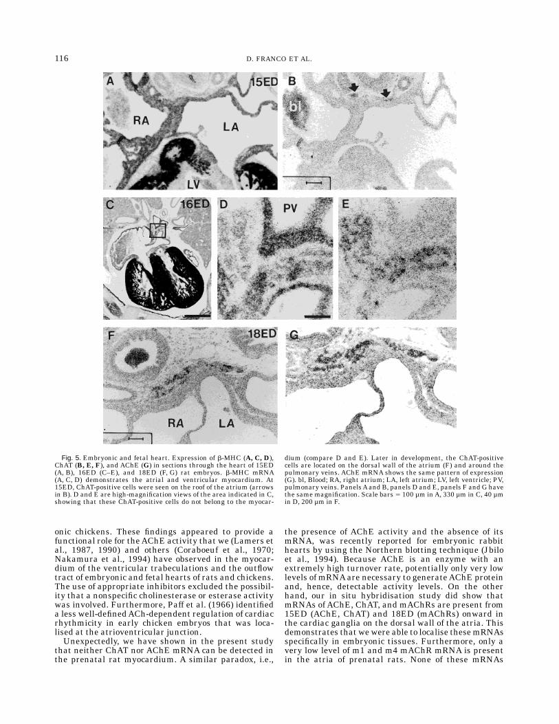
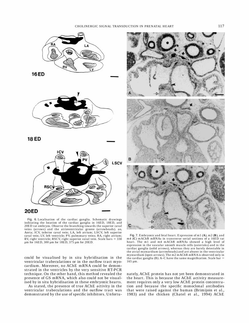
The myocardium of the outflow tract (OFT) and theventricular trabeculations do not contain detectablelevels of AChE, ChAT, or mAChR mRNAs during anystage of development (Fig. 4B–D). The first cells contain-ing AChE and ChAT mRNAs were observed in thedorsal wall of the atrium at 15ED (Fig. 5B), but thosecells were not myocardial cells, as demonstrated bycomparison with the expression of b-MHC mRNA (Fig.5A). From 16ED onward, AChE and ChAT mRNAsshowed a well-defined pattern, which, based on immu-nohistochemistry with monoclonal antibodies againstneurofilament proteins and Leu-7 antigens, corre-sponded to the cardiac ganglia. The cardiac gangliadeveloped several branches towards the superior cavalvein and the interventricular groove and around thepulmonary veins (Fig. 5E–G). Their final distributionpattern was achieved around 20ED (Fig. 6).Muscarinic receptors (m1, m2, andm4) mRNAs could
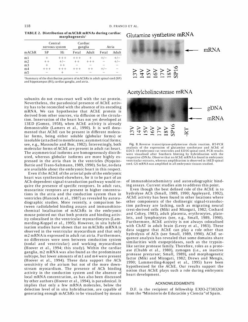
be clearly demonstrated from 18ED onward in thecardiac ganglia (Fig. 7A–C). The m1 and m4 subtypeswere also observed in the vascular smooth muscle cellsand lung buds (Fig. 7A,C). The m1 and m4 subtypeswere slightly more abundant than the m2 subtype inthe cardiac ganglia during the fetal period (Fig. 7B). Onthe other hand, no detectable level above backgroundwas observed for the m3 and m5 mAChRs. The atrialmyocardium shows a just detectable hybridisation sig-nal for them1 andm4mAChRmRNAs, but this was notthe case for the ventricular myocardium. No differencesregarding the mAChR mRNAs were found in the devel-oping conduction system (SA and AV nodes, and ven-tricular conduction system) compared to the surround-ing myocardium. The pattern of expression of themAChRs during the fetal and adult periods is sum-marised in Table 2.
RT-PCR
AChE is an enzyme with an extremely high turnoverrate, so, potentially, only very low levels of protein, andhence mRNA, are necessary to generate detectableactivity levels. To obtain more definitive data about theabsence or presence of AChE mRNA during early ratheart development, we applied RT-PCR to total RNAextracts from embryonic ventricles. Embryonic ven-
tricles were used to avoid possible contamination withthe cardiac ganglia. Furthermore, the OFT was re-moved to exclude nerves in the arterial pole of theheart. In effect, we therefore determined whether or notthe trabeculations containAChE mRNA.AChE and GSprimers were tested in adult rat spinal cord, adult basalnuclei, and 16ED spinal cord, total RNAgenerating theexpected fragments.No AChE mRNA could be detected in extracts of
ventricles between 13ED and 18ED (Fig. 8A). RT-PCRof GS mRNA, which served as a control of the RTprocedure, showed that the RNA was of sufficientquality to allow the detection of low-abundance mRNAs(Fig. 8B). Therefore, both the in situ hybridisation andthe RT-PCR data show that no mRNA coding for the
Fig. 3. Neonatal heart. Expression of m1 (A), m2 (B), and m4 (C)mAChR mRNAs in transverse sections of a 10-day-old rat heart.Observe that the expression of the mAChRs is restricted to the cardiacganglia (arrows) and is not observed in the myocardial cells. am, atrialmyocardium; tr, trachea. A–C have the same magnification. Scalebar 5 100 µm.
114 D. FRANCO ET AL.
AChE is present in the myocardium of the embryonicrat.
DISCUSSION
Within the embryonic heart, five different compart-ments can be distinguished according to their electro-physiological characteristics. These segments are aslow-conducting inflow tract, the fast-conducting atria,a slow-conducting atrioventricular canal, the fast-conducting ventricles, and a slow-conducting outflowtract (Arguello et al., 1986; De Jong et al., 1992). Inaddition, the upstream venous part of the heart ischaracterised by short contractions, whereas the down-stream arterial part is characterised by long-lasting
contractions (De Jong et al., 1992; Moorman and Lam-ers, 1994). Each of these compartments therefore ac-quires a distinct molecular phenotype that reflects thisfunctional heterogeneity (Moorman and Lamers, 1994).The characterisation of the mechanisms underlying
the differences in the contraction-relaxation cycle in thearterial and venous poles of the heart is far fromcomplete. Changes in the free intracellular calciumconcentrations are an important effector regulatingcontraction-relaxation states within myocytes. Thegroup of Drews (Oettling et al., 1989, 1992; Lohmann etal., 1991) has identified the transient presence of amuscarinic, inositiol triphosphate-dependent calcium-mobilising pathway in isolated cardiomyocytes of embry-
Fig. 4. Embryonic and fetal heart. Expression of b-MHC (A,A8),ChAT (B,B8), AChE (C), and m3 mAChR (D) mRNAs in sagittal(A,A8,B,B8) and transverse (C, D) sections of 13ED rat hearts. A8 andB8 are high-magnification views of the rectangles in A and B, respec-tively. Observe the localisation of ChAT mRNA (B) in the spinal cord
(arrows), whereas it is absent in the outflow tract myocardium (OFT)and ventricular trabeculations (arrowheads). AChE and m3 mAChRmRNA were nor observed in the ventricular trabeculations. Panels Aand B, panels A8 and B8, and panels C and D have the samemagnification. Scale bars 5 330 µm inA, 200 µm inA8, 120 µm in C.
115CHOLINERGIC SIGNAL TRANSDUCTION IN PRENATAL HEART
onic chickens. These findings appeared to provide afunctional role for theAChE activity that we (Lamers etal., 1987, 1990) and others (Coraboeuf et al., 1970;Nakamura et al., 1994) have observed in the myocar-dium of the ventricular trabeculations and the outflowtract of embryonic and fetal hearts of rats and chickens.The use of appropriate inhibitors excluded the possibil-ity that a nonspecific cholinesterase or esterase activitywas involved. Furthermore, Paff et al. (1966) identifieda less well-definedACh-dependent regulation of cardiacrhythmicity in early chicken embryos that was loca-lised at the atrioventricular junction.Unexpectedly, we have shown in the present study
that neither ChAT nor AChE mRNA can be detected inthe prenatal rat myocardium. A similar paradox, i.e.,
the presence of AChE activity and the absence of itsmRNA, was recently reported for embryonic rabbithearts by using the Northern blotting technique (Jbiloet al., 1994). Because AChE is an enzyme with anextremely high turnover rate, potentially only very lowlevels of mRNAare necessary to generateAChE proteinand, hence, detectable activity levels. On the otherhand, our in situ hybridisation study did show thatmRNAs of AChE, ChAT, and mAChRs are present from15ED (AChE, ChAT) and 18ED (mAChRs) onward inthe cardiac ganglia on the dorsal wall of the atria. Thisdemonstrates that wewere able to localise thesemRNAsspecifically in embryonic tissues. Furthermore, only avery low level of m1 and m4 mAChR mRNA is presentin the atria of prenatal rats. None of these mRNAs
Fig. 5. Embryonic and fetal heart. Expression of b-MHC (A, C, D),ChAT (B, E, F), and AChE (G) in sections through the heart of 15ED(A, B), 16ED (C–E), and 18ED (F, G) rat embryos. b-MHC mRNA(A, C, D) demonstrates the atrial and ventricular myocardium. At15ED, ChAT-positive cells were seen on the roof of the atrium (arrowsin B). D and E are high-magnification views of the area indicated in C,showing that these ChAT-positive cells do not belong to the myocar-
dium (compare D and E). Later in development, the ChAT-positivecells are located on the dorsal wall of the atrium (F) and around thepulmonary veins. AChE mRNA shows the same pattern of expression(G). bl, Blood; RA, right atrium; LA, left atrium; LV, left ventricle; PV,pulmonary veins. PanelsAand B, panels D and E, panels F and G havethe same magnification. Scale bars 5 100 µm inA, 330 µm in C, 40 µmin D, 200 µm in F.
116 D. FRANCO ET AL.
could be visualised by in situ hybridisation in theventricular trabeculations or in the outflow tract myo-cardium. Moreover, no AChE mRNA could be demon-strated in the ventricles by the very sensitive RT-PCRtechnique. On the other hand, this method revealed thepresence of GS mRNA, which also could not be visual-ised by in situ hybridisation in these embryonic hearts.As stated, the presence of true AChE activity in the
ventricular trabeculations and the outflow tract wasdemonstrated by the use of specific inhibitors. Unfortu-
nately, AChE protein has not yet been demonstrated inthe heart. This is because the AChE activity measure-ment requires only a very lowAChE protein concentra-tion and because the specific monoclonal antibodiesthat were raised against the human (Brimijoin et al.,1983) and the chicken (Chatel et al., 1994) AChE
Fig. 6. Localisation of the cardiac ganglia. Schematic drawingsindicating the location of the cardiac ganglia in 16ED, 18ED, and20ED rat embryos. Observe the branching towards the superior cavalveins (arrows) and the atrioventricular groove (arrowheads). ao,Aorta; ICV, inferior caval vein; LA, left atrium; LSCV, left superiorcaval vein; LV, left ventricle; PV, pulmonary veins; RA, right atrium;RV, right ventricle; RSCV, right superior caval vein. Scale bars 5 330µm for 16ED, 300 µm for 18ED, 375 µm for 20ED.
Fig. 7. Embryonic and fetal heart. Expression of m1 (A), m2 (B), andm4 (C) mAChR mRNAs in transverse serial sections of a 16ED ratheart. The m1 and m4 mAChR mRNAs showed a high level ofexpression in the vascular smooth muscle cells (asterisks) and in thecardiac ganglia (solid arrows), whereas they are barely detectable inthe atrial myocardium (arrowheads) and are absent in the ventricularmyocardium (open arrows). The m2mAChRmRNA is observed only inthe cardiac ganglia (B). A–C have the same magnification. Scale bar 5165 µm.
117CHOLINERGIC SIGNAL TRANSDUCTION IN PRENATAL HEART
subunits do not cross-react well with the rat protein.Nevertheless, the paradoxical presence of AChE activ-ity has to be reconciled with the absence of its encodingmRNA. We can hypothesise that AChE protein isderived from other sources, via diffusion or the circula-tion. Innervation of the heart has not yet developed at13ED (Gomez, 1958), when AChE activity is alreadydemonstrable (Lamers et al., 1990). It is well docu-mented that AChE can be present in different molecu-lar forms, being either soluble (globular forms) orinsoluble (attached tomembranes; asymmetrical forms;see, e.g., Massoulie and Bon, 1982). Interestingly, bothmolecular forms ofAChE are present in adult rat heart.The asymmetrical isoforms are homogeneously distrib-uted, whereas globular isoforms are more highly ex-pressed in the atria than in the ventricles (Nyquist-Battie and Trans-Saltmann, 1989, 1990). So far, no dataare available about the embryonic heart in this respect.Even if theAChE of the arterial pole of the embryonic
heart was synthesised elsewhere, for it to be part of anACh-dependent signal-transduction pathway would re-quire the presence of specific receptors. In adult rats,muscarinic receptors are present in higher concentra-tions in the atria and the conduction system than inventricles (Hancock et al., 1987) as revealed by autora-diographic studies. More recently, a comparison be-tween radiolabeled binding assays and immunohisto-chemical localisation of mAChRs in the embryonicmouse pointed out that both protein and binding activ-ity colocalised in the ventricular myocardiocytes (Lam-merding-Koppel et al., 1995). However, in situ hybrid-isation studies have shown that no mAChRs mRNA isobserved in the ventricular myocardium and that onlym2 mRNA is expressed in adult rat atria. Furthermore,no differences were seen between conduction system(nodal and ventricular) and working myocardium(Hoover et al., 1994; this study). Within the cardiacganglia, m2 mRNA was also found as the predominantsubtype, but lower amounts of m1 and m4 were present(Hoover et al., 1994). These data support the AChsensitivity of the atrium but not of the more down-stream myocardium. The presence of ACh bindingactivity in the conduction system and the absence oflocal mRNA concentration, as has also been discussedby other authors (Hoover et al., 1994), is paradoxical. Itimplies that only a few mRNA molecules, below thedelection level of in situ hybridisation, are capable ofgenerating enough mAChRs to be visualised by means
of immunohistochemistry and autoradiographic bind-ing assays. Current studies aim to address this point.Even though the best defined role of the AChE is to
hydrolyse ACh (Small, 1989, 1990; Appleyard, 1992),AChE activity has been found in other locations whereother components of the cholinergic signal-transduc-tion pathway are lacking, such as migrating neuralcrest-derived cells (Miki and Mizoguti, 1982; Cochardand Coltey, 1983), adult placenta, erythrocytes, plate-lets, and lymphocytes (see, e.g., Small, 1989, 1990).Furthermore, AChE activity is not always colocalisedwith ChAT in adult brain (Levey et al., 1983). Thesedata suggest that AChE can play a role other thanhydrolysis of ACh (see Small, 1989, 1990). AChE se-quence analysis has revealed that some domains sharesimilarities with exopeptidases, such as the trypsin-like serine protease family. Therefore, roles as a prote-ase (Chubb et al., 1980), zymogen (i.e., an inactiveprotease precursor; Small, 1989), and morphogeneticfactor (Miki and Mizoguti, 1982; Drews and Mengis,1990; Lammerding-Koppel et al., 1995) have beenhypothesised for the AChE. Our results support thenotion that AChE plays such a role during embryonicheart development.
ACKNOWLEDGMENTS
D.F. is the recipient of fellowship EX93-27383269from the ‘‘Ministerio de Educacion y Ciencia’’ of Spain.
Fig. 8. Reverse transcriptase-polymerase chain reaction. RT-PCRanalysis of the expression of glutamine synthetase and AChE ofED13–18 embryonic rat ventricles and ED16 spinal cord. PCR resultswere visualised after Southern blotting by hybridisation with therespective cDNAs. Observe that noAChEmRNAis found in embryonicventricular extracts, whereas amplification is observed in 16ED spinalcord. GS mRNAwas amplified in all embryonic tissues studied.
TABLE 2. Distribution ofmAChRmRNAs during cardiacmorphogenesis1
mAChR
Centralnervous system
Cardiacganglia Atria
SP Hi Fetal Adult Fetal Adult
m1 1 111 111 1 1/2 1/2m2 11 1/2 11 111 2 1/2m3 1 11 2 2 2 2m4 111 111 111 11 1/2 2m5 2 1 2 2 2 2
1Summary of the distribution pattern ofmAChRs in adult spinal cord (SP)and hippocampus (Hi), cardiac ganglia, and atria.
118 D. FRANCO ET AL.
LITERATURE CITED
Aatsinki, J.T., J.T. Lakkakorpi, E.M. Pietila, and H.J. Rajaniemi 1994A coupled one-step reverse transcription PCR procedure forgeneration of full-length open ready frames. Biotechniques, 16:282–288.
Appleyard, M.E. 1992 Secreted acetylcholinesterase: Non-classicalaspects of a classical enzyme. TINS, 15:485–490.
Arguello, C.J., J. Alanis, O. Pantoja, and B. Valenzuela 1986 Electro-physiological and ultraestructural study of the atrioventricularcanal during the development of the chick embryo. J. Mol. Cell.Cardiol. 18:499–510.
Barnett, J.V., S.M. Shamah, B. Lassegue, K.K. Griengling, and J.B.Galper 1990 Development of muscarinic-cholinergic stimulationof inositol phosphate production in cultures embryonic chickatrial cells. Biochem. J., 271:443–448.
Brice, A., B. Berrard, B. Raynaud, S. Ansieau, T. Coppola, W.J. Weber,and J. Mallet 1989 Complete sequence of a cDNA encoding anactive rat choline acetyltransferase: A tool to investigate theplasticity of cholinergic phenotype expression. J. Neurosci. Res.,23:266–273.
Brimijoin, S., K.P. Mintz, and M.C. Alley 1983 Production andcharacterization of separate monoclonal antibodies to humanacetylcholinesterase and butyrylcholinesterase. Molec. Pharma-col., 24:513–520.
Chatel, J.M., J. Eichler, F.M. Vallette, S. Bon, J. Massoulie, and J.Grassi 1994 Two-site immunoradiometric assay of chicken acetyl-cholinesterases:active and inactive molecular forms in brain andmuscle. J. Neurochem., 63:1111–1118.
Chomszynki, P., and N. Sacchi 1987 Single-step method of RNAisolation by acid guanidium thiocyanate-phenol-chloroform extrac-tion. Anal. Biochem., 162:156–159.
Chubb, I.W., A.J. Hodgson, and G.H. White 1980 Acetylcholinesterasehydrolizes substance P. Neuroscience, 5:2065–2072.
Cochard, P., and P. Coltley 1983 Cholinergic traits in the neural crest:Acetylcholinesterase in crest cells of the chick embryo. Dev. Biol.,98:221–238.
Coraboeuf, E., G. Obrecht-Coutris, and G. Le Douarin 1970 Acetylcho-line and the embryonic heart. Am. J. Cardiol., 25:285–291.
De Jong, F., T. Ophof, A.A.M. Wilde, M.J. Janse, R. Charles, W.H.Lamers, and A.F.M. Moorman 1992 Persisting zones of slowimpulse conduction in developing chicken hearts. Circ. Res.,71:240–250.
Drews, U., and W. Mengis 1990 Contraction wave in the chickblastoderm induced by muscarinic stimulation. Anat. Embryol.,182:447–454.
Eckenstein, F., and M.V. Sofroniew 1983 Identification of centralcholinergic neurons containing both choline acetyltransferase andacetylcholinesterase and of the central neurons containing onlyacetylcholinesterase. J. Neurosci., 3:2286–2291.
Gomez, H. 1958 The development of the innervation of the heart in therat embryo. Anat. Rec., 130:53–65.
Hancock, J.C., D.B. Hoover, and M.W. Hougland 1987 Distribution ofmuscarinic receptors and acetylcholinesterase in the rat heart. J.Auton. Nerv. Syst., 19:59–66.
Hoover, D.B., R.H. Baisden, and S.X. Xi-Moy 1994 Localisation ofmuscarinic receptor mRNAs in rat heart and intrinsic cardiacganglia by in situ hybridisation. Circ. Res., 75:813–820.
Hosey, M.M. 1992 Diversity of structure, signaling and regulationwithin the family of muscarinic cholinergic receptors. FASEB J.,6:845–852.
Hulme, E.C., N.J.M. Birdsall, and N.J. Buckely 1990 Muscarinicreceptor subtypes. Annu. Rev. Pharmacol. Toxicol., 30:633–673.
Jbilo, O., Y. L’Hermite, V. Talesa, J.P. Toutant, and A. Chatonnet 1994Acetylcholinesterase and butyrylcholinesterase expression in adultrabbit tissues and during development. Eur. J. Biochem., 225:115–124.
Lamers, W.H., W.J.C. Geerts, and A.F.M. Moorman 1990 Distributionpattern of acetylcholinesterase in early embryonic chicken hearts.Anat. Rec., 228:297–305.
Lamers, W.H., A. te Korstschot, J.A. Los, and A.F.M. Moorman 1987Acetylcholinesterase in prenatal rat heart: A marker for earlydevelopment of the cardiac conductive tissue?Anat. Rec., 217:361–370.
Lammerding-Koppel, M., A. Greiner-Shroder, U. Drews 1995 Musca-rinic receptors in the prenatal mouse embryo. Comparison ofM35-immunohistochemistry with [3H]quinuclidinyl benzylate au-toradiography. Histochemistry, 103:301–310.
Landwehrmeyer, B., A. Probst, J.M. Palacios, and G. Mengod 1993Expression of acetylcholinesterase messenger RNA in humanbrain: an in situ hybridisation study. Neuroscience, 57:615–634.
Lauterborn, J.C., P.J. Isackson, R. Montalvo, and C.M. Gall 1993 Insitu hybridisation localisation of choline acetyltransferase mRNAin adult rat brain and spinal cord. Mol. Brain Res., 17:59–69.
Levey, A.I., B.H. Wainer, D.B. Rye, E.J. Mufson, and M.-M. Mesulam1983 Choline acetyltransferase immunoreactive neurons intrisicto rodent cortex and distinction from acetylcholinesterase-positive neurons. Neuroscience, 13:341–353.
Lohmann, F., U. Drews, F. Donie, and G. Reiser 1991 Chick embryomuscarinic and purinergic receptors activate cytosolic Ca11 viaphosphatidylinositol metabolism. Exp. Cell Res., 197:326–329.
Marvin, M.J., K. Hermsmeyer, R. McDonald, L.M. Roskoski, and R.Roskoski 1980 Ontogenesis of cholinergic innervation in the ratheart. Circ. Res., 46:690–695.
Massoulie, J., and S. Bon 1982 The molecular forms of cholinesteraseand acetylcholinesterase in vertebrates. Annu. Rev. Neurosci.,5:57–106.
Masters, S.B., M.W. Martin, T.K. Harden, and J.H. Brown 1985Pertussis toxin does not inhibit muscarinic-receptor mediatedphosphatidylinositol hydrolysis or calciummobilisation. Biochem.J., 227:933–937.
Mei, L., W.R. Roeske, and H.I. Yamamura 1989 Molecular pharmacol-ogy of muscarinic receptor heterogeneity. Life Sci., 45:1831–1851.
Melton, D.A., P.A. Krieg, M.R. Rebagliati, T. Maniatis, K. Zinn, andM.R. Green 1984 Efficient in vitro synthesis of biologically activeRNA and RNA hybridisation probes from plasmids containing abacteriophage SP6 promoter. Nucleic Acids Res., 12:7035–7056.
Miki, A., and H. Mizoguti 1982 Proliferating ability, morphologicaldevelopment and acetylcholinesterase activity of the neural tubecells in early chick embryos. Histochemistry, 76:303–314.
Moorman, A.F.M., and W.H. Lamers 1994 Molecular anatomy of thedeveloping heart. Trends in Cardiovascular Medicine. 4:257–264.
Moorman, A.F.M., J.M.L. Vermeulen, K. Schwartz, W.H. Lamers, andK.R. Boheler 1995 Patterns of expression of sarcoplasmaticreticulum Ca11 ATPase and phospholamban mRNA during ratheart development. Circ. Res., 76:616–635.
Nakamura, T., T. Ikeda, I. Shimokawa, Y. Inoue, T. Suematsu, H.Sakai, K. Iwasaki, and T. Matsuo 1994 Distribution of acetylcho-linesterase activity in the rat embryonic heart with reference toHNK-1 immunoreactivity in the conduction tissue. Anat Em-bryol., 190:367–373.
Nyquist-Battie, C., and K. Trans-Saltmann 1989 Regional distributionof the molecular forms of acetylcholinesterase in adult rat heart.Circ. Res., 65:55–62.
Nyquist-Battie, C., and K.T. Trans-Saltmann 1990 Changes in acetyl-cholinesterase molecular form expression during rat heart devel-opment. Ann. N.Y. Acad. Sci., 436–437.
Oettling, G., U. Gotz, and U. Drews 1992 Characterisation of the Ca11
influx into embryonic cells after stimulation of the embryonicmuscarinic receptor. J. Dev. Physiol., 17:147–155.
Oettling, G., H. Schmidt, and U. Drews 1989 An embryonic Ca11
mobilizing muscarinic system in the chick embryo heart. J. Dev.Physiol., 12:85–94.
Oh, J.D., N.J.Woolf,A. Roghani, R.H. Edwards, and L.L. Butcher 1992Cholinergic neurons in the rat central nervous system demon-strated by in situ hybridisation of choline acetyltransferasemRNA. Neuroscience, 47:807–822.
Paff, G.H., R.J. Boucek, and T.P. Glander 1966 Acetylcholinesterase-acetylcholine, an enzyme system essential to rhythmicity in thepreneural embryonic chick heart. Anat. Rec., 154:675–681.
Peralta, E.G., A. Ashkenazi, J.W. Winslow, D.H. Smith, J. Ramachad-ran, and D.J. Capon 1987 Distinct primary structures, ligand-binding properties and tissue-specific expression of four humanmuscarinic acetylcholine receptors. EMBO J., 6:3923–3929.
Sambrook, J., E.F. Fritsch, and T. Maniatis 1989 Molecular Cloning. ALaboratory Manual, 2nd Ed. Cold Spring Harbor LaboratoryPress, Cold Spring Harbor, NY.
Small, D.H. 1989 Acetylcholinesterases: Zymogens of neuropeptideprocessing enzymes? Neuroscience, 29:241–249.
Small, D.H. 1990 Non-cholinergic actions of acetylcholinesterases:Proteases regulating cell growth and development? TIBS, 15:213–216.
Soreq, H., R. Ben-Aziz, C.A. Prody, S. Seidman, A. Gnatt, L. Neville, J.Lieman-Hurwitz, E. Lev-Lehman, D. Ginzberg, Y. Lapidot-Lifson,and H. Zakut 1990 Molecular cloning and construction of thecoding region for human acetylcholinesterase reveals a G 1 C richattenuating structure. Proc. Natl. Acad. Sci. USA, 87:9688–9692.
119CHOLINERGIC SIGNAL TRANSDUCTION IN PRENATAL HEART

Soreq, H., D. Patinkin, E. Lev-Lehman, M. Grifman, D. Ginzberg, F.Eckstein, and H. Zakut 1994 Antisense oligonucleotide inhibitionof acetylcholinesterase gene expression induces progenitor cellexpansion and suppresses hematopoietic apoptosis ex vivo. Proc.Natl. Acad. Sci. USA, 91:7907–7911.
Tang, W., andA.G. Gilman 1992Adenylyl cyclases. Cell, 70:869–872.van der Zande, L., W.Th. Labruyere, A.C.Arnberg, R.H. Wilson,A.J.W.
van den Bogoert, A.T. Das, D.A.J. van Oorschot, C. Frijters, R.Charles, A.F.M. Moorman, and W.H. Lamers 1989 Isolation andcharacterization of the rat glutamine synthetase-encoding gene.Gene, 87:225–232.
Vilaro, M.T., K.-H. Wiederhold, J.M. Palacios, and G. Mengod 1992Muscarinic M2 receptor mRNA expression and receptor bindingin cholinergic and non-cholinergic cells in the rat brain: a correla-tive study using in situ hybridisation histochemistry and receptorautoradiography. Neuroscience, 47:367–393.
Wessels, A., J.L.M. Vermeulen, S. Viragh, F. Kalman, W.H. Lamersand A.F.M. Moorman 1991 Spatial distribution of ‘‘tissue-specific’’antigens in the developing human heart and skeletal muscle: II)An immunohistochemical analysis of myosin heavy chain isoformexpression patterns in the embryonic heart. Anat. Rec., 229:355–368.
120 D. FRANCO ET AL.