Embed Size (px)
Citation preview

Jpn J Ophthalmol 43, 458–465 (1999)© 1999 Japanese Ophthalmological Society 0021-5155/99/$–see front matterPublished by Elsevier Science Inc. PII S0021-5155(99)00116-1
Expression of Stress-ResponseProtein 60 in Iritis Associated With
Experimental Autoimmune Encephalomyelitis
Takeshi Kumagami,* Shinsuke Kato,
†
Ryoko Ishikura,* Masao Nagata,* Akihiko Tamai* and Eisaku Ohama
†
*Department of Ophthalmology and
†
Division of Neuropathology, Institute of Neurological Sciences, Faculty of Medicine, Tottori University, Yonago, Japan
Purpose:
To study the expression of stress-response proteins in the inflamed iris of rats withexperimental autoimmune encephalomyelitis (EAE).
Methods:
EAE was induced in Lewis rats by immunization with homogenized spinal cord ofthe guinea pig emulsified in complete Freund’s adjuvant (CFA) (group EAE). Control ratsincluded those immunized with only CFA (group CFA) and those that were untreated(group Normal). Immunohistochemical study for the localization of stress-response protein(srp) 27, srp 60, srp 72, ubiquitin, and
a
B-crystallin was performed.
Results:
All rats in group EAE developed iritis, whereas none of the rats in group CFA andgroup Normal developed iritis. No expression of ubiquitin,
a
B-crystallin, srp 27, srp 60, orsrp 72 was seen in the epithelium of the iris in group CFA rats. In the eyes of rats in groupEAE, srp 60 was expressed in the epithelium of the iris in 20 of 22 (90.9%), ubiquitin in 4 of22 (18.2%), and
a
B-crystallin in 3 of 22 (13.6%). In the group Normal rats, only ubiquitinwas expressed in the epithelium of the iris in 1 of 6 (16.7%) eyes examined.
Conclusions:
These results suggest that srp 60 may be a potential uveitogenic antigen in theiris in EAE.
Jpn J Ophthalmol 1999;43:458–465
© 1999 Japanese Ophthalmological Society
Key Words:
Experimental autoimmune encephalomyelitis, immunochemistry, iritis,
posterior iris epithelium, stress-response protein 60.
Introduction
Experimental autoimmune encephalomyelitis (EAE)is an inflammatory, demyelinating disease of thecentral nervous system (CNS) induced by immuniza-tion with myelin antigens. It is generally consideredto be a useful animal model for studying the immu-nopathogenesis of multiple sclerosis. The clinical as-sociation of multiple sclerosis and anterior uveitis,which is characterized by inflammation of the iris oriritis, is well known in humans.
1–6
It is also wellrecognized that iritis is associated with EAE in al-
bino rabbits,
7,8
rhesus monkeys,
9
and Lewis rats.
10,11
However, the reasons for such an association are stillunknown.
Stress-response proteins (srps) are induced inprokaryotic and eukaryotic species under variousconditions of stress.
12–15
These srps have been stud-ied extensively for their physiological roles in themaintenance of self-integration at the cellular level.Srps are named and classified in different familiesaccording to their apparent molecular mass. The srp60 kDa family (srp 60 kDa or srp 60), which have re-tained a uniquely high level of sequence conserva-tion during evolution, is the focus of interest as a po-tential antigen in autoimmune diseases
16,17
such asjuvenile arthritis,
l8
multiple sclerosis,
19,20
Behçet’sdisease,
21
and autoimmune hepatitis.
22
In the presentstudy, we examined the expression of srps in the iris,the most anterior component of the uvea, in EAE.
Received: February 2, 1998Correspondence and reprint requests to: Takeshi KUMA-
GAMI, MD, Department of Ophthalmology, Faculty of Medi-cine, Tottori University, 36-1 Nishimachi, Yonago, Tottori 683-8504, Japan

T. KUMAGAMI ET AL.
459
EXPRESSION OF SRP 60 IN IRIS IN EAE
To the best of our knowledge, such an investigationhas not been reported previously.
Materials and Methods
Animals
Forty female Lewis rats, aged 8–9 weeks andweighing 155–170 g, were purchased from Seiwa Ex-perimental Animal Farm (Fukuoka). Water andfood pellets were given ad libitum. Treatment of theanimals adhered to the ARVO Statement for theUse of Animals in Ophthalmic and Vision Research,and the Guidelines for Animal Experiments in theFaculty of Medicine, Tottori University.
Induction of EAE
The induction of EAE was a modified version
23
ofthe one described by Feurer et al.
24
In brief, 1 g ofguinea pig spinal cord and 1 mL of 0.01 mol/L phos-phate-buffered saline (PBS, pH 7.4) were homoge-nized. The homogenate was then emulsified with 2 mLof Difco’s Bacto complete Freund’s adjuvant (CFA)supplemented with 40 mg of
Mycobacterium tuberculo-sis
H37 Ra (Difco Laboratories, Detroit, MI, USA).Twenty-seven rats were immunized with 0.2 mL of thisencephalitogenic emulsion by intradermal injectioninto both hind footpads (group EAE). Thirteen ratswere used as controls: 3 rats were uninjected (groupNormal) and 10 rats were inoculated with an emulsionof CFA and PBS (1 mL/1 mL) using the same dosageand method as in group EAE (group CFA).
Evaluation of EAE and Iritis
Rats were weighed and assessed daily for clinicalsigns of EAE and iritis. EAE was characterized clin-ically by scoring the severity of limb paralysis ac-cording to the scale of Kato and Nakamura
25
: 0
5
normal; l
5
limp tail; 2
5
weakness of hind legs ormild ataxia; 3
5
hind limb paralysis and severeataxia; 4
5
severe hind limb paralysis accompaniedby urinary incontinence; and 5
5
severe quadriplegiaor a morbid state.
Eyes were examined for ocular signs by slit-lampbiomicroscopy. Because iritis is characterized bywhite membranous infiltrates confined to the iris, in-flammation was scored as follows: 0
5
normal; 1
5
iris hyperemia around the pupillary margin; 2
5
fo-cal infiltrates; 3
5
infiltrates present in less than 25%of the anterior surface of the iris; 4
5
involvement ofan area less than 50%; and 5
5
involvement of anarea greater than 50%.
Histopathological and Immunohistochemical Examinations
At various times, experimental animals were anes-thetized with an intraperitoneal injection of sodiumpentobarbital (50 mg/kg body weight) and perfusedwith fixative consisting of 4% paraformaldehyde in0.1 mol/L cacodylate buffer (pH 7.3). The enucle-ated eyes were embedded in paraffin and 6
m
m-thicksections were prepared for hematoxylin and eosinstaining and immunohistochemical studies. The his-topathological severity of the iritis was evaluated bygrading the degree of infiltration of inflammatorycells, mostly mononuclear cells, into the iris (4 de-grees, from
2
to
111
) and fibrin exudate on theanterior surface of the iris (as
2
or
1
).The sources of the primary antibodies and the di-
lution used are listed in Table 1. The primary poly-clonal antibodies against ubiquitin and
a
B-crystallinwere supplied by Dr. S.-H. C. Yen (Albert EinsteinCollege of Medicine, New York, NY, USA)
26
andDr. J. E. Goldman (Columbia University College ofPhysicians and Surgeons, New York, NY, USA),
27
respectively.Sections were deparaffinized, and the endogenous
peroxidase activity was quenched by incubation for30 minutes with 0.3% H
2
O
2
. After washing, normalsera homologous with the second antibody wereused as blocking reagents. Sections were then incu-bated with each primary antibody overnight at 4
8
C,and control sections were exposed to PBS. Boundantibodies were visualized by the avidin-biotin-per-oxidase complex (ABC, pH 7.4) system using
Table 1.
Primary Antibodies Used in Present Study
Antibody Against Clonality Dilution Source (Reference)
Srp 60 M 1: 800 Clone LK-2 (StressGen, Victoria, Canada)Ubiquitin P 1: 800 Dr. S.-H.C. Yen
26
a
B-crystallin P 1: 500 Dr. J.E. Goldman
27
Srp 27 M Ready to use Clone G3.1 (BioGenex, San Ramon, CA, USA)Srp 72 M 1: 500 Code 3B6 (Amersham International, Little Chalfont, UK)
Srp: stress-response protein, M: monoclonal, P: polycolonal.

460
Jpn J OphthalmolVol 43: 458–465, 1999
Vectastain ABC kits (Vector Laboratories, Burlin-game, CA, USA), and 3, 3
9
-diaminobenzidine tet-rahydrochloride (DAB; DAKO, Glostrup, Den-mark) as the final chromogen.
Ultastructural Examination
Ultrastructural studies were performed at the peakof iritis in the EAE rats. Under general anesthesia byan intraperitoneal injection of sodium pentobarbital(50 mg/kg body weight), experimental animals wereperfused with a fixative consisting of 2% paraformal-dehyde and 5% glutaraldehyde supplemented with0.1 mol/L cacodylate buffer (pH 7.4) containing 8%
sucrose. The eyes were enucleated and the anteriorsegment of the eye (the cornea, iris, and ciliary body)was taken for study. After postosmication and dehy-dration, the tissues were embedded in epoxy resin andultrathin sections were cut with a diamond knife andan ultramicrotome (Type MT 6000-XT; RMC, Tuc-son, AZ, USA). The ultrathin sections were double-stained with uranyl acetate and lead citrate, and ex-amined with a transmission electron microscope(Type H-300; Hitachi, Tokyo).
Figure 1. Clinical course of experimental autoimmune en-cephalomyelitis (j ) and iritis (h) after immunization ofLewis rats with guinea pig spinal cord emulsified in com-plete Freund’s adjuvant. j: 27 to 2 (number of rats exam-ined); h: 54 to 4 (number of eyes examined).
Figure 2. Anterior segment of eye of experimental autoim-mune encephalomyelitis rat at peak of iritis. White mem-branous filtrates are observed on anterior surface of iris. Inaddition, iritis is characterized by engorged iris vessels, ir-regularity of pupillary margin, and miosis with posteriorsynechiae.
Table 2.
Histopathological Findings in Iris of Rats in Group EAE
Time Course of Disease Process
Peak of Iritis
(n
5
22)
1st Attack of EAE (n
5
10)
1st Remission of EAE (n
5
4 )
2nd Attack of EAE (n
5
10)
2nd Remission of EAE (n
5
8)
Infiltration of mononuclear cells
111
†
22 (100)* 0 (0) 0 (0) 0 (0) 0 (0)
11
0 (0) 3 (30.0) 0 (0) 0 (0) 0 (0)
1
0 (0) 4 (40.0) 2 (50.0) 3 (30.0) 2 (25.0)
2
0 (0) 3 (30.0) 2 (50.0) 7 (70.0) 6 (75.0)Exudation of fibrin
1
‡
22 (100) 0 (0) 0 (0) 0 (0) 0 (0)
2
0 (0) 10 (100) 4 (100) 10 (100) 8 (100)
EAE: experimental autoimmune encephalomyelitis, n: number of eyes examined. Rats in Group EAE: Lewis rats immunized withhomogenized spinal cord of guinea pig in Complete Freund’s adjuvant.
* Percentage of eyes examined.
†
Degree of infiltration of mononuclear cells into iris graded from
2
to
111
.
‡
Exudation of fibrin on anterior surface of iris rated:
1
, none;
1
, present.
T. KUMAGAMI ET AL.
461
EXPRESSION OF SRP 60 IN IRIS IN EAE
Results
Clinical Findings
The rats in group EAE suffered two attacks ofEAE (Figure 1), as previously reported.
23
The firstpeak of EAE appeared at 13 days postinoculationand the second peak appeared at 25 days postinocu-lation on the average. EAE developed with a scoreof 3.8
6
0.3 (mean
6
SE) in the first attack and 2.7
6
0.5 in the second attack.However, iritis appeared only in one attack and
the peak was observed at 11 days postinoculation onthe average (Figure 1). The iritis score was 3.7
6
0.1for this single peak. In group EAE, all rats devel-oped bilateral iritis. The duration of the iritis variedconsiderably, the shortest episode being 5 days andthe longest, 12 days. Iritis was characterized clini-cally by a white membranous infiltration on the irissurface, engorged iris vessels, irregularity of thepupillary margin, posterior synechiae, and miosis(Figure 2).
The time course of the disease process could be di-vided into five periods: the first peak of iritis, thefirst attack of EAE, the first remission of EAE, thesecond attack of EAE, and the second remission ofEAE. We therefore made histopathological studieson the eyes of the EAE rats in the five periods.
In group CFA, none of the rats developed eitherEAE or iritis.
Histopathological Findings
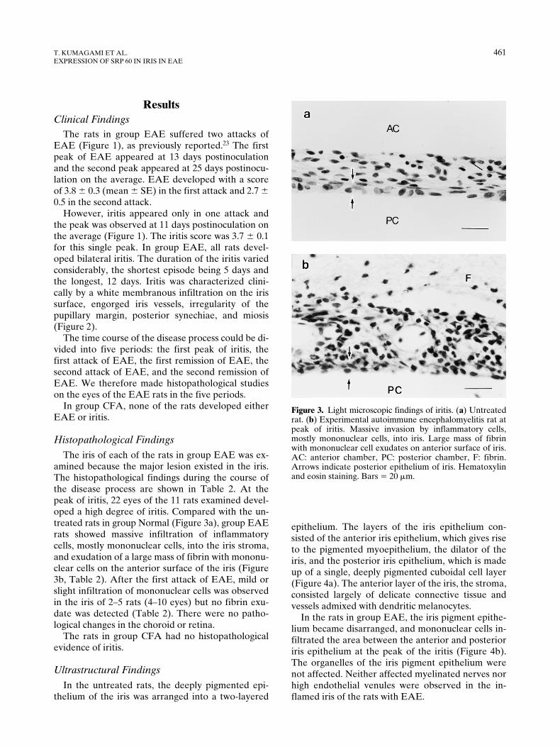
The iris of each of the rats in group EAE was ex-amined because the major lesion existed in the iris.The histopathological findings during the course ofthe disease process are shown in Table 2. At thepeak of iritis, 22 eyes of the 11 rats examined devel-oped a high degree of iritis. Compared with the un-treated rats in group Normal (Figure 3a), group EAErats showed massive infiltration of inflammatorycells, mostly mononuclear cells, into the iris stroma,and exudation of a large mass of fibrin with mononu-clear cells on the anterior surface of the iris (Figure3b, Table 2). After the first attack of EAE, mild orslight infiltration of mononuclear cells was observedin the iris of 2–5 rats (4–10 eyes) but no fibrin exu-date was detected (Table 2). There were no patho-logical changes in the choroid or retina.
The rats in group CFA had no histopathologicalevidence of iritis.
Ultrastructural Findings
In the untreated rats, the deeply pigmented epi-thelium of the iris was arranged into a two-layered
epithelium. The layers of the iris epithelium con-sisted of the anterior iris epithelium, which gives riseto the pigmented myoepithelium, the dilator of theiris, and the posterior iris epithelium, which is madeup of a single, deeply pigmented cuboidal cell layer(Figure 4a). The anterior layer of the iris, the stroma,consisted largely of delicate connective tissue andvessels admixed with dendritic melanocytes.
In the rats in group EAE, the iris pigment epithe-lium became disarranged, and mononuclear cells in-filtrated the area between the anterior and posterioriris epithelium at the peak of the iritis (Figure 4b).The organelles of the iris pigment epithelium werenot affected. Neither affected myelinated nerves norhigh endothelial venules were observed in the in-flamed iris of the rats with EAE.
Figure 3. Light microscopic findings of iritis. (a) Untreatedrat. (b) Experimental autoimmune encephalomyelitis rat atpeak of iritis. Massive invasion by inflammatory cells,mostly mononuclear cells, into iris. Large mass of fibrinwith mononuclear cell exudates on anterior surface of iris.AC: anterior chamber, PC: posterior chamber, F: fibrin.Arrows indicate posterior epithelium of iris. Hematoxylinand eosin staining. Bars 5 20 mm.

462
Jpn J OphthalmolVol 43: 458–465, 1999
Immunohistochemical Findings
No expression of srp 60 was seen in the epitheliumof the iris of the rats in either group CFA or groupNormal (Figure 5a). When control sections withEAE were incubated with PBS, no reaction productswere seen (Table 3). Srp 60, however, was stronglyexpressed but limited to the posterior cuboidal epi-thelium of the iris at the peak of iritis (Figure 5b) in20 of 22 (90.9%) eyes in the group EAE rats. Therewas a gradual decrease in the expression of srp 60and iritis during the clinical course of EAE.
At the peak of iritis, ubiquitin and
a
B-crystallinwere also expressed in the posterior epithelium ofthe iris in 4 of 22 (18.2%) and 3 of 22 (13.6%) eyes ofthe rats in group EAE, respectively. Interestingly,
ubiquitin was expressed in the iris epithelium in 1 of6 (16.7%) eyes examined from the untreated rats ingroup Normal. No expression of srp 27 or srp 72 wasseen in the iris epithelium of the rats in groups EAE,CFA, or Normal.
No expression of the srps was seen in tissues of therat other than the iris epithelium or the inflamma-tory cells of the eyes examined in the EAE, CFA,and Normal groups.
Discussion
Experimental autoimmune encephalomyelitis isan organ-specific, cell-mediated inflammatory au-toimmune disease of the CNS. It is elicited by theimmunization of susceptible rat species with anemulsified suspension of CFA and either CNS tissue,myelin basic protein (MBP), proteolipid proteins, or
Figure 4. Electron micrographs of epithelium of the iris. (a)Untreated rat, iris epithelium shows well arranged two-lay-ered epithelium. (b) Experimental autoimmune encephalo-myelitis rat at peak of iritis, iris epithelium is disarrangedwith infiltrating mononuclear cells (arrowheads). PC: pos-terior chamber. Bars 5 5 mm.
Figure 5. Expression of stress-response protein (srp) 60 iniris (light microscopy). (a) Untreated rat. No immunoreac-tion to antibody against srp 60 is observed in iris. (b) Exper-imental autoimmune encephalomyelitis rat at peak of iritis.Srp 60 is strongly expressed in epithelium of iris but limitedto posterior cuboidal epithelium of iris. AC: anterior cham-ber, PC: posterior chamber. Arrows indicate posterior epi-thelium of iris. Srp 60 immunostaining. Bars 5 20 mm.

T. KUMAGAMI ET AL.
463
EXPRESSION OF SRP 60 IN IRIS IN EAE
peptide fragments of the proteins. It is regarded tobe a model of multiple sclerosis, although multiplesclerosis remains an extremely complex disease ofunknown etiology. There is no spontaneous animalequivalent that includes all aspects of its pathogen-esis.
23
EAE is a disease characterized histologicallyby a cellular infiltration of mononuclear cells intothe CNS.
28
In the present study, EAE was induced in Lewisrats by immunization with homogenized spinal cordof the guinea pig emulsified with CFA. All rats withEAE developed iritis, whereas no rats sensitizedwith only CFA developed iritis (group Normal). Thisindicates that the combination of spinal cord andCFA induced the iritis in the Lewis rats.
Verhagen et al
11
showed that MBP was uveitoge-nic in Lewis rats. They found that the iritis could beadoptively transferred by MBP-specific lines andclones. From this, they suggested that the myelinatednerves of the iris may possess CNS characteristics orat least some similarity to the MBP-specific T cells.
However, Shikishima et al
10
demonstrated ultra-structurally that the myelinated nerves remained in-tact in the inflamed iris associated with EAE inLewis rats. High endothelial venules, which are re-sponsible for the transcellular emigration of lympho-cytes in various inflammatory diseases or in experi-mental models, perform a large part in the perivascularinflammatory process in the iris, retina, optic nerve,and CNS in EAE. In our study, the iritis at the peakof EAE was characterized histopathologically bymassive infiltration of inflammatory cells, mostlymononuclear cells, into the iris stroma, and exuda-tion of a large mass of fibrin with mononuclear cellson the anterior surface of the iris. As observed by
electron microscopy, the mononuclear cells invadedthe area between the disarranged anterior and poste-rior iris epithelium. However, neither affected myeli-nated nerves in the iris nor high endothelial venuleswere observed in the inflamed iris in the rats withEAE. These findings present an enigma for the oc-currence and development of iritis associated withEAE, but this discrepancy could be attributed to thedifference in dose and content of the CFA supple-mented with
M. tuberculosis
or
Bordetella pertussis.
The immunohistochemical study revealed thatubiquitin,
a
B-crystallin, srp 27, srp 60, or srp 72 werenot expressed in tissues other than the iris pigmentepithelium or in the inflammatory cells in the rats ingroup EAE. This indicates that the iris epithelium isa target tissue and manifests the immunoreaction, ie,iritis.
No expression of the srps was seen in the epithe-lium of the iris in the rats in group CFA. In contrast,in the group EAE, srp 60 was highly expressed in theposterior cuboidal epithelium of the iris at the peakof iritis in 20 of the 22 (90.9%) of the eyes examined,ubiquitin in 4 of the 22 (18.2%), and
a
B-crystallin in3 of the 22 (13.6%) eyes. In the untreated, groupNormal rats, only ubiquitin was expressed in the epi-thelium of the iris in 1 of the 6 (16.7%) eyes examined.
In the group EAE rats, srp 60 in the iris epithe-lium also showed a gradual decrease in its expressionduring the clinical course of EAE and iritis, indicat-ing the possibility that srp 60 may be a potentialuveitogenic antigen in the iris in EAE. Our ultra-structural findings revealed that srp 60 became a tar-get of the inflammatory cells; mononuclear cells in-filtrated the area between the disarranged two-layered pigment epithelium of the iris.
Table 3. Expression of Stress-Response Proteins (Srps) in Iris Epithelium of Rats in Groups EAE, CFA, and Normal
Time Course of Disease Process (Group EAE)*
Srps
Peak ofIritis
(n 5 22)
1st Attackof EAE(n 5 10)
1st Remissionof EAE (n 5 4)
2nd Attackof EAE(n 5 10)
2nd Remissionof EAE(n 5 8)
Group†
CFA(n 5 20)
Group‡
Normal(n 5 6)
Srp 60 20 (90.9) 6 (60.0) 6 (25.0) 2 (20.0) 1 (12.5) 0 (0) 0 (0)Ubiquitin 4 (18.2) 1 (10.0) 0 (0) 0 (0) 0 (0) 0 (0) 1 (16.7)aB-crystallin 3 (13.6) 1 (10.0) 0 (0) 1 (10.0) 0 (0) 0 (0) 0 (0)Srp 27 0 (0) 0 (0) 0 (0) 0 (0) 0 (0) 0 (0) 0 (0)Srp 72 0 (0) 0 (0) 0 (0) 0 (0) 0 (0) 0 (0) 0 (0)
EAE: experimental autoimmune encephalomyelitis, CFA: complete Freund’s adjuvant, n: number ofeyes examined.
Values in parentheses indicate percentage of eyes examined.*Rats in Group EAE: Lewis rats immunized with homogenized spinal cord of guinea pig in CFA.†Rats in Group CFA: rats immunited only with CFA.‡Rats in Group Normal: untreated rats (normal controls).

464 Jpn J OphthalmolVol 43: 458–465, 1999
In the lesion of the spinal cord of EAE rats, the in-flammatory cells expressed srp 60 during the acutephase, and glial cells expressed srp 60 during thechronic phase.29 This expression pattern of srp 60 isdifferent from that in the iris of EAE rats. In the ratswith EAE, srp 60 was expressed in a limited sense inthe posterior cuboidal epithelium of the iris, whichcorresponds to the inner, retinal layer of the opticcup, regardless of the phase of EAE and iritis. Thisdiscrepancy in the pattern of srp expression may beattributed to the difference between bulbar lesionand spinal lesion.
In addition, small amounts of ubiquitin and aB-crystallin were expressed in the epithelium of the irisat the peak of iritis in the rats with EAE. Smallamounts of ubiquitin were also expressed in the epi-thelium of the iris in the untreated rats. It is knownthat ubiquitin plays a role in the management of de-generated proteins as the ubiquitin pathway for pro-tein degeneration,30 and that aB-crystallin functionsas a molecular chaperone.31 Thus, it appears thatthese proteins were induced for natural environmen-tal stress and/or protective reaction against inflam-matory stress conditions, ie, an attack of the inflam-matory cells.7
In conclusion, our results suggest that srp 60 maybe a potential uveitogenic antigen to the iris in EAE.However, further immunohistochemical studies areneeded to support a definite conclusion because wehave not examined whether immunization of ratswith srp 60 will induce EAE and iritis. Further stud-ies are also needed to disclose how srp 60, althoughexpressed in a limited sense in the posterior epithe-lium of the iris in the Lewis rats, can develop iritis.
The authors thank Dr. K Tamura (Institute of Neurological Sci-ences, Faculty of Medicine, Tottori University) for his skillful tech-nical assistance. We also thank Dr. S.-H.C. Yen for the supply ofthe antibody against ubiquitin, and Dr. J.E. Goldman, for the sup-ply of the antibody against aB-crystallin.
This article appeared in the Nippon Ganka Gakkai Zasshi (J JpnOphthalmol Soc) 1997;101:299–304. It appears here in modifiedform after peer review and editing for this journal.
References1. Breger BC, Leopold IH. The incidence of uveitis in multiple
sclerosis. Am J Ophthalmol 1966;62:540–5.
2. Giles CL. Peripheral uveitis in patients with multiple sclerosis.Am J Ophthalmol 1970;70:17–9.
3. Porter R. Uveitis in association with multiple sclerosis. Br JOphthalmol 1972;56:478–81.
4. Bamford CR, Ganley JP, Sibley WA, Laguna JF. Uveitis,perivenous sheathing and multiple sclerosis. Neurology 1978;28:119–24.
5. Bachman DM, Rosenthal AR, Beckingsale AB. Granuloma-tous uveitis in neurological disease. Br J Ophthalmol1985;69:192–6.
6. Rothova A, Buitenhuis HJ, Meenken C, et al. Uveitis and sys-temic disease. Br J Ophthalmol 1992;76:137–41.
7. Bullington SJ, Waksman BH. Uveitis in rabbits with experi-mental allergic encephalomyelitis. Results produced by injec-tion of nervous tissue and adjuvants. Arch Ophthalmol1958;59:435–45.
8. Wray SH, Cogan DG, Arnason BGW. Experimental allergicencephalomyelitis. Passive transfer by the intraocular injec-tion of sensitized cells. Arch Neurol 1976;33:183–5.
9. Hayreh SS. Experimental allergic encephalomyelitis. II. Reti-nal and other ocular manifestations. Invest Ophthalmol VisSci 1981;21:270–81.
10. Shikishima K, Lee WR, Behan WMH, Foulds WS. Uveitisand retinal vasculitis in acute experimental allergic encepha-lomyelitis in the Lewis rat: an ultrastructural study. Exp EyeRes 1993;56:167–75.
11. Verhagen C, Mor F, Cohen IR. T cell immunity to myelin ba-sic protein induces anterior uveitis. J Neuroimmunol 1994;53:65–71.
12. Tissières A, Mitchell HK, Tracy UM. Protein synthesis in sali-vary glands of Drosophila melanogaster: relation to chromo-some puffs. J Mol Biol 1974;84:389–98.
13. Collins PL, Hightower LE. Newcastle disease virus stimulatesthe cellular accumulation of stress (heat shock) mRNAs andproteins. J Virol 1982;44:703–7.
14. Lanks KW. Modulators of the eukaryotic heat shock re-sponse. Exp Cell Res 1986;165:1–10.
15. Lindquist S, Craig EA. The heat shock proteins. Annu RevGenet 1988;22:631–77.
16. Haregewoin A, Soman G, Hom RC, Finberg RW. Humangd1 T cells respond to mycobacterial heat-shock protein. Na-ture 1989;340:309–12.
17. Haregewoin A, Singh B, Gupta RS, Finberg RW. A mycobac-terial heat-shock protein-responsive gd T cell clone also re-sponds to the homologous human heat-shock protein: a possi-ble link between infection and autoimmunity. J Infect Dis1991;163:156–60.
18. Boog CJP, de Graeff-Meeder ER, Lucassen MA, et al. Twomonoclonal antibodies generated against human hsp 60 showreactivity with synovial membranes of patients with juvenilechronic arthritis. J Exp Med 1992;175:1805–18.
19. Selmaj K, Brosnan CF, Raine CS. Colocalization of lympho-cytes bearing gd T-cell receptor and heat shock protein hsp651 oligodendrocytes in multiple sclerosis. Proc Natl AcadSci USA 1991;88:6452–6.
20. Selmaj K, Brosnan CF, Raine CS. Expression of heat vitro:implications for multiple sclerosis. Neurology 1992;42:795–800.
21. Lehner T, Lavery E, Smith R, van der Zee R, Mizushima Y,Shinnick T. Association between the 65-kilodalton heat shockprotein, Streptococcus sanguis, and the corresponding anti-bodies in Behçet’s syndrome. Infect Immun 1991;59:1434–41.
22. Lohse AW, Dienes HP, Herkel J, Hermann E, van Eden W,zem Buschenfelde K-HM. Expression of the 60 kDa heatshock protein in normal and inflamed liver. J Hepatol1993;19:159–66.
23. Hosoda Y, Kato S, Ohama E. Suppression of relapsing exper-imental allergic encephalomyelitis by mizoribine: clinical, his-tological and immunohistochemical studies. Neuropathology1996;16:15–20.

T. KUMAGAMI ET AL. 465EXPRESSION OF SRP 60 IN IRIS IN EAE
24. Feurer C, Prentice DE, Cammisuli S. Chronic relapsing ex-perimental allergic encephalomyelitis in the Lewis rat. J Neu-roimmunol 1985;10:159–66.
25. Kato S, Nakamura H. Suppression of acute experimental al-lergic encephalomyelitis by synthetic serum thymic factor.Clinical, histopathological,and immunohistochemical studies.Acta Neuropathol 1988;75:337–44.
26. Lee S, Park YD, Yen S-HC, Ksiezak-Reding H, Goldman JE,Dickson DW. A study of infantile motor neuron disease withneurofilament and ubiquitin immunocytochemistry. Neurope-diatrics 1989;20:107–11.
27. Iwaki T, Kume-Iwaki A, Goldman JE. Cellular distribution ofaB-crystallin in non-lenticular tissues. J Histochem Cytochem1990;38:31–9.
28. Hickey WF, Gonatas NK, Kimura H, Wilson DB. Identifica-tion and quantitation of T lymphocyte subsets found in thespinal cord of the Lewis rat during acute experimental allergicencephalomyelitis. J Immunol 1988;131:2805–9.
29. Gao YL, Brosnan CF, Raine CS. Experimental autoimmuneencephalomyelitis. Qualitative and semiquantitative differ-ences in heat shock protein 60 expression in the central ner-vous system. J Immunol 1995;154:3548–56.
30. Hershko A. The ubiquitin pathway for protein degeneration.Trends Biochem Sci 1991;16:265–8.
31. Horwitz J. a-Crystallin can function as a molecular chaper-one. Proc Natl Acad Sci USA 1992;89:10449-53.