Embed Size (px)
Citation preview

Expression of rat aldehyde reductase AKR7A1: influence of age andsex and tissue-specific inducibility�
Anne Granta, Louise Staffasb, Louise Mancowizb, Vincent P. Kellyc, Margaret M. Mansond,Joseph W. DePierreb, John D. Hayesc, Elizabeth M. Ellisa,*
aDepartment of Bioscience & Biotechnology and Department of Pharmaceutical Sciences, University of Strathclyde, 204, George Street,Glasgow G1 1XW, Scotland, UK
bUnit for Biochemical Toxicology, Department of Biochemistry Wallenberg Laboratory, Stockholm University, Stockholm S-10691, SwedencBiomedical Research Centre, Ninewells Hospital and Medical School, University of Dundee, Dundee DD1 9SY, Scotland, UKdCentre for Mechanisms of Human Toxicity, Hodgkin Building, University of Leicester, PO Box 138, Leicester LE1 9HN, UK
Received 19 February 2001; accepted 2 July 2001
Abstract
The regulation of the aldo-keto reductase AKR7A1 was examined in the livers of male and female rats during development by usingWestern blots, and its contribution to carbonyl metabolism was assessed by using enzyme assays. Hepatic levels of AKR7A1 are low infetal rats and rise to a peak at around 6 weeks of age in animals of both sexes. Higher levels of the enzyme are found in adult male rat liverthan in adult female rat liver. The reductase, therefore, appears to be subject to sex-specific regulation. The effect of growth hormone inmediating this difference in expression was examined by using hypophysectomized animals whose serum growth hormone levels had beenfeminized by continuous administration. Results demonstrate that such treatment leads to a reduction in AKR7A1 expression. AKR7A1 wasfound to be constitutively expressed in rat tissues such as liver, kidney, small intestine, and testis, but it was not detected in nasal mucosa,skeletal muscle, heart, adrenal gland, brain, or spleen. However, AKR7A1 was inducible by the synthetic antioxidant ethoxyquin in liver,kidney, and small intestine, but not in the other tissues examined. These results show that levels of this important detoxication enzyme varyconsiderably according to age and sex and that dietary antioxidants can also influence its level in several tissues. © 2001 Elsevier ScienceInc. All rights reserved.
Keywords: Aldehyde reductase; Developmental regulation; Fetal expression; Sex-specific expression; Growth hormone
1. Introduction
Aldehydes and ketones are ubiquitous in the environ-ment, and some of these compounds have been shown tocause damage to protein, DNA, and membranes, leading totoxicity and mutagenicity [1]. Most organisms have evolved
protective mechanisms to detoxify carbonyl-containingcompounds. Enzymatic defences include the glutathioneS-transferases, which can conjugate glutathione (GSH) toreactive carbonyls [2]; aldehyde dehydrogenases, which canoxidize aldehydes to acids [3]; and enzymes, which canreduce aldehydes and ketones to alcohols [4]. This latterbiotransformation is catalysed by enzymes that fall intothree separate families: the aldo-keto reductases [4]; thealcohol dehydrogenases, and NAD(P)H:quinone oxi-doreductase (NQO1; also called DT-diaphorase). The aldo-keto reductases comprise a large superfamily of NAD(P)H-dependent reductases, members of which have been isolatedfrom a variety of species and tissues [5,6]. Several membersof this family have been identified in the rat, includingaldehyde reductase (AKR1A3) [7], aldose reductase(AKR1B4) [8], and 3�-hydroxysteroid dehydrogenase(AKR1C9) [9,10]. In addition to their potential roles indetoxification, aldo-keto reductases can reduce sugars, ste-
� This work was funded partly by a grant from the Association forInternational Cancer Research. EME was supported by a Beit MemorialFellowship and VPK was supported by a Biomedical Research CentreStudentship. The experiments performed in Stockholm were supported bythe Knut and Alice Wallenberg Foundation.
* Corresponding author. Tel.: �44-141-548-2122; fax: �44-141-553-4124.
E-mail address: [email protected] (E.M. Ellis).Abbreviations: 2-CBA, 2-carboxybenzaldehyde; 4-NBA, 4-nitrobenz-
aldehyde; 9,10-PQ, 9,10-phenanthrenequinone; NQO1, NAD(P)H:quinoneoxidoreductase; GH, growth hormone; GSH, glutathione; and SDS, sodiumdodecyl sulfate.
Biochemical Pharmacology 62 (2001) 1511–1519
0006-2952/01/$ – see front matter © 2001 Elsevier Science Inc. All rights reserved.PII: S0006-2952(01)00771-7

roids, and prostaglandins, but because of their overlappingsubstrate specificity, definitive functions have been difficultto ascribe.
We have previously identified and characterized an al-dehyde reductase from rat liver (AKR7A1) that is capableof detoxifying a dialdehyde metabolite of aflatoxin B1 [11,12]. This enzyme is also capable of reducing a range ofcytotoxic and mutagenic aldehydes and ketones, includingproducts of lipid peroxidation such as 4-hydroxynonenaland hexanal [13]. We have shown that the hepatic level ofthis enzyme, unlike most other members of the aldo-ketoreductase family, is increased in rats after dietary exposureto certain xenobiotics. Such inducing agents include thephenolic antioxidants butylated hydroxyanisole andethoxyquin, as well as naturally occurring compounds in-cluding coumarin [12,14]. Levels of AKR7A1 are also el-evated in the liver of rats raised on a selenium-deficient diet[15]. Modulation of the level of AKR7A1 correlates withprofound differences in susceptibility to chemical carcino-genesis: animals with high levels of AKR7A1 in the liverappear to be more resistant to the hepatocarcinogen afla-toxin B1 than are animals with low levels of AKR7A1 [11].
This study was designed to investigate whether factorssuch as age and sex also influence the expression ofAKR7A1, as well as to examine the tissue-specific induc-ibility of this enzyme. Such information can provide in-sights into factors that may affect susceptibility to the ef-fects of toxic aldehydes and ketones, as well suggestingadditional biological roles for this important enzyme innormal cellular processes.
2. Materials and methods
2.1. Animals
Developmental expression of AKR7A1 was examined inmale and female Fischer 344 rats that were purchased fromHarlan Olac Ltd. (Oxon, England). Such animals were fedad lib a control diet comprising RM1 diet (SDS Ltd.,Edinburgh, Scotland) [15]. Livers were prepared from fetalanimals (17 days gestation) or animals at the ages of 2, 4, 6,8, 10, and 12 weeks. Tissue-specific expression was exam-ined in the same animals. For the enzyme induction exper-iments, 10-week-old animals were fed on a RM1 diet con-taining 0.5% (w/w) ethoxyquin for the 10 days immediatelybefore being killed.
The growth hormone regulation study was carried outusing Sprague-Dawley rats obtained from Mollegaards Avs-laboratorium (Denmark). Sham-operated animals were usedas controls, and hypophysectomy was performed at 6 weeksof age. Hormone treatment was initiated 4 weeks later, asdescribed previously by Staffas et al. [16]. To compensatefor thyroid and adrenal gland deficiency in the hypophysec-tomized rats, thyroxine and cortisone acetate (Sigma-Al-drich, Poole, Dorset, UK) were administered daily, at doses
of 10 �g and 500 �g/kg body weight respectively, as de-scribed previously, starting 2 days before growth hormonetreatment [16]. Growth hormone (a gift of Pharmacia andUpjohn, Stockholm, Sweden) was administered at a rate of50 m-units/hr and kg body weight for 7 days [16].
Animals were killed by decapitation; livers were re-moved and placed on ice. Tissue extracts were either pre-pared immediately, or tissues were frozen at �70°C forfurther analysis. Thin slices (1–2 mm) of various tissueswere fixed in ice cold acetone before sectioning for immu-nohistochemistry.
2.2. Sodium dodecyl sulfate-polyacrylamide gelelectrophoresis and western blotting
Cell extracts were prepared from livers by homogeniza-tion in 20 mM sodium phosphate buffer, pH 7.0. Solublefractions were obtained by centrifugation at 9,000 � g for20 min (4°C). Sodium dodecyl sulfate-polyacrylamide gelelectrophoersis (SDS-PAGE) was performed by the methodof Laemmli [17], and proteins were subsequently trans-ferred to nitrocellulose membranes (Merck, Poole, Dorset,UK). Immunoreactive proteins were identified by usingantisera that had been raised previously in female NewZealand White rabbits using recombinantly expressed aldo-keto reductases or NQO1 as immunogens [14,18]. Immunecomplexes were detected by enhanced chemiluminescenceand exposure to X-ray film. After scanning, relativeamounts of each antigen were quantitated by measuringpixel densities by using National Institutes of Health imageanalysis software (http://rsb.info.nih.gov/nih-image/).
2.3. Immunohistochemistry
Immunohistochemistry, using the polyclonal antiseraraised to AKR7A1 at a dilution of 1:500, was carried out onacetone-fixed tissues as described previously [19].
2.4. Protein and enzyme assays
Protein concentrations were measured by using themethod of Bradford [20]. Aldehyde and ketone reductaseactivity was measured as described previously [13] by mea-suring the rate of disappearance of the cofactor [eitherNADPH or NADH] at 340 nm and at 25°C. Assays werecarried out in 0.1 M sodium phosphate buffer, pH 6.8, byusing a Beckman DU650 UV spectrophotometer with alight path of 1 cm.
2.5. Statistical analysis
Statistical analysis was carried out by using InStat soft-ware (GraphPad, San Diego, CA). Pairwise sets of data
1512 A. Grant et al. / Biochemical Pharmacology 62 (2001) 1511–1519

were analysed by applying an unpaired t test. Three or morecomparisons were made by using one-way ANOVA (anal-ysis of variance) and the significance determined for eachresult by using Dunnett’s multiple comparison post-test.
3. Results
3.1. Ontogenic regulation of AKR1A7 expression in the liver
Previous studies have shown that, whereas some carbon-yl-reducing enzymes are expressed in the livers of adult rats,others, such as aldose reductase (AKR1B4), are only ex-pressed in fetal liver [21]. These levels may influence sen-sitivity to toxic aldehydes and ketones. To investigatewhether there is any variation in the expression of AKR7A1during development, liver extracts prepared from male andfemale Fischer 344 rats of different ages were examined byusing western blots probed with a polyclonal antibody to-ward purified AKR7A1. Fig. 1 shows that AKR7A1 wasdetectable in fetal rats and that levels were comparativelylow in 2-week-old male and female animals. In both sexes,levels of AKR7A1 rose dramatically in the weeks after birthand peaked just before puberty at around 6 weeks, beforedecreasing in adult rats. Levels in adult male animals re-mained consistently higher than in female animals. Thesedata are in contrast with the findings for aldehyde reductase(AKR1A3), the level of which increased only slightly inpubescent rats. NQO1 was undetectable in fetal rat liver, butwas increased after birth in both male and female animals.In females, the level of this protein decreased significantlyin adulthood.
To assess the impact of the levels of AKR7A1 on thedetoxication capacity of rat liver, extracts were assayed forthe ability to reduce a range of aldehydes and ketones, someof which are toxic and known to be substrates of AKR7A1.The results, documented in Table 1, show that levels ofreductase activity toward 2-CBA (a substrate that is rela-tively specific for AKR7A family members when measuredby using NADH as cofactor [18]) reach a maximum at 6
Fig. 1. Ontogenic regulation of AKR7A1. Extracts from 200 mg frozenliver tissue from male or female animals aged 2, 4, 6, 8, 10, 12, 14, or 16weeks or from fetal (F; 17 days gestation) livers were prepared as describedin Materials and Methods in 20 mM sodium phosphate buffer pH 7.0.Protein (10 �g) from each extract was separated by SDS-PAGE andtransferred to nitrocellulose membranes. Western blots were probed withantisera and were processed employing enhanced chemiluminescence.Quantitation for each blot was standardized against controls, and theaverage pixel density of each band determined. (A) Mean values ofAKR7A1 levels in 3 different animals. (B) Representative western blotsprobed with antisera raised against AKR7A1, AKR1A3, or NQO1.
Table 1Levels of the activities of carbonyl reducing enzymes in rat liver during developmenta
Cofactor: NADH NADPH
Substrate: 2-CBA(nmol min�1 mg�1)
2-CBA(nmol min�1 mg�1)
4-NBA(nmol min�1 mg�1)
9,10-PQ(nmol min�1 mg�1)
Age (weeks) Male Female Male Female Male Female Male Female
Fetal 2.3 � 0.2b 4.8 � 2.5b 9.9 � 1.9b 13.3 � 3.7b
2 6.5 � 1.7* 5.6 � 1.0** 4.8 � 1.1 3.6 � 0.9 35.0 � 4.7 37.8 � 4.1 57.6 � 6.7** 60.7 � 4.54 8.8 � 0.4 13.9 � 3.9* 6.8 � 1.4 4.4 � 0.2 35.0 � 11.5 39.0 � 4.1 54.7 � 5.4** 57.5 � 3.76 16.0 � 1.9* 12.8 � 1.8 7.4 � 0.1 4.7 � 0.2 34.1 � 8.0 36.8 � 1.4 51.6 � 1.0** 58.7 � 2.78 14.6 � 1.1 11.2 � 1.8 8.3 � 1.3 4.7 � 0.4 34.3 � 6.5 41.6 � 3.6 48.0 � 1.9 60.8 � 8.310 8.3 � 1.7 9.5 � 2.1 5.6 � 0.4 4.2 � 0.3 28.0 � 10.9 38.5 � 6.5 39.2 � 6.2 61.4 � 3.2*12 12.9 � 2.3 11.8 � 1.3 7.9 � 0.3 4.9 � 0.2 32.1 � 12.6 37.4 � 1.8 43.3 � 6.6 51.0 � 0.914 11.0 � 1.0 9.2 � 1.1 6.2 � 0.7 4.5 � 0.5 26.8 � 10.3 37.1 � 4.5 38.9 � 3.6 49.3 � 2.616 11.6 � 2.6 8.6 � 1.5 6.0 � 0.7 4.9 � 0.1 25.3 � 11.1 36.4 � 4.5 36.3 � 5.7 49.7 � 8.4
a Cytosols were prepared from the livers of Fischer 344 rats at the developmental stages indicated and assayed as described in Materials and Methods usingthe substrates indicated. Values represent the means � SEM, for 3 animals.
b Fetal samples were not sexed.* Activities are significantly different, P � 0.05, from 16 week animals of the same sex; ** P � 0.01.2-CBA, 2-carboxybenzaldehyde; 4-NBA, 4-nitrobenzaldehyde; 9,10-PQ, 9,10-phenanthrenequinone.
1513A. Grant et al. / Biochemical Pharmacology 62 (2001) 1511–1519
weeks of age in male animals. This correlates well with thelevels of AKR7A1 detected on western blots.
3.2. Sex-specific regulation of AKR7A1
During this and previous studies, the levels of AKR7A1protein in the liver have consistently been found to behigher in males than in female rats. To establish the basis forthis sex-specific regulation, we examined the expression ofAKR7A1 in Sprague-Dawley rats that had been hypophy-sectomized and subsequently treated with Growth Hormone(GH). The continuous administration of GSH to hypophy-sectomized animals of either sex was designed to feminizethe pattern of expression, as has been described previously[16].
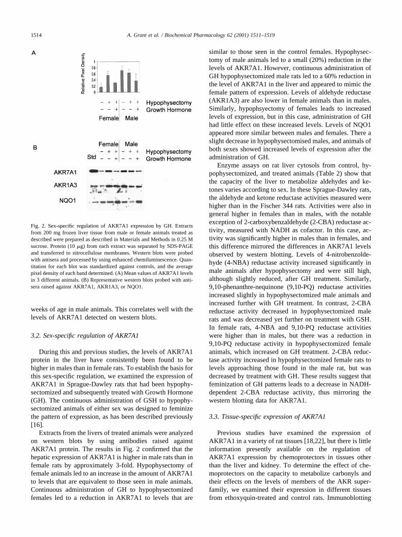
Extracts from the livers of treated animals were analyzedon western blots by using antibodies raised againstAKR7A1 protein. The results in Fig. 2 confirmed that thehepatic expression of AKR7A1 is higher in male rats than infemale rats by approximately 3-fold. Hypophysectomy offemale animals led to an increase in the amount of AKR7A1to levels that are equivalent to those seen in male animals.Continuous administration of GH to hypophysectomizedfemales led to a reduction in AKR7A1 to levels that are
similar to those seen in the control females. Hypophysec-tomy of male animals led to a small (20%) reduction in thelevels of AKR7A1. However, continuous administration ofGH hypophysectomized male rats led to a 60% reduction inthe level of AKR7A1 in the liver and appeared to mimic thefemale pattern of expression. Levels of aldehyde reductase(AKR1A3) are also lower in female animals than in males.Similarly, hypophsyectomy of females leads to increasedlevels of expression, but in this case, administration of GHhad little effect on these increased levels. Levels of NQO1appeared more similar between males and females. There aslight decrease in hypophysectomised males, and animals ofboth sexes showed increased levels of expression after theadministration of GH.
Enzyme assays on rat liver cytosols from control, hy-pophysectomized, and treated animals (Table 2) show thatthe capacity of the liver to metabolize aldehydes and ke-tones varies according to sex. In these Sprague-Dawley rats,the aldehyde and ketone reductase activities measured werehigher than in the Fischer 344 rats. Activities were also ingeneral higher in females than in males, with the notableexception of 2-carboxybenzaldehyde (2-CBA) reductase ac-tivity, measured with NADH as cofactor. In this case, ac-tivity was significantly higher in males than in females, andthis difference mirrored the differences in AKR7A1 levelsobserved by western blotting. Levels of 4-nitrobenzolde-hyde (4-NBA) reductase activity increased significantly inmale animals after hypophysectomy and were still high,although slightly reduced, after GH treatment. Similarly,9,10-phenanthre-nequinone (9,10-PQ) reductase activitiesincreased slightly in hypophysectomized male animals andincreased further with GH treatment. In contrast, 2-CBAreductase activity decreased in hypophysectomized malerats and was decreased yet further on treatment with GSH.In female rats, 4-NBA and 9,10-PQ reductase activitieswere higher than in males, but there was a reduction in9,10-PQ reductase activity in hypophysectomized femaleanimals, which increased on GH treatment. 2-CBA reduc-tase activity increased in hypophysectomized female rats tolevels approaching those found in the male rat, but wasdecreased by treatment with GH. These results suggest thatfeminization of GH patterns leads to a decrease in NADH-dependent 2-CBA reductase activity, thus mirroring thewestern blotting data for AKR7A1.
3.3. Tissue-specific expression of AKR7A1
Previous studies have examined the expression ofAKR7A1 in a variety of rat tissues [18,22], but there is littleinformation presently available on the regulation ofAKR7A1 expression by chemoprotectors in tissues otherthan the liver and kidney. To determine the effect of che-moprotectors on the capacity to metabolize carbonyls andtheir effects on the levels of members of the AKR super-family, we examined their expression in different tissuesfrom ethoxyquin-treated and control rats. Immunoblotting
Fig. 2. Sex-specific regulation of AKR7A1 expression by GH. Extractsfrom 200 mg frozen liver tissue from male or female animals treated asdescribed were prepared as described in Materials and Methods in 0.25 Msucrose. Protein (10 �g) from each extract was separated by SDS-PAGEand transferred to nitrocellulose membranes. Western blots were probedwith antisera and processed by using enhanced chemiluminescence. Quan-titation for each blot was standardized against controls, and the averagepixel density of each band determined. (A) Mean values of AKR7A1 levelsin 3 different animals. (B) Representative western blots probed with anti-sera raised against AKR7A1, AKR1A3, or NQO1.
1514 A. Grant et al. / Biochemical Pharmacology 62 (2001) 1511–1519

(Fig. 3) confirmed that AKR7A1 is expressed constitutivelyat relatively high levels in liver, kidney, small intestine, andtestis. This tissue distribution differs from that of either ofthe two related human AKR7A enzymes, which are ex-pressed at high levels in liver, kidney, small intestine, andpancreas (both AKR7A2 and AKR7A3); skeletal muscle,heart, testes, and ovary (AKR7A2); and in thyroid andadrenals (AKR7A3) [23,24]. A novel rat enzyme that ishighly related to AKR7A1 has recently been identified [18,25] and exhibits a tissue distribution similar to that ofhuman AKR7A2 and AKR7A3.
The rat AKR7A1 is inducible in liver, as previouslydocumented, and is slightly inducible in kidney. Important,the data in Fig. 3 show that AKR7A1 is also inducible in thesmall intestine. The reductase does not appear to be induc-ible in the testes or any other tissue we have examined todate.
Enzyme assays on cytosols prepared from tissues iso-lated from ethoxyquin-treated and control rats were alsocarried out (Table 3). These show that there was significant
increase in 2-CBA reductase activity in the liver, kidney,and testis of animals treated with ethoxyquin, when mea-sured by using NADH as cofactor. There was also an in-crease in 2-CBA reductase activity in the small intestine,adrenals, and brain, although these were not statisticallysignificant. Treatment with ethoxyquin also appears to leadto a significant increase in 4-NBA reductase activity in theheart, although this does not correlate with increasedAKR7A1 levels as determined by western blots.
Immunohistochemical staining of kidney, small intes-tine, and testis revealed those areas of these tissues whereAKR7A1 is expressed (Fig. 4). In the kidney, staining forAKR7A1 was pronounced in the collecting ducts (Fig. 4a).In the small intestine, staining in the villi (distinct frombasal alkaline phosphatase) was observed. Staining was alsoobserved in the testis. In addition, although AKR7A1 is notdetectable in skeletal muscle when using western blots, wewere able to identify discrete areas of staining in the nervebundles, suggesting that the enzyme may fulfill a function inthese highly specialized cells.
Fig. 3. Ethoxyquin induction of AKR7A1 in rat tissues. Extracts of the frozen tissues indicated from adult male rats aged 10 weeks were prepared as describedin Materials and Methods in 20 mM sodium phosphate buffer, pH 7.0. Protein (10 �g) from each extract was separated by SDS-PAGE and transferred tonitrocellulose membranes. Western blots were probed with antisera raised against AKR7A1, AKR7A2, or NQO1 and were processed employing enhancedchemiluminescence. Blots were carried out on extracts from 3 animals; representative blots are shown. (C) Control animals. (E) Ethoxyquin-treated animals.
Table 2Effects of hypophysectomy and treatment with growth hormone on hepatic carbonyl reducing activitya
Cofactor: NADH NADPH
Substrate: 2-CBA(nmol min�1 mg�1)
2-CBA(nmol min�1 mg�1)
4-NBA(nmol min�1 mg�1)
9,10-PQ(nmol min�1 mg�1)
Treatment Sex: Male Female Male Female Male Female Male Female
Sham operated (control) 28.7 � 3.6 8.2 � 2.2 3.6 � 2.0 4.8 � 2.0 86 � 4 133 � 19 116 � 5 238 � 56Hypopysectomy 16.1 � 5.5* 19.8 � 5.3* 1.2 � 0.8 4.6 � 0.9 142 � 25** 158 � 16 133 � 16 177 � 4**Hypophysectomy � growth hormone 8.8 � 2.0** 9.2 � 5.7 3.7 � 1.1 7.0 � 3.5 133 � 12* 148 � 7 176 � 14** 196 � 11
a Cytosols were prepared from the livers of Sprague-Dawley rats given the treatments indicated and assayed as described in Materials and Methods usingthe substrates indicated. Values represent the mean of assays � SEM, carried out on livers from 3 separate animals.
* Activities are significantly different, P � 0.05, from control animals of the same sex; ** P � 0.01.2-CBA, 2-carboxybenzaldehyde; 4-NBA, 4-nitrobenzaldehyde; 9,10-PQ, 9,10-phenanthrenequinone.
1515A. Grant et al. / Biochemical Pharmacology 62 (2001) 1511–1519

4. Discussion
The important role played by AKR7A1 in the metabo-lism of aflatoxin B1 metabolites has been clearly demon-strated [11,24,26]. Its regulation by dietary constituents hasbeen shown to contribute to the detoxication of the hepato-carcinogen in exposed animals [11,27]. In the present study,we have shown that AKR7A1 is also regulated by otherfactors, including age and sex, and that is inducible in thesmall intestine.
Age-specific regulation of hepatic enzymes includingother drug-metabolizing enzymes, such as cytochromesP-450 [28], has been observed previously. Another aldo-keto reductase known to be regulated ontogenically in liveris aldose reductase, which is expressed in fetal, but not inadult, rat liver and in certain tumors [21]. Aldose reductaseis also regulated ontogenically in rat kidney, where its levelsincrease after birth [29]. The results presented here showthat AKR7A1 is elevated approximately 8-fold in 6-week-old rats compared to 2-week-old animals. This affects theability of the liver to metabolize aldehydes and ketones.Table 1 shows clearly that hepatic activity toward 2-CBA,which is a relatively specific substrate for AKR7A sub-family members, is much lower in fetal rats than in prepu-bescent rats; interestingly, this effect is most obvious whenNADH is employed as a cofactor.
Levels of GH are known to vary dramatically in animalsof different ages [30]. In female rats, GH secretion is epi-sodic in 4-week-old rats, but in 13-week-old animals, thissecretion is more irregular. In male rats, the episodic natureof secretion is established at 4 weeks of age, (with higherpeak serum levels of GH than in female animals of the sameage and between peaks, periods of negligible levels), andthis pulsatile pattern of expression is maintained through to
maturity. Only at 6 weeks of age is the pattern of secretionin male and female rats similar [30].
These short absences of GH are required to obtain themale-specific expression of certain hepatic enzymes such ascytochromes P450, whereas the more continuous secretion ofGH induces the female expression pattern [28,31]. This GHpulse-regulated gene expression is mediated by the STAT5family of transcription factors [32]. We have shown here thatrats in which GH secretion is pulsatile express higher hepaticlevels of AKR7A1 than do those in which GH secretion isirregular and/or results in lower peak serum levels.
To investigate whether it is the pattern of GH secretion orthe peak levels of GH that affect AKR7A1 expression, weexamined levels of AKR7A1 in male and female adult ratsin which the pituitary had been surgically removed and towhich GH was administered continuously to mimic thefemale pattern of expression [16].
Hypophysectomy was shown to lead to higher levels ofAKR7A1 expression in female rats, and continuous admin-istration of GH lead to a reduction in these levels, suggest-ing that continuous administration of GH can repress theexpression of AKR7A1. Hypophysectomy of male animalshas very little effect on the higher level of AKR7A1 expres-sion. It is possible that the pulsatile nature of GH secretionnormally present in males is unable to elicit the same degree ofrepression as is the continuous but lower level of GH secretionseen in females. In support of this hypothesis, continuousadministration of GH to hypophysectomized male rats wasfound to lower the expression of AKR7A1.
This hypothesis based on our present data cannot explainthe low levels of AKR7A1 expression observed in 2-week-old rats, where GH secretion is known to be episodic in bothmale and female rats, although resulting in much lower
Table 3Inducibility of carbonyl reducing enzyme activity by ethoxyquin treatment in various male rat tissuesa
Cofactor: NADH NADPH
Substrate 2-CBA(nmol min�1 mg�1)
2-CBA(nmol min�1 mg�1)
4-NBA(nmol min�1 mg�1)
9,10-PQ(nmol min�1 mg�1)
Tissue Control Treated Control Treated Control Treated Control Treated
Liver 19.4 � 0.9 40.8 � 7.9** 5.2 � 2.5 9.0 � 2.9 39.1 � 11.0 42.0 � 6.8 60.3 � 6.3 74.2 � 3.6*Kidney 25.8 � 2.1 33.9 � 1.1** 5.8 � 0.8 9.8 � 1.6* 71.6 � 6.6 80.4 � 15.3 43.3 � 4.9 52.6 � 2.7*Nasal 2.6 � 0.7 2.0 � 0.8 2.0 � 0.8 2.5 � 0.4 5.35 � 3.9 7.1 � 2.9 46.8 � 4.4 40.8 � 5.6Small intestine 1.7 � 0.6 4.8 � 2.3 1.2 � 0.7 3.2 � 1.5 15.4 � 6.3 21.8 � 12.5 29.0 � 7.4 28.7 � 3.4Skeletal muscle 3.5 � 2.7 2.4 � 1.1 1.1 � 0.5 1.8 � 0.7 4.0 � 1.2 5.9 � 5.5 6.5 � 2.7 6.3 � 1.1Heart 9.0 � 1.9 8.7 � 4.2 2.4 � 1.0 3.0 � 0.5 7.1 � 2.3 14.2 � 0.9** 19.2 � 3.9 19.9 � 6.1Pancreas 1.9 � 0.3 2.7 � 0.7 1.1 � 1.6 5.2 � 2.3 6.1 � 4.8 8.5 � 4.7 6.3 � 1.4 9.7 � 2.9Testis 2.1 � 0.3 3.7 � 0.8* 1.6 � 0.5 2.6 � 1.6 16.9 � 12.1 17.1 � 6.7 13.6 � 3.3 15.1 � 1.6Adrenal 4.0 � 1.8 10.1 � 3.8 4.3 � 1.8 5.2 � 2.0 19.5 � 11.4 25.4 � 11.6 49.8 � 13.3 77.6 � 16.7Lung 2.0 � 0.9 1.9 � 0.7 2.5 � 0.3 2.9 � 0.6 12.2 � 5.7 7.7 � 2.5 29.8 � 5.8 22.3 � 9.6Brain 2.6 � 0.8 4.0 � 1.1 2.0 � 1.3 3.0 � 0.9 8.1 � 0.9 10.0 � 3.5 15.0 � 3.0 19.4 � 7.1Spleen 1.8 � 0.4 2.4 � 1.2 2.4 � 0.2 3.2 � 1.4 9.5 � 1.8 10.7 � 4.2 10.1 � 2.3 8.3 � 3.3
a Cytosols were prepared from tissues of Fischer 344 rats at 10 weeks of age, after treatment with ethoxyquin for 2 weeks. Extracts were assayed asdescribed in Materials and Methods using the substrates indicated. Values represent the means � SEM, for 3 animals.
* Activities are significantly different, P � 0.05, from control animals; ** P � 0.01.2-CBA, 2-carboxybenzaldehyde; 4-NBA, 4-nitrobenzaldehyde; 9,10-PQ, 9,10-phenanthrenequinone.
1516 A. Grant et al. / Biochemical Pharmacology 62 (2001) 1511–1519

serum levels [30]. It is likely that additional regulatorymechanisms influence AKR7A1 expression in neonatal rats.
The effect of growth hormone on the expression ofanother important detoxication enzyme, glutathione S-trans-ferase GSTA5, has been examined previously. AlthoughAKR7A1 and GSTA5 are co-induced in the liver byethoxyquin, GSTA5 shows a pattern of regulation by GHdistinct from that of AKR7A1 [16]. In this case, levels ofGSTA5 are normally much higher in females than in males.Hypophysectomy increases the levels of GSTA5 in both
sexes. Administration of GH to hypophysectomized animalsdecreases the levels of GSTA5 in both male and female rats,although the downregulation in males is not as marked asthat in females. This further illustrates the complexity ofenzyme regulation by GH, which can both upregulate anddownregulate the levels of enzymes, for example, cyto-chrome P450 (for review see [33] and for GST isoenzymes[16,34].
The expression of rat 3-� hydroxysteroid dehydroge-nase/dihydrodiol dehydrogenase (AKR1C9), another mem-
Fig. 4. Immunohistochemical localization of AKR7A1 in rat tissues. Tissues from rats were sectioned and probed with antibodies raised against rat AKR7A1.Detection was achieved by using alkaline phosphatase conjugated secondary antibody. (a) Kidney (arrows indicate collecting ducts). (b) Testis. (c) Smallintestine without antibody (arrow indicates endogenous alkaline phosphatase). (d) Small intestine with antibodies to AKR7A1. (e) Skeletal muscle (arrowsindicate nerve bundles).
1517A. Grant et al. / Biochemical Pharmacology 62 (2001) 1511–1519

ber of the aldo-keto reductase family, has also been reportedto exhibit sex-specific regulation in the liver and othertissues [35–37]. Female animals express 2-fold higher lev-els of AKR1C9 in the liver than did males, and hypophy-sectomy leads to a reduction in expression in both male andfemale animals, although this reduction is not apparentimmediately after surgery, indicating an indirect effect.Treatment of male rats with estrogen leads to increasedlevels of AKR1C9 expression, suggesting that this enzymeis directly regulated in the liver by estrogen.
Mechanisms of regulation of protein levels in rat liveroften involves elevated levels of mRNA which may be dueto increased rates of transcription. We have found that thelevel of AKR7A1 mRNA is higher in male rat liver (datanot shown), and we will now investigate whether this re-flects a higher rate of transcription of the AKR7A1 gene. Byusing mice in which the genes for Stat5a and Stat5b havebeen disrupted, it has been possible to demonstrate that thelevels of these two transcription factors are important in theregulation of certain GSH-regulated cytochromes P-450[38]. Identification and analysis of 5�-flanking regions of theAKR7A1 gene may reveal whether the promoter containsconsensus binding sites for these factors.
Among the other liver enzymes subject to sex-specifcregulation, many are involved in steroid metabolism [39,40]. It is not clear why such expression would be ob-served for an enzyme whose only known role to date hasbeen assumed to be in detoxication. To understand moreabout the role of AKR7A1 in the whole animal, weexamined its expression in a range of different tissuesfrom both control and ethoxyquin-treated rats. As ex-pected, AKR7A1 is inducible in the liver and kidney, butunexpectedly it is also inducible in the small intestine.Surprisingly, this protein is also expressed at a high levelin the testis, but is not inducible in this tissue. These datasupport our hypothesis that AKR7A1 is a key defensiveenzyme in the rat: all of the tissues where this protein iseither highly expressed and/or inducible represent loca-tions where exogenously-derived toxic aldehydes or ke-tones are expected to be encountered (e.g. the liver,kidney, and small intestine) or where the ability of thecell to detoxify endogenous aldehydes is critical (as inthe testis). It is also possible that AKR7A1 plays someother endogenous role in the testes, possibly in connec-tion with the metabolism of hormones. AKR7A1 is notinducible by ethoxyquin in the testis. There is substantialevidence that ethoxyquin regulates transcription in theliver via an antioxidant response element in the promoterof regulated genes [41– 43]. Possibly, ethoxyquin doesnot reach the testis or is not metabolized to a form thatleads to the induction of gene expression or in this tissue,the signalling pathways or transcription factors requiredfor such induction are not present.
In conclusion, we have shown that levels of AKR7A1 areregulated in rat liver by age and sex and that GH may in partexplain aspects of this regulation. These differences have
consequences for the metabolism of aldehydes and ketonesby the liver in animals of different ages and sex. In addition,expression of AKR7A1 appears to be regulated only inliver, kidney, and testes, supporting its role as an inducibledetoxication enzyme.
Acknowledgments
We thank Professor Iain Hunter for use of the Beckmanspectrophotometer and Dr. Derek Jamieson for helpful com-ments.
References
[1] Marnett LJ, Hurd HK, Hollstein MC, Levin DE, Esterbauer H, AmesBN. Naturally occurring carbonyl compounds are mutagens in Sal-monella tester strain TA104. Mutat Res 1985;148:25–34.
[2] Hayes JD, Pulford DJ. The glutathione S-transferase supergene fam-ily: regulation of GST and the contribution of the isoenzymes tocancer chemoprotection and drug resistance. Crit Rev Biochem MolBiol 1995:30:445–600.
[3] Bosron WF, Li T-K. Alcohol dehydrogenase. In: Jakoby WB, editor.Enzymatic basis of detoxication. vol. 1. New York: Academic Press,1980. p. 231–44.
[4] Wermuth B. Aldo-keto reductases. In: Flynn TG, Weiner H, editors.Enzymology of carbonyl metabolism. vol. 2. New York: Alan R Liss,1985. p. 209–30.
[5] Jez JM, Flynn TG, Penning TM. A new nomenclature for the aldo-keto reductase superfamily. Biochem Pharmacol 1997;54:639–47.
[6] AKR Homepage. http://www.med.upenn.edu/akr. Accessed 15th Oc-tober, 2001.
[7] Takahashi M, Fujii J, Teshima T, Suzuki K, Shiba T, Taniguchi N.Identity of a major 3-deoxyglucosone-reducing enzyme with alde-hyde reductase in rat liver established by amino acid sequencing andcDNA expression. Gene 1993;127:249–53.
[8] Carper D, Nishimura C, Shinohara T, Dietzchold B, Wistow G, CraftC, Kador P, Kinoshita JH. Aldose reductase and �-crystallin belong tothe same protein superfamily as aldehyde reductase. FEBS Lett 1987;220-209–13.
[9] Penning T, Mukharji I, Barrows S, Talalay P. Purification and prop-erties of a 3a-hydroxysteroid dehydrogenase of rat liver cytosol andits inhibition by anti-inflammatory drugs. Biochem J 1984;222:601–11.
[10] Pawlowski J, Huizinga M, Penning T. Cloning and sequencing of thecDNA for rat liver 3a-hydroxysteroid/dihydrodiol dehydrogenase.J Biol Chem 1991;266:8820–5.
[11] Hayes JD, Judah DJ, Neal GE. Resistance to aflatoxin-B1 is associ-ated with the expression of a novel aldo-keto reductase which hascatalytic activity towards a cytotoxic aldehyde-containing metaboliteof the toxin. Cancer Res 1993;53:3887–94.
[12] Ellis EM, Judah DJ, Neal GE, Hayes JD. An ethoxyquin-induciblealdehyde reductase from rat liver that metabolizes aflatoxin B1 de-fines a subfamily of aldo-keto reductases. Proc Natl Acad Sci USA1993;90:10350–4.
[13] Ellis EM, Hayes JD. Substrate specificity of an aflatoxin-metaboliz-ing aldehyde reductase. Biochem J 1995;312:535–41.
[14] Kelly VP, Ellis EM, Manson MM, Chanas SA, Moffat GJ, McLeodR, Judah DJ, Neal GE, Hayes JD. Chemoprevention of aflatoxin B1
hepatocarcinogenesis by coumarin, a natural benzopyrone that is apotent inducer of AFB1-aldehyde reductase, the glutathione S-trans-ferase A5 and P1 subunits, and NAD(P)H:quinone oxidoreductase inrat liver. Cancer Res 2000;60:957–69.
[15] McLeod R, Ellis EM, Arthur JR, Neal GE, Judah DJ, Manson MM,Hayes JD. Protection conferred by selenium deficiency against afla-
1518 A. Grant et al. / Biochemical Pharmacology 62 (2001) 1511–1519

toxin B1 in the rat is associated with the hepatic expression of analdo-keto reductase and a glutathione S-transferase subunit that me-tabolize the mycotoxin. Cancer Res 1997;57:4257–66.
[16] Staffas L, Ellis EM, Hayes JD, Lundgren B, DePierre JW, MankowitzL. Growth hormone- and testosterone-dependent regulation of gluta-thione transferase subunit A5 in rat liver. Biochem J 1998;332:763–8.
[17] Laemmli UK. Cleavage of structural proteins during the assembly ofthe head of bacteriophage T4. Nature 1970;227:680–5.
[18] Kelly V, Ireland L, Ellis E, Hayes J. Purification from rat liver of anovel constitutively expressed member of the aldo-keto reductase 7family that is widely distributed in extra-hepatic tissues. Biochem J2000;348:389–400.
[19] Green J, Cook N, Manson M. Monoclonal antibodies against ratkidney gamma-glutamyl transpeptidase show species and tissue spec-ificity. Biochem J 1986;238:913–7.
[20] Bradford MM. A rapid and sensitive method for the quantitation ofmicrogram quantities of protein utilizing the principle of protein-dyebinding. Anal Biochem 1976;72:248–4.
[21] Zeindl-Eberhart E, Jungblat PR, Otto A, Rabes HM. Identification oftumour-associated protein variants during rat hepatocarcinogenesis.Aldose reductase. J Biol Chem 1997;269:14589–94.
[22] McLellan LI, Judah DJ, Neal GE, Hayes JD. Regulation of aflatoxinB1-metabolizing aldehyde reductase and glutathione S-transferase bychemoprotectors. Biochem J 1994;300:117–24.
[23] Ireland LS, Harrison DJ, Neal GE, Hayes JD. Molecular cloning,expression and catalytic activity of a human AKR7 member of thealdo-keto reductase superfamily: evidence that the major 2-carboxy-benzaldehyde reductase from human liver is a homologue of rataflatoxin B1 aldehyde reductase. Biochem J 1998:332:21–34.
[24] Knight LP, Primiano EA, Groopman JD, Kensler TW, Sutter T.cDNA cloning, expression and activity of a second human aflatoxinB1-metabolising member of the aldo-keto reductase superfamily,AKR7A3. Carcinogenesis 1999;7:1215–23.
[25] Nishi N, Shoji H, Miyanaka H, Nakamura T. Androgen-regulatedexpression of a novel member of the aldo-keto reductase superfamilyin regrowing rat prostate. Endocrinol 2000;141:3194–9.
[26] Judah DJ, Hayes JD, Yang JC, Lian LY, Roberts GCK, Farmer PB,Lamb JH, Neal GE. A novel aldehyde reductase with activity towardsa metabolite of aflatoxin B1 is expressed in rat liver during carcino-genesis and following the administration of an antioxidant. BiochemJ 1993;292:13–8.
[27] Eaton DL, Gallagher EP. Mechanisms of aflatoxin carcinogenesis.Ann Rev Pharmacol 1994;34:135–74.
[28] Mode A, Wiersma-Larsson E, Strom A. A dual role of growth hormoneas a feminizing and masculinizing factor in the control of sex-specificcytochrome P-450 isozymes in rat liver. J Endocrinol 1989;120:311–7.
[29] Freund N, Prieur B, Bismuth J, Delaval E. Effect of hyperglycemia onthe polyol pathway in rat kidney during the perinatal period. EurJ Biochem 1996;242:86–9.
[30] Eden S. Age- and sex-related differences in episodic growth factorhormone secretion in the rat. Endocrinol 1979;105:555–60.
[31] Waxman DJ, Pampori NA, Ram PA, Agrawal AK, Shapiro BH.Interpulse interval in circulating growth hormone patterns regulatessexually dimorphic expression of hepatic cytochrome P450. Proc NatlAcad Sci USA 1991;88:6868–72.
[32] Davey HD, Wilkins RJ, Waxman DJ. Stats signalling in sexually dimor-phic gene expression and growth pattern. Am J Genet 1999;65:959–65.
[33] Shapiro BH, Agrawal AK, Pampori NA. Gender differences in drugmetabolism regulated by growth hormone. Int J Biochem Cell Biol1995;27:9–20.
[34] Staffas L, Mankowitz L, Soderstrom M, Blanck A, Porsch-HallstromI, Sundberg C, Mannervik B, Olin B, Rydstrom J, DePierre JW.Further characterization of hormonal regulation of glutathione trans-ferase in rat liver and adrenal glands. Sex differences and demonstra-tion that growth hormone regulates the hepatic levels. Biochem J1992;286:65–72.
[35] Smithgall TE, Penning T. Sex differences in indomethacin-sensitive3�-hydroxysteroid dehydrogenase of rat liver cytosol. Cancer Res1985;45:4946–9.
[36] Penning T, Isaacson K, Lyttle C. Hormonal regulation of 3-� hydrox-ysteroid/dibydrodiol dehydrogenase in rat liver cytosol. BiochemPharmacol 1992;43:1148–54.
[37] Hou YT, Xia W, Pawlowski JE, Penning TM. Rat dihydrodiol dehy-drogenase—complexity of gene structure and tissue-specific and sex-ually dimorphic gene expression. Cancer Res 1994;54:247–55.
[38] Park SH, Liu XW, Hennighausen L, Davey HW, Waxmen DJ. Dis-tinctive roles of STAT5a and STAT5b in sexual dimorphism ofhepatic P450 gene expression—impact of Stat5 gene disruption.J Biol Chem 1999;274:7421–30.
[39] Mode A, Norstedt G, Simic B, Eneroth P, Gustafsson J-A. Continu-ous infusion of growth hormone feminizes hepatic steroid metabolismin the rat. Endocrinol 1981;108:2103–8.
[40] Zaphiopoulos P, Mode A, Norstedt G, Gustafsson J-A. Regulation ofsexual differentiation in drug and steroid metabolism. Trends Phar-macol Sci 1989;10:149–53.
[41] Hayes JD, Ellis EM, Neal GE, Harrison DJ, Manson MM. Cellularresponse to cancer chemopreventive agents: contribution of the anti-oxidant responsive element to the adaptive response to oxidative andchemical stress. Biochem Soc Symp 1999;64:141–68.
[42] Rushmore T, Morton M, Pickett C. The antioxidant response element.Activation by oxidative stress and identification of the DNA consen-sus sequence required for functional activity. J Biol Chem 1991;266:11632–9.
[43] Hayes JD, Chanas SA, Henderson CJ, McMahon M, Sun C, MoffatGJ, Wolf CR, Yamamoto M. The Nrf2 transcription factor contributesto both the basal expression of glutathione S-transferases in mouseliver and to their induction by the chemopreventive synthetic anti-oxidants butylated hydroxyanisole and ethoxyquin. Biochem SocTrans 2000;28:33–41.
1519A. Grant et al. / Biochemical Pharmacology 62 (2001) 1511–1519