Embed Size (px)
Citation preview

EXPRESSION OF PYRUVATE DECARBOXYLASE IN A GRAM POSITIVE HOST: Sarcina ventriculi PYRUVATE DECARBOXYLASE VERSUS OTHER KNOWN
PYRUVATE DECARBOXYLASES
By
LEEANN TALARICO BLALOCK
A DISSERTATION PRESENTED TO THE GRADUATE SCHOOL OF THE UNIVERSITY OF FLORIDA IN PARTIAL FULFILLMENT
OF THE REQUIREMENTS FOR THE DEGREE OF DOCTOR OF PHILOSOPHY
UNIVERSITY OF FLORIDA
2003
i

This dissertation is dedicated to my mother, Sandra Lee, without whom none of this
would be possible. I would also like to dedicate it to my husband, Timothy Blalock, for
his love and encouragement during the course of this work
ii

ACKNOWLEDGMENTS
I would like to express my deepest gratitude to my mentor, Dr. Julie Maupin-
Furlow, for her training and guidance throughout the course my work. Her experience
and advice in this endeavor have been indispensable to my success. I would also like to
sincerely thank the members of my doctoral thesis committee: Dr. Lonnie O. Ingram, Dr.
K.T. Shanmugam, Dr. Jon Stewart, and Dr. Greg Luli. Their advice was crucial to the
success of my research.
I would also like to extend my appreciation to Dr. Kwang-Myung Cho, Dr. K.C.
Raj, Dr. Heather Wilson, Dr. Adnan Hasona, Dr. Han Tao, Dr. Yilei Qian, Steve
Kaczowka, Gosia Gil, Chris Reuter, Angelina Toral, Jason Cesario, Brea Duval, Uyen
Le, Jennifer Timotheé, and Angel Sampson for all of the experimental advice, friendship,
and support that they have shown me during my time at the University of Florida.
Finally, I would like to thank my mother, Sandra Lee; my husband, Dr. Timothy
Blalock; my grandmother, Jane Lee; my godmother, Enid Causey; and my family:
Michelle Murillo, Michael Murillo, Cherie and Sabas Murillo, Jeanne and Al Crews, Tina
Johnson, Christina Johnson, and Carl Johnson for the invaluable support they have
offered to me over the years. Lastly, I would like to thank Dr. Nathan Griggs, who
piqued my interest in research and gave me the knowledge I needed to pursue my goals.
iii
TABLE OF CONTENTS
page
ACKNOWLEDGEMENTS................................................................................................iii
LIST OF TABLES............................................................................................................ vii
LIST OF FIGURES ......................................................................................................... viii
KEY TO ABBREVIATIONS..............................................................................................x
ABSTRACT...................................................................................................................... xii
CHAPTER 1 LITERATURE REVIEW ..............................................................................................1
1 Industrial Importance of Pyruvate Decarboxylase......................................................... Pyruvate Decarboxylase Catalyzes the Production of Bioethanol...........................1 Pyruvate Decarboxylase Catalyzes the Production of PAC ....................................4 Production of PAC by Yeast ....................................................................................5 Production of PAC in a Cell Free System ...............................................................7 Distribution of Pyruvate Decarboxylase........................................................................8 PDC in Fungi and Yeast ..........................................................................................8 PDC in Bacteria .....................................................................................................12 PDC in Plants.........................................................................................................14 Structure of Pyruvate Decarboxylase...........................................................................16 Subunits of PDC ....................................................................................................17 Cofactors of PDC...................................................................................................18 Kinetics of PDC .....................................................................................................19 Catalytic Residues of PDC.....................................................................................20 Alternative Substrates of PDC...............................................................................21 Study Rationale and Design.........................................................................................22 2 CLONING AND EXPRESSION OF pdc, AND CHARACTERIZATION OF
PYRUVATE DECARBOXYLASE FROM Sarcina ventriculi. ...........................23
23 Introduction.................................................................................................................. 24 Materials and Methods................................................................................................. Materials ................................................................................................................24 Bacterial Strains and Media ...................................................................................25 DNA Isolation........................................................................................................25
iv

Cloning of the S. ventriculi pdc Gene....................................................................25 Nucleotide and Protein Sequence Analyses...........................................................27 Production of S. ventriculi PDC in Recombinant E. coli.......................................28 Purification of the S. ventriculi PDC Protein.........................................................28 Activity Assays ......................................................................................................29 Molecular Mass and Amino Acid Sequence Analyses ..........................................30 31 Results and Discussion ................................................................................................ PDC Operon in S. ventriculi ..................................................................................31 PDC Protein Sequence in S. ventriculi .................................................................32 Production of S. ventriculi PDC Protein ................................................................35 Properties of the S. ventriculi PDC Protein from Recombinant E. coli .................36 39 Conclusion ................................................................................................................... 3 OPTIMIZATION OF SvPDC EXPRESSION IN A GRAM POSITIVE HOST .........56 56 Introduction.................................................................................................................. 58 Materials and Methods................................................................................................. Materials ................................................................................................................58 Bacterial Strains and Media ...................................................................................58 DNA Isolation........................................................................................................58 Cloning of the Sarcina ventriculi pdc Gene into Expression Vector pWH1520...58 Gram-positive Ethanol (PET) Operon ...................................................................59 Protoplast Formation and Transformation of B. megaterium ................................59 Production of SvPDC in Recombinant Hosts.........................................................60 Purification of the S. ventriculi PDC Protein.........................................................60 Activity Assays and Protein Electrophoresis Techniques .....................................61 62 Results.......................................................................................................................... SvPDC Expression Vector for B. megaterium .......................................................62 Production and Purification of SvPDC from B. megaterium .................................62 Determination of Optimum Conditions for SvPDC Activity.................................63 Kinetics of SvPDC Produced in B. megaterium.....................................................63 Thermostability of SvPDC Produced in B. megaterium ........................................64 Generation of a Gram-positive Ethanol Production Operon..................................65 65 Discussion.................................................................................................................... 4 EXPRESSION OF PDCs IN A GRAM POSITIVE BACTERIAL HOST, B.
megaterium ............................................................................................................77 77 Introduction.................................................................................................................. 78 Materials and Methods................................................................................................. Materials ................................................................................................................78 Bacterial Strains and Media ...................................................................................79 Protoplast Formation and Transformation of B. megaterium ................................79 DNA Isolation and Cloning ...................................................................................79 Production of PDC Proteins in Recombinant B. megaterium................................80 Activity Assays and Protein Electrophoresis Techniques .....................................81 RNA Isolation ........................................................................................................81
v

RNA Quantification...............................................................................................82 Pulse Chase ............................................................................................................82 83 Results..........................................................................................................................
Discussion....................................................................................................................
5
LIST OF REFERENCES.................................................................................................
BIOGRAPHICAL SKETCH ...........................................................................................122
Construction of Gram-positive PDC Expression Plasmids ...................................83 Expression of PDC In Recombinant B. megaterium .............................................84 Analysis of PDC Transcript Levels .......................................................................85 PDC Protein Stability in Recombinant B. megaterium..........................................86 87 GENERAL DISCUSSION AND CONCLUSIONS....................................................98
102
vi

LIST OF TABLES
Table page 2-1 Strains and plasmids used for production of PDC from S. ventriculi in E. coli .......41 2-2 Amino acid composition of PDC proteins ................................................................43 2-3 Codon usage of S. ventriculi (Sv) and Z. mobilis (Zm) pdc genes ...........................44 3-1 Strains, plasmids, and primers used in Chapter 3 .....................................................68 3-2 Purification of SvPDC from B. megaterium .............................................................69 4-1 Strains, plasmids and primers used in Chapter 4 ......................................................89 4-2 PDC activity of B. megaterium strains transformed with pdc expression plasmids ....................................................................................................................91 4-3 Codon usage of PDC genes and B. megaterium genome .........................................92
vii

LIST OF FIGURES
Figure page 2-1 A partial map of restriction endonuclease sites for a 7-kb BclI genomic DNA
fragment from S. ventriculi .......................................................................................46 2-2 Nucleic acid and predicted amino acid sequence of the S. ventriculi pdc gene........47 2-3 Multiple amino acid sequence alignment of S. ventriculi PDC with other PDC
protein sequences ......................................................................................................49 2-4 Relationships between selected PDCs ......................................................................50 2-5 Relationships between pyruvate decarboxylase (PDC), indole pyruvate
decarboxylase (IPD), α-ketoisocaproate decarboxylase (KID), and homologues (ORF) ........................................................................................................................52
2-6 S. ventriculi PDC protein synthesis in recombinant E. coli ......................................54 2-7 Pyruvate dependant activity of the S. ventriculi PDC purified from recombinant E.
coli.............................................................................................................................55 3-1 S. ventriculi PDC protein synthesized in recombinant E. coli and B. megaterium...70 3-2 pH profile for S. ventriculi PDC activity ..................................................................71 3-3 Effect of temperature on S. ventriculi PDC ..............................................................72 3-4 Effect of Pyruvate concentration on S. ventriculi PDC synthesized in recombinant
E. coli, and B. megaterium........................................................................................73 3-5 Thermostability of recombinant S. ventriculi PDC...................................................74 3-6 Effect of pH on the thermostability of the S. ventriculi PDC produced in B.
megaterium ...............................................................................................................75 3-7 Induction of S. ventriculi PDC and G. stearothermophilus ADH in B. megaterium ...........................................................................................................76
viii
4-1 Strategy used to construct plasmids for expression of S. ventriculi pdc in recombinant B. megaterium ......................................................................................94
4-2 PDC proteins synthesized in recombinant B. megaterium........................................95 4-3 Levels of pdc-specific transcripts in recombinant B. megaterium............................96 4-4 PDC protein thermostability in recombinant B. megaterium....................................97
ix
KEY TO ABBREVATIONS
ADH alcohol dehydrogenase
Ap Acetobacter pasteurianus
bp base pairs
DNA deoxyribonucleic acid
Km Michaelis Constant for enzyme activity
MES 2-[N-morpholino]ethanesulfonic acid
NADH nicotinamide adenine dinucleotide
ORF open reading frame
PAC (R)-phenylacetylcarbinol (R-1-hydroxy-1-phenylpropane-2-one)
PDC pyruvate decarboxylase
PET portable ethanol operon
PVDF polyvinylidene difluoride
RNA ribonucleic acid
Sc Saccharomyces cerevisiae
SDS-PAGE sodium dodecyl sulfate-polyacrylamide gel electrophoresis
Sv Sarcina ventriculi
TPP thiamine pyrophosphate
U Unit of enzyme activity defined as the amount of enzyme that generates 1 µmol of product (acetaldehyde) per minute
x

Vmax maximal rate of enzyme activity
Zm Zymomonas mobilis
Zp Zymobacter palmae
xi
Abstract of Dissertation Presented to the Graduate School of the University of Florida in Partial Fulfillment of the Requirements for the Degree of Doctor of Philosophy
EXPRESSION OF PYRUVATE DECARBOXYLASE IN A GRAM POSITIVE HOST:
Sarcina ventriculi PYRUVATE DECARBOXYLASE VERSUS OTHER KNOWN PYRUVATE DECARBOXYLASES
by
LeeAnn Talarico Blalock
December 2003
Chair: Julie A. Maupin-Furlow Major Department: Microbiology and Cell Science
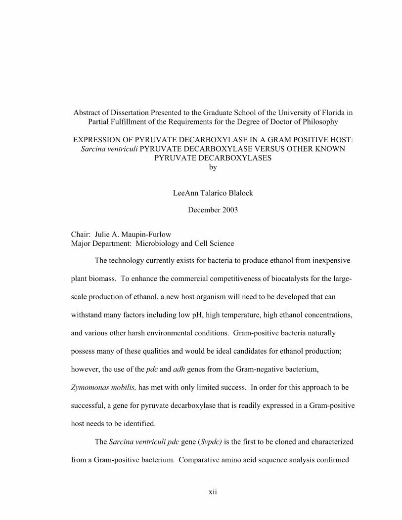
The technology currently exists for bacteria to produce ethanol from inexpensive
plant biomass. To enhance the commercial competitiveness of biocatalysts for the large-
scale production of ethanol, a new host organism will need to be developed that can
withstand many factors including low pH, high temperature, high ethanol concentrations,
and various other harsh environmental conditions. Gram-positive bacteria naturally
possess many of these qualities and would be ideal candidates for ethanol production;
however, the use of the pdc and adh genes from the Gram-negative bacterium,
Zymomonas mobilis, has met with only limited success. In order for this approach to be
successful, a gene for pyruvate decarboxylase that is readily expressed in a Gram-positive
host needs to be identified.
The Sarcina ventriculi pdc gene (Svpdc) is the first to be cloned and characterized
from a Gram-positive bacterium. Comparative amino acid sequence analysis confirmed
xii

that SvPDC is quite distant from Z. mobilis PDC (ZmPDC) and plant PDC enzymes.
Elucidation of the sequence of the Svpdc sequence also led to the identification of a new
subfamily of PDCs.
The Svpdc gene was expressed at low levels in recombinant E. coli due to
differences in the codon usage in the hosts and the Sarcina ventriculi pdc. Expression
was improved by the addition of supplemental tRNA genes and facilitated the
purification and biochemical characterization of the recombinant SvPDC enzyme. This
dramatic difference in codon usage suggested that the Svpdc gene was an ideal candidate
for engineering high-level PDC production in low G+C Gram-positive bacteria. To
confirm this, expression of pdc genes from distantly related organisms (i.e. Z. mobilis,
Acetobacter pasteurianus, and Saccharomyces cerevisiae) were compared to that of the
Svpdc in recombinant Bacillus megaterium. SvPDC protein and activity levels were
several-fold higher in recombinant B. megaterium compared to the other PDCs examined.
Transcript levels using quantitative reverse transcriptase polymerase chain reaction and
protein stability using pulse-chase indicated that SvPDC was expressed at higher levels
than other PDCs tested due to its optimal codon usage. This is the first PDC expressed at
high levels in Gram-positive hosts.
xiii

CHAPTER 1 LITERATURE REVIEW
Industrial Importance of Pyruvate Decarboxylase
Pyruvate Decarboxylase Catalyzes the Production of Bioethanol
In 2001 the United States produced 1.77 billion gallons of fuel ethanol of which
90% was produced by fermentation of corn by yeast (1). The demand for fuel ethanol is
expected to more than double in the next few years because it will replace the fuel
oxygenate methyl tertiary butyl ether (MTBE), a known carcinogen which has been
linked to ground water contamination and has proven difficult to remove from the
environment (2). Fuel ethanol production in 2001 consumed over 5% of the corn crop,
and it has been estimated that fuel ethanol production will reach more than 4 billion
gallons per year by 2006 (1, 2). The use of corn as a feedstock for the production of
ethanol has led to several problems. Because corn is also used for food, this feedstock
has a higher price than alternative feedstocks that are considered waste products from
various processes (3). The use of corn also leads to controversy over sacrificing a food
product for fuel production (3). However, production of ethanol from non-food sources
(bioethanol) can provide a useful alternative to the current method of disposing of
lignocellulosic wastes such as rice straw and wood wastes that were historically burned
but now must be disposed of in a more environmentally friendly and much more costly
manner (3). Utilizing these waste products for bioethanol production not only provides
1

2
an inexpensive feedstock but benefits the environment by disposing of this material in an
environmentally friendly manner and producing a clean burning fuel source (3).
Organisms traditionally used for ethanol fermentation do not have the ability to
metabolize pentoses. Considerable research has been performed to identify naturally
occurring organisms that can ferment pentoses (4). Because yeast have been the
traditional organisms used for ethanol production and they produce high ethanol yields,
research focused on identifying yeast that could metabolize pentose sugars (5). Several
yeast strains have been identified that are capable of utilizing xylose for ethanol
production: Pachysolen tannophilus (6-9), Pichia stipitis (10-14), and Candida shehatae
(10, 11, 15). Unfortunately, these yeast produce only low levels of ethanol from xylose
and exhibit a multitude of problems including low ethanol tolerance, utilization of the
ethanol produced, inability to utilize and metabolize arabinose, and production of xylitol
(6-15). Attempts have also been made to isolate yeast that ferment arabinose, but the
yeast which have been isolated produce very low levels of ethanol (4.1 g/liter) (16).
Attempts to improve yeast strains for ethanol production have also been pursued
by engineering recombinant strains. S. cerevisiae has been the primary focus of this
research because the corn ethanol industry is already familiar with this organism, it
produces high levels of ethanol, and it has been shown to be resistant to high levels of
ethanol (5). Attempts to engineer a xylose utilization pathway from bacteria into S.
cerevisiae has been unsuccessful (17-21). A more promising strategy has been to
engineer the xylose-utilization genes from other yeast into S. cerevisiae (22, 23). One of
the most successful recombinant strains of S. cerevisiae to utilize xylose has been a strain
engineered with a plasmid that contained three xylose-metabolizing genes: a xylose

3
reductase gene and xylitol dehydrogenase gene from Pichia stipitis, and a xylulokinase
gene from S. cerevisiae (23). This strain produced ethanol at 22 g/liter which was a vast
improvement over strains expressing bacterial xylose utilization genes (23). While
recombinant yeast have been engineered to utilize xylose, there have been no successful
attempts to engineer arabinose utilization into these organisms.
Many bacteria naturally possess the ability to ferment hexose and pentose sugars,
but produce a variety of fermentation endproducts (4). In bacteria, pentose and hexose
sugars are metabolized to pyruvate. Ingram et al. (24) have demonstrated that funneling
this pyruvate to ethanol is possible by the use of the pyruvate decarboxylase and alcohol
dehydrogenase II from Z. mobilis. The low Km of the Z. mobilis PDC (0.4 mM pyruvate)
competes favorably with the other enzymes for pyruvate in the cell and causes large
amounts of acetaldehyde to be made, which is then converted to ethanol by alcohol
dehydrogenase (24). A portable ethanol production operon (PET) was generated that
contained the Z. mobilis PDC transcriptionally coupled to the Z. mobilis alcohol
dehydrogenase II (24). The PET operon was used to successfully engineer enteric
bacteria for ethanol production including Klebsiella oxytoca (25), Erwinia chrysanthemi
(26), Enterobacter cloacae (27), and many strains of E. coli (28, 29). The PET operon
has also been successfully used to engineer a wide range of other organisms including
tobacco (30, 31) and cyanobacteria (32). Attempts have also been made to engineer
Gram-positive bacteria to produce ethanol using the PET operon, but this strategy has
been unsuccessful due to poor expression of the Z. mobilis genes (33-35). The recent
cloning and characterization of a PDC from the Gram-positive bacterium Sarcina
ventriculi and its subsequent high level expression in the Gram-positive bacterium,

4
Bacillus megaterium, will enable the development of new Gram-positive biocatalysts for
the production of ethanol (this study).
Pyruvate Decarboxylase Catalyzes the Production of PAC
In 1921, while examining the biotransformation of benzaldehyde to benzyl
alcohol by fermenting Brewer’s yeast (Saccharomyces uvarum), Neuberg and Hirsh
discovered that after 3-5 days no sugar or benzaldehyde remained. Furthermore, the
amount of benzyl alcohol produced was not proportional to the amount of substrate
consumed (36). They later determined that the byproduct of this reaction was (R)-
phenylacetylcarbinol (PAC) and named the enzyme that catalyzed its synthesis
“carboligase” (37, 38).
The process of PAC formation by Brewer’s yeast was patented in 1932, making it
one of the first chiral intermediates to be produced on an industrial scale by
biotransformation (39, 40). PAC is the first chiral intermediate in the production of L-
ephedrine and pseudoephedrine, which are the major ingredients in several commonly
used decongestants and antiasthmatics as well as having a possible use in control of
obesity (41, 42).
Several studies have confirmed that the enzyme catalyzing the production of PAC
is pyruvate decarboxylase (EC 4.1.1.1) (43-46). Pyruvate decarboxylase (PDC) catalyzes
two different reactions: non-oxidative decarboxylation of α-ketoacids to the
corresponding aldehyde (47-50) and the “carboligase” side reaction forming the
hydroxyketones (51, 52). In the acycloin-type condensation reaction, an active aldehyde
in the active site is condensed with a second aldehyde as a cosubstrate (40). The

5
cosubstrate is acetaldehyde in vivo, but can be another aldehyde when supplied externally
(40). In the production of PAC, benzaldehyde is the cosubstrate fed to yeast cells (40).
Production of PAC by Yeast
The industrial production of PAC has historically utilized yeast cells, primarily
Saccharomyces sp. and Candida sp. Many efforts to improve yeast PAC production have
focused on increasing yields of PAC through alteration of the fermentation conditions
and medium (53-55).
When S. carlsbergensis is grown on sucrose, acetaldehyde, and benzaldehyde the
highest initial rate of biotransformation and the highest production of PAC were detected
in the cells with the lowest PDC activity. This led to the suggestion that production of
PAC is limited only by the intracellular pools of pyruvate and that biotransformation of
PAC ceases due to low levels of pyruvate before benzaldehyde mediated inactivation of
PDC occurs. Addition of pyruvate did not increase the rate of PDC synthesis but did
increase the overall production of PAC (55).
The current industrial process for the production of PAC uses a two-stage fed-
batch process. In the first stage, the yeast are grown under partial fermentative conditions
to induce the production of PDC and allow intracellular accumulation of pyruvate. In the
second stage, the biotransformation takes place with feeding of noninhibiting levels of
benzaldehyde. Using this strategy a PAC accumulation level of 22 g/L has been reached
(56).
This strategy, however, is hindered by side-reactions within the cells as well as
the sensitivity of the cells to benzaldehyde and the fermentative products (57). Besides
the PAC production, yeast cells also typically reduce up to 16% to 50% of the

6
benzaldehyde to benzyl alcohol (36-38). The production of benzyl alcohol is primarily
due to the action of alcohol dehydrogenases and other oxidoreductases in the cell (58-60).
Other byproducts are also produced including acetoin, benzoic acid, benzoin, butan-2,3-
dione (diketone), trans-cinnamaldehyde, 2-hydroxypropiophenone, and 1-phenyl-propan-
2,3-dione (acetylbenzoyl) (61, 62). In addition to the formation of these side-products,
PAC is also enzymatically reduced to (1R, 2S)-1-phenyl-1,2-propane-diol (54).
At benzaldehyde concentrations above 16 mM the viability of the yeast cells is
diminished, and PAC production is completely inhibited above 20 mM (63). If the level
of benzaldehyde drops below 4mM, benzyl alcohol becomes the primary product (63).
Comparison of intracellular and extracellular benzaldehyde levels shows that the
membrane maintains a permeability barrier (9.4 mM), which results in lower levels of
benzaldehyde in the cell and may protect intracellular proteins. At concentrations above
9.4 mM benzaldehyde, the barrier appears to falter and intracellular enzymes are
inactivated (59). The yeast PDC, however, is resistant to denaturation by benzaldehyde
at levels up to 66 mM benzaldehyde and is also fairly resistant to final PAC concentration
(59). Thus it was concluded that the modification of cell permeability by benzaldehyde
decreases PAC production by causing release of the cofactors necessary for the
carboligation reaction (i.e. Mg2+ and TPP) and not by inactivation of PDC (59).
Because of these limitations, it would be beneficial to genetically engineer an
organism for PAC production that is more resistant to benzaldehyde and does not
catalyze multiple side reactions. Alternatively, a cell free system may be a viable
alternative to the use of whole cells for PAC production.

7
Production of PAC in a Cell Free System
Utilization of isolated PDC for the biotransformation of pyruvate to PAC has only
recently been pursued as an alternative to the use of whole cells. A distinct advantage to
using a cell free system as opposed to cells as a source of PDC is that the oxidoreductases
responsible for the conversion of benzaldehyde to benzyl alcohol as well as the
cytotoxicity of benzaldehyde can be avoided (58, 59, 63, 64).
The first attempt to use partially purified PDC for the conversion of pyruvate to
PAC compared the efficiencies of PDCs from Z. mobilis and S. carlsbergensis (65). This
study proved that both PDCs can be used for production of PAC, however the Z. mobilis
PDC has a much lower affinity for benzaldehyde (65).
In another study, a high concentration of benzaldehyde was used with partially
purified PDC from Candida utilis (66). At a benzaldehyde levels of 200 mM, a PAC
level of 190mM (28.6 g/L) was obtained which was considerably higher than previously
reported values. Shin and Rogers (67) later determined that the factor limiting
conversion of pyruvate and benzaldehyde to PAC was the inactivation of PDC by
benzaldehyde. This inactivation was determined to be first order with respect to
benzaldehyde and exhibited a square root dependency on time.
Stability of the PDC used for the production of PAC is an important factor in the
success of the biotransformation. Previous studies have shown that S. cerevisiae PDC
exhibits a high carboligase activity, but shows only low stability when isolated (65). The
PDC from Z. mobilis has been shown to have low carboligase activity with respect to the
yeast enzyme but high stability (65, 68). It was determined that mutating residues within
the Z. mobilis PDC enhanced its carboligase activity (68-70). The Pohl lab (71, 72) used

8
the Z. mobilis PDC mutants to produce PAC in an enzyme-membrane reactor. This
continuous reaction system utilized acetaldehyde and benzaldehyde in an equimolar ratio.
At a substrate concentration of 50 mM of both aldehydes, a PAC volume production of
81 g L-1d-1 was obtained with higher yields possible by use of a series of membrane
reactors.
Use of cell free systems for the production of PAC is relatively new, having only
started in 1988 (65), as opposed to the biotransformation using whole cells which began
in 1932 (40). At the moment, the most promising PDCs for production of PAC are
variants of Z. mobilis PDC that enable benzaldehyde to access the active site (68-70). In
cell free systems, the primary factor limiting production of PAC is the availability of
PDC enzymes that can withstand the reaction conditions, mainly inactivation by
aldehydes. Until recently, Z. mobilis PDC was the only known PDC from bacteria. This
enzyme has been shown to be more stable when compared to the yeast PDCs and
alteration of as little as one amino acid enhanced carboligase activity (70). Recently
characterized PDCs from bacteria are likely to have beneficial qualities for the production
of PAC.
Distribution of Pyruvate Decarboxylase
PDC has been identified in a wide variety of plants and fungi, but is rare in
bacteria. The following section identifies the organisms known to encode PDC and
describes the known function of the enzyme in that organism.
PDC in Fungi and Yeast
Several fungal PDCs have been identified. These PDCs from filamentous fungi
appear to be active when the organism experiences anoxic conditions (73-75). It is

9
through PDC that the cell has the ability to regenerate NAD+ through the production of
acetaldehyde that is then converted to ethanol by alcohol dehydrogenase.
In Neurospora crassa PDC forms large cytoplasmic filaments that can measure 8-
10 nm in length (73). The appearance of these filaments in the cell has been shown to
correspond to increased levels of pdc mRNA and increased PDC activity levels within the
cell (73). Disassembly of the filaments enables recovery of active PDC indicating that
the filaments are an active storage form of the enzyme (73). This PDC is particularly
interesting in that the amino acid sequence is more closely related to bacterial PDCs than
to yeast PDCs while the kinetics are more similar to other fungal PDCs (73).
A gene encoding a putative pdc was isolated from a genomic DNA library of
Aspergillus parasiticus (74). The A. parasiticus PDC deduced amino acid sequence was
shown to have 37% similarity to the PDC1 from Saccharomyces cerevisiae, which was
the highest to any PDC and showed that it is quite different from previously characterized
PDCs (74). The organisms A. parasiticus, Aspergillus niger, and Aspergillus nidulans
were tested for the production of ethanol in shake flask cultures. Ethanol was detected
indicating a response to anoxic conditions even though they are obligate aerobes (74).
Although this showed that A. nidulans produced ethanol under anoxic conditions (74), the
reasearchers did not test for PDC activity in cell lysate. Lockington et al. (75) showed
that mycelia subjected to anoxic stress had elevated levels of PDC activity. The gene for
PDC was isolated and sequenced from A. nidulans (75) and the deduced amino acid
sequence from this gene was shown to have highest similarity (37%) to the A. parasiticus
PDC (75). This study showed that production of PDC in the cell is regulated at the level
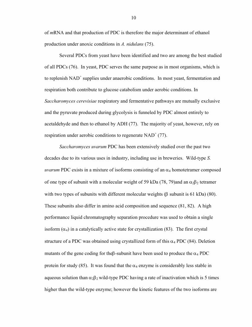
10
of mRNA and that production of PDC is therefore the major determinant of ethanol
production under anoxic conditions in A. nidulans (75).
Several PDCs from yeast have been identified and two are among the best studied
of all PDCs (76). In yeast, PDC serves the same purpose as in most organisms, which is
to replenish NAD+ supplies under anaerobic conditions. In most yeast, fermentation and
respiration both contribute to glucose catabolism under aerobic conditions. In
Saccharomyces cerevisiae respiratory and fermentative pathways are mutually exclusive
and the pyruvate produced during glycolysis is funneled by PDC almost entirely to
acetaldehyde and then to ethanol by ADH (77). The majority of yeast, however, rely on
respiration under aerobic conditions to regenerate NAD+ (77).
Saccharomyces uvarum PDC has been extensively studied over the past two
decades due to its various uses in industry, including use in breweries. Wild-type S.
uvarum PDC exists in a mixture of isoforms consisting of an α4 homotetramer composed
of one type of subunit with a molecular weight of 59 kDa (78, 79)and an α2β2 tetramer
with two types of subunits with different molecular weights (β subunit is 61 kDa) (80).
These subunits also differ in amino acid composition and sequence (81, 82). A high
performance liquid chromatography separation procedure was used to obtain a single
isoform (α4) in a catalytically active state for crystallization (83). The first crystal
structure of a PDC was obtained using crystallized form of this α4 PDC (84). Deletion
mutants of the gene coding for theβ-subunit have been used to produce the α4 PDC
protein for study (85). It was found that the α4 enzyme is considerably less stable in
aqueous solution than α2β2 wild-type PDC having a rate of inactivation which is 5 times
higher than the wild-type enzyme; however the kinetic features of the two isoforms are
11
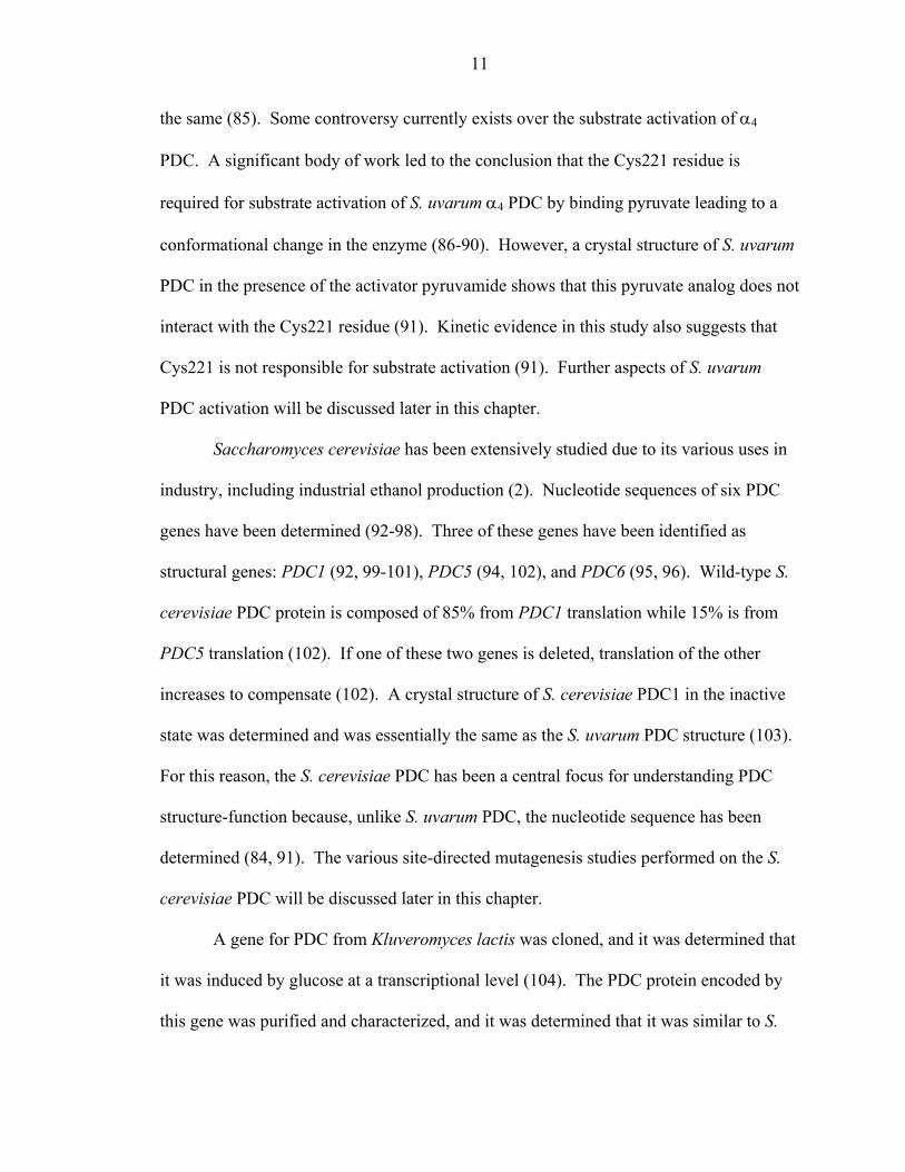
the same (85). Some controversy currently exists over the substrate activation of α4
PDC. A significant body of work led to the conclusion that the Cys221 residue is
required for substrate activation of S. uvarum α4 PDC by binding pyruvate leading to a
conformational change in the enzyme (86-90). However, a crystal structure of S. uvarum
PDC in the presence of the activator pyruvamide shows that this pyruvate analog does not
interact with the Cys221 residue (91). Kinetic evidence in this study also suggests that
Cys221 is not responsible for substrate activation (91). Further aspects of S. uvarum
PDC activation will be discussed later in this chapter.
Saccharomyces cerevisiae has been extensively studied due to its various uses in
industry, including industrial ethanol production (2). Nucleotide sequences of six PDC
genes have been determined (92-98). Three of these genes have been identified as
structural genes: PDC1 (92, 99-101), PDC5 (94, 102), and PDC6 (95, 96). Wild-type S.
cerevisiae PDC protein is composed of 85% from PDC1 translation while 15% is from
PDC5 translation (102). If one of these two genes is deleted, translation of the other
increases to compensate (102). A crystal structure of S. cerevisiae PDC1 in the inactive
state was determined and was essentially the same as the S. uvarum PDC structure (103).
For this reason, the S. cerevisiae PDC has been a central focus for understanding PDC
structure-function because, unlike S. uvarum PDC, the nucleotide sequence has been
determined (84, 91). The various site-directed mutagenesis studies performed on the S.
cerevisiae PDC will be discussed later in this chapter.
A gene for PDC from Kluveromyces lactis was cloned, and it was determined that
it was induced by glucose at a transcriptional level (104). The PDC protein encoded by
this gene was purified and characterized, and it was determined that it was similar to S.
12
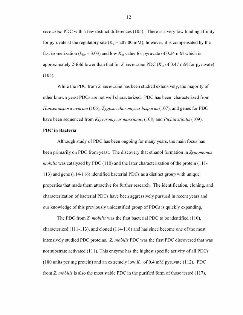
cerevisiae PDC with a few distinct differences (105). There is a very low binding affinity
for pyruvate at the regulatory site (Ka = 207.00 mM); however, it is compensated by the
fast isomerization (kiso = 3.03) and low Km value for pyruvate of 0.24 mM which is
approximately 2-fold lower than that for S. cerevisiae PDC (Km of 0.47 mM for pyruvate)
(105).
While the PDC from S. cerevisiae has been studied extensively, the majority of
other known yeast PDCs are not well characterized. PDC has been characterized from
Hanseniaspora uvarum (106), Zygosaccharomyces bisporus (107), and genes for PDC
have been sequenced from Klyveromyces marxianus (108) and Pichia stipitis (109).
PDC in Bacteria
Although study of PDC has been ongoing for many years, the main focus has
been primarily on PDC from yeast. The discovery that ethanol formation in Zymomonas
mobilis was catalyzed by PDC (110) and the later characterization of the protein (111-
113) and gene (114-116) identified bacterial PDCs as a distinct group with unique
properties that made them attractive for further research. The identification, cloning, and
characterization of bacterial PDCs have been aggressively pursued in recent years and
our knowledge of this previously unidentified group of PDCs is quickly expanding.
The PDC from Z. mobilis was the first bacterial PDC to be identified (110),
characterized (111-113), and cloned (114-116) and has since become one of the most
intensively studied PDC proteins. Z. mobilis PDC was the first PDC discovered that was
not substrate activated (111). This enzyme has the highest specific activity of all PDCs
(180 units per mg protein) and an extremely low Km of 0.4 mM pyruvate (112). PDC
from Z. mobilis is also the most stable PDC in the purified form of those tested (117).

13
This protein is readily expressed at high levels in E. coli (113, 114). A high resolution
crystal structure of Z. mobilis PDC was obtained, and it was shown that the tight packing
of the subunits in the dimers of the tetramer prevents large conformational changes and
locks the enzyme in an active state (117). This crystal structure also showed how a
previously characterized mutant, Trp392Ala, improved synthesis of PAC by Z. mobilis
PDC (70) by relieving the steric hindrance caused by bulky amino acid side chains in the
active site cavity (117). Extensive site-directed mutagenesis studies have been performed
on Z. mobilis PDC (70, 110, 118-126). These studies will be discussed later in this
chapter. The Z. mobilis PDC enzyme has been successfully used to engineer a wide
variety of organisms for ethanol production (4, 30-32, 34, 127-129) and has also been
modified for the efficient production of PAC in recombinant hosts (68, 70-72, 123).
Acetobacter pasteurianus utilizes PDC in a unique way (130). While all other
known PDC proteins function only in anaerobic fermentation to ethanol, the A.
pasteurianus PDC actually functions only in oxidative metabolism (130). In A.
pasteurianus, this enzyme functions to cleave the central metabolite pyruvate into
acetaldehyde and CO2, after which the acetaldehyde is oxidized to the final product,
acetic acid (130). Upon comparison of the deduced amino acid sequence, it was shown
that the A. pasteurianus PDC is most closely related to the Z. mobilis PDC (130).
The most recently discovered bacterial PDC is from Zymobacter palmae (131).
The Z. palmae PDC protein composed approximately 1/3 of the soluble protein when
produced in recombinant E. coli (131). It was hypothesized that the high level of PDC
protein produced is due to similar codon usage of this pdc gene and the E. coli genome
(131). The Km for pyruvate (0.24 mM) of the Z. palmae PDC is the lowest of all bacterial

14
PDCs and is equivalent to the lowest Km for pyruvate reported for all PDCs (0.24 mM
pyruvate for the PDC from K. lactis) (105, 131). This enzyme also has the highest Vmax
(130 units per mg protein) of recombinant bacterial PDC proteins purified using similar
conditions (131). The high level of Z. palmae PDC produced in recombinant E. coli
combined with the biochemical characteristics of this enzyme make it an exciting enzyme
for the development of new biocatalysts for fuel ethanol production (131).
In 1992, Lowe and Zeikus (132) purified a PDC from Sarcina ventriculi. This
was only the second PDC from bacteria to be characterized and unlike Z. mobilis PDC it
was substrate activated (132). The gene for this PDC was cloned and expressed
recombinantly in E. coli (133). Production of this protein in recombinant E. coli was
low, probably due to large differences in codon usage, therefore augmentation with
accessory tRNAs was necessary (133). The deduced amino acid sequence of S. ventriculi
PDC differs from the Z. mobilis PDC and the SvPDC appears to have diverged from a
common ancestor that included most fungal PDCs and bacterial indole-3-pyruvate
decarboxylases (133). The purified enzyme is biphasic with a Km of 2.8 mM and 10 mM
for pyruvate for the high and low affinity sites, respectively (133). Expression of S.
ventriculi PDC is higher in Bacillus megaterium when compared to S. cerevisiae PDC1,
Z. mobilis PDC, and Acetobacter pasteurianus PDC, indicating that it will be a useful
tool in the engineering of Gram-positive bacteria for ethanol production (this study).
PDC in Plants
In plants, PDC serves to convert pyruvate to acetaldehyde. The acetaldehyde is
then converted to ethanol by alcohol dehydrogenase. In this manner these two enzymes
catalyze a pathway in which NAD+ is regenerated under anaerobic conditions such as
15
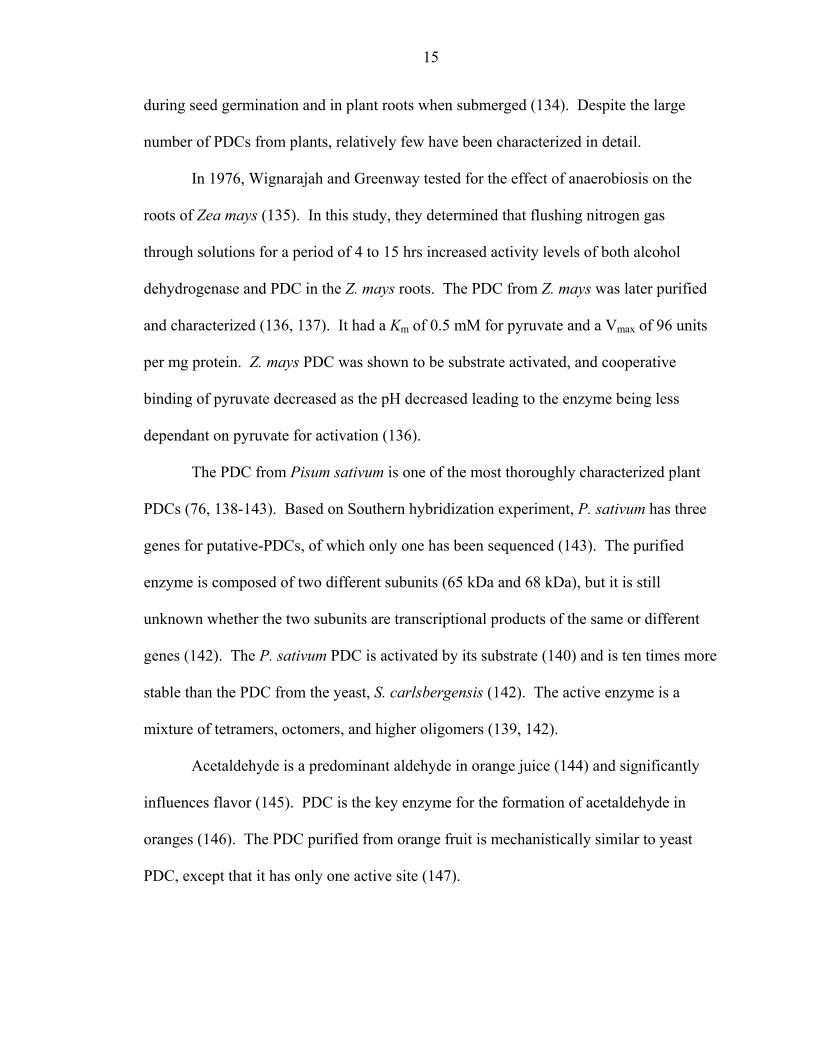
during seed germination and in plant roots when submerged (134). Despite the large
number of PDCs from plants, relatively few have been characterized in detail.
In 1976, Wignarajah and Greenway tested for the effect of anaerobiosis on the
roots of Zea mays (135). In this study, they determined that flushing nitrogen gas
through solutions for a period of 4 to 15 hrs increased activity levels of both alcohol
dehydrogenase and PDC in the Z. mays roots. The PDC from Z. mays was later purified
and characterized (136, 137). It had a Km of 0.5 mM for pyruvate and a Vmax of 96 units
per mg protein. Z. mays PDC was shown to be substrate activated, and cooperative
binding of pyruvate decreased as the pH decreased leading to the enzyme being less
dependant on pyruvate for activation (136).
The PDC from Pisum sativum is one of the most thoroughly characterized plant
PDCs (76, 138-143). Based on Southern hybridization experiment, P. sativum has three
genes for putative-PDCs, of which only one has been sequenced (143). The purified
enzyme is composed of two different subunits (65 kDa and 68 kDa), but it is still
unknown whether the two subunits are transcriptional products of the same or different
genes (142). The P. sativum PDC is activated by its substrate (140) and is ten times more
stable than the PDC from the yeast, S. carlsbergensis (142). The active enzyme is a
mixture of tetramers, octomers, and higher oligomers (139, 142).
Acetaldehyde is a predominant aldehyde in orange juice (144) and significantly
influences flavor (145). PDC is the key enzyme for the formation of acetaldehyde in
oranges (146). The PDC purified from orange fruit is mechanistically similar to yeast
PDC, except that it has only one active site (147).

16
Ipomoea batatas (sweet potato) produces PDC in its roots (148-150). This PDC
is substrate activated, has a Km of 0.6 mM, and is inhibited by phosphate (149). Pyruvate
decarboxylation is the rate-limiting step in alcoholic fermentations in sweet potato roots
based on the finding that PDC activity is 21- to 28-fold less than ADH activity under
aerobic conditions, but 6- to 8-fold less than ADH under anaerobic conditions (150).
PDC has also been characterized from Triticum aestivum (wheat) (81, 82, 151-
154), Oryza sativa (rice) (155-160), and Vicia faba (fava bean) (161). PDC has been
shown to be produced in but not characterized from Capsicum annuum (bell pepper) fruit
(162), Echinochloa crus-galli (barnyard grass) (163), Nicotiana tobacum (tobacco) (164),
Vitis vinifera (grape) (165), Lycopersicon esculentum (tomato) (166), Lepidium latifolium
(167), Populus deltoides (Eastern cottonwood) (168), Glycine max (soybean) (168), and
Arabidopsis thaliana (169, 170)
Structure of Pyruvate Decarboxylase
The crystal structures of Z. mobilis (117), S. uvarum (84, 91) and S. cerevisiae
(103) PDCs have been invaluable when studying PDC proteins for use and engineering
for industrial application. By comparison of deduced amino acid sequences and
biochemical characteristics it has been shown that the A. pasteurianus and Z. palmae
PDCs are more closely related to Z. mobilis PDC (130, 131); whereas, the S. ventriculi
PDC is more closely related to S. cerevisiae PDC1 (133). Because the majority of the
bacterial PDC proteins were only recently discovered (130, 131, 133) there has not been
sufficient time for detailed structural analysis of these enzymes. However, the crystal
structure and mutagenesis analysis of the well characterized Z. mobilis, S. uvarum and S.

17
cerevisiae PDC proteins can give important and useful information about the structure of
the newly identified bacterial PDC proteins.
Subunits of PDC
The quaternary structure of most PDCs is a tetramer with an apparent molecular
weight of 240 kDa (79, 105, 111, 130-133, 148, 151, 155, 171), with the exception of
PDCs forming larger complexes: A. pateurianus (130), Z. mays (135), P. sativum (139,
142), T. aestivum (81), and N. crassa (73). The association of the subunits has been
determined to be pH-dependant with optimal pH for catalytic activity and subunit
association of between pH 5.0 and pH 6.7 (108, 113, 131, 132, 147, 155). Until recently
it was believed that the tetramer was the only active conformation (172), but a recent
study showed that both dimers and tetramers of ScPDC1 had comparable specific activity
(173). This study, however, determined a difference in the dissociation constant for the
regulatory substrate by one order of magnitude among the two forms indicating that
binding of the substrate to the regulatory site is influenced by oligomerization (173). In
contrast, the subunit interactions of the Z. mobilis PDC are different than those of S.
cerevisiae PDC1 (117). Unlike the S. cerevisiae PDC1, Z. mobilis PDC is not controlled
by allosteric regulation. The reason for this difference is elucidated in the crystal
structures (117). Z. mobilis PDC dimers are packed tightly together and lock the enzyme
in an activated form so that large conformational changes are not possible or necessary
for enzyme activity as they are in S. cerevisiae PDC1 (91, 117). This tight packing of the
dimers also explains the extreme stability of the Z. mobilis PDC in comparison to the S.
cerevisiae PDC1 (174). This data is also in agreement with the differences in the
thermostabilities of the bacterial PDCs. The Z. mobilis, A. pasteurianus, and Z. palmae

18
PDCs have temperature optima of 60°C, while S. ventriculi PDC has a temperature
optimum of 32°C and is completely inactive at 60°C (131). Structural differences in the
subunit interactions may be responsible for the instability of S. ventriculi PDC at high
temperatures. Analysis of S. ventriculi PDC thermostability throughout a range of pH
shows that the enzyme is more stable between pH 5.0 and pH 5.5 indicating that
protonation of an amino acid side chain may stabilize the subunit interactions at high
temperatures (this study).
Cofactors of PDC
Both Mg2+ and thiamin diphosphate (TPP) are required cofactors for the action of
PDC (175, 176). It has been demonstrated that TPP dissociates from PDC under alkaline
conditions, but it is difficult if not impossible to remove Mg2+ completely from the
enzyme (137, 177-179). Mg2+ can be replaced by other divalent cations, such as Mn2+,
Ni2+, Co2+, and Ca2+ (176). The substitution of Mg2+ with these other cations does not
affect the Vmax of the enzyme, but it does affect the stability of the reconstructed
holoenzyme (180, 181). A TPP derivative retaining the N-1’-4’amino system functions
properly with full binding capacity therefore proving that this is the functional group
necessary for activity of the PDC (182, 183). The Z. mobilis PDC retains its tetrameric
state even after the TPP and Mg2+ cofactors are removed (178). This is also true of S.
ventriculi PDC, Z. palmae PDC, and S. cerevisiae PDC1, but not of A. pasteurianus PDC
(131). A. pasteurianus PDC forms both tetramers and octomers of similar specific
activity and dissociates into dimers after cofactor extraction (131). Tetrameric
configuration and activity are restored upon addition of the cofactors (131). The
dissociation of the subunits is consistent with the behavior of other PDCs upon cofactor

19
removal (76). Residues responsible for binding the cofactors, as determined through X-
ray crystallography studies, are conserved throughout yeast and bacterial PDC proteins
(91, 103, 131).
Kinetics of PDC
There are currently two distinct groups of PDC proteins based on kinetics. All
known PDC proteins, except those from Gram-negative bacteria, are allosterically
regulated (76). The substrate activation behavior of S. cerevisiae PDC has been studied
in detail through site-directed mutagenesis and crystal structure analysis (47, 86-91, 103,
184-191). Initial studies of the S. uvarum PDC determined that a cysteine residue was
most likely responsible for the substrate activation behavior. In these studies, irreversible
activation of the enzyme, exhibited by disappearance of the lag phase in product
formation, was achieved by utilization of thiol specific reagents (80, 192-194). Use of a
PDC1-PDC6 fusion protein that contained Cys221 as its only cysteine residue suggested
that the Cys221 residue was responsible for the substrate activation behavior of S.
cerevisiae PDC (86). Site-directed mutagenesis of the Cys221 and/or Cys222 to serine
showed that the enzyme could no longer be activated by the substrate (87). Steady state
kinetics studies were also used to bolster the argument for Cys221 as the site of substrate
activation (88, 89). Although crystal structures of S. uvarum and S. cerevisiae PDC were
determined in the presence and absence of effectors (84, 103, 187, 195), these crystal
structures were not of high enough quality to determine where the activator molecules
bound the enzyme . More recently, Lu et al. obtained a high resolution crystal structure
of S. uvarum PDC in the presence of pyruvamide and determined that pyruvamide did not
bind at or near Cys221 (91). This study also used kinetics to show that the Cys221Ser

20
was in fact still substrate activated (91); however, this data was later refuted by Wei et
al. (90) who used solvent kinetic isotope effect to reaffirm that their original assertion
that Cys221Ser does shift the enzyme into an active conformation. Lu et al. (91)
determined the residues that bind pyruvamide in the regulatory site of the crystal
structure of PDC1 as Tyr157 and Arg 224. Sergeinko et al. (191), however, argues that
pyruvamide should not be considered to form an active conformation of the enzyme and
may actually represent an inhibitory mode of binding. It is interesting to note that plant
PDCs and the S. ventriculi PDC are substrate activated, yet the Cys221 equivalent is not
conserved in these proteins while equivalent residues for Tyr157 and Arg224 are
conserved (121, 131, 133).
The Gram-negative bacterial PDC proteins are the only known PDCs that exhibit
Michaelis-Menten kinetics (111-113, 130, 131). These PDCs also have high affinity for
the substrate pyruvate with a Km of 0.24 mM pyruvate for Z. palmae PDC, 0.39 mM
pyruvate for A. pasteurianus PDC, and 0.43 mM pyruvate for Z. mobilis PDC (131). The
Gram-negative bacterial PDCs also have the highest Vmax values of all PDCs (68, 131).
The low Km and high Vmax of Z. mobilis PDC have already been exploited successfully
to engineer biocatalysts for fuel ethanol production (4).
Catalytic Residues of PDC
All crystal structures of TPP dependent enzymes have a glutamate residue close to
the N-1’ of TPP that promotes the ionization of the C-2 proton of TPP (121). Candy et
al. demonstrated that substitution of Glu50 with either aspartate or glutamine yields an
enzyme with 3.0% and 0.5% remaining catalytic activity of the wild-type enzyme,
respectively (119). Each of these mutants also displays a decreased affinity for both

21
cofactors (119). The equivalent glutamate in yeast, Glu51, is also essential for catalytic
activity (196). Only 0.04% catalytic activity of the wild-type enzyme remains upon
substitution of Glu51 with glutamine and binding of TPP to the protein is slow (196).
The Z. mobilis PDC crystal structure reveals that amino acid side chains Asp27, His113,
His114, Thr388, and Glu473 are in the vicinity of the active site and are conserved
among PDC proteins (117). This data corresponds well with the crystal structure data
and site-directed mutagenesis studies of the S. cerevisiae, Z. mobilis, and S. uvarum
PDCs (91, 120, 122, 195, 197).
Alternative Substrates of PDC
As discussed previously in this chapter, Neuberg and Hirsch (36, 38) first
discovered that yeast could catalyze the formation of PAC when benzaldehyde was added
to the medium. This reaction was later determined to be catalyzed by PDC (43-46). It
has since been shown that the yeast PDCs are much more efficient at carboligase
reactions than the PDC from Z. mobilis (65). The reason for this difference is believed to
be the size of the active site cleft which is smaller in the Z. mobilis PDC than its yeast
counterparts (117). Bruhn et al. found that the mutation of only one amino acid increased
carboligase activity by Z. mobilis PDC by 4-fold when compared to wild-type (70). The
crystal structure of Z. mobilis PDC showed other large side chains that were possible sites
for mutagenesis to increase carboligase activity (117). Pohl et al. have since made these
mutations and found a wide variety of carboligase activities catalyzed by these PDC
variants, including one in which the stereochemistry has been changed to form (S)-
phenylacetylcarbinol (123).

22
PDC from Brewer’s yeast catalyzes the formation of acetoin through two separate
mechanisms (198-200). Acetoin is produced by the aldol-type condensation reaction
between two molecules of acetaldehyde or by the addition of acetaldehyde to an
intermediate formed between pyruvate and thiamin pyrophosphate (198-200).
Besides pyruvate, yeast PDCs have been shown to accept longer aliphatic α-keto acids
like α -keto butanoic acid, α -keto pentanoic acid, branched aliphatic α -keto acids, as
well as α -keto-phenylpropanoic acid (benzoylformate) and various phenyl-substituted
derivatives of the latter (69, 201, 202). Only C4 and C5-keto acids have been shown to
be substrates for PDC from Z. mobilis (65).
Study Rationale and Design
Engineering Gram-positive bacteria for ethanol production has been difficult due
to the absence of suitably expressed pdc genes. A PDC was previously purified and
characterized from the Gram-positive bacterium S. ventriculi; however the gene was not
cloned (132). It was expected that S. ventriculi PDC will be expressed at high levels in
Gram-positive hosts due to its origination from a Gram-positive bacterium. To test this
possibility the pdc gene from S. ventriculi was cloned, sequenced, and characterized.
SvPDC was expressed in recombinant E. coli and the protein was biochemically
characterized. SvPDC was expressed in a Gram-positive host, B. megaterium. SvPDC
production in B. megaterium was analyzed and optimal conditions for SvPDC activity in
B. megaterium were determined. Expression analysis and optimization of a variety of
PDCs (i.e. Z. mobilis, A. pasteurianus, S. cerevisiae, and S. ventriculi) in B. megaterium
were performed to determine the optimal PDC for ethanol production in Gram-positive
bacterial hosts.
23
CHAPTER 2 CLONING AND EXPRESSION OF pdc, AND CHARACTERIZATION OF
PYRUVATE DECARBOXYLASE FROM Sarcina ventriculi
Introduction
PDC (EC 4.1.1.1) serves as the key enzyme in all homo-ethanol fermentations.
This enzyme catalyzes the non-oxidative decarboxylation of pyruvate to acetaldehyde
and carbon dioxide using Mg2+ and thiamine pyrophosphate (TPP) as cofactors.
Acetaldehyde is subsequently reduced to ethanol by alcohol dehydrogenase (ADH,
EC1.1.1.1) during the regeneration of NAD+. PDC is widespread among plants, absent in
animals, and rare in prokaryotes. Prior to this study, the only bacterial pdc gene described
was from the Gram-negative α-proteobacterium Zymomonas mobilis (114-116, 203). Z.
mobilis PDC was purified to homogeneity, crystallized, and extensively characterized
(121). This enzyme has also been purified from an unusual Gram-positive organism,
Sarcina ventriculi (132).
S. ventriculi is an obligate anaerobe that grows from pH 2 to pH 10, fermenting
hexose and pentose sugars to produce acetate, ethanol, formate, CO2 and H2 (204, 205).
In this organism, the relative production of ethanol and acetate vary with environmental
pH. Under acidic conditions where acetic acid is toxic to cells, ethanol is the primary
product (205). At neutral pH and above, a near equimolar mixture of ethanol and acetate
are produced with low levels of formate (206). These changes in fermentation profiles
3
2
24
have been attributed to changes in the levels of two enzymes that metabolize pyruvate,
PDC and pyruvate dehydrogenase (205, 206).
The properties of the S. ventriculi PDC are very different from those of the Z.
mobilis enzyme. Unlike the Michaelis-Menten kinetics of Z. mobilis PDC (111, 116), the
S. ventriculi enzyme is activated by pyruvate (132), similar to PDC enzymes from yeast
and higher plants. S. ventriculi PDC was reported to have an unusually high Km for
pyruvate (13 mM) compared to Km values of 0.3 mM to 4.4 mM for other PDC enzymes
(111, 116, 207). The phenylalanine content of purified S. ventriculi PDC was reported to
be 4-fold to 5-fold higher than that of other PDC enzymes suggesting significant
differences in primary structure (132).
To further examine the unusual nature of the S. ventriculi PDC, this gene was
cloned, sequenced, and expressed in recombinant E. coli. This approach provided the
primary amino acid sequence and facilitated PDC purification for further kinetic and
biophysical characterization.
Materials and Methods
Materials
Biochemicals were purchased from Sigma Chemical Co. (St. Louis, Mo.). Other
organic and inorganic analytical grade chemicals were from Fisher Scientific (Atlanta,
Ga.). Restriction endonucleases and DNA-modifying enzymes were from New England
BioLabs (Beverly, Mass.). Oligonucleotides were from Sigma-Genosys (The
Woodlands, Tex.). Digoxigenin-11-dUTP (2’-deoxyuridine-5’-triphosphate coupled by
an 11-atom spacer to digoxigenin), alkaline phosphatase conjugated antibody raised
against digoxigenin, and nylon membranes for colony and plaque hybridizations were
25
from Roche Molecular Biochemicals (Indianapolis, Ind.). Positively charged membranes
for Southern hybridization were from Ambion (Austin, Tex.).
Bacterial Strains and Media
Table 2-1 lists the E. coli strains used in this study including strains TB-1 and
DH5α that were used for routine recombinant DNA experiments. E. coli strain SE2309
was used to create a genomic DNA library in plasmid pBR322. E. coli strains ER1647,
LE392, and BM25.8 were used in conjunction with λBlueSTAR for a genomic DNA
library. E. coli strains BL21(DE3), BL21-CodonPlus-RIL, and BL21-CodonPlus-
RIL/pSJS1240 were used to examine the expression of the S. ventriculi pdc gene from
plasmid pJAM419. E. coli strains were grown in Luria-Bertani (LB) medium and
supplemented with antibiotics as appropriate (30 mg of chloramphenicol per liter, 100 mg
of carbenicillin per liter, 100 mg of ampicillin per liter, and/or 50 mg of spectinomycin
per liter). S. ventriculi strain Goodsir was cultivated as described previously (205).
DNA Isolation
Plasmid DNA was isolated and purified using a Quantum Prep Plasmid Miniprep
Kit from BioRad (Hercules, Ca.). DNA fragments were eluted from 0.8% SeaKem GTG
agarose (FMC Bioproducts, Rockland, Me.) using either Ultrafree-DA filters from
Millipore (Bedford, Md.) or the QIAquick gel extraction kit from Qiagen (Valencia, Ca.).
S. ventriculi genomic DNA was isolated and purified as described previously (208).
Cloning of the S. ventriculi pdc Gene
A degenerate oligonucleotide (5’-AARGARGTNAAYGTNGARCAYA-
TGTTYGGNGT-3’) was synthesized based on the N-terminal amino acid sequence of
PDC purified from S. ventriculi (132)(where, R is A or G; N is A, C, G, or T; Y is C or
T). This oligonucleotide was labeled at the 3’-end using terminal transferase with

26
digoxigenin-11-dUTP and dATP as recommended by the supplier (Roche Molecular
Biochemicals) and was used to screen genomic DNA from S. ventriculi.
For Southern analysis, genomic DNA was digested with BglI, EcoRI, or HincII, separated
by 0.8% agarose electrophoresis, and transferred to positively charged nylon membranes
(209). Membranes were equilibrated at 58ºC for 2 h in 5× SSC (1× SSC is 0.15 M NaCl
plus 0.015 M sodium citrate) containing 1% blocking reagent (Roche Molecular
Biochemicals), 0.1% N-lauroylsarcosine, and 0.02% SDS. After the probe (0.2 pmol per
ml) and Poly(A) (0.01mg per ml) were added, membranes were incubated at 58ºC for
18.5 h. Membranes were washed twice with 2 X SSC containing 0.1% SDS (5 min per
wash) at 25ºC and twice with 0.5 X SSC containing 0.1% SDS (15 min per wash) at
58ºC. Signals were visualized using colorimetric detection according to supplier (Roche
Molecular Biochemicals).
For generation of a genomic library in plasmid pBR322, S. ventriculi
chromosomal DNA was digested with HincII and fractionated by electrophoresis. The
2.5- to 3.5-kb HincII DNA fragments were ligated into the EcoRV site of pBR322 and
transformed into E. coli SE2309. Colonies were screened with the degenerate
oligonucleotide by colorimetric detection. By this method, plasmid pJAM400 that carries
a HincII fragment containing 1,350 bp of the pdc gene was isolated.
The λBlueSTAR Vector System (Novagen) was used to create an additional
genomic library to facilitate isolation of the full-length pdc gene from S. ventriculi.
Genomic DNA was digested with BclI, separated by electrophoresis in 0.8% agarose, and
the 6.5- to 8.5-kb fragments were ligated with the λBlueSTAR BamHI arms. In vitro
packaging and plating of phage was performed according to the supplier (Novagen). A

27
DNA probe was generated using an 800-bp EcoRI fragment of the pdc gene from
pJAM400 that was labeled with digoxigenin-11-dUTP using the random primed method
as recommended by the supplier (Roche Molecular Biochemicals). Plaques were
screened using colorimetric detection. Cre-loxP-mediated subcloning was used to
circularize the DNA of the positive plaques by plating λBlueSTAR phage with E. coli
BM25.8 that expresses Cre recombinase (Novagen). The circularized plasmid pJAM410
was then purified and electroporated into E. coli DH5α.
For generation of a pdc expression vector, the promoterless pdc gene was
subcloned into pET21d after amplification from pJAM413 (Table 2-1) by the polymerase
chain reaction (PCR). Primers were designed for directional insertion using BspHI (oligo
1) and XhoI (oligo 2) restriction sites. The resulting fragment was ligated into compatible
NcoI and XhoI sites of pET21d (Novagen) to produce pJAM419 (Figure 2-1). The fidelity
of the pdc gene was confirmed by DNA sequencing.
Nucleotide and Protein Sequence Analyses
DNA fragments of plasmids pJAM400 and pJAM410 (Figure 2-1) were
subcloned into plasmid vector pUC19 for determining the pdc sequence using the
dideoxy termination method (210) and a LI-COR (Lincoln, Neb.) automated DNA
sequencer (DNA Sequencing Facility, Department of Microbiology and Cell Science,
University of Florida). The nucleotide sequence of the S. ventriculi pdc gene and
surrounding DNA was deposited in the GenBank database (accession number
AF354297).
Genepro 5.0 (Riverside Scientific, Seattle, WA), ClustalW version 1.81 (211),
Treeview version 1.5 (212), and MultiAln (213) were used for DNA and/or protein

28
sequence alignments and comparisons. Deduced amino acid sequences were compared to
protein sequences available in the GenBank, EMBL, and SwissProt databases at the
National Center for Biotechnology Information (Bethesda, Md.) using the BLAST
network server (214). The Dense Alignment Surface (DAS) method was used for the
prediction of transmembrane α-helices (215).
Production of S. ventriculi PDC in Recombinant E. coli
Plasmid pJAM419 was transformed into E. coli BL21-CodonPlus-RIL containing
plasmid pSJS1240 (Table 2-1). Expression of the pdc gene in this plasmid is regulated
by the bacteriophage T7 RNA polymerase-promoter system (Novagen). Freshly
transformed cells were inoculated into LB medium containing ampicillin, spectinomycin,
and chloramphenicol and grown at 37ºC (200 rpm) until cells reached an O.D.600nm of 0.6
to 0.8 (mid-log phase). Transcription/translation of pdc was initiated by the addition of 1
mM isopropyl-γ-D-thiogalactopyranoside (IPTG). Cells were harvested after 2-3 h by
centrifugation at 5000 × g (10 min, 4ºC) and stored at –70ºC or in liquid nitrogen.
Purification of the S. ventriculi PDC Protein
All purification buffers contained 1 mM TPP and 1mM MgSO4 unless indicated
otherwise. Recombinant E. coli cells (14.8 g wet wt) were thawed in 6 volumes (wt/vol)
of 50 mM Na-PO4 buffer at pH 6.5 (Buffer A) and passed through a French pressure cell
at 20,000 lb per in2. Cell debris was removed by centrifugation at 16,000 × g (20 min,
4ºC). Supernatant was removed and filtered through a 45 µm filter membrane. Filtrate
(692 mg protein) was applied to a Q Sepharose Fast Flow 26/10 column (Pharmacia) that
was equilibrated with Buffer A containing 300 mM NaCl. The SvPDC did not bind and
eluted in the wash. The wash fractions containing PDC activity (326 mg protein) were
29
precipitated with 80% (NH4)2SO4. Protein was dissolved in Buffer A, dialyzed against
buffer A (4ْC, 16 h), and filtered (.45 µm membrane). The filtrate (287 mg) was applied
to a Q Sepharose column equilibrated with Buffer A and developed with a linear NaCl
gradient (0 to 400 mM NaCl in 220 ml of Buffer A). PDC active fractions eluted at 230
to 300 mM NaCl and were pooled. The pooled sample (23 mg) was applied to a 5 ml
Bio-scale hydroxyapatite type I column (BioRad) that was equilibrated with 5 mM Na-
PO4 buffer at pH 6.5 (Buffer B). The column was washed with 15 ml Buffer B and
developed with a linear Na-PO4 gradient (5 to 500 mM Na-PO4 at pH 6.5 in 75 ml).
Protein fractions (11.4 mg) with PDC activity eluted at 200 to 300 mM Na-PO4 and were
pooled. For further purification, portions of this material (0.25 to 0.5 mg protein per 0.25
to 0.5 ml) were applied to a Superdex 200 HR 10/30 column (Pharmacia) equilibrated in
50 mM Na-PO4 at pH 6.5 with 150 mM NaCl and 10% glycerol in the presence or
absence of 1 mM MgSO4 and 1 mM TPP.
Activity Assays
PDC activity was assayed by monitoring the pyruvate-dependent reduction of
NAD+ with baker’s yeast alcohol dehydrogenase (ADH) (Sigma) as a coupling enzyme at
pH 6.5 as previously described (115), with the following modifications. Buffered
enzyme (100 µl) was added to a final volume of 1 ml containing 0.15 mM NADH, 0.1
mM TPP, 0 to 25 mM pyruvate, and 10 U ADH in 50 mM potassium-MES (2-[N-
morpholino]ethanesulfonic acid) buffer with 5 mM MgCl2 at pH 6.5. Since this assay
does not distinguish PDC from NADH oxidizing enzymes such as lactate dehydrogenase,
activity of cell lysate was estimated by correcting for control reactions performed in the
absence of added ADH. One unit of enzyme activity is defined as amount of enzyme that
30
oxidizes 1 µmol of NADH per min. Thermostability was determined by incubating
purified PDC in 50 mM Na-PO4 buffer at pH 6.5 with 1 mM TPP and 1 mM MgCl2 for
90 min and then assaying for activity with 10 mM pyruvate under standard conditions.
Protein concentration was determined using Bradford protein reagent with bovine serum
albumin as the standard (BioRad).
Molecular Mass and Amino Acid Sequence Analyses
Subunit molecular mass was estimated by reducing and denaturing sodium
dodecyl sulfate-polyacrylamide gel electrophoresis (SDS-PAGE) using 12%
polyacrylamide gels which were stained with Coomassie blue R-250. The molecular
weight standards for SDS-PAGE were: phosphorylase b (97.4 kDa), bovine serum
albumin (66.2 kDa), ovalbumin (45 kDa), carbonic anhydrase (31 kDa), trypsin inhibitor
(21.5 kDa), and lysozyme (14.4 kDa). For determination of native molecular mass,
samples were applied to a Superdex 200 HR 10/30 column equilibrated with 50 mM Na-
PO4 buffer at pH 6.5 with 150 mM NaCl, 10% glycerol, and no added cofactors.
Molecular mass standards included: serum albumin (66-kDa), alcohol dehydrogenase
(150 kDa), α -amylase (200 kDa), apoferritin (443 kDa), and thyroglobulin (669 kDa).
The N-terminal sequence was determined for PDC protein purified from
recombinant E. coli. The protein was separated by SDS-PAGE and electroblotted onto a
polyvinylidene difluoride (PVDF) membrane (Immobilon-P). The sequence was
determined by automated Edman degradation at the protein chemistry core facility of the
University of Florida Interdisciplinary Center for Biotechnology Research.
31
Results and Discussion
PDC Operon in S. ventriculi
The N-terminal amino acid sequence of the PDC protein purified from S.
ventriculi (132) was used to generate a degenerate oligonucleotide for hybridization to
genomic DNA. This approach facilitated the isolation of a 7.0-kb BclI genomic DNA
fragment from S. ventriculi. The fragment was further subcloned in order to sequence
both strands of a 3,886 bp HincII-to-HincII region that hybridized to the oligonucleotide
probe (Figure 2-1). Analysis of the DNA sequence revealed an open reading frame
(ORF) of 1,656 bp encoding a protein with an N-terminus identical to that of the
previously purified S. ventriculi PDC (Figure 2-2). The ORF is therefore designated pdc.
A canonical Shine-Dalgrano sequence is present 7 bp upstream of the pdc translation start
codon. In addition, a region 82 to 110 bp upstream of pdc has limited identity to the
eubacterial –35 and –10 promoter consensus sequence. Downstream (43 bp) of the pdc
translation stop codon is a region predicted to form a stem-loop structure followed by an
AT-rich region, consistent with a ρ-independent transcription terminator. Thus, the S.
ventriculi pdc appears to be transcribed as a monocistronic operon like the Z. mobilis pdc
gene (115).
A partial ORF was identified 722 bp upstream of pdc which encodes a 177 amino
acid protein fragment (ORF1*) (Figure 2-1). ORF1* has identity (28-29 %) to several
hypothetical membrane proteins (GenBank accession numbers CAC11620, CAC24018,
CAA22902) and is predicted to form several transmembrane spanning domains (data not
shown).

32
PDC Protein Sequence in S. ventriculi
The S. ventriculi pdc gene apparently encodes a protein of 552 amino acids
(including the N-terminal methionine) with a calculated pI of 5.16 and anhydrous
molecular mass of 61,737 Da. Consistent with other Z. mobilis and fungal PDC proteins,
the N-terminal extension of up to 47 amino acids that is common to plant PDC proteins is
not conserved in the S. ventriculi PDC protein. Although the pI of the purified S.
ventriculi PDC protein has not been experimentally determined, the calculated pI is
consistent with the acidic pH optimum of 6.3 to 6.7 for stability and activity of this
enzyme (132). The amino acid composition of the protein deduced from the S. ventriculi
pdc gene is similar to that determined for the S. cerevisiae PDC1 and Z. mobilis pdc
genes (Table 2-2). A notable exception is the alanine composition of Z. mobilis PDC,
which is 1.8- to 2.2-fold higher than the composition of S. cerevisiae PDC1 and S.
ventriculi PDC. Although the phenylalanine composition of the deduced S. ventriculi
PDC protein is consistent with the other PDC proteins, it is almost 3.6-fold less than the
composition previously reported for the purified S. ventriculi enzyme (132). The reason
for this discrepancy remains to be determined.
The amino acid sequence of S. ventriculi PDC was aligned with the sequences of
the yeast (Sc) PDC1 and Z. mobilis (Zm) PDC proteins, both of which have been
analyzed by X-ray crystallography (76, 84, 103, 117) (Figure 2-3). The conserved motif
of TPP-dependent enzymes identified by Hawkins et al. (216) and known to be involved
in Mg2+-TPP cofactor binding is highly conserved in all three PDC proteins. Other
amino acid residues located within a 0.4 nm distance of the Mg2+ and TPP binding site of
the two crystallized PDC proteins are also conserved in the S. ventriculi PDC protein.
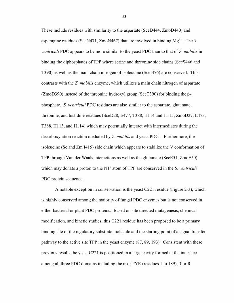
33
These include residues with similarity to the aspartate (SceD444, ZmoD440) and
asparagine residues (SceN471, ZmoN467) that are involved in binding Mg2+. The S.
ventriculi PDC appears to be more similar to the yeast PDC than to that of Z. mobilis in
binding the diphosphates of TPP where serine and threonine side chains (SceS446 and
T390) as well as the main chain nitrogen of isoleucine (SceI476) are conserved. This
contrasts with the Z. mobilis enzyme, which utilizes a main chain nitrogen of aspartate
(ZmoD390) instead of the threonine hydroxyl group (SceT390) for binding the β-
phosphate. S. ventriculi PDC residues are also similar to the aspartate, glutamate,
threonine, and histidine residues (SceD28, E477, T388, H114 and H115; ZmoD27, E473,
T388, H113, and H114) which may potentially interact with intermediates during the
decarboxylation reaction mediated by Z. mobilis and yeast PDCs. Furthermore, the
isoleucine (Sc and Zm I415) side chain which appears to stabilize the V conformation of
TPP through Van der Waals interactions as well as the glutamate (SceE51, ZmoE50)
which may donate a proton to the N1’ atom of TPP are conserved in the S. ventriculi
PDC protein sequence.
A notable exception in conservation is the yeast C221 residue (Figure 2-3), which
is highly conserved among the majority of fungal PDC enzymes but is not conserved in
either bacterial or plant PDC proteins. Based on site directed mutagenesis, chemical
modification, and kinetic studies, this C221 residue has been proposed to be a primary
binding site of the regulatory substrate molecule and the starting point of a signal transfer
pathway to the active site TPP in the yeast enzyme (87, 89, 193). Consistent with these
previous results the yeast C221 is positioned in a large cavity formed at the interface
among all three PDC domains including the α or PYR (residues 1 to 189), β or R

34
(residues 190 to 356), and γ or PP (residues 357 to 563) domains (84). However, recent
high-resolution structural analysis of the brewer’s yeast PDC crystallized in the presence
of pyruvamide, a pyruvate analogue, enabled localization of the activator binding site and
revealed that cysteine does not play a direct role in this binding (91). Additionally,
kinetic studies using stopped-flow techniques revealed that the C221A variant of yeast
PDC was still substrate activated, and the lag phase of product formation did not
disappear with progressive thiol oxidation (91). Instead, tyrosine (Y157) and arginine
(R224) residues form hydrogen bonds with the amide group of pyruvamide. Both of
these residues are conserved in the S. ventriculi PDC and not found in the Z. mobilis
enzyme which displays Michaelis-Menten kinetics. These results suggest that residues of
the S. ventriculi PDC protein may allosterically bind the substrate activator with a
mechanism common to the majority of fungal PDC proteins. Interestingly, these two
residues that bind pyruvamide in the yeast enzyme are not universally distributed among
the substrate activated PDC enzymes, most notably the plant PDCs.
Phylogenetic analysis was performed to compare the PDC proteins and other
TPP-dependent enzymes including indole-3-pyruvate decarboxylase (IPD), the E1
component of pyruvate dehydrogenase (E1), acetolactate synthase (ALS), and
transketolase (TK) (Figure 2-4). The comparison reveals that all of these proteins are
related in primary sequence and that there is a significant clustering of the sequences into
families based on specific enzyme function. Of these, the S. ventriculi PDC appears most
closely related to eubacterial IPD proteins as well as the majority of fungal PDC proteins.
In contrast, the Z. mobilis PDC protein is most closely related to plant PDCs in addition
to a couple of out-grouping fungal PDCs. Thus, it appears that the IPD protein family

35
has close evolutionary roots with the PDC family and specifically the S. ventriculi PDC
protein. In addition, the distant relationship of the S. ventriculi and Z. mobilis PDC
proteins is consistent with the biochemical differences between these two enzymes (111,
116, 132, 207). In contrast to Z. mobilis PDC which may have originated by the
horizontal transfer of a plant pdc gene, S. ventriculi PDC appears to have diverged early
during evolution and last shared a common ancestor with most eubacterial IPD and
fungal PDC enzymes.
The unique nature of the S. ventriculi PDC enabled us to search for previously
unknown PDC-like proteins (Figure 2-5). A new subfamily of hypothetical PDC proteins
from Gram-positive bacteria has now been identified for further study due to their
similarity to the newly identified S. ventriculi PDC. This subfamily includes PDC
proteins from Gram-positive organisms including two bacilli, Bacillus anthracis and
Bacillus cereus.
Production of S. ventriculi PDC Protein
Unlike Z. mobilis pdc, the codon usage of the pdc of S. ventriculi dramatically
differs from that of E. coli (Table 2-3). In particular, the pdc gene of S. ventriculi
requires elevated use of tRNAAUA and tRNAAGA both of which are relatively rare in E.
coli. This is in contrast to the Z. mobilis pdc gene, which does not use the AUA codon
and has only minimal use of the AGA codon. This suggests that production of the S.
ventriculi PDC protein in recombinant E. coli may be limited by mRNA translation.
To further investigate this, the levels of the tRNA genes that are rare in E. coli
were modified during pdc expression by including multiple copies of these genes on the

36
chromosome (E. coli strain BL21-CodonPlus-RIL) and/or on a complementary plasmid
(pSJS1240) (Table 2-1). These modified E. coli strains were transformed with plasmid
pJAM419 which carries the S. ventriculi pdc gene positioned 8 bp downstream of an
optimized Shine-Dalgrano consensus sequence and controlled at the transcriptional-level
by T7 RNA polymerase promoter and terminator sequences from plasmid vector pET21d.
Detectable levels of PDC activity (0.16 U per mg protein at 5 mM pyruvate) were
observed after induction of pdc transcription in E. coli host strains with additional
chromosomal and/or plasmid copies of the ileU/ileX, argU and leuW genes encoding the
rare tRNAAUA, tRNAAGG/AGA, and tRNACUA. A 5- to 10-fold increase in the level of a 58-
kDa protein with a molecular mass comparable to the S. ventriculi PDC (Figure 2-6) was
produced in these strains compared to a similar E. coli strain without added tRNA genes
[BL21(DE3)] (data not shown). These results suggest that the S. ventriculi PDC protein
is synthesized in recombinant E. coli and that the high percentage of AUA and AGA
codons of the pdc gene probably limits translation.
Interestingly, additional proteins of 43 and 27 kDa were also observed when the
PDC protein was synthesized in E. coli compared to control strains (Figure 2-6). The
origin of these proteins remains to be determined. They may be fragments of PDC
generated by proteolysis or truncated PDC produced from errors in
translation/transcription. Alternatively, increased production of PDC may increase the
levels of acetaldehyde, which can be toxic to the cell, and may subsequently induce the
levels of proteins in response to this stress.
Properties of the S. ventriculi PDC Protein from Recombinant E. coli
The S. ventriculi PDC protein was purified over 136-fold from a recombinant E.
coli. The N-terminal amino acid sequence of this protein (MKITIAEYLLXR, where X is
37
an unidentified amino acid) was identical to the sequence of PDC purified from S.
ventriculi (132). Both PDC proteins have an N-terminal methionine residue, which
suggests that this residue is not accessible for cleavage by either the S. ventriculi or E.
coli aminopeptidases.
The thermostability of the purified S. ventriculi PDC was examined in the
presence of 1 mM cofactors TPP and Mg2+ at pH 6.5. Enzyme activity was stable up to
42ْC but was abolished after incubation for 60 to 90 min at temperatures of 50ْC and
above. This is consistent with the significant loss of PDC activity observed when a
thermal treatment step (60º C for 30 min) was included in the purification (data not
shown). In contrast, methods used to purify Z. mobilis and other PDC proteins (76)
typically incorporate thermal treatment to remove unwanted proteins. These results
suggest that the recombinant S. ventriculi PDC protein is not as thermostable as other
PDC proteins including that of Z. mobilis.
PDC proteins have been shown to bind TPP and Mg2+ cofactors with high affinity
at slightly acidic pH (76). Consistent with this, the recombinant S. ventriculi PDC retains
full activity after incubation at 37ْC for 90 min in the presence of 25 mM EGTA or
EDTA in pH 6.5 buffer without cofactors. This is similar to the PDC protein purified
directly from S. ventriculi which is fully active after similar treatment with metal
chelators.
The recombinant S. ventriculi enzyme displays sigmoidal kinetics (Figure 2-7)
suggesting substrate activation similar to the fungal and plant PDC proteins (76). This
contrasts with the Z. mobilis PDC which is the only PDC protein known to display
Michaelis-Menten kinetics. The recombinant S. ventriculi PDC has a Km of 2.8 mM for

38
pyruvate and Vmax of 66 U per mg protein for acetaldehyde production (Figure 2-7). This
Km value is almost 5-fold less than the Km (13 mM) observed for the PDC purified from
S. ventriculi (132); however, it is within the range of Km values determined for many of
the fungal and plant PDCs including those purified from S. cerevisiae (1 to 3 mM)(184,
217), Zygosaccharomyces bisporus (1.73 mM) (107), orange (0.8 to 3.2 mM) (147), and
wheat germ (3 mM) (81). The Km value for the recombinant PDC protein is several-fold
higher than those values reported for the PDCs of Z. mobilis (0.3 to 0.4 mM) (112, 113)
and rice (0.25 mM)(155).
The reason for the apparent discrepancy in affinity for pyruvate between the PDC
purified from S. ventriculi and that from recombinant E. coli may in part be due to the
type of buffer used in the enzyme assay (sodium hydrogen maleate vs. potassium-MES
buffer pH 6.5, respectively). The Z. mobilis PDC, which was reported to have a Km of
4.4 mM for pyruvate was determined in Tris-maleate buffer at pH 6 (111) while the Km
values of 0.3 to 0.4 mM were from assays using potassium-MES buffer at pH 6 (112) and
sodium citrate buffer at pH 6.5, respectively (113). It is also possible that the different
methods used for purification of the S. ventriculi PDC protein may have influenced its
affinity. In our study, the cofactors TPP and Mg2+ were included in the buffers for all
purification steps. This differs from the initial steps used for purification of PDC from S.
ventriculi (132) which may have resulted in partial loss of cofactors and decreased
affinity of the enzyme for its substrate, pyruvate. If so, it is more likely TPP than Mg2+
since metal chelators do not influence the activity of either the PDC purified from
recombinant E. coli (described above) or the enzyme purified from S. ventriculi at pH 6.5
(132). An additional possibility is that synthesis of PDC in E. coli may have modified the

39
affinity of the enzyme through misincorporation of amino acids due to a high percentage
of rare codons in the gene.
At pH 6.5, the recombinant PDC forms a 235-kDa homotetramer consisting of a
58-kDa protein, as determined by Superdex 200 gel filtration chromatography and SDS-
12% PAGE electrophoresis (see methods). Exclusion of the cofactors from the buffer
during gel filtration chromatography at pH 6.5 did not alter the tetramer configuration or
enzyme activity, suggesting that the cofactors are tightly bound. The configuration of the
PDC complex is consistent with that purified from S. ventriculi as well as the majority of
those isolated from fungi, plants, and Z. mobilis. There are however, plant PDCs which
have been reported to form larger complexes including the PDC from Neurospora crassa
which forms aggregated filaments of 8-10 nm (73) as well as the PDC from Pisum
sativum which forms up to 960 kDa complexes (142).
Conclusion
Based on this study, the S. ventriculi PDC protein appears to share similar
primary sequence structure to TPP-dependent enzymes and is highly related to the fungal
PDC and eubacterial IPD enzymes. The close relationship of the S. ventriculi and fungal
PDC structures is consistent with the similar biochemical properties of these enzymes.
Both types of enzymes display substrate cooperativity with similar affinities for pyruvate.
The structure and biochemistry of the S. ventriculi PDC, however, dramatically contrast
with the only other bacterial PDC (Z. mobilis) that has been characterized. The Z. mobilis
PDC is closely related to plants in primary structure; however, it is the only PDC enzyme
known to display Michaelis-Menten kinetics.

40
This study also demonstrates the synthesis of active, soluble S. ventriculi PDC
protein in recombinant E. coli. Only two other genes, the Z. mobilis pdc and S. cerevisiae
PDC1 genes, have been reported to synthesize PDC protein in recombinant bacteria (114,
115, 218). Of these, at least 50% of the S. cerevisiae PDC1 forms insoluble inclusions in
E. coli and thus has not been useful in engineering bacteria for high-level ethanol
production (218). Due to codon bias, accessory tRNA is essential for efficient production
of S. ventriculi PDC in recombinant E. coli. However, the low G+C codon usage of the
S. ventriculi pdc gene should broaden the spectrum of bacteria that can be engineered as
hosts for high-level production of PDC protein and the engineering of homo-ethanol
pathways (4). The S. ventriculi PDC is unique among previously characterized bacterial
PDCs. This has enabled the identification of a new subfamily of PDC-like proteins from
Gram-positive bacteria that will broaden the host range of future endeavors utilizing
Gram-positive bacterial hosts.

41
Table 2-1. Strains and plasmids used for production of PDC from S. ventriculi in E. coli. Strain or plasmid Phenotype, genotype, description, PCR
primers Source
S. ventriculi Goodsir
American Type Culture Collection 55887
American Type Culture Collection (Manassas, Va.)
E. coli TB-1 F- ara ∆(lac-proAB) rpsL (Strr) [φ80lac ∆(lacZ)M15] thi hsdR(rk
- mk+)
New England BioLabs (Beverly, Mass.)
E. coli DH5α F- recA1 endA1 hsdR17(rk- mk
+) supE44 thi-1 gyrA relA1
Life Technologies (Rockville, Md.)
E. coli SE2309 F- e14-(McrA-) endA1 supE44 thi-1 relA1? rfbD1? spoT1? ∆(mcrC-mrr)114::IS10 pcnB80 zad2084::Tn10
provided by K. T. Shanmugam (Univ. of Fl.)
E. coli ER1647 F- fhuA2∆(lacZ) r1 supE44 trp31 mcrA1272::Tn10(Tetr) his-1 rpsL104 (Strr)xyl-7 mtl-2 metB1 ∆(mcrC-mrr)102::Tn10(Tetr) recD1040
Novagen (Madison, Wi.)
E. coli LE392 F- e14-(McrA-) hsdR514(rk- mk
+) supE44 supF58 lacY1 or ∆(lacIZY)6 galK2 galT22 metB1 trpR55
Novagen
BM25.8 supE thiD(lac-proAB) [F’ traD36 proA+B+ lacIqZ∆M15] λimm434(kanR) P1 (CmR) hsdR (rK12
-mK12+)
Novagen
E. coli BL21-CodonPlus-RIL
F– ompT hsdS(rB– mB
–) dcm+ Tetr gal λ (DE3) endA Hte [argU ileY leuW Camr] (an E.coli B strain)
Stratagene (La Jolla, Ca.)
E. coli BL21(DE3)
F- ompT gal[dcm] [lon] hsdSB (rB-mB
-; an E. coli B strain) with λDE3, a λ prophage carrying the T7 RNA polymerase gene
Novagen
pBR322 Apr, Tcr; cloning vector New England Biolabs pUC19 Apr; cloning vector New England Biolabs pBlueSTAR-1 Apr; plasmid derived from
λBlueSTAR-1 Novagen
pET21d Apr; expression vector Novagen pSJS1240 Spr; derivative of pACYC184 with E.
coli ileX and argU (219)
pJAM400 Apr; two 3-kb HincII fragments of S. ventriculi genomic DNA ligated into the EcoRV site of pBR322; carries only 1350 bp of pdc
This study
pJAM410 Apr; 7-kb fragment of S. ventriculi genomic DNA with the complete pdc gene in pBlueSTAR-1
This study
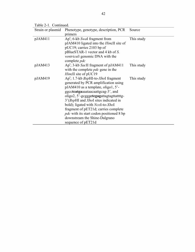
42
Table 2-1. Continued. Strain or plasmid Phenotype, genotype, description, PCR
primers Source
pJAM411 Apr; 6-kb SwaI fragment from pJAM410 ligated into the HincII site of pUC19; carries 2103 bp of pBlueSTAR-1 vector and 4 kb of S. ventriculi genomic DNA with the complete pdc
This study
pJAM413 Apr; 3-kb SacII fragment of pJAM411 with the complete pdc gene in the HincII site of pUC19
This study
pJAM419 Apr; 1.7-kb BspHI-to-XhoI fragment generated by PCR amplification using pJAM410 as a template, oligo1, 5’-ggcctcatgaaaataacaattgcag-3’, and oligo2, 5’-gcgggctcgagattagtagttattttg-3’(BspHI and XhoI sites indicated in bold); ligated with NcoI-to-XhoI fragment of pET21d; carries complete pdc with its start codon positioned 8 bp downstream the Shine-Dalgrano sequence of pET21d
This study

43
Table 2-2. Amino acid composition of PDC proteins. Composition expressed as % residues per mol enzyme predicted from the gene sequence (g) or chemically determined from the purified enzyme (e). Abbreviations: Sv, Sarcina ventriculi PDC; Sc, Saccharomyces cerevisiae PDC1; Zm, Zymomonas mobilis PDC; ND, not determined. References: Sv(e) (132), Sv(g) (this study), Sc(g) (99), Zm (g) (113). Amino Acid
Composition (mol%) Sv(e) Sv(g) Sc(g) Zm(g)
Asx 8.7 9.8 10.2 10.2 Glx 10.7 12.0 8.9 8.6 Ser 5.5 6.5 6.0 4.2 Gly 7.3 7.1 7.8 8.1 His 1.1 1.6 2.0 2.1 Arg 4.1 4.0 2.7 3.0 Thr 6.1 6.5 7.7 4.6 Ala 7.2 6.9 8.2 15.0 Pro 2.7 2.7 4.6 4.8 Tyr 3.2 4.0 3.1 3.9 Val 6.8 8.0 7.5 7.8 Met 2.1 2.7 2.4 1.9 Ile 6.1 7.1 6.6 4.9 Leu 7.8 8.2 9.7 8.8 Phe 16.2 4.7 4.2 3.2 Lys 4.4 6.9 6.2 6.3 Cys ND 0.9 1.1 1.2 Trp ND 0.5 1.3 1.2

44
Table 2-3. Codon usage of S. ventriculi (Sv) and Z. mobilis (Zm) pdc genes.
Amino Acid
Codon
Zm*
Sv
E.
coli† Ala GCA 28.1 32.5 20.1
GCC 35.1 0 25.5 GCG 12.3 1.8 33.6 GCU 73.8 34.4 15.3
Arg AGA 1.8 39.8 2.1 CGC 15.8 0 22.0 CGG 1.8 0 5.4 CGU 12.3 0 20.9
Asn AAC 47.5 19.9 21.7 AAU 10.5 27.1 17.7
Asp GAC 28.1 3.6 19.1 GAU 10.5 47.0 32.1
Cys UGC 28.1 1.8 6.5 UGU 10.5 7.2 5.2
Gln CAA 0 28.9 15.3 CAG 17.6 0 28.8
Glu GAA 61.5 85.0 39.4 GAG 3.5 5.4 17.8
Gly GGA 1.8 50.6 8.0 GGC 19.3 0 29.6 GGG 0 0 11.1 GGU 56.2 19.9 24.7
His CAC 8.8 3.6 9.7 CAU 14.1 12.7 12.9
Ile AUA 0 39.8 4.4 AUC 35.1 9.0 25.1 AUU 14.1 21.7 30.3
Leu CUA 0 7.2 3.9 CUC 19.3 0 11.1 CUG 33.4 0 52.6 CUU 12.3 14.5 11.0 UUA 1.8 59.7 13.9 UUG 5.4 0 13.7
Lys AAA 33.4 63.3 33.6 AAG 31.6 5.4 10.3
Met AUG 21.1 27.1 27.9 Phe UUC 31.6 18.1 16.6
UUU 0 28.9 22.3 Pro CCA 3.5 18.1 8.4
CCC 3.5 0 5.5 CCG 29.9 1.8 23.2 CCU 10.5 7.2 7.0

45
d
Table 2-3. Continue Amino AcidCodon
Zm*
Sv
E.
coli† Ser AGC 14.1 12.7 16.1
AGU 7.0 12.7 8.8 UCA 1.8 32.5 7.2 UCC 15.8 0 8.6 UCU 5.3 7.2 8.5
Thr ACA 0 34.4 7.1 ACC 28.1 0 23.4 ACG 10.5 0 14.4 ACU 8.8 30.7 9.0
Trp UGG 12.3 5.4 15.2 Tyr UAC 12.3 7.2 12.2
UAU 26.4 32.5 16.2 Val GUA 0 36.2 10.9
GUC 26.4 0 15.3 GUG 7.0 0 26.4 GUU 43.9 43.4 18.3
*Codon usage for amino acids represented as frequency per thousand bases. Stop codons are not indicated. CGA and AGG codons for Arg, UCG for Ser, and GGG for Gly were not used for either of the pdc genes. Abbreviations: Zm, Zymomonas mobilis; Sv, Sarcina ventriculi. †Average usage in frequency per thousand bases for genes in E. coli K-12. Highlighted
are codons for accessory tRNAs essential for high-level synthesis of S. ventriculi PDC in recombinant E. coli.
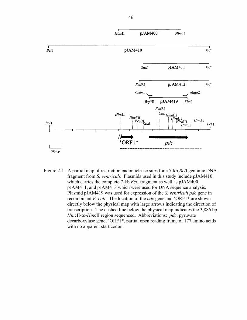
46
Figure 2-1. A partial map of restrictiofragment from S. ventricuwhich carries the completpJAM411, and pJAM413 Plasmid pJAM419 was usrecombinant E. coli. The directly below the physicatranscription. The dashedHincII-to-HincII region sedecarboxylase gene; ‘ORFwith no apparent start cod
‘
n endonuclease sites for a 7-kb BclI genomic DNA li. Plasmids used in this study include pJAM410 e 7-kb BclI fragment as well as pJAM400, which were used for DNA sequence analysis. ed for expression of the S. ventriculi pdc gene in location of the pdc gene and ‘ORF1* are shown l map with large arrows indicating the direction of line below the physical map indicates the 3,886 bp quenced. Abbreviations: pdc, pyruvate 1*, partial open reading frame of 177 amino acids
on.

47
-35 -10 AAATTTAAAAATAACATCAGATAAATCGTTTATATTAATTTTTACTAAAAGCTATTTAAA 60 ttgaca-------N17-------tataat SD GGTGTATTATATATACATAGTTTATCTTATAAATAAAAAATGAATTGGAGGAAATACATA 120 ATGAAAATAACAATTGCAGAATACTTATTAAAAAGATTAAAAGAAGTAAATGTAGAGCAT 180 M K I T I A E Y L L K R L K E V N V E H 20 M K I I I A E Y L L K R L K E V N V E H ATGTTTGGAGTTCCTGGAGATTATAACTTAGGATTTTTAGATTATGTTGAAGATTCTAAA 240 M F G V P G D Y N L G F L D Y V E D S K 40 M F G V P G D Y N L G F L D Y V GATATTGAATGGGTTGGAAGCTGTAATGAACTTAATGCAGAATATGCAGCAGATGGATAT 300 D I E W V G S C N E L N A E Y A A D G Y 60 GCAAGACTTAGAGGATTTGGTGTAATACTTACAACTTATGGAGTTGGTTCACTTAGTGCA 360 A R L R G F G V I L T T Y G V G S L S A 80 ATAAATGCTACAACAGGTTCATTTGCAGAAAATGTTCCAGTATTACATATATCAGGTGTA 420 I N A T T G S F A E N V P V L H I S G V 100 CCATCAGCTTTAGTTCAACAAAACAGAAAGCTAGTTCACCATTCAACTGCTAGAGGAGAA 480 P S A L V Q Q N R K L V H H S T A R G E 120 TTCGACACTTTTGAAAGAATGTTTAGAGAAATAACAGAATTTCAATCAATCATAAGCGAA 540 F D T F E R M F R E I T E F Q S I I S E 140 TATAATGCAGCTGAAGAAATCGATAGAGTTATAGAATCAATATATAAATATCAATTACCA 600 Y N A A E E I D R V I E S I Y K Y Q L P 160 GGTTATATAGAATTACCAGTTGATATAGTTTCAAAAGAAATAGAAATCGACGAAATGAAA 660 G Y I E L P V D I V S K E I E I D E M K 180 CCGCTAAACTTAACTATGAGAAGCAACGAGAAAACTTTAGAGAAATTCGTAAATGATGTA 720 P L N L T M R S N E K T L E K F V N D V 200 AAAGAAATGGTTGCAAGCTCAAAAGGACAACATATTTTAGCTGATTATGAAGTATTAAGA 780 K E M V A S S K G Q H I L A D Y E V L R 220 GCTAAAGCTGAAAAAGAATTAGAAGGATTTATAAATGAAGCAAAAATCCCAGTAAACACT 840 A K A E K E L E G F I N E A K I P V N T 240 Figure 2-2. Nucleic acid and predicted amino acid sequence of the S. ventriculi pdc gene.
DNA is shown in the 5’- to 3’-direction. Predicted amino acid sequences are shown in single-letter code directly below the first base of each codon. The N-terminal sequence previously determined for the purified PDC protein is shown directly below the sequence predicted for PDC. A putative promoter is double underlined with the –35 and –10 eubacterial promoter consensus sequence indicated in lower-case letters below the DNA sequence. A presumed ribosome-binding site is underlined. The translation stop codon is indicated by an asterisk. A stem-loop structure which may facilitate ρ-independent transcription termination is indicated by arrows below the DNA sequence.
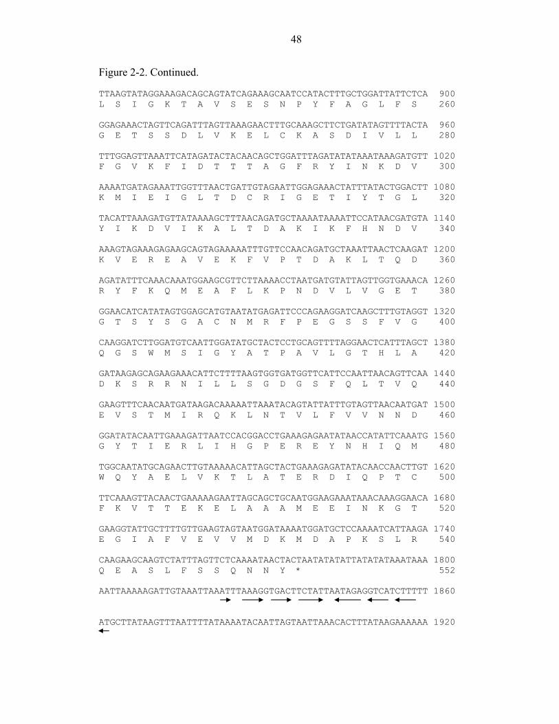
48
Figure 2-2. Continued. TTAAGTATAGGAAAGACAGCAGTATCAGAAAGCAATCCATACTTTGCTGGATTATTCTCA 900 L S I G K T A V S E S N P Y F A G L F S 260 GGAGAAACTAGTTCAGATTTAGTTAAAGAACTTTGCAAAGCTTCTGATATAGTTTTACTA 960 G E T S S D L V K E L C K A S D I V L L 280 TTTGGAGTTAAATTCATAGATACTACAACAGCTGGATTTAGATATATAAATAAAGATGTT 1020 F G V K F I D T T T A G F R Y I N K D V 300 AAAATGATAGAAATTGGTTTAACTGATTGTAGAATTGGAGAAACTATTTATACTGGACTT 1080 K M I E I G L T D C R I G E T I Y T G L 320 TACATTAAAGATGTTATAAAAGCTTTAACAGATGCTAAAATAAAATTCCATAACGATGTA 1140 Y I K D V I K A L T D A K I K F H N D V 340 AAAGTAGAAAGAGAAGCAGTAGAAAAATTTGTTCCAACAGATGCTAAATTAACTCAAGAT 1200 K V E R E A V E K F V P T D A K L T Q D 360 AGATATTTCAAACAAATGGAAGCGTTCTTAAAACCTAATGATGTATTAGTTGGTGAAACA 1260 R Y F K Q M E A F L K P N D V L V G E T 380 GGAACATCATATAGTGGAGCATGTAATATGAGATTCCCAGAAGGATCAAGCTTTGTAGGT 1320 G T S Y S G A C N M R F P E G S S F V G 400 CAAGGATCTTGGATGTCAATTGGATATGCTACTCCTGCAGTTTTAGGAACTCATTTAGCT 1380 Q G S W M S I G Y A T P A V L G T H L A 420 GATAAGAGCAGAAGAAACATTCTTTTAAGTGGTGATGGTTCATTCCAATTAACAGTTCAA 1440 D K S R R N I L L S G D G S F Q L T V Q 440 GAAGTTTCAACAATGATAAGACAAAAATTAAATACAGTATTATTTGTAGTTAACAATGAT 1500 E V S T M I R Q K L N T V L F V V N N D 460 GGATATACAATTGAAAGATTAATCCACGGACCTGAAAGAGAATATAACCATATTCAAATG 1560 G Y T I E R L I H G P E R E Y N H I Q M 480 TGGCAATATGCAGAACTTGTAAAAACATTAGCTACTGAAAGAGATATACAACCAACTTGT 1620 W Q Y A E L V K T L A T E R D I Q P T C 500 TTCAAAGTTACAACTGAAAAAGAATTAGCAGCTGCAATGGAAGAAATAAACAAAGGAACA 1680 F K V T T E K E L A A A M E E I N K G T 520 GAAGGTATTGCTTTTGTTGAAGTAGTAATGGATAAAATGGATGCTCCAAAATCATTAAGA 1740 E G I A F V E V V M D K M D A P K S L R 540 CAAGAAGCAAGTCTATTTAGTTCTCAAAATAACTACTAATATATATTATATATAAATAAA 1800 Q E A S L F S S Q N N Y * 552 AATTAAAAAGATTGTAAATTAAATTTAAAGGTGACTTCTATTAATAGAGGTCATCTTTTT 1860 ATGCTTATAAGTTTAATTTTATAAAATACAATTAGTAATTAAACACTTTATAAGAAAAAA 1920

49
Figure 2-3. Multiple amino acid sequence alignment of S. ventriculi PDC with other PDC protein sequences. Abbreviations with GenBank or SwissProt accession numbers: Sce, S. cerevisiae P06169; Sve, S. ventriculi; Zmo, Z. mobilis P06672. Identical amino acid residues are shaded in inverse print. Functionally conserved and semi-conserved amino acid residues are shaded in gray. Dashes indicate gaps introduced in protein sequence alignment. Indicated above the sequences are amino acid residues within a 0.4 nm distance of the Mg2+ and TPP binding site of yeast PDC1 (84)(▼), the Cys221 residue originally postulated to be required for pyruvate activation of yeast PDC1 (●), and the Tyr157 and Arg224 residues which form hydrogen bonds with allosteric activators such as pyruvamide (■). The underlined sequence is a conserved motif identified in TPP-dependent enzymes (216).
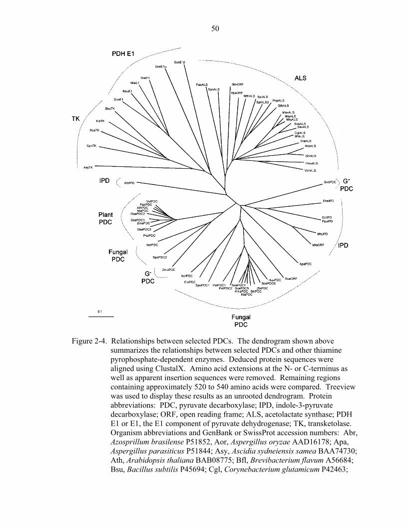
50
Figure 2-4. Relationships between selected PDCs. The dendrogram shown above
summarizes the relationships between selected PDCs and other thiamine pyrophosphate-dependent enzymes. Deduced protein sequences were aligned using ClustalX. Amino acid extensions at the N- or C-terminus as well as apparent insertion sequences were removed. Remaining regions containing approximately 520 to 540 amino acids were compared. Treeview was used to display these results as an unrooted dendrogram. Protein abbreviations: PDC, pyruvate decarboxylase; IPD, indole-3-pyruvate decarboxylase; ORF, open reading frame; ALS, acetolactate synthase; PDH E1 or E1, the E1 component of pyruvate dehydrogenase; TK, transketolase. Organism abbreviations and GenBank or SwissProt accession numbers: Abr, Azosprillum brasilense P51852, Aor, Aspergillus oryzae AAD16178; Apa, Aspergillus parasiticus P51844; Asy, Ascidia sydneiensis samea BAA74730; Ath, Arabidopsis thaliana BAB08775; Bfl, Brevibacterium flavum A56684; Bsu, Bacillus subtilis P45694; Cgl, Corynebacterium glutamicum P42463;

51
Cpn, Chlamydophila pneumoniae H72020; Dra, Deinococcus radiodurans A75387 (ALS), A75541 (E1); Ecl, Enterobacter cloacae P23234; Eco, E. coli CAA24740; Ehe, Erwinia herbicola AAB06571; Eni, Aspergillus (Emericella) nidulans P87208; Fan, Fragaria x ananassa AAG13131; Ghi, Gossypium hirsutum S60056; Gth, Guillardia theta NP_050806; Huv, Hanseniaspora uvarum P34734; Kla, Kluyveromyces lactis Q12629 (PDC), Q12630 (TK); Kma, Kluyveromyces marxianus P33149; Mav, Mycobacterium avium Q59498; Mja, Methanococcus jannaschii Q57725; Mle, Mycobacterium leprae CAC31122 (ORF), 033112 (ALS), CAC30602 (E1); Mth, Methanobacterium thermoautotrophicum A69081 (ORF), C69059 (ALS); Mtu, Mycobacterium tuberculosis E70814 (IPD), 053250 (ALS); Ncr, N. crassa P33287; Nta, Nicotiana tabacum P51846 (PDC), P09342 (ALS); Osa, Oryza sativa P51847 (PDC1), P51848 (PDC2), P51849 (PDC3); Pae, Pseudomonas aeruginosa G83123; Pmu, Pasteurella multocida AAK03712; Pop, Prophyra purpurea NP_053940; Ppu, Pseudomonas putida AAG00523; Psa, P. sativum P51850; Pst, Pichia stipitis AAC03164 (PDC1), AAC03165 (PDC2); Rca, Rhodobacter capsulatus JC4637; Reu, Ralstonia eutropha Q59097; Sav, Streptomyces avermitilis AAA93098; Sce, S. cerevisiae P06169 (PDC1), P16467 (PDC5), P26263 (PDC6), Q07471 (ORF), NP_011105 (E1α), NP_009780 (E1β); Sco, Streptomyces coelicolor T35828; Skl, Saccharomyces kluyveri AAF78895; Spl, Spirulina platensis P27868; Spo, Schizosaccharomyces pombe Q09737 (PDC1), Q92345 (PDC2); Sve, S. ventriculi AF354297; Syn, Synechocystis sp. BAA17984; Vch, Vibrio cholerae A82375; Vvi, Vitis vinifera AAG22488; Zma, Zea mays P28516; Zbi, Zygosaccharomyces bisporus CAB65554; Zmo, Z. mobilis P06672. Scale bar represents 0.1 nucleotide substitutions per site.

52
Newly discovered Gram-positive PDC-like proteins
Figure 2-5. Relationships between pyruvate decarboxylase (PDC), indole pyruvate decarboxylase (IPD), α-ketoisocaproate decarboxylase (KID), and homologues (ORF). Abbreviations: Abr, Azospirillum brasilense; Ali, Azospirillum lipoferum; Aor, Aspergillus oryzae; Apa, Aspergillus parasiticus; Ape, A. pasteurianus; Ath, Arabidopsis thaliana; Ban, Bacillus anthracis; Bce, Bacillus cereus; Bcp, Burkholderia cepacia; Bfu, Burkholderia fugorum; Cac, Clostridium acetobutylicum; Cgl, Candida glabrata; Ecl, Enterobacter cloacae; Eni, Emericella nidulans; Fan, Fragaria x ananassa; Huv, Hanseniaspora uvarum; Kla, Kluyveromyces
53
lactis; Kma, Kluyveromyces marxianus; Kpn, Klebsiella pneumoniae; Lla, Lactococcus lactis; Mac, Methanosarcina acetovorans; Mba, Methanosarcina barkeri; Mbo, Mycobacterium bovis; Mle, Mycobacterium leprae; Mlo, Mesorhizobium loti; Mpe, Mycoplasma penetrans; Mtu, Mycobacterium tuberculosis; Ncr, Neurospora crassa; Npu, Nostoc punctiforme; Nta, Nicotiana tabacum; Osa, Oryza sativa; Pag, Pantoea agglomerans; Ppu, Pseudomonas putida; Psa, Pisum sativum; Pst, Pichia stipitis; Rpl, Rhodopseudomonas palustris; Rru, Rhodospirillum rubrum; Sau, Staphylococcus aureus; Sba, Saccharomyces bayanus; Sep, Staphylococcus epidermidis; Sty, Salmonella typhimurium; Styp, Salmonella typhi; Sce, S. cerevisiae; Skl, Saccharomyces kluyveri; Spo, Schizosaccharomyces pombe; Sve, S. ventriculi; Vvi, Vitis vinifera; Zma, Zea mays; Zbi, Zygosaccharomyces bisporus; Zmo, Z. mobilis; Zpa; Z. palmae; G+, Gram-positive; G-, Gram-negative; C, cyanobacteria; A, archaea; P, plants; F, fungi and yeast; Bar, 0.1 nucleotide substitutions per site; ORF, open reading frame with no enzyme information.
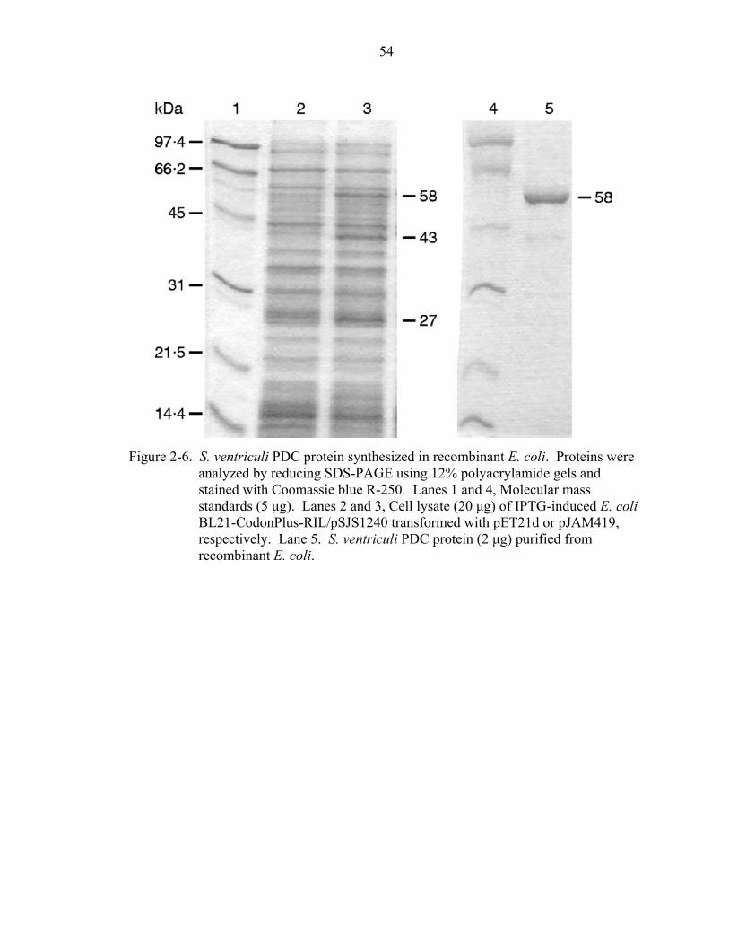
54
Figure 2-6. S. ventriculi PDC protein synthesized in recombinant E. coli. Proteins were
analyzed by reducing SDS-PAGE using 12% polyacrylamide gels and stained with Coomassie blue R-250. Lanes 1 and 4, Molecular mass standards (5 µg). Lanes 2 and 3, Cell lysate (20 µg) of IPTG-induced E. coli BL21-CodonPlus-RIL/pSJS1240 transformed with pET21d or pJAM419, respectively. Lane 5. S. ventriculi PDC protein (2 µg) purified from recombinant E. coli.

55
Figure 2-7. Pyruvate dependant activity of the S. ventriculi PDC purified from recombinant E. coli. The data represent mean results from triplicate determinations of PDC activity by the ADH coupled assay using 1 µg of purified enzyme in 1 ml final assay volume as described in methods section. SvPDC assayed in K-MES ( ) and Maleate buffer (O).

56
CHAPTER 3 OPTIMIZATION OF Sarcina ventriculi PDC EXPRESSION IN A GRAM POSITIVE
HOST
Introduction
Our previous work has shown that the PDC from the Gram-positive bacterium S.
ventriculi (SvPDC) was poorly expressed in E. coli (133). Addition of accessory tRNAs
was necessary for a ten-fold increase in protein production. The elevated levels of
protein produced upon addition of accessory tRNAs facilitated the purification of the
SvPDC. While the protein produced and purified from E. coli enabled the initial
characterization of SvPDC, it was not the optimal host due to low levels of SvPDC
produced.
Therefore, it was necessary to determine if there was a host that had similar codon
usage to that of SvPDC so that limited tRNAs would not hinder over expression of the
protein. Because S. ventriculi is a low-G+C Gram-positive bacterium, it was reasoned
that a low-G+C Gram-positive host would be most suitable for expression of this protein
in large quantities.
The Gram-positive bacterium Bacillus megaterium WH320 was examined as a
potential host for engineering high-level synthesis of PDC. B. megaterium has several
advantages over other bacilli including the availability of a shuttle plasmid (pWH1520)
for xylose inducible expression of foreign genes cloned downstream of the xylA
promoter. Another advantage is that the alkaline proteases, often responsible for the
degradation of foreign proteins in recombinant bacilli, are not produced in B. megaterium
6
5
57
(220, 221). In contrast to E. coli, the tRNAs for AUA and AGA are abundant in B.
megaterium suggesting that factors limiting PDC production will be optimal in this host.
In this study, SvPDC was over expressed in and purified from B. megaterium.
The biochemical characteristics and optimum conditions for activity of the SvPDC
enzyme purified from B. megaterium were determined. Due to the expression of SvPDC
in B. megaterium, a plasmid was also constructed containing a Gram-positive ethanol
production operon in which production of SvPDC and Geobacillus stearothermophilus
alcohol dehydrogenase (ADH) (222) are transcriptionally coupled and expression of these
proteins was demonstrated.

58
Materials and Methods
Materials
Biochemicals were purchased from Sigma Chemical Company (St. Louis, MO).
Other organic and inorganic analytical-grade chemicals were purchased from Fisher
Scientific (Marietta, GA). Restriction enzymes were from New England Biolabs
(Beverly, MA). Oligonucleotides were obtained from QIAgen Operon (Valencia, CA).
Bacillus megaterium Protein Expression System was purchased from MoBiTec (Marco
Islands, FL). Rnase-free water and solutions were obtained from Ambion (Austin, TX).
Bacterial Strains and Media
Strains and plasmids used in this study are listed in Table 3-1. E. coli DH5α was
used for routine recombinant DNA experiments. B. megaterium WH320 was used for
protein production. Growth and transformation of B. megaterium were performed
according to the manufacturer (MoBiTec). All strains were grown in Luria-Bertani (LB)
medium supplemented with antibiotics as appropriate (ampicillin 100 mg per liter, or
tetracycline 12.5 mg per liter) at 37°C and 200 rpm.
DNA Isolation
Plasmid DNA was isolated and purified from E. coli using the QIAprep Spin
Miniprep Kit (QIAgen). DNA was eluted from 0.8% (w/v) SeaKem GTG agarose
(BioWhittaker Molecular Applications) gels using the QIAquick gel elution kit
(QIAgen).
Cloning of the Sarcina ventriculi pdc Gene Into Expression Vector pWH1520
Plasmid pJAM420 was constructed using the following methods. The BspEI-to-
XbaI fragment of plasmid pJAM419 was ligated with 7.7-kb SpeI-to-XmaI fragment of

59
plasmid vector pWH1520. This resulted in generation of the B. megaterium expression
plasmid pJAM420 that carried the S. ventriculi pdc gene, along with the Shine-Dalgrano
site and T7 transcriptional terminator of the original pET21d vector. The pdc gene was
positioned to interrupt the B. megaterium xylA gene (xylA’) of plasmid pWH1520 and to
generate a stop codon within xylA’. The Shine-Dalgrano site of the inserted pdc gene was
positioned directly downstream of the xylA’ stop codon to allow for translational coupling
in which the ribosomes would presumably terminate at the stop codon for xylA’ and then
reinitiate at the pdc start codon.
Gram-positive Ethanol Operon (PET).
To construct the Gram-positive PET operon, the HindIII-to-MfeI fragment of
pLOI1742 containing the adh gene from G. stearothermophilus (222) was blunt-end
ligated into the BlpI site of pJAM420 using Vent Polymerase (New England Biolabs).
This resulted in the generation of plasmid pJAM423 which was designed to facilitate the
translational coupling of the S. ventriculi pdc gene with the G. stearothermophilus adh
gene. The xylA promoter is upstream of the Svpdc and the terminator now follows the
adh gene.
Protoplast Formation and Transformation of B. megaterium.
A 1.0% (v/v) inoculum of B. megaterium WH320 cells was grown in LB to an
OD600nm of 0.6 units (early-log phase). Protoplasts were formed according to Puyet et al.
(223) with the following variations. Cells were treated with 10 µg per ml lysozyme for
20 min at 37°C. Protoplasts were stored at –70°C. Transformation of the protoplasts was
performed according to the B. megaterium protein expression kit manual (MoBiTec).
60
Production of SvPDC In Recombinant Hosts.
Production of SvPDC in E. coli was performed as previously described (133).
SvPDC protein was synthesized in B. megaterium WH320 cells using pJAM420. A 1.0%
(v/v) overnight inoculum of recombinant B. megaterium cells was grown in LB
supplemented with tetracycline to an OD600nm of about 0.3 units (early-log phase).
Transcription from the xylA’ promoter was induced with 0.5% (w/v) xylose for 3 h. Cells
were harvested by centrifugation at 5000 × g (10 min, 4°C) and stored at -70°C.
Purification of the S. ventriculi PDC Protein.
All purification buffers contained 1 mM TPP and 1mM MgSO4 unless indicated
otherwise. Purification of SvPDC from E. coli was performed as previously described
(133). Recombinant B. megaterium cells (15 g wet wt) were thawed in 6 volumes (w/v)
of 50 mM Na-PO4 buffer at pH 6.5 (Buffer A) and passed through a French pressure cell
at 20,000 lb per in2. Cell debris was removed by centrifugation at 16,000 × g (20 min,
4ºC). Supernatant was filtered through a .45 µm membrane. Filtrate (372.3 mg protein)
was applied to a Q Sepharose Fast Flow 26/10 column (Pharmacia) that was equilibrated
with Buffer A. A linear gradient was applied from 0 mM to 400 mM NaCl. Fractions
containing PDC activity eluted at 250 to 300 mM NaCl were pooled. Pooled fractions
were applied to a 5 ml Bio-scale hydroxyapatite type I column (BioRad) that was
equilibrated with 5 mM Na-PO4 buffer at pH 6.5 (Buffer B). The column was washed
with 15 ml Buffer B and developed with a linear Na-PO4 gradient (5 to 500 mM Na-PO4
at pH 6.5 in 75 ml). Protein fractions with PDC activity were eluted at 300 to 530 mM
Na-PO4 and were pooled (1mg protein per ml). For further purification, portions of this
material (0.25 to 0.5 ml) were applied to a Superdex 200 HR 10/30 column (Pharmacia)

61
equilibrated in 50 mM Na-PO4 at pH 6.5 with 150 mM NaCl and 10% glycerol in the
presence or absence of 1 mM MgSO4 and 1 mM TPP.
Activity Assays and Protein Electrophoresis Techniques.
PDC activity was assayed by monitoring the pyruvic acid-dependant reduction of
NAD+ with alcohol dehydrogenase (ADH) as a coupling enzyme at pH 6.5, as previously
described (224). Sample was added to a final volume of 1 ml containing 0.15 mM
NADH, 0.1 mM TPP, 50.0 mM pyruvate, and 10 U ADH in 50 mM K-MES buffer at pH
6.5 with 5 mM MgCl2. The reduction of NAD+ was monitored in a 1 cm path length
cuvette at 340 nm over a 5 min period using a BioRad SmartSpec 300 (BioRad). Protein
concentration was determined using BioRad Protein assay dye with bovine serum
albumin as the standard according to supplier (BioRad).
The pH optimum of SvPDC was assayed in buffers suitable to maintain the
desired pH. The temperature optimum of SvPDC was assayed using a Beckman DU640
(Beckman) spectrophotometer with a circulating water bath.
Thermostability of SvPDC was assayed by incubating purified enzyme in lysis
buffer at a concentration of 0.02 µg of protein per µl for 90 min. After incubation,
samples were assayed at room temperature.
Molecular masses were estimated by reducing and denaturing SDS-PAGE using
12% polyacrylamide gels. Proteins were stained using the Rapid Fairbanks method
(225). The molecular mass standards were phosphorylase b (97.4 kDa), serum albumin
(66.2 kDa), ovalbumin (45 kDa), carbonic anhydrase (31 kDa), trypsin inhibitor (21.5
kDa), and lysozyme (14.4 kDa).

62
Results
SvPDC Expression Vector for B. megaterium.
Previous studies have shown that SvPDC is poorly expressed in E. coli (133).
Because SvPDC is from a Gram-positive bacterium, we decided to use a Gram-positive
bacterial expression system for high-level production of SvPDC. A fragment of plasmid
pJAM419, used previously for expression of SvPDC in E. coli (133), was isolated that
contained a Shine-Dalgrano sequence, the S. ventriculi pdc gene, and the T7 terminator.
This fragment was cloned into pWH1520 in such a way that the xylose isomerase gene
(xylA) of pWH1520 was truncated to form a stop codon after 30 codons. The Shine-
Dalgrano sequence upstream of SvPDC was positioned so that a xylose-inducible
transcriptional coupling occurred between xylA’ and Svpdc. The resulting plasmid,
pJAM420, was used for expression of SvPDC in B. megaterium.
Production and Purification of SvPDC from B. megaterium.
Based on SDS-PAGE, expression of SvPDC is notably higher when expressed in
B. megaterium compared to E. coli (Figure 3-1). The high levels of SvPDC protein
produced in B. megaterium facilitated a 22-fold purification of the protein from this host,
with 8.35 mg purified protein from 15 g of cells (wet wt.) (Table 3-2). This is in contrast
to purification of SvPDC from E. coli, which began with 14.8 g of cells and only yielded
0.2 mg purified protein (unpublished data).
Purified SvPDC from B. megaterium was determined to be a 235 kDa
homotetramer of 58 kDa subunits as determined by Superdex 200 gel filtration
chromatography and SDS-12% PAGE electrophoresis. These results correlate with
previous studies (132, 133).

63
Determination of Optimum Conditions for SvPDC Activity.
It is important when assaying any enzyme to determine its optimum conditions.
PDCs are routinely assayed at pH 6.5 (178, 218) and pH 6.0 (94, 112). While these pH
values are acceptable for the Z. mobilis and S. cerevisiae PDC proteins, they may give
misleading kinetic values for SvPDC. Our research found that the recombinant SvPDC
from B. megaterium had a pH optimum in the range of pH 6.5 to pH 7.4 (Figure 3-2).
This pH optimum is quite high when compared to the PDCs from Z. mobilis (pH 6.0), S.
cerevisiae PDC1 (pH 5.4-5.8), A. pasteurianus (pH 5.0-5.5), and Z. palmae (pH 5.5-6.0)
(113, 131, 226). The pH optimum of SvPDC purified from recombinant E. coli has
previously been shown to be between pH 6.3 to 6.7 (131), which is higher than the other
bacterial PDCs and also different to the pH optimum determined for the B. megaterium
purified protein.
The temperature optimum of the SvPDC from B. megaterium was determined to
be 32°C (Figure 3-3). This temperature differs greatly from the Z. mobilis, Z. palmae,
and A. pasteurianus PDCs, which have temperature optima of 60°C (131). There is,
however, an approximately 2.5-fold increase in activity of the SvPDC at 32°C compared
to room temperature. This increase in activity is comparable to that observed when the
Gram-negative bacterial PDCs were assayed at their optimal temperatures (131).
Kinetics of SvPDC Produced in B. megaterium.
The recombinant SvPDC from B. megaterium displayed sigmoidal kinetics
(Figure 3-4). The recombinant SvPDC from B. megaterium had a Km of 3.9 mM for
pyruvate and a Vmax of 98 U per mg of protein when assayed at pH 6.5 and room
temperature. When assayed at optimal conditions, 32°C and pH 6.72, there was an

64
increase in both Km (6.3 mM for pyruvate) and Vmax (172 U per mg protein). These
results suggest that a change in conformation mediated by an increase in pH and/or
temperature reduces the affinity of the enzyme for pyruvate but increases the overall
activity of the enzyme. Further study is necessary to determine the cause of this
phenomenon.
Thermostability of SvPDC Produced in B. megaterium.
Previous studies showed that SvPDC is not as thermostable as the other bacterial
PDC proteins (131, 133). In order to determine if production of the SvPDC protein in a
sub optimal host was responsible for this, thermostability of SvPDC produced in E. coli
was compared to that produced in B. megaterium (Figure 3-5). When assayed for
thermostability, the SvPDC produced in B. megaterium retained 30% activity after
incubation at 50°C while the protein purified from E. coli only had 0.95% residual
activity after incubation at 50°C. These results indicate that SvPDC is more thermostable
when produced in B. megaterium compared to E. coli. Misincorporation of amino acids
due to use of rare codons and/or misfolding of the SvPDC protein may have occurred
when the enzyme was produced in E. coli and may account for this reduction in
thermostability.
During the biochemical characterization of SvPDC, we discovered that pH had a
drastic effect on the thermostability of this enzyme (Figure 3-6). While the optimal pH
for activity of the SvPDC is pH 6.72, this is not optimal for its thermostability. At pH 6.5
the SvPDC enzyme has only 3% of original activity remaining after incubation at 60°C
while samples incubated at pH 5.0 to pH 5.5 have 94% to 97% activity remaining. It was

65
also determined that SvPDC retained 100% activity when stored at pH 5.5 for two weeks
compared to 62 % when stored at 4°C at pH 6.5 (data not shown).
Generation of a Gram-positive Ethanol Production Operon.
B. megaterium WH320 is capable of growth when tested in xylose minimal
medium. The strain is also able to grow at temperatures up to 42°C and at a low pH of
5.0. This strain appears to be a suitable candidate to perform preliminary tests on ethanol
production with a portable pyruvate to ethanol operon (PET) and may prove useful in
large-scale ethanol production under acidic conditions. To construct a Gram-positive
PET operon, the adh gene from G. stearothermophilus (222) was cloned behind the S.
ventriculi pdc gene in the B. megaterium pWH1520 expression vector. This vector was
chosen based on successful overproduction of S. ventriculi PDC (Figure 3-7). The
resulting PET plasmid, pJAM423, was transformed into B. megaterium. After xylose
induction, a considerable portion of the cell lysate of this strain was composed of the S.
ventriculi PDC and G. stearothermophilus ADH proteins (Figure 3-7). The ethanol
production of this construct was tested in the presence of 0.5% xylose. HPLC analysis
showed that ethanol production was doubled from that of a strain with pWH1520 alone,
but levels were still quite low (20mM)(data not shown). PDC has already been shown to
be very active in cell lysate, but further analysis needs to be performed to determine if the
ADH is active.
Discussion
The SvPDC protein is poorly expressed in recombinant E. coli (133). Therefore,
we reasoned that a host more similar to S. ventriculi might express this PDC at higher
levels. B. megaterium was chosen as a host because it has several benefits over other

66
Gram-positive expression systems. These include a xylose inducible expression vector
and absence of alkaline proteases that are often responsible for degradation of foreign
proteins (220, 221). Augmentation of the host, B. megaterium, with accessory tRNAs
was not necessary for high-level SvPDC production. This high yield of SvPDC protein
facilitated the 22-fold purification. The SvPDC protein was more active when produced
in B. megaterium compared to E. coli. We believe that the difference in activity is
primarily due to differences in the rate of misincorporation of amino acids based on
codon usage.
The SvPDC protein produced in B. megaterium has a higher Vmax (98 U per mg
protein) at RT than when produced by E. coli (66 U per mg protein). The SvPDC
produced in B. megaterium is also more thermostable than the E. coli produced protein.
Choosing the correct host appears to have affected the quality of SvPDC protein that was
recovered. These results indicate that differences can occur in the biochemical properties
of recombinant protein based on host.
In this study, we discovered that the pH of the incubation buffer has an effect on
the thermostability of SvPDC. Low pH stabilized SvPDC at higher temperatures. These
results suggest that residues of SvPDC gain a charge between pH 5.0–5.5 that allows the
tetramer conformation to remain stable at higher temperatures. This is an important
discovery because it gives insight into residues that can be altered in future experiments
in order to engineer SvPDC to be more thermostable at cytosolic pH.
The current portable production of ethanol (PET) operon consists of the pdc and
adh genes from Zymomonas mobilis, a Gram-negative organism (24, 25, 129, 227). Past
research to engineer a Gram-positive host for ethanol production has focused on using

67
this PET operon, but these attempts have met with limited success (33-35, 228) primarily
due to poor expression of the PDC. We have shown that SvPDC is expressed at high
levels in B. megaterium, a Gram-positive host. Our construction and expression of the
Gram-positive ethanol production operon using the SvPDC and G. stearothermophilus
ADH has demonstrated that recombinant PDC and ADH production no longer limit
ethanol production in Gram-positive biocatalysts.
Our research shows that selection of host for recombinant production of proteins
can affect the quality and stability of the recombinant protein. We have also
demonstrated that SvPDC has qualities that make it unique among bacterial PDCs,
including its substrate activation and elevated pH optimum. SvPDC is the only bacterial
PDC that is not thermostable, but our results indicate that alteration of charged residues
may facilitate the engineering of thermostable SvPDC variants. Lastly, we have created a
Gram-positive ethanol production operon that will be useful in engineering future Gram-
positive hosts for ethanol production.

68
Table 3-1. Strains, plasmids, and primers used in Chapter 3. Strain or Plasmid Phenotype or genotype, PCR primers Source E. coli DH5α F- recA1 endA1 hsdR17 (rk
- mk+) supE44 thi-1
gyrA relA1 GibcoBRL (Gathers-burg, Md.)
E. coli BL21-CodonPlus-RIL
F– ompT hsdS(rB– mB
–) dcm+ Tetr gal λ (DE3) endA Hte [argU ileY leuW Camr] (an E.coli B strain)
Stratagene (La Jolla, CA.)
B. megaterium WH320
lac- xyl+ MoBiTec
pSJS1240 Spr; derivative of pACYC184 with E. coli ileX and argU
(219)
pET21d Apr; expression vector for replication in E. coli Novagen
pWH1520 Apr Tc r; shuttle expression vector for replication in E. coli and B. megaterium
(220)
pJAM419 Apr; pET21d derivative encoding SvPDC (133) pJAM420 Apr Tc r; 1.9-kb BspEI-to-XbaI fragment of
pJAM419 ligated with the SpeI-to-XmaI fragment of pWH1520; used for synthesis of SvPDC in B. megaterium
This study
pLOI1742 Plasmid containing the G. stearothermophilus adh gene
L. Yomano
pJAM423 Apr Tc r; 1.8-kb HindIII-to-MfeI fragment of pLOI1742 blunt-end ligated into the BlpI site of pJAM420; xylose-inducible Gram-positive ethanol production operon
This study

69
Table 3-2. Purification of SvPDC from B. megaterium.
Step Protein (mg) Sp. Act. (U per mg protein) Purification Fold Percent
Yield Cell Lysate 372.3 3.85 1.00 100 Q-Sepharose 38.26 20.34 5.28 54 Hydroxyapatite 15.70 24.83 6.45 27 Superdex 200 8.35 84.18 21.87 49

70
Figure 3-1. S. ventricul
megateriumpolyacrylamMolecular mBL21-CodoIPTG inducmegateriumuninduced,
97.4
66.2i .
ne
r
45
21.5
14.4
PDC protein synthesize Proteins were analyzedide gels and stained withass standard (5 µg). LanPlus-RIL transformed wd, respectively. Lanes 5WH320 transformed witespectively.
58
kDa
1 2 3 4 5 6d in recombinant E. col by reducing SDS-PAG Coomassie blue R-25es 2 and 3, Cell lysateith pJAM419/pSJS124
and 6, Cell lysate (20 µh pJAM420 xylose ind
58
i and B. E using 12%
0. Lanes 1 and 4, (20 µg) of E.coli 0 uninduced and g) of B. uced and

71
020406080
100120
4 6 8
pH
Sp. A
ct. (
U/m
g)
10
Figure 3-2. pH profile for S. ventriculi PDC activity.

72
0
50
100
150
200
0 10 20 30 40 5
Temperature °C
Sp. A
ct. (
U/m
g)
0
Figure 3-3. Effect of temperature on S. ventriculi PDC activity.

73
020406080
100120140
0 2 4 6 8 10 12 14
Pyruvate (mM)
Sp.A
ct. (
U/m
g)
Figure 3-4. Effect of pyruvate concentration on S. ventriculi PDC synthesized in
recombinant E. coli (■), and B. megaterium (▲) at 25°C and pH 6.5. S. ventriculi PDC at 32°C and pH 6.72 from recombinant B. megaterium (♦). The data represent mean results from triplicate determinations of PDC activity.

74
0%
20%
40%
60%
80%
100%
120%
40 50 60
Temperature (°C)
Rel
ativ
e A
ctiv
ity (U
/mg)
Figure 33-5. Thermostability of recombinant S. ventriculi PDC produced in B. megaterium (♦) and E. coli CodonPlus with plasmid pSJS1240 (■).

75
0%
20%
40%
60%
80%
100%
120%
140%
160%
0 10 20 30 40 50 60 70
Temperature (°C )
Rel
ativ
e A
ctiv
ity
Figure 3-6. Effect of pH on the thermostability of the S. ventriculi PDC produced in B. megaterium. Thermostability was tested at a pH 5.0 (□), pH 5.5 (♦), pH 6.5 (■), and pH 7.5 (▲).

76
1 2 3 4 kDa
97.4
66.2S. ventriculi PDC
45G. stearothermophilus ADH
31
21.5
14.4Figure 3-7. Induction of S. ventriculi PDC and G. stearothermophilus ADH in B.
megaterium. Proteins were seperated by reducing SDS-PAGE using 12% polyacrylamide gels and stained with Coomassie blue R-250. Lanes 1, Molecular mass standard (5 µg). Lanes 2, Cell lysate (20 µg) of B. megaterium transformed with pWH1520 induced with xylose. Lanes 3 and 4, Cell lysate (20 µg) of B. megaterium WH320 transformed with pJAM423 uninduced and xylose induced, respectively.

77
CHAPTER 4 EXPRESSION OF PDCs IN THE GRAM-POSITIVE BACTERIAL HOST,
B. megaterium
Introduction
PDC (PDC, EC 4.1.1.1) is a central enzyme in ethanol fermentation and catalyzes
the non-oxidative decarboxylation of pyruvate to acetaldehyde with release of carbon
dioxide. The acetaldehyde generated from this reaction is then converted to ethanol by
alcohol dehydrogenase (ADH, EC1.1.1.1). The recombinant production of these two
enzymes (PDC and ADH) converts intracellular pools of pyruvate to ethanol. The
current portable production of ethanol (PET) operon used to engineer this conversion
consists of the pdc and adh genes from Zymomonas mobilis, a Gram-negative organism
(24, 25, 129, 227). While this strategy has been highly successful in the modification of
Gram-negative bacteria for ethanol production, improvements in host strains are
necessary (4, 129).
To enhance the commercial competitiveness of biocatalysts for the large-scale
production of ethanol, the hosts must withstand low pH, high temperature, high salt, high
sugar, high ethanol, and various other harsh conditions. Many of these qualities are not
found in Gram-negative bacteria and must be introduced through metabolic engineering.
In contrast, Gram-positive bacteria naturally possess many desirable traits for the
industrial production of ethanol (228); however, modifying them for ethanol production
has met with only limited success. Several attempts to engineer the PET operon into
77
78
Gram-positive organisms have resulted in low levels of PDC activity and only small
elevations in ethanol production (33-35, 228).
Prior to this work, construction of PET operons for engineering high-level
synthesis of ethanol in recombinant Gram-positive bacteria has been limited by the
availability of bacterial pdc genes. Recently, however, the cloning and DNA sequence of
a pdc gene from the Gram-positive bacterium, S. ventriculi (Sv), was described (133).
Synthesis of the SvPDC protein in recombinant Escherichia coli was low but enhanced
by augmentation with accessory tRNAs (133). Based on these results, it is hypothesized
that reduced translation due to differences in codon usage can be a major factor in
limiting PDC production in recombinant bacterial hosts.
In this study, pdc genes from diverse organisms (i.e., S. ventriculi, Z. mobilis,
Acetobacter pasteurianus and Saccharomyces cerevisiae) with differing GC content were
expressed in recombinant Bacillus megaterium. Superior levels of active SvPDC were
produced in this host. Assessment of the mRNA transcript levels and rates of protein
degradation in these recombinant strains revealed that the differences in PDC were at the
level of protein synthesis. This is the first report of high level PDC production in a
recombinant Gram-positive host and reveals that SvPDC is an ideal candidate for the
metabolic engineering of ethanol production in this desirable group of organisms.
Materials and Methods
Materials
Biochemicals were purchased from Sigma (St. Louis, Mo.). Other organic and
inorganic analytical-grade chemicals were from Fisher Scientific (Atlanta, Ga.).
Restriction enzymes were from New England Biolabs (Beverly, Mass.).

79
Oligonucleotides were from QIAgen Operon (Valencia, Ca.) and Integrated DNA
Technologies (Coralville, Ind.). Bacillus megaterium Protein Expression System was
from MoBiTec (Marco Islands, Fla.). Rnase-free water and solutions were from Ambion
(Austin, Tx.).
Bacterial Strains and Media
Strains and plasmids used in this study are listed in Table 4-1. E. coli DH5α was
used for routine recombinant DNA experiments. B. megaterium WH320 was used for
PDC production, pulse-chase, and transcript analysis. Strains were grown in Luria-
Bertani (LB) medium unless otherwise indicated. Medium was supplemented with 2%
(wt/vol) glucose and antibiotics (ampicillin 100 mg per liter, kanamycin 30 mg per liter,
or tetracycline 15 mg per liter) as needed. All strains were grown at 37ºC and 200 rpm.
Isolated colonies of B. megaterium were grown overnight in liquid medium and used as a
1.0% (vol/vol) inoculum into fresh medium unless otherwise indicated.
Protoplast Formation and Transformation of B. megaterium.
B. megaterium WH320 was grown to an O.D.600nm of 0.6 units (early-log phase).
Protoplasts were generated according to Puyet et al. (223) with the following
modifications. Cells were treated with lysozyme (10 µg per ml) for 20 min. Protoplasts
were stored at –70°C and transformed according to MoBiTec.
DNA Isolation and Cloning
Plasmid DNA was isolated and purified from E. coli using the QIAprep Spin
Miniprep Kit (QIAgen). DNA was eluted from 0.8% (wt/vol) SeaKem GTG agarose
(Cambrex Corp., East Rutherford, NJ) gels using the QIAquick gel elution kit (QIAgen).
To generate the B. megaterium expression plasmids (pJAM420, pJAM430, pJAM432,

80
and pJAM435), similar strategies were used (Figure 4-1) (Table 4-1). For example,
plasmid pJAM420 was constructed as follows. A BspHI-to-XhoI DNA fragment with the
complete S. ventriculi pdc gene was generated by PCR amplification and cloned into the
NcoI and XhoI sites of plasmid pET21d (133). The 1.9-kb XbaI-to-BspEI DNA fragment
of the resulting plasmid (pJAM419) was ligated into the SpeI and XmaI sites of plasmid
pWH1520. This resulted in generation of a pWH1520-based expression plasmid
(pJAM420) that carried the S. ventriculi pdc gene, along with the Shine-Dalgrano site and
T7 transcriptional terminator of the original pET21d vector. The pdc gene was
positioned to interrupt the B. megaterium xylA gene (xylA’) of plasmid pWH1520 and to
generate a stop codon within xylA’. The Shine-Dalgrano site originally from pET21d of
upstream of the inserted pdc gene was positioned directly downstream of the xylA’ stop
codon to allow for translational coupling in which the ribosomes would terminate at the
stop codon for xylA’ and then reinitiate at the pdc start codon.
Production of PDC Proteins In Recombinant B. megaterium.
PDC proteins were independently synthesized in B. megaterium WH320 cells
using the expression plasmids described above. Cells were grown to an O.D.600 nm of 0.3
units (early-log phase). Transcription from the xylA’ promoter was induced by addition
of xylose (0.5% [wt/vol]). Cells were harvested after 3 h by centrifugation (5,000 × g, 10
min, 4°C) and stored at -80°C. Cell pellets (0.5 g) were thawed in 6 volumes (wt/vol) of
50 mM Na2HPO4 buffer at pH 6.5 containing 1 mM MgSO4 and 1 mM TPP. Cells were
passed through a French pressure cell at 20,000 lb per in2. Debris was removed by
centrifugation (16,000 × g, 20 min, 4°C). Cell lysate was immediately assayed for
activity.
81
Activity Assays and Protein Electrophoresis Techniques
PDC activity was assayed by monitoring the pyruvic acid-dependant reduction of
NAD+ with alcohol dehydrogenase (ADH) as a coupling enzyme at pH 6.5, as previously
described (115). Cell lysate (10 µl) was added to a final volume of 1 ml containing 0.15
mM NADH, 0.1 mM thiamine pyrophosphate, 50.0 mM pyruvate, and 10 U ADH in 50
mM K-MES buffer at pH 6.5 with 5 mM MgCl2. The reduction of NAD+ was monitored
in a 1 cm path length cuvette at 340 nm over a 5 min period using a BioRad SmartSpec
300 (BioRad). Protein concentration was determined using BioRad Protein assay dye
with bovine serum albumin as the standard according to supplier (BioRad).
Protein molecular masses were analyzed by reducing and denaturing SDS-PAGE
using 12% polyacrylamide gels that were stained by heating with Coomassie blue R-250
(225). Molecular mass standards were phosphorylase b (97.4 kDa), serum albumin (66.2
kDa), ovalbumin (45 kDa), carbonic anhydrase (31 kDa), trypsin inhibitor (21.5 kDa),
and lysozyme (14.4 kDa).
RNA Isolation
Cultures were grown in triplicate to an O.D.600 nm of 0.3 units (early-log phase).
Transcription of pdc was induced for 15 min with 0.5% (wt/vol) xylose. Total RNA was
isolated using the RNeasy miniprep kit. Samples were treated with lysozyme and On-
column DNase as recommended by supplier (QIAgen). The removal of DNA from RNA
samples was confirmed by performing PCR using Jumpstart Taq Readymix in the
absence of reverse transcriptase (Sigma). Quality and quantity of RNA were determined
by 0.8% agarose gel electrophoresis and absorbance at 260 nm, respectively.

82
RNA Quantifications
The MAXIscript T7 In vitro transcription kit (Ambion) was used to generate
transcript from the E. coli expression vectors (pJAM419, pJAM429, pJAM431, and
pScPDC1). Nuc-Away spin columns (Ambion) were used to remove unincorporated
nucleotides. RNA products expressed in vitro were used to generate standard curves of
absolute copy number for each experiment. Transcript levels were analyzed using
quantitative real time reverse transcriptase PCR with an Icycler (BioRad). Total RNA
(100 pg) was used as a template with the primers listed in Table 4-1. RNA
Quantification reactions were performed using the QuantiTect SYBR Green 1-step RT-
PCR kit according to supplier (QIAgen). All data had PCR efficiency of 90 to 100% and
were analyzed using the Icycler software version 3.0.6070 (BioRad) and Microsoft Excel.
Pulse Chase
Recombinant B. megaterium strains were grown in minimal medium (10 g
sucrose, 2.5 g K2HPO4, 2.5 g KH2PO4, 1.0 g (NH4)2HPO4, 0.2 g MgSO4·7H2O, 10 mg
FeSO4·7H2O, 7 mg MnSO4·H2O in 985 ml dH2O at pH 7.0) supplemented with
tetracycline (MM Tet) using a 1% (vol/vol) inoculum. Cells were grown to an O.D.600nm
of 0.3 units (early-log phase) and recombinant gene transcription was induced for 15 min
with 0.5% (wt/vol) xylose. Cells were harvested by centrifugation (5000 × g, 10 min,
25°C) and resuspended in 2 ml of MM Tet supplemented with 0.5% xylose and 50 µCi
per ml L-[35S]-methionine (DuPont-NEN). Cells were incubated for 15 min (37°C, 200
rpm) and harvested as above. Cell pellets were resuspended in MM Tet supplemented
with 0.5% xylose and 5mM L-methionine with or without chloramphenicol (15 mg per L)
and incubated (37°C, 200 rpm). Aliquots (0.5 ml) were withdrawn after 5, 10, 15, 30, 60,

83
90, 120, 150, and 180 min of incubation and immediately added to 50 µl stop solution (75
mM NaCl, 25 mM EDTA, 20 mM Tris pH 7.5, and 1 mg chloramphenicol per ml). Cells
were incubated on ice (5 min), harvested at 16,000 × g (10 min, 25ºC), and stored at –
80°C.
Cell pellets were subjected to 3 cycles of freeze-thaw (–80°C and 0°C) to weaken
the cell membrane. Pellets were resuspended to an O.D.600nm of 0.0134 units per µl Lysis
solution (75 mM NaCl, 25 mM EDTA, 20 mM Tris pH 7.5, and 0.2 mg lysozyme per ml)
and incubated (25ºC, 15 min). Samples (O.D.600nm of 0.02 units per lane) were boiled (20
min) in SDS-PAGE loading dye (BioRad) and separated by SDS-PAGE. Gels were dried
and exposed to X-ray film. A VersaDoc Model 1000 with Quantity One Software
(BioRad) was used for densitometric readings.
Results
Construction of Gram-positive PDC Expression Plasmids
Previous work suggested that codon usage effects the synthesis of PDCs in Gram-
negative bacteria (133). To determine if this was the factor responsible for limiting PDC
expression in Gram-positive bacteria, four PDC genes with different G+C content and
codon usage were chosen for expression analysis. These included the S. ventriculi pdc
gene (Svpdc) that is poorly expressed in E. coli and is the only known PDC from a Gram-
positive bacterium. In addition, the Saccharomyces cerevisiae PDC1 (ScPDC1) was
chosen because the encoded protein is closely related to SvPDC (130, 133) and is
currently used in corn-to-ethanol production (2). The Acetobacter pasteurianus (130)
and Zymomonas mobilis (111-115) pdc genes (Appdc and Zmpdc) were also used. These

84
latter two genes are from Gram-negative bacteria and have high levels of expression and
activity in Gram-negative hosts (130, 131, 133).
To construct the expression plasmids, the pdc genes were initially cloned into
pET vectors (Figure 4-1)(Table 4-1). DNA fragments containing the pdc gene of interest
and the Shine-Dalgrano and T7-terminator from the pET plasmid were cloned into the B.
megaterium expression plasmid pWH1520. This generated a truncation of the xylA gene,
which encodes xylose isomerase, and allowed for induction of pdc expression by xylose
in B. megaterium.
Expression of PDC In Recombinant B. megaterium
After 3 h induction of recombinant pdc gene expression, the levels of PDC protein
produced in the B. megaterium strains were estimated by SDS-PAGE (Figure 4-2). High-
levels of SvPDC protein were evident and estimated to account for 5% of soluble protein
based on Coomassie blue R-250 stained gels. In contrast, only low-level synthesis of
ZmPDC, ApPDC, and ScPDC1 were apparent. To determine if the PDC proteins were
produced in an active form, cell lysate of the recombinant B. megaterium strains was
assayed for PDC activity (Table 4-2). The SvPDC had the highest specific activity in cell
lysate, with 5.29 U per mg protein. Thus, approximately 5% of the total soluble protein
was active SvPDC, consistent with the levels of SvPDC protein estimated by SDS-PAGE.
In contrast, the specific activity of the ZmPDC and ScPDC was 5-fold and 10-fold lower
than SvPDC, respectively. Previous studies have determined the specific activity of
ZmPDC to be 6.2 to 8 U per mg protein (113, 131) when produced in recombinant E.
coli, in contrast with 1.1 U per mg in this study. There was no detectable activity for the
ApPDC protein.
85
It was previously reported that purified SvPDC from recombinant E. coli and
reported specific activities in cell lysate of 0.16 U per mg from BL21-CodonPlus-RIL
augmented with accessory tRNAs for the AUA and AGA codons (133). No tRNA
augmentation was necessary in recombinant B. megaterium and yet there was a 33-fold
increase in the specific activity in cell lysate. These results demonstrate that SvPDC is
not only produced in very high quantity, but is produced in an active form within the B.
megaterium host cell. This is quite remarkable because it is the first report of high levels
of PDC production in a recombinant Gram-positive bacterium. These results indicate that
B. megaterium is a better host for production of the SvPDC while it is sub optimal for the
production of the Gram-negative PDCs, ZmPDC and ApPDC, which were expressed
more efficiently in E. coli.
Analysis of PDC Transcript Levels
The factors responsible for low-level production of PDC protein in recombinant
Gram-positive bacteria are unknown (33-35, 228). In order to determine if transcription
and/or mRNA degradation were limiting production of PDC in Gram-positive hosts, we
analyzed pdc transcript levels for the various recombinant B. megaterium strains. Total
RNA was isolated and quantitative reverse transcriptase PCR was performed to
determine if transcript levels correlated with PDC production (Figure 4-3). The transcript
levels were similar for all four pdc genes, ranging from 12 to 24% of total RNA, with the
transcript for Zmpdc the lowest and Scpdc1 the highest. There was not an abundance of
Svpdc transcript compared to the other pdc gene transcripts. Thus, the pdc-specific
mRNA levels did not correlate with the levels of PDC protein in the recombinant B.
megaterium strains. These results indicate that the level of transcript is not the factor

86
influencing protein levels of PDC in the cell. This is not unexpected due to the use of the
same inducible promoter, transcription terminator, and vector for the construction of all
four pdc gene expression plasmids.
PDC Protein Stability In Recombinant B. megaterium
Gram-positive bacteria, particularly the bacilli, are well known for an abundance
of proteases (229). This is often a problem when producing heterologous proteins in
these hosts (229-231). To determine if protein degradation was responsible for limiting
PDC production in B. megaterium, pulse-chase analysis was performed. The SvPDC and
ZmPDC were chosen for analysis based on the availability of antibodies. After induction
of pdc transcription (15 min), protein was labeled with L-[35S]-methionine (15 min) and
chased with excess unlabeled L-methionine. This enabled the rate of protein degradation
after induction of pdc gene transcription to be monitored over a period of several hours
(Figure 4-4).
During the initial half-hour, the rate of degradation of recombinant PDC protein
ranged from 1.3 to 3% of labeled PDC protein per min. The degradation of SvPDC was
at a higher rate than that of ZmPDC. After these elevated initial rates, however,
degradation of both SvPDC and ZmPDC were similar at 0.48% and 0.44% labeled PDC
protein per minute, respectively. In contrast, samples that had chloramphenicol, a protein
synthesis inhibitor, present during the entire chase exhibited no degradation of the PDC
proteins. It is, therefore, interesting to note that the protease or proteases responsible for
the degradation of the PDC proteins are induced during the induction of the recombinant
proteins.

87
This data proves that degradation of recombinant PDC proteins occurs at very
similar rates, yet the amounts of the SvPDC present after 3 h induction is dramatically
different when visualized on SDS-PAGE gel (Figure 4-1). Protein degradation is,
therefore, not a factor influencing the levels of active PDC protein in B. megaterium.
Discussion
For production of ethanol in Gram-positive bacteria to become a viable fuel
alternative it will be necessary to find a PDC that can be expressed at high enough levels
to rapidly funnel pyruvate to acetaldehyde. Until now, there has not been a PDC that has
been expressed well in a recombinant Gram-positive bacterium (33-35, 228).
In this study, B. megaterium expression vectors were designed in such a way to
transcribe all four pdc genes at similar rates by using the same xylA promoter, Shine-
Dalgrano sequence, and T7 terminator. Using this approach, the S. ventriculi PDC was
expressed at high levels in the recombinant Gram-positive host. The SvPDC protein
levels and activity were at least 5-fold higher than when the Z. mobilis, A. pasteurianus,
or S. cerevisiae PDC proteins were expressed. To assess the biological reason for these
differences, quantitative reverse transcriptase PCR and pulse-chase experiments were
performed. Similar levels of pdc-specific transcript and similar rates of PDC protein
degradation were determined. Thus, in the Gram-positive host examined in this study,
protein synthesis limited the production of PDC proteins from yeast and Gram-negative
bacterial genes.
It was previously demonstrated that addition of accessory tRNAs is necessary for
enhancement of protein levels of SvPDC in E. coli by ten-fold (133). This is not the case
when ApPDC and ZmPDC are expressed in E. coli. Both PDCs are produced at very high
88
levels in this Gram-negative host without the addition of accessory tRNA. In B.
megaterium, however, SvPDC is expressed at very high levels, while expression of
ApPDC and ZmPDC is poor. The results of the expression of the PDC proteins in E. coli
and B. megaterium indicate that codon usage of the pdc genes is one of the primary
factors influencing expression of these proteins in Gram-positive hosts (131, 133) (Table
4-3). The contrasting codon usage of the pdc genes used in this study becomes evident
when analyzing the % G+C in the wobble position. B. megaterium has a wobble position
% G+C of 30.8%. The S. ventriculi pdc gene has the lowest % G+C in the wobble
position at 12.3%, the A. pasteurianus pdc gene has the highest at 74.2%, and the Z.
mobilis and S. cerevisiae pdc genes have similar percentages at 54.6% and 51.5%,
respectively. These values vary quite dramatically and correspond with the general trend
of efficiency of expression in B. megaterium demonstrated by these results. Previous
studies have shown that changing rare codons to codons optimal for the recombinant host
can increase protein levels. For example, expression of cyt2Aa1 of Bacillus thuringiensis
in Pichia pastoris was improved (232) and production of antigen 85A from
Mycobacterium tuberculosis in E. coli was increased 54-fold (233).
Thus, future research is aimed at engineering Gram-positive hosts for ethanol
production using the only known PDC that is expressed well in a Gram-positive host,
SvPDC. Alternatively, a pdc gene with optimized codon usage could be synthesized for
high-level production of alternative PDCs.
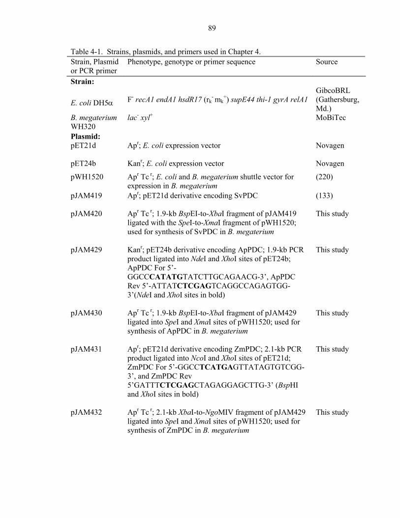
89
Table 4-1. Strains, plasmids, and primers used in Chapter 4. Strain, Plasmid or PCR primer
Phenotype, genotype or primer sequence Source
Strain:
E. coli DH5α
F- recA1 endA1 hsdR17 (rk
- mk+) supE44 thi-1 gyrA relA1
GibcoBRL (Gathersburg, Md.)
B. megaterium WH320
lac- xyl+ MoBiTec
Plasmid: pET21d
Apr; E. coli expression vector
Novagen
pET24b Kanr; E. coli expression vector Novagen
pWH1520 Apr Tc r; E. coli and B. megaterium shuttle vector for expression in B. megaterium
(220)
pJAM419 Apr; pET21d derivative encoding SvPDC
(133)
pJAM420 Apr Tc r; 1.9-kb BspEI-to-XbaI fragment of pJAM419 ligated with the SpeI-to-XmaI fragment of pWH1520; used for synthesis of SvPDC in B. megaterium
This study
pJAM429 Kanr; pET24b derivative encoding ApPDC; 1.9-kb PCR product ligated into NdeI and XhoI sites of pET24b; ApPDC For 5’-GGCCCATATGTATCTTGCAGAACG-3’, ApPDC Rev 5’-ATTATCTCGAGTCAGGCCAGAGTGG-3’(NdeI and XhoI sites in bold)
This study
pJAM430 Apr Tc r; 1.9-kb BspEI-to-XbaI fragment of pJAM429 ligated into SpeI and XmaI sites of pWH1520; used for synthesis of ApPDC in B. megaterium
This study
pJAM431 Apr; pET21d derivative encoding ZmPDC; 2.1-kb PCR product ligated into NcoI and XhoI sites of pET21d; ZmPDC For 5’-GGCCTCATGAGTTATAGTGTCGG-3’, and ZmPDC Rev 5’GATTTCTCGAGCTAGAGGAGCTTG-3’ (BspHI and XhoI sites in bold)
This study
pJAM432 Apr Tc r; 2.1-kb XbaI-to-NgoMIV fragment of pJAM429 ligated into SpeI and XmaI sites of pWH1520; used for synthesis of ZmPDC in B. megaterium
This study

90
Table 4-1. Continued. Strain, Plasmid or PCR primer
Phenotype, genotype or primer sequence Source
pScPDC1 Apr; pET22b derivative encoding ScPDC1 with a 6xHIS tag
(90)
pJAM435
Apr Tc r; 1.9-kb BspEI-to-XbaI fragment of pScPDC1 ligated into SpeI and XmaI sites of pWH1520; used for synthesis of ScPDC1 in B. megaterium
This study
RT Primersa:
Svpdc For 5’-AATCGAAATGAAACCGCTAA-3’ This study Svpdc Rev 5’-TGAGCTTGCAACCATTTCTTTTA-3’ This study Appdc For 5’-CGCGCCCAACAGCAATGATCA-3’ This study Appdc Rev 5’-GGGCGGAGTGAGCGTCGGTAAT-3’ This study Zmpdc For 5’-TGGCGAACTGGCAGAAGCTATCA-3’ This study Zmpdc Rev 5’-CGCGCTTACCCCATTTGACCA-3’ This study Scpdc1 For 5’-CACGGTCCAAAGGCTCAATACAA-3’ This study Scpdc1 Rev 5’-CCGGTGGTAGCGACTCTGTGG-3’ This study
aAbbreviations: RT, reverse transcriptase; For, forward primer; Rev, reverse
primer; Sv, S. ventriculi; Ap, A. pasteurianus; Zm, Z. mobilis; Sc, S. cerevisiae.
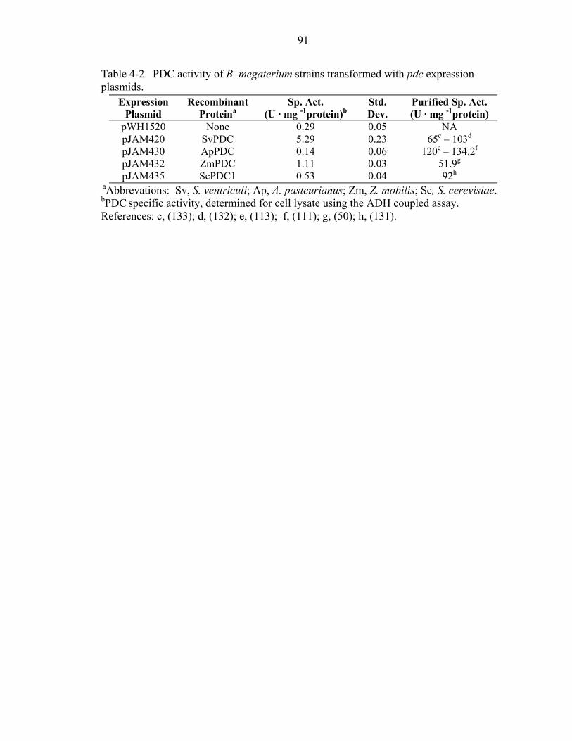
91
Table 4-2. PDC activity of B. megaterium strains transformed with pdc expression plasmids.
Expression Plasmid
Recombinant Proteina
Sp. Act. (U · mg -1protein)b
Std. Dev.
Purified Sp. Act. (U · mg -1protein)
pWH1520 None 0.29 0.05 NA pJAM420 SvPDC 5.29 0.23 65c – 103d
pJAM430 ApPDC 0.14 0.06 120e – 134.2f
pJAM432 ZmPDC 1.11 0.03 51.9g
pJAM435 ScPDC1 0.53 0.04 92h
aAbbrevations: Sv, S. ventriculi; Ap, A. pasteurianus; Zm, Z. mobilis; Sc, S. cerevisiae. bPDC specific activity, determined for cell lysate using the ADH coupled assay. References: c, (133); d, (132); e, (113); f, (111); g, (50); h, (131).
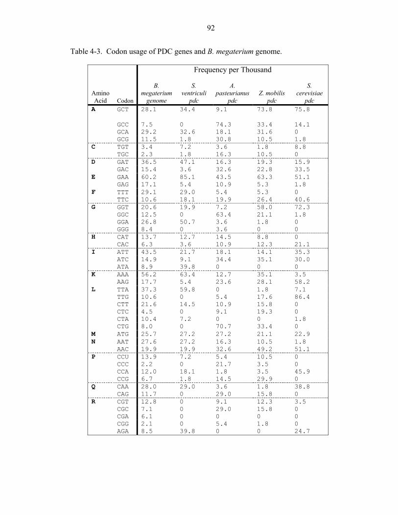
92
Table 4-3. Codon usage of PDC genes and B. megaterium genome.
Frequency per Thousand
Amino Acid Codon
B. megaterium
genome
S. ventriculi
pdc
A. pasteurianus
pdc Z. mobilis
pdc
S. cerevisiae
pdc A GCT 28.1 34.4 9.1 73.8 75.8
GCC 7.5 0 74.3 33.4 14.1 GCA 29.2 32.6 18.1 31.6 0 GCG 11.5 1.8 30.8 10.5 1.8 C TGT 3.4 7.2 3.6 1.8 8.8 TGC 2.3 1.8 16.3 10.5 0 D GAT 36.5 47.1 16.3 19.3 15.9 GAC 15.4 3.6 32.6 22.8 33.5 E GAA 60.2 85.1 43.5 63.3 51.1 GAG 17.1 5.4 10.9 5.3 1.8 F TTT 29.1 29.0 5.4 5.3 0 TTC 10.6 18.1 19.9 26.4 40.6 G GGT 20.6 19.9 7.2 58.0 72.3 GGC 12.5 0 63.4 21.1 1.8 GGA 26.8 50.7 3.6 1.8 0 GGG 8.4 0 3.6 0 0 H CAT 13.7 12.7 14.5 8.8 0 CAC 6.3 3.6 10.9 12.3 21.1 I ATT 43.5 21.7 18.1 14.1 35.3 ATC 14.9 9.1 34.4 35.1 30.0 ATA 8.9 39.8 0 0 0 K AAA 56.2 63.4 12.7 35.1 3.5 AAG 17.7 5.4 23.6 28.1 58.2 L TTA 37.3 59.8 0 1.8 7.1 TTG 10.6 0 5.4 17.6 86.4 CTT 21.6 14.5 10.9 15.8 0 CTC 4.5 0 9.1 19.3 0 CTA 10.4 7.2 0 0 1.8 CTG 8.0 0 70.7 33.4 0 M ATG 25.7 27.2 27.2 21.1 22.9 N AAT 27.6 27.2 16.3 10.5 1.8 AAC 19.9 19.9 32.6 49.2 51.1 P CCU 13.9 7.2 5.4 10.5 0 CCC 2.2 0 21.7 3.5 0 CCA 12.0 18.1 1.8 3.5 45.9 CCG 6.7 1.8 14.5 29.9 0 Q CAA 28.0 29.0 3.6 1.8 38.8 CAG 11.7 0 29.0 15.8 0 R CGT 12.8 0 9.1 12.3 3.5 CGC 7.1 0 29.0 15.8 0 CGA 6.1 0 0 0 0 CGG 2.1 0 5.4 1.8 0 AGA 8.5 39.8 0 0 24.7
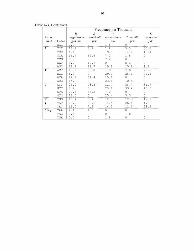
93
Table 4-3. Continued. Frequency per Thousand
Amino Acid Codon
B. megaterium
genome
S. ventriculi
pdc
A. pasteurianus
pdc Z. mobilis
pdc
S. cerevisiae
pdc AGG 2.4 0 1.8 0 0 S TCT 16.7 7.2 1.8 5.3 35.3 TCC 4.4 0 19.9 14.1 19.4 TCA 15.7 32.6 7.2 1.8 0 TCG 5.0 0 7.2 0 0 AGT 9.9 12.7 0 5.3 0 AGC 11.2 12.7 19.9 15.8 1.8 T ACT 10.5 30.8 1.8 7.0 26.5 ACC 6.2 0 29.0 28.1 49.4 ACA 24.1 34.4 10.9 0 0 ACG 14.2 0 23.6 10.5 0 V GTT 25.0 43.5 12.7 38.7 31.7 GTC 8.2 0 23.6 33.4 40.6 GTA 27.3 36.2 7.2 0 0 GTG 12.5 0 25.4 5.3 0 W TGG 10.4 5.4 12.7 12.3 12.3 Y TAT 23.9 32.6 14.5 26.4 1.8 TAC 11.0 7.2 14.5 12.3 28.2 Stop TAA 2.4 1.8 0 0 3.5 TAG 0.4 0 0 1.8 0 TGA 0.5 0 1.8 0 0
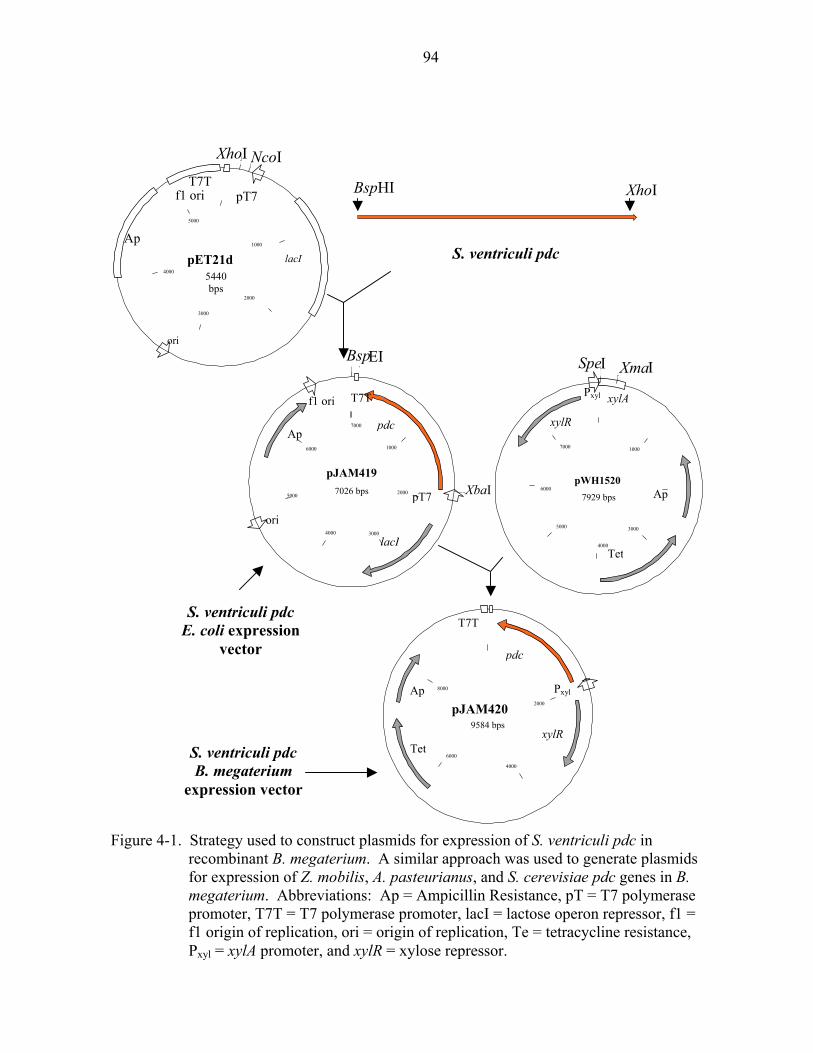
94
pET21d5440 bps
4000 3000
S. ventriculi pdc B. megaterium
expression vector
S. ventriculi pdc E. coli expression
vector
S. ventriculi pdc
Ap
TetxylR
Pxyl
pdc
T7T
8000
6000
4000
2000
9584 bpspJAM420
Pxyl
xylR
Ap
Xma
xylA ISpeI
7000
6000
5000
4000
Tet
3000
1000
7929 bps
pWH1520
f1 ori
Ap
ori
lacI
pT7
pdc
T7T
XbaI
Bsp,
EI
7000
6000
5000 2000
1000
7026 bps
pJAM419
XhoIBspHI
Ap
ori
lacI
pT7T7T
f1 ori
NcoIXhoI
5000
4000
3000
2000
1000
Figure 4-1. Strategy used to construct plasmids for expression of S. ventriculi pdc in recombinant B. megaterium. A similar approach was used to generate plasmids for expression of Z. mobilis, A. pasteurianus, and S. cerevisiae pdc genes in B. megaterium. Abbreviations: Ap = Ampicillin Resistance, pT = T7 polymerase promoter, T7T = T7 polymerase promoter, lacI = lactose operon repressor, f1 = f1 origin of replication, ori = origin of replication, Te = tetracycline resistance, Pxyl = xylA promoter, and xylR = xylose repressor.
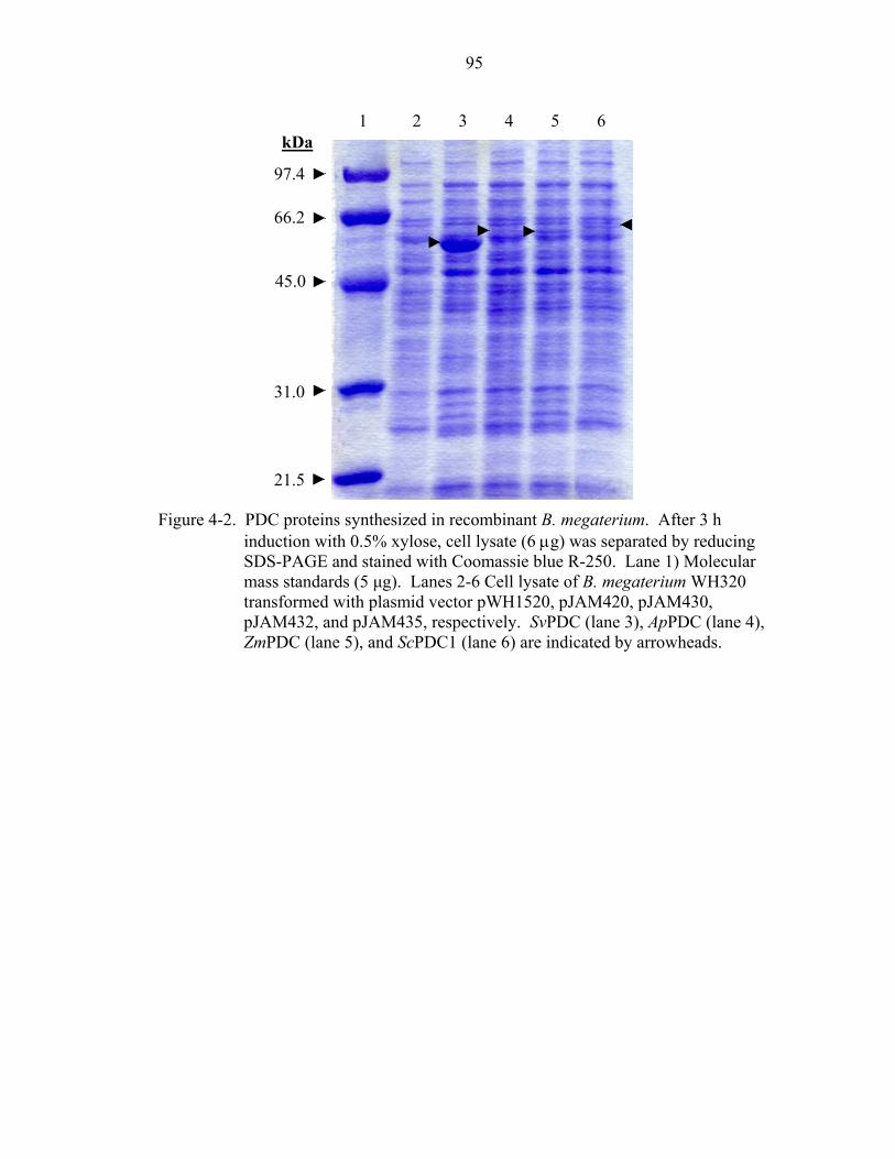
95
Figure 4-2. PD
indSDmatranpJAZm
97.4
66.2 45.0 31.0 21.5C proteins suction with S-PAGE anss standardssformed wiM432, and
PDC (lane 5
1
ynthesiz0.5% xyd staine (5 µg). th plasmpJAM4), and S
2
ed in rlose, c
d with Lanesid vec
35, rescPDC1
3
ecombell lysCooma 2-6 Ctor pWpectiv (lane
4
inant Bate (6 µssie b
ell lysaH1520
ely. Sv 6) are
5
. megg) wa
lue R-2te of B, pJAPDC (indica
6
kDaaterium. After 3 h s separated by reducing 50. Lane 1) Molecular . megaterium WH320
M420, pJAM430, lane 3), ApPDC (lane 4), ted by arrowheads.

96
0%
10%
20%
30%
40%
50%
60%
70%
80%
90%
100%
Sc Sv Ap Zm
% T
otal
RN
A
Figure 4-3. Levels of pdc-specific transcripts in recombinant B. megaterium. Transcript
levels were measured in triplicate using real time quantitative reverse transcription. Abbreviations for genes expressed in recombinant B. megaterium are Sc (S. cerevisiae pdc1), Sv (S. ventriculi pdc), Ap (A. pasteurianus pdc), and Zm (Z. mobilis pdc).
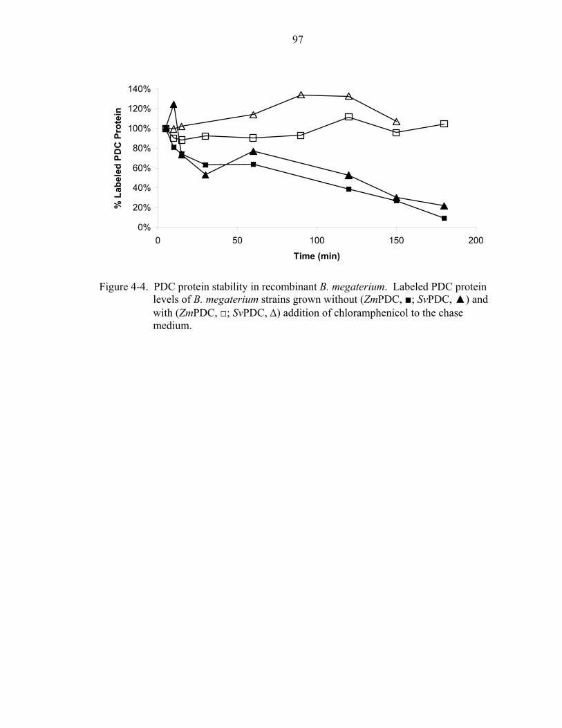
97
0%
20%
40%
60%
80%
100%
120%
140%
0 50 100 150 200
Time (min)
% L
abel
ed P
DC
Pro
tein
Figure 4-4. PDC protein stability in recombinant B. megaterium. Labeled PDC protein
levels of B. megaterium strains grown without (ZmPDC, ■; SvPDC, ▲) and with (ZmPDC, □; SvPDC, ∆) addition of chloramphenicol to the chase medium.

98
CHAPTER 5 GENERAL DISCUSSION AND CONCLUSIONS
The S. ventriculi pdc is the first pyruvate decarboxylase to be cloned from a
Gram-positive bacterium. S. ventriculi PDC protein appears to share similar primary
sequence structure to TPP-dependent enzymes and is highly related to the fungal PDC
and eubacterial IPD enzymes. The close relationship of the S. ventriculi and fungal PDC
structures is consistent with the similar biochemical properties of these enzymes. Both
types of enzymes display substrate cooperativity with similar affinities for pyruvate. The
structure and biochemistry of the S. ventriculi PDC, however, dramatically contrast with
the only other bacterial PDC (Z. mobilis) that has been characterized. The Z. mobilis
PDC is closely related to plants in primary structure; however, it is the only PDC enzyme
known to display Michaelis-Menten kinetics.
This study also demonstrates the synthesis of active, soluble S. ventriculi PDC
protein in recombinant E. coli. Only two other genes, the Z. mobilis pdc and S. cerevisiae
PDC1 genes, have been reported to synthesize PDC protein in recombinant bacteria (114,
115, 218). Of these, at least 50% of the S. cerevisiae PDC1 forms insoluble inclusions in
E. coli and thus has not been useful in engineering bacteria for high-level ethanol
production (218). Due to codon bias, accessory tRNA is essential for efficient production
of S. ventriculi PDC in recombinant E. coli. However, the low G+C codon usage of the
S. ventriculi pdc gene should broaden the spectrum of bacteria that can be engineered as
hosts for high-level production of PDC protein and the engineering of homo-ethanol
98

99
pathways (4). The S. ventriculi PDC is unique among previously characterized bacterial
PDCs. This has enabled the identification of a new subfamily of PDC-like proteins from
Gram-positive bacteria that will broaden the host range of future endeavors utilizing
Gram-positive bacterial hosts.
The SvPDC protein is poorly expressed in recombinant E. coli (133). Therefore,
we reasoned that a host more similar to S. ventriculi might express this PDC at higher
levels. B. megaterium was chosen as a host because it has several benefits over other
Gram-positive expression systems. These include a xylose inducible expression vector
and absence of alkaline proteases that are often responsible for degradation of foreign
proteins (220, 221). Augmentation of the host, B. megaterium, with accessory tRNAs
was not necessary for high-level SvPDC production. The SvPDC protein was more active
when produced in B. megaterium compared to E. coli.
The SvPDC protein produced in B. megaterium has a higher Vmax (98 U per mg
protein) at RT than when produced by E. coli (66 U per mg protein). The SvPDC
produced in B. megaterium is also more thermostable than the E. coli produced protein.
Choosing the correct host appears to have affected the quality of SvPDC protein that was
recovered. These results indicate that differences can occur in the biochemical properties
of recombinant protein based on host.
In this study, we discovered that the pH of the incubation buffer has an effect on
the thermostability of SvPDC. Low pH stabilized SvPDC at higher temperatures. These
results suggest that residues of SvPDC gain a charge between pH 5.0–5.5 that allows the
tetramer conformation to remain stable at higher temperatures. This is an important

100
discovery because it gives insight into residues that can be altered in future experiments
in order to engineer SvPDC to be more thermostable at cytosolic pH.
The portable production of ethanol (PET) operon used in E. coli consists of the
pdc and adh genes from Zymomonas mobilis, a Gram-negative organism (24, 25, 129,
227). Past research to engineer a Gram-positive host for ethanol production has focused
on using this PET operon, but these attempts have met with limited success (33-35, 228)
primarily due to poor expression of the PDC. We have shown that SvPDC is expressed at
high levels in B. megaterium, a Gram-positive host. Our construction and expression of
the Gram-positive ethanol production operon using the SvPDC and G.
stearothermophilus ADH has demonstrated that recombinant PDC and ADH production
no longer limit ethanol production in Gram-positive biocatalysts.
Our research shows that selection of host for recombinant production of proteins
can affect the quality and stability of the recombinant protein. We have also
demonstrated that SvPDC has qualities that make it unique among bacterial PDCs,
including its substrate activation and elevated pH optimum. SvPDC is the only bacterial
PDC that is not thermostable, but our results indicate that alteration of charged residues
may facilitate the engineering of thermostable SvPDC variants. Lastly, we have created a
Gram-positive ethanol production operon that will be useful in engineering future Gram-
positive hosts for ethanol production.
B. megaterium expression vectors were designed in such a way to transcribe all
four pdc genes at similar rates by using the same xylA promoter, Shine-Dalgrano
sequence, and T7 terminator. Using this approach, the S. ventriculi PDC was expressed
at high levels in the recombinant Gram-positive host. The SvPDC protein levels and

101
activity were at least 5-fold higher than when the Z. mobilis, A. pasteurianus, or S.
cerevisiae PDC proteins were expressed. To assess the biological reason for these
differences, quantitative reverse transcriptase PCR and pulse-chase experiments were
performed. Similar levels of pdc-specific transcript and similar rates of PDC protein
degradation were determined. Thus, in the Gram-positive host examined in this study,
protein synthesis limited the production of PDC proteins from yeast and Gram-negative
bacterial genes.
It was previously demonstrated that addition of accessory tRNAs is necessary for
enhancement of protein levels of SvPDC in E. coli by ten-fold (133). This is not the case
when ApPDC and ZmPDC are expressed in E. coli. Both PDCs are produced at very high
levels in this Gram-negative host without the addition of accessory tRNA. In B.
megaterium, however, SvPDC is expressed at very high levels, while expression of
ApPDC and ZmPDC is low. The results of the expression of the PDC proteins in E. coli
and B. megaterium indicate that codon usage of the pdc genes is one of the primary
factors influencing expression of these proteins in Gram-positive hosts (131, 133).
Thus, this research has now identified a PDC that is expressed at high levels
within a Gram-positive bacterial host. Codon usage has also been identified as a major
factor to consider when attempting to produce recombinant PDC. Future efforts to
engineer Gram-positive hosts for ethanol production now have a PDC available that has
been proven to be expressed at high levels or alternatively to synthesize a pdc with codon
usage that will lead to optimal expression in the host.
102
LIST OF REFERENCES
1. MacDonald, T., G. Yowell, and M. McCormack. 2001. US Ethanol Industry; Production Capacity OutlookCalifornia Energy Commission.
2. Dien, B. S., R. J. Bothast, N. N. Nichols, and M. A. Cotta. 2002. The US Corn Ethanol Industry: An Overview of Current Technology and Future Prospects. Int.Sugar J. 104:204-213.
3. Wyman, C. E. 2001. Twenty Years of Trials, Tribulations, and Research Progress in Bioethanol Technology - Selected Key Events Along the Way. Appl.Biochem.Biotechnol. 91-3:5-21.
4. Ingram, L. O., H. C. Aldrich, A. C. Borges, T. B. Causey, A. Martinez, F. Morales, A. Saleh, S. A. Underwood, L. P. Yomano, S. W. York, J. Zaldivar, and S. Zhou. 1999. Enteric Bacterial Catalysts for Fuel Ethanol Production. Biotechnol.Prog. 15:855-866.
5. Bothast, R. J., N. N. Nichols, and B. S. Dien. 1999. Fermentations With New Recombinant Organisms. Biotechnol.Prog. 15:867-875.
6. Deverell, K. F. 1983. Ethanol-Production from Wood Hydrolysates Using Pachysolen tannophilus. Biotechnol.Lett. 5:475-480.
7. Neirinck, L., R. Maleszka, and H. Schneider. 1982. Ethanol Production From Mixed Sugars by Pachysolen tannophilus. Biotechnol.Bioeng.Symp. 12:161-169.
8. Neirinck, L., R. Maleszka, and H. Schneider. 1982. Alcohol Production from Sugar Mixtures by Pachysolen tannophilus. Biotechnol.Bioeng. 161-169.
9. Slininger, P. J., R. J. Bothast, J. E. Vancauwenberge, and C. P. Kurtzman. 1982. Conversion of D-Xylose to Ethanol by the Yeast Pachysolen tannophilus. Biotechnol.Bioeng. 24:371-384.
10. Dupreez, J. C., M. Bosch, and B. A. Prior. 1986. The Fermentation of Hexose and Pentose Sugars by Candida shehatae and Pichia stipitis. Appl.Microbiol.Biotechnol. 23:228-233.
11. Dupreez, J. C., M. Bosch, and B. A. Prior. 1986. Xylose Fermentation by Candida shehatae and Pichia stipitis - Effects of pH, Temperature and Substrate Concentration. Enzyme Microb.Technol. 8:360-364.
102
103
12. Delgenes, J. P., R. Moletta, and J. M. Navarro. 1988. Continuous Production of Ethanol from A Glucose, Xylose and Arabinose Mixture by A Flocculant Strain of Pichia stipitis. Biotechnol.Lett. 10:725-730.
13. Delgenes, J. P., R. Moletta, and J. M. Navarro. 1988. The Ethanol Tolerance of Pichia stipitis Y-7124 Grown on A D-Xylose, D-Glucose and L-Arabinose Mixture. J.Ferment.Technol. 66:417-422.
14. Delgenes, J. P., R. Moletta, and J. M. Navarro. 1988. Fermentation of D-Xylose, D-Glucose and L-Arabinose Mixture by Pichia stipitis Y-7124 - Sugar Tolerance. Appl.Microbiol.Biotechnol. 29:155-161.
15. Jeffries, T. W. and H. K. Sreenath. 1988. Fermentation of Hemicellulosic Sugars and Sugar Mixtures by Candida shehatae. Biotechnol.Bioeng. 31:502-506.
16. Dien, B. S., C. P. Kurtzman, B. C. Saha, and R. J. Bothast. 1996. Screening for L-arabinose Fermenting Yeasts. Appl.Biochem.Biotechnol. 57-8:233-242.
17. Sarthy, A. V., B. L. Mcconaughy, Z. Lobo, J. A. Sundstrom, C. E. Furlong, and B. D. Hall. 1987. Expression of the Escherichia coli Xylose Isomerase Gene in Saccharomyces cerevisiae. Appl.Environ.Microbiol. 53:1996-2000.
18. Gong, C. S., L. F. Chen, M. C. Flickinger, L. C. Chiang, and G. T. Tsao. 1981. Production of Ethanol from D-Xylose by Using D-Xylose Isomerase and Yeasts. Appl.Environ.Microbiol. 41:430-436.
19. Amore, R., M. Wilhelm, and C. P. Hollenberg. 1989. The Fermentation of Xylose - An Analysis of the Expression of Bacillus and Actinoplanes Xylose Isomerase Genes in Yeast. Appl.Microbiol.Biotechnol. 30:351-357.
20. Moes, C. J., I. S. Pretorius, and W. H. vanZyl. 1996. Cloning and expression of the Clostridium thermosulfurogenes D-xylose isomerase gene (xylA) in Saccharomyces cerevisiae. Biotechnol.Lett. 18:269-274.
21. Walfridsson, M., X. M. Bao, M. Anderlund, G. Lilius, L. Bulow, and B. HahnHagerdal. 1996. Ethanolic Fermentation of Xylose with Saccharomyces cerevisiae Harboring the Thermus thermophilus xylA gene, Which Expresses an Active Xylose (glucose) Isomerase. Appl.Environ.Microbiol. 62:4648-4651.
22. Otero, R. R. C., F. C. Wahlbom, W. H. van Zyl, and B. Hahn-Hagerdal. 2002. A Physiological Comparison Between an Industrial, Recombinant, Xylose-Utilizing Saccharomyces sp and Pichia stipitis. Yeast 19:284.
23. Ho, N. W. Y., Z. D. Chen, and A. P. Brainard. 1998. Genetically Engineered Sacccharomyces Yeast Capable of Effective Cofermentation of Glucose and Xylose. Appl.Environ.Microbiol. 64:1852-1859.

104
24. Ingram, L. O., T. Conway, D. P. Clark, G. W. Sewell, and J. F. Preston. 1987. Genetic Engineering of Ethanol Production in Escherichia coli. Appl.Environ.Microbiol. 53:2420-2425.
25. Ohta, K., D. S. Beall, J. P. Mejia, K. T. Shanmugam, and L. O. Ingram. 1991. Metabolic Engineering of Klebsiella oxytoca M5A1 for Ethanol Production from Xylose and Glucose. Appl.Environ.Microbiol. 57:2810-2815.
26. Beall, D. S. and L. O. Ingram. 1993. Genetic-Engineering of Soft-Rot Bacteria for Ethanol-Production from Lignocellulose. J.Ind.Microbiol. 11:151-155.
27. Dell, DM, Grunden, A, Shanmugam, KT, and Ingram LO. Genetic Engineering of Enterobacter cloacae AC1 for Ethanol Production. Abst.Am.Soc.Microbiol.Gen.Meeting O-40, 426. 1997.
28. Alterthum, F. and L. O. Ingram. 1989. Efficient Ethanol Production from
Glucose, Lactose, and Xylose by Recombinant Escherichia coli. Appl.Environ.Microbiol. 55:1943-1948.
29. Ohta, K., F. Alterthum, and L. O. Ingram. 1990. Effects of Environmental Conditions on Xylose Fermentation by Recombinant Escherichia coli. Appl.Environ.Microbiol. 56:463-465.
30. Bucher, M., R. Braendle, and C. Kuhlemeier. 1994. Ethanolic Fermentation in Transgenic Tobacco Expressing Zymomonas mobilis Pyruvate Decarboxylase. EMBO J. 13:2755-2763.
31. Tadege, M., R. Brandle, and C. Kuhlemeier. 1998. Anoxia Tolerance in Tobacco Roots: Effect of Overexpression of Pyruvate Decarboxylase. Plant J. 14:327-335.
32. Deng, M. D. and J. R. Coleman. 1999. Ethanol Synthesis by Genetic Engineering in Cyanobacteria. Appl.Environ.Microbiol. 65:523-528.
33. Barbosa, M. F. S. and L. O. Ingram. 1994. Expression of the Zymomonas mobilis alcohol dehydrogenase II (adhB) and pyruvate decarboxylase (pdc) genes in Bacillus. Curr.Microbiol. 28:279-282.
34. Gold, R. S., M. M. Meagher, S. Tong, R. W. Hutkins, and T. Conway. 1996. Cloning and expression of the Zymomonas mobilis "production of ethanol" genes in Lactobacillus casei. Curr.Microbiol. 33:256-260.
35. Nichols, N. N., B. S. Dien, and R. J. Bothast. 2003. Engineering Lactic Acid Bacteria with Pyruvate Decarboxylase and Alcohol Dehydrogenase Genes for Ethanol Production from Zymomonas mobilis. J.Ind.Microbiol.Biotechnol. 30:315-321.

105
36. Neuberg, C. and J. Hirsch. 1921. Biochem Zeitschr 115:282-310. as sited in reference number 69.
37. Neuberg, C. and H. Ohle. 1922. Biochem Zeitschr 127:327-339. as sited in reference number 69.
38. Neuberg, C. and L. Lieberman. 1921. Biochem Zeitschr 121:322-325. as sited in reference number 69.
39. Hildebrandt, G and Klavehn, W. [U.S. Patent Num. 1,956,950]. 1934. as sited in reference number 69.
40. Hildebrandt, G and Klavehn, W. [German Patent Num. 548 459]. 1932. as sited in reference number 69.
41. Astrup, A., L. Breum, S. Toubro, P. Hein, and F. Quaade. 1992. The Effect and Safety of An Ephedrine Caffeine Compound Compared to Ephedrine, Caffeine and Placebo in Obese Subjects on An Energy Restricted Diet: A Double-Blind Trial. Int.J.Obesity 16:269-277.
42. Astrup, A., B. Buemann, N. J. Christensen, S. Toubro, G. Thorbek, O. J. Victor, and F. Quaade. 1992. The Effect of Ephedrine Caffeine Mixture on Energy-Expenditure and Body-Composition in Obese Women. Metab.Clin.Exp. 41:686-688.
43. Singer, T. and J. Pensky. 1951. Acetoin Synthesis by Highly Purified Alpha-Carboxylase. Arch.Biochem.Biophys. 31:457-459.
44. Singer, T. P. and J. Pensky. 1952. Isolation and Properties of the Alpha-Carboxylase of Wheat Germ. J.Biol.Chem. 196:375-388.
45. Dirscherl, W. 1931. Hoppe-Seyler's Z.Physiol.Chem. 201:47-77. as sited in reference number 69.
46. Langenbeck, W., H. Wrede, and W. Schlockermann. 1934. Hoppe-Seyler's Z.Physiol.Chem. 227:263. as sited in reference number 69.
47. Hübner, G., S. König, A. Schellenberger, and M. H. Koch. 1990. An X-ray Solution Scattering Study of the Cofactor and Activator Induced Structural Changes in Yeast Pyruvate Decarboxylase (PDC). FEBS Lett. 266:17-20.
48. Barman, T. 1969. Pyruvate Dehydrogenase, p. 701. In Enzyme Handbook. Springer-Verlag, New York.
49. Juni, E. 1961. Evidence for A 2-Site Mechanism for Decarboxylation of Alpha-Keto Acids by Alpha-Carboxylase. J.Biol.Chem. 236:2302-&.
106
50. Ullrich, J., J. H. Wittorf, and C. J. Gubler. 1966. Molecular Weight and Coenzyme Content of Pyruvate Decarboxylase from Brewer's Yeast. Biochim.Biophys.Acta 113:595-604.
51. Crout, D. H. G., H. Dalton, D. W. Hutchinson, and M. Miyagoshi. 1991. Studies on Pyruvate Decarboxylase - Acyloin Formation from Aliphatic, Aromatic and Heterocyclic Aldehydes. J.Chem.Soc.-Perk.Trans. 1329-1334.
52. Kren, V., D. H. G. Crout, H. Dalton, D. W. Hutchinson, W. Konig, M. M. Turner, G. Dean, and N. Thomson. 1993. Pyruvate Decarboxylase - A New Enzyme for the Production of Acyloins by Biotransformation. J.Chem.Soc.Chem.Commun. 341-343.
53. Zwickau: Isis-Chemie KG. [German Patent Num. 1,543,691]. 1966. 54. Groger, D., SCHMAUDE.HP, and K. Mothes. 1966. Untersuchungen Uber Die
Gewinnung Von I-Phenylacetylcarbinol. Z.Allg.Mikrobiol. 6:275-297.
55. Vojtisek, V. and J. Netrval. 1982. Effect of Pyruvate Decarboxylaxe Activity and of Pyruvate Concentration on the Production of 1-Hydroxy-1-Phenylpropanone in Saccharomyces carlsbergensis. Folia Microbiol. 27:173-177.
56. Wang, B., H. Shin, and P. L. Rogers. 1994. p. 249. In Better Living Through Innovative Biochemical Engineering. Continental Press, Singapore.
57. Csuk, R. and B. I. Glanzer. 1991. Bakers-Yeast Mediated Transformations in Organic-Chemistry. Chem.Rev. 91:49-97.
58. Nikolova, P. and O. P. Ward. 1991. Production of L-Phenylacetyl Carbinol by Biotransformation - Product and By-Product Formation and Activities of the Key Enzymes in Wild-Type and Adh Isoenzyme Mutants of Saccharomyces cerevisiae. Biotechnol.Bioeng. 38:493-498.
59. Long, A. and O. P. Ward. 1989. Biotransformation of Benzaldehyde by Saccharomyces cerevisiae - Characterization of the Fermentation and Toxicity Effects of Substrates and Products. Biotechnol.Bioeng. 34:933-941.
60. Smith, P. F. and D. Hendlin. 1953. Mechanism of Phenylacetylcarbinol Synthesis by Yeast. J.Bacteriol. 65:440-445.
61. Becvarova, H. and O. Hanc. 1963. Production of Phenylacetylcarbinol by Various Yeast Species. Folia Microbiol. 8:42-&.
62. Voets, J. P., E. J. Vandamme, and C. Vlerick. 1973. Some Aspects of Phenylacetylcarbinol Biosynthesis by Saccharomyces cerevisiae. Z.Allg.Mikrobiol. 13:355-366.

107
63. Agarwal, S. C., S. K. Basu, V. C. Vora, J. R. Mason, and S. J. Pirt. 1987. Studies on the Production of L-Acetyl Phenyl Carbinol by Yeast Employing Benzaldehyde As Precursor. Biotechnol.Bioeng. 29:783-785.
64. Long, A. and O. P. Ward. 1989. Biotransformation of Aromatic-Aldehydes by Saccharomyces cerevisiae - Investigation of Reaction-Rates. J.Ind.Microbiol. 4:49-53.
65. Bringer-Meyer, S. and H. Sahm. 1988. Acetoin and Phenylacetylcarbinol Formation by the Pyruvate Decarboxylases of Zymomonas mobilis and Saccharomyces carlsbergensis. Biocatalysis 1:321-331.
66. Shin, H. S. and P. L. Rogers. 1995. Biotransformation of Benzeldehyde to L-Phenylacetylcarbinol, an Intermediate in L-Ephedrine Production, by Immobilized Candida utilis. Appl.Microbiol.Biotechnol. 44:7-14.
67. Shin, H. S. and P. L. Rogers. 1996. Production of L-phenylacetylcarbinol (L-PAC) from Benzaldehyde Using Partially Purified Pyruvate Decarboxylase (PDC). Biotechnol.Bioeng. 49:52-62.
68. Pohl, M. 1997. Protein Design on Pyruvate Decarboxylase (PDC) by Site-Directed Mutagenesis. Application to Mechanistical Investigations, and Tailoring PDC for the Use in Organic Synthesis. Adv.Biochem.Eng Biotechnol. 58:15-43.
69. Iding, H., P. Siegert, K. Mesch, and M. Pohl. 1998. Application of α-keto Acid Decarboxylases in Biotransformations. Biochim.Biophys.Acta 1385:307-322.
70. Bruhn, H., M. Pohl, J. Grotzinger, and M. R. Kula. 1995. The Replacement of Trp392 by Alanine Influences the Decarboxylase/Carboligase Activity and Stability of Pyruvate Decarboxylase from Zymomonas mobilis. Eur.J.Biochem. 234:650-655.
71. Goetz, G., P. Iwan, B. Hauer, M. Breuer, and M. Pohl. 2001. Continuous Production of (R)-Phenylacetylcarbinol in an Enzyme-Membrane Reactor Using a Potent Mutant of Pyruvate Decarboxylase from Zymomonas mobilis. Biotechnol.Bioeng. 74:317-325.
72. Iwan, P., G. Goetz, S. Schmitz, B. Hauer, M. Breuer, and M. Pohl. 2001. Studies on the Continuous Production of (R)-(-)-Phenylacetylcarbinol in an Enzyme-Membrane Reactor. J.Mol.Catal., B Enzym. 11:387-396.
73. Alvarez, M. E., A. L. Rosa, E. D. Temporini, A. Wolstenholme, G. Panzetta, L. Patrito, and H. J. Maccioni. 1993. The 59-kDa Polypeptide Constituent of 8-10-nm Cytoplasmic Filaments in Neurospora crassa is a Pyruvate Decarboxylase. Gene 130:253-258.

108
74. Sanchis, V., I. Vinas, I. N. Roberts, D. J. Jeenes, A. J. Watson, and D. B. Archer. 1994. A Pyruvate Decarboxylase Gene from Aspergillus parasiticus. FEMS Microbiol.Lett. 117:207-210.
75. Lockington, R. A., G. N. Borlace, and J. M. Kelly. 1997. Pyruvate Decarboxylase and Anaerobic Survival in Aspergillus nidulans. Gene 191:61-67.
76. König, S. 1998. Subunit Structure, Function and Organisation of Pyruvate Decarboxylases from Various Organisms. Biochim.Biophys.Acta 1385:271-286.
77. Gancedo, C. and R. Serrano. 1989. Energy Yielding Metabolism, p. 205. In A. Rose and J. Harrisin (eds.), The Yeast vol. 3. Academic Press, New York.
78. König, S., D. Svergun, M. H. Koch, G. Hübner, and A. Schellenberger. 1992. Synchrotron Radiation Solution X-ray Scattering Study of the pH Dependence of the Quaternary Structure of Yeast Pyruvate Decarboxylase. Biochemistry 31:8726-8731.
79. Kuo, D. J., G. Dikdan, and F. Jordan. 1986. Resolution of Brewers' Yeast Pyruvate Decarboxylase into Two Isozymes. J.Biol.Chem. 261:3316-3319.
80. Sieber, M., S. Konig, G. Hubner, and A. Schellenberger. 1983. A Rapid Procedure for the Preparation of Highly Purified Pyruvate Decarboxylase from Brewer's Yeast. Biomed.Biochim.Acta 42:343-349.
81. Zehender, H., D. Trescher, and J. Ullrich. 1987. Improved Purification of Pyruvate Decarboxylase from Wheat Germ. Its Partial Characterisation and Comparison with the Yeast Enzyme. Eur.J.Biochem. 167:149-154.
82. Zehender, H. and J. Ullrich. 1985. Amino Acid Composition of α- and β-chains of Yeast and Wheat Germ Pyruvate Decarboxylase. FEBS Lett. 180:51-54.
83. Farrenkopf, B. and F. Jordan. 1992. Resolution of Brewers Yeast Pyruvate Decarboxylase into Multiple Isoforms With Similar Subunit Structure and Activity Using High-Performance Liquid Chromatography. Protein Express.Purif. 3:101-107.
84. Dyda, F., W. Furey, S. Swaminathan, M. Sax, B. Farrenkopf, and F. Jordan. 1993. Catalytic Centers in the Thiamin Diphosphate Dependent Enzyme Pyruvate Decarboxylase at 2.4-Å resolution. Biochemistry 32:6165-6170.
85. Killenberg-Jabs, M., S. König, S. Hohmann, and G. Hübner. 1996. Purification and Characterisation of the Pyruvate Decarboxylase from a Haploid Strain of Saccharomyces cerevisiae. Biol.Chem.Hoppe Seyler 377:313-317.

109
86. Zeng, X., B. Farrenkopf, S. Hohmann, F. Dyda, W. Furey, and F. Jordan. 1993. Role of Cysteines in the Activation and Inactivation of Brewers' Yeast pyruvate decarboxylase investigated with a PDC1-PDC6 fusion protein. Biochemistry 32:2704-2709.
87. Baburina, I., Y. Gao, Z. Hu, F. Jordan, S. Hohmann, and W. Furey. 1994. Substrate Activation of Brewers' Yeast Pyruvate Decarboxylase is Abolished by Mutation of Cysteine 221 to Serine. Biochemistry 33:5630-5635.
88. Baburina, I., G. Dikdan, F. Guo, G. I. Tous, B. Root, and F. Jordan. 1998. Reactivity at the Substrate Activation Site of Yeast Pyruvate Decarboxylase: Inhibition by Distortion of Domain Interactions. Biochemistry 37:1245-1255.
89. Baburina, I., H. Li, B. Bennion, W. Furey, and F. Jordan. 1998. Interdomain Information Transfer During Substrate Activation of Yeast Pyruvate Decarboxylase: the Interaction Between Cysteine 221 and Histidine 92. Biochemistry 37:1235-1244.
90. Wei, W., M. Liu, and F. Jordan. 2002. Solvent Kinetic Isotope Effects Monitor Changes in Hydrogen Bonding at the Active Center of Yeast Pyruvate Decarboxylase Concomitant with Substrate Activation: The Substituent at Position 221 Can Control the State of Activation. Biochemistry 41:451-461.
91. Lu, G., D. Dobritzsch, S. Baumann, G. Schneider, and S. König. 2000. The Structural Basis of Substrate Activation in Yeast Pyruvate Decarboxylase. A Crystallographic and Kinetic Study. Eur.J.Biochem. 267:861-868.
92. Schaaff, I., J. B. Green, D. Gozalbo, and S. Hohmann. 1989. A Deletion of the PDC1 Gene for Pyruvate Decarboxylase of Yeast Causes a Different Phenotype than Previously Isolated Point Mutations. Curr.Genet. 15:75-81.
93. Schmitt, H. D. and F. K. Zimmermann. 1982. Genetic-Analysis of the Pyruvate Decarboxylase Reaction in Yeast Glycolysis. J.Bacteriol. 151:1146-1152.
94. Seeboth, P. G., K. Bohnsack, and C. P. Hollenberg. 1990. pdc1o Mutants of Saccharomyces cerevisiae Give Evidence for an Additional Structural PDC Gene: Cloning of PDC5, a Gene Homologous to PDC1. J.Bacteriol. 172:678-685.
95. Hohmann, S. 1991. PDC6, a Weakly Expressed Pyruvate Decarboxylase Gene from Yeast, is Activated When Fused Spontaneously Under the Control of the PDC1 Promoter. Curr.Genet. 20:373-378.
96. Hohmann, S. 1991. Characterization of PDC6, a Third Structural Gene for Pyruvate Decarboxylase in Saccharomyces cerevisiae. J.Bacteriol. 173:7963-7969.

110
97. Hohmann, S. 1993. Characterisation of PDC2, a Gene Necessary for High Level Expression of Pyruvate Decarboxylase Structural Genes in Saccharomyces cerevisiae. Mol.Gen.Genet. 241:657-666.
98. Wright, A. P., H. L. Png, and B. S. Hartley. 1989. Identification, Cloning and Characterisation of a New Gene Required for Full Pyruvate Decarboxylase Activity in Saccharomyces cerevisiae. Curr.Genet. 15:171-175.
99. Kellermann, E., P. G. Seeboth, and C. P. Hollenberg. 1986. Analysis of the Primary Structure and Promoter Function of a Pyruvate Decarboxylase Gene (PDC1) from Saccharomyces cerevisiae. Nucleic Acids Res. 14:8963-8977.
100. Butler, G. and D. J. McConnell. 1988. Identification of an Upstream Activation Site in the Pyruvate Decarboxylase Structural Gene (PDC1) of Saccharomyces cerevisiae. Curr.Genet. 14:405-412.
101. Kellermann, E. and C. P. Hollenberg. 1988. The Glucose-and Ethanol-Dependent Regulation of PDC1 from Saccharomyces cerevisiae are Controlled by Two Distinct Promoter Regions. Curr.Genet. 14:337-344.
102. Hohmann, S. and H. Cederberg. 1990. Autoregulation May Control the Expression of Yeast Pyruvate Decarboxylase Structural Genes PDC1 and PDC5. Eur.J.Biochem. 188:615-621.
103. Arjunan, P., T. Umland, F. Dyda, S. Swaminathan, W. Furey, M. Sax, B. Farrenkopf, Y. Gao, D. Zhang, and F. Jordan. 1996. Crystal Structure of the Thiamin Diphosphate-Dependent Enzyme Pyruvate Decarboxylase from the Yeast Saccharomyces cerevisiae at 2.3 Å resolution. J.Mol.Biol. 256:590-600.
104. Bianchi, M. M., L. Tizzani, M. Destruelle, L. Frontali, and M. Wesolowski-Louvel. 1996. The 'Petite-Negative' Yeast Kluyveromyces lactis Has a Single Gene Expressing Pyruvate Decarboxylase Activity. Mol.Microbiol. 19:27-36.
105. Krieger, F., M. Spinka, R. Golbik, G. Hubner, and S. Konig. 2002. Pyruvate Decarboxylase from Kluyveromyces lactis - An Enzyme With an Extraordinary Substrate Activation Behaviour. Eur.J.Biochem. 269:3256-3263.
106. Holloway, P. and R. E. Subden. 1994. The Nucleotide Sequence and Initial Characterization of Pyruvate Decarboxylase from the yeast Hanseniaspora uvarum. Yeast 10:1581-1589.
107. Neuser, F., H. Zorn, U. Richter, and R. G. Berger. 2000. Purification, Characterisation and cDNA Sequencing of Pyruvate Decarboxylase from Zygosaccharomyces bisporus. Biol.Chem. 381:349-353.
108. Holloway, P. and R. E. Subden. 1993. The Isolation and Nucleotide Sequence of the Pyruvate Decarboxylase Gene from Kluyveromyces marxianus. Curr.Genet. 24:274-277.

111
109. Lu, P., B. P. Davis, and T. W. Jeffries. 1998. Cloning and Characterization of Two Pyruvate Decarboxylase Genes from Pichia stipitis CBS 6054. Appl.Environ.Microbiol. 64:94-97.
110. Dawes, E. A., D. W. Ribbons, and P. J. Large. 1966. The Route of Ethanol Formation in Zymomonas mobilis. Biochem.J. 98:795-803.
111. Hoppner, T. C. and H. W. Doelle. 1983. Purification and Kinetic Characteristics of Pyruvate Decarboxylase and Ethanol Dehydrogenase from Zymomonas mobilis in Relation to Ethanol Production. Eur.J.Appl.Microbiol.Biotechnol. 17:152-157.
112. Bringer-Meyer, S., K.-L. Schimz, and H. Sahm. 1986. Pyruvate Decarboxylase from Zymomonas mobilis. Isolation and Partial Characterization. Arch.Microbiol. 146:105-110.
113. Neale, A. D., R. K. Scopes, R. E. Wettenhall, and N. J. Hoogenraad. 1987. Pyruvate Decarboxylase of Zymomonas mobilis: Isolation, Properties, and Genetic Expression in Escherichia coli. J.Bacteriol. 169:1024-1028.
114. Braü, B. and H. Sahm. 1986. Cloning and Expression of the Structural Gene for Pyruvate Decarboxylase of Zymomonas mobilis in Escherichia coli. Arch.Microbiol. 144:296-301.
115. Conway, T., Y. A. Osman, J. I. Konnan, E. M. Hoffmann, and L. O. Ingram. 1987. Promoter and Nucleotide Sequences of the Zymomonas mobilis Pyruvate Decarboxylase. J.Bacteriol. 169:949-954.
116. Neale, A. D., R. K. Scopes, R. E. Wettenhall, and N. J. Hoogenraad. 1987. Nucleotide Sequence of the Pyruvate Decarboxylase Gene from Zymomonas mobilis. Nucleic Acids Res. 15:1753-1761.
117. Dobritzsch, D., S. König, G. Schneider, and G. Lu. 1998. High Resolution Crystal Structure of Pyruvate Decarboxylase from Zymomonas mobilis. Implications for Substrate Activation in Pyruvate Decarboxylases. J.Biol.Chem. 273:20196-20204.
118. Diefenbach, R. J., J. M. Candy, J. S. Mattick, and R. G. Duggleby. 1992. Effects of Substitution of Aspartate-440 and Tryptophan-487 in the Thiamin Diphosphate Binding Region of Pyruvate Decarboxylase from Zymomonas mobilis. FEBS Lett. 296:95-98.
119. Candy, J. M., J. Koga, P. F. Nixon, and R. G. Duggleby. 1996. The Role of Residues Glutamate-50 and Phenylalanine-496 in Zymomonas mobilis Pyruvate Decarboxylase. Biochem.J. 315:745-751.
120. Schenk, G., F. J. Leeper, R. England, P. F. Nixon, and R. G. Duggleby. 1997. The Role of His113 and His114 in Pyruvate Decarboxylase from Zymomonas mobilis. Eur.J.Biochem. 248:63-71.

112
121. Candy, J. M. and R. G. Duggleby. 1998. Structure and Properties of Pyruvate Decarboxylase and Site-Directed Mutagenesis of the Zymomonas mobilis Enzyme. Biochim.Biophys.Acta 1385:323-338.
122. Chang, A. K., P. F. Nixon, and R. G. Duggleby. 1999. Aspartate-27 and Glutamate-473 are Involved in Catalysis by Zymomonas mobilis Pyruvate Decarboxylase. Biochem.J. 339:255-260.
123. Pohl, M., P. Siegert, K. Mesch, H. Bruhn, and J. Grotzinger. 1998. Active Site Mutants of Pyruvate Decarboxylase from Zymomonas mobilis--a Site-Directed Mutagenesis Study of L112, I472, I476, E473, and N482. Eur.J.Biochem. 257:538-546.
124. Chang, A. K., P. F. Nixon, and R. G. Duggleby. 2000. Effects of Deletions at the Carboxyl Terminus of Zymomonas mobilis Pyruvate Decarboxylase on the Kinetic Properties and Substrate Specificity. Biochemistry 39:9430-9437.
125. Wu, Y. G., A. K. Chang, P. F. Nixon, W. Li, and R. G. Duggleby. 2000. Mutagenesis at Asp27 of Pyruvate Decarboxylase from Zymomonas mobilis Effect on its Ability to Form Acetoin and Acetolactate. Eur.J.Biochem. 267:6493-6500.
126. Huang, C. Y., A. K. Chang, P. F. Nixon, and R. G. Duggleby. 2001. Site-Directed Mutagenesis of the Ionizable Groups in the Active Site of Zymomonas mobilis Pyruvate Decarboxylase: Effect on Activity and pH Dependence. Eur.J.Biochem. 268:3558-3565.
127. Arfman, N., V. Worrell, and L. O. Ingram. 1992. Use of the tac Promoter and lacIq for the Controlled Expression of Zymomonas mobilis Fermentative Genes in Escherichia coli and Zymomonas mobilis. J.Bacteriol. 174:7370-7378.
128. Wood, B. E. and L. O. Ingram. 1992. Ethanol Production From Cellobiose, Amorphous Cellulose, and Crystalline Cellulose by Recombinant Klebsiella oxytoca Containing Chromosomally Integrated Zymomonas mobilis Genes for Ethanol Production and Plasmids Expressing Thermostable Cellulase Genes from Clostridium thermocellum. Appl.Environ.Microbiol. 58:2103-2110.
129. Ingram, L. O., P. F. Gomez, X. Lai, M. Moniruzzaman, B. E. Wood, L. P. Yomano, and S. W. York. 1998. Metabolic Engineering of Bacteria for Ethanol Production. Biotechnol.Bioeng. 58:204-214.
130. Raj, K. C., L. O. Ingram, and J. A. Maupin-Furlow. 2001. Pyruvate Decarboxylase: a Key Enzyme for the Oxidative Metabolism of Lactic Acid by Acetobacter pasteurianus. Arch.Microbiol. 176:443-451.

113
131. Raj, K. C., L. A. Talarico, L. O. Ingram, and J. A. Maupin-Furlow. 2002. Cloning and Characterization of the Zymobacter palmae Pyruvate Decarboxylase Gene (pdc) and Comparison to Bacterial Homologues. Appl.Environ.Microbiol. 68:2869-2876.
132. Lowe, S. E. and J. G. Zeikus. 1992. Purification and Characterization of Pyruvate Decarboxylase from Sarcina ventriculi. J.Gen.Microbiol. 138:803-807.
133. Talarico, L. A., L. O. Ingram, and J. A. Maupin-Furlow. 2001. Production of the Gram-Positive Sarcina ventriculi Pyruvate Decarboxylase in Escherichia coli . Microbiology 147:2425-2435.
134. Davies, D. D., S. Grego, and P. Kenworthy. 1974. The Control of the Production of Lactate and Ethanol by Higher Plants. Planta 118:297-310.
135. Wignarajah, K. and H. Greenway. 1976. Effect of Anaerobiosis on Activities of Alcohol Dehydrogenase and Pyruvate Decarboxylase in Roots of Zea mays. New Phytol. 77:575-584.
136. Lee, T. C. and P. J. Langston-Unkefer. 1985. Pyruvate Decarboxylase from Zea mays L. I. Purification and Partial Characterization from Mature Kernals and Anaerobically Treated Roots. Plant Physiol. 79:242-247.
137. Langston-Unkefer, P. J. and T. C. Lee. 1985. Pyruvate Decarboxylase from Zea mays L. 2. Examination of Hysteretic Kinetics. Plant Physiol. 79:436-440.
138. Leblová, S. and J. Valik. 1980. Partial Characterization of Pyruvate Decarboxylase Isolated from Germinating Pea Seeds. Aust.J.Plant Physiol. 7:35-39.
139. König, S., D. I. Svergun, V. V. Volkov, L. A. Feigin, and M. H. Koch. 1998. Small-Angle X-ray Solution-Scattering Studies on Ligand-Induced Subunit Interactions of the Thiamine Diphosphate Dependent Enzyme Pyruvate Decarboxylase from Different Organisms. Biochemistry 37:5329-5334.
140. Dietrich, A. and S. König. 1997. Substrate Activation Behaviour of Pyruvate Decarboxylase from Pisum sativum cv. Miko. FEBS Lett. 400:42-44.
141. Leblová, S. and J. Valik. 1981. The Effect of Metals on Isolated Pea Pyruvate Decarboxylase. Biolôgia Plantarum (Praha) 23:81-85.
142. Mücke, U., S. König, and G. Hübner. 1995. Purification and Characterisation of Pyruvate Decarboxylase from Pea Seeds (Pisum sativum cv. Miko). Biol.Chem.Hoppe Seyler 376:111-117.

114
143. Mücke, U., T. Wohlfarth, U. Fiedler, H. Bäumlein, K. P. Rücknagel, and S. König. 1996. Pyruvate Decarboxylase from Pisum sativum. Properties, Nucleotide and Amino Acid Sequences. Eur.J.Biochem. 237:373-382.
144. Kirschner, J. and JM. Miller. 1957. Volatile Water-Soluble and Oil Constituents of Valencia Orange Juice. J.Agric.Food Chem. 5:283.
145. Ahmed, EM, Dennison, RA, and Shaw, PE. Flavor Enhancing Potential of Selected Orange Oil and Essence Components. 10-10-1973. Winter Haven, FL. USDA Citrus Chemistry and Technology Conference.
146. Roe, B. and J. H. Bruemmer. 1974. Enzyme-Mediated Aldehyde Change in
Orange Juice. J.Agri.Food Chem. 22:285-288.
147. Raymond, W. R., J. B. Hostettler, K. Assar, and C. Varsel. 1979. Orange Pyruvate Decarboxylase: Isolation and Mechanistic Studies. J.Food Sci. 44:777-781.
148. Oba, K. and I. Uritani. 1975. Purification and Characterization of Pyruvate Decarboxylase from Sweet Potato Roots. J.Biochem (Tokyo) 77:1205-1213.
149. Ôba, K. and I. Uritani. 1982. Pyruvate Decarboxylase from Sweet Potato Roots. Methods Enzymol. 90 Pt E:528-532.
150. Huang, Y. H., D. H. Picha, and A. W. Kilili. 2002. Atmospheric Oxygen Level Influences Alcohol Dehydrogenase and Pyruvate Decarboxylase Activities in Sweetpotato Roots. J.Plant Physiol. 159:129-136.
151. Balla, H. and J. Ullrich. 1980. Wheat Germ Pyruvate Decarboxylase Improved Purification and Properties in Comparison With the Yeast Enzyme. Hoppe Seylers.Z.Physiol Chem. 361:1265.
152. Kluger, R., G. Gish, and G. Kauffman. 1984. Interaction of Thiamin Diphosphate and Thiamin Thiazolone Diphosphate with Wheat-Germ Pyruvate Decarboxylase. J.Biol.Chem. 259:8960-8965.
153. Ullrich, J. 1982. Structure-Function Relationships in Pyruvate Decarboxylase of Yeast and Wheat Germ. Ann.N.Y.Acad.Sci. 378:287-305.
154. Davies, D. D. and H. Asker. 1985. The Enzymatic Decarboyxlation of Hydroxypyruvate Associated with Purified Pyruvate Decarboxylase from Wheat Germ. Phytochemistry 24:231-234.
155. Rivoal, J., B. Ricard, and A. Pradet. 1990. Purification and Partial Characterization of Pyruvate Decarboxylase from Oryza sativa L. Eur.J.Biochem. 194:791-797.

115
156. Hossain, M. A., J. D. McGee, A. Grover, E. S. Dennis, W. J. Peacock, T. K. Hodges, and E. Dennis. 1994. Nucleotide Sequence of a Rice Genomic Pyruvate Decarboxylase Gene that Lacks Introns: a Pseudo-Gene? [published erratum appears in Plant Physiol 1995 Jun;108(2):881]. Plant Physiol 106:1697-1698.
157. Hossain, M. A., E. Huq, T. K. Hodges, and E. Hug. 1994. Sequence of a cDNA from Oryza sativa (L.) Encoding the Pyruvate Decarboxylase 1 Gene. Plant Physiol 106:799-800.
158. Huq, E., M. A. Hossain, and T. K. Hodges. 1995. Cloning and Sequencing of a cDNA Encoding Pyruvate Decarboxylase 2 Gene (Accession no. U27350) from Rice. Plant Physiol. 109:722.
159. Hossain, M. A., E. Huq, A. Grover, E. S. Dennis, W. J. Peacock, and T. K. Hodges. 1996. Characterization of Pyruvate Decarboxylase Genes from Rice. Plant Mol.Biol. 31:761-770.
160. Kato-Noguchi, H. 2003. Anoxia Tolerance in Rice Roots Acclimated by Several Different Periods of Hypoxia. J.Plant Physiol. 160:565-568.
161. Leblová, S. and J. Martinec. 1987. Isolation and Characterization of Pyruvate Decarboxylase from Germinating Bean Seeds (Vicia faba). Biolôgia (Bratislava) 42:1181-1189.
162. Imahori, Y., M. Kota, Y. Ueda, M. Ishimaru, and K. Cachin. 2002. Regulation of Ethanolic Fermentation in Bell Pepper Fruit Under Low Oxygen Stress. Postharvest Biol.Technol. 25:159-167.
163. Fukao, T., R. A. Kennedy, Y. Yamasue, and M. E. Rumpho. 2003. Genetic and Biochemical Analysis of Anaerobically-Induced Enzymes During Seed Germination of Echinochloa crus-galli Varieties Tolerant and Intolerant of Anoxia. J.Exp.Bot. 54:1421-1429.
164. Bucher, M., K. A. Brander, S. Sbicego, T. Mandel, and C. Kuhlemeier. 1995. Aerobic Fermentation in Tobacco Pollen. Plant Mol.Biol. 28:739-750.
165. Or, E., J. Baybik, A. Sadka, and A. Ogrodovitch. 2000. Fermentative Metabolism in Grape Berries: Isolation and Characterization of Pyruvate Decarboxylase cDNA and Analysis of its Expression Throughout Berry Development. Plant Sci. 156:151-158.
166. Imahori, Y., K. Matushita, M. Kota, Y. Ueda, M. Ishimaru, and K. Chachin. 2003. Regulation of Fermentative Metabolism in Tomato Fruit Under Low Oxygen Stress. J.Hort.Sci.Biotechnol. 78:386-393.
167. Chen, H. J. and R. G. Qualls. 2003. Anaerobic Metabolism in the Roots of Seedlings of the Invasive Exotic Lepidium latifolium. Environ.Exper.Bot. 50:29-40.

116
168. Kimmerer, T. W. 1987. Alcohol Dehydrogenase and Pyruvate Decarboxylase Activity in Leaves and Roots of Eastern Cottonwood (Populus deltoides Bartr.) and Soybean (Glycine max L.). Plant Physiol. 84:1210-1213.
169. Kursteiner, O., I. Dupuis, and C. Kuhlemeier. 2003. The Pyruvate Decarboxylase1 Gene of Arabidopsis is Required During Anoxia But Not Other Environmental Stresses. Plant Physiol. 132:968-978.
170. Ismond, K. P., R. Dolferus, M. De Pauw, E. S. Dennis, and A. G. Good. 2003. Enhanced Low Oxygen Survival in Arabidopsis Through Increased Metabolic Flux in the Fermentative Pathway. Plant Physiol. 132:1292-1302.
171. Gounaris, A. D., I. Turkenkopf, S. Buckwald, and A. Young. 1971. Pyruvate Decarboxylase. I. Protein Dissociation Into Subunits Under Conditions in Which Thiamine Pyrophosphate is Released. J.Biol.Chem. 246:1302-1309.
172. König, S., D. Svergun, M. H. Koch, G. Hübner, and A. Schellenberger. 1993. The Influence of the Effectors of Yeast Pyruvate Decarboxylase (PDC) on the Conformation of the Dimers and Tetramers and Their pH-Dependent Equilibrium. Eur.Biophys.J. 22:185-194.
173. Killenberg-Jabs, M., A. Jabs, H. Lilie, R. Golbik, and G. Hubner. 2001. Active Oligomeric States of Pyruvate Decarboxylase and Their Functional Characterization. Eur.J.Biochem. 268:1698-1704.
174. Pohl, M., J. Grötzinger, A. Wollmer, and M. R. Kula. 1994. Reversible Dissociation and Unfolding of Pyruvate Decarboxylase from Zymomonas mobilis. Eur.J.Biochem. 224:651-661.
175. Green, D. E., D. Herbert, and V. Subrahmanyan. 1941. Carboxylase. J.Biol.Chem. 138:327-339.
176. Schellenberger, A. 1967. Angew.Chem. 79:1050-1061.
177. Morey, A. V. and E. Juni. 1968. Studies on the Nature of the Binding of Thiamine Pyrophosphate to Enzymes. J.Biol.Chem. 243:3009-3019.
178. Diefenbach, R. J. and R. G. Duggleby. 1991. Pyruvate Decarboxylase from Zymomonas mobilis. Structure and Re-Activation of Apoenzyme by the Cofactors Thiamin Diphosphate and Magnesium Ion. Biochem.J. 276:439-445.
179. Vaccaro, J. A., E. J. Crane, III, T. K. Harris, and M. W. Washabaugh. 1995. Mechanism of Reconstitution of Brewers' Yeast Pyruvate Decarboxylase With Thiamin Diphosphate and Magnesium. Biochemistry 34:12636-12644.
180. Schellenberger, A. and G. Hübner. 1967. Mechanismus and Kinetik Der Rekombination und Daraus Abgeletete Bindungsverhaltnisse im Aktiven Zentrum Der Hefe-PyruvatdecArboxylase. Hoppe Seylers.Z.Physiol.Chem. 348:491.
117
181. Eppendorfer, S., S. König, R. Golbik, H. Neef, K. Lehle, R. Jaenicke, A. Schellenberger, and G. Hübner. 1993. Effects of Metal Ions, Thiamine Diphosphate Analogues and Subunit Interactions on the Reconstitution Behaviour of Pyruvate Decarboxylase from Brewer's Yeast. Biol.Chem.Hoppe Seyler 374:1129-1134.
182. Kuo, D. J. and F. Jordan. 1983. Direct Spectroscopic Observation of a Brewer's Yeast Pyruvate Decarboxylase-Bound Enamine Intermediate Produced From a Suicide Substrate. Evidence for Nonconcerted Decarboxylation. J.Biol.Chem. 258:13415-13417.
183. Kuo, D. J. and F. Jordan. 1983. Active site Directed Irreversible Inactivation of Brewers' Yeast Pyruvate Decarboxylase by the Conjugated Substrate Analogue (E)-4-(4- chlorophenyl)-2-oxo-3-butenoic Acid: Development of a Suicide Substrate. Biochemistry 22:3735-3740.
184. Hübner, G., R. Weidhase, and A. Schellenberger. 1978. The Mechanism of Substrate Activation of Pyruvate Decarboxylase: A First Approach. Eur.J.Biochem. 92:175-181.
185. Gish, G., T. Smyth, and R. Kluger. 1988. Thiamin Diphosphate Catalysis - Mechanistic Divergence As A Probe of Substrate Activation of Pyruvate Decarboxylase. J.Amer.Chem.Soc. 110:6230-6234.
186. Baburina, I., D. J. Moore, A. Volkov, A. Kahyaoglu, F. Jordan, and R. Mendelsohn. 1996. Three of Four Cysteines, Including That Responsible For Substrate Activation, Are Ionized at pH 6.0 in Yeast Pyruvate Decarboxylase: Evidence From Fourier Transform Infrared and Isoelectric Focusing Studies. Biochemistry 35:10249-10255.
187. Furey, W., P. Arjunan, L. Chen, F. Dyda, T. Umland, S. Swaminathan, M. Sax, F. Jordan, B. Farrenkopf, Y. Gao, and D. Zhang. 1996. Multiple Modes of Tetramer Assembly and Insight Into Allosteric Activation Revealed by X-ray Crystal Structures of Pyruvate Decarboxylase, p. 103-124. In H. Bisswanger and A. Schellenberger (eds.), Biochemistry and Physiology of Thiamine Diphosphate Enzymes. Proc. Int. Meet. 4th. A.u.C. Intemann Wissenschaftlicher-Verlag, Prien, Germany.
188. Li, H. and F. Jordan. 1999. Effects of Substitution of Tryptophan 412 in the Substrate Activation Pathway of Yeast Pyruvate Decarboxylase. Biochemistry 38:10004-10012.
189. Li, H., W. Furey, and F. Jordan. 1999. Role of Glutamate 91 in Information Transfer During Substrate Activation of Yeast Pyruvate Decarboxylase. Biochemistry 38:9992-10003.
118
190. Wang, J., R. Golbik, B. Seliger, M. Spinka, K. Tittmann, G. Hubner, and F. Jordan. 2001. Consequences of a Modified Putative Substrate-Activation Site on Catalysis by Yeast Pyruvate Decarboxylase. Biochemistry 40:1755-1763.
191. Sergienko, E. A. and F. Jordan. 2002. New Model for Activation of Yeast Pyruvate Decarboxylase by Substrate Consistent with the Alternating Sites Mechanism: Demonstration of the Existence of Two Active Forms of the Enzyme. Biochemistry 41:3952-3967.
192. Shellenberger, A., G. Hubner, and M. Sieber. 1988. p. 113-121. In A. Schellenberger and R. Schowen (eds.), Thiamine Pyrophosphate Biochemistry. CRC Press, Boca Raton, FL.
193. Hübner, G., S. König, and A. Schellenberger. 1988. The Functional Role of Thiol Groups of Pyruvate Decarboxylase from Brewer's Yeast. Biomed.Biochim.Acta 47:9-18.
194. Jordan, F., O. Akinoyosoye, G. Dikdan, Z. Kudzin, and D. Kuo. 1988. p. 79-92. In A. Schellenberger and R. Schowen (eds.), Thiamine Pyrophosphate Biochemistry. CRC Press, Boca Raton, FL.
195. Lu, G., D. Dobritzsch, S. König, and G. Schneider. 1997. Novel Tetramer Assembly of Pyruvate Decarboxylase from Brewer's Yeast Observed in a New Crystal Form. FEBS Lett. 403:249-253.
196. Killenberg-Jabs, M., S. König, I. Eberhardt, S. Hohmann, and G. Hübner. 1997. Role of Glu51 for Cofactor Binding and Catalytic Activity in Pyruvate Decarboxylase from Yeast Studied by Site-Directed Mutagenesis. Biochemistry 36:1900-1905.
197. Jordan, F., N. Nemeria, F. Guo, I. Baburina, Y. Gao, A. Kahyaoglu, H. Li, J. Wang, J. Yi, J. R. Guest, and W. Furey. 1998. Regulation of Thiamin Diphosphate-Dependent 2-Oxo Acid Decarboxylases by Substrate and Thiamin Diphosphate.Mg(II) - Evidence for Tertiary and Quaternary Interactions. Biochim.Biophys.Acta 1385:287-306.
198. Chen, G. C. and F. Jordan. 1984. Brewers-Yeast Pyruvate Decarboxylase Produces Acetoin from Acetaldehyde - A Novel Tool to Study the Mechanism of Steps Subsequent to Carbon-Dioxide Loss. Biochemistry 23:3576-3582.
199. Stivers, J. T. and M. W. Washabaugh. 1993. Catalysis of Acetoin Formation by Brewers Yeast Pyruvate Decarboxylase Isozymes. Biochemistry 32:13472-13482.
200. Stivers, J. T. and M. W. Washabaugh. 1993. Catalysis of Acetoin Formation by Brewer Yeast Pyruvate Decarboxylase: Steady-State Kinetics, Steady-State Primary Deuterium and Tritium Kinetic Isotope Effects, and Activation by Pyruvamide. FASEB J. 7:A1198.

119
201. Suomalainen, H. and T. Linnahalme. 1966. Metabolites of Alpha-Ketomonocarboxylic Acids Formed by Dried Bakers and Brewers Yeast. Arch.Biochem.Biophys. 114:502-511.
202. Lehmann, H., G. Fischer, G. Hübner, K. D. Kohnert, and A. Schellenberger. 1973. The Influence of Steric and Electronic Parameters on the Substrate Behavior of α-Oxo Acids to Yeast Pyruvate Decarboxylase. Eur.J.Biochem. 32:83-87.
203. Reynen, M. and H. Sahm. 1988. Comparison of the Structural Genes for Pyruvate Decarboxylase in Different Zymomonas mobilis Strains. J.Bacteriol. 170:3310-3313.
204. Stephenson, M. P. and E. A. Dawes. 1971. Pyruvic Acid and Formic Acid Metabolism in Sarcina ventriculi and the Role of Ferredoxin. J.Gen.Microbiol. 69:331-343.
205. Goodwin, S. and J. G. Zeikus. 1987. Physiological Adaptations of Anaerobic Bacteria to Low pH: Metabolic Control of Proton Motive Force in Sarcina ventriculi. J.Bacteriol. 169:2150-2157.
206. Lowe, S. E. and J. G. Zeikus. 1991. Metabolic Regulation of Carbon and Electron Flow as a Function of pH During Growth of Sarcina ventriculi. Arch.Microbiol. 155:325-329.
207. Ullrich, J. and I. Donner. 1970. Kinetic Evidence for Two Active Sites in Cytoplasmic Yeast Pyruvate Decarboxylase. Hoppe Seylers.Z.Physiol Chem. 351:1026-1029.
208. Harwood, C. R. and S. M. Cutting. 1990. In Molecular Biological Methods for Bacillus. Wiley, New York.
209. Southern, E. M. 1975. Detection of Specific Sequences Among DNA Fragments Separated by Gel Electrophoresis. J.Mol.Biol. 98:503-517.
210. Sanger, F., S. Nicklen, and A. R. Coulson. 1977. DNA Sequencing With Chain-Terminating Inhibitors. Proc.Natl.Acad.Sci.U.S.A. 74:5463-5467.
211. Thompson, J. D., D. G. Higgins, and T. J. Gibson. 1994. CLUSTAL W: Improving the Sensitivity of Progressive Multiple Sequence Alignment Through Sequence Weighting, Position-Specific Gap Penalties and Weight Matrix Choice. Nucleic Acids Res. 22:4673-4680.
212. Page, R. D. 1996. TreeView: An Application to Display Phylogenetic Trees on Personal Computers. Comput.Appl.Biosci. 12:357-358.
213. Corpet, F. 1988. Multiple Sequence Alignment with Hierarchical Clustering. Nucleic Acids Res. 16:10881-10890.

120
214. Altschul, S. F., W. Gish, W. Miller, E. W. Myers, and D. J. Lipman. 1990. Basic Local Alignment Search Tool. Journal of Molecular Biology 215:403-410.
215. Cserzo, M., E. Wallin, I. Simon, G. von Heijne, and A. Elofsson. 1997. Prediction of Transmembrane Alpha-Helices in Prokaryotic Membrane Proteins: The Dense Alignment Surface Method. Protein Eng 10:673-676.
216. Hawkins, C. F., A. Borges, and R. N. Perham. 1989. A Common Structural
Motif in Thiamin Pyrophosphate-Binding Enzymes. FEBS Lett. 255:77-82.
217. Boiteux, A. and B. Hess. 1970. Allosteric Properties of Yeast Pyruvate Decarboxylase. FEBS Lett. 9:293-296.
218. Candy, J. M., R. G. Duggleby, and J. S. Mattick. 1991. Expression of Active Yeast Pyruvate Decarboxylase in Escherichia coli. J.Gen.Microbiol. 137:2811-2815.
219. Kim, K. K., R. Kim, and S. H. Kim. 1998. Crystal Structure of a Small Heat-Shock Protein. Nature 394:595-599.
220. Rygus, T. and W. Hillen. 1991. Inducible High-Level Expression of Heterologous Genes in Bacillus megaterium Using the Regulatory Elements of the Xylose-Utilization Operon. Appl.Microbiol.Biotechnol. 35:594-599.
221. Meinhardt, F., U. Stahl, and W. Ebeling. 1989. Highly Efficient Expression of Homologous and Heterologous Genes in Bacillus megaterium. Appl.Microbiol.Biotechnol. 30:343-350.
222. Sakoda, H. and T. Imanaka. 1992. Cloning and Sequencing of the Gene Coding for Alcohol Dehydrogenase of Bacillus stearothermophilus and Rational Shift of the Optimum pH. J.Bacteriol. 174:1397-1402.
223. Puyet, A., H. Sandoval, P. López, A. Aguilar, J. F. Martin, and M. Espinosa. 1987. A Simple Medium for Rapid Regeneration of Bacillus subtilis Protoplasts Transformed with Plasmid DNA. FEMS Microbiol.Lett. 40:1-5.
224. Conway, T., Y. A. Osman, and L. O. Ingram. 1987. Gene Expression in Zymomonas mobilis: Promoter Structure and Identification of Membrane Anchor Sequences Forming Functional lacZ' Fusion Proteins. J.Bacteriol. 169:2327-2335.
225. Wong, C. 2000. Heating Greatly Speeds Coomassie Blue Staining and Destaining. Biotechniques 29:544.
226. Jordan, F., D. J. Kuo, and E. U. Monse. 1978. A pH-Rate Determination of the Activity-pH Profile of Enzymes. Application to Yeast Pyruvate Decarboxylase Demonstrating the Existence of Multiple Ionizable Groups. Anal.Biochem. 86:298-302.
121
227. Ohta, K., D. S. Beall, J. P. Mejia, K. T. Shanmugam, and L. O. Ingram. 1991. Genetic Improvement of Escherichia coli for Ethanol Production: Chromosomal Integration of Zymomonas mobilis Genes Encoding Pyruvate Decarboxylase and Alcohol Dehydrogenase II. Appl.Environ.Microbiol. 57:893-900.
228. Gold, R. S., M. M. Meager, R. Hutkins, and T. Conway. 1992. Ethanol Tolerance and Carbohydrate-Metabolism in Lactobacilli. J.Indust.Microbiol. 10:45-54.
229. Wong, S. L. 1995. Advances in the Use of Bacillus subtilis for the Expression and Secretion of Heterologous Proteins. Curr.Opin.Biotechnol. 6:517-522.
230. Miyagi, T., K. Kaneichi, R. I. Aminov, Y. Kobayashi, K. Sakka, S. Hoshino, and K. Ohmiya. 1995. Enumeration of Transconjugated Ruminococcus albus and Its Survival in the Goat Rumen Microcosm. Appl.Environ.Microbiol. 61:2030-2032.
231. Wong, S. L., R. Q. Ye, and S. Nathoo. 1994. Engineering and Production of Streptokinase in A Bacillus subtilis Expression-Secretion System. Appl.Environ.Microbiol. 60:517-523.
232. Gurkan, C. and D. J. Ellar. 2003. Expression of the Bacillus thuringiensis Cyt2Aa I toxin in Pichia pastoris Using a Synthetic Gene Construct. Biotechnol.Appl.Biochem. 38:25-33.
233. Lakey, D. L., R. K. R. Voladri, K. M. Edwards, C. Hager, B. Samten, R. S. Wallis, P. F. Barnes, and D. S. Kernodle. 2000. Enhanced Production of Recombinant Mycobacterium tuberculosis Antigens in Escherichia coli by Replacement of Low-Usage Codons. Infect.Immun. 68:233-238.

122
BIOGRAPHICAL SKETCH
LeeAnn Talarico Blalock was born on April 12, 1977, in Charleston, South
Carolina. She grew up in Hanahan, South Carolina, with her mother, Sandra Lee. Upon
graduation from Stratford High School in Goose Creek, South Carolina, she attended
Charleston Southern University on a Board of Trustees Academic Scholarship. She
graduated cum laude with a major in biology and a minor in chemistry. She is a member
of Beta Beta Beta biological honor society and Alpha Chi academic honor society. While
attending college, LeeAnn gained experience working in microbiology, chemistry, and
biotechnology laboratories, as well as tutoring college students in biology and chemistry.
In August 1999, LeeAnn was accepted as a graduate student in the Department of
Microbiology and Cell Science at the University of Florida. She worked in the laboratory
of Dr. Julie Maupin-Furlow on the expression of pyruvate decarboxylase in Gram-
positive bacteria. In November 2002, she received the President’s Award for Graduate
Student Oral Presentation at the Southeastern Branch of the American Society for
Microbiology annual meeting. In December 2003, LeeAnn will be conferred the degree
of Doctor of Philosophy. Upon graduation, LeeAnn will move with her husband,
Timothy Blalock, to Boston, Massachusetts, where she will be a Postdoctoral Fellow with
Dr. Dennis Kasper in the Department of Microbiology and Molecular Genetics at
Harvard Medical School.
122