Embed Size (px)
Citation preview
Li et al. Journal of Inflammation (2015) 12:24 DOI 10.1186/s12950-015-0072-5
RESEARCH Open Access
Expression of hedgehog signal pathway inarticular cartilage is associated with the severityof cartilage damage in rats with adjuvant-inducedarthritisRong Li1,2, Li Cai3*, Cheng-mu Hu1,2, Ting-ni Wu1,2 and Jun Li1,2
Abstract
Background: Cartilage damage is a crucial step in rheumatoid arthritis (RA) disease progress while its molecularmechanisms are not fully understood. Here we investigated the expression of hedgehog (Hh) signal pathway inarticular cartilage of adjuvant-induced arthritis (AIA) rats and its possible pathological role in cartilage damage.
Methods: 30 rats were divided into sham and AIA group (n = 15). Complete Freund’s adjuvant was used to induceAIA. Secondary paw swelling was measured on day 10, 14, 18, 22 and 26 after induction. Rats were sacrificed onday 26 and knee joints and cartilage tissues were collected. Paw swelling, cartilage histopathologic changes andOARSI scores were used to evaluate AIA in rats. The protein expression of Hh signal related genes (Shh, Ptch1, Smoand Gli1) in cartilage were assayed by immunohistochemistry. The mRNA levels of Shh, Ptch1, Smo, Gli1, type-IIcollagen (COII) and aggrecan in cartilage were assayed by real-time PCR. In vitro study, cultured AIA chondrocyteswere treated with cyclopamine (a specific inhibitor of Hh signal) and the mRNA levels of Hh signal and ECMcomponents (COII and aggrecan) were measured by real-time PCR.
Results: Immunohistochemical results revealed that Shh, Ptch1, Smo and Gli1 proteins showed higher expression inthe articular cartilage of AIA rats than those of sham rats. Real-time PCR results confirmed that Shh, Ptch1, Smo andGli1 mRNA levels in cartilage tissues of AIA rats were significantly increased compared with those of sham rats(1.6, 1.4, 1.6, 2.0 fold, respectively). The mRNA levels of Shh, Ptch1, Smo, and Gli1 were associated with the severityof cartilage damage (indicated by OARSI scores, COII and aggrecan mRNA levels in cartilage). In vitro, cyclopamineeffectively decreased the mRNA levels of Shh, Ptch1, Smo and Gli1, and increased the mRNA levels of COII andaggrecan in AIA chondrocytes, suggesting Hh signal inhibition might directly promote ECM production.
Conclusions: Our findings present certain experimental evidence that Hh signal pathway is involved in thepathogenesis of cartilage damage in RA.
Keywords: Adjuvant-induced arthritis, Articular cartilage, Hedgehog signal pathway, Inflammation, Rheumatoid arthritis
BackgroundRheumatoid arthritis (RA) is a chronic inflammatory dis-ease that often leads to serious disability resulting fromcartilage and joint destruction [1,2]. Articular cartilagedamage is related to irreversible physical disability of RAand especial attention should be taken to therapeutic
* Correspondence: [email protected] of Pathology, School of Basic Medicine, Anhui MedicalUniversity, 81 Meishan Road, Hefei 230032, Anhui, ChinaFull list of author information is available at the end of the article
© 2015 Li et al.; licensee BioMed Central. ThisAttribution License (http://creativecommons.oreproduction in any medium, provided the orDedication waiver (http://creativecommons.orunless otherwise stated.
interference with cartilage damage [3]. However, moststudies on the pathogenetic mechanisms of RA focusedon synovial hyperplasia and inflammatory cell infiltration,the pathogenesis of cartilage damage has not receivedmuch attention. In physiological condition, cartilagehomeostasis is maintained by a balance between synthesisand degradation of extracellular matrix (ECM) that iscomposed of fibrils containing type-II collagen (COII) andproteoglycans [4]. In RA, many pathological factors in-cluding proinflammatory cytokines, aggrecanases, matrix
is an Open Access article distributed under the terms of the Creative Commonsrg/licenses/by/4.0), which permits unrestricted use, distribution, andiginal work is properly credited. The Creative Commons Public Domaing/publicdomain/zero/1.0/) applies to the data made available in this article,

Li et al. Journal of Inflammation (2015) 12:24 Page 2 of 9
metalloproteinases and nitric oxide result in the imbalancebetween anabolic and catabolic processes of ECM, finallyinduce cartilage destruction [5]. Yet, the detailed molecu-lar mechanisms underlying cartilage damage in RA arenot fully understood.Hedgehog (Hh) signal pathway regulates chondrocyte
growth and differentiation during development andafter birth [6,7]. Hh protein family consists of Indianhedgehog, Desert hedgehog and Sonic hedgehog (Shh,the best studied Hh protein with broadest expressionpattern) [8]. Extracellular Hh protein binds to patchedhomologue 1 (Ptch1) and releases the inhibition ofPtch1 on Smoothened (Smo). Signaling by Smo resultsin the activation of transcription factors encoded byglioma-associated oncogene homolog (Gli) and conse-quent induction of Hh target genes (Gli1, Ptch1), andthe levels of these transcripts are regarded as surrogatemarkers of Hh pathway activity [9,10].Although the etiologies and pathologies of RA and
osteoarthritis (OA) are different, the progressive cartil-age damage is a marked feature of both diseases. Thepathological role of Hh signal in cartilage damage hasbeen demonstrated in OA disease. Hh signal was appar-ently activated in osteoarthritic cartilage and its blockagecould be used as a therapeutic approach to inhibit cartil-age degeneration in OA [11]. Hh signal expression corre-lated with OA progression and changes in chondrocytemorphology and gene expression was consistent with car-tilage degradation in OA cartilage [12]. Interestingly, a re-cent study revealed that Shh signal pathway was activatedin synovium of RA patients and in cultured fibroblast-likesynoviocytes from RA patients in vitro, suggesting shh sig-nal as a novel therapeutic target in RA [13]. However,there is no direct available study observing the expressionof Hh signal in articular cartilage and its possible role incartilage damage of RA. It is unknown whether the similarpathological mechanisms of Hh signal in OA apply to thecartilage damage in RA.In the present study, we investigated the expression of
Hh signal related genes (Shh, Ptch1, Smo and Gli1) at pro-tein and mRNA levels in the articular cartilage of rats withadjuvant-induced arthritis (AIA). Then we correlatedmRNA levels of Shh, Ptch1, Smo and Gli1 with the sever-ity of cartilage damage in AIA rats. In vitro, we observedthe potential effect of cyclopamine (a specific inhibitor ofSmo) on mRNA expression of Hh signal and ECM com-ponents (COII, aggrecan) in cultured AIA chondrocytes.
MethodsExperimental induction of AIA in rats and evaluationMale Sprague–Dawley rats (three months old, 140–160 g)were obtained from Experimental Animal Center of AnhuiMedical University. The rats were housed in plasticcages under standardized conditions of temperature and
humidity. After 7-day acclimatization, the rats were ran-domly divided into sham control and AIA experimentalgroup (n = 15). Complete Freund’s adjuvant (CFA) wasprepared by suspending heat-killed Mycobacteriumbutyricium (Detroit Laboratory, MI, USA) in sterile par-affin oil (10 mg/mL). Rats in AIA group received a sin-gle intradermal injection of 0.1 mL CFA into the righthind paw [14,15]. The equal volume of saline was givento rats in sham group. The injection day was regardedas day 0. The volume of non-injected (left) hind pawwas measured by a plethysmograph apparatus on day 0(basic value) and day 10, 14, 18, 22, 26 respectively. Thesecondary paw swelling was defined as volume changeson day 0 and each time point (ΔmL). All protocols wereapproved by Ethic Committee and Animal ExperimentalCommittee of Anhui Medical University.
Tissue preparationAnimals were euthanized on day 26 after AIA induction.The left knee joints were removed, trimmed, fixed in 4%paraformaldehyde and then decalcified in 10% EDTA for2 weeks. The tissues were embedded in paraffin and sliced(5 μm) for the histological examination and immunohisto-chemical analysis. Fresh cartilage tissues quickly obtainedfrom right knee joints were used to perform real-timePCR and isolate articular chondrocytes.
Histological examination of articular cartilage damageParaffin sections of knee joints were stained withhematoxylin and eosin (HE) and evaluated by twotrained observers unknown the sample source. The se-verity of cartilage damage to the femoral condyle andtibial plateau was semi-quantified with OsteoarthritisResearch Society International (OARSI) scores [16,17],which is defined as assessment of combined disease grade(0–6 points) and disease stage (0–4 points), with a com-bined assessment of disease severity and extent (0–24points). The average score of three specimens from eachknee joint was used for statistical analysis. Safranin Ostaining of knee joints sections was also performed toassay proteoglycans expression in articular cartilage.
Immunohistochemical analysis of Hh signal related genes(Shh, Ptch1, Smo and Gli1)The procedures of immunohistochemistry were per-formed with classical protocols [18]. Briefly, tissue sec-tions were hydrated, rinsed and microwave-treated incitrate-buffered saline. Sections were treated with 3%hydrogen peroxide followed by incubated in 5% goatserum. Subsequently, sections were incubated overnightat 4°C with primary antibodies (Santa Cruz, CA, USA)against Shh, Ptch1, Smo and Gli1 (1: 100). Sections wereincubated with biotinylated IgG (1:200) and avidin-biotin horseradish peroxidase complex (1:200) at 37°C

Li et al. Journal of Inflammation (2015) 12:24 Page 3 of 9
for 30 min. Sections were visualized with diaminobenzi-dine, dehydrated, hyalinized in xylene and mounted withneutral gum. Staining scores were calculated by semi-quantitative optical analysis. Cell membranes, cytoplasm,and/or nuclei that contained yellow or brown granuleswere considered positively stained cells. Five fields perslide were evaluated the number of positive cells and totalcells, and the positive expression rates were calculated.The positive expression rate (%) was defined as (numberof positive cells/total cells) × 100%. The proportion ofpositive cells was graded as follows: negative expression(−), < 5% positive cells; weakly positive (+), 5-25% positivecells; moderately positive (++), 25-75% positive cells;strongly positive (+++), > 75% positive cells [19].
RNA isolation and real-time quantitative PCR (Q-PCR)Total RNA was extracted from cartilage tissues usingTrizol method according to the protocol (Invitrogen,CA, USA). cDNA were synthesized by a RevertAid FirstStrand cDNA Synthesis Kit (Thermo Scientific, PA,USA). Q-PCR was performed by SYBR Green PCR Kit(Applied Biosystems, USA) and an ABI Prism 7000 Se-quence Detector system in 25 μl volume for 40 cycles:15 s at 95°C; 60 s at 64°C (Shh, Smo, COII) or 60°C(Ptch1, Gli1, aggrecan). The primer sequences for targetgenes were synthesized by Sangon Biotech Company(Shanghai, China) and listed in Table 1. The 2-ΔΔCt
method was used to determine relative amount ofmRNA [20], and the result from each sample was nor-malized against that of β-actin. The relative amplifica-tion efficiencies of the primers were tested and shownto be similar.
Table 1 Primer sequences used in the study
Gene 5′-3′ primer sequence
Shh Forward: TCCGATGTGTTCCGTTACC
Reverse: AACCTTGCCTGCTGTTGC
Ptch1 Forward: CACCAAGTGATTGTGGAAGC
Reverse: CTGTTGCCGAGAGTTCAAGG
Smo Forward: ATGCGTGTTTCTTTGTGGGC
Reverse: ACACAGGATAGGGTCTCGCT
Gli1 Forward: AACTCCACGAGCACACAGG
Reverse: GGCAGTCCGTCTCATACACA
COII Forward: TCAAGTCGCTGAACAACCAG
Reverse: GTCTCCGCTCTTCCACTCTG
aggrecan Forward: GCAGCACAGACACTTCAGGA
Reverse: CCCACTTTCTACAGGCAAGC
β-actin Forward: TTGCTGACAGGATGCAGAA
Reverse: ACCAATCCACACAGAGTACTT
Isolation of articular chondrocytes and identificationArticular chondrocytes were isolated and prepared bythe method of trypsin and collagenase digestion as de-scribed previously [21,22]. Briefly, small minced cartilagefrom knee joint (about 1 mm3) was digested with 0.25%trypsin for 30 min and with 0.2% type II collagenase for3 h in cell incubator, respectively. Then the isolated cellswere pipetted through 200-mesh nylon mesh into a sterilecentrifuge tube. After wash with PBS, the freshly isolatedchondrocytes were suspended in DMEM supplementedwith 10% fetal calf serum (FBS) and incubated in a flatbottomed culture bottle at 37°C, 5% CO2 for 5 days. Ad-herent cells were trypsinized, split and recultured inmedium. The chondrocytes of passages 1–3 were used inour studies. The cultured chondrocytes were identified byimmunocytochemical stain of COII and toluidine bluestaining of glycosaminoglycan. The chondrocytes werefixed with 4% paraformaldehyde and permeated with 0.5%Triton X-100. The detailed procedure of immunocyto-chemical stain of COII was similar to the immunohisto-chemical steps described above. Some chondrocytes wereincubated with 1% toluidine blue dye solution at 37°C for30 min, washed subsequently with distilled water, 95%ethanol and xylol, and mounted.
Shh, Ptch1, Smo, Gli1, COII and aggrecan mRNA levels incultured chondrocytesThe cultured cells were divided into different groups in-cluding chondrocytes from sham rats (sham chondro-cytes), chondrocytes from AIA rats (AIA chondrocytes)and AIA chondrocytes with cyclopamine treatment (AIAchondrocytes + cyclopamine-10 μM). Cyclopamine (LCLaboratories, USA) was dissolved at 20 g/L in ethanol
Product size (bp) GenBank accession
100 NM_017221.1
102 NM_053566.1
133 NM_012807.1
106 NM_001191910.1
116 NM_012929.1
137 NM_022190.1
101 NM_031144.3
Li et al. Journal of Inflammation (2015) 12:24 Page 4 of 9
and diluted to the final concentration using DMEM with10% FBS. Cultured articular chondrocytes were seededin 6-well plates at a density of 2 × 10⁸ cell/L and incu-bated at 37°C, 5% CO2 for 24 h growth, then the culturemedium was aspirated and the cells were treated with1 mL vehicle or cyclopamine (10 μM) for another 48 h.The total RNA was extracted from the cultured articu-lar chondrocytes. cDNA was synthesized and used forPCR. The levels of Shh, Ptch1, Smo, Gli1, COII andaggrecan mRNA were detected by real-time PCR asmentioned above.
Statistical analysisStatistical analysis was performed by SPSS software.Values are presented as mean ± SEM. The data were ana-lyzed by Independent-Samples T test or One-Way analysisof variance followed by LSD post hoc test. Correlation be-tween mRNA levels of Shh, Ptch1, Smo, Gli1 in cartilageand the severity of cartilage damage of AIA rats (indicatedby OARSI scores, COII and aggrecan mRNA levels) weredetermined by Pearson’s correlation test. p < 0.05 was con-sidered to be statistically significant.
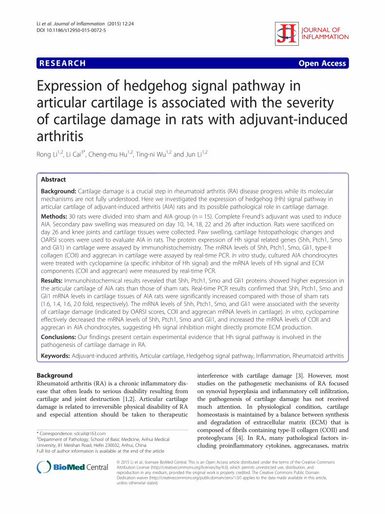
ResultsEvaluation of AIA in ratsTypical photos of non-injected hind paw from shamand AIA rats were taken on day 26 after AIA induction(Figure 1a,b). There was a significant increase of sec-ondary hind paw swelling (i.e. secondary inflammation)on different time points (days 14, 18, 22, 26) (Figure 1g).
Figure 1 Evaluation of AIA in rats. (a, b) Representative photos of non-inhistopathologic photos of knee joints sections with HE staining (×100). (e, f) RO staining (×100). (g) The changes of secondary hind paw swelling on differecartilage of knee joints. Data are mean ± SEM (n = 15). **p < 0.01 compared wi
Photomicrographs of knee joint paraffin sections withHE staining illustrated the severity of cartilage damagein AIA rats. No cartilage destruction was seen in theknee joints from sham rats (Figure 1c). On the contrary,typical pathological characteristics of cartilage damageincluding cartilage loss, articular cartilage zone thinnessand articular surface roughness were apparently foundin AIA rats (Figure 1d). Safranin O staining further re-vealed that proteoglycans were positively expressed inarticular cartilage of the sham rats (Figure 1e) whilewere faintly observed in the AIA rats (Figure 1f ). Inaddition, OARSI scores on articular cartilage in AIAgroup were obviously higher than those in sham group,indicating the articular cartilage damage of AIA rats(Figure 1h).
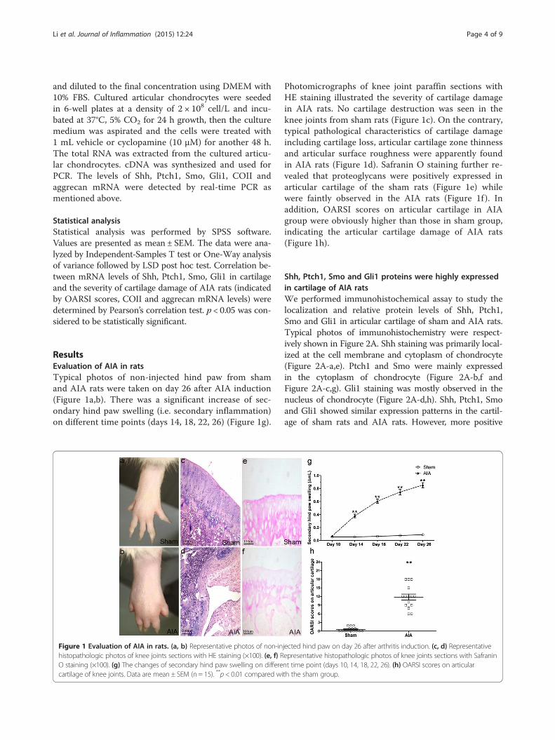
Shh, Ptch1, Smo and Gli1 proteins were highly expressedin cartilage of AIA ratsWe performed immunohistochemical assay to study thelocalization and relative protein levels of Shh, Ptch1,Smo and Gli1 in articular cartilage of sham and AIA rats.Typical photos of immunohistochemistry were respect-ively shown in Figure 2A. Shh staining was primarily local-ized at the cell membrane and cytoplasm of chondrocyte(Figure 2A-a,e). Ptch1 and Smo were mainly expressedin the cytoplasm of chondrocyte (Figure 2A-b,f andFigure 2A-c,g). Gli1 staining was mostly observed in thenucleus of chondrocyte (Figure 2A-d,h). Shh, Ptch1, Smoand Gli1 showed similar expression patterns in the cartil-age of sham rats and AIA rats. However, more positive
jected hind paw on day 26 after arthritis induction. (c, d) Representativeepresentative histopathologic photos of knee joints sections with Safraninnt time point (days 10, 14, 18, 22, 26). (h) OARSI scores on articularth the sham group.

Figure 2 Immunohistochemistry analyses for Shh, Ptch1, Smo and Gli1 in articular cartilage of knee joints. (A) Typical images of Shh,Ptch1, Smo and Gli1 expression in articular cartilage from sham and AIA rats (×100). Shh (a), Ptch1 (b), Smo (c) and Gli1 (d) were expressed atlow levels in articular cartilage from the sham rats, while relatively strong positive staining of Shh (e), Ptch1 (f), Smo (g) and Gli1 (h) were seen inthe AIA rats. Negative controls were enclosed to show the antibody specificity (i, j, k and l). (B) The positive expression rates of Shh, Ptch1, Smoand Gli1 in articular cartilage from sham and AIA rats. The positive expression rate is defined as (number of positive cells/total cells) × 100%. Dataare mean ± SEM (n = 15). **p < 0.01 compared with the sham group.
Li et al. Journal of Inflammation (2015) 12:24 Page 5 of 9
stained chondrocytes of the above proteins were seen inthe cartilage of AIA rats (Figure 2A-e,f,g and h). Negativecontrols were also enclosed to show the antibody
specificity (Figure 2A-i,j,k and l). We performed a statis-tical analysis on the positive expression rates of interestproteins in cartilage from the sham and AIA group. Our
Li et al. Journal of Inflammation (2015) 12:24 Page 6 of 9
results showed that the positive expression rates of Shh,Ptch1, Smo and Gli1 in AIA rats were all significantly in-creased compared with those in sham rats (Figure 2B).The results of semi-quantitative analyses of Shh, Ptch1,
Smo and Gli1 expressions in cartilage of the sham andAIA rats were shown in Table 2. Our results indicated thatthe rate of moderately (++) and strongly (+++) positive ex-pression of Shh, Ptch1, Smo and Gli1 (80%, 53.3%, 86.7%and 73.3% respectively) in AIA rats was higher than thatin the sham rats (33.3%, 20%, 40% and 20% respectively).However, no strongly (+++) positive expression of Ptch1was found in the sham and AIA group.
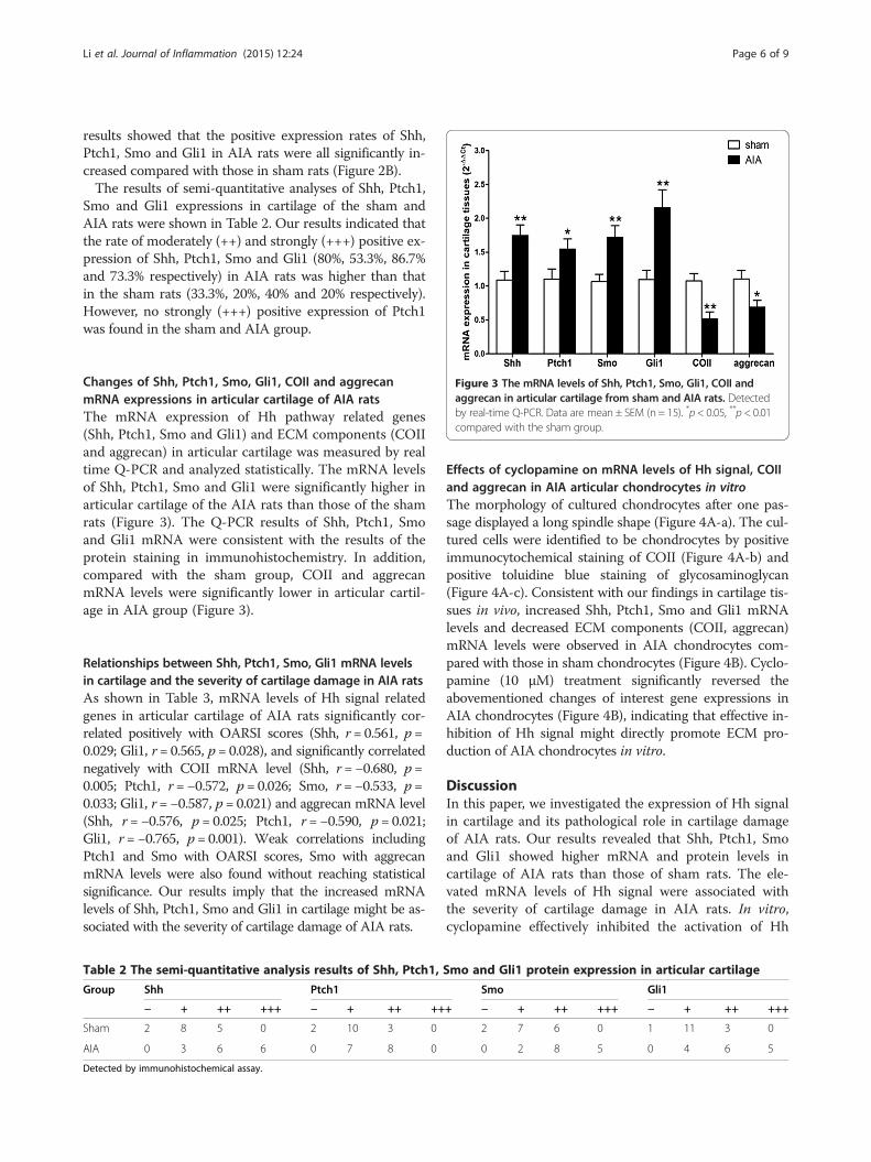
Figure 3 The mRNA levels of Shh, Ptch1, Smo, Gli1, COII andaggrecan in articular cartilage from sham and AIA rats. Detectedby real-time Q-PCR. Data are mean ± SEM (n = 15). *p < 0.05, **p < 0.01compared with the sham group.
Changes of Shh, Ptch1, Smo, Gli1, COII and aggrecanmRNA expressions in articular cartilage of AIA ratsThe mRNA expression of Hh pathway related genes(Shh, Ptch1, Smo and Gli1) and ECM components (COIIand aggrecan) in articular cartilage was measured by realtime Q-PCR and analyzed statistically. The mRNA levelsof Shh, Ptch1, Smo and Gli1 were significantly higher inarticular cartilage of the AIA rats than those of the shamrats (Figure 3). The Q-PCR results of Shh, Ptch1, Smoand Gli1 mRNA were consistent with the results of theprotein staining in immunohistochemistry. In addition,compared with the sham group, COII and aggrecanmRNA levels were significantly lower in articular cartil-age in AIA group (Figure 3).
Relationships between Shh, Ptch1, Smo, Gli1 mRNA levelsin cartilage and the severity of cartilage damage in AIA ratsAs shown in Table 3, mRNA levels of Hh signal relatedgenes in articular cartilage of AIA rats significantly cor-related positively with OARSI scores (Shh, r = 0.561, p =0.029; Gli1, r = 0.565, p = 0.028), and significantly correlatednegatively with COII mRNA level (Shh, r = −0.680, p =0.005; Ptch1, r = −0.572, p = 0.026; Smo, r = −0.533, p =0.033; Gli1, r = −0.587, p = 0.021) and aggrecan mRNA level(Shh, r = −0.576, p = 0.025; Ptch1, r = −0.590, p = 0.021;Gli1, r =−0.765, p = 0.001). Weak correlations includingPtch1 and Smo with OARSI scores, Smo with aggrecanmRNA levels were also found without reaching statisticalsignificance. Our results imply that the increased mRNAlevels of Shh, Ptch1, Smo and Gli1 in cartilage might be as-sociated with the severity of cartilage damage of AIA rats.
Table 2 The semi-quantitative analysis results of Shh, Ptch1,
Group Shh Ptch1
− + ++ +++ − + ++ ++
Sham 2 8 5 0 2 10 3 0
AIA 0 3 6 6 0 7 8 0
Detected by immunohistochemical assay.
Effects of cyclopamine on mRNA levels of Hh signal, COIIand aggrecan in AIA articular chondrocytes in vitroThe morphology of cultured chondrocytes after one pas-sage displayed a long spindle shape (Figure 4A-a). The cul-tured cells were identified to be chondrocytes by positiveimmunocytochemical staining of COII (Figure 4A-b) andpositive toluidine blue staining of glycosaminoglycan(Figure 4A-c). Consistent with our findings in cartilage tis-sues in vivo, increased Shh, Ptch1, Smo and Gli1 mRNAlevels and decreased ECM components (COII, aggrecan)mRNA levels were observed in AIA chondrocytes com-pared with those in sham chondrocytes (Figure 4B). Cyclo-pamine (10 μM) treatment significantly reversed theabovementioned changes of interest gene expressions inAIA chondrocytes (Figure 4B), indicating that effective in-hibition of Hh signal might directly promote ECM pro-duction of AIA chondrocytes in vitro.
DiscussionIn this paper, we investigated the expression of Hh signalin cartilage and its pathological role in cartilage damageof AIA rats. Our results revealed that Shh, Ptch1, Smoand Gli1 showed higher mRNA and protein levels incartilage of AIA rats than those of sham rats. The ele-vated mRNA levels of Hh signal were associated withthe severity of cartilage damage in AIA rats. In vitro,cyclopamine effectively inhibited the activation of Hh
Smo and Gli1 protein expression in articular cartilage
Smo Gli1
+ − + ++ +++ − + ++ +++
2 7 6 0 1 11 3 0
0 2 8 5 0 4 6 5
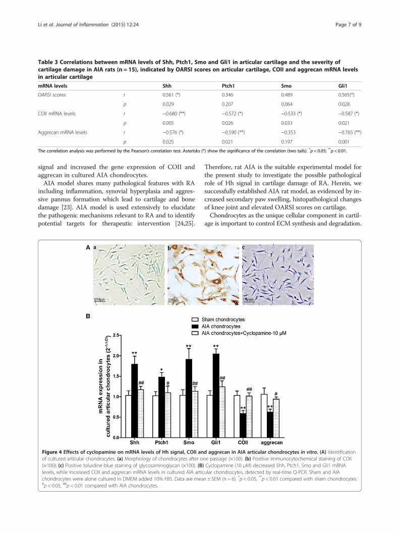
Table 3 Correlations between mRNA levels of Shh, Ptch1, Smo and Gli1 in articular cartilage and the severity ofcartilage damage in AIA rats (n = 15), indicated by OARSI scores on articular cartilage, COII and aggrecan mRNA levelsin articular cartilage
mRNA levels Shh Ptch1 Smo Gli1
OARSI scores r 0.561 (*) 0.346 0.489 0.565(*)
p 0.029 0.207 0.064 0.028
COII mRNA levels r −0.680 (**) −0.572 (*) −0.533 (*) −0.587 (*)
p 0.005 0.026 0.033 0.021
Aggrecan mRNA levels r −0.576 (*) −0.590 (**) −0.353 −0.765 (**)
p 0.025 0.021 0.197 0.001
The correlation analysis was performed by the Pearson’s correlation test. Asterisks (*) show the significance of the correlation (two tails). *p < 0.05; **p < 0.01.
Li et al. Journal of Inflammation (2015) 12:24 Page 7 of 9
signal and increased the gene expression of COII andaggrecan in cultured AIA chondrocytes.AIA model shares many pathological features with RA
including inflammation, synovial hyperplasia and aggres-sive pannus formation which lead to cartilage and bonedamage [23]. AIA model is used extensively to elucidatethe pathogenic mechanisms relevant to RA and to identifypotential targets for therapeutic intervention [24,25].
Figure 4 Effects of cyclopamine on mRNA levels of Hh signal, COII anof cultured articular chondrocytes. (a) Morphology of chondrocytes after o(×100); (c) Positive toluidine blue staining of glycosaminoglycan (×100). (B)levels, while increased COII and aggrecan mRNA levels in cultured AIA articchondrocytes were alone cultured in DMEM added 10% FBS. Data are mea#p < 0.05, ##p < 0.01 compared with AIA chondrocytes.
Therefore, rat AIA is the suitable experimental model forthe present study to investigate the possible pathologicalrole of Hh signal in cartilage damage of RA. Herein, wesuccessfully established AIA rat model, as evidenced by in-creased secondary paw swelling, histopathological changesof knee joint and elevated OARSI scores on cartilage.Chondrocytes as the unique cellular component in cartil-
age is important to control ECM synthesis and degradation.
d aggrecan in AIA articular chondrocytes in vitro. (A) Identificationne passage (×100). (b) Positive immunocytochemical staining of COIICyclopamine (10 μM) decreased Shh, Ptch1, Smo and Gli1 mRNAular chondrocytes, detected by real-time Q-PCR. Sham and AIAn ± SEM (n = 6). *p < 0.05, **p < 0.01 compared with sham chondrocytes.

Li et al. Journal of Inflammation (2015) 12:24 Page 8 of 9
Chondrocytes injury may be of pathogenetic signifi-cance in arthritis development including RA and OA[26]. Hh signal regulates chondrocyte proliferation anddifferentiation, finally influences the synthesis of colla-gen and aggrecan in ECM [27,28]. Some studies usingknockout mice revealed that activation of Hh pathwaycaused a remarkable decrease of cartilage thickness andproteoglycans content, while Hh inhibition increasedcartilage thickness and proteoglycans content [29,30].Over-activated Hh signal in chondrocytes promotedchondrocyte hypertrophy and cartilage damage in OA[11,12]. In the current study, immunohistochemistry re-sults showed that Hh signal related proteins (Shh,Ptch1, Smo and Gli1) were expressed in a similar pat-tern in articular cartilage of sham and AIA rats (i.e. Shhat cell membrane and cytoplasm, Ptch1 and Smo atcytoplasm, and Gli1 at nucleus). However, these pro-teins were highly expressed in articular cartilage of AIArats, while their expression levels were relatively lowerin sham rats. Real-time PCR results revealed that themRNA levels of Shh, Ptch1, Smo and Gli1 genes weresignificantly elevated in cartilage of AIA rats than thoseof sham rats. In addition, correlation analyses were per-formed to elucidate whether the increased mRNA levelsof Hh signal related genes were related to the severity ofcartilage damage of AIA rats. Our results indicated thatShh and Gli1 correlated positively with OARSI scores,Shh, Ptch1, Smo and Gli1 correlated negatively withCOII mRNA levels, and Shh, Ptch1 and Gli1 correlatednegatively with aggrecan mRNA levels. The above re-sults suggested that Hh signal was over-activated in thearticular cartilage of AIA rats and might play an import-ant role in cartilage damage.Cyclopamine is a steroidal alkaloid obtained from sep-
aration and extraction of the corn lily (Veratrum califor-nicum). As a classical specific inhibitor of Hh pathwayby directly binding to Smo, it has been extensively usedin vivo and in vitro experiments [31]. In the presentstudy, we successfully isolated and prepared articular chon-drocytes which were identified by positively stain of COIIand glycosaminoglycan. Then we investigated the possibleeffect of cyclopamine on mRNA expression of Hh signalrelated genes and ECM components (COII and aggrecan)in cultured AIA articular chondrocytes. Consistent withour findings in cartilage tissues in vivo, the mRNA levels ofShh, Ptch1, Smo and Gli1 were significantly elevated, whilethe mRNA levels of COII and aggrecan were remarkablyreduced in cultured AIA chondrocytes than those in shamchondrocytes. Cyclopamine treatment significantly sup-pressed the activation of Hh signal and increased COII,aggrecan mRNA expressions in cultured AIA chondro-cytes. It is well known that COII is the main structural col-lagen in cartilage and aggrecan is the major structuralproteoglycans in articular cartilage ECM [32]. Our results
suggested that inhibition of Hh signal by cyclopaminemight directly promote ECM production of AIA chondro-cytes in vitro.
ConclusionsTaken together, our study revealed that Hh signal pathwaywas over-activated in the articular cartilage of AIA rats,and the up-regulated Hh signal was apparently associatedwith the severity of cartilage damage. In vitro, inhibition ofHh signal by cyclopamine could promote COII and aggre-can mRNA expressions in cultured AIA chondrocytes.The present study suggests, for the first time, that Hh sig-nal pathway underlies the pathogenesis of cartilage de-struction in RA. Further research is needed to investigatethe potential therapeutical effect of cyclopamine and itsmolecular mechanisms on AIA rats.
AbbreviationsAIA: Adjuvant-induced arthritis; CFA: Complete Freund’s adjuvant; COII: Type-IIcollagen; ECM: Extracellular matrix; FBS: Fetal calf serum; FLS: Fibroblast-likesynoviocytes; Gli1: Glioma-associated oncogene homolog 1; Hh: Hedgehog;OA: Osteoarthritis; Ptch1: Patched homologue 1; RA: Rheumatoid arthritis;Shh: Sonic hedgehog; Smo: Smoothened.
Competing interestsThe authors declare that they have no competing interests.
Authors’ contributionsLC and JL designed the study and wrote the protocol and the first draft of themanuscript. CH managed the literature searches and the statistical analyses. TWperformed animal model experiments and immunohistochemistry analysis. RLperformed the in vitro study and the gene expression experiments, and wroteparts of the manuscript. All authors read and approved the final manuscript.
AcknowledgementsThis study was supported by National Natural Science Foundation of China(81102273), Specialized Research Fund for the Doctoral Program of HigherEducation (20113420120005), Program for the Young and Middle-agedAcademic Technology Leaders of Anhui Medical University (201309) andProgram for the Top Young Talents of Anhui Medical University.
Author details1School of Pharmacy, Anhui Medical University, 81 Meishan Road, Hefei230032, Anhui, China. 2Key Laboratory for Bioactivity of Natural Medicine ofAnhui Province, Hefei 230032, Anhui, China. 3Department of Pathology,School of Basic Medicine, Anhui Medical University, 81 Meishan Road, Hefei230032, Anhui, China.
Received: 7 August 2014 Accepted: 20 March 2015
References1. Andreas K, Lubke C, Haupl T, Dehne T, Morawietz L, Ringe J, et al. Key
regulatory molecules of cartilage destruction in rheumatoid arthritis: anin vitro study. Arthritis Res Ther. 2008;10:R9.
2. Nishioku T, Dohgu S, Koga M, Machida T, Watanabe T, Miura T, et al.Cyclophilin A secreted from fibroblast-like synoviocytes is involved in theinduction of CD147 expression in macrophages of mice with collagen-inducedarthritis. J Inflamm (Lond). 2012;9:44.
3. Aletaha D, Funovits J, Smolen JS. Physical disability in rheumatoid arthritis isassociated with cartilage damage rather than bone destruction. Ann RheumDis. 2011;70:733–9.
4. Yasuda T. Cartilage destruction by matrix degradation products. Mod Rheumatol.2006;16:197–205.

Li et al. Journal of Inflammation (2015) 12:24 Page 9 of 9
5. Marotte H, Gineyts E, Miossec P, Delmas PD. Effects of infliximab therapy onbiological markers of synovium activity and cartilage breakdown in patientswith rheumatoid arthritis. Ann Rheum Dis. 2009;68:1197–200.
6. Day TF, Yang Y. Wnt and hedgehog signaling pathways in bonedevelopment. J Bone Joint Surg Am. 2008;90 Suppl 1:19–24.
7. Mak KK, Bi Y, Wan C, Chuang PT, Clemens T, Young M, et al. Hedgehogsignaling in mature osteoblasts regulates bone formation and resorption bycontrolling PTHrP and RANKL expression. Dev Cell. 2008;14:674–88.
8. Berman DM, Karhadkar SS, Maitra A, Montes De Oca R, Gerstenblith MR,Briggs K, et al. Widespread requirement for Hedgehog ligand stimulation ingrowth of digestive tract tumours. Nature. 2003;425:846–51.
9. Pasca di Magliano M, Hebrok M. Hedgehog signalling in cancer formationand maintenance. Nat Rev Cancer. 2003;3:903–11.
10. Wilson CW, Chuang PT. Mechanism and evolution of cytosolic Hedgehogsignal transduction. Development. 2010;137:2079–94.
11. Lin AC, Seeto BL, Bartoszko JM, Khoury MA, Whetstone H, Ho L, et al.Modulating hedgehog signaling can attenuate the severity of osteoarthritis.Nat Med. 2009;15:1421–5.
12. Wei F, Zhou J, Wei X, Zhang J, Fleming BC, Terek R, et al. Activation ofIndian hedgehog promotes chondrocyte hypertrophy and upregulation ofMMP-13 in human osteoarthritic cartilage. Osteoarthritis Cartilage.2012;20:755–63.
13. Wang M, Zhu S, Peng W, Li Q, Li Z, Luo M, et al. Sonic hedgehog signalingdrives proliferation of synoviocytes in rheumatoid arthritis: a possible noveltherapeutic target. J Immunol Res. 2014;2014:401903.
14. Narendhirakannan RT, Limmy TP. Anti-inflammatory and anti-oxidantproperties of Sida rhombifolia stems and roots in adjuvant induced arthriticrats. Immunopharmacol Immunotoxicol. 2012;34:326–36.
15. Suresh P, Kavitha Ch N, Babu SM, Reddy VP, Latha AK. Effect of ethanolextract of Trigonella foenum graecum (Fenugreek) seeds on Freund’sadjuvant-induced arthritis in albino rats. Inflammation. 2012;35:1314–21.
16. Custers RJ, Creemers LB, Verbout AJ, van Rijen MH, Dhert WJ, Saris DB.Reliability, reproducibility and variability of the traditional Histologic/Histochemical Grading System vs the new OARSI Osteoarthritis CartilageHistopathology Assessment System. Osteoarthritis Cartilage. 2007;15:1241–8.
17. Gerwin N, Bendele AM, Glasson S, Carlson CS. The OARSI histopathologyinitiative - recommendations for histological assessments of osteoarthritis inthe rat. Osteoarthritis Cartilage. 2010;18 Suppl 3:S24–34.
18. Cai L, Yan XB, Chen XN, Meng QY, Zhou JN. Chronic all-trans retinoic acidadministration induced hyperactivity of HPA axis and behavioral changes inyoung rats. Eur Neuropsychopharmacol. 2010;20:839–47.
19. Yoshikawa R, Nakano Y, Tao L, Koishi K, Matsumoto T, Sasako M, et al.Hedgehog signal activation in oesophageal cancer patients undergoingneoadjuvant chemoradiotherapy. Br J Cancer. 2008;98:1670–4.
20. Livak KJ, Schmittgen TD. Analysis of relative gene expression data usingreal-time quantitative PCR and the 2(−Delta Delta C(T)) Method. Methods.2001;25:402–8.
21. Ponce A. Expression of voltage dependent potassium currents in freshlydissociated rat articular chondrocytes. Cell Physiol Biochem. 2006;18:35–46.
22. Yuan FL, Chen FH, Lu WG, Li X, Li JP, Li CW, et al. Inhibition of acid-sensingion channels in articular chondrocytes by amiloride attenuates articularcartilage destruction in rats with adjuvant arthritis. Inflamm Res.2010;59:939–47.
23. Bevaart L, Vervoordeldonk MJ, Tak PP. Evaluation of therapeutic targets inanimal models of arthritis: how does it relate to rheumatoid arthritis?Arthritis Rheum. 2010;62:2192–205.
24. Shah SU, Ashraf N, Soomro ZH, Shah MR, Kabir N, Simjee SU. Theanti-arthritic and anti-oxidative effect of NBD (6-nitro-1,3-benzodioxane) inadjuvant-induced arthritis (AIA) in rats. Inflamm Res. 2012;61:875–87.
25. Meyer DM, Jesson MI, Li X, Elrick MM, Funckes-Shippy CL, Warner JD, et al.Anti-inflammatory activity and neutrophil reductions mediated by theJAK1/JAK3 inhibitor, CP-690,550, in rat adjuvant-induced arthritis. J Inflamm(Lond). 2010;7:41.
26. Temenoff JS, Mikos AG. Review: tissue engineering for regeneration ofarticular cartilage. Biomaterials. 2000;21:431–40.
27. Lin L, Shen Q, Xue T, Duan X, Fu X, Yu C. Sonic hedgehog improvesredifferentiation of dedifferentiated chondrocytes for articular cartilagerepair. PLoS One. 2014;9:e88550.
28. Maeda Y, Nakamura E, Nguyen MT, Suva LJ, Swain FL, Razzaque MS, et al.Indian Hedgehog produced by postnatal chondrocytes is essential formaintaining a growth plate and trabecular bone. Proc Natl Acad Sci U S A.2007;104:6382–7.
29. Beaupre GS, Stevens SS, Carter DR. Mechanobiology in the development,maintenance, and degeneration of articular cartilage. J Rehabil Res Dev.2000;37:145–51.
30. Mak KK, Kronenberg HM, Chuang PT, Mackem S, Yang Y. Indian hedgehogsignals independently of PTHrP to promote chondrocyte hypertrophy.Development. 2008;135:1947–56.
31. Taipale J, Chen JK, Cooper MK, Wang B, Mann RK, Milenkovic L, et al. Effectsof oncogenic mutations in Smoothened and Patched can be reversed bycyclopamine. Nature. 2000;406:1005–9.
32. Roughley PJ. The structure and function of cartilage proteoglycans. Eur CellMater. 2006;12:92–101.
Submit your next manuscript to BioMed Centraland take full advantage of:
• Convenient online submission
• Thorough peer review
• No space constraints or color figure charges
• Immediate publication on acceptance
• Inclusion in PubMed, CAS, Scopus and Google Scholar
• Research which is freely available for redistribution
Submit your manuscript at www.biomedcentral.com/submit