Embed Size (px)
Citation preview

DISSERTATION
Titel der Dissertation
The VEGF-regulated transcription factor HLX controls the expression of guidance cues and negatively regulates
sprouting of endothelial cells
Verfasserin
Mag. rer. nat. Julia Testori
angestrebter akademischer Grad
Doktorin der Naturwissenschaften (Dr.rer.nat.)
Wien, Februar 2011
Studienkennzahl lt. Studienblatt: A 091 441
Dissertationsgebiet lt. Studienblatt: Genetik - Mikrobiologie
Betreuerin / Betreuer: Univ.-Prof. Dr. Erhard Hofer

Danksagung
Mein Dank gilt meinem Betreuer Erhard Hofer, der mir die Möglichkeit gegeben hat,
an einem interessanten Thema zu arbeiten, und mich dabei immer unterstützt hat.
Weiters danke ich meinen Kolleginnen und Kollegen, Caterina Sturtzel, Dorit Reiche,
Karoline Lipnik, Bettina Strasser, Renate Hofer-Warbinek, Thomasz Bobrzynski,
Susanne Sattler, Irene Karas und Bernhard Schweighofer, die mir immer mit Rat und
Tat zur Seite standen und mit denen der Laboralltag immer wieder unterhaltsam und
motivierend war.
Ganz besonders bedanken möchte ich mich bei meinen Eltern, die mir dieses
Studium ermöglicht haben und mich all die Jahre immer unterstützt haben.
Vor allem danke ich meinem Ehemann Hannes, ohne den ich das niemals geschafft
hätte, für seine Geduld und Unterstützung.
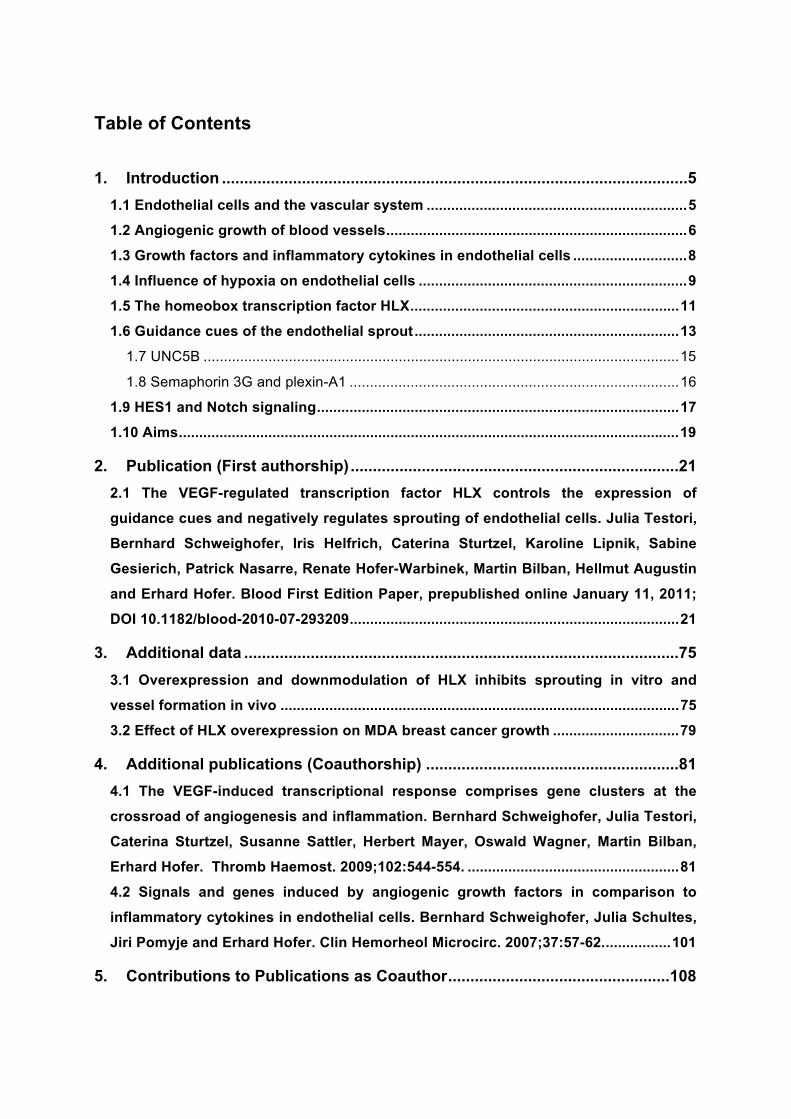
Table of Contents
1. Introduction .........................................................................................................5
1.1 Endothelial cells and the vascular system ................................................................5
1.2 Angiogenic growth of blood vessels..........................................................................6
1.3 Growth factors and inflammatory cytokines in endothelial cells ............................8
1.4 Influence of hypoxia on endothelial cells ..................................................................9
1.5 The homeobox transcription factor HLX..................................................................11
1.6 Guidance cues of the endothelial sprout.................................................................13
1.7 UNC5B .....................................................................................................................15
1.8 Semaphorin 3G and plexin-A1 .................................................................................16
1.9 HES1 and Notch signaling.........................................................................................17
1.10 Aims...........................................................................................................................19
2. Publication (First authorship)..........................................................................21
2.1 The VEGF-regulated transcription factor HLX controls the expression of
guidance cues and negatively regulates sprouting of endothelial cells. Julia Testori,
Bernhard Schweighofer, Iris Helfrich, Caterina Sturtzel, Karoline Lipnik, Sabine
Gesierich, Patrick Nasarre, Renate Hofer-Warbinek, Martin Bilban, Hellmut Augustin
and Erhard Hofer. Blood First Edition Paper, prepublished online January 11, 2011;
DOI 10.1182/blood-2010-07-293209.................................................................................21
3. Additional data ..................................................................................................75
3.1 Overexpression and downmodulation of HLX inhibits sprouting in vitro and
vessel formation in vivo ..................................................................................................75
3.2 Effect of HLX overexpression on MDA breast cancer growth ...............................79
4. Additional publications (Coauthorship) .........................................................81
4.1 The VEGF-induced transcriptional response comprises gene clusters at the
crossroad of angiogenesis and inflammation. Bernhard Schweighofer, Julia Testori,
Caterina Sturtzel, Susanne Sattler, Herbert Mayer, Oswald Wagner, Martin Bilban,
Erhard Hofer. Thromb Haemost. 2009;102:544-554. ....................................................81
4.2 Signals and genes induced by angiogenic growth factors in comparison to
inflammatory cytokines in endothelial cells. Bernhard Schweighofer, Julia Schultes,
Jiri Pomyje and Erhard Hofer. Clin Hemorheol Microcirc. 2007;37:57-62.................101
5. Contributions to Publications as Coauthor..................................................108

5.1 The VEGF-induced transcriptional response comprises gene clusters at the
crossroad of angiogenesis and inflammation.............................................................108
5.2 Signals and genes induced by angiogenic growth factors in comparison to
inflammatory cytokines in endothelial cells ................................................................109
6. Discussion.......................................................................................................110
References..............................................................................................................119
Table of Figures .....................................................................................................123
Abstract...................................................................................................................124
Zusammenfassung ................................................................................................125
Curriculum Vitae ....................................................................................................126
Introduction
5
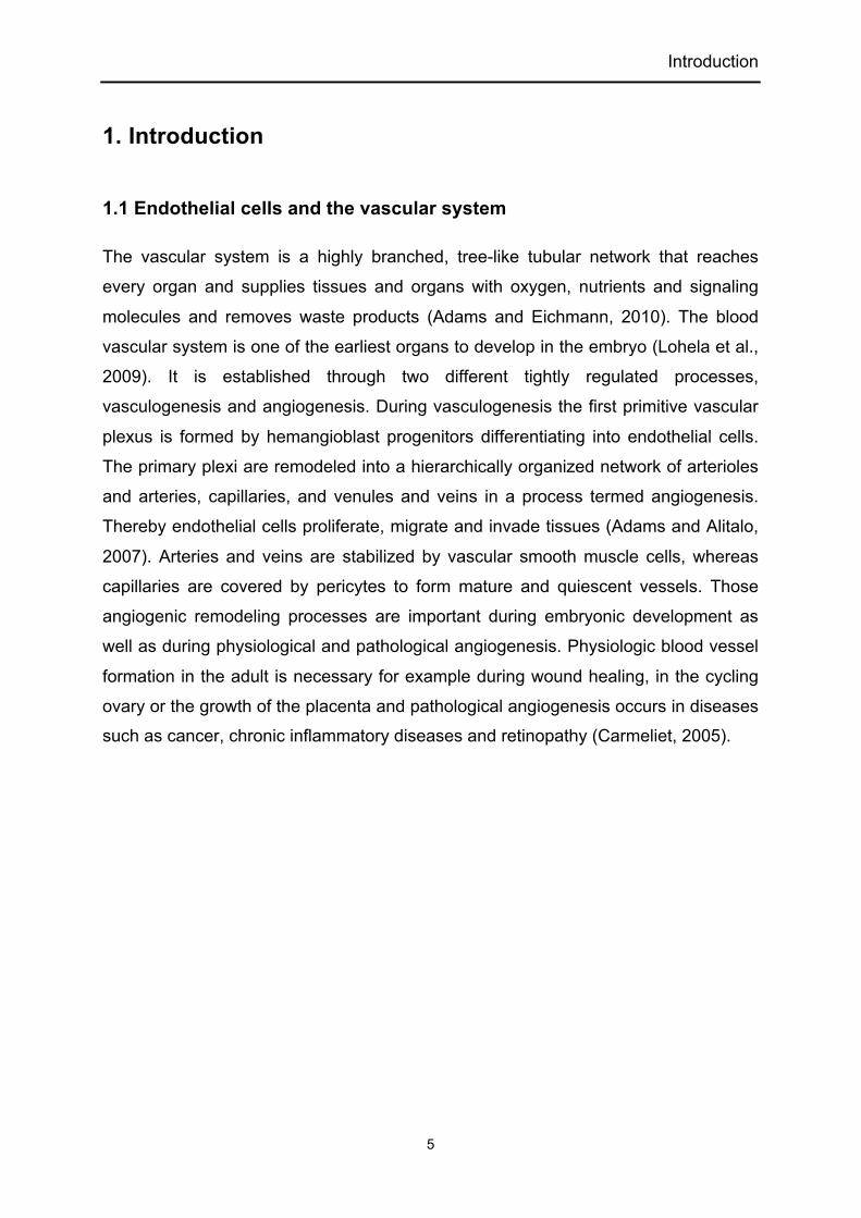
1. Introduction
1.1 Endothelial cells and the vascular system
The vascular system is a highly branched, tree-like tubular network that reaches
every organ and supplies tissues and organs with oxygen, nutrients and signaling
molecules and removes waste products (Adams and Eichmann, 2010). The blood
vascular system is one of the earliest organs to develop in the embryo (Lohela et al.,
2009). It is established through two different tightly regulated processes,
vasculogenesis and angiogenesis. During vasculogenesis the first primitive vascular
plexus is formed by hemangioblast progenitors differentiating into endothelial cells.
The primary plexi are remodeled into a hierarchically organized network of arterioles
and arteries, capillaries, and venules and veins in a process termed angiogenesis.
Thereby endothelial cells proliferate, migrate and invade tissues (Adams and Alitalo,
2007). Arteries and veins are stabilized by vascular smooth muscle cells, whereas
capillaries are covered by pericytes to form mature and quiescent vessels. Those
angiogenic remodeling processes are important during embryonic development as
well as during physiological and pathological angiogenesis. Physiologic blood vessel
formation in the adult is necessary for example during wound healing, in the cycling
ovary or the growth of the placenta and pathological angiogenesis occurs in diseases
such as cancer, chronic inflammatory diseases and retinopathy (Carmeliet, 2005).

Introduction
6
Figure 1. Murine embryonic vasculature
Murine developing vasculature on embryonic day 9.5 was stained for CD31 (PECAM) and analyzed with optical projection tomography. BA, branchial arteries; DA, dorsal aorta; ICA, intercarotid artery; ISV, intersomitic vessels; OFT, outflow tract; PCV, posterior cardinal vein; RV, right ventricle. Reprinted by permission from Macmillan Publishers Ltd: Nature (Coultas et al., 2005), copyright 2005.
1.2 Angiogenic growth of blood vessels
The growth factor VEGF-A is the major trigger of angiogenesis. Binding to its
receptor VEGFR2 leads to endothelial cell differentiation, proliferation and sprouting.
Some endothelial cells within the capillary wall are selected for sprouting and
become motile and invasive and initiate sprouting. These cells are the tip cells which
lead the growing sprout. They sense the environment for guidance cues using their
continuously searching filopodia. Those specialized cells resemble the axonal growth
cone in the nervous system in structure and function to a high degree. The growing
sprout is guided by a VEGF-A gradient and by other attractive and repulsive
guidance cues in the matrix or on guidepost cells in the tissue. The subsequent stalk
cells trail behind the tip cells. They proliferate, form junctions, lay down extracellular

Introduction
7
matrix and form a lumen by the fusion of vacuoles. A third group of specialized
endothelial cells are the so called phalanx cells that are the most quiescent cells that
line the vessels once the new vessel branches have been consolidated. They are
already covered by pericytes, have tight junctions and are embedded in a thick
basement membrane. The specialized phalanx cells are engaged in optimizing blood
flow, tissue perfusion and oxygenation. The expression of platelet-derived growth
factor (PDGFB) by the tip cells leads to the recruitment of mural cells, vascular
smooth muscle cells and pericytes that express the PDGF receptor ß (Adams and
Alitalo, 2007; De Smet et al., 2009).
Figure 2. Vascular sprouts are guided by endothelial tip cells.
Tip cell (green) lead the growing sprout and extend their filopodia toward stimuli in the tissue environment (red gradient). Proliferating stalk cells (purple) trail behind, elongating the sprout, while phalanx cells (gray) remain quiescent and form a tight barrier. Reprinted by permission from Wolters Kluwer Health: Arterioscler Thromb Vasc Biol (De Smet et al., 2009), copyright 2009.

Introduction
8
Figure 3. Angiogenic sprouting and blood vessel growth. Sprouting is initiated upon stimulating signals such as VEGF-A from the tissue-environment. Some endothelial cells (yellow and green) differentiate into tip cells extending filopodia, sensing the tissue surroundings, migrating and invading. The stalk cells (red) trail behind and form the sprout stalk. To form new connections, the tip cells contact other growing sprouts or established vessels. These cell bridges (orange) are altered into perfused vessels with a lumen. Eventually further sprouting initiates elsewhere by tip cells (yellow, green) and additional endothelial cells proliferate (purple). Reprinted by permission from Cold Spring Harb Laboratory Press: (Adams and Eichmann, 2010), copyright 2010.
1.3 Growth factors and inflammatory cytokines in endothelial cells
Vascular endothelial growth factor-A (VEGF-A) is a key molecule for the initiation and
direction of sprouting angiogenesis. In hypoxic areas cells express VEGF-A and this
leads to the formation of a VEGF-A gradient which serves as a directional and
chemoattractive cue for endothelial sprouts. VEGF-A binds to and activates via the
receptor tyrosine kinase VEGFR2/KDR/Flk1 the major angiogenic signaling pathway
(Eilken and Adams, 2010). In addition, it can also bind VEGFR1, which seems to act
as a negative regulator of angiogenesis during development. VEGF-A is part of a
large family of angiogenic molecules which includes placental growth factor (PlGF),
VEGF-B, VEGF-C and VEGF-D. In addition to angiogenesis, VEGF-A is essential for

Introduction
9
chemotaxis and differentiation of endothelial precursor cells and vasculogenesis.
Alternative splicing of VEGF-A can generate several different isoforms and those can
regulate blood-vessel growth and patterning of the vasculature. The splice variants
differ in their matrix binding and co-receptor binding capability. VEGF-A121 is freely
diffusible, VEGF-A165 the predominant angiogenic form and VEGF-A189 is tightly
matrix-bound. The b-isoform even has anti-angiogenic properties (Adams and Alitalo,
2007; Lohela et al., 2009).
Basic fibroblast growth factor (bFGF) is another important angiogenic growth factor
for endothelial cells. bFGF binds and signals mainly through the receptor tyrosine
kinase FGF receptor-1. bFGF can trigger basal lamina degradation, migration,
proliferation and has effects on morphogenesis and vessel maturation during
angiogenic processes (Presta et al., 2005).
In comparison to VEGF-A, epidermal growth factor (EGF) represents a more general
growth factor inducing proliferation and survival in many different cell types. EGF and
the HER or erbB receptors are prototypes of growth factors and receptor tyrosine
kinases and are of wide importance for the development and proliferation of epithelial
and many other cell types. In endothelial cells EGF can mediate proliferation and a
gene repertoire much smaller than VEGF-A, which is the essential factor for directed
endothelial sprouting angiogenesis (Citri and Yarden, 2006; Schweighofer et al.,
2009; Sini et al., 2005).
Angiogenesis often takes place in inflammatory surroundings, e.g. during wound
healing and in tumors, which are created by immune cells invading the damaged or
malignant tissue. In response to cytokines or histamines, endothelial cells are
activated to express a large number of inflammatory genes like cytokines, enzymes
and adhesion molecules implicated in immune cell recruitment, activation and tissue
repair. The cytokine IL-1 signals through the type 1 IL-1 receptor activating the
transcription factor nuclear factor-kappaB (NF- B) and thereby the transcription of
proinflammatory genes (Pober and Sessa, 2007; Schweighofer et al., 2009).
1.4 Influence of hypoxia on endothelial cells
A decrease in oxygen tension leads to tissue hypoxia, which attracts angiogenic
sprouting into those areas (Germain et al., 2010). Three hypoxia-inducible factor-

Introduction
10
proteins (HIF-1 , HIF-2 and HIF-3 ) show oxygen-regulated protein stability and
together with HIFß (ARNT) activate HIF target genes in response to hypoxia through
binding to hypoxia response elements. These transcription factors induce the
expression of hundreds of genes important for the regulation of cell survival,
metabolism and angiogenesis. During normoxia prolyl hydroxylases (PHDs)
hydroxylate HIF- s in an oxygen-dependent manner and this induces HIFs to interact
with VHL (von Hippel-Lindau) and its degradation through the proteasome. Factor
inhibiting HIFs (FIH) also hydroxylates HIF-1 and thereby inhibits the interaction of
HIF-1 with its transcriptional coactivator p300 (Fraisl et al., 2009). In the case of
hypoxia the hydroxylases cannot hydroxylate the HIF factors, which results in
stabilization, increase in concentration and efficient interaction of HIFs with p300.
HIF-2 , which is highly expressed in vascular endothelial cells, seems to be the
predominant form responsible for modulation of vascular function and angiogenesis.
HIF-1 is more broadly expressed throughout most tissues and has a role in
mediating the paracrine effects of angiogenesis inducing angiogenic factors such as
VEGF-A (Fong, 2009).
Hypoxia upregulates growth factors, provisional extracellular matrix (ECM)
components and vascular basement membrane proteins in endothelial cells and
therefore regulates mechanical and biological properties of vascular basement
membrane and ECM. Among the proteins upregulated by hypoxia are the
thrombospondins (TSPs), a family of secreted matricellular proteins that function as
adapter proteins to guide ECM synthesis and remodeling. Furthermore, angiopoietin-
like 4 (ANGPTL4) is expressed by hypoxic endothelial cells and promotes
angiogenesis and lipid metabolism. These cells further induce Type IV collagen,
heparin sulfate proteoglycans (HSPGs), and several ECM-modifying enzymes (LOX,
LOXL2, PLID1,2 and P4HA1,2) (Germain et al., 2010).

Introduction
11
Figure 4. Schematic drawing of a filopodial-extending tip cell (dark gray) migrating
toward the hypoxic area (gray gradient) Tip cells migrate extending their filopodia on an interstitial fibronectin network (black strings) and guide the endothelial sprout. The provisional extracellular matrix is remodeled at the front of the tip cell (gray network) and the basement membrane (black line) is deposited at the back of the tip cell as well as at stalk cells (white) and surrounding pericytes (light gray). Reprinted by permission from Wolters Kluwer Health: Current Opinion in Hematology (Germain et al., 2010), copyright 2010.
1.5 The homeobox transcription factor HLX
HLX (H2.0-like homeobox protein) is an evolutionary highly conserved homeobox
transcription factor which was originally detected in the visceral musculature of
Drosophila (H2.0) (Barad et al., 1991). The homeodomain is a 60-amino-acid DNA-
binding domain showing considerable variations in primary sequence within the
different homeobox genes. However, the three-dimensional structure is conserved
and consists of three -helices and an unstructured amino-terminal arm (Abate-
Shen, 2002). The largest family of homeobox genes is the HOX family, which
consists of four clusters (A-D) and comprises 39 genes. They were discovered to be
the mammalin orthologs of homeotic genes of Drosophila. Most homeobox families
are unclustered, diverged homeobox genes, where the members are dispersed
throughout the genome. Homeobox transcription factors are developmental
regulators that are essential for cell proliferation, cell differentiation and migration in
many cell types. These properties are responsible for their critical role in regulating
pattern formation and organogenesis during embryogenesis. Several members of the

Introduction
12
homeobox family have also been implicated in the formation of the embryonic
vascular system as well as in vascular remodeling in the adult and have a role in
many pathologies including atherosclerosis and tumor angiogenesis (Douville and
Wigle, 2007; Gorski and Walsh, 2003).
Human HLX was first isolated from a mitogen-activated human B-lymphocyte cDNA
library using probes designed to hybridize homeodomains. HLX is further expressed
in bone marrow-derived CD34+ cells and in vitro differentiation results in down-
regulation of HLX expression (Deguchi and Kehrl, 1991). Furthermore, in naïve CD4
T lympohcytes HLX interacts with T-bet to promote heritable Th1 gene induction.
This genetic interaction initiates optimal induction of interferon-g expression and the
Ifng locus then undergoes DNA methylation and stable chromatin remodeling,
thereby becoming independent of T-bet activity (Mullen et al., 2002). HLX expression
is dispensable for the maintenance of a transcriptional permissive ifng gene, since
this signature gene activity of helper T cells becomes epigenetically fixed (Martins et
al., 2005). In resting naïve CD4 T cells, HLX furthermore genetically controls IL-4R
level at the transcriptional level and thereby determines the ration of Th1 and Th2 cell
differentiation. Transgenic HLX down-regulates IL4-R and IL-4 signaling and
enhances Th1 response (Mikhalkevich et al., 2006).
In contrast to CD4 T cells, HLX expression is induced in monokine-activated NK cells
with delayed kinetics compared to IFN- and ectopic expression of HLX negatively
regulated IFN- production. This inhibition is at least partly achieved through the
accelerated dephosphorylation and proteasomal degradation of STAT4, a key
transcription factor for IFN- (Becknell et al., 2007).
The homeobox gene HLX is also expressed in the placental vasculature, exhibiting a
higher expression in placental microvascular endothelial cells when compared to
macrovascular human umbilical vein endothelial cells (HUVEC). The heterogeneity of
expression of HLX and other homeobox genes probably reflects differences of
function in those endothelial cells. In the placenta, microvascular endothelial cells are
important for the formation of the placental vasculature and they vascularize the
cotyledons of the placenta, which are essential for maternal-fetal gas and nutrient
exchange and play a role in placental disorders (Murthi et al., 2007).

Introduction
13
During placental development abnormal throphoblast development is associated with
clinically significant pregnancy disorders such as fetal growth restriction and
preeclampsia. HLX is expressed in proliferating and migrating human trophoblast
cells in the early placenta, showing a nuclear localization in the villous
cytotrophoblast cells and the extravillous trophoblasts in the proximal region of the
cell column and a cytoplasmic expression in interstitial trophoblasts and in the
endovascular trophoblasts. In human fetal growth restriction HLX expression is
significantly decreased. Moreover, HLX is a regulator of colony- stimulating-factor-1
dependent trophoblast proliferation and of hepatocyte growth factor/c-met- mediated
trophoblast migration (Murthi et al., 2006; Rajaraman et al., 2010; Rajaraman et al.,
2007; Rajaraman et al., 2009; Rajaraman et al., 2008).
Furthermore, in endometrial epithelium HLX is expressed in both the proliferative and
secretory phase and might be required for the transcriptional control of genes
necessary for endometrial cell differentiation and to control epithelial-mesenchymal
cell interaction in the endometrium (Quinn et al., 1998).
In mouse development the HLX-/- genotype is lethal around day 15 of embryonic
development on a mixed genetic background and the embryos display anemia and
severe hypoplasia of the liver and the gut. During murine embryogenesis HLX is
most prominently expressed in the visceral mesenchyme of the developing liver, gall
bladder and gut and regulates a mesenchymal-epithelial interaction that is required
for early aspects of enteric nervous system development (Bates et al., 2006; Hentsch
et al., 1996).
1.6 Guidance cues of the endothelial sprout
The angiogenic sprout expresses similar receptors for guidance as the nerve growth
cone and responds to similar attractive and repulsive cues (Adams and Eichmann,
2010). Proteins of the ROBO, uncoordinated 5 (UNC5), plexin or neuropilin families
as well as of the Eph receptor tyrosine kinase family are expressed on tip cell
filopodia and serve as receptors to sense and respond to the corresponding
guidance cues. These include proteins of the SLIT, netrin, semaphorin and ephrin
families that are either secreted in the tissue environment or are transmembrane
proteins on guidepost cells. These ligand-receptor interactions lead either to

Introduction
14
attraction or repulsion of the tip cell filopodia and the growing sprout is thus guided to
form new connections.
The receptors ROBO4, plexin-D1 and UNC5B are predominately expressed in the
vasculature, whereas many of the other isoforms are both expressed on nerve and
endothelial cells.
Roundabouts (ROBOs) are single-pass transmembrane receptors for SLITs. ROBO4
-/- mice show that ROBO4 is necessary for blood vessel integrity and they have
increased retinal vascular permeability and hypervascularization during oxygen-
induced retinopathy. However, whether ROBO4 binds to SLITs during blood vessel
regulation is still unclear. Furthermore, expression of ROBO1 in the vasculature and
interactions with SLITs have been reported to affect endothelial cell migration.
The netrin receptor UNC5B is vascular-specific and primarily expressed in arterial
endothelial cells, sprouting capillaries and tip cells. Binding of netrins leads to
repulsion of endothelial sprouts. It was further reported that netrin-4, which is
upregulated in endothelial cells by long-term VEGF-A induction, binds to neogenin
associated with UNC5B and mediates repulsion. Additional proposed binding
partners are repulsive guidance molecule (RGM) for neogenin and fibronectin and
leucine rich transmembrane protein 3 (FLRT3) for UNC5B (Adams and Eichmann,
2010).
Another class of guidance molecules are the semaphorins that are characterized by
a so-called Sema domain. The class III semaphorins are secreted molecules and
bind to neuropilin, which again interacts with the signal transducing plexins. An
exception to this rule is SEMA3E that directly binds plexin-D1 and mediates
repulsion. Most of these signaling molecules have a preferential repulsive regulatory
role on endothelial tip cells. However, neuropilins can also bind VEGF-A and then
have an attractive guidance function upon interaction with VEGFR.
Moreover, the Eph receptor tyrosine kinases, transmembrane proteins with a single
cytoplasmic kinase domain bind to ephrins, which are cell surface proteins, and
thereby regulate different processes in the vasculature. Especially the interaction of
ephrin-B2 ligand with the EphB4 receptor is important for the regulation of endothelial

Introduction
15
cell migration and angiogenesis leading to different outcomes under diverse
circumstances (Adams and Eichmann, 2010).
Figure 5. Axon guidance receptor expression in endothelial cells Schematic representation of the four families of axon guidance cues and their receptors. Predominantly endothelial-expressed receptors are labeled in red, receptors with shared expression in the nervous and the vascular system in blue and molecules with no (known) expression in the vascular system in black. Note that in each axon guidance receptor family, at least one member is expressed in the vasculature. Reprinted by permission from Cold Spring Harb Laboratory Press: Developmental Cell (Adams and Eichmann, 2010), copyright 2010.
1.7 UNC5B
The uncoordinated5 (UNC5) molecule family are transmembrane receptors that
contain two immunoglobulin and two thrombospondin-like domains in the
extracellular region and a zona occluden 5 domain, a DCC-binding domain and a
death domain in the intracellular region. The receptor UNC5B is selectively
expressed in the vasculature and strongly expressed in growing capillaries, tip cells
and arterial endothelial cells (Larrivee et al., 2009). UNC5B -/- mice show excessive
capillary branching and an increase in expression of tip cell filopodia, which indicates
a repulsive function in the growing sprout. Upon binding of its ligand netrin-1 the
filopodia retract and sprouting is inhibited. Netrins are secreted matrix-binding

Introduction
16
proteins, related to the basement-membrane molecule laminin and were first
discovered to be guidance molecules for the nerve growth cone (Adams and
Eichmann, 2010; Larrivee et al., 2007). Netrins can be either attractive when
interacting with the receptors deleted in colorectal cancer (DCC) or repulsive through
the UNC5 receptors or UNC5-DCC heterodimers. The cytoplasmic signaling domain
of UNC5B contains a death domain which has been shown to induce apoptosis in the
absence of the ligand. This effect could explain the hypervascularization seen in
UNC5B -/- mice. However, there is only little endothelial cell apoptosis detectable in
normal growing sprouts (Lu et al., 2004).
Netrin-4 is another signaling molecule interacting with UNC5B and was shown to
negatively regulate angiogenesis by binding to neogenin, which recruits UNC5B
(Lejmi et al., 2008). Netrin-4 has also been described to act as a lymphangiogenic
factor, but this induction of lymphangiogenesis and enhanced metastasis is
independent of neogenin or UNC5B (Larrieu-Lahargue et al., 2010). In neurons
repulsive guidance molecule (RGM) binds to neogenin and UNC5B and this
interaction leads to repulsion and growth cone collapse. Fibronectin and leucine rich
transmembrane protein 3 is also a possible binding partner of UNC5B in xenopus
embryos (Karaulanov et al., 2009). It might therefore be supposed that UNC5B in
endothelial cells has several so far unknown interaction partners that exert additional
influence on vessel guidance and angiogenesis.
1.8 Semaphorin 3G and plexin-A1
Semaphorins are a large family of transmembrane and secreted proteins that contain
a highly-conserved Sema domain. The cell-associated semaphorins bind to plexins,
whereas the secreted class III semaphorins usually bind to neuropilins and the signal
is transduced by the plexins. However, secreted SEMA3E can independently of
neuropilins directly bind plexin-D1, which is preferentially expressed in developing
blood vessel endothelial cells, and the resulting signal is important for vascular
patterning.
Class III semaphorins are secreted by several cell types, including tumor cells, where
they act to inhibit tumor growth and angiogenesis. The inhibition of cell motility and
migration of tumor as well as endothelial cells is mediated by inducing collapse of the
actin cytoskeleton through neuropilins and plexins. SEMA3G is a secreted class III

Introduction
17
semaphorin with higher affinity for Neuropilin 2 than Neuropilin 1, which induces
signaling through the plexin-A1-4 (Gaur et al., 2009). SEMA3G has recently been
found to be expressed primarily in the vasculature (Kutschera et al., 2010).
Plexins are large single-pass transmembrane receptors that regulate cellular
organization and migration. The plexin receptor family consists of nine members: four
type A plexins (A1,A2, A3 and A4), three type B plexins (B1, B2, B3) and plexins- C1
and D1. The activation of the plexin receptors through the neuropilins upon binding of
the class III semaphorins induces actin depolymerisation and cytoskeletal collapse
(Gaur et al., 2009). Plexins are characterized by the presence of a split cytoplasmic
GTPase-activating protein (GAP) domain on the intracellular part and by a Sema
domain and PSI and glycine-proline rich motifs in the extracellular part. By interaction
with neuropilin 1 plexin-A1 mediates signal transduction of SEMA3A-D and by
interaction with neuropilin 2 it mediates signaling induced by SEMA3B, C, D, F and
G.
1.9 HES1 and Notch signaling
The Notch pathway is an evolutionary conserved signaling system which is essential
for normal embryonic development, the regulation of tissue homeostasis and the
maintenance of stem cells in adults. The Notch receptor is a heterodimeric protein
and upon binding of its ligands, Jagged 1,2, Delta-like 1,3 or 4, called DSL ligands,
the Notch extracellular domain is transendocytosed into the signal-sending cell. This
leads to the cleavage of the remaining transmembrane Notch receptor part by ADAM
(a desintegrin and metalloprotease) and -secretase, which releases NICD (Notch
intracellular domain). NICD directly translocates to the nucleus, interacts with the
transcription factor CSL, triggering transcriptional activation of Notch target genes.
This interaction removes a corepressor complex containing histone deacetylse and
replaces it instead with a transcriptional activation complex including NICD,
Mastermind-like and histone acetyltransferases such as p300, which switches on
expression of basic helix-loop-helix proteins such as Hairy/Enhancer of split (HES),
Hes-related proteins (Hey/HRT/HERP) and Notch-regulated ankyrin repeat protein
(Nrarp). The HES and HEY genes are transcriptional repressors of their own
expression and additional downstream genes (Phng and Gerhardt, 2009). The HES1
transcriptional repressor plays an important role in the development of the nervous

Introduction
18
system, sensory organs (eye, inner ear), pancreas and endocrine cells, as well as
lymphocytes (Fischer and Gessler, 2007).
Figure 6. The Notch Signaling Pathway
The Notch receptor consists of an extracellular and a membrane-bound intracellular fragment which interact through noncovalent interactions. The DSL ligand binds to the Notch extracellular domain and dissociates the subunits through endocytosis into the signal-sending cell. The remaining membrane-bound receptor is proteolyzed by ADAM and -secretase, which releases the NICD. The NICD translocates to the nucleus to remove HDAc and trigger transcriptional activation of Notch target genes such as Hes and Hey. CSL, CBF, Suppressor of hairless, LAG-1; DSL, Delta, Serrate, LAG-2; HDAc, Histone deacetylase; MAML, Mastermind-like; HAc, Histone acetyltransferase; NECD, Notch extracellular domain; NICD, Notch intracellular domain; ADAM, a disintegrin and metalloprotease; Ub, ubiquitin. Reprinted by permission from Elsevier: Developmental Cell (Phng and Gerhardt, 2009), copyright 2009.
During angiogenesis endothelial cells are exposed to a gradient of VEGF-A165 which
promotes the formation of tip cells and their extension of filopodia. However, only a
fraction of endothelial cells acquires tip cell behavior and others become stalk cells.
The Notch pathway, which is important for cell fate determination and differentiation
processes, regulates the tip or stalk cell decision in endothelial cells. VEGF-A
upregulates the Notch ligand Dll4 and tip cells express highest levels. Dll4 binds and
activates Notch in the neighboring stalk cells downregulating VEGF receptor 2 and 3
expression and thereby tip cell phenotype. Jagged1 is most strongly expressed in
stalk cells and is a positive regulator of angiogenesis opposing the Dll4-Notch
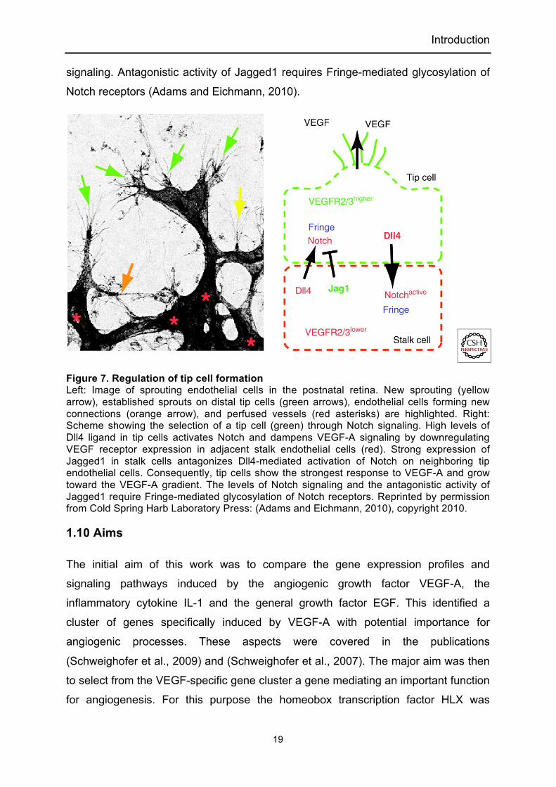
Introduction
19
signaling. Antagonistic activity of Jagged1 requires Fringe-mediated glycosylation of
Notch receptors (Adams and Eichmann, 2010).
Figure 7. Regulation of tip cell formation Left: Image of sprouting endothelial cells in the postnatal retina. New sprouting (yellow arrow), established sprouts on distal tip cells (green arrows), endothelial cells forming new connections (orange arrow), and perfused vessels (red asterisks) are highlighted. Right: Scheme showing the selection of a tip cell (green) through Notch signaling. High levels of Dll4 ligand in tip cells activates Notch and dampens VEGF-A signaling by downregulating VEGF receptor expression in adjacent stalk endothelial cells (red). Strong expression of Jagged1 in stalk cells antagonizes Dll4-mediated activation of Notch on neighboring tip endothelial cells. Consequently, tip cells show the strongest response to VEGF-A and grow toward the VEGF-A gradient. The levels of Notch signaling and the antagonistic activity of Jagged1 require Fringe-mediated glycosylation of Notch receptors. Reprinted by permission from Cold Spring Harb Laboratory Press: (Adams and Eichmann, 2010), copyright 2010.
1.10 Aims
The initial aim of this work was to compare the gene expression profiles and
signaling pathways induced by the angiogenic growth factor VEGF-A, the
inflammatory cytokine IL-1 and the general growth factor EGF. This identified a
cluster of genes specifically induced by VEGF-A with potential importance for
angiogenic processes. These aspects were covered in the publications
(Schweighofer et al., 2009) and (Schweighofer et al., 2007). The major aim was then
to select from the VEGF-specific gene cluster a gene mediating an important function
for angiogenesis. For this purpose the homeobox transcription factor HLX was

Introduction
20
investigated and its biological functions in endothelial cells and in sprouting
angiogenesis were defined. The obtained data show that HLX mediates a genetic
program to differentially regulate the expression of repulsive guidance cues in
normoxia and hypoxia. Details of this study are described in the first authorship
publication (Testori et al., 2011) and in the additional data section.

Publication (First authorship)
21
2. Publication (First authorship)
2.1 The VEGF-regulated transcription factor HLX controls the expression
of guidance cues and negatively regulates sprouting of endothelial cells.
Julia Testori, Bernhard Schweighofer, Iris Helfrich, Caterina Sturtzel, Karoline
Lipnik, Sabine Gesierich, Patrick Nasarre, Renate Hofer-Warbinek, Martin
Bilban, Hellmut Augustin and Erhard Hofer. Blood First Edition Paper,
prepublished online January 11, 2011; DOI 10.1182/blood-2010-07-293209
[This research was originally published in Blood Online. Testori, J., Schweighofer, B.,
Helfrich, I., Sturtzel, C., Lipnik, K., Gesierich, S., Nasarre, P., Hofer-Warbinek, R.,
Bilban, M., Augustin, H.G., Hofer, E. The VEGF-regulated transcription factor HLX
controls the expression of guidance cues and negatively regulates sprouting of
endothelial cells. Blood. Prepublished January 11, 2011; DOI 10.1182/blood-2010-
07-293209.]
doi:10.1182/blood-2010-07-293209 Prepublished online Jan 11, 2011;
Patrick Nasarre, Renate Hofer-Warbinek, Martin Bilban, Hellmut G. Augustin and Erhard Hofer Julia Testori, Bernhard Schweighofer, Iris Helfrich, Caterina Sturtzel, Karoline Lipnik, Sabine Gesierich,
guidance cues and negatively regulates sprouting of endothelial cellsThe VEGF-regulated transcription factor HLX controls the expression of
http://bloodjournal.hematologylibrary.org/misc/rights.dtl#repub_requestsInformation about reproducing this article in parts or in its entirety may be found online at:
http://bloodjournal.hematologylibrary.org/misc/rights.dtl#reprintsInformation about ordering reprints may be found online at:
http://bloodjournal.hematologylibrary.org/subscriptions/index.dtlInformation about subscriptions and ASH membership may be found online at:
. Hematology; all rights reservedCopyright 2011 by The American Society of 20036.the American Society of Hematology, 2021 L St, NW, Suite 900, Washington DC Blood (print ISSN 0006-4971, online ISSN 1528-0020), is published weekly by
For personal use only. on January 11, 2011. Bibliothek der MedUniWien (149592) at www.bloodjournal.orgFrom
22

The VEGF-regulated transcription factor HLX controls the expression of
guidance cues and negatively regulates sprouting of endothelial cells
Julia Testori1, Bernhard Schweighofer1,5, Iris Helfrich4,6, Caterina Sturtzel1, Karoline
Lipnik1, Sabine Gesierich4, Patrick Nasarre4,7, Renate Hofer-Warbinek1, Martin
Bilban2,3, Hellmut G. Augustin4 and Erhard Hofer1
Short title: HLX-mediated regulation of guidance proteins
1Department of Vascular Biology and Thrombosis Research, Center for Physiology
and Pharmacology, 2Clinical Department for Medical and Chemical Laboratory
Diagnostics, Medical University of Vienna, 3Ludwig Boltzmann Institute for Clinical
and Experimental Oncology, Vienna, Austria and 4Joint Research Division Vascular
Biology, Medical Faculty Mannheim (CBTM), Heidelberg University, and German
Cancer Research Center, Heidelberg (DKFZ-ZMBH Alliance), Germany
Present addresses: 5 Department of Cell Biology
The Scripps Research Institute
La Jolla, CA, USA 6 Department of Dermatology
University Hospital Essen
Essen, Germany 7 Department of Hematology/Oncology
Medical University of South Carolina,
Charleston, SC, USA
Blood First Edition Paper, prepublished online January 11, 2011; DOI 10.1182/blood-2010-07-293209
Copyright © 2011 American Society of Hematology
For personal use only. on January 11, 2011. Bibliothek der MedUniWien (149592) at www.bloodjournal.orgFrom
23

2
Corresponding author:
Dr. Erhard Hofer
Department of Vascular Biology and Thrombosis Research
Center for Physiology and Pharmacology
Medical University of Vienna
Lazarettgasse 19, A-1090 Vienna
Austria
Tel.: +43-1-40160-33111
Fax: +43-1-40160-933100
E-mail: [email protected]
Category: Vascular Biology
For personal use only. on January 11, 2011. Bibliothek der MedUniWien (149592) at www.bloodjournal.orgFrom
24

3
Abstract
The HLX gene encoding a diverged homeobox transcription factor has been found to
be upregulated by VEGF-A in endothelial cells. We have now investigated the gene
repertoire induced by HLX and its potential biological function. HLX strongly
increased the transcripts for several repulsive cell guidance proteins including
UNC5B, plexin A1 and semaphorin 3G. In addition, genes for transcriptional
repressors such as HES1 were upregulated. In line with these findings, adenoviral
overexpression of HLX inhibited endothelial cell migration, sprouting and vessel
formation in vitro and in vivo, whereas proliferation was unaffected. This inhibition of
sprouting was caused to a significant part by HLX-mediated upregulation of UNC5B
as shown by shRNA-mediated downmodulation of the respective mRNA. VEGF-A
stimulation of endothelial cells induced elevated levels of HLX over longer time
periods resulting in especially high upregulation of UNC5B mRNA as well as an
increase in cells displaying UNC5B at their surface. However, induction of HLX was
strongly reduced and UNC5B upregulation completely abrogated when cells were
exposed to hypoxic conditions. These data suggest that HLX may function to balance
attractive with repulsive vessel guidance by upregulating UNC5B and to
downmodulate sprouting under normoxic conditions.
For personal use only. on January 11, 2011. Bibliothek der MedUniWien (149592) at www.bloodjournal.orgFrom
25

4
Introduction
Vascular endothelial growth factor-A (VEGF-A) is the major trigger of vasculogenesis
and angiogenesis during embryogenesis and blood vessel formation in the adult1,2. It
has also been implicated in pathological angiogenesis in diseases such as cancer,
chronic inflammatory disorders and retinopathy.3 Whereas several peptide products
are generated from the VEGF-A gene by differential splicing, the available data
suggest that isoform VEGF-A165 is the predominant form responsible for the major
angiogenic effects.4
The gene repertoire induced by VEGF-A mainly via VEGF receptor-2 has been
investigated by several groups,5-7 however the transcription factors upregulated by
VEGF-A and how they mediate its specific and unique biological functions remain
largely uncharacterized. We have recently identified a group of genes selectively or
at least preferentially induced by VEGF-A in endothelial cells, when compared to a
more general growth factor such as EGF or inflammatory mediators such as IL-1.8
Most prominent upregulation in the described manner was found for the transcription
factors NR4A2 (nuclear receptor subfamily 4, group A, member 2), EGR3 (early
growth response 3), HLX (H2.0-like homeobox 1) and MEF2C (myocyte enhancer
factor 2) suggesting their involvement in specific VEGF-A-triggered responses.5
In the current study we have focused on the investigation of HLX, which is an
evolutionary highly conserved homeobox transcription factor originally detected in the
visceral musculature of Drosophila.9 Furthermore, HLX was shown to be expressed
in bone marrow-derived CD34+ cells,10 lymphocytes11-13 as well as in endothelial
cells.14 Expression in type 1 helper T lymphocytes seems to play an essential role to
establish heritable expression of the γ-IFN gene by epigenetic mechanisms.11,12 In
mouse development, the HLX-/- genotype is lethal around day 15 of embryonic
development on a mixed genetic background, the embryos displaying severe
hypoplasia of the gut and liver.15,16 In this context, it has been proposed that, during
visceral organogenesis, HLX regulates a mesenchymal-epithelial interaction that is
required for early aspects of enteric nervous system development.16
There is accumulating evidence that several mechanisms are shared between the
development of the nervous and vascular systems.17 During angiogenesis endothelial
tip cells, which lead the growing sprout, sense the environment by dynamically
extending filopodia towards attractive signals and retracting them from repulsive
For personal use only. on January 11, 2011. Bibliothek der MedUniWien (149592) at www.bloodjournal.orgFrom
26

5
ones. In this regard endothelial sprouts use similar guidance cues as growing nerve
fibers do for directional growth and network formation.17 Guidance molecules with a
similar role in endothelial tip cells and axonal growth cones include the semaphorins
and plexin receptors as well as the secreted netrins and uncoordinated-5 (UNC5)
receptors. However, signals and transcription factors that regulate the expression of
these guidance cues have not yet been established.
We have now investigated the genes controlled by the transcription factor HLX in
endothelial cells. Our data demonstrate that HLX is a specific regulator of cell
guidance molecules such as UNC5B, plexin A1 (PLXNA1) and semaphorin 3G
(SEMA3G), that presumably display repulsive functions in vessel guidance and/or
may prevent inappropriate sprouting. Furthermore, HLX also induces transcriptional
repressors including HES1, SNAI2 and BCOR. Following overexpression of HLX we
observed an inhibition of migration of endothelial cells, decreased sprouting and
reduced vessel formation. This inhibition of sprouting was in part due to UNC5B
upregulated by HLX. Furthermore, we show that VEGF-A can increase UNC5B
expression in an HLX-dependent way, however induction of HLX and consecutively
UNC5B was strongly reduced or prevented under hypoxic conditions. We propose
that HLX serves to counterbalance the effects of attractive guidance cues by
upregulating the expression of repulsive guidance molecules such as UNC5B.
For personal use only. on January 11, 2011. Bibliothek der MedUniWien (149592) at www.bloodjournal.orgFrom
27

6
Materials and methods
Cell culture and materials
Human umbilical vein endothelial cells (HUVEC) were isolated as described
previously18 and cultured in EGM-2 medium (Clonetics, Lonza, Walkersville, MD).
HUVEC of passage 3 to 5 were used for experiments.
To apply hypoxic conditions the cell culture plates were transferred to a modular
incubation chamber (Billups-Rothenberg, Inc., Del Mar, CA). This was inflated with
10% CO2 compensated with 90% N2 for 10 min, closed tightly and kept at 37°C for
the length of the experiment. Cultures to be analyzed within the first 2 h after VEGF
addition were preincubated under hypoxic conditions for 4 h.
HEK293 cells (ATCC No. CRL-1573) were cultured in MEMα medium with 10% NCS
(both from Invitrogen, Carlsbad, CA). 293T cells (ATCC No. CRL-11268) were
cultured in DMEM with 10% FCS (both from Invitrogen).
VEGF-A165 and bFGF were obtained from PromoKine (Heidelberg, Germany) or R&D
Systems (Merck, Darmstadt, Germany).
Construction of recombinant adenoviruses and transduction of cells
A cDNA clone of human HLX (IRALp962M1922Q, http://www.imagenes-bio.de/) was
obtained from RZPD (Heidelberg, Germany). The coding region together with a Flag-
tag sequence at the 3´-end was cloned into the pACCMVplpASR+ expression
plasmid19 (pAC.HLX). The plasmids pAC.HLX and pJM17 (Microbix Biosystems,
Toronto, CAN), were cotransfected into HEK293 cells by the calcium phosphate
method (Stratagene, LaJolla, CA). Primary adenoviruses generated were subcloned
and purified after amplification in HEK293 cells using CsCl-ultracentrifugation20. The
HLX sequence in the viral preparations was confirmed by sequencing. Viral titre was
determined using the Adeno-X Rapid Titer Kit (Clontech, Mountain View, CA). An
empty control adenovirus20 was also grown and purified as described above.
The UNC5B encoding adenovirus was a generous gift of Drs. Noriaki Kitamura and
Hirofumi Arakawa (National Cancer Center, Tokyo, Japan).
Production of recombinant lentiviruses and transduction of cells
Plasmids (Mission shRNA pLKO.1-puro) containing the corresponding shRNA
sequences targeted to HLX and UNC5B and a control shRNA plasmid were obtained
For personal use only. on January 11, 2011. Bibliothek der MedUniWien (149592) at www.bloodjournal.orgFrom
28

7
from Sigma-Aldrich. The shRNA plasmids together with two packaging vectors
(pMD2.G and psPAX.1) were cotransfected into 293T cells. Supernatants were
harvested after 24 and 48 h and used for infection mixed with medium in a ratio of
1:1 or 1:2.
RNA preparation
HUVEC were infected in subconfluent state with control (Ad.con) and HLX encoding
adenovirus (Ad.HLX) using multiplicity of infections (MOI) of 20 to 40. Then cells
were incubated with RNAlater (Ambion, Austin, TX) for 1 min, lysed with Trizol
(Invitrogen, Carlsbad, CA) and RNA was extracted.
Real-time RT-PCR analysis
Total RNA (2 µg) was used to synthesize cDNA with Superscript II Reverse
Transcriptase and oligodT primer (both from Invitrogen). mRNA levels were
measured using realtime RT-PCR detecting SYBR GreenI in a LightCycler (Roche
Diagnostics GmbH, Mannheim, Germany). Values were normalized to β2-
microglobulin mRNA as internal standard. Oligonucleotide primers were designed
using the Primer3 software (http://frodo.wi.mit.edu/primer3/) and are listed in
Supplemental Table 2.
Affymetrix microarray analysis
cRNA was prepared from total RNA, hybridized to the Human Genome U133 Plus
2.0 Array (Affymetrix, Santa Clara, CA) and arrays were scanned according to the
manufacturer’s protocols (Affymetrix support site,
www.affymetrix.com/support/index.affx) as described.21 RMA (Robust multiarray
average) signal extraction and normalization were performed as described
(http://www.bioconductor.org/).22
Western Blot Analysis
Cell pellets were lysed in Laemmli buffer. Proteins were separated by SDS-
polyacrylamide gel electrophoresis (SDS-PAGE) and transferred to Immobilon-P
membranes (Millipore, Bedford, MA). The membrane was blocked with 5% skim milk
/ PBS and 0.1% Tween 20 (PBST) and incubated overnight with the primary antibody
at 4 °C. The membrane was washed and incubated with peroxidase-conjugated
For personal use only. on January 11, 2011. Bibliothek der MedUniWien (149592) at www.bloodjournal.orgFrom
29

8
secondary antibody for 1 h. The membrane was incubated with ECL Plus reagent
(GE Healthcare, Buckinghamshire, UK) and exposed to X-ray film. Bands were
quantified in scanned film images by measuring tonality using Adobe Photoshop
software.
Antibodies used were: mouse anti-human HLX (Abnova, Taipei, Taiwan), mouse
anti-human GAPDH (Chemicon, Billerica, MA, USA), and secondary horseradish
peroxidase-conjugated ECL sheep-anti-mouse antibodies (GE Healthcare).
Flow cytometry
Following treatment with accutase (PAA, Pasching, Austria) cells were harvested,
fixed with 4% paraformaldehyde, permeabilized with 0.05% Triton-X-100 (Sigma-
Aldrich, St.Louis, MO) and blocked with 5% BSA-PBS. Cells were stained with 5
µg/ml of the humanized UNC5B antibody #4 (Genentech, San Francisco, CA) for 1 h
at 4°C followed by the secondary antibody Alexa Fluor® 647 goat anti-human IgG
(Invitrogen). Binding was assessed using a FACSCalibur (BD Biosciences).
Immunocytochemistry
HUVEC were grown in fibronectin-coated chamber slides. Cells were fixed with 4%
paraformaldehyde and blocked with 1% BSA/PBS. To reveal HLX cells were in
addition permeabilized with 0.1% TritonX100/PBS. Slides were stained with: affinity-
purified rabbit anti-human HLX antibodies13 raised against a GST-HLX fusion protein
(1:500 dilution); anti-human UNC5B antibody #4 (20 μg/ml, Genentech); secondary
Alexa Fluor 568 goat anti-rabbit IgG at a dilution of 1:5000 or Alexa Fluor 647 goat
anti-human IgG at 1:5000 (Molecular Probes, Invitrogen).
Proliferation assay
HUVEC were infected with adenoviruses using a MOI of 8. The cells were trypsinized
on the following day and seeded into 96-well plates (5000 cells/well). Proliferation
was determined by measuring the total protein content using the sulforhodamineB
(SRB) colorimetric assay.23 Experiments were performed with 5 wells per data point.
Transwell migration assay
One day after infection 5x10⁴ HUVEC were seeded into the upper well of a
gelatinized transwell with a pore size of 8 µm (Corning, Lowell, MA). Following the
For personal use only. on January 11, 2011. Bibliothek der MedUniWien (149592) at www.bloodjournal.orgFrom
30

9
addition of 50 ng/ml VEGF-A165/ml to the lower chamber, the transwell was incubated
for 4 h at 37°C and the migration of cells towards the lower chamber was determined.
Cells were fixed with methanol and stained with 1 µg/ml 4´,6-Diamidin-2-phenylindol
(DAPI, Sigma-Aldrich, Taufkirchen, Germany). Cells in the lower chamber were
counted in 5 randomly chosen microscopic fields photographed using a Nikon
Diaphot TMD microscope and a CCD camera (Kappa GmbH, Gleichen, Germany).
Generation of endothelial cell spheroids
HUVEC were transduced at a MOI of 10 to 20 or infected with 0.5 to 1ml of lentivirus.
Cells were then used for the spheroid-based in vitro or in vivo assays. Cells were
used 24 h after infection with the adenoviruses or 48 h after infection with the
lentiviruses to generate spheroids. Transduction efficiency of all viruses was regularly
controlled.
Spheroids of a defined cell number were generated from non-transduced or
transduced cells as described in Korff et al (2004).24 Briefly, HUVEC were suspended
in medium containing 0.25% (wt/vol) methylcellulose and grown in hanging drops
overnight. Single spheroid with a defined cell number of approximately 400 (for the in
vitro assay) or 100 (for the in vivo assay) cells/spheroid were obtained.
Spheroid-based in vitro angiogenesis assay
HUVEC spheroids were embedded into rat collagen gels as described25 with the
following modifications: Fifty spheroids of 400 cells per spheroid were mixed with
80% Methocel / 20% FCS. 0.1 ml ECGM (basal medium; PromoCell) without or
containing VEGF or bFGF (25 ng/ml each) was layered onto the top of the formed
gel. After 24 h sprouts were quantified by measuring the total length grown out of
each spheroid on pictures taken on the Nikon microscope.
In vivo implantation of spheroids
Spheroids were harvested and carefully suspended in 500 µl Matrigel (growth factor
reduced; BD Biosciences) and fibrinogen (2 mg/ml; Calbiochem, Merck, Darmstadt,
Germany) containing VEGF plus bFGF (500 ng/ml each). Gel formation was initiated
by the addition of thrombin (0.4 U; Calbiochem).
Spheroid suspensions containing about 1000 spheroids of 100 cells each were
injected subcutaneously on each side lateral to the abdominal midline region into 4-6
For personal use only. on January 11, 2011. Bibliothek der MedUniWien (149592) at www.bloodjournal.orgFrom
31

10
week old SCID mice. Two plugs were implanted per mouse, each experimental group
consisted of 8 mice. After 14 days, mice were sacrificed, the harvested plugs were
fixed overnight (0.5 g/l calcium acetate, 5 g/l zinc acetate, 5 g/l zinc chloride in 0.1 M
Tris, pH 7.4) and analyzed by immunohistochemistry. Animal procedures were
carried out in accordance to guidelines of the local committee for animal
experimentation (DKFZ Heidelberg and RP Karlsruhe, Germany).
Immunohistochemistry
Zink fixed Matrigel plugs were embedded in paraffin. Plugs were sectioned at 8 µm.
Primary antibodies used were: mouse-anti-human CD34 (QBEND10, Novocastra,
Newcastle, UK); sheep-anti-human CD31 (Dako, Glostrup, Denmark). Antigen
retrieval for anti-human-CD34 stainings was performed by digestion with Proteinase
K (Sigma-Aldrich, Taufkirchen, Germany) at 37°C for 15 min. The secondary
antibodies donkey-anti-sheep Alexa 488 (Invitrogen), and biotinylated goat-anti-
mouse (Zymed, Invitrogen) were detected by streptavidin-peroxidase conjugate
(Dako) and DAB substrate chromogen system (Dako).
Quantificationof the human grafted vasculature and statistical analysis
Sections were prepared across the entire implant size and three sections were
selected from the beginning, the center and the end. MVD (microvessel density)
analysis was performed using immunohistochemistry for human CD31 and CD34.
Images of the complete plug area were taken using an Olympus IX50 microscope
and assembled by multiple alignment (Cell-P, Olympus). Fluorescent signals of the
complete plug matrix were calculated as vessel number per mm².
For personal use only. on January 11, 2011. Bibliothek der MedUniWien (149592) at www.bloodjournal.orgFrom
32

11
Results
HLX upregulates genes for cell guidance proteins and transcriptional
repressors
The gene encoding the homeobox transcription factor HLX has previously been
identified by us to be induced by VEGF in endothelial cells suggesting the
involvement of HLX in VEGF-mediated transcriptional responses.5 We therefore
investigated the transcriptomic program induced by HLX. An adenoviral expression
construct encoding a Flag-tagged HLX cDNA was generated and used to transduce
HUVEC in parallel with control adenovirus. mRNA was extracted from cells between
4 and 32 h after infection and subjected to Affymetrix microarray analysis (GEO
GSE13054). 53 genes were identified to be induced more than 2.5 fold 16 h after
infection (see Supplemental Table 1). This group of genes was analyzed according to
the categories surface receptors, secreted proteins, cytoplasmic signaling proteins
and transcription factors. Thereby it became apparent that among the strongest
induced genes were those for guidance receptors with proposed repulsive functions
in the guidance of endothelial sprouts, namely UNC5B and plexin A1. Furthermore,
transcripts for a secreted guidance molecule, SEMA3G, and for several transcription
factors with repressive function were found to be upregulated. One example is the
repressor HES1, which has been described as a direct target gene of the Notch
signaling pathway. Table 1 displays the most strongly induced genes, which were
confirmed by real-time RT-PCR. The kinetics of the upregulation of mRNAs for
UNC5B, PLXNA1, SEMA3G and HES1 as measured by real-time RT-PCR shows
that the relative upregulation was most pronounced for UNC5B (Figure 1A). The
accumulation of recombinant HLX protein after transduction with Ad.HLX is depicted
in Figure 1B.
VEGF most prominently upregulates UNC5B in an HLX-dependent way
To evaluate whether VEGF upregulates genes for repellent guidance molecules,
which could be mediated by HLX as a secondary transcriptional response to VEGF,
we examined mRNA levels for UNC5B, PLXNA1, SEMA3G and HES1 by real-time
RT-PCR. From these preferentially UNC5B mRNA was found to be increasingly
For personal use only. on January 11, 2011. Bibliothek der MedUniWien (149592) at www.bloodjournal.orgFrom
33

12
upregulated following VEGF stimulation starting between 2 and 4 h, being 10-fold
increased at 16 h and reaching levels several hundred-fold of the starting values
between 24 and 56 h (Figure 2A, Supplemental Figure 1A). This upregulation of
UNC5B mRNA was reflected in the appearance of a fraction of cells displaying
UNC5B at their surface as shown by flow cytometry (Figure 2C) and
cytoimmunochemistry (Figure 2D).
In the same samples HLX mRNA already displayed a rapid peak value after 1 hour of
stimulation and thereafter stayed 5- to 10-fold elevated up to 56 h. HLX protein was
increased starting at 4 h and reaching 10-fold higher levels at later time points as
shown by Western blot analysis (Figure 2B). The VEGF induced HLX accumulated in
the nuclei (Figure 2D). It therefore seemed possible that VEGF-induced HLX is
involved in the upregulation of UNC5B. We further investigated how general the
upregulation of HLX by VEGF would be in endothelial cells of different origin. This
indicated that VEGF indeed induces HLX in a broad range of different endothelial cell
types obtained from various sources including large vessel and microvascular
endothelial cells. The induction rates were comparable to those observed with
different HUVEC isolates ranging from 5- to 30-fold with a tendency of somewhat
lower inducibility in microvascular cells (Supplemental Figure 1B).
Next we tested whether HLX would directly mediate the observed upregulation of
UNC5B by VEGF. Indeed, downmodulation of HLX using a respective shRNA
expressed from a lentiviral vector reduced the VEGF-induced accumulation of
UNC5B mRNA and protein by more than 80% (Figure 2E). This showed that UNC5B
upregulation by VEGF is mediated via HLX.
HLX inhibits migration of endothelial cells, but proliferation is unaffected
Considering that HLX appeared to preferentially upregulate UNC5B and other
repellent guidance cues and a transcriptional repressor of the Notch pathway, we
next investigated to what extent HLX would affect proliferation and/or migration of
endothelial cells. When HUVEC were infected with HLX encoding adenoviruses, no
significant influence of HLX overexpression on the growth of cells could be detected
in comparison to control adenovirus infected or non-infected cells (Figure 3A).
However, in a transwell system, migration towards VEGF was inhibited by about 80%
For personal use only. on January 11, 2011. Bibliothek der MedUniWien (149592) at www.bloodjournal.orgFrom
34
13
as compared to cells infected with control adenovirus or non-infected cells (Figure
3B).
We have further assessed the potential triggering of apoptosis by HLX
overexpression, but no signs of apoptosis were detectable 4 days after infection
(Supplemental Figure 2). This suggests that, in endothelial cells, HLX specifically
interferes with a migratory mechanism, while it does not affect proliferation and
apoptotic signaling pathways.
HLX and UNC5B strongly inhibit sprouting of endothelial cells in vitro
To investigate the effect of HLX on angiogenesis and particularly on sprouting of
endothelial cells, we first used an in vitro angiogenesis assay, which evaluates sprout
formation of cells embedded as spheroids in a collagen matrix. In this assay HLX
overexpression strongly inhibited sprouting of cells induced by VEGF or bFGF
(Figure 4A and B). When decreasing MOI of virus infection were used, the inhibiting
effect diminished in a dose-dependent manner (Supplemental Figure 3).
To evaluate whether UNC5B expression could mediate the inhibition of sprouting we
first used an adenovirus for overexpression of UNC5B. Indeed, ectopic UNC5B
expression was able to strongly reduce sprouting activity (Figure 4C). This indicated
that the inhibitory effect of HLX overexpression is at least in part mediated via
UNC5B.
Inhibition of sprouting by HLX is mediated to a significant part by UNC5B
To establish to which extent UNC5B would contribute to the inhibition of sprouting
observed after HLX overexpression we used lentiviral expression of shRNA for the
downmodulation of UNC5B mRNA. On average a 60% downmodulation of the
UNC5B mRNA was achieved after VEGF induction at the 56 h time point
(Supplemental Figure 4A). The results show that a reduction in the HLX-mediated
upregulation of UNC5B led to significantly decreased inhibition of sprouting (Figure
4D). This shows that the inhibitory effect of HLX overexpression is at least to a
significant part caused by UNC5B upregulation.
It was further intriguing, that also in the VEGF-induced control samples
downmodulation of UNC5B caused a strong increase of sprouting activity as
For personal use only. on January 11, 2011. Bibliothek der MedUniWien (149592) at www.bloodjournal.orgFrom
35

14
evidenced by the cumulative sprout length depicted in Figure 4D. This effect was
primarily composed of a higher number of sprouts (Supplemental Figure 4B)
suggesting that UNC5B upregulated in VEGF-induced endothelial cells reduces
sprout initiation.
Upregulation of HLX and UNC5B is strongly reduced under hypoxic conditions
Furthermore, we tested the hypothesis that a potential reason for the upregulation of
inhibitory pathways and molecules by VEGF would be the downmodulation of
sprouting in a negative feed-back loop when normoxic conditions are restored after
neovascularization. Therefore we comparatively evaluated the effects of VEGF on
HLX and UNC5B mRNAs under normoxic and hypoxic conditions. The obtained
results show that under hypoxia the VEGF-inducible levels of HLX mRNA were
strongly reduced, especially at later time points, and the upregulation of UNC5B
mRNA was completely prevented (Figure 5A). In contrast hypoxia induced VEGF
mRNA as previously shown.26
When we performed the spheroid sprouting assay under hypoxic conditions we
observed a significant increase of sprouting supporting that hypoxia has an additive
effect on VEGF-induced sprouting. Hypoxia- plus VEGF-induced sprouting could be
inhibited by overexpression of HLX, however the relative inhibition appeared less
pronounced when compared to the inhibition under normoxic conditions (Figure 5B).
This suggests that HLX-mediated induction of UNC5B is involved in downmodulation
of sprouting under normoxic conditions and hypoxia would facilitate sprouting by
reducing UNC5B expression.
Overexpression of HLX inhibits vessel formation in vivo
To test whether overexpression of HLX would exert an inhibiting effect on
angiogenesis in vivo we further employed an in vivo angiogenesis assay with human
endothelial cells in mice27. In this assay, HUVEC spheroids infected with HLX or
control adenoviruses were suspended in a mixture of Matrigel and fibrinogen
supplemented with growth factors and implanted subcutaneously into SCID mice.
Grafted human endothelial cells vascularize the plug and anastomose with the
mouse vasculature to give rise to a perfused vascular network (see Supplemental
For personal use only. on January 11, 2011. Bibliothek der MedUniWien (149592) at www.bloodjournal.orgFrom
36

15
Figure 5). Plugs were harvested after 14 days and analyzed by
immunohistochemistry.
A significant reduction in the density and length of vessels was observed in vascular
networks originating from HLX overexpressing HUVEC compared to control cells
(Figure 6). The inhibition was similarily pronounced as the inhibition of sprouting in
vitro supporting that HLX plays a comparable role in vivo.
For personal use only. on January 11, 2011. Bibliothek der MedUniWien (149592) at www.bloodjournal.orgFrom
37

16
Discussion
Several members of the homeobox family have been implicated in the development
of the cardiovascular system as well as in vascular remodeling in the adult and have
been proposed to be important to many pathologies including atherosclerosis and
tumor angiogenesis.28-30
We have recently identified HLX,5,8 a diverged homeobox gene, so far not known to
be involved in vascular remodeling, as being preferentially upregulated in endothelial
cells by VEGF-A and to some extent also by another main angiogenic mediator,
bFGF.5,31 HLX has previously been observed to be expressed in HUVEC, in placental
endothelial cells and in trophoblasts.6,14,32,33 However, no precise function and
detailed mechanism of action has yet been defined for HLX in the vascular system.
Since our experiments neither identified an induction of HLX expression by
inflammatory mediators nor by other more general growth factors such as EGF,5 we
proposed that HLX might mediate a specific function of VEGF-A and bFGF related to
angiogenesis and/or vascular remodeling.
In the present study, we have undertaken an analysis of downstream target genes of
HLX by gene profiling following overexpression in endothelial cells (Supplemental
Table 1). It was intriguing that HLX can be a prominent inducer of genes implicated in
the negative regulation of the sprouting process, including genes for repulsive
guidance cues such as UNC5B, PLXNA1 and SEMA3G as well as a transcriptional
repressor, HES1 (Table 1).
UNC5B belongs to the Uncoordinated-5 family of transmembrane receptors which
consists of four members. From these UNC5A plays a pivotal role in neurons for
axon guidance, whereas UNC5B is largely restricted to the vascular system.34 It has
been demonstrated that upon binding to its soluble ligand Netrin-1, angiogenesis is
inhibited.17,35 A primarily repulsive role of UNC5B and its ligands in vessel formation
is further indicated by the finding that disruption of the UNC5B gene in mice leads to
increased extensions of tip cell filopodia, excessive vessel branching and abnormal
navigation.34 UNC5B is expressed in growing embryonic vessels and is then
downregulated in the quiescent adult vasculature, but reexpressed upon stimulation
of sprouting angiogenesis, e.g. in tumor angiogenesis.
PLXNA1 and SEMA3G are members of another complex system regulating axonal
and vessel guidance.36,37 SEMA3G has recently been found to be a primarily
For personal use only. on January 11, 2011. Bibliothek der MedUniWien (149592) at www.bloodjournal.orgFrom
38
17
endothelial cell expressed isoform.38 In general, members of the class 3 semaphorin
family bind to neuropilin-1 or neuropilin-2 in a complex with plexins as co-
receptors.37,39 As neuropilins are also coreceptors for various VEGF isoforms these
interactions interfere with VEGF receptor signaling and mediate repulsive signaling
via the plexin receptors.
The transcriptional repressor HES1 is a hallmark of the activated Notch pathway.40
This could indicate a link to tip and stalk cell differentiation as it has been shown that
tip cells via Dll4 activate the Notch receptor in neighboring cells. Thereby stalk cells
are inhibited to sprout and are prevented from becoming additional tip cells.41,42
Since HLX induces HES1, it is possible that this might support the stalk cell
phenotype.
Based on further functional data that, in line with the repulsive and repressive
functions of the described molecules, HLX overexpression strongly inhibited
sprouting of endothelial cells, we started a systematic analysis to define which of the
genes would be most significantly involved in HLX- and VEGF-regulated
mechanisms. The obtained data showed that especially UNC5B is intimately linked in
expression and function to HLX and VEGF-A. First, from the four genes analyzed by
realtime RT-PCR, which included UNC5B, PLXNA1, SEMA3G and HES1,
overexpression of HLX upregulated UNC5B mRNA several hundred-fold, which was
by far the strongest effect (Figure 1). Second, we observed that in VEGF-A-
containing cultures UNC5B mRNA displayed a continuous several hundred-fold
upregulation over several days, whereas PLXNA1, SEMA3G and HES1 mRNAs
increased only modestly around five-fold (Figure 2A). The VEGF-induction of UNC5B
mRNA was also reflected in the appearance of UNC5B protein at the surface of a
fraction of the cells (Figure 2C). UNC5B upregulation was preceded by the induction
of continuously elevated level of HLX mRNA resulting in over 10-fold nuclear
accumulation of HLX protein at 24 h (Figure 2B and 2D). This was in line with the
possibility that HLX is involved in upregulating transcription of the UNC5B gene.
Importantly, downmodulation of HLX mRNA by shRNA reduced VEGF-inducible
UNC5B mRNA and protein by over 80 % (Figure 2E). This directly confirmed that
HLX indeed mediates the VEGF-triggered UNC5B induction to a large extent.
Since our data showed that HLX overexpression caused a strong inhibition of
migration (Figure 3B) and sprouting of endothelial cells (Figure 4A and B), we first
tested the possibility that UNC5B upregulated by HLX might be decisively involved.
For personal use only. on January 11, 2011. Bibliothek der MedUniWien (149592) at www.bloodjournal.orgFrom
39
18
Indeed, the obtained data showed that overexpression of UNC5B alone using a
recombinant adenovirus caused a strong inhibition of sprouting (Figure 4C). Vice
versa, downmodulation of UNC5B in HLX overexpressing cells significantly reduced
inhibition of sprouting restoring activity to the level of control virus infected cells
(Figure 4D). This confirmed that UNC5B expression by itself can cause significant
inhibition of sprouting and at least a significant part of the HLX-mediated inhibition. In
this context, it is important to mention that downmodulation of UNC5B not only
increased sprouting in HLX suppressed endothelial cells, but also in VEGF-induced
control cells reaching 3-fold higher sprouting levels (Figure 4D and Supplemental
Figure 4B). This may indicate that HLX-mediated inherent expression of UNC5B in
VEGF-induced cells reduces sprouting activity. This is in accordance with previous
reports that UNC5B is preferentially expressed in growing vessels and capillaries.35
Based on these data and considering a primarily repulsive role, one can principally
speculate on two non-exclusive potential roles of VEGF induced HLX and UNC5B for
sprouting angiogenesis. It might be possible that UNC5B expression is generally
needed to counterbalance the effects of attractive guidance cues. This may be
necessary to fine-tune sprouting by preventing excessive sprout initiation under
VEGF stimulation and/or to give appropriate direction to the growing sprout by
reducing branching. This possibility is in line with the finding that downmodulation of
UNC5B in VEGF induced cells additively increases the number of sprouts
(Supplemental Figure 4B).
Furthermore, it might be possible that UNC5B is an inbuilt inhibitory feed-back
mechanism that serves to adapt sprouting activity to physiological needs. Such a
possibility could be the modulation of UNC5B expression by a hypoxic gradient. It
has been previously shown that hypoxia via the transcription factor HIF1α can lead to
the up- and downregulation of several hundred genes.26 We therefore tested the
hypothesis that HLX and UNC5B expression in the presence of VEGF could be
modulated by hypoxia. Indeed, we find that under hypoxic conditions VEGF induction
of HLX mRNA is strongly reduced and accumulation of UNC5B mRNA is prevented.
Furthermore, hypoxia increased sprouting additively to VEGF. It is tempting to
speculate that this is due to the reduced capacity of the cells to produce UNC5B
under hypoxic conditions. These data strongly suggest that one function of the
VEGF/HLX/UNC5B induction axis may be to prevent inappropriate sprouting under
normoxic conditions and to adapt sprouting activity to an hypoxic gradient.
For personal use only. on January 11, 2011. Bibliothek der MedUniWien (149592) at www.bloodjournal.orgFrom
40

19
We have further used an endothelial spheroid xenografting assay to evaluate
whether HLX might similarly function in vivo and exert a negative effect on vessel
formation. In this assay HUVEC spheroids are implanted in a Matrigel/fibrinogen
matrix below the skin of SCID mice. The human cells can normally give rise to a
perfused vascular network fused with murine vessels as indicated by the presence of
erythrocytes in the human vessel parts (Supplemental Figure 5). Comparable to the
in vitro assay, HLX expression strongly reduced the capacity of the cells to form
vessels supporting that HLX exerts similar inhibitive function in vivo.
This work defines for the first time a transcription factor apparently controling a
genetic program for the expression of several repulsive molecules. Whereas the data
described here show that UNC5B is most closely linked to VEGF induction as well as
the inhibitory effects of HLX, it will be further interesting to determine to what extent
PLXNA1 and SEMA3G might function in a comparable or rather different way.
Considering these newly described properties of HLX, we propose that the factor
might be a potent inhibitor of pathologic angiogenesis and could be used as a novel
principle to inhibit tumor angiogenesis.
For personal use only. on January 11, 2011. Bibliothek der MedUniWien (149592) at www.bloodjournal.orgFrom
41

20
Acknowledgements
We thank Drs. Brian Becknell and Michael A. Caligiuri for a generous gift of rabbit
anti-HLX antibodies, Drs. Noriaki Kitamura and Hirofumi Arakawa for adenoviruses
expressing human UNC5B and Genentech Inc. for antibodies to human UNC5B.
Further, we are grateful to Dr. Johann Wojta and Christoph Kaun for a gift of several
macro- and microvascular enothelial cells, the midwives of the Wilhelminen Hospital
Vienna for continuous supply with umbilical cords and Maria Witkowsky for reliable
and expert technical assistance in the isolation and culture of endothelial cells. Our
thanks belong also to the members of the molecular vascular biology group of the
Medical University of Vienna, for technical help and discussions. We acknowledge
the support of this work by grants of the Austrian Science Foundation (P21291-B11)
and the European Commission (Health 222995). CS is a recipient of a DOC-
fFORTE-fellowship of the Austrian Academy of Sciences.
Authorship contributions
JT did the majority of detailed planning, experimental work and analysis as well as
writing of parts of the manuscript, BS, IH, CS, KL, SG, PN, RHW and MB performed
experiments and analyzed data. HGA contributed in the design of and conclusions
from the vessel formation experiments. EH perceived and designed the overall
research work and wrote the majority of the paper.
Disclosure of conflicts of interest
The authors declare no competing financial interests.
For personal use only. on January 11, 2011. Bibliothek der MedUniWien (149592) at www.bloodjournal.orgFrom
42

21
References
1. Lohela M, Bry M, Tammela T, Alitalo K. VEGFs and receptors involved in
angiogenesis versus lymphangiogenesis. Curr Opin Cell Biol. 2009;21(2):154-
165.
2. Adams RH, Alitalo K. Molecular regulation of angiogenesis and
lymphangiogenesis. Nat Rev Mol Cell Biol. 2007;8(6):464-478.
3. Loges S, Roncal C, Carmeliet P. Development of targeted angiogenic medicine.
J Thromb Haemost. 2009;7(1):21-33.
4. Ferrara N. Vascular endothelial growth factor: basic science and clinical
progress. Endocr Rev. 2004;25(4):581-611.
5. Schweighofer B, Testori J, Sturtzel C, et al. The VEGF-induced transcriptional
response comprises gene clusters at the crossroad of angiogenesis and
inflammation. Thromb Haemost. 2009;102(3):544-554.
6. Minami T, Horiuchi K, Miura M, et al. Vascular endothelial growth factor- and
thrombin-induced termination factor, Down syndrome critical region-1,
attenuates endothelial cell proliferation and angiogenesis. J Biol Chem.
2004;279(48):50537-50554.
7. Liu D, Jia H, Holmes DI, Stannard A, Zachary I. Vascular endothelial growth
factor-regulated gene expression in endothelial cells: KDR-mediated induction
of Egr3 and the related nuclear receptors Nur77, Nurr1, and Nor1. Arterioscler
Thromb Vasc Biol. 2003;23(11):2002-2007.
8. Hofer E, Schweighofer B. Signal transduction induced in endothelial cells by
growth factor receptors involved in angiogenesis. Thromb Haemost.
2007;97(3):355-363.
9. Barad M, Erlebacher A, McGinnis W. Despite expression in embryonic visceral
mesoderm, H2.0 is not essential for Drosophila visceral muscle morphogenesis.
Dev Genet. 1991;12(3):206-211.
10. Deguchi Y, Kehrl JH. Selective expression of two homeobox genes in CD34-
positive cells from human bone marrow. Blood. 1991;78(2):323-328.
11. Mikhalkevich N, Becknell B, Caligiuri MA, Bates MD, Harvey R, Zheng WP.
Responsiveness of naive CD4 T cells to polarizing cytokine determines the ratio
of Th1 and Th2 cell differentiation. J Immunol. 2006;176(3):1553-1560.
For personal use only. on January 11, 2011. Bibliothek der MedUniWien (149592) at www.bloodjournal.orgFrom
43

22
12. Mullen AC, Hutchins AS, High FA, et al. Hlx is induced by and genetically
interacts with T-bet to promote heritable T(H)1 gene induction. Nat Immunol.
2002;3(7):652-658.
13. Becknell B, Hughes TL, Freud AG, et al. Hlx homeobox transcription factor
negatively regulates interferon-gamma production in monokine-activated natural
killer cells. Blood. 2007;109(6):2481-2487.
14. Murthi P, So M, Gude NM, Doherty VL, Brennecke SP, Kalionis B. Homeobox
genes are differentially expressed in macrovascular human umbilical vein
endothelial cells and microvascular placental endothelial cells. Placenta.
2007;28(2-3):219-223.
15. Hentsch B, Lyons I, Li R, et al. Hlx homeo box gene is essential for an inductive
tissue interaction that drives expansion of embryonic liver and gut. Genes Dev.
1996;10(1):70-79.
16. Bates MD, Dunagan DT, Welch LC, Kaul A, Harvey RP. The Hlx homeobox
transcription factor is required early in enteric nervous system development.
BMC Dev Biol. 2006;6:33.
17. Larrivee B, Freitas C, Suchting S, Brunet I, Eichmann A. Guidance of vascular
development: lessons from the nervous system. Circ Res. 2009;104(4):428-
441.
18. Wojta J, Hoover RL, Daniel TO. Vascular origin determines plasminogen
activator expression in human endothelial cells. Renal endothelial cells produce
large amounts of single chain urokinase type plasminogen activator. J Biol
Chem. 1989;264(5):2846-2852.
19. Gomez-Foix AM, Coats WS, Baque S, Alam T, Gerard RD, Newgard CB.
Adenovirus-mediated transfer of the muscle glycogen phosphorylase gene into
hepatocytes confers altered regulation of glycogen metabolism. J Biol Chem.
1992;267(35):25129-25134.
20. Lucerna M, Mechtcheriakova D, Kadl A, et al. NAB2, a corepressor of EGR-1,
inhibits vascular endothelial growth factor-mediated gene induction and
angiogenic responses of endothelial cells. J Biol Chem. 2003;278(13):11433-
11440.
21. Bilban M, Ghaffari-Tabrizi N, Hintermann E, et al. Kisspeptin-10, a KiSS-
1/metastin-derived decapeptide, is a physiological invasion inhibitor of primary
human trophoblasts. J Cell Sci. 2004;117(Pt 8):1319-1328.
For personal use only. on January 11, 2011. Bibliothek der MedUniWien (149592) at www.bloodjournal.orgFrom
44
23
22. Bilban M, Heintel D, Scharl T, et al. Deregulated expression of fat and muscle
genes in B-cell chronic lymphocytic leukemia with high lipoprotein lipase
expression. Leukemia. 2006;20(6):1080-1088.
23. Vichai V, Kirtikara K. Sulforhodamine B colorimetric assay for cytotoxicity
screening. Nat Protoc. 2006;1(3):1112-1116.
24. Korff T, Krauss T, Augustin HG. Three-dimensional spheroidal culture of
cytotrophoblast cells mimics the phenotype and differentiation of
cytotrophoblasts from normal and preeclamptic pregnancies. Exp Cell Res.
2004;297(2):415-423.
25. Korff T, Augustin HG. Tensional forces in fibrillar extracellular matrices control
directional capillary sprouting. J Cell Sci. 1999;112 ( Pt 19):3249-3258.
26. Manalo DJ, Rowan A, Lavoie T, et al. Transcriptional regulation of vascular
endothelial cell responses to hypoxia by HIF-1. Blood. 2005;105(2):659-669.
27. Alajati A, Laib AM, Weber H, et al. Spheroid-based engineering of a human
vasculature in mice. Nat Methods. 2008;5(5):439-445.
28. Cantile M, Schiavo G, Terracciano L, Cillo C. Homeobox genes in normal and
abnormal vasculogenesis. Nutr Metab Cardiovasc Dis. 2008;18(10):651-658.
29. Douville JM, Wigle JT. Regulation and function of homeodomain proteins in the
embryonic and adult vascular systems. Can J Physiol Pharmacol.
2007;85(1):55-65.
30. Gorski DH, Walsh K. Control of vascular cell differentiation by homeobox
transcription factors. Trends Cardiovasc Med. 2003;13(6):213-220.
31. Presta M, Dell'Era P, Mitola S, Moroni E, Ronca R, Rusnati M. Fibroblast
growth factor/fibroblast growth factor receptor system in angiogenesis. Cytokine
Growth Factor Rev. 2005;16(2):159-178.
32. Rajaraman G, Murthi P, Leo B, Brennecke SP, Kalionis B. Homeobox gene
HLX1 is a regulator of colony stimulating factor-1 dependent trophoblast cell
proliferation. Placenta. 2007;28(10):991-998.
33. Rajaraman G, Murthi P, Pathirage N, Brennecke SP, Kalionis B. Downstream
targets of homeobox gene HLX show altered expression in human idiopathic
fetal growth restriction. Am J Pathol. 2010;176(1):278-287.
34. Lu X, Le Noble F, Yuan L, et al. The netrin receptor UNC5B mediates guidance
events controlling morphogenesis of the vascular system. Nature.
2004;432(7014):179-186.
For personal use only. on January 11, 2011. Bibliothek der MedUniWien (149592) at www.bloodjournal.orgFrom
45

24
35. Larrivee B, Freitas C, Trombe M, et al. Activation of the UNC5B receptor by
Netrin-1 inhibits sprouting angiogenesis. Genes Dev. 2007;21(19):2433-2447.
36. Gaur P, Bielenberg DR, Samuel S, et al. Role of class 3 semaphorins and their
receptors in tumor growth and angiogenesis. Clin Cancer Res.
2009;15(22):6763-6770.
37. Neufeld G, Kessler O. The semaphorins: versatile regulators of tumour
progression and tumour angiogenesis. Nat Rev Cancer. 2008;8(8):632-645.
38. Kutschera S, Weber H, Weick A, et al. Differential Endothelial Transcriptomics
Identifies Semaphorin 3G as a Vascular Class 3 Semaphorin. Arterioscler
Thromb Vasc Biol. 2010.
39. Serini G, Valdembri D, Zanivan S, et al. Class 3 semaphorins control vascular
morphogenesis by inhibiting integrin function. Nature. 2003;424(6947):391-397.
40. Fischer A, Gessler M. Delta-Notch--and then? Protein interactions and
proposed modes of repression by Hes and Hey bHLH factors. Nucleic Acids
Res. 2007;35(14):4583-4596.
41. Roca C, Adams RH. Regulation of vascular morphogenesis by Notch signaling.
Genes Dev. 2007;21(20):2511-2524.
42. Phng LK, Gerhardt H. Angiogenesis: a team effort coordinated by notch. Dev
Cell. 2009;16(2):196-208.
For personal use only. on January 11, 2011. Bibliothek der MedUniWien (149592) at www.bloodjournal.orgFrom
46

25
Table 1: Selected genes upregulated by HLX in endothelial cells. Total RNA was isolated from HUVEC after infection with HLX encoding (Ad.HLX) and control adenoviruses (Ad.con) for 4, 8, 16 and 32 h. RNA was subjected to microarray analysis using the Affymetrix Human Genome U133 Plus 2.0 Array. Changes in gene expression induced in Ad.HLX relative to Ad.con infected cultures are displayed as fold induction at the given time points. The full set of Affymetrix data is available in the GEO database under GSE13054.
Ad.HLX/Ad.con UniGene ID Entrez
Gene Gene Symbol
Gene Title 4h 8h 16h 32h Surface Receptors Hs.585457 219699 UNC5B unc-5 homolog B (C. elegans) 1.41 2.23 5.71 7.14 Hs.432329 5361 PLXNA1 plexin A1 1.15 2.96 4.61 8.38 Hs.132781 9466 IL27RA interleukin 27 receptor, alpha 0.91 1.86 4.18 5.53 Secreted proteins Hs.9315 56944 OLFML3 olfactomedin-like 3 1.09 1.72 4.62 23.68 Hs.59729 56920 SEMA3G sema domain, immunoglobulin domain (Ig),
short basic domain, secreted, (semaphorin) 3G
1.27 2.28 2.88 4.89
Cytoplasmic (signaling) proteins Hs.417050 8900 CCNA1 cyclin A1 0.78 2.15 3.79 2.96 Hs.2128 1847 DUSP5 dual specificity phosphatase 5 1.07 2.38 3.76 3.54 Hs.381167 1992 SERPINB1 serpin peptidase inhibitor, clade B
(ovalbumin), member 1 0.93 1.33 3.15 4.25
Transcription factors Hs.360174 6591 SNAI2 snail homolog 2 (Drosophila) 1.58 3.87 4.51 6.91 Hs.525704 3725 JUN Jun oncogene 1.48 1.51 4.06 3.14 Hs.250666 3280 HES1 hairy and enhancer of split 1, (Drosophila) 2.13 3.63 3.49 4.37 Hs.659681 54880 BCOR BCL6 co-repressor 2.01 2.54 3.45 5.37
F
or personal use only. on January 11, 2011.
Bibliothek der M
edUniW
ien (149592) at
ww
w.bloodjournal.org
From

26
Figure legends
Figure 1. HLX upregulates genes for repellent cell guidance molecules and a
transcriptional repressor of the Notch signaling pathway.
(A) Real-time RT-PCR analysis of UNC5B, PLXNA1, SEMA3G and HES1 mRNA:
HUVEC were transduced for 4, 8, 16 and 32 h with recombinant adenoviruses
encoding HLX (Ad.HLX) or control viruses (Ad.con) using an MOI of 20. Total RNA
was isolated and real-time RT-PCR performed as described in the methods section.
Values were normalized to ß2-microglobulin mRNA as internal standard. Mean
values +/- standard deviation (SD) calculated from triplicate wells are depicted as fold
induction of the mRNA levels obtained after infection with Ad.HLX (■) or Ad.con (○)
as compared with non-infected cells. Results of one representative experiment of
three independently performed are shown.
(B) Western blot analysis of HLX: Total cell lysates were prepared from HUVEC non-
transduced (0 h) or tranduced with Ad.HLX (4, 8, 16 and 32 h) or with Ad.con for 32 h
using an MOI of 20. Cells were harvested and proteins were separated by SDS-
PAGE. Membranes were probed with anti-HLX and anti-GAPDH antibodies.
Figure 2. VEGF upregulates HLX and UNC5B in endothelial cells.
(A) Real-time RT-PCR analysis of UNC5B, PLXNA1, SEMA3G, HES1 and HLX
mRNA: HUVEC were cultured to density, starved overnight in EBM-2 medium without
supplements and then induced with 100 ng VEGF/ml for 1, 2, 4, 8, 16, 32 and 56 h.
Total RNA was isolated and mRNA levels were determined by real-time RT-PCR. All
values were normalized to ß2-microglobulin mRNA as internal standard. Results
displayed represent the mean of fold induction +/- standard error of the mean (SEM)
of the respective mRNA levels calculated from triplicate wells. One representative
experiment of three performed is shown.
(B) Western blot analysis of HLX: Total cell lysates were prepared from HUVEC
induced with 100 ng/ml VEGF for 4, 6 and 24 h and HUVEC transduced with
adenoviruses encoding HLX (Ad.HLX) for 72 h. Cells were harvested and proteins
were separated by SDS-PAGE. Membranes were probed with anti-HLX and anti-
GAPDH antibodies. The left part displays one exemplary blot, the right part the
quantification obtained from three independent experiments.
For personal use only. on January 11, 2011. Bibliothek der MedUniWien (149592) at www.bloodjournal.orgFrom
48

27
(C) Flow cytometry analysis of UNC5B surface expression after VEGF induction:
HUVEC starved as described above were induced with VEGF-A for 56 h or kept
without stimulation. Cells were detached from the plates, stained with UNC5B and
Alexa Fluor® 647-labelled goat anti-human IgG antibodies and subjected to flow
cytometry. The left part displays representative dot blots of untreated, 56 h VEGF-
induced and control samples transduced for 56 h with an UNC5B adenovirus. The
right part shows the quantification of three independent experiments performed in
triplicates. Mean percentage of positive cells +/- SEM calculated from at least three
independent experiments is displayed.
(D) Immunocytochemistry analysis of HLX and UNC5B after VEGF induction:
HUVEC were induced for 24 and 48 h, cells were fixed and stained with anti-HLX and
anti-UNC5B antibodies. The upper part displays the VEGF induction, the lower part a
control experiment using transduction with HLX and UNC5B expressing
adenoviruses.
(E) Upregulation of UNC5B by VEGF-A is HLX-dependent: HUVEC transduced with
lentiviruses containing shRNA.con and shRNA.HLX for 48 h were starved overnight
and then induced with VEGF for 24, 48 or 56 h as indicated. Cells were harvested
and UNC5B mRNA and protein analyzed by realtime RT-PCR and flow cytometry,
respectively. The left part displays fold induction of UNC5B or HLX mRNA +/- SD in
comparison to non-induced control shRNA cells as calculated from one experiment
representative of four performed in triplicates. The right part displays percent UNC5B
positive cells +/- SD as measured by flow cytometry in a representative experiment
performed in triplicates.
Figure 3. HLX does not affect the proliferation of endothelial cells, whereas
migration is strongly reduced.
(A) Proliferation assay: HUVEC were infected with HLX encoding adenoviruses
(Ad.HLX ▲) or control viruses (Ad.con ■) using an MOI of 8 or remained non-
infected (w/o �). After 24 h 5x10³ cells were seeded per well into 96-well plates. 0, 1,
2 and 3 days after seeding the protein content was measured at OD492 using the
sulforhodamineB (SRB) assay. The values display the increase in protein content
over the indicated time period +/- SD calculated from 5 wells each. One experiment
of three with similar results is shown.
For personal use only. on January 11, 2011. Bibliothek der MedUniWien (149592) at www.bloodjournal.orgFrom
49

28
(B) Migration assay: HUVEC were transduced with Ad.HLX or Ad.con with a MOI of 8
each or remained non-infected. Three days after infection cells were seeded into a
transwell system and the migration of the cells from the upper chamber towards the
lower chamber containing medium with 50 ng VEGF/ml or without VEGF was scored
4 h after seeding of the cells. Cells migrated into the lower chamber were visualized
by staining with DAPI. Five random microscopic fields per transwell were
photographed at a magnification of 10-fold and the stained cells counted. Values
display the percentage of migrated cells in the individual samples in comparison to
the VEGF-induced migration in samples containing non-infected cells arbitrarily set to
100%. Mean values were calculated from two independent experiments with
triplicates each +/- SD. Inhibition of migration by Ad.HLX was significant as
calculated by t test (** P < .005, *** P < .001).
Figure 4. HLX causes strong inhibition of sprouting which is mediated to a
significant part by UNC5B.
HUVEC were infected with Ad.HLX, Ad.UNC5B or Ad.con. The following day
spheroids of infected and non-infected endothelial cells were generated as described
in Materials and Methods. The spheroids were embedded into collagen gels and
induced with VEGF or bFGF (25 ng/ml) or cultured without cytokine stimulation. After
24 h the spheroids were fixed and photographic images were taken for quantification.
(A) Representative images of spheroids generated from Ad.HLX or Ad.con infected
cells (10 MOI) and induced with VEGF (+) or left untreated (-) are displayed. (B)
Quantification of inhibition of sprouting by overexpression of HLX: analyses of sprout
lengths were performed by measuring the total lengths of sprouts for 10 spheroids
each on microscopic images using the ImageJ software
(http://www.uhnres.utoronto.ca/facilities/wcif/imagej/). Results are displayed as mean
values +/- SEM of the fold induction of sprout lengths observed when compared to
non-induced samples infected with control viruses (arbitrarily set to 1). One
representative experiment out of four performed is shown. (C) Quantification of
inhibition of sprouting by overexpression of UNC5B: Analyses were as described
above. One representative experiment out of three is shown. (D) Quantification of
increase in sprouting by downmodulation of UNC5B in endothelial cells infected with
Ad.con or Ad.HLX: HUVEC were first transduced with lentiviruses for control shRNA
or shRNA targeted against UNC5B. After 1 d cells were in addition infected with 15
For personal use only. on January 11, 2011. Bibliothek der MedUniWien (149592) at www.bloodjournal.orgFrom
50
29
MOI of Ad.con or Ad.HLX. The following day spheroids were prepared and the next
day incorporated into collagen gels in the presence of VEGF-A. Total sprout lengths
of 15 spheroids each were measured. The shown values are calculated from four
independent experiments and display percent sprout length +/-SEM in comparison to
the samples transduced with Ad.con and shRNA.con. *** P < .001, t- test.
Figure 5: Under hypoxic conditions VEGF-mediated upregulation of HLX and
UNC5B mRNA is strongly reduced whereas sprouting activity is increased.
(A) Comparison of VEGF-A effects on HLX, UNC5B and VEGF-A mRNA under
hypoxic and normoxic conditions: Starved HUVEC were treated with VEGF-A and
were kept under normoxic or hypoxic conditions for 4, 8, 16 and 32 h. The sample for
the 1 h value was kept under hypoxic conditions already for 4 h before addition of
VEGF. Cells were harvested, the RNA isolated and subjected to realtime RT-PCR
analysis. One represenative experiment of three performed in triplicates is displayed.
The values depict the mean of triplicates +/- SEM.
(B) Comparison of sprouting activity under hypoxic and normoxic conditions: HUVEC
infected with Ad.con or Ad.HLX (20 MOI each) for 1 day were used in the spheroid
sprouting assay. Normoxic or hypoxic conditions were employed immediately after
embedding into the collagen gel concomitant with VEGF addition. Results are
displayed as fold incuction +/- SEM of total sprout length in comparison to spheroids
incubated at normoxic conditions without VEGF. ** P < .005, *** P < .001, t-test.
Figure 6. HLX reduces vessel formation in an endothelial spheroid
xenografting assay in vivo.
HUVEC were infected with HLX encoding adenoviruses and control viruses with an
MOI of 20. The infected cells were used to generate endothelial cell spheroids
consisting of about 100 cells/spheroid. Spheroids were suspended in a
Matrigel/fibrinogen mixture containing 500 ng/ml VEGF and bFGF. Following addition
of thrombin the spheroid suspension was injected subcutaneously into SCID mice (2
plugs/mouse, 8 mice/experimental group). Plugs were harvested after 14 days, fixed
and analyzed by immunohistochemistry using CD31 or CD34 or antibodies. (A)
Representative images of sections of plugs containing HUVEC infected with Ad.HLX
or Ad.con and stained with anti-human CD34 antibodies or anti-human CD31
antibodies plus hematoxylin. (B) Evaluation of vessel formation was performed by
For personal use only. on January 11, 2011. Bibliothek der MedUniWien (149592) at www.bloodjournal.orgFrom
51

30
measuring the vessel areas stained for CD34 on 16 images of 8 sections. The
microvessel density (MVD) per mm2 +/- SEM is depicted for plugs containing vessels
with Ad.HLX (black bar) and Ad.con (white bar) infected HUVEC. * P < .05, t-test.
For personal use only. on January 11, 2011. Bibliothek der MedUniWien (149592) at www.bloodjournal.orgFrom
52

Ad.con Ad.HLX
10
20
30
10 20 30 40
PLXNA1 mRNA
200
mR
NA
(fold
indu
ctio
n)
400
10 20 30 40
UNC5B mRNA
time (h)
SEMA3G mRNA
10 20 30 40
4
6
2
mR
NA
(fold
indu
ctio
n)
Fig. 1
4
8
12
10 20 30 40
HES1 mRNA
A
B
HLX
GAPDH
0 h 4 h 8 h 16 h32 h
32 h
Ad.HLX
Ad.con
time (h)
time (h) time (h)
For personal use only. on January 11, 2011. Bibliothek der MedUniWien (149592) at www.bloodjournal.orgFrom

)noit
time (h)VEGF
)noitcudnidlof(
AN
Rm
Fig. 2
HLX
GAPDH
h0 h4 h6VEGF
XLH.dA
A
B
cudnidlof(
AN
Rm
UNC5B mRNA
800
400
20 40 60
PLXNA1 mRNA
HES1 mRNA
)noitcudnidlof(
AN
Rm
time (h)
VEGF
SEMA3G mRNA
20 40 60
20 40 60
20 40 60
4
8
12
16
2010 30time (h)
VEGF
)noitcudni dlof( nietorp
h 42
HLX protein
VEGF
VEGF
time (h)
time (h)
time (h)VEGF20 40
10
30
60
HLX mRNA
10
30
10
30
10
30
A+B
For personal use only. on January 11, 2011. Bibliothek der MedUniWien (149592) at www.bloodjournal.orgFrom
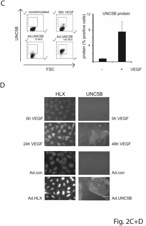
C
D
F i g . 2C+D
FSC
B5C
NU
104 10
4
104
104
10310
3
103
103
Ad.UNC5B 10 MOI
Ad.UNC5B 100 MOI
nonstimulated 56h VEGF UNC5B protein
- + VEGF
)sllec evitisop %( nietorp
4
8
12
HLX UNC5B
0h VEGF
24h VEGF
0h VEGF
48h VEGF
Ad.con
Ad.HLX
Ad.con
Ad.UNC5B10µm
For personal use only. on January 11, 2011. Bibliothek der MedUniWien (149592) at www.bloodjournal.orgFrom

For personal use only. on January 11, 2011. Bibliothek der MedUniWien (149592) at www.bloodjournal.orgFrom

0
0.02
0.04
0 1 2 3
w/oAd.conAd.HLX
days
DO( tnetnoc nietor
P294)
40
80
120
- +
w/oAd.conAd.HLX
)%( sllec detargi
MA
B
Fig. 3
VEGF
**
***
For personal use only. on January 11, 2011. Bibliothek der MedUniWien (149592) at www.bloodjournal.orgFrom
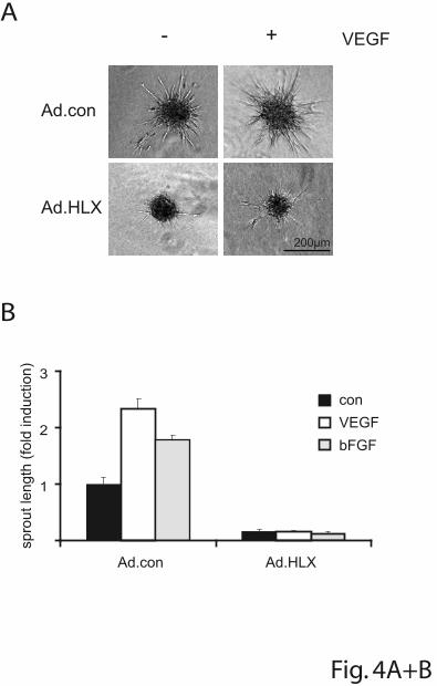
Fig. 4A+B
B
200µm
Ad.HLX
Ad.con
VEGF)noitcudni
dlof(htgneltuorps
1
2
3
Ad.con Ad.HLX
conVEGFbFGF
A- +
For personal use only. on January 11, 2011. Bibliothek der MedUniWien (149592) at www.bloodjournal.orgFrom

Fig. 4C+D
D
shRNA con
shRNA UNC5B
)noitcudnidlof(
htgneltuorps
Ad.con Ad.HLX
* **
* **
1
3
2
4
C)noitcudni
dlof(htgneltuorps
100 MOI
4
1
2
3
Ad.con Ad.UNC5B50 MOI
Ad.UNC5B
conVEGF
100 MOI
For personal use only. on January 11, 2011. Bibliothek der MedUniWien (149592) at www.bloodjournal.orgFrom

)noitcudnidlof(
AN
Rm
Fig. 5
time (h) time (h)20 4010 3020 4010 30
20
40
20
10
HLX mRNA UNC5B mRNA
Normoxia
Hypoxia
VEGF VEGF
A
B
Ad.con
Ad.HLX
aixomroN
aixopyH
noninduced VEGF
aixomroN
aixopyH
2
4
)noitcudnidlof(
htgneltuorps
VEGF-A mRNA
20
10
)noitcudnidlof(
AN
Rm
20 4010 30 VEGFtime (h)
* **
* **
**
**
For personal use only. on January 11, 2011. Bibliothek der MedUniWien (149592) at www.bloodjournal.orgFrom

10
20
30
40
*
Ad. con Ad.HLX
Ad.conAd.HLXm
m( D
VM
2)
ME
S -/+
CD31HE
Fig. 6
CD34
100µm100µm
50µm 50µm
A
B
For personal use only. on January 11, 2011. Bibliothek der MedUniWien (149592) at www.bloodjournal.orgFrom

Ad.HLX/Ad.con Ad.con/non infected UniGene ID
Entrez Gene
Gene Symbol Gene Title 4h 8h 16h 32h 4h 8h 16h 32h
Hs.585457 219699 UNC5B unc-5 homolog B (C. elegans) 1.41 2.23 5.71 7.14 0.69 1.17 1.21 2.61 Hs.9315 56944 OLFML3 olfactomedin-like 3 1.09 1.72 4.62 23.68 1.00 0.87 1.23 2.33 Hs.432329 5361 PLXNA1 plexin A1 1.15 2.96 4.61 8.38 1.11 1.38 1.22 1.40 Hs.360174 6591 SNAI2 snail homolog 2 (Drosophila) 1.58 3.87 4.51 6.91 1.00 1.37 2.14 2.40 Hs.132781 9466 IL27RA interleukin 27 receptor, alpha 0.91 1.86 4.18 5.53 1.25 1.17 1.57 1.57 Hs.525704 3725 JUN Jun oncogene 1.48 1.51 4.06 3.14 1.07 1.84 0.69 0.67 Hs.532626 23093 TTLL5 tubulin tyrosine ligase-like family, member 5 0.92 1.43 3.93 2.17 0.97 1.18 0.40 1.25 Hs.417050 8900 CCNA1 cyclin A1 0.78 2.15 3.79 2.96 1.00 0.85 0.95 0.40 Hs.2128 1847 DUSP5 dual specificity phosphatase 5 1.07 2.38 3.76 3.54 1.19 1.13 1.72 2.49 Hs.593413 7852 CXCR4 chemokine (C-X-C motif) receptor 4 1.22 1.46 3.74 11.61 1.11 0.95 0.16 0.03 Hs.657797 --- --- CDNA FLJ12055 fis, clone HEMBB1002049 0.97 1.29 3.70 2.22 0.93 1.70 0.36 1.12 Hs.356216 54855 FAM46C family with sequence similarity 46, member C 0.98 1.43 3.70 2.85 1.07 1.24 1.45 1.68 Hs.530443 387763 LOC387763 hypothetical LOC387763 0.94 2.51 3.52 3.15 1.15 1.06 0.92 1.10 Hs.250666 3280 HES1 hairy and enhancer of split 1, (Drosophila) 2.13 3.63 3.49 4.37 1.22 1.28 1.41 1.88 Hs.702013 10687 PNMA2 paraneoplastic antigen MA2 0.94 1.82 3.49 4.41 0.87 0.84 0.41 0.07 Hs.659681 54880 BCOR BCL6 co-repressor 2.01 2.54 3.45 5.37 1.00 1.50 0.84 1.40 Hs.653792 --- --- Transcribed locus 0.68 1.35 3.30 0.93 0.87 0.71 1.73 3.13 Hs.437075 9586 CREB5 cAMP responsive element binding protein 5 0.88 1.64 3.28 10.81 0.98 1.38 0.81 0.73 Hs.655332 10894 LYVE1 lymphatic vessel endothelial hyaluronan receptor 1 0.97 1.32 3.17 1.55 0.59 0.34 0.45 0.70 Hs.381167 1992 SERPINB1 serpin peptidase inhibitor, clade B (ovalbumin), member 1 0.93 1.33 3.15 4.25 0.94 0.64 0.97 1.44 Hs.654568 3880 KRT19 keratin 19 0.72 1.18 3.05 3.76 0.83 0.55 0.64 0.88 Hs.76152 358 AQP1 aquaporin 1 (Colton blood group) 1.22 3.35 3.03 3.33 0.54 0.24 0.44 0.51 Hs.647068 6009 RHEB Ras homolog enriched in brain 0.76 1.53 2.96 1.70 0.92 1.14 0.78 1.17 Hs.59729 56920 SEMA3G sema domain, immunoglobulin domain (Ig), short basic
domain, secreted, (semaphorin) 3G 1.27 2.28 2.88 4.89 1.10 0.89 1.08 0.77
Hs.438489 27109 ATP5S ATP synthase, H+ transporting, mitochondrial F0 complex, subunit s (factor B)
1.02 1.60 2.85 2.80 0.90 1.04 0.44 0.77
Hs.459265 3669 ISG20 interferon stimulated exonuclease gene 20kDa 1.09 1.90 2.83 2.88 0.77 0.71 0.92 1.16 Hs.523852 595 CCND1 cyclin D1 1.04 0.92 2.83 8.14 0.92 1.49 1.41 0.82
62

Hs.93675 11067 C10orf10 chromosome 10 open reading frame 10 1.45 1.48 2.83 7.98 0.88 0.86 0.27 0.06 Hs.122514 51312 SLC25A37 solute carrier family 25, member 37 1.23 1.20 2.83 2.74 0.83 1.07 0.65 0.92 Hs.657642 --- --- Transcribed locus 1.07 2.73 2.82 2.09 0.92 1.06 0.58 0.77 Hs.535713 727811
/// 9034 CCRL2 /// LOC727811
chemokine (C-C motif) receptor-like 2 /// similar to chemokine (C-C motif) receptor-like 2
0.98 2.13 2.80 3.69 0.83 0.64 1.05 0.95
Hs.654678 --- --- Transcribed locus 0.88 1.11 2.79 2.09 0.87 1.29 0.96 0.64 Hs.459538 220 ALDH1A3 aldehyde dehydrogenase 1 family, member A3 0.84 1.49 2.78 7.81 1.03 1.05 0.80 0.64 Hs.25960 4613 MYCN v-myc myelocytomatosis viral related oncogene,
neuroblastoma derived (avian) 1.54 3.75 2.78 10.33 1.02 0.87 0.71 0.43
Hs.592409 --- --- Transcribed locus 0.69 1.23 2.77 4.81 1.12 0.96 1.11 1.83 Hs.96 5366 PMAIP1 phorbol-12-myristate-13-acetate-induced protein 1 1.45 1.70 2.75 1.53 0.99 1.24 0.73 0.80 Hs.585118 1903 EDG3 endothelial differentiation, sphingolipid G-protein-coupled
receptor, 3 1.14 2.41 2.73 6.17 0.99 1.05 0.93 0.54
Hs.593413 7852 CXCR4 chemokine (C-X-C motif) receptor 4 1.15 1.73 2.72 3.77 0.97 0.68 0.28 0.08 Hs.494557 195828 ZNF367 zinc finger protein 367 0.75 1.04 2.67 2.64 1.07 0.92 0.36 0.11 Hs.658524 --- --- Transcribed locus 1.43 1.20 2.66 3.03 1.05 2.05 0.82 1.52 Hs.591251 154 ADRB2 adrenergic, beta-2-, receptor, surface 0.94 2.02 2.64 4.45 1.04 0.77 1.21 1.13 Hs.110571 4616 GADD45B growth arrest and DNA-damage-inducible, beta 1.20 1.68 2.64 4.26 0.77 0.82 0.35 0.19 Hs.593318 --- --- Full-length cDNA clone CS0DI001YP15 of Placenta Cot
25-normalized of Homo sapiens (human) 0.96 1.41 2.63 4.67 1.06 1.05 1.12 1.52
Hs.107125 83483 PLVAP plasmalemma vesicle associated protein 1.43 1.79 2.63 2.56 0.82 0.91 1.65 2.89 Hs.19985 --- --- Transcribed locus 0.89 1.30 2.62 2.38 0.78 0.62 0.39 0.72 Hs.203637 5357 PLS1 plastin 1 (I isoform) 0.61 1.41 2.59 1.36 1.21 1.02 0.65 0.64 Hs.520819 3638 INSIG1 insulin induced gene 1 0.92 1.78 2.59 2.78 0.92 1.09 2.20 1.11 Hs.659130 --- --- MRNA; cDNA DKFZp564A023 (from clone
DKFZp564A023) 1.32 0.93 2.59 0.98 0.92 1.32 0.52 2.09
Hs.491582 5327 PLAT plasminogen activator, tissue 0.98 1.42 2.57 4.27 1.18 1.07 1.44 1.31 Hs.460 467 ATF3 activating transcription factor 3 1.45 2.70 2.57 4.86 1.00 1.47 1.49 1.65 Hs.42645 9120 SLC16A6 solute carrier family 16, member 6 (monocarboxylic acid
transporter 7) 1.19 2.39 2.55 2.97 0.98 0.92 0.49 0.88
Hs.59332 200734 SPRED2 sprouty-related, EVH1 domain containing 2 0.80 1.80 2.51 4.25 1.01 1.02 0.76 0.46 Hs.25590 6781 STC1 stanniocalcin 1 1.06 0.94 2.51 1.07 0.92 1.12 1.22 1.46
63

Table S1. Genes upregulated by HLX in endothelioal cells more than 2-fold. Total RNA was isolated from
HUVEC after infection with HLX encoding adenoviruses (Ad.HLX) and control (empty) adenoviruses (Ad.con) for 4, 8, 16 and 32h.
Non-infected cells were used as control for virus effects. The RNA was subjected to microarray analysis using the Affymetrix
Human Genome U133 Plus 2.0 Array. Gene expression changes for HLX adenovirus relative to control adenovirus infected
cultures are displayed as fold induction in the Ad.HLX/Ad.con lane, for control adenovirus infected relative to non-infected cultures
in the Ad.con/non-infected lane. The full set of Affymetrix data is available in the GEO database under under GSE13054. All genes
induced more than 2.5-fold at the 16 h time point are listed according to decreasing HLX induction values. Genes occuring several
times in the original Affymetrix array dataset are only given once using their highest value.
64

Primer Sequence (5’ – 3’ direction)
UNC5B-forward CTG GGA CCT TAT GCC TTC AA
UNC5B-reverse CGC TTT GGT GGC AAA GTA AT
PlexinA1-forward TGT CTG GAT TCA GTG GCT CA
PlexinA1-reverse GGT TCT GCG GTG TTA AAG GA
HES1-forward ACG CAG TGT CAC CTT CCA G
HES1-reverse AGA GTC CGG AGG GAA GAG AG
HLX1-forward CTAGCCAGCCGAACACTTCT
HLX1-reverse CGTCCTAAGTGCCTTTGAGC
Sema3G-forward ACGGAGCACAATAGCACCTT
Sema3G-reverse AGACTCGCTCGTCCGTCTT
VEGF-A-forward GTGGGCCTTGCTCAGAGCGG
VEGF-A-reverse AGGCTCCTTCCTCCTGCCCG
Table S2. PCR primers used in Realtime RT-PCR
65

Gene shRNA sequence (5’ – 3’ direction)
HLX CCGGCGCATCTCTAGATCCCATTAACTCGAGTTAATGGGATCTAGAGATGCGTTTTT
UNC5B CCGGCAGAAGATATGCAACAGCCTACTCGAGTAGGCTGTTGCATATCTTCTGTTTTTG
control CCGGCAACAAGATGAAGAGCACCAACTCGAGTTGGTGCTCTTCATCTTGTTGTTTTT
Table S3. shRNA sequences in Mission shRNA pLKO.1-puro
66

Figure S1. Induction of UNC5B and HLX mRNA in HUVEC and other
endothelial cells of different origin (A) UNC5B is starting to be upregulated at 2 h after VEGF induction. HUVEC
were cultured to density, starved overnight in EBM-2 medium without
supplements and then induced with 100 ng VEGF/ml. Total RNA was isolated
and mRNA levels were determined by real-time RT-PCR. All values were
normalized to ß2-microglobulin mRNA as internal standard. Results are
displayed for the early time points from 1 to 16 h after VEGF addition and
represent the mean of fold induction +/- standard error of the mean (SEM) of
the respective mRNA levels calculated from triplicate wells. One
representative experiment of three performed is shown.
(B) HLX is induced by VEGF-A in endothelial cells of different origin.
Various endothelial cells isolated from different tissues including Human Aorta
Endothelial Cells (HAEC), Human Pulmonary Artery Endothelial Cells
(HPAEC), Human Retina Endothelial Cells (HREC), Human Skin
Microvascular Endothelial Cells (HSMEC), Human Saphenous Vein
Endothelial Cells (HSVEC) and HUVEC as well as Human Smooth Muscle
Cells (HSMC) were grown to density, starved overnight and induced with
VEGF-A for 1 h. RNA was isolated from the cells and subjected to realtime
RT-PCR analysis. Results are displayed as fold induction +/-SEM from one
experiment performed in triplicates.
Figure S2. Infection of HUVEC with Ad.HLX does not induce apoptosis HUVEC seeded in a chamber slide (Nunc, Naperville, IL, USA) were infected
with HLX and control adenoviruses with a MOI of 8. 24h later the cells were
fixed, permeabilized and stained according to the In Situ Cell Death Detection
Kit (TMR red, Roche, Mannheim, Germany). As a positive control one well
was treated with 100 U/ml DNase I for 10 min to induce DNA strand breaks.
The cells were photographed using a Nikon Diaphot TMD microscope and a
CCD camera (Kappa GmbH, Gleichen, Germany).
67

Figure S3. HLX reduces sprout formation in vitro in a dose-dependent manner HUVEC were infected with HLX and control adenoviruses using a MOI of 10,
3.3, 1 or 0.1. The following day spheroids of infected and noninfected
endothelial cells were prepared consisting of about 400 cells/spheroid as
described in Materials and Methods. The spheroids were embedded in
collagen gels and stimulated with VEGF or bFGF (25 ng/ml) or left without
cytokine stimulation. After 24 h the spheroids were fixed with PFA and the
length of sprouts was analysed for 10 spheroids per group. The results are
displayed as fold induction of sprout length compared to cells infected with
control viruses without stimulation + standard error of the mean (SEM)
calculated from triplicate wells.
Figure S 4. Downmodulation of UNC5B mRNA by shRNA increases the number of VEGF-inducible sprouts from endothelial spheroids.(A) Downmodulation of UNC5B mRNA by shRNA: HUVEC were transduced
with lentiviruses expressing shRNA targeted against UNC5B or control
shRNA. Then they were starved overnight and induced with VEGF-A for 56 h.
RNA was isolated and subjected to realtime RT-PCR. Results are displayed
as percent UNC5B mRNA +/- SEM calculated from triplicate wells.
(B) Increased number of sprouts following downmodulation of UNC5B in
VEGF-induced spheroids: HUVEC were first transduced with lentiviruses for
control shRNA or shRNA targeted against HLX. After 2 d cells were in
addition infected with 15 MOI of Ad.con or Ad.HLX. The following day
spheroids were prepared and incorporated into collagen gels in the presence
of VEGF-A. Sprouting of spheroids was evaluated the next day. The shown
values are calculated from four independent experiments and display average
sprout number per spheroid +/-SEM in comparison to the samples transduced
with Ad.con and shRNA. con.
68

Figure S5. Perfusion of vessels generated in the in vivo endothelial spheroid xenografting assaySections obtained from plugs generated with endothelial spheroids infected
with control adenovirus. Arrows indicate red blood cells in the luminal areas of
vessels containing human endothelial cells. Zink fixed Matrigel plugs were embedded in paraffin and sectioned at 8 µm,
deparaffinized and rehydrated. The primary antibody, mouse-anti-human
CD31 (Dako, Glostrup, Denmark) was detected by the following secondary
antibody: biotinylated anti-mouse (Vector Laboratories, Burlingame, USA).
The incubation with the secondary antibody was followed by the incubation
with streptavidin-peroxidase conjugate (Vector Laboratories) and developed
using DAB substrate chromogen system (Dako). Afterwards the sections were
stained with Hämalaun (Merck) and Eosin.
69
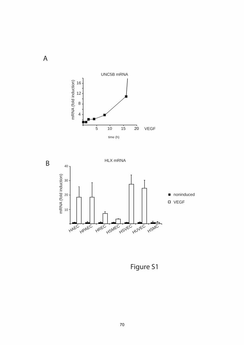
Figure S1
HLX mRNA40
10
20
30
mR
NA
(fold
indu
ctio
n)
HAECHPAEC
HRECHSMEC
HSVECHUVEC
HSMC
noninduced
VEGF
A
B
20
time (h)
10
4
8
12
16
VEGF5 15
)noitcudnidlof(
AN
Rm
UNC5B mRNA
70
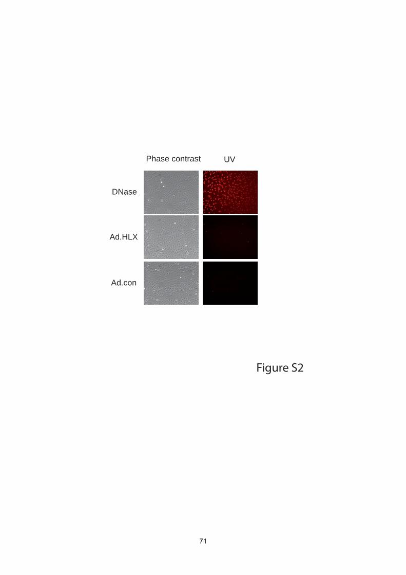
DNase
Phase contrast UV
Ad.HLX
Ad.con
Figure S2
71

Figure S3
VEGF bFGFnoninduced
Ad.con
Ad.HLX
MOI
80
40
100 4
60
40
2040
80
MOI MOI2 6 8 100 42 6 8 100 42 6 8
spro
ut le
ngth
(µm
)
72

shRNA conshRNA UNC5B
UNC5B mRNA
60
120
mR
NA
(%)
Figure S4
A
B shRNA conshRNA UNC5B
shRNA conshRNA UNC5B
spro
ut n
umbe
r
10
20
30
aver
age
spro
ut le
ngth
(pix
el)
0
30
60
73

F i gure S5
hCD31 DAB staining + HE staining arrow: red blood cells
100 µm 100 µm
74
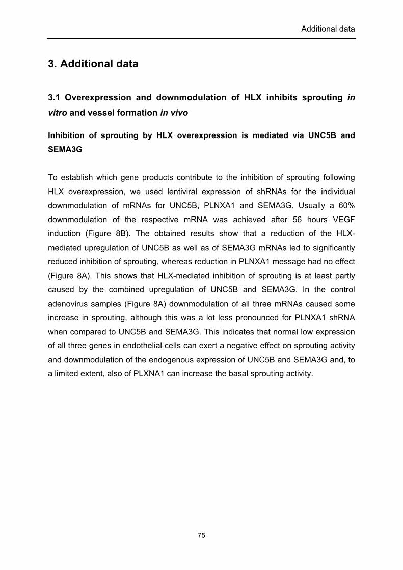
Additional data
75
3. Additional data
3.1 Overexpression and downmodulation of HLX inhibits sprouting in
vitro and vessel formation in vivo
Inhibition of sprouting by HLX overexpression is mediated via UNC5B and
SEMA3G
To establish which gene products contribute to the inhibition of sprouting following
HLX overexpression, we used lentiviral expression of shRNAs for the individual
downmodulation of mRNAs for UNC5B, PLNXA1 and SEMA3G. Usually a 60%
downmodulation of the respective mRNA was achieved after 56 hours VEGF
induction (Figure 8B). The obtained results show that a reduction of the HLX-
mediated upregulation of UNC5B as well as of SEMA3G mRNAs led to significantly
reduced inhibition of sprouting, whereas reduction in PLNXA1 message had no effect
(Figure 8A). This shows that HLX-mediated inhibition of sprouting is at least partly
caused by the combined upregulation of UNC5B and SEMA3G. In the control
adenovirus samples (Figure 8A) downmodulation of all three mRNAs caused some
increase in sprouting, although this was a lot less pronounced for PLNXA1 shRNA
when compared to UNC5B and SEMA3G. This indicates that normal low expression
of all three genes in endothelial cells can exert a negative effect on sprouting activity
and downmodulation of the endogenous expression of UNC5B and SEMA3G and, to
a limited extent, also of PLXNA1 can increase the basal sprouting activity.

Additional data
76
A
B
Figure 8. Effects of downmodulation of UNC5B, PLXNA1 and SEMA3G by the
respective shRNA.
(A) Inhibition of sprouting by HLX is reduced by downmodulation of UNC5B or SEMA3G. HUVEC were transduced with lentiviruses for control shRNA or shRNA targeted against UNC5B, PLXNA1 or SEMA3G. After 1 d cells were in addition infected with 15 MOI of Ad.con or Ad.HLX. The following day cells were used for the spheroid sprouting assay. Total sprout lengths of 15 spheroids each were measured. Results are displayed as fold induction +/- SEM in comparison to the sprout lengths obtained for non-induced spheroids prepared from control shRNA and control adenovirus transduced cells. One representative experiment of two performed is shown. ***P < .001, t test (B) Downmodulation of UNC5B, PLXNA1 and SEMA3G mRNA by the respective shRNAs. HUVEC were transduced with lentiviruses expressing shRNA targeted against UNC5B, PLXNA1 or SEMA3G for 2 days. Then they were starved overnight and induced with VEGF for 56 h. RNA was isolated and subjected to realtime RT-PCR. Results are displayed as percent UNC5B, PLXNA1 or SEMA3G mRNA +/- SD calculated from triplicate wells.
Reduction of HLX expression by shRNA reduces sprouting
We next asked how the reduction of HLX by lentiviral expression of a corresponding
shRNA would affect the sprouting activity. Surprisingly we found that
downmodulation of HLX by shRNA also exerted an inhibitive effect on sprouting,
although the achieved inhibition seemed less pronounced when compared to
overexpression of HLX (Figure 9A). When a dilution series of the shRNA lentivirus

Additional data
77
was tested, the inhibition diminished in a dose-dependent way (Figure 9B). This
shows that a minimal expression level of HLX is necessary for sprouting, which might
be due to the HLX-mediated induction of a so far non-identified protein that would
support sprouting. This indicates that overexpression of HLX as well as levels of HLX
reduced below a certain threshold can lead to inhibition of sprouting.
A
B
Figure 9. Downmodulation of HLX reduces sprouting.
(A) HUVEC were transduced with lentiviruses for control shRNA or shRNA targeted against HLX. After 2 d cells were used for the spheroid sprouting assay. Total sprout lengths of 15 spheroids each were calculated. The shown values are calculated from four independent experiments and display percent sprout length +/-SEM in comparison to the control shRNA samples stimulated with VEGF. (B) Downmodulation of HLX using shRNA lentiviruses reduces sprout formation in vitro in a dose-dependent manner: HUVEC were transduced using different volumes of lentiviral stock preparations for two days. Then cells were used for spheroid sprouting assay. The results show fold induction of total sprout length +/- SEM as calculated from 15 spheroids for each condition.

Additional data
78
Downmodulation of HLX can inhibit vessel formation in vivo
To confirm that the inhibitory effect observed on sprouting in vitro would also be
reflected in vessel formation in vivo, a spheroid xenografting assay was performed.
Indeed, in line with the in vitro sprouting results a reduction of HLX mRNA by a
corresponding shRNA similarly reduced vessel density as well as vessel length in
vivo supporting the importance of appropriate HLX levels to vessel formation (Figure
10).
Figure 10. Downmodulation of HLX reduces vessel formation in an endothelial
spheroid xenografting assay in vivo
HUVEC were transduced with lentiviruses containing control or HLX shRNA. Stably shRNA expressing cells were selected with puromycin for 5 days. The infected cells were used to generate endothelial cell spheroids consisting of about 100 cells/spheroid. Spheroids were suspended in a Matrigel/fibrinogen mixture containing 500 ng/ml VEGF and bFGF. Following addition of thrombin the spheroid suspension was injected subcutaneously into SCID mice (2 plugs/mouse, 8 mice/experimental group). Plugs were harvested, fixed and analyzed by immunohistochemistry using CD34 antibodies after 20 days. MVD per mm2 +/- SEM was calculated from five representative sections of five plugs.

Additional data
79
3.2 Effect of HLX overexpression on MDA breast cancer growth
Based on the upregulation of repulsive guidance molecules by HLX and the
connected inhibition of vessel growth we predicted that overexpression of HLX
throughout the tumor tissue including tumor vessels might lead to inhibition of tumor
growth. To evaluate such a potential inhibition we used a human MDA breast cancer
model in SCID beige mice. In this model HLX was overexpressed in tumor and
endothelial cells by 3 consecutive injections of Ad.HLX into the tumor tissue on days
7, 11 and 15 after injection of tumor cells. This indeed led to a significant inhibition in
tumor growth. Following the first injection of Ad.HLX no further increase in tumor
volume was observed (Figure 11). This might be due to the expression of HLX in
infected endothelial cells of the tumor vessels, which should lead to reduced
neovascularization as observed before for the murine spheroid xenografting assay.
However, as adenoviruses would equally infect tumor cells, the inhibition of tumor
growth could also be a direct or indirect effect of the expression of HLX in tumor
cells. This possibility was tested by analyzing MDA breast cancer cells following
infection with HLX adenoviruses in cell culture. Whereas no effect on the proliferation
of the tumor cells was observed (data not shown), the analysis of guidance molecule
expression revealed an increased expression of SEMA3G by the tumor cells
overexpressing HLX (Figure 12). UNC5B and PLXNA1 were not expressed in the
tumor cells (data not shown). It is therefore possible that increased secretion of
SEMA3G into the tumor tissue environment might contribute to diminished
endothelial sprouting and vessel growth in the growing tumor, thereby blocking
adequate oxygen and nutrient supply resulting in inhibition of tumor growth. However,
it remains possible that SEMA3G also exerts direct negative effects on tumor cells in
vivo (Gaur et al., 2009). Further experiments will be required to establish the exact
mode of action of the astonishingly strong effect of HLX on tumor growth.

Additional data
80
Figure 11. Overexpression of HLX inhibits tumor growth of breast cancer cells 5 x 10⁶ MDA-MB-231 cells were injected subcutaneously into the flanks of SCID/beige (C.B-Igh1b/IcrHsd-PrkdcscidLystbg) mice. On day 7, 11 and 15 after the tumor cell application 1x10⁸ PFU of Ad.HLX or Ad.con (expressing GFP) or the same volume of PBS was injected into the grown tumor. The tumor volume was established during the whole experiment by measuring the tumor size using a caliper rule. The tumor volume was calculated according to the formula: tumor volume= tumor length x tumor width x tumor width/2. The experiment was terminated on day 18 after tumor cell injection. Results display tumor volume from one representative experiment of two performed with at least 5 mice per group and show tumor volume (mm3) +/- SEM.
Figure 12. Overexpression of HLX induces SEMA3G mRNA expression in MDA cancer
cells
MDA-MB-231 cancer cells were transduced with a MOI of 20 of Ad.HLX and Ad.con (expressing GFP) for 16 and 32h. Cells were harvested, the RNA isolated and subjected to real-time RT-PCR analysis. Values were normalized to ß2-microglobulin mRNA as internal standard.

Additional publications (Coauthorship)
81
4. Additional publications (Coauthorship)
4.1 The VEGF-induced transcriptional response comprises gene clusters
at the crossroad of angiogenesis and inflammation.
Bernhard Schweighofer, Julia Testori, Caterina Sturtzel, Susanne Sattler,
Herbert Mayer, Oswald Wagner, Martin Bilban, Erhard Hofer.
Thromb Haemost. 2009;102:544-554.

544
© 2009 Schattauer GmbH, Stuttgart
The VEGF-induced transcriptional response comprises gene clusters at the crossroad of angiogenesis and inflammation Bernhard Schweighofer1; Julia Testori1; Caterina Sturtzel1; Susanne Sattler1; Herbert Mayer4; Oswald Wagner2,3; Martin Bilban2,3; Erhard Hofer1 1Department of Vascular Biology and Thrombosis Research, Center for Biomolecular Medicine and Pharmacology, Medical University of Vienna, Vienna, Austria; 2Clinical Department for Medical and Chemical Laboratory Diagnostics, Medical University of Vienna, Vienna, Austria; 3Ludwig Boltzmann Institute for Clinical and Experimental Oncology, Vienna, Austria; 4Present address: Austrian Science Foundation, Vienna, Austria
Summary VEGF-A is the major trigger of vasculogenesis and physiologic angiogenesis. We have investigated to which extent the gene repertoire induced by VEGF-A in endothelial cells is distinct from that of other growth factors and inflammatory cytokines. Genes upregulated in human umbilical vein endothelial cells treated with VEGF, EGF or IL-1 were compared by microarray analysis and clusters characteristic for individual or com-binations of inducers were defined. VEGF-A upregulated in com-parison to EGF a five-fold larger gene repertoire, which surpris-ingly overlapped to 60% with the inflammatory repertoire of IL-1. As shown by real-time RT-PCR for selected genes, VEGF-induction was mostly mediated by VEGF receptor-2 and the ca-pacity of VEGF-A to induce genes in common with IL-1 largely
Keywords VEGF-A, endothelial cells, angiogenesis, inflammation, gene repertoire
depended on activation of the calcineurin/NFAT pathway, since cyclosporin A inhibited this induction. Another angiogenic growth factor, bFGF, did not share a comparable induction of in-flammatory genes, but partially induced a small group of genes in common with VEGF-A, which were not regulated by EGF. Thus, the data display that VEGF-A induces a distinct gene repertoire, which, contrasting with other growth factors such as EGF or bFGF, includes an inherent inflammatory component possibly contributing to the cross-regulation of angiogenesis and inflam-mation as further indicated by the VEGF-mediated induction of leukocyte adhesion. Furthermore, a small group of genes selec-tively induced by VEGF-A with potential importance for angio-genesis is defined.
Thromb Haemost 2009; 102: 544–554
Cardiovascular Biology and Cell Signalling
Correspondence to: Prof. Dr. Erhard Hofer Department of Vascular Biology and Thrombosis Research Center for Biomolecular Medicine and Pharmacology Medical University of Vienna Lazarettgasse 19, A-1090 Vienna, Austria Tel.: +43 1 40160 33111, Fax: +43 1 40160 933100 E-mail: [email protected]
Received: December 19, 2008 Accepted after major revision: June 1, 2009
Prepublished online: July 3, 2009
doi:10.1160/TH08-12-0830
Introduction Vascular endothelial growth factor-A (VEGF-A) is the primary inducer of vascular development during embryogenesis and physiological blood vessel formation in the adult. It is also a cau-sative factor of pathologic angiogenesis associated with a multi-tude of diseases including cancer, chronic inflammatory diseases and retinopathy (1, 2). VEGF-A is normally produced in tissues in response to low oxygen tension and forms a chemotactic gradient sensed by endothelial cells in preexisting vessels, which respond by growing sprouts leading to the formation of new cap-illaries (3, 4). Several peptide products are generated by differen-tial splicing from the VEGF-A gene such as VEGF-A121,
VEGF-A165 and VEGF-A189 or the inhibitory variant VEGF-A165b. However, isoform VEGF-A165 seems to be the predomi-nant form responsible for major angiogenic effects (5).
The responses induced by VEGF-A in endothelial cells in-clude promotion of survival, proliferation, migration and in-vasion of surrounding tissue as well as the formation of the three-dimensional vascular tube. Furthermore, VEGF-A induces vas-cular leakage and is also known as vascular permeability factor (2). These biological effects of VEGF are thought to be mediated mainly by VEGF receptor-2 (VEGFR-2), whereas VEGF recep-tor-1 (VEGFR-1) transduces only a weak intracellular signal and was initially proposed to function in endothelial cells primarily as a decoy receptor and negative regulator of angiogenesis (6). In
82
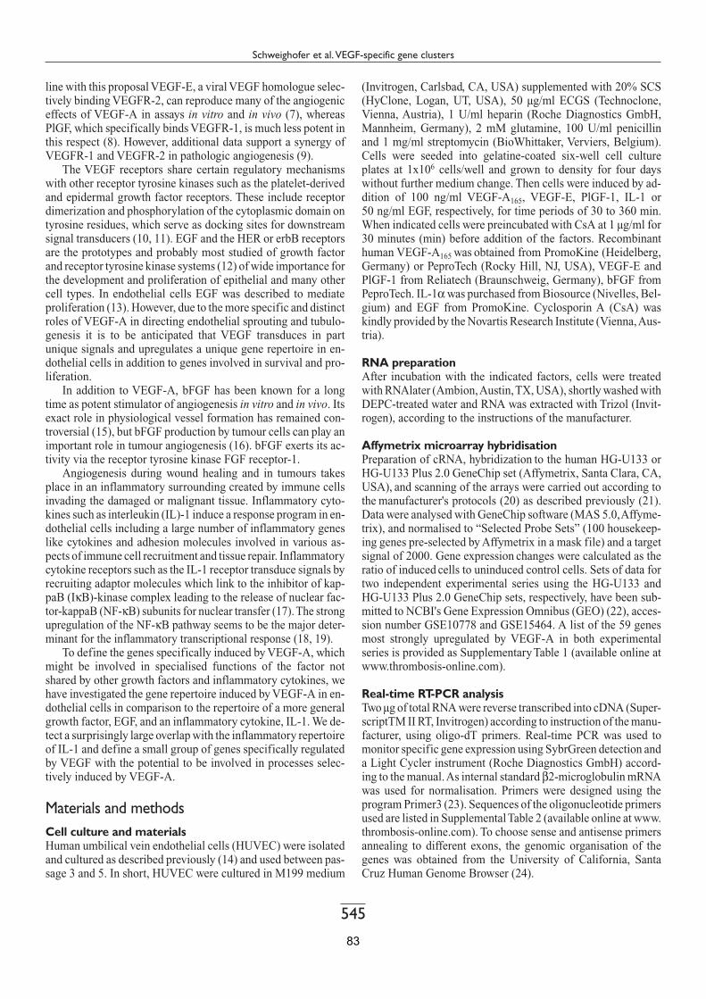
545
Schweighofer et al. VEGF-specific gene clusters
line with this proposal VEGF-E, a viral VEGF homologue selec-tively binding VEGFR-2, can reproduce many of the angiogenic effects of VEGF-A in assays in vitro and in vivo (7), whereas PlGF, which specifically binds VEGFR-1, is much less potent in this respect (8). However, additional data support a synergy of VEGFR-1 and VEGFR-2 in pathologic angiogenesis (9).
The VEGF receptors share certain regulatory mechanisms with other receptor tyrosine kinases such as the platelet-derived and epidermal growth factor receptors. These include receptor dimerization and phosphorylation of the cytoplasmic domain on tyrosine residues, which serve as docking sites for downstream signal transducers (10, 11). EGF and the HER or erbB receptors are the prototypes and probably most studied of growth factor and receptor tyrosine kinase systems (12) of wide importance for the development and proliferation of epithelial and many other cell types. In endothelial cells EGF was described to mediate proliferation (13). However, due to the more specific and distinct roles of VEGF-A in directing endothelial sprouting and tubulo-genesis it is to be anticipated that VEGF transduces in part unique signals and upregulates a unique gene repertoire in en-dothelial cells in addition to genes involved in survival and pro-liferation.
In addition to VEGF-A, bFGF has been known for a long time as potent stimulator of angiogenesis in vitro and in vivo. Its exact role in physiological vessel formation has remained con-troversial (15), but bFGF production by tumour cells can play an important role in tumour angiogenesis (16). bFGF exerts its ac-tivity via the receptor tyrosine kinase FGF receptor-1.
Angiogenesis during wound healing and in tumours takes place in an inflammatory surrounding created by immune cells invading the damaged or malignant tissue. Inflammatory cyto-kines such as interleukin (IL)-1 induce a response program in en-dothelial cells including a large number of inflammatory genes like cytokines and adhesion molecules involved in various as-pects of immune cell recruitment and tissue repair. Inflammatory cytokine receptors such as the IL-1 receptor transduce signals by recruiting adaptor molecules which link to the inhibitor of kap-paB (IκB)-kinase complex leading to the release of nuclear fac-tor-kappaB (NF-κB) subunits for nuclear transfer (17). The strong upregulation of the NF-κB pathway seems to be the major deter-minant for the inflammatory transcriptional response (18, 19).
To define the genes specifically induced by VEGF-A, which might be involved in specialised functions of the factor not shared by other growth factors and inflammatory cytokines, we have investigated the gene repertoire induced by VEGF-A in en-dothelial cells in comparison to the repertoire of a more general growth factor, EGF, and an inflammatory cytokine, IL-1. We de-tect a surprisingly large overlap with the inflammatory repertoire of IL-1 and define a small group of genes specifically regulated by VEGF with the potential to be involved in processes selec-tively induced by VEGF-A.
Materials and methods Cell culture and materials Human umbilical vein endothelial cells (HUVEC) were isolated and cultured as described previously (14) and used between pas-sage 3 and 5. In short, HUVEC were cultured in M199 medium
(Invitrogen, Carlsbad, CA, USA) supplemented with 20% SCS (HyClone, Logan, UT, USA), 50 µg/ml ECGS (Technoclone, Vienna, Austria), 1 U/ml heparin (Roche Diagnostics GmbH, Mannheim, Germany), 2 mM glutamine, 100 U/ml penicillin and 1 mg/ml streptomycin (BioWhittaker, Verviers, Belgium). Cells were seeded into gelatine-coated six-well cell culture plates at 1x10⁶ cells/well and grown to density for four days without further medium change. Then cells were induced by ad-dition of 100 ng/ml VEGF-A165, VEGF-E, PlGF-1, IL-1 or 50 ng/ml EGF, respectively, for time periods of 30 to 360 min. When indicated cells were preincubated with CsA at 1 µg/ml for 30 minutes (min) before addition of the factors. Recombinant human VEGF-A165 was obtained from PromoKine (Heidelberg, Germany) or PeproTech (Rocky Hill, NJ, USA), VEGF-E and PlGF-1 from Reliatech (Braunschweig, Germany), bFGF from PeproTech. IL-1α was purchased from Biosource (Nivelles, Bel-gium) and EGF from PromoKine. Cyclosporin A (CsA) was kindly provided by the Novartis Research Institute (Vienna, Aus-tria).
RNA preparation After incubation with the indicated factors, cells were treated with RNAlater (Ambion, Austin, TX, USA), shortly washed with DEPC-treated water and RNA was extracted with Trizol (Invit-rogen), according to the instructions of the manufacturer.
Affymetrix microarray hybridisation Preparation of cRNA, hybridization to the human HG-U133 or HG-U133 Plus 2.0 GeneChip set (Affymetrix, Santa Clara, CA, USA), and scanning of the arrays were carried out according to the manufacturer's protocols (20) as described previously (21). Data were analysed with GeneChip software (MAS 5.0, Affyme-trix), and normalised to “Selected Probe Sets” (100 housekeep-ing genes pre-selected by Affymetrix in a mask file) and a target signal of 2000. Gene expression changes were calculated as the ratio of induced cells to uninduced control cells. Sets of data for two independent experimental series using the HG-U133 and HG-U133 Plus 2.0 GeneChip sets, respectively, have been sub-mitted to NCBI's Gene Expression Omnibus (GEO) (22), acces-sion number GSE10778 and GSE15464. A list of the 59 genes most strongly upregulated by VEGF-A in both experimental series is provided as Supplementary Table 1 (available online at www.thrombosis-online.com).
Real-time RT-PCR analysis Two µg of total RNA were reverse transcribed into cDNA (Super-scriptTM II RT, Invitrogen) according to instruction of the manu-facturer, using oligo-dT primers. Real-time PCR was used to monitor specific gene expression using SybrGreen detection and a Light Cycler instrument (Roche Diagnostics GmbH) accord-ing to the manual. As internal standard β2-microglobulin mRNA was used for normalisation. Primers were designed using the program Primer3 (23). Sequences of the oligonucleotide primers used are listed in Supplemental Table 2 (available online at www.thrombosis-online.com). To choose sense and antisense primers annealing to different exons, the genomic organisation of the genes was obtained from the University of California, Santa Cruz Human Genome Browser (24).
83

546
Schweighofer et al. VEGF-specific gene clusters
Figure 1: VEGF-A induced genes compared to IL-1α and EGF induction pat-terns in HUVEC. The hier-archical clustering of all genes induced in HUVEC by VEGF-A, more than three-fold at one of the indicated time points, is displayed as a heatmap in comparison to the IL-1α and the EGF gene ex-pression pattern. Genes were clustered with EPCLUST (http://www.ebi.ac.uk/micro-array-srv/EP/) using Pearson’s correlation coefficient as dis-tance measure and average linkage. Induction of ex-pression is indicated by shades of red, repression by shades of green indicating ap-proximately 10– to 10–1-fold regulation. To visually separate the induction patterns of the individual factors, the null control value (ctrl) is shown in black before the respective induction.
84

547
Schweighofer et al. VEGF-specific gene clusters
Clustering analysis First, genes as represented by Affymetrix “probe sets” showing absolute calls of „absence“, difference calls of „no change“ as determined by the Affymetrix MAS 5.0 software, less than three-fold upregulation or a signal intensity of less than 60 for all VEGF-induced data points were excluded from further analysis. Then the genes selected for more than three-fold upregulation by VEGF in two independent induction series were subjected to real-time RT-PCR analyses to confirm over three-fold upregu-lation for at least one time point and genes displaying lower regu-lation were again abandoned. For the remaining fifty-nine genes cluster analysis was performed using the Expression Profile data CLUSTering and analysis (EPCLUST) program (25) using lin-ear correlation based distance (Pearson, centered) and average linkage (UPGMA).
Flow cytometry HUVEC stimulated for six hours (h) with VEGF-A165, IL-1 or EGF were harvested following short trypsin treatment, washed and incubated with PBS containing 5% FCS for 15 min. Then 5 µg/ml mouse anti-human VCAM1 antibody (R&D systems, Minneapolis, MN, USA) was added and the cells incubated for 30 min on ice. Samples were washed and incubated for another 30 min with FITC-labeled secondary anti-mouse antibody (Dako, Denmark). After a final washing step, flow cytometry was performed using a FACSCalibur (Becton-Dickinson, San Jose, CA, USA). The number of VCAM1-positive cells was de-duced in comparison to uninduced cells.
Adhesion assay HUVEC induced for 4, 6 or 8 h with VEGF-A165, IL-1 or EGF were incubated with 3,3'-dioctadecyl-oxacarbocyanine perchlor-ate (DiO)-labeled (Invitrogen) HL-60 cells on a shaker for 30 min (15 min horizontal, 15 min vertical shaking) at 37°C in a 5% CO2 atmosphere. Non-adherent HL-60 cells were removed by washing with phosphate-buffered saline and adhering HL-60 cells and HUVEC were detached by trypsin treatment. The per-centage of DiO-labeled HL-60 cells in the cell mixture was de-termined by flow cytometry using a FACSCalibur.
Results A VEGF-A-specific gene signature: clustering of VEGF-induced genes To delineate genes induced by VEGF-A and involved in VEGF-specific functions during angiogenesis and/or endothelial differ-entiation we comparatively analysed the gene repertoires up-regulated in endothelial cells by VEGF-A, EGF or IL-1. The se-lection of these factors was based on the assumption that VEGF-A, the main trigger of vasculogenesis and angiogenesis, would induce gene clusters in addition to the gene repertoire up-regulated by EGF, a growth factor which promotes proliferation in endothelial cells and is a general component of the growth supplements used for primary endothelial cell cultures (26). The VEGF-induced genes should further be distinguished from the IL-1 response, which should primarily include genes involved in endothelial inflammation.
Quiescent HUVEC were treated in parallel with VEGF-A or EGF for time periods ranging from 30 to 360 min and total RNA was isolated from the cultures and subjected to Affymetrix microarray analysis. The obtained results were analysed includ-ing a separate comparable experiment with IL-1 using identical culture conditions and the same Affymetrix chips (27). Fifty-nine genes were selected to be reproducibly upregulated by VEGF-A more than three-fold as described in Methods (see Supplementary Table 1 available online at www.thrombosis-online.com).
The clustering of the gene repertoire induced by VEGF-A more than three-fold is displayed in Figure 1. Two smaller clusters of genes, one regulated similarly by VEGF-A, EGF or IL-1, another preferentially by VEGF-A, and a larger cluster of genes induced by VEGF-A as well as IL-1 can be distinguished. The distribution of the genes in the VEGF-specific, the VEGF/IL-1 and the VEGF/EGF/IL-1 clusters is schematically indicated in the Venn diagram of Figure 2. The regulation of all genes of the VEGF-specific gene cluster and of about half of the genes of the other two clusters were further tested by real-time RT-PCR using RNA from three independent induction experiments and found to closely resemble the results of the microarray analysis (Table 1).
The VEGF-selective gene cluster defines a group of genes induced by VEGF-A, but not by EGF or IL1 Whereas the VEGF/EGF/IL-1 cluster contains to a large extent immediate early genes reaching peak levels between 30 and 60 min of induction such as the transcription factors FOS, JUNB
Figure 2: Venn Diagram schematically displaying the percen-tage of genes in each of the three clusters. Percentage numbers are calculated by comparing the number of genes in the respective groups (VEGF/EGF/IL-1 or generally activated cluster; VEGF/IL-1 or in-flammation-related cluster; VEGF-specific cluster) to the total number of VEGF-induced genes. As indicated by the diagram, the number of genes induced by IL-1 is higher than the number of genes induced by VEGF-A. EGF induced a much smaller repertoire than VEGF-A.
85

548
Schweighofer et al. VEGF-specific gene clusters
and EGR-1, the VEGF-specific gene cluster comprises rather genes upregulated with peak values between 60 and 360 min and includes transcription factors, intracellular signalling molecules as well as secreted proteins and receptors. In the VEGF-specific group the earliest and most highly VEGF-upregulated genes are the transcription factors NR4A2 (Nurr1) and EGR-3, which could function in the induction of secondary genes of import-ance for angiogenesis. It further includes genes with more de-layed induction kinetics such as the transcription factors HLX1,
MEF2C and PER1, the secreted IGFBP3 and the chemokine re-ceptor CCRL1 (Fig. 3A and Table 1).
To establish whether the upregulation of these genes is me-diated via VEGFR-2 or VEGFR-1 we used further VEGF-E as a specific trigger of VEGFR-2 and PlGF as a factor binding spe-cifically to VEGFR-1. As shown in Figure 3B, VEGF-E was as potent as VEGF-A in the upregulation of VEGF-cluster genes, whereas PlGF had no significant effects.
Group Relative VEGF induction
At timepoint Gene symbol
Induction by bFGF
VEGF inducibility in prescence of CsA
SVI (VEGF)
1300 60 NR4A2 – (26) – (260)
120 60 EGR3 + (25) + (38)
13 60 IGFBP3 – – (3)
7 60 HLX1 ++ (5) +++ (7)
2 60 CREM +++(2) +
6 150 PER1 ++(3) n/d
4 150 MEF2C + (2) ++ (3)
2 150 CCRL1 + –
2 150 THBD + +
36 360 NDRG1 + (9) ++ (23)
35 30 FOS +++ (35) ++++ (192)
25 30 EGR1 ++++ (60) ++++ (39)
8 60 BHLHB2 + (3) ++++ (8)
4 60 KLF4 ++ (3) n/d
Relative mRNA induction by VEGF is given in -fold of uninduced control. Time points displayed are those when peak levels were observed for the respective mRNA and are given in minutes following induction. For bFGF induction and VEGF inducibility in presence of CsA the percentage of the comparable VEGF value is indicated by: – <25%; + 25–50%; ++ 50–75%; +++ 75–100%; ++++ >>100% of VEGF induction. The values in parantheses display the approximate -fold upregulation observed in comparison to the corresponding untreated control values. Values are given only for at least two-fold or larger changes. SVI, selec-tively VEGF induced; IR, inflammation related; GA, generally activated; n/d, not determined.
19 360 DNAJB9 n/d n/d
5 360 MYCN ++ (3) +++ (4)
IR (VEGF + IL1)
225 30 FOSB – (25) ++++ (1240)
105 30 ATF3 – (2) +++ (92)
51 30 CXCL2 – ++ (27)
30 30 NEDD9 – (3) ++ (17)
11 30 KCNJ2 – (3) + (5)
9 30 NFKBIZ – +++ (7)
80 60 DSCR1 – –
40 60 IL8 – (2) +++ (34)
17 60 PTGS2 – (2) – (4)
10 60 ITGAV – – (2)
100 150 F3 – (2) ++ (72)
4 150 CNKSR3 – (2) + (2)
380 360 VCAM1 – – (60)
60 360 SELE – +++ (70)
16 360 STC1 – (4) + (6)
GA (VEGF+IL1+EGF)
130 30 EGR2 ++ (73) +++ (88)
Table 1: Real-time RT-PCR based grouping of VEGF-induced genes displaying bFGF induction and CsA inhibition. Genes were grouped according to inducibil-ity by VEGF-A, IL-1 and EGF. Genes representing individual groups were further sorted in relation to 1) fold induction and 2) the time point when maximum induction is ob-served. Furthermore, induc-tion by bFGF is shown as per-centage of the VEGF-A induc-tion, and the capacity of VEGF-A to induce the genes in the presence of CsA is de-picted. Data display the aver-age from at least three inde-pendent experiments.
86
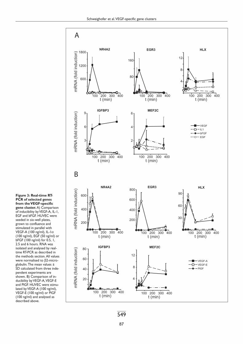
549
Schweighofer et al. VEGF-specific gene clusters
Figure 3: Real-time RT-PCR of selected genes from the VEGF-specific gene cluster. A) Comparison of inducibility by VEGF-A, IL-1, EGF and bFGF. HUVEC were seeded in six-well plates, grown to confluence and stimulated in parallel with VEGF-A (100 ng/ml), IL-1α (100 ng/ml), EGF (50 ng/ml) or bFGF (100 ng/ml) for 0.5, 1, 2.5 and 6 hours. RNA was isolated and analysed by real-time RT-PCR as described in the methods section. All values were normalised to β2-micro-globulin. The mean values ± SD calculated from three inde-pendent experiments are shown. B) Comparison of in-ducibility by VEGF-A, VEGF-E and PlGF. HUVEC were stimu-lated by VEGF-A (100 ng/ml), VEGF-E (100 ng/ml) or PlGF (100 ng/ml) and analysed as described above.
87

550
Schweighofer et al. VEGF-specific gene clusters
The VEGF/IL-1 gene cluster comprises the majority of VEGF-induced genes Surprisingly the largest group is the gene cluster upregulated by VEGF-A as well as IL-1, but not by EGF. This cluster contains a heterogeneous group of genes displaying peak values between 30 to 360 min. It comprises a whole group of classical inflamma-tory genes such as CXCL2, IL8, PTGS2 (COX-2), tissue factor and the adhesion molecules VCAM1 and E-selectin. Whereas VEGF-A induction of IL8, PTGS2 and tissue factor is largely comparable to the induction rate observed for IL-1, induction of VCAM1, E-selectin and CXCL2 by VEGF was approximately 10-fold and 100-fold less pronounced than IL-1 induction, re-spectively. In addition transcription factors such as ATF3, members of cytoplasmic signalling pathways such as NEDD9 and NFKBIZ and the ion channel KCNJ2 belong to this cluster (Table 1 and Supplementary Fig. 1A available online at www.thrombosis-online.com).
To test for the involvement of VEGFR-2 in the upregulation we evaluated VEGF-E and PlGF effects on selected inflamma-tory genes. Again VEGF-E could completely or in part repro-duce VEGF-A effects, whereas PlGF did not display detectable activity (Supplementary Fig. 1B available online at www.thrombosis-online.com).
To confirm that also the corresponding inflammatory pro-teins are upregulated in the cells, we have analysed VCAM1 ex-pression by flow cytometry. As shown in Figure 4, VCAM1 ex-pression was significantly induced by VEGF-A on the surface of the cells to about 25% of the level observed following treatment with IL-1. This correlates well with the observed VCAM1 mRNA induction (Supplementary Fig. 1A available online at www.thrombosis-online.com). In contrast, EGF did not induce any detectable VCAM1. Furthermore, we have tested whether VEGF-A induction would also mediate corresponding leukocyte adhesion in an assay using HL60 cells. Indeed, treatment of HUVEC with VEGF-A induced a significant increase in the ad-hesion of HL60 to the HUVEC monolayer. Adhesion was about 15% of the level obtained with IL-1. Again EGF did not induce increased adhesion.
The upregulation of the majority of the VEGF-specific and inflammatory cluster genes depends on the transcription factor NFAT Since we have previously described that VEGF-A strongly in-duces a pathway via PLC-γ and calcineurin/NFAT activation (14, 28), which is in accordance with the reports of others (29–31), we have been interested to determine, whether the upregulation of the VEGF/IL-1 cluster is due to NFAT activation and distin-guished from the upregulation of the VEGF-specific and the VEGF/EGF/IL-1 cluster. Alternatively, it would also be possible, that similar to the induction by inflammatory cytokines (18), VEGF-A could use NF-κB for the upregulation of the VEGF/IL-1 cluster genes. To test the involvement of calcineurin/NFAT we have used a specific inhibitor of calcineurin, cyclosporin A (CsA) (32). The effects of CsA on the upregulation of selected VEGF cluster genes is displayed in Figure 5 and summarised in Table 1. The data clearly show that the VEGF induction of most of the genes of the VEGF-specific cluster can be inhibited by CsA, which is similar to the inhibition of VEGF/IL-1 cluster
genes (Supplementary Fig. 1B available online at www.thrombosis-online.com). In contrast, none of the genes inducible by all three factors could be inhibited, but rather displayed in most cases an enhancement of their VEGF-induction in the presence of CsA (Table 1). This shows that calcineurin/NFAT does not contribute to, but rather negatively regulates the induction of the immediate early genes upregulated by all three factors. However, NFAT is an important component in the induction of the major-
Figure 4: Expression of VCAM1 and adherence of leukocytes to monolayers of VEGF-induced HUVEC. A) Flow cytometry analysis displaying VCAM1 expression on VEGF-A- or IL-1-induced HUVEC. Cells were treated VEGF-A, IL-1 or EGF for 6 hours, harvested and stained with anti-VCAM1 antibodies and corresponding FITC-labelled secondary antibodies. Filled histograms display treated, open histogram uninduced cells. B) Quantification of VCAM1-positive cells. The number of VCAM1 expressing cells was determined from the flow cytometry data by setting the appropriate gate in comparison to uninduced cells. C) Assay displaying adhesion of HL60 cells to VEGF-A- or IL1-induced HUVEC. HUVEC treated for 4, 6 and 8 hours with VEGF, IL-1 or EGF were incubated with DiO-labeled HL60 cells. The number of DiO-la-beled adherent HL60 cells were determined following detachment of all cells by flow cytometry and displayed as percentage of total cells. Mean values and standard deviations are calculated from two experiments per-formed with triplicate wells.
88

551
Schweighofer et al. VEGF-specific gene clusters
ity of the genes of the VEGF/IL-1 cluster as well as the VEGF-specific cluster. To exclude a significant contribution of NF-κB we in parallel tested a potential cytoplasmic/nuclear trans-location of the p65/RelA subunit of NF-κB in reponse to VEGF-A (Supplementary Fig. 2 available online at www.thrombosis-online.com). In line with our previous results displaying absence of IκB degradation following VEGF-A treatment (28), no significant translocation of NF-κB was observed.
bFGF does not upregulate inflammatory VEGF/IL-1 cluster genes, but part of the VEGF cluster genes In addition to VEGF-A, bFGF is a growth factor with strong ca-pacity to induce angiogenesis in vitro and in vivo. Although the extent of its participation in physiological angiogenesis is not clarified, it has been described as a major contributor to pathol-ogic tumour angiogenesis. Therefore we have evaluated to which extent bFGF would be able to induce genes of the selected VEGF repertoire by real-time RT-PCR. As summarised in Table 1, bFGF, as expected, upregulated all generally induced genes. It could also induce in part VEGF-specific cluster genes (Fig. 3A); however, it was unable to significantly induce any of the inflam-matory VEGF/IL-1 cluster genes (Supplementary Fig. 1 avail-able online at www.thrombosis-online.com). This is in accord-ance with the possibility of a functional overlap of VEGF-A and bFGF in the induction of specific angiogenesis-related re-sponses.
Discussion In comparison to other growth factors there are unique responses mediated by VEGF-A signalling, which comprise the triggering of progenitor cell differentiation towards the endothelial cell lin-eage, directing tip cell filopodia extension (3), formation of a three-dimensional vascular tube, and regulation of vascular per-meability (2). Although several forms of VEGF-A have been de-scribed to be produced by differential splicing, which may act in concert, it is generally assumed that isoform VEGF-A165 is the major form, which seems to be to a large degree responsible for gene regulation important for vasculogenesis and angiogenesis of blood vessels (5). The functions exerted by VEGF-A appear not to be inducible to the same extent and quality by other growth factors and cytokines. It has therefore to be anticipated that the unique properties of VEGF-A are mediated via a distinct capac-ity to signal and to induce a unique gene signature in comparison to other factors. VEGF-A has been shown to bind to VEGFR-1 as well as VEGFR-2, both receptors being expressed on HUVEC (33). However, whereas binding to VEGFR-2 results in the strong induction of downstream signalling pathways such as PLC-γ and PI3-kinase, VEGFR-1 can only trigger much weaker intracellular signals (8) and little gene regulation (see below). Given the fact that the VEGFR-2 contains 19 tyrosine residues in its cytoplasmic domain, which in part seem to be phosphorylated in a dynamic way dependent on the activity state of the cell (11),
Figure 5: Effect of the cal-cineurin inhibitor CsA on the VEGF-mediated induc-tion of selected genes of the VEGF-specific cluster. HUVEC were seeded in six-well plates and grown to con-fluence. Following preincu-bation with CsA (1 µg/ml) for 30 minutes the cells were in-duced with VEGF-A (100 ng/ml) for 0.5, 1, 2.5 and 6 hours. Real-time PCR data are ob-tained and displayed as de-scribed for Figure 3.
89

552
Schweighofer et al. VEGF-specific gene clusters
it is likely the qualitatively and quantitatively differential dock-ing of signal transducers which mediates a unique gene signature and responses important for specific VEGF-A165 functions.
Indeed, a group of about 20% of the VEGF-induced genes were selectively induced by VEGF-A and not at all or to a much lower extent by EGF or IL-1. Addressing the question whether the upregulation of these genes was mediated by VEGFR-2 and/or VEGFR-1 we have tested by real-time RT-PCR the regulation of selected genes in response to VEGF-E and PlGF, two factors binding selectively to VEGFR-2 or VEGFR-1, respectively. Clearly, VEGF-E resulted in an upregulation of these genes com-parable to VEGF-A, whereas PlGF had little if any effect on gene expression suggesting that VEGFR-2 triggering by itself can me-diate the majority of the observed transcriptional response. We propose that these genes regulated by VEGF-A via VEGFR-2 are induced to fulfill a function exerted predominantly by VEGF-A, but not by EGF or IL-1. The most strongly upregulated genes in this group are NR4A2 (Nurr1) and EGR-3. NR4A2 is a member of the NR4A subfamily of orphan nuclear receptors and has,
among others, been previously implicated in nerve cell develop-ment (35). It could be involved in processes of axon guidance as well as angiogenesis via regulation of neuropilin levels (36), EGR-3 is unique among the EGR family, in that it is prefer-entially upregulated by VEGF and partially sensitive to NFAT in-hibition, whereas EGR-1 and EGR-2 seem to be more generally activated and induced without NFAT contribution. EGR-3 has been previously described as an important regulatory factor in T lymphocytes (37) and certain nerve cells, e.g. to be essential for adaptation to stress and novelty (38). It should be interesting to define the secondary response genes regulated by NR4A2 and EGR-3, as, while this work was in progress, it has been reported that NR4A2 as well as EGR-3 induction are a prerequisite for VEGF-mediated endothelial proliferation, migration and tubu-logenesis in vitro (39).
Three additional transcription factors selectively induced by VEGF-A are the homeobox gene HLX1, the MADS box factor MEF2C and the period family member PER1. In accordance with the proposed importance of the VEGF cluster genes for angio-genic processes, our recent results support that HLX1 is a spe-cific regulator of the expression of guidance receptors involved in endothelial sprouting (Testori et al., in preparation). MEF2C has been previously shown to be required for vascular development (40) and PER1 extends the list of VEGF-regulated genes, which reported functions in nerve cells (41). Another gene of interest from the VEGF-specific group encoding a secreted protein is IGFBP-3. Although several available reports are conflicting in re-gard of pro- or antiangiogenic effects of IGFBP-3, two recent re-ports suggest a role for IGFBP3 in endothelial progenitor cell mi-gration, differentiation and capillary formation (42).
The contribution of inflammatory mediators to angiogenic processes is a debated issue. There is considerable evidence of cross-regulation of inflammation and angiogenesis in many pa-thologies (43). Increased angiogenesis is frequently observed in chronic inflammatory diseases and excessive angiogenesis and inflammation is the hallmark of malignant tumours. Inflamma-tory cytokines such as IL-1 as well as VEGF-A may therefore play an important role in the complex link between inflammation and angiogenesis. It was therefore of interest to investigate to which extent the inflammatory signalling cascades and gene sig-natures overlap with or are distinct from angiogenic VEGF-me-diated signalling and function.
Surprisingly, 60% of the VEGF cluster genes are IL-1-regu-lated genes and are not induced by EGF, indicating that the VEGF-A response in contrast to other growth factors contains a significant inflammatory component. It appears that mainly VEGFR-2 is mediating this property of VEGF-A as VEGF-E, but not PlGF, could mediate induction of selected genes from this group. The inflammatory gene cluster encodes proteins with a typical inflammatory function such as PTGS2/COX-2, which via synthesis of prostaglandins plays a prominent role in inflam-matory processes (44), or VCAM1, which mediates leukocyte adhesion (45). As we have previously shown it also includes as one of the most strongly VEGF-upregulated genes the tissue fac-tor gene (14, 28), which as a primary initiator of the extrinsic co-agulation pathway is an important component of the repair mechanisms of vascular cells after inflammatory or mechanical injury. Whereas some genes such as PTGS2/COX-2 and tissue
What is known about this topic? − VEGF-A is the major trigger of angiogenesis and vascu-
logenesis and VEGF-regulated genes have been described by several authors.
− However, it remains so far undefined which part of the gene repertoire induced by VEGF is specifically induced by VEGF-A165, necessary for angiogenesis and cannot be induced by other growth factors or cytokines and to what degree the VEGF repertoire overlaps with that of the other factors.
− Furthermore, the major key regulatory transcription fac-tors mediating the VEGF-response and angiogenesis re-main largely undefined.
What does this paper add? − We have directly compared in parallel experiments the
gene repertoires induced by the "angiogenesis-inducing" growth factor VEGF-A, the "proliferation-inducing" growth factor EGF and the "inflammation-inducing" cy-tokine IL-1.
− The obtained data define for the first time i) a group of genes selectively regulated by VEGF-A
which we propose to fulfill specific roles for angiogen-esis which can not be contributed by the other factors,
ii) the data display an unexpected large overlap of the VEGF repertoire with that of inflammatory cytokines (which is in contrast to the EGF-repertoire) suggesting that VEGF has itself some inflammatory capacity,
iii) define the transcription factor NFAT as a mediator not only of a major part of the inflammatory component of VEGF induction, but also as an important component of the "VEGF-specific" gene induction and
iv) suggest several additional transcription factors as key mediators of specific VEGF effects such as HLX1 and MEF2C.
90

553
Schweighofer et al. VEGF-specific gene clusters
factor are upregulated to a similar extent by VEGF-A and IL-1, others such as VCAM1 are much stronger induced by IL-1 than by VEGF-A.
To minimise the possibility that culture conditions using sparse cells would induce the upregulation of inflammatory genes, we have used for induction densely grown HUVEC monolayers for which cell to cell contact should mediate contact inhibition via VE-cadherin (46). Although we can not com-pletely exclude that the cell culture conditions would facilitate the induction of inflammatory genes, nevertheless we believe that the data clearly show the principal capacity of VEGF to in-duce genes with inflammatory functions. In the case of VCAM1, which is involved in immune cell adhesion during extravasation (47), we have demonstrated that the protein is expressed on the cell surface of endothelial cells following VEGF-A induction. Furthermore, significantly increased leukocyte adhesion can be observed as to be expected for cells with upregulated adhesion molecules such as VCAM1.
Taken together it appears that VEGF-A preferentially in-duces a significant subfraction of inflammatory genes, each to a specific extent, which may vary from IL-1 induction. This may indicate a role of these inflammatory genes for neovasculari-sation occurring during repair processes. Given the intrinsic in-flammatory component in VEGF-A signalling, it is possible, that VEGF-A by itself is capable to promote inflammation, as it may be necessary for efficient tissue repair and angiogenesis. Fur-thermore, it may be that inflammatory cytokine signalling may considerably lower the threshold for angiogenic stimulation and vice versa, which could facilitate the initiation and progression of chronic inflammatory diseases.
It needs to be mentioned that whereas many of the genes of the VEGF-specific and of the inflammatory cluster are upregu-lated with peak values between 30 and 60 min after addition of the factors, some genes such as MEF2C and PER1 of the VEGF-specific cluster or VCAM1 and E-selectin of the inflammatory cluster display more delayed induction kinetics with mRNA peak values between 2 and 6 h. It is likely that the induction of these genes involve secondary gene regulatory events presum-ably requiring some of the transcription factors induced with im-mediate early kinetics.
We have previously analysed signalling cascades triggered by VEGF-A and EGF in endothelial cells and have shown that a major difference is that VEGF preferentially activates pathways leading via Ca++/calcineurin and PKC/MEK/ERK to transcrip-tion factors such as NFAT and EGR-1, respectively. In contrast, EGF was not capable to induce NFAT at all and upregulated EGR-1 in a PKC-independent way, probably via RAS-mediated MEK/ERK activation (14, 28). Given the strong induction of NFAT by VEGF-A and the inability of EGF to induce NFAT, it is likely that NFAT is a predominant factor endowing VEGF-A with the potency to induce a several fold larger gene repertoire when compared to EGF. The sensitivity of the majority of the VEGF- and VEGF/IL-1 cluster genes to the specific calcineurin inhibitor CsA (32) strongly supports this possibility. Given the short preincubation time of 30 min and the rapid VEGF-me-diated induction of a major part of the genes within 1–2 h the CsA-mediated inhibition should be due to the direct inhibition of
calcineurin and NFAT activation and the influence of secondary CsA effects as reported after longer incubation periods of 24 h (48) is less likely.
It is well established that inflammatory cytokines such as IL-1 strongly induce the NF-κB pathway, which is essential for the upregulation of most of the inflammatory response genes (18, 19). It is therefore conceivable that the VEGF/IL-1 cluster genes comprise a group of genes with NFAT as well as NF-κB binding sites in their promoters. VEGF-A would then prefer-entially use NFAT and IL-1 NF-κB for the induction of these genes. Results of a TOUCAN promoter analysis (49) of the genes of the VEGF/IL-1 cluster supported this possibility (B. Schweig-hofer and E. Hofer, unpublished results).
Another growth factor long known to have a role in repair as-sociated angiogenesis and pathologic tumour angiogenesis is bFGF (15), which in endothelial cells induces signals mainly via the receptor tyrosine kinase FGF receptor-1. Whereas part of the experimental evidence suggests that bFGF induces vasculari-sation indirectly via upregulation of the VEGF/VEGFR system, other data have shown a direct dependence of tube formation in embryonic explants on bFGF and a synergistic activity of bFGF with VEGF in tumour angiogenesis (15). In our study bFGF was competent to upregulate in part genes from the VEGF cluster, which we propose to fulfill specific roles in angiogenesis.
As shown in Figure 2, about 20% of the VEGF-induced genes were similarly induced by VEGF-A, EGF and IL-1. This group of generally induced genes, which includes for example members of the Fos, Jun and EGR families, are presumably necessary for general responses triggered by all three factors and induced by a wide variety of different growth factors and cyto-kines. The promoters of these genes are characterized by the presence of a high number of serum response factor (SRF) bind-ing sites (B. Schweighofer and E. Hofer, unpublished observa-tion) suggesting their predominant activation by the MEK/ERK MAP-kinase pathway (50). These immediate early response genes encode mainly transcription factors involved in survival and proliferation responses and/or necessary in part as co-factors to activate secondary transcriptional responses together with the more inducer-specific transcription factors such as NFAT or NF-κB. For example, it has been shown that AP-1 functions by inter-acting with NFAT as a necessary factor in the upregulation of NFAT-controlled genes such as IL-2 (51) and we have recently shown that EGR-1 similarly can functionally interact with NFAT to induce tissue factor expression by VEGF-A (14).
From the multitude of genes regulated by VEGF-A (30) this work has defined a small group selectively upregulated by VEGF-A, but not by EGF or IL-1. It is intriguing that this group contains several genes previously implicated in nerve cell differ-entiation and axon guidance which could provide additional examples for the proposed analogy of endothelial with neuron sprouting and guidance mechanisms (52). Based on the hypoth-esis that the VEGF cluster genes may have a role in specific func-tions induced by VEGF-A and essential for endothelial differ-entiation, angiogenic sprouting and/or tubulogenesis, it is possi-ble that these genes will constitute preferential targets to inter-fere with angiogenesis as it has already been indicated for some examples by the reports of others (39).
91
554
Schweighofer et al. VEGF-specific gene clusters
References 1. Carmeliet P. Angiogenesis in health and disease. Nat Med 2003; 9: 653–660. 2. Ferrara N, Gerber HP, LeCouter J. The biology of VEGF and its receptors. Nat Med 2003; 9: 669–676. 3. Gerhardt H, Golding M, Fruttiger M, et al. VEGF guides angiogenic sprouting utilizing endothelial tip cell filopodia. J Cell Biol 2003; 161: 1163–1177. 4. Pugh CW, Ratcliffe PJ. Regulation of angiogenesis by hypoxia: role of the HIF system. Nat Med 2003; 9: 677–684. 5. Ferrara N. Vascular endothelial growth factor: basic science and clinical progress. Endocrine Rev 2004; 25: 581–611. 6. Hiratsuka S, Minowa O, Kuno J, et al. Flt-1 lacking the tyrosine kinase domain is sufficient for normal de-velopment and angiogenesis in mice. Proc Natl Acad Sci USA 1998; 95: 9349–9354. 7. Shibuya M. Vascular endothelial growth factor re-ceptor-2: its unique signalling and specific ligand, VEGF-E. Cancer Sci 2003; 94: 751–756. 8. Fischer C, Mazzone M, Jonckx B, et al. FLT1 and its ligands VEGFB and PlGF: drug targets for anti-an-giogenic therapy? Nature Rev 2008; 8: 942–956. 9. Autiero M, Waltenberger J, Communi D, et al. Role of PlGF in the intra- and intermolecular cross talk be-tween the VEGF receptors Flt1 and Flk1. Nat Med 2003; 9: 936–943. 10. Hofer E, Schweighofer B. Signal transduction in-duced in endothelial cells by growth factor receptors in-volved in angiogenesis. Thromb Haemost 2007; 97: 355–363. 11. Olsson AK, Dimberg A, Kreuger J, et al. VEGF re-ceptor signalling – in control of vascular function. Nat Rev Mol Cell Biol 2006; 7: 359–371. 12. Citri A, Yarden Y. EGF-ERBB signalling: towards the systems level. Nat Rev Mol Cell Biol 2006; 7: 505–516. 13. Sini P, Wyder L, Schnell C, et al. The antitumor and antiangiogenic activity of vascular endothelial growth factor receptor inhibition is potentiated by ErbB1 blockade. Clin Cancer Res 2005; 11: 4521–4532. 14. Schabbauer G, Schweighofer B, Mechtcheriakova D, et al. Nuclear factor of activated T cells and early growth response-1 cooperate to mediate tissue factor gene induction by vascular endothelial growth factor in endothelial cells. Thromb Haemost 2007; 97: 988–997. 15. Presta M, Dell'Era P, Mitola S, et al. Fibroblast growth factor/fibroblast growth factor receptor system in angiogenesis. Cytokine Growth Factor Rev 2005; 16: 159–178. 16. Compagni A, Wilgenbus P, Impagnatiello MA, et al. Fibroblast growth factors are required for efficient tumor angiogenesis. Cancer Res 2000; 60: 7163–7169. 17. Pober JS, Sessa WC. Evolving functions of en-dothelial cells in inflammation. Nat Rev Immunol 2007; 7: 803–815. 18. Denk A, Goebeler M, Schmid S, et al. Activation of NF-kappa B via the Ikappa B kinase complex is both essential and sufficient for proinflammatory gene ex-pression in primary endothelial cells. J Biol Chem 2001; 276: 28451–28458.
19. Winsauer G, de Martin R. Resolution of inflam-mation: intracellular feedback loops in the endothe-lium. Thromb Haemost 2007; 97: 364–369. 20. Affymetrix Support Site [http://wwwaffymetrix-com/support/indexaffx]. 21. Bilban M, Ghaffari-Tabrizi N, Hintermann E, et al. Kisspeptin-10, a KiSS-1/metastin-derived decapep-tide, is a physiological invasion inhibitor of primary human trophoblasts. J Cell Sci 2004; 117: 1319–1328. 22. Gene Expression Omnibus. [http://www.ncbi.nlm.nih.gov/geo/]. 23. Primer3. [http://primer3.sourceforge.net/]. 24. UCSC Genome Bioinformatics. [http://ge-nome.ucsc.edu/]. 25. EPCLUST (Expression Profile data CLUSTering and analysis). [http://www.bioinf.ebc.ee/EP/EP/EP-CLUST/]. 26. Wu SQ, Minami T, Donovan DJ, et al. The proximal serum response element in the Egr-1 promoter me-diates response to thrombin in primary human endothe-lial cells. Blood 2002; 100: 4454–4461. 27. Mayer H, Bilban M, Kurtev V, et al. Deciphering regulatory patterns of inflammatory gene expression from interleukin-1-stimulated human endothelial cells. Arterioscler Thromb Vasc Biol 2004; 24: 1192–1198. 28. Mechtcheriakova D, Schabbauer G, Lucerna M, et al. Specificity, diversity, and convergence in VEGF and TNF-alpha signalling events leading to tissue factor up-regulation via EGR-1 in endothelial cells. Faseb J 2001; 15: 230–242. 29. Hesser BA, Liang XH, Camenisch G, et al. Down syndrome critical region protein 1 (DSCR1), a novel VEGF target gene that regulates expression of inflam-matory markers on activated endothelial cells. Blood 2004; 104: 149–158. 30. Minami T, Horiuchi K, Miura M, et al. Vascular en-dothelial growth factor- and thrombin-induced ter-mination factor, Down syndrome critical region-1, at-tenuates endothelial cell proliferation and angiogen-esis. J Biol Chem 2004; 279: 50537–50554. 31. Sakurai Y, Ohgimoto K, Kataoka Y, et al. Essential role of Flk-1 (VEGF receptor 2) tyrosine residue 1173 in vasculogenesis in mice. Proc Natl Acad Sci USA 2005; 102: 1076–1081. 32. Ho S, Clipstone N, Timmermann L, et al. The mechanism of action of cyclosporin A and FK506. Clin Immunol Immunopathol 1996; 80: S40–45. 33. Waltenberger J, Claesson-Welsh L, Siegbahn A, et al. Different signal transduction properties of KDR and Flt1, two receptors for vascular endothelial growth fac-tor. J Biol Chem 1994; 269: 26988–26995. 34. Shibuya M. Differential roles of vascular endothe-lial growth factor receptor-1 and receptor-2 in angio-genesis. J Biochem Mol Biol 2006; 39: 469–478. 35. Zetterstrom RH, Solomin L, Jansson L, et al. Dopa-mine neuron agenesis in Nurr1-deficient mice. Science 1997; 276: 248–250. 36. Hermanson E, Borgius L, Bergsland M, et al. Neur-opilin1 is a direct downstream target of Nurr1 in the de-veloping brain stem. J Neurochem 2006; 97: 1403–1411.
37. Safford M, Collins S, Lutz MA, et al. Egr-2 and Egr-3 are negative regulators of T cell activation. Nat Immunol 2005; 6: 472–480. 38. Gallitano-Mendel A, Izumi Y, Tokuda K, et al. The immediate early gene early growth response gene 3 me-diates adaptation to stress and novelty. Neuroscience 2007; 148: 633–643. 39. Liu D, Evans I, Britton G, et al. The zinc-finger transcription factor, early growth response 3, mediates VEGF-induced angiogenesis. Oncogene 2008; 27: 2989–2998. 40. Lin Q, Lu J, Yanagisawa H, et al. Requirement of the MADS-box transcription factor MEF2C for vascular development. Development 1998; 125: 4565–4574. 41. Shearman LP, Zylka MJ, Weaver DR, et al. Two period homologs: circadian expression and photic regulation in the suprachiasmatic nuclei. Neuron 1997; 19: 1261–1269. 42. Chang KH, Chan-Ling T, McFarland EL, et al. IGF binding protein-3 regulates hematopoietic stem cell and endothelial precursor cell function during vascular development. Proc Natl Acad Sci USA 2007; 104: 10595–10600. 43. Jackson JR, Seed MP, Kircher CH, et al. The code-pendence of angiogenesis and chronic inflammation. Faseb J 1997; 11: 457–465. 44. Anderson GD, Hauser SD, McGarity KL, et al. Se-lective inhibition of cyclooxygenase (COX)-2 reverses inflammation and expression of COX-2 and interleukin 6 in rat adjuvant arthritis. J Clin Invest 1996; 97: 2672–2679. 45. Ley K, Laudanna C, Cybulsky MI, et al. Getting to the site of inflammation: the leukocyte adhesion cas-cade updated. Nat Rev Immunol 2007; 7: 678–689. 46. Cavallaro U, Liebner S, Dejana E. Endothelial cad-herins and tumor angiogenesis. Exp Cell Res 2006; 312: 659–667. 47. Bevilacqua MP. Endothelial-leukocyte adhesion molecules. Ann Rev Immunol 1993; 11: 767–804. 48. Shah G, Middleton FA, Gentile KL, et al. Cyclo-sporine inhibition of angiogenesis involves the tran-scription factor HESR1. J Surg Res 2008; 149: 171–176. 49. Aerts S, Thijs G, Coessens B, et al. Toucan: decip-hering the cis-regulatory logic of coregulated genes. Nucleic Acids Res 2003; 31: 1753–1764. 50. Chai J, Jones MK, Tarnawski AS. Serum response factor is a critical requirement for VEGF signalling in endothelial cells and VEGF-induced angiogenesis. Faseb J 2004; 18: 1264–1266. 51. Wolfe SA, Zhou P, Dotsch V, et al. Unusual Rel-like architecture in the DNA-binding domain of the tran-scription factor NFATc. Nature 1997; 385: 172–176. 52. Carmeliet P, Tessier-Lavigne M. Common mechan-isms of nerve and blood vessel wiring. Nature 2005; 436: 193–200.
Acknowledgements We thank the members of the Department of Vascular Biology and Throm-bosis Research for help and discussion and Sylvia Molzer and Eva Maria
Muzik for expert technical support with GeneChip experiments. This work was supported by grants of the Austrian Science Fund (NFN-S94–3) and the European Commission (LSHC-CT-2005–5181178).
92
Supplementary Figures and Tables to Schweighofer et al. “The VEGF-induced transcriptional response comprises gene clusters at the crossroad of angiogenesis and inflammation“ (Thromb Haemost 2009; 102.3)
Supplementary Figure 1. Induction pattern and CsA sensitivity of selected genes from
the VEGF/EGF/IL-1 and VEGF/IL-1 clusters. A) Induction pattern for VEGF, EGF, IL-1 and
bFGF. HUVEC were seeded in 6-well plates, grown to confluence and stimulated in parallel
with VEGF (100 ng/ml), IL-1α (100 ng/ml), EGF (50 ng/ml) or bFGF (100 ng/ml) for 0.5, 1,
2.5 and 6 h. RNA was isolated and subjected to realtime RT-PCR analysis as described in
the Methods. All values were normalized to β2-microglobulin. One representative experiment
of three performed with similar results is shown. B) Induction pattern for VEGF-A, VEGF-E
and PlGF. HUVEC were stimulated by VEGF-A (100 ng/ml), VEGF-E (100 ng/ml) or PlGF
(100 ng/ml) and analyzed as described above. C) Effects of CsA on induction pattern.
HUVEC were seeded in 6-well plates and grown to confluence. The cells were preincubated
with CsA (1 µg/ml) for 30 minutes and induced with VEGF (100 ng/ml) for 0.5, 1, 2.5 and 6 h.
All values were normalized to β2-microglobulin. One representative experiment of three
performed with similar results is shown.
Supplementary Figure 2: In contrast to IL-1α VEGF fails to induce significant
cytoplasmic-nuclear translocation of NF-κB. Immunofluorescence of nuclear
accumulation of NF-κB. HUVEC were seeded on LabTek 8-well tissue culture chamber
slides (NUNC, Wiesbaden, Germany) coated with 100 µg/ml fibronectin (Becton Dickinson,
Heidelberg, Germany) for 1 h. After treatment with VEGF (100 ng/ml) or IL-1 (100 ng/ml) for
20 min the cells were fixed in 4% paraformaldehyde/PBS for 10min. Permeabilization was
achieved by addition of PBS containing 0.5% Triton X-100 for 5 minutes at RT, anti-NF-B
p65 rabbit antibodies sc-109 from Santa Cruz Biotechnology (Santa Cruz, CA) were diluted
1:500 in PBS/1% BSA and incubated for 1 h at RT. The cells were washed four times in
PBS/1% BSA. Secondary Alexa 488 goat anti-rabbit antibodies from Molecular Probes
(Eugene, OR), diluted 1:10000 were used for visualization followed by washing of the cells 5
times in PBS/1%BSA. For nuclear staining, Hoechst (Sigma, St.Luis, MO) diluted 1:1000 was
93

Schweighofer et al., VEGF-specific gene clusters
added in the last washing step. Cover slips were mounted in fluorescent mounting medium
(Dako, Glostrup, Denmark) and analyzed on a Nikon Diaphot TMD microscope. Images were
taken by a CCD camera (Kappa GmbH, Gleichen, Germany).
Supplementary Table 1: List of all genes induced by VEGF over three-fold. VEGF
induced genes are sorted according to their fold induction value at the 60 min time point and
dependent on their classification as SVI (selectively VEGF induced), IR (inflammation
related) and GA (generally activated). Furthermore, the corresponding fold induction values
for EGF and IL-1 is given. The displayed genes were selected according to the following
criteria. Firstly, Affymetrix probe sets showing absolute calls of "absence", difference calls of
"no change", less than 3-fold upregulation or a signal intensity of less than 60 for all VEGF-
induced data points were excluded. Secondly, only genes upregulated in two arrays obtained
from two independent experimental induction series were included in the selection. Thirdly,
the VEGF induction of few genes could not be verified by realtime RT-PCR and these were
again excluded from the selected list.
Supplementary Table 2: Primers used for realtime RT-PCR. Forward and reverse primers
as used for realtime RT-PCR are displayed.
94

t (min) t (min)
t (min) t (min)
A
Supplemental Figure 1A and B
100 200 300 400 100 200 300 400
40
80
40
100 200 300 400
80
100
200 PTGS2 DSCR1
VCAM1
VEGF-AVEGF-EPlGFt (min)
t (min)
mRN
A (fo
ld in
duct
ion)
mRN
A (fo
ld in
duct
ion)
100 200 300 400
10
30
t (min)
CNKSR3
t (min)
mRN
A (fo
ld in
duct
ion)
mRN
A (fo
ld in
duct
ion)
B

C
t (min) t (min)
t (min) t (min)
Supplemental Figure 1C
mRN
A (fo
ld in
duct
ion)
mRN
A (fo
ld in
duct
ion)

p65 merged Hoechst
control
VEGF 20‘
IL-1 20‘
Supplemental Figure 2

VEGF EGF IL1UniGene ID Entrez Gene Gene Symbol David Gene Name 30min 60min 150min 360min 30min 60min 150min 360min 30min 60min 150min 360min Classification
HS.165258 4929 NR4A2 Nuclear receptor subfamily 4, group A, member 2 16.0 36.8 8.6 1.1 1.1 4.3 3.0 0.7 1.0 5.3 0.9 0.4 SVIHS.534313 1960 EGR3 Early growth response 3 6.5 21.1 1.1 1.1 1.4 1.9 0.8 0.8 8.6 24.3 1.9 2.5 SVIHS.445534 5187 PER1 Period homolog 1 (Drosophila) 2.0 8.6 2.3 1.0 0.9 1.7 0.7 0.7 0.8 2.8 0.5 0.8 SVIHS.450230 3486 IGFBP3 Insulin-like growth factor binding protein 3 2.3 8.0 21.1 9.2 2.0 2.6 3.7 2.8 1.2 1.0 4.3 9.8 SVIHS.82071 10370 CITED2 Cbp/p300-interacting transactivator 2 2.8 4.3 3.5 1.4 1.5 1.6 1.7 1.1 1.1 0.7 0.8 0.5 SVIHS.200250 1390 CREM CAMP responsive element modulator 2.3 3.5 8.0 2.3 1.1 1.7 2.1 1.2 1.1 1.7 1.6 0.7 SVIHS.74870 3142 HLX1 H2.0-like homeobox 1 (Drosophila) 1.3 3.5 3.5 3.2 1.1 0.9 1.6 1.1 1.1 0.7 1.4 0.9 SVIHS.25960 4613 MYCN V-myc myelocytomatosis viral related oncogene, neuroblastoma derived 2.8 2.5 7.5 3.5 0.4 1.3 2.3 0.9 0.9 0.3 0.4 0.4 SVIHS.6790 4189 DNAJB9 Dnaj (hsp40) homolog, subfamily B, member 9 1.7 2.1 3.2 2.0 1.7 1.6 1.4 1.6 1.1 1.0 1.5 1.5 SVIHS.372914 10397 NDRG1 N-myc downstream regulated gene 1 1.1 1.9 3.2 1.9 1.2 1.1 1.3 1.1 1.1 1.0 0.9 1.5 SVIHS.2030 7056 THBD Thrombomodulin 1.1 1.6 3.7 0.9 0.6 0.9 1.6 0.8 1.4 1.6 0.5 0.3 SVIHS.310512 51554 CCRL1 Chemokine (C-C motif) receptor-like 1 0.9 1.5 3.7 2.0 0.8 1.1 1.4 1.0 1.1 0.6 0.6 0.8 SVIHS.654474 4208 MEF2C MADS box transcription enhancer factor 2, polypeptide c 1.1 1.3 4.3 1.6 1.1 1.1 1.3 0.6 0.9 1.0 0.5 0.5 SVIHS.62192 2152 F3 Coagulation factor III (thromboplastin, tissue factor) 3.0 29.9 6.5 5.7 1.0 0.7 0.8 1.0 29.9 128.0 73.5 24.3 IRHS.196384 5743 PTGS2 Prostaglandin-endoperoxide synthase 2 8.0 21.1 3.5 1.6 2.8 3.7 1.3 1.2 8.0 7.5 13.9 10.6 IRHS.282326 1827 DSCR1 Down syndrome critical region gene 1 4.3 13.0 8.6 4.9 1.1 1.5 1.2 1.1 4.9 10.6 5.7 2.5 IRHS.460 467 ATF3 Activating transcription factor 3 5.3 12.1 1.2 1.0 2.1 2.1 1.0 0.9 13.9 13.9 8.0 3.0 IRHS.75678 2354 FOSB Fbj murine osteosarcoma viral oncogene homolog B 4.3 11.3 1.3 1.2 1.2 3.0 1.1 0.9 24.3 36.8 1.1 1.2 IRHS.75765 2920 CXCL2 Chemokine (C-X-C motif) ligand 2 6.5 9.8 2.3 0.9 3.0 1.9 0.8 0.8 10.6 10.6 10.6 13.0 IRHS.319171 64332 NFKBIZ Nuclear factor of kappa light polypeptide gene enhancer in B-cells inhibitor, zeta isoform A 3.5 8.0 1.1 0.9 1.9 1.9 0.7 0.9 8.0 12.1 8.6 8.6 IRHS.444947 10221 TRIB1 Tribbles homolog 1 (Drosophila) 2.8 7.5 1.0 1.2 2.0 2.8 0.9 0.9 6.5 5.7 3.0 2.5 IRHS.624 3576 IL8 Interleukin 8 3.5 6.5 2.0 0.4 1.6 1.2 0.7 0.7 4.9 7.5 8.6 8.6 IRHS.432453 1326 MAP3K8 Mitogen-activated protein kinase kinase kinase 8 1.1 5.3 6.5 1.7 0.8 0.7 1.1 1.3 1.5 4.3 7.5 2.6 IRHS.524430 3164 NR4A1 Nuclear receptor subfamily 4, group A, member 1 2.3 5.3 2.6 1.0 1.1 0.9 0.5 0.2 4.0 6.5 0.8 1.1 IRHS.534115 9510 ADAMTS1 Adam metallopeptidase with thrombospondin type 1 motif, 1 1.6 5.3 3.2 2.3 1.1 1.9 1.2 0.9 2.1 4.0 3.0 4.3 IRHS.23388 81575 APOLD1 Apolipoprotein l domain containing 1 2.6 5.3 1.4 0.8 1.5 1.5 0.5 0.9 1.7 1.5 1.5 2.6 IRHS.279522 8013 NR4A3 Nuclear receptor subfamily 4, group A, member 3 0.8 4.3 1.5 1.4 0.8 0.6 0.9 0.8 0.9 24.3 9.2 14.9 IRHS.440829 1052 CEBPD Ccaat/enhancer binding protein (C/EBP), delta 1.4 4.0 5.3 1.5 1.2 1.4 1.3 1.0 3.5 8.0 12.1 6.1 IRHS.89690 2921 CXCL3 Chemokine (C-X-C motif) ligand 3 1.7 3.7 2.1 1.1 1.3 1.3 0.4 1.1 24.3 42.2 36.8 39.4 IRHS.799 1839 HBEGF Heparin-binding EGF-like growth factor 1.9 3.5 3.7 1.6 1.4 1.6 1.6 0.9 2.3 4.0 1.6 2.8 IRHs.436029 --- 236685_at --- 1.5 3.5 1.9 0.7 0.9 1.1 0.9 0.9 11.3 9.8 9.2 3.5 IRHS.789 2919 CXCL1 Chemokine (C-X-C motif) ligand 1 2.0 3.2 1.5 0.8 1.3 1.9 1.0 1.2 4.6 5.7 6.1 5.7 IRHS.1547 3759 KCNJ2 Potassium inwardly-rectifying channel, subfamily J, member 2 1.7 3.2 1.6 0.5 0.8 0.9 0.8 0.5 1.4 1.1 1.7 1.6 IRHS.37982 4739 NEDD9 Neural precursor cell expressed, developmentally down-regulated 9 4.0 3.2 1.9 1.5 1.7 1.3 0.5 0.7 1.6 1.6 1.3 1.0 IRHS.150557 687 KLF9 Kruppel-like factor 9 1.7 3.0 2.6 1.2 1.2 1.5 0.7 0.7 1.2 2.8 2.0 1.4 IRHS.1722 3552 IL1A Interleukin 1, alpha 1.7 3.0 0.9 0.8 1.3 1.4 0.3 0.8 16.0 17.1 8.6 9.2 IRHS.73853 650 BMP2 Bone morphogenetic protein 2 1.2 2.8 2.8 1.7 0.9 1.2 1.1 1.4 1.4 2.6 2.6 2.5 IRHS.25590 6781 STC1 Stanniocalcin 1 1.3 2.8 5.7 2.1 0.9 0.8 1.0 0.6 1.3 2.1 1.7 3.5 IRHS.303649 6347 CCL2 Chemokine (C-C motif) ligand 2 1.4 2.6 3.2 0.6 1.5 1.3 0.8 1.0 1.7 2.1 2.1 2.1 IRHS.89546 6401 SELE Selectin E (endothelial adhesion molecule 1) 0.9 2.3 7.0 2.1 0.8 0.7 0.8 0.4 24.3 42.2 48.5 39.4 IRHS.109225 7412 VCAM1 Vascular cell adhesion molecule 1 1.1 2.1 12.1 3.2 1.1 0.9 1.3 1.0 3.0 17.1 29.9 17.1 IRHS.1048 4254 KITLG Kit ligand 0.8 1.4 3.0 1.5 0.8 1.1 1.7 0.9 1.1 3.7 4.0 7.5 IRHS.159223 4665 NAB2 NGFI-A binding protein 2 (EGR1 binding protein 2) 1.1 1.4 4.0 1.2 0.5 1.0 1.4 0.9 0.6 1.9 2.1 0.9 IRHS.436873 3685 ITGAV Integrin, alpha v (vitronectin receptor, alpha polypeptide, antigen cd51) 0.4 1.2 2.8 0.4 0.1 0.2 0.3 0.1 1.5 3.2 2.5 0.9 IRHS.16064 154043 CNKSR3 CNKSR family member 3 1.0 1.1 4.9 1.6 1.0 0.9 1.1 0.8 1.1 3.7 8.6 2.6 IRHS.510172 3097 HIVEP2 Human immunodeficiency virus type I enhancer binding protein 2 1.1 1.1 4.9 1.6 0.9 1.1 1.3 1.1 1.2 1.2 5.7 3.0 IRHS.437075 9586 CREB5 CAMP responsive element binding protein 5 0.9 1.1 3.5 0.9 1.1 0.8 1.9 1.3 1.1 1.6 4.6 2.8 IRHS.326035 1958 EGR1 Early growth response 1 26.0 22.6 1.9 0.8 18.4 9.8 0.5 0.8 19.7 5.3 1.1 1.1 GAHS.1395 1959 EGR2 Early growth response 2 10.6 21.1 1.2 1.4 7.0 4.6 1.9 0.5 6.5 3.2 2.6 0.6 GAHS.25647 2353 FOS V-fos FBJ murine osteosarcoma viral oncogene homol. 32.0 13.9 1.0 1.1 42.2 4.3 0.9 0.8 90.5 13.0 1.3 1.1 GAHS.376206 9314 KLF4 Kruppel-like factor 4 (gut) 5.7 12.1 2.8 2.1 4.9 6.1 1.7 1.9 1.7 1.6 2.6 1.5 GAHS.534052 7538 ZFP36 Zinc finger protein 36, C3H type, homolog (Mouse) 10.6 7.5 2.5 1.5 6.1 1.4 0.2 0.4 26.0 6.5 3.2 0.9 GAHS.171695 1843 DUSP1 Dual specificity phosphatase 1 11.3 6.1 4.0 0.6 2.8 0.6 0.7 0.6 5.3 2.5 1.1 0.3 GAHS.171825 8553 BHLHB2 Basic helix-loop-helix domain containing, class B, 2 3.0 5.7 2.3 1.1 2.5 3.2 1.6 1.0 2.8 6.1 1.4 1.5 GAHS.517617 23764 MAFF V-maf musculoaponeurotic fibrosarcoma oncogene homolog f (Avian) 2.3 4.9 2.8 2.3 2.1 2.8 1.1 1.4 1.9 4.3 1.9 2.5 GAHS.25292 3726 JUNB Jun B proto-oncogene 3.0 4.6 1.6 1.0 3.5 1.9 0.7 0.8 13.0 13.0 4.9 2.6 GAHS.2128 1847 DUSP5 Dual specificity phosphatase 5 1.6 3.7 2.8 1.1 1.5 2.0 1.1 0.7 2.8 4.6 1.4 1.5 GAHS.78944 5997 RGS2 Regulator of G-protein signalling 2, 24kda 1.6 3.5 2.5 1.1 1.6 2.6 1.2 0.9 0.7 0.7 2.6 1.4 GAHS.435001 7071 KLF10 Kruppel-like factor 10 1.7 3.2 0.7 0.9 3.5 2.1 1.4 1.4 9.2 1.1 1.4 0.7 GA
Supplemental Table 1

Supplemental Table 2: PCR primers 5’-3’ Primer Sequence ATF3-forward GTGCCGAAACAAGAAGAAGG ATF3-reverse TCTGGAGTCCTCCCATTCTG beta-2-microglobulin-forward GATGAGTATGCCTGCCGTGTG beta-2-microglobulin-reverse CAATCCAAATGCGGCATCT beta-actin-forward GTGATGGTGGGCATGGGTCA beta-actin-reverse TTAATGTCACGCACGATTTCCC BHLHB2-forward GGCATAGCACGGTAGTGGTT BHLHB2-reverse TCAGACCTTGGTTTGGTTCC CCRL1-forward CTTGGTTGCAGTGGTGCTTA CCRL1-reverse AGTGGTCCTGGGTACCCTTC CNKSR3-forward GACTGGTGGCATTGTTCCTT CNKSR3-reverse GCCACGTTATTGCAAAGTCA CREM-forward GCCCTTTCTACCATCTCACG CREM-reverse GGACAGCTCCAGACCACCTA CXCL2-forward CTCAAGAATGGGCAGAAAGC CXCL2-reverse CTTCAGGAACAGCCACCAAT DNAJB9-forward AAAATAAGAGCCCGGATGCT DNAJB9-reverse TGACTGCTCAAAAGAACTTCCA DSCR1_E4-forward TAGCTCCCTGATTGCCTGT DSCR1_E5-reverse GGAGAAGGGGTTGCTGAAGT EGR1-forward AGCCCTACGAGCACCTGAC EGR1-reverse TGGGTTGGTCATGCTCACTA EGR2-forward AGTTGGGTCTCCAGGTTGTG EGR2-reverse AGCAAAGCTGCTGGGATATG EGR3-forward GACAATCTGTACCCCGAGGA EGR3-reverse TCCCAAGTAGGTCACGGTCT EGR4-forward ATGAGAAGAAACGGCACAGC EGR4-reverse GCTCAGAGAGAAGCGAAGGA F3-forward CCGAACAGTTAACCGGAAGA F3-reverse TCAGTGGGGAGTTCTCCTTC FOSB-forward CGTGCTGCATGAAAAACATT FOSB-reverse CGCACACACACACATCCATA HLX1-forward CTCCAACCTGCAGAGGAAAG HLX1-reverse GGTTCTGGAACCACACCTTC IGFBP3-forward GCTACAGCATGCAGAGCAAG IGFBP3-reverse CTGCTGGTCATGTCCTTGG IL8-forward CTCTTGGCAGCCTTCCTGATT IL8-reverse TATGCACTGACATCTAAGTTCTTTAGCA ITGAV-forward ACCAGAGCCTGCATCAAAAT ITGAV-reverse CAAGTTGCATCTGGGGAACT JunB-forward CCTTCCACCTCGACGTTTAC JunB-reverse TTCCACAGTACGGTGCAGAG KCNJ2-forward GGGTCTTGGGAATTCTGGTT KCNJ2-reverse TGGGAGCCTTGTGGTTCTAC KLF4-forward GCGGCAAAACCTACACAAAG KLF4-reverse CCCCGTGTGTTTACGGTAGT MEF2C-forward CATAACATGCCACCATCTGC MEF2C-reverse CGTGTGTTGTGGGTATCTCG MYCN-forward CGCAAAAGCCACCTCTCATTA
99

MYCN-reverse TCCAGCAGATGCCACATAAGG NDRG1-forward CTCGCTGAGGCCTTCAAGTA NDRG1-reverse GGGTGCCATCCAGAGAAGT NEDD9-forward CAGAAATTCAGG GAGCTGGA NEDD9-reverse GCACGTGGACAAGTTTTCTG NFKBIZ-forward ACTCGGAACTTGGAGAACGA NFKBIZ-reverse AATACGGTGGAGCTCTCTGC NR4A2-forward TTTCTGCCTTCTCCTGCATT NR4A2-reverse GTGGCACCAAGTCTTCCAAT PER1-forward AGCCTCGGTTTTCTGAGGAC PER1-reverse CCAGTCCATCCAGCTCTGA PTGS-forward ATCACAGGCTTCCATTGACC PTGS-reverse CAGGATACAGCTCCACAGCA SELE-forward CGCTGTAAAATCTTGGCACA SELE-reverse CTGTGGGCATTCAACATCTG STC1-forward AGCTGAATGTGTGCAGCATC STC1-reverse ATCACATTCCAGCAGGCTTC THBD-forward CGGGTTGTGTGTCTGTTCAC THBD-reverse AGCCCTCCATGCATCTCATA VCAM-forward AAAAGCGGAGACAGGAGACA VCAM-reverse CCCTTCATGTTGGCTTTTCT
100

Additional publications (Coauthorship)
101
4.2 Signals and genes induced by angiogenic growth factors in
comparison to inflammatory cytokines in endothelial cells.
Bernhard Schweighofer, Julia Schultes, Jiri Pomyje and Erhard Hofer.
Clin Hemorheol Microcirc. 2007;37:57-62.

Clinical Hemorheology and Microcirculation 37 (2007) 57–62 57IOS Press
Signals and genes induced by angiogenicgrowth factors in comparison toinflammatory cytokines in endothelial cells
Bernhard Schweighofer, Julia Schultes, Jirí Pomyje ∗ and Erhard Hofer ∗∗
Department of Vascular Biology and Thrombosis Research, Center for Biomolecular Medicine andPharmacology, Medical University of Vienna, Vienna, Austria
Abstract. The evaluation of signaling pathways leading to gene induction by VEGF-A and IL-1 in endothelial cells supports theimportance of the NF-κB pathway for the IL-1-induced gene repertoire, whereas VEGF-A is a strong and preferential triggerof signals via PLC-γ. This leads (i) via Ca++ to the activation of calcineurin and NFAT and (ii) via PKC and the MEK/ERKMAPK pathway to the upregulation of EGR-1. Part of the VEGF-triggered gene induction depends on a cooperation of thetranscription factors NFAT and EGR-1. Gene activation via PLC-γ provides VEGF with the potency to induce a wide spectrumof genes including many also upregulated by IL-1. A gene upregulated by VEGF and IL-1 is the DSCR-1 gene, which encodesan inhibitor of calcineurin. DSCR1 is induced by NFAT or NF-κB and limits Ca++ signaling in a negative feed-back loop.Similarly, NAB2, a corepressor of EGR-1, is induced by EGR-1 and limits EGR-1 effects. Adenoviral overexpression of DSCR1or NAB2 inhibited part of VEGF-induced gene expression and reduced sprouting in angiogenesis models.
1. Introduction
The endothelium-specific growth factors important for vasculogenesis and angiogenesis include fivemembers of the VEGF family and four members of the angiopoietin family [23]. They exert their specificactivities through binding to three forms of VEGF receptors and two forms of Tie receptors. VEGF-Asignaling mainly via VEGFR-2 (VEGF receptor-2) mediates the major growth and differentiation ac-tivities for vascular endothelial and progenitor cells and is the primary factor initiating physiologicalsprouting angiogenesis [4]. VEGF-C, based on its ability to bind to the lymphatic-specific VEGFR-3, isimportant for the formation of the lymphatic system [1]. PlGF (placenta growth factor) and VEGF-B,which bind to VEGFR-1, and VEGF-D binding to VEGFR-2 and -3 are the remaining isoforms of theVEGF family. They appear to have more subtile roles in adult vascular remodeling, coronary vascular-ization or the lymphatic system, respectively. Ang-1 by binding to the Tie-2 receptor and potentiallyco-activating Tie-1 [19] has a role in vascular remodeling events, probably optimizing the manner inwhich endothelial cells integrate with supporting cells. Ang-2 on the other hand appears to antagonizeAng-1 action by blocking its binding to Tie-2 [5,23].
*Present address: Jirí Pomyje, MD, PhD, IVAX Pharmaceuticals Ltd., Andel City, Radlická 1c, 150 00 Praha 5, CzechRepublic.
**Corresponding author: Dr. Erhard Hofer, Department of Vascular Biology and Thrombosis Research, Center forBiomolecular Medicine and Pharmacology, Medical University of Vienna, Lazarettgasse 19, A-1090 Vienna, Austria. Tel.:+43 1 40160 33111; Fax: +43 1 40160 339100; E-mail: [email protected]
1386-0291/07/$17.00 2007 – IOS Press and the authors. All rights reserved
102

58 B. Schweighofer et al. / Angiogenic signal transduction and induced genes
The VEGF and Tie receptors are transmembrane proteins with intrinsic tyrosine kinase activity intheir cytoplasmic domains. It is still largely unclear to what extent the multitude of different tyrosinesin the cytoplasmic tails (the VEGFR-2 contains 19 tyrosine residues) are differentially phosphorylated,bind different SH-2 domain-containing proteins and thus lead to the activation of receptor-specific in-tracellular signaling and gene induction pattern. It seems, that in addition to differences in the temporaland spatial expression of the various receptors, a receptor-specific signaling pattern is responsible for thedifferential effects of the various growth factors and receptors which govern the differentiation, growth,tube formation and maturation aspects involved in new vessel formation.
Endothelial cells express further a number of receptors found on many different cell types, whichcan either synergize or interfere with signals of the endothelial-specific receptors. Among those are thebFGF receptor and members of the PDGF and EGF receptor families [2,24]. There is further consid-erable evidence that receptors for inflammatory cytokines play important roles and participate in thecross-regulation of inflammation and angiogenesis in many pathologies [10,18]. Whereas the main an-giogenic factors as well as PDGF and EGF mediate their effects via tyrosine kinase receptors, binding ofinflammatory cytokines to their receptors generates signals by recruiting cytoplasmic adaptor moleculeswhich link to the IκB-kinase complex. The IL-1 receptor recruits first the adaptor protein MyD88 tothe cytoplasmic part of the IL-1 receptor [16] and MyD88 bridges via IRAK and TRAF6 to the proteinkinases TAK-1 and NIK and these activate the IκB-kinase complex. When IKK-2 (IκB kinase-2) in thecomplex is activated it phosphorylates IκB leading to ubiquitinylation and proteasomal degradation ofIκB which releases the NFκB subunits for nuclear transfer [6].
In an attempt to define the specificities in signaling and gene induction of the different receptorspresent on endothelial cells, which contribute to the angiogenic and inflammatory response, we have sofar investigated in detail the signal transduction pathways initiating at VEGFR-2 and compared them tothe signals of the EGFR and the IL-1 receptor. This was done by directly studying induced signalingpathways and how they interact to trigger the induction of selected genes. Initially, we have used thetissue factor (TF) gene as a model of a gene induced by VEGF and IL-1. Then we have investigated bymicroarray analyses the complete gene repertoires induced by the same receptors and to what extent theyoverlap or lead to the induction of distinct genes. Furthermore, we have tested whether the adenoviraloverexpression of natural feed-back inhibitors detected in our screens, such as DSCR-1 and NAB2,could be used to inhibit VEGF-triggered gene expression and angiogenesis models.
2. Tissue factor as a model for an IL-1 and VEGF-regulated gene
To determine specificities of VEGF-mediated signaling and gene induction we have investigated thedownstream signaling of VEGFR-2 and the gene repertoire induced in comparison to another, non-endothelial specific growth factor receptor, the EGFR, and the inflammatory cytokines TNF-α and IL-1[11–13]. Our initial data had shown that EGR-1 is an important transcription factor for VEGF-mediatedgene induction in endothelial cells [11]. We have then analyzed signals at the MAP kinase level origi-nating from the VEGFR-2 and leading to EGR-1. The obtained results showed that in endothelial cellsVEGF-A mainly activates ERK1/2 and p38, but only trace amounts of JNK. The upregulation of EGR-1by VEGF could be completely blocked by a specific MEK inhibitor, but not by a p38 inhibitor, demon-strating the upregulation of EGR-1 via the MEK/ERK MAP-kinase cascade [13]. Furthermore, EGR-1upregulation was blocked by a PKC inhibitor suggesting that PKC was involved in VEGF-mediatedMEK/ERK and EGR-1 induction. This is in contrast to EGF-induced EGR-1, which cannot be inhibitedby PKC inhibitors.
103

B. Schweighofer et al. / Angiogenic signal transduction and induced genes 59
Fig. 1. Signal tranduction by VEGF-A/VEGFR2 in comparison to IL-1/IL-1R. Major characteristic pathways activated byVEGF-A/VEGFR2 and the IL-1 receptor in endothelial cells are shown. VEGF-A couples via Tyr1175 to PLC-γ [20,21]activating the Ca++/calcineurin and PKC/MAPK pathways. This leads to the induction of the transcription factors NFAT andEGR-1, which are important for part of the VEGF-induced gene repertoire and the angiogenic/proliferative response [8,11,13,14,25]. The IL-1 receptor induces genes to a large extent via NFκB [17,22]. A significant fraction of the NF-κB-induced genesappears to be also upregulated by VEGF-A-induced NFAT. VEGF-A does not trigger NF-κB activation to a significant extent[13]. Adenoviral overexpression of natural feed-back inhibitors inhibits gene induction via these pathways, e.g. IκB interfereswith NFκB, NAB2 with EGR-1 and DSCR1 with calcineurin/NFAT activation. Alternatively, calcineurin/NFAT induction canbe blocked by cyclosporin A (CsA).
The TF gene promoter was used in these studies as we have shown that TF is an example of agene/protein upregulated not only by inflammatory cytokines [15], but also by VEGF-A in endothe-lial cells [11]. Therefore TF seems to be a valid model to delineate principal signaling pathways leadingto gene activation during the angiogenic and inflammatory activation of endothelial cells. Whereas wehad previously shown that NFκB activation is essential for TF gene induction by TNF-α [22] and a spe-cific p65/c-Rel heterodimer regulates the TF promoter [15], we found NFκB not activated by VEGF-A[11,13]. However, the obtained data displayed that VEGF-A is a strong and preferential trigger of signalsvia PLC-γ, PKC and Ca++. On the one hand PKC-α and PKC-ε are activated and lead to the inductionof the MEK/ERK MAP-kinase pathway and consecutively to the upregulation of EGR-1. The Ca++
signals, on the other hand, activate calcineurin and the transcription factor NFAT (see Fig. 1). NFATcooperatively with EGR-1 activates the TF gene in endothelial cells [25]. NFAT has been previouslyshown to bind to a site closely overlapping with the NFκB binding site in the TF promoter [3]. Based onthese data it appeared that full transcriptional response of the TF and potentially other genes in responseto VEGF requires EGR-1 and NFAT.
3. VEGF-regulated gene repertoire in comparison to EGF and IL1
In the next step we have investigated this more closely by Affymetrix microarray analysis of thegenes induced by VEGF-A in endothelial cells in comparison to the genes induced by EGF and IL-1(B. Schweighofer and E. Hofer, in preparation). The obtained data show that VEGF-A induces a largespectrum of more than 100 genes. It is a characteristic feature of the VEGF-A-induced gene repertoirethat about 40% of these genes are also upregulated by IL-1. In contrast, this fraction of genes cannot beupregulated by EGF. It seems that gene activation via PLC-γ, PKC and calcineurin provides VEGF-Awith the competence to regulate many genes in common with IL-1. The EGFR, which can not inducethe same genes, does not trigger PLC-γ and NFAT in endothelial cells, but rather induces the MAPKpathway solely via Ras [25]. Based on these data it is possible that VEGFR-2 signaling and gene reg-ulation includes an inflammatory component, which at least in part is caused by the upregulation of agroup of genes with NFAT and NF-κB binding sites in their promoters, IL-1 uses NF-κB and VEGF
104

60 B. Schweighofer et al. / Angiogenic signal transduction and induced genes
NFAT for their upregulation. Furthermore, about 10% of the genes are specifically regulated by VEGFand not by EGF or IL-1 and we propose that these might be important for the strong angiogenic proper-ties of the factor. One example of a specifically VEGF and not EGF or IL-1 regulated gene is HLX1, adiverged human homeobox gene, which we have chosen for further analysis (J. Schultes and E. Hofer,in preparation).
4. Inhibition of angiogenesis by modulating these signals/genes
Another aspect of our work deals with the identification of natural feed-back inhibitors of endothelialcell activation, which could be used to inhibit angiogenic gene induction. As a matter of fact the groupsof Aird and Gerber [7,14] and our laboratory (B. Schweighofer and E. Hofer, in preparation) find thatamong the genes most strongly upregulated by VEGF is the DSCR1 gene, which encodes an inhibitor ofcalcineurin. It is induced by NFAT likely to shut down calcineurin and consecutive NFAT activation in anegative feed-back loop. Another example is NAB2, a specific corepressor of EGR-1, which is inducedby EGR-1 and limits EGR-1 effects [8]. As suggested by the cooperative gene regulation observed forEGR-1 and NFAT, adenoviral overexpression of DSCR1 and NAB2 inhibited VEGF-induced expressionof genes with potential importance for angiogenesis reflected in reduced sprouting and tubule formationin angiogenesis models [8,14].
In contrast to the positive effects on proliferation and angiogenesis of VEGF-A induced transientexpression of EGR-1, sustained adenoviral overexpression of EGR-1 itself led to preferential strong in-duction of negative feed-back mechanisms including upregulation of transcriptional inhibitors such asNAB2 as well as anti-angiogenic, anti-proliferative and pro-apoptotic genes. EGR-1 expressing aden-oviruses applied to endothelial cells inhibited not only angiogenesis in cellular and animal models, butfurthermore inhibited tumor growth in a murine fibrosarcoma model [9]. This may serve as an exam-ple, that the modulation of natural inhibitive feed-back pathways could be exploited for inhibition ofangiogenesis.
5. Conclusions
Whereas NF-κB is the essential transcription factor in the inflammatory response mediated by IL-1,EGR-1 and NFAT are important transcription factors in VEGF-A/VEGFR2-mediated gene regulation.Both factors seem to cooperatively activate transcription of part of the VEGF-induced gene repertoire.EGR-1 is upregulated by VEGF via PLC-γ, PKCα/ε and the MEK/ERK MAP kinase pathway, NFATis activated via PLC-γ, Ca++ and calcineurin. In contrast to VEGFR-2, some tyrosine kinase receptors,such as the EGFR, can not activate NFAT. VEGF via VEGFR-2 is competent to induce a large generepertoire, 40% of which overlaps with IL1 induced genes. It appears that at least part of the genesinduced by VEGF in common with IL-1 are regulated by VEGF via NFAT and by IL-1 via NF-κB. Thisproperty of VEGF to induce a large spectrum of genes including genes also regulated by inflammatorymediators and a smaller number of VEGF-specific genes could be important for the strong angiogenicproperties of the factor. Modulation of natural inhibitory feed-back loops leading via EGR-1 to NAB2and via calcineurin/NFAT to DSCR1 by adenoviral overexpression of the respective inhibitors leads toinhibition of in vitro and in vivo angiogenesis models.
105

B. Schweighofer et al. / Angiogenic signal transduction and induced genes 61
Acknowledgements
We are grateful to our colleagues of the Department of Vascular Biology for important help and dis-cussions. The work of the authors was supported by grants of the Austrian Science Fund (NFN-S94-3)and the European Commission (LSHC-CT-2005-518178).
References
[1] K. Alitalo, T. Tammela and T.V. Petrova, Lymphangiogenesis in development and human disease, Nature 438 (2005),946–953.
[2] D.N. Amin, K. Hida, D.R. Bielenberg and M. Klagsbrun, Tumor endothelial cells express epidermal growth factor receptor(EGFR) but not ErbB3 and are responsive to EGF and to EGFR kinase inhibitors, Cancer Res. 66 (2006), 2173–2180.
[3] A.L. Armesilla, E. Lorenzo, P. Gomez del Arco, S. Martinez-Martinez, A. Alfranca and J.M. Redondo, Vascular endothe-lial growth factor activates nuclear factor of activated T cells in human endothelial cells: a role for tissue factor geneexpression, Mol. Cell. Biol. 19 (1999), 2032–2043.
[4] P. Carmeliet, Angiogenesis in health and disease, Nat. Med. 9 (2003), 653–660.[5] U. Fiedler, Y. Reiss, M. Scharpfenecker, V. Grunow, S. Koidl, G. Thurston, N.W. Gale, M. Witzenrath, S. Rosseau,
N. Suttorp, A. Sobke, M. Herrmann, K.T. Preissner, P. Vajkoczy and H.G. Augustin, Angiopoietin-2 sensitizes endothelialcells to TNF-alpha and has a crucial role in the induction of inflammation, Nat. Med. 12 (2006), 235–239.
[6] S. Ghosh and M. Karin, Missing pieces in the NF-kappaB puzzle, Cell 109 Suppl (2002), S81–96.[7] B.A. Hesser, X.H. Liang, G. Camenisch, S. Yang, D.A. Lewin, R. Scheller, N. Ferrara and H.P. Gerber, Down syndrome
critical region protein 1 (DSCR1), a novel VEGF target gene that regulates expression of inflammatory markers on acti-vated endothelial cells, Blood 104 (2004), 149–158.
[8] M. Lucerna, D. Mechtcheriakova, A. Kadl, G. Schabbauer, R. Schafer, F. Gruber, Y. Koshelnick, H.D. Muller, K. Iss-brucker, M. Clauss, B.R. Binder and E. Hofer, NAB2, a corepressor of EGR-1, inhibits vascular endothelial growthfactor-mediated gene induction and angiogenic responses of endothelial cells, J. Biol. Chem. 278 (2003), 11433–11440.
[9] M. Lucerna, J. Pomyje, D. Mechtcheriakova, A. Kadl, F. Gruber, M. Bilban, Y. Sobanov, G. Schabbauer, J. Breuss,O. Wagner, M. Bischoff, M. Clauss, B.R. Binder and E. Hofer, Sustained expression of early growth response protein-1blocks angiogenesis and tumor growth, Cancer Res. 66 (2006), 6708–6713.
[10] G. Majno, Chronic inflammation: links with angiogenesis and wound healing, Am. J. Pathol. 153 (1998), 1035–1039.[11] D. Mechtcheriakova, A. Wlachos, H. Holzmuller, B.R. Binder and E. Hofer, Vascular endothelial cell growth factor-
induced tissue factor expression in endothelial cells is mediated by EGR-1, Blood 93 (1999), 3811–3823.[12] D. Mechtcheriakova, M. Clauss and E. Hofer, Specificity, diversity and convergence in angiogenic and inflammatory
signaling in endothelial cells, in: Vascular Endothelium: Source and Target of Inflammatory mediators, J.D. Catravas,A.D. Callow, U.S. Ryan and M. Simionescu, eds, IOS Press, Amsterdam, 2001, pp. 211–226.
[13] D. Mechtcheriakova, G. Schabbauer, M. Lucerna, M. Clauss, R. De Martin, B.R. Binder and E. Hofer, Specificity, di-versity, and convergence in VEGF and TNF-alpha signaling events leading to tissue factor up-regulation via EGR-1 inendothelial cells, Faseb J. 15 (2001), 230–242.
[14] T. Minami, K. Horiuchi, M. Miura, M.R. Abid, W. Takabe, N. Noguchi, T. Kohro, X. Ge, H. Aburatani, T. Hamakubo,T. Kodama and W.C. Aird, Vascular endothelial growth factor- and thrombin-induced termination factor, Down syndromecritical region-1, attenuates endothelial cell proliferation and angiogenesis, J. Biol. Chem. 279 (2004), 50537–50554.
[15] T. Moll, M. Czyz, H. Holzmuller, R. Hofer-Warbinek, E. Wagner, H. Winkler, F.H. Bach and E. Hofer, Regulation of thetissue factor promoter in endothelial cells. Binding of NF kappa B-, AP-1-, and Sp1-like transcription factors, J. Biol.Chem. 270 (1995), 3849–3857.
[16] L.A. O’Neill, Signal transduction pathways activated by the IL-1 receptor/toll-like receptor superfamily, Curr. Top. Mi-crobiol. Immunol. 270 (2002), 47–61.
[17] W. Oitzinger, R. Hofer-Warbinek, J.A. Schmid, Y. Koshelnick, B.R. Binder and R. de Martin, Adenovirus-mediated ex-pression of a mutant IkappaB kinase 2 inhibits the response of endothelial cells to inflammatory stimuli, Blood 97 (2001),1611–1617.
[18] G. Rajashekhar, A. Willuweit, C.E. Patterson, P. Sun, A. Hilbig, G. Breier, A. Helisch and M. Clauss, Continuous en-dothelial cell activation increases angiogenesis: evidence for the direct role of endothelium linking angiogenesis andinflammation, J. Vasc. Res. 43 (2006), 193–204.
[19] P. Saharinen, K. Kerkela, N. Ekman, M. Marron, N. Brindle, G.M. Lee, H. Augustin, G.Y. Koh and K. Alitalo, Multipleangiopoietin recombinant proteins activate the Tie1 receptor tyrosine kinase and promote its interaction with Tie2, J. Cell.Biol. 169 (2005), 239–243.
106

62 B. Schweighofer et al. / Angiogenic signal transduction and induced genes
[20] Y. Sakurai, K. Ohgimoto, Y. Kataoka, N. Yoshida and M. Shibuya, Essential role of Flk-1 (VEGF receptor 2) tyrosineresidue 1173 in vasculogenesis in mice, Proc. Natl. Acad. Sci. USA 102 (2005), 1076–1081.
[21] T. Takahashi, S. Yamaguchi, K. Chida and M. Shibuya, A single autophosphorylation site on KDR/Flk-1 is essentialfor VEGF-A-dependent activation of PLC-gamma and DNA synthesis in vascular endothelial cells, Embo J. 20 (2001),2768–2778.
[22] C.J. Wrighton, R. Hofer-Warbinek, T. Moll, R. Eytner, F.H. Bach and R. de Martin, Inhibition of endothelial cell activationby adenovirus-mediated expression of I kappa B alpha, an inhibitor of the transcription factor NF-kappa B, J. Exp. Med.183 (1996), 1013–1022.
[23] G.D. Yancopoulos, S. Davis, N.W. Gale, J.S. Rudge, S.J. Wiegand and J. Holash, Vascular-specific growth factors andblood vessel formation, Nature 407 (2000), 242–248.
[24] P.J. Zeller, T.C. Skalak, A.M. Ponce and R.J. Price, In vivo chemotactic properties and spatial expression of PDGF indeveloping mesenteric microvascular networks, Am. J. Physiol. Heart Circ. Physiol. 280 (2001), H2116–2125.
[25] G. Schabbauer, B. Schweighofer, D. Mechtcheriakova, M. Lucerna, B.R. Binder and E. Hofer, Thromb. Haemost. (2007),in press.
107

Contributions to Publications as Coauthor
108
5. Contributions to Publications as Coauthor
5.1 The VEGF-induced transcriptional response comprises gene clusters
at the crossroad of angiogenesis and inflammation
Bernhard Schweighofer, Julia Testori, Caterina Sturtzel, Susanne Sattler, Herbert
Mayer, Oswald Wagner, Martin Bilban, Erhard Hofer.
Thromb Haemost. 2009;102:544-554.
This publication shows the transcription response induced by VEGF-A in comparison
to the gene repertoire induced by the growth factor EGF and the inflammatory
cytokine IL-1. VEGF-A induced a large repertoire of genes and it overlapped to 60%
with the inflammatory repertoire induced by IL-1. About 20% of VEGF-upregulated
genes were selectively induced by VEGF including genes such as NR4A2, EGR3,
MEF2C and HLX, forming a potential pool of preferentially angiogenesis-related
genes. This work further demonstrates that the VEGF-induction of the majority of
genes is mediated by VEGF receptor-2 and not VEGF receptor-1. A small cluster of
genes was induced by all three factors, VEGF-A, IL-1 and EGF, and constitutes
general immediate early response genes presumably activated by a wide range of
factors. In addition, we demonstrated that a significant portion of the VEGF-cluster
and VEGF-/IL-1-cluster genes were activated through the calcineurin/NFAT pathway.
The finding that VEGF is able to induce a large portion of genes with an inflammatory
component, was also confirmed by the ability of VEGF to induce leukocyte adhesion.
My contribution to this work was primarily related to a more precise analysis of the
VEGF-specific gene cluster leading to the identification and selection of HLX as a
potential relevant gene for angiogenesis. It was our goal to select genes which
would fulfill the requirement to be inducible by more than one highly angiogenic
factor, but not by inflammatory cytokines or growth factors which are not primary
inducers of angiogenesis. For this reason I applied secondary selection criteria
analyzing the inducibility of the VEGF cluster genes by bFGF, the second major
inducer of angiogenesis. Among all genes of the cluster HLX turned out to be
similarly induced by VEGF and bFGF, whereas most of the other genes were much

Contributions to Publications as Coauthor
109
less inducible by bFGF. These data are included in this publication and have led to
the further analysis of HLX.
5.2 Signals and genes induced by angiogenic growth factors in
comparison to inflammatory cytokines in endothelial cells
Bernhard Schweighofer, Julia Schultes, Jiri Pomyje and Erhard Hofer.
Clin Hemorheol Microcirc. 2007;37:57-62.
This publication summarizes results of several manuscripts on the signaling
pathways induced by VEGF-A and IL-1 in endothelial cells. In this regard with the
tissue factor gene as a model, it was shown that VEGF-A binding to VEGF receptor-2
leads, via PLC- activation, to the induction of calcineurin/NFAT on the one hand,
and to induction of PKC and downstream MEK/ERK/EGR-1 on the other hand. Gene
upregulation triggered by VEGF is to a significant extent caused by a cooperation of
the two induced transcription factors NFAT and EGR-1. Furthermore, within a
negative feed-back loop NFAT upregulates DSCR1, an inhibitor of calcineurin, and
EGR-1 upregulates its own corepressor NAB2. By adenoviral overexpression of the
natural feedback inhibitors DSCR1 and NAB2 VEGF-mediated gene expression and
angiogenic sprouting activity could be reduced.
In contrast to VEGF, IL-1 signals primarily through the NF- B pathway activating NF-
B-responsive promoters. As shown by Affymetrix microarray analysis, VEGF
induces an astonishingly large repertoire of inflammatory genes overlapping with the
IL-1 repertoire. This is presumably caused by the presence of partially overlapping
NFAT and NF- B binding in these inflammatory genes. VEGF uses NFAT and IL-1
NF- B for the regulation of these genes.
My contribution to this publication was an analysis of the genes induced by VEGF as
well as IL-1.

Discussion
110
6. Discussion
Comparison of gene induction by the angiogenic growth factor VEGF-A and
the inflammatory cytokine IL-1 in endothelial cells
VEGF-A is the major trigger of vasculogenesis as well as angiogenesis and signaling
mainly via VEGF receptor 2 induces a gene repertoire which is important for its
specific function during endothelial sprouting and vessel formation (Adams and
Alitalo, 2007; Lohela et al., 2009). Endothelial cells further express other signaling
receptors that are found widely on other cell types such as the epidermal growth
factor (EGF) receptor. EGF has a more general role stimulating proliferation and
growth (Citri and Yarden, 2006; Sini et al., 2005). During wound healing and in
tumors there is a cross-regulation between inflammation and angiogenesis (Pober
and Sessa, 2007). Inflammatory cytokines such as interleukin (IL)-1 induce a
response program in endothelial cells upregulating inflammatory genes like cytokines
and adhesion molecules important for immune cell recruitment and tissue repair. The
signaling pathways leading to gene induction induced by VEGF-A and IL-1 in
endothelial cells differ, although they are able to induce many genes in common. The
IL-1 receptor recruits the adapter protein MyD88 that binds to the cytoplasmic
domain of the IL-1 receptor and attracts the proteins IRAK and TRAF6. Those
proteins mediate the signal through TAK-1 and NIK activating the I B-kinase
complex. The IKK-2 (I B-kinase-2) is activated and phosphorylates I B, which is
subsequently ubiquitinylated and degraded by the proteasome. Thereby the NF B
subunits are released and then translocate to the nucleus activating transcription
(Schweighofer et al., 2007).
In contrast to the IL-1 receptor leading to the activation and translocation of NF B,
VEGFR2 has an intrinsic tyrosine kinase activity in its cytoplasmic domain and via
SH2 domains attracts PLC- . PLC- activates the Ca++/calcineurin and PKC/MAPK
pathways, which leads to the induction of the transcription factors NFAT and EGR-1.
The tissue factor (TF) gene is an example of an IL-1 and VEGF-regulated gene and
we could show that TF is upregulated by VEGF through activation of PKC and Ca++.
PKC induces the MEK/ERK MAP-kinase pathway leading to upregulation of EGR-1.

Discussion
111
Ca++ activates calcineurin and the transcription factor NFAT. Both transcription
factors cooperatively activate TF transcription. IL-1 on the other hand upregulates TF
through NF B activation (Schweighofer et al., 2007).
To investigate the complete gene repertoire induced by VEGF-A, IL-1 and EGF in
endothelial cells we used microarray analysis. We found that VEGF induces a large
spectrum of genes, which is five-fold larger in comparison to the repertoire induced
by EGF. Moreover, it overlapped to 60% with the inflammatory repertoire of IL-1
(Figure 2 in (Schweighofer et al., 2009)). This shows that the VEGF gene repertoire
has a large inflammatory component including genes regulating leukocyte adhesion
and coagulation during tissue repair, which is likely important for the crossregulation
between angiogenesis and inflammation occurring during repair processes. About
20% of the VEGF-induced genes were to a similar degree induced by EGF as well as
IL-1. This cluster of genes comprises to a large extent immediate early genes which
reach peak levels between 30 and 60 min and are presumably part of a general
response triggered by a wide variety of factors (Table 1 of (Schweighofer et al.,
2009)).
Cluster of VEGF-selective genes
Since VEGF exerts a unique response in endothelial cells, it was anticipated that it
would also induce a specific gene expression pattern. Indeed, 20% of the VEGF-
induced genes were VEGF-specific and were not induced by EGF or IL-1. To define
whether the upregulation of these genes is mediated through VEGFR1 or VEGFR2
we used two growth factors, VEGF-E and PlGF, binding selectively to VEGFR2 and
VEGFR1, respectively. Only VEGF-E induced the VEGF-specific genes to the same
extent as VEGF-A, therefore upregulation of those genes has to be mediated through
VEGFR2 (Figure 3 in (Schweighofer et al., 2009)). The two most strongly induced
genes of the VEGF-specific cluster are the nuclear receptor NR4A2 (Nurr1) and the
transcription factor EGR-3. NR4A2 has been described to be involved in nerve cell
development and to regulate neuropilin, which could also be a function in endothelial
cells (Hermanson et al., 2006; Zetterstrom et al., 1997). EGR3 was previously shown
to be important as a transcriptional regulator in T lymphocytes and nerve cells
(Gallitano-Mendel et al., 2007; Safford et al., 2005). This can now be extended to
endothelial cells as it was reported that both NR4A2 and EGR3 are engaged in

Discussion
112
endothelial proliferation, migration and tubulogenesis mediated by VEGF in vitro (Liu
et al., 2008).
In addition, the transcription factors HLX, a homeobox gene, MEF2C, a MADS box
factor and PER1, a period family member were selectively upregulated by VEGF.
MEF2C was previously reported to be required during vascular development (Lin et
al., 1998) and PER1 was shown to have a role in nerve cells (Shearman et al., 1997)
(Figure 3 in (Schweighofer et al., 2009)). It is intriguing that several of the VEGF-
specific genes have been described to be important for nerve cell development and
function, which may indicate certain functional similarities of nerve cells with
endothelial cells.
In regard to HLX, we found this factor to be induced not only by VEGF, but also by
another growth factor important for angiogenesis, and that is bFGF (Presta et al.,
2005; Schweighofer et al., 2009). Based on its inducibility by the two major
angiogenic factors VEGF-A and bFGF, and the inability of EGF or IL-1 to induce
HLX, we proposed that HLX would be an excellent candidate for an interesting gene
potentially mediating a specific and important function of VEGF-A and bFGF related
to angiogenesis or vascular remodeling.
HLX (H2.0-like homeobox protein) is a diverged homeobox transcription factor, which
has so far been shown to be expressed in T lymphocytes, NK cells, placental
endothelial cells, trophoblasts and HUVEC (Becknell et al., 2007; Mullen et al., 2002;
Murthi et al., 2007; Rajaraman et al., 2010; Rajaraman et al., 2007). However, it has
previously not been implicated in angiogenesis or vascular remodeling. Homeobox
transcription factors are important regulators during developmental processes
including pattern formation and organogenesis and several are involved in the
formation of the embryonic vascular system and vascular remodeling in the adult.
Moreover, they have been reported to influence many pathologies including
atherosclerosis, Alzheimer’s disease and tumor angiogenesis (Abate-Shen, 2002;
Douville and Wigle, 2007; Gorski and Walsh, 2003).
Discussion
113
Upregulation of genes for cell guidance molecules by the homeobox
transcription factor HLX
To get initial information on the potential function of HLX in endothelial cells we first
investigated downstream target genes of the homeobox transcription factor. To this
end we performed microarray analysis of endothelial cells overexpressing HLX
(supplemental Table 1 of (Testori et al., 2011)). Interestingly the most strongly
upregulated genes included genes for repulsive guidance cues involving UNC5B,
PLXNA1 and SEMA3G and a transcriptional repressor HES1 (Table 1 of (Testori et
al., 2011)). Some of these genes have been implicated in the guidance of the axonal
growth cone and of the endothelial sprout, which have recently been shown to be
modulated by the same families of guidance proteins. Similar to axonal growth
cones, endothelial sprouts need to navigate through tissue sensing and exploring the
environment with their filopodia to establish a stereotyped vascular branching pattern
(Adams and Eichmann, 2010).
Uncoordinated-5B (UNC5B) belongs to a family of four transmembrane receptors,
UNC5A - D. UNC5B was shown to be selectively expressed in the vasculature,
strongly in growing capillaries and preferentially in tip cells compared to stalk cells
during sprouting angiogenesis. The knockdown of the gene in mice leads to
excessive capillary branching and an increase in expression of tip cell filopodia
indicating a repulsive function in the growing endothelial sprout (Adams and
Eichmann, 2010; Larrivee et al., 2009). Upon binding of netrin-1, a secreted matrix-
binding protein, to UNC5B on tip cells, the searching filopodia retract and thereby
capillary branching is negatively regulated and sprouting is inhibited.
Semaphorins and plexins belong to another large protein family important for vessel
guidance. Semaphorins can be either transmembrane or secreted proteins and
contain the highly-conserved Sema domain. SEMA3G is a member of the secreted
class III semaphorins that bind to neuropilins which occur in a complex with plexin-
A1-4. The signal is transduced by the plexin molecule (Gaur et al., 2009). SEMA3G
has been recently found to be expressed primarily in the vasculature (Kutschera et
al., 2010). Generally class III semaphorins have been shown to be expressed by
several cell types, including tumor cells, which can lead to inhibition of angiogenesis
and tumor growth. Semaphorin signaling has been demonstrated to be important for
Discussion
114
vascular patterning by mediating endothelial cell repulsion and restricting blood
vessel growth (Adams and Eichmann, 2010).
The transcription repressor HES1 is a direct target gene of the Notch signaling
pathway (Fischer and Gessler, 2007; Phng and Gerhardt, 2009). The Notch pathway
is an important regulator of cell fate decisions and differentiation of various cell types
and was recently defined to regulate the tip versus stalk cell phenotype in endothelial
cells. By displaying Dll4, tip cells activate the Notch signaling pathway in neighboring
cells, thereby inhibiting their sprouting and inducing their stalk cell phenotype.
Disruption of the Notch signaling pathway leads to deregulated angiogenesis
involving increased endothelial cell sprouting, ectopic branches and higher sprouting
motility (Adams and Eichmann, 2010). Given that HLX upregulates HES1, it can be
supposed that this affects the negative regulation of sprouting and inhibition of tip cell
formation.
Induction of HLX and UNC5B by VEGF
Following the analysis of the microarray data of genes upregulated by HLX, we
further defined the genes that would be most significantly involved in the functions of
VEGF-induced HLX. We investigated the HLX-mediated upregulation of UNC5B,
PLXNA1, SEMA3G and HES1 by real-time RT-PCR and confirmed that UNC5B was
the most strongly upregulated gene with induction levels of several hundred fold
(Figure 1 in (Testori et al., 2011)). The guidance receptor was also induced by VEGF
starting after a few hours and reaching levels of several hundred fold after several
days. In comparison to UNC5B, PLXNA1, SEMA3G and HES1 showed only very low
inducibility by VEGF (Figure 2A in (Testori et al., 2011)). In addition, in the case of
UNC5B the upregulation of the corresponding protein on the cell surface of
endothelial cells could be shown (Figure 2C in (Testori et al., 2011)). The kinetics of
UNC5B upregulation seemed to follow the upregulation of HLX protein, which
increased over several hours and was more than 10-fold after 24h, showing nuclear
accumulation (Figure 2B, 2D in (Testori et al., 2011)). Moreover, the downmodulation
of HLX by using shRNA inhibited the VEGF-inducibility of UNC5B significantly over
80% (Figure 2E in (Testori et al., 2011)). This confirms that HLX mediates the VEGF-
stimulated induction of UNC5B.

Discussion
115
Effect of HLX on endothelial proliferation, migration and sprouting
Considering the fact that the described upregulated proteins exhibit a repressive and
repulsive function we investigated whether HLX overexpression would mediate a
corresponding biological effect in endothelial cells. Indeed, we observed a strong
inhibition of migration (Figure 3B) and sprouting of endothelial cells. (Figure 4A, B). In
contrast, the proliferation of cells overexpressing HLX was not affected and the cells
showed no signs of apoptosis (Figure 3A, supplemental Figure 2, all in (Testori et al.,
2011)).
Contribution of HLX-induced genes to the inhibition of sprouting
To determine which of the HLX-upregulated genes would mediate the observed
inhibition of sprouting, we first used overexpression of UNC5B by a recombinant
adenovirus. This definitely led to a strong inhibition of sprouting and reduction of
sprout length (Figure 4C in (Testori et al., 2011)), which showed that UNC5B has the
potential to mediate the inhibitive effects of HLX. Conversely, the downmodulation of
UNC5B through a corresponding shRNA in HLX-overexpressing cells was able to
reduce the inhibition of sprouting by HLX. Sprouting was restored to the same level
as observed for control adenovirus infected cells (Figure 4D in (Testori et al., 2011)).
We further tested whether some of the other downstream target genes, SEMA3G or
PLXNA1, would also cause part of HLX-induced inhibition of sprouting. The
downmodulation with the respective shRNAs showed that in addition to shRNA for
UNC5B also SEMA3G shRNA reduced the sprouting inhibition of HLX. However,
knockdown of PLXNA1 had no effect (additional Figure 8A). This confirms that the
combined upregulation of SEMA3G and UNC5B can cause a significant part of
sprouting inhibition observed for HLX. Surprisingly, in control adenovirus infected
samples the downmodulation of all three mRNA caused an increase in sprouting,
showing about 3-fold higher sprout lengths, but this effect was stronger for UNC5B
and SEMA3G than for PLXNA1 (additional Figure 8A, Figure 4D in (Testori et al.,
2011)). This indicates that endogenous expression of UNC5B and SEMA3G and to
some degree also of PLNXA1 suppresses basal endothelial cell sprouting activity.
It has been reported that UNC5B is selectively expressed in growing capillaries and
becomes re-expressed during sprouting angiogenesis (Larrivee et al., 2007).
Therefore a potential hypothesis for the function of the VEGF-triggered and HLX-

Discussion
116
mediated regulation of UNC5B is that the repulsive UNC5B is necessary to
counterbalance the activation of attractive guidance cues to prevent excessive
sprouting activity. It is possible that the expression of UNC5B is necessary for the
growing sprout to be able to react to repulsive guidance cues to achieve directed
sprouting in a tissue environment by reducing branching and preventing excessive
sprout initiation. Our findings that the downmodulation of basal and VEGF-induced
UNC5B expression increased the number of sprouts and thus sprout initiation,
support the above mentioned role for UNC5B (supplemental Figure 4B in (Testori et
al., 2011)).
Influence of hypoxia on VEGF induction and endothelial sprouting
A second non-exclusive hypothesis might be that UNC5B is a natural feed-back
mechanism that serves to adapt sprouting to physiological needs and inhibits
inappropriate sprouting. First we hypothesized that UNC5B expression might be
regulated by a hypoxic gradient to inhibit angiogenic sprouting under restored
normoxic conditions. Recently it was shown that hypoxia can induce the up- and
downregulation of several hundred genes. This is mediated by the transcription factor
HIF-1, which is a heterodimeric protein composed of HIF-1 and the constitutively
expressed HIF-1 . Hypoxia regulated cytokines/growth factors, receptors, signaling
proteins and transcription factors accounted for the largest group of genes (Manalo et
al., 2005). Consequently, we analyzed whether hypoxia would influence the VEGF-
induced upregulation of HLX and UNC5B. Under hypoxic conditions, the VEGF
induction of HLX was indeed strongly reduced and the upregulation of UNC5B by
VEGF was completely abrogated (Figure 5A). Moreover, we found that sprouting is
increased additively to VEGF induction under hypoxic conditions (Figure 5B, both in
(Testori et al., 2011)). The VEGF-induced sprouting under hypoxic conditions could
still be inhibited by HLX overexpression, but the relative inhibition was less
pronounced when compared to sprouting inhibition under normoxia. This enhanced
sprouting activity under hypoxic conditions might be due to the inability to express
UNC5B. Furthermore, the HLX-mediated UNC5B induction by VEGF may be
important to prevent inappropriate sprouting under restored normoxic conditions and
adjust sprouting activity to a hypoxic gradient.

Discussion
117
Overexpression of HLX in an in vivo endothelial spheroid xenografting assay
In order to assess whether HLX would mediate a similar inhibitory effect on
endothelial cells in vivo and reduce sprouting and vessel formation, we used an
endothelial xenografting assay. HUVEC transfected with HLX were implanted
together with a Matrigel/fibrinogen matrix and growth factors below the skin of SCID
mice. The human endothelial cells then normally establish a perfused vascular
network, which is connected to murine invaded vessels (supplemental Figure 5 in
(Testori et al., 2011)). In accordance with the in vitro findings, HLX overexpression
significantly inhibited vessel formation and led to a strong reduction of vessel density
(Figure 6 in (Testori et al., 2011)). This shows that HLX exerts a similar repulsive and
inhibitive function on angiogenesis in vivo.
Downmodulation of HLX
We further investigated the downmodulation of HLX by shRNA and its effect on
sprouting activity. Surprisingly, the reduction of HLX expression also led to an
inhibition of sprouting activity, although the sprouting reduction (additional Figure 9A,
B) seemed to be less pronounced than the inhibition achieved by overexpression of
HLX. The inhibition was reduced in a dilution series of the shRNA in a dose-
dependent manner. Consequently, a minimal expression level of HLX seems to be
essential for regulated, inducible sprouting activity. This could be caused by the HLX-
mediated induction of a so far unidentified protein that would be essential for
sprouting. There are candidates in our gene profiling data such as the HLX-induced
water channel aquaporin-1, which has been reported to regulate endothelial cell
migration and angiogenesis by enhancing lamellipodia dynamics (De Smet et al.,
2009). The identification of proteins responsible for the shRNA HLX effect will
however need additional experiments.
We then used the endothelial spheroid xenografting assay to assess whether the
reduction of HLX expression levels by shRNA would lead to inhibition of vessel
formation in vivo. Indeed downmodulation of HLX significantly reduced vessel
formation and vessel density in vivo. This implicates that appropriate HLX expression
levels are important for the capability of endothelial cells to sprout in a regulated
manner (additional Figure 10).

Discussion
118
Overexpression of HLX in an in vivo MDA breast cancer model
Since HLX overexpression led to an inhibition of sprouting and hence angiogenesis,
we tested whether this would also have an influence on tumor growth. To this end,
we used the MDA-MB-231 breast cancer model. The MDA-MB-231 cells were
administered under the skin of SCID beige mice and tumors grown for 7 days. Then
HLX encoding adenoviruses were injected into the growing tumors in a way that HLX
expression was obtained throughout the tumor mass. This led to a strong reduction
of tumor growth and a reduction of tumor volume (additional Figure 11). It is possible
that this reduction is caused by infection of endothelial cells, which as described
before, should prevent further vessel growth. Alternatively the infection of the tumor
cells directly or indirectly might lead to the observed inhibition. Our results in this
regard indicate that HLX has no direct effect on MDA-MB-231 proliferation, however,
we detected an increase in expression of SEMA3G mRNA by the tumor cells.
(additional Figure 12) This is in accordance with recent reports that SEMA3G can be
secreted by several cell types including certain tumor types and has an inhibitory
effect on tumor growth and angiogenesis (Gaur et al., 2009).
In summary, this study characterized a homeobox transcription factor, specifically
induced by VEGF that mediates a genetic program resulting in the expression of
several repressive and repulsive molecules including UNC5B, SEMA3G, PLXNA1
and HES1. We could show that HLX most prominently mediates the VEGF-induction
of the repulsive guidance receptor UNC5B and that UNC5B is responsible for a
significant part of the sprouting inhibition observed for HLX overexpression. In
addition, we found a similar influence of HLX-induced SEMA3G on the sprouting
inhibition achieved by HLX overexpression. Surprisingly, downmodulation of HLX
also inhibited sprouting. This suggests that an appropriate HLX level is important for
the appropriate regulation of sprouting angiogenesis. In regard to tumor angiogenesis
we showed in a breast cancer model that HLX significantly inhibits tumor growth.
Consequently our data propose that the homeobox protein HLX is a strong inhibitor
of pathological angiogenesis and this could be exploited as a novel therapeutic
principle to inhibit tumor angiogenesis and growth.

References
119
References
Abate-Shen, C. (2002). Deregulated homeobox gene expression in cancer: cause or consequence? Nat Rev Cancer 2, 777-785.
Adams, R.H., and Alitalo, K. (2007). Molecular regulation of angiogenesis and lymphangiogenesis. Nat Rev Mol Cell Biol 8, 464-478.
Adams, R.H., and Eichmann, A. (2010). Axon guidance molecules in vascular patterning. Cold Spring Harb Perspect Biol 2, a001875.
Barad, M., Erlebacher, A., and McGinnis, W. (1991). Despite expression in embryonic visceral mesoderm, H2.0 is not essential for Drosophila visceral muscle morphogenesis. Dev Genet 12, 206-211.
Bates, M.D., Dunagan, D.T., Welch, L.C., Kaul, A., and Harvey, R.P. (2006). The Hlx homeobox transcription factor is required early in enteric nervous system development. BMC Dev Biol 6, 33.
Becknell, B., Hughes, T.L., Freud, A.G., Blaser, B.W., Yu, J., Trotta, R., Mao, H.C., Caligiuri de Jesus, M.L., Alghothani, M., Benson, D.M., Jr., et al. (2007). Hlx homeobox transcription factor negatively regulates interferon-gamma production in monokine-activated natural killer cells. Blood 109, 2481-2487.
Carmeliet, P. (2005). Angiogenesis in life, disease and medicine. Nature 438, 932-936.
Citri, A., and Yarden, Y. (2006). EGF-ERBB signalling: towards the systems level. Nat Rev Mol Cell Biol 7, 505-516.
Coultas, L., Chawengsaksophak, K., and Rossant, J. (2005). Endothelial cells and VEGF in vascular development. Nature 438, 937-945.
De Smet, F., Segura, I., De Bock, K., Hohensinner, P.J., and Carmeliet, P. (2009). Mechanisms of vessel branching: filopodia on endothelial tip cells lead the way. Arterioscler Thromb Vasc Biol 29, 639-649.
Deguchi, Y., and Kehrl, J.H. (1991). Selective expression of two homeobox genes in CD34-positive cells from human bone marrow. Blood 78, 323-328.
Douville, J.M., and Wigle, J.T. (2007). Regulation and function of homeodomain proteins in the embryonic and adult vascular systems. Can J Physiol Pharmacol 85, 55-65.
Eilken, H.M., and Adams, R.H. (2010). Dynamics of endothelial cell behavior in sprouting angiogenesis. Curr Opin Cell Biol 22, 617-625.
Fischer, A., and Gessler, M. (2007). Delta-Notch--and then? Protein interactions and proposed modes of repression by Hes and Hey bHLH factors. Nucleic Acids Res 35, 4583-4596.
Fong, G.H. (2009). Regulation of angiogenesis by oxygen sensing mechanisms. J Mol Med 87, 549-560.
Fraisl, P., Mazzone, M., Schmidt, T., and Carmeliet, P. (2009). Regulation of angiogenesis by oxygen and metabolism. Dev Cell 16, 167-179.

References
120
Gallitano-Mendel, A., Izumi, Y., Tokuda, K., Zorumski, C.F., Howell, M.P., Muglia, L.J., Wozniak, D.F., and Milbrandt, J. (2007). The immediate early gene early growth response gene 3 mediates adaptation to stress and novelty. Neuroscience 148, 633-643.
Gaur, P., Bielenberg, D.R., Samuel, S., Bose, D., Zhou, Y., Gray, M.J., Dallas, N.A., Fan, F., Xia, L., Lu, J., et al. (2009). Role of class 3 semaphorins and their receptors in tumor growth and angiogenesis. Clin Cancer Res 15, 6763-6770.
Germain, S., Monnot, C., Muller, L., and Eichmann, A. (2010). Hypoxia-driven angiogenesis: role of tip cells and extracellular matrix scaffolding. Curr Opin Hematol 17, 245-251.
Gorski, D.H., and Walsh, K. (2003). Control of vascular cell differentiation by homeobox transcription factors. Trends Cardiovasc Med 13, 213-220.
Hentsch, B., Lyons, I., Li, R., Hartley, L., Lints, T.J., Adams, J.M., and Harvey, R.P. (1996). Hlx homeo box gene is essential for an inductive tissue interaction that drives expansion of embryonic liver and gut. Genes Dev 10, 70-79.
Hermanson, E., Borgius, L., Bergsland, M., Joodmardi, E., and Perlmann, T. (2006). Neuropilin1 is a direct downstream target of Nurr1 in the developing brain stem. J Neurochem 97, 1403-1411.
Karaulanov, E., Bottcher, R.T., Stannek, P., Wu, W., Rau, M., Ogata, S., Cho, K.W., and Niehrs, C. (2009). Unc5B interacts with FLRT3 and Rnd1 to modulate cell adhesion in Xenopus embryos. PLoS One 4, e5742.
Kutschera, S., Weber, H., Weick, A., De Smet, F., Genove, G., Takemoto, M., Prahst, C., Riedel, M., Mikelis, C., Baulande, S., et al. (2010). Differential Endothelial Transcriptomics Identifies Semaphorin 3G as a Vascular Class 3 Semaphorin. Arterioscler Thromb Vasc Biol 31, 151-159.
Larrieu-Lahargue, F., Welm, A.L., Thomas, K.R., and Li, D.Y. (2010). Netrin-4 induces lymphangiogenesis in vivo. Blood 115, 5418-5426.
Larrivee, B., Freitas, C., Suchting, S., Brunet, I., and Eichmann, A. (2009). Guidance of vascular development: lessons from the nervous system. Circ Res 104, 428-441.
Larrivee, B., Freitas, C., Trombe, M., Lv, X., Delafarge, B., Yuan, L., Bouvree, K., Breant, C., Del Toro, R., Brechot, N., et al. (2007). Activation of the UNC5B receptor by Netrin-1 inhibits sprouting angiogenesis. Genes Dev 21, 2433-2447.
Lejmi, E., Leconte, L., Pedron-Mazoyer, S., Ropert, S., Raoul, W., Lavalette, S., Bouras, I., Feron, J.G., Maitre-Boube, M., Assayag, F., et al. (2008). Netrin-4 inhibits angiogenesis via binding to neogenin and recruitment of Unc5B. Proc Natl Acad Sci U S A 105, 12491-12496.
Lin, Q., Lu, J., Yanagisawa, H., Webb, R., Lyons, G.E., Richardson, J.A., and Olson, E.N. (1998). Requirement of the MADS-box transcription factor MEF2C for vascular development. Development 125, 4565-4574.
Liu, D., Evans, I., Britton, G., and Zachary, I. (2008). The zinc-finger transcription factor, early growth response 3, mediates VEGF-induced angiogenesis. Oncogene
27, 2989-2998.
Lohela, M., Bry, M., Tammela, T., and Alitalo, K. (2009). VEGFs and receptors involved in angiogenesis versus lymphangiogenesis. Curr Opin Cell Biol 21, 154-165.

References
121
Lu, X., Le Noble, F., Yuan, L., Jiang, Q., De Lafarge, B., Sugiyama, D., Breant, C., Claes, F., De Smet, F., Thomas, J.L., et al. (2004). The netrin receptor UNC5B mediates guidance events controlling morphogenesis of the vascular system. Nature
432, 179-186.
Manalo, D.J., Rowan, A., Lavoie, T., Natarajan, L., Kelly, B.D., Ye, S.Q., Garcia, J.G., and Semenza, G.L. (2005). Transcriptional regulation of vascular endothelial cell responses to hypoxia by HIF-1. Blood 105, 659-669.
Martins, G.A., Hutchins, A.S., and Reiner, S.L. (2005). Transcriptional activators of helper T cell fate are required for establishment but not maintenance of signature cytokine expression. J Immunol 175, 5981-5985.
Mikhalkevich, N., Becknell, B., Caligiuri, M.A., Bates, M.D., Harvey, R., and Zheng, W.P. (2006). Responsiveness of naive CD4 T cells to polarizing cytokine determines the ratio of Th1 and Th2 cell differentiation. J Immunol 176, 1553-1560.
Mullen, A.C., Hutchins, A.S., High, F.A., Lee, H.W., Sykes, K.J., Chodosh, L.A., and Reiner, S.L. (2002). Hlx is induced by and genetically interacts with T-bet to promote heritable T(H)1 gene induction. Nat Immunol 3, 652-658.
Murthi, P., Doherty, V., Said, J., Donath, S., Brennecke, S.P., and Kalionis, B. (2006). Homeobox gene HLX1 expression is decreased in idiopathic human fetal growth restriction. Am J Pathol 168, 511-518.
Murthi, P., So, M., Gude, N.M., Doherty, V.L., Brennecke, S.P., and Kalionis, B. (2007). Homeobox genes are differentially expressed in macrovascular human umbilical vein endothelial cells and microvascular placental endothelial cells. Placenta 28, 219-223.
Phng, L.K., and Gerhardt, H. (2009). Angiogenesis: a team effort coordinated by notch. Dev Cell 16, 196-208.
Pober, J.S., and Sessa, W.C. (2007). Evolving functions of endothelial cells in inflammation. Nat Rev Immunol 7, 803-815.
Presta, M., Dell'Era, P., Mitola, S., Moroni, E., Ronca, R., and Rusnati, M. (2005). Fibroblast growth factor/fibroblast growth factor receptor system in angiogenesis. Cytokine Growth Factor Rev 16, 159-178.
Quinn, L.M., Kilpatrick, L.M., Latham, S.E., and Kalionis, B. (1998). Homeobox genes DLX4 and HB24 are expressed in regions of epithelial-mesenchymal cell interaction in the adult human endometrium. Mol Hum Reprod 4, 497-501.
Rajaraman, G., Murthi, P., Brennecke, S.P., and Kalionis, B. (2010). Homeobox gene HLX is a regulator of HGF/c-met-mediated migration of human trophoblast-derived cell lines. Biol Reprod 83, 676-683.
Rajaraman, G., Murthi, P., Leo, B., Brennecke, S.P., and Kalionis, B. (2007). Homeobox gene HLX1 is a regulator of colony stimulating factor-1 dependent trophoblast cell proliferation. Placenta 28, 991-998.
Rajaraman, G., Murthi, P., Pathirage, N., Brennecke, S.P., and Kalionis, B. (2009). Downstream targets of homeobox gene HLX show altered expression in human idiopathic fetal growth restriction. Am J Pathol 176, 278-287.

References
122
Rajaraman, G., Murthi, P., Quinn, L., Brennecke, S.P., and Kalionis, B. (2008). Homeodomain protein HLX is expressed primarily in cytotrophoblast cell types in the early pregnancy human placenta. Reprod Fertil Dev 20, 357-367.
Safford, M., Collins, S., Lutz, M.A., Allen, A., Huang, C.T., Kowalski, J., Blackford, A., Horton, M.R., Drake, C., Schwartz, R.H., et al. (2005). Egr-2 and Egr-3 are negative regulators of T cell activation. Nat Immunol 6, 472-480.
Schweighofer, B., Schultes, J., Pomyje, J., and Hofer, E. (2007). Signals and genes induced by angiogenic growth factors in comparison to inflammatory cytokines in endothelial cells. Clin Hemorheol Microcirc 37, 57-62.
Schweighofer, B., Testori, J., Sturtzel, C., Sattler, S., Mayer, H., Wagner, O., Bilban, M., and Hofer, E. (2009). The VEGF-induced transcriptional response comprises gene clusters at the crossroad of angiogenesis and inflammation. Thromb Haemost 102, 544-554.
Shearman, L.P., Zylka, M.J., Weaver, D.R., Kolakowski, L.F., Jr., and Reppert, S.M. (1997). Two period homologs: circadian expression and photic regulation in the suprachiasmatic nuclei. Neuron 19, 1261-1269.
Sini, P., Wyder, L., Schnell, C., O'Reilly, T., Littlewood, A., Brandt, R., Hynes, N.E., and Wood, J. (2005). The antitumor and antiangiogenic activity of vascular endothelial growth factor receptor inhibition is potentiated by ErbB1 blockade. Clin Cancer Res 11, 4521-4532.
Testori, J., Schweighofer, B., Helfrich, I., Sturtzel, C., Lipnik, K., Gesierich, S., Nasarre, P., Hofer-Warbinek, R., Bilban, M., Augustin, H.G., et al. (2011). The VEGF-regulated transcription factor HLX controls the expression of guidance cues and negatively regulates sprouting of endothelial cells. Blood. Prepublished January 11, 2011; DOI 10.1182/blood-2010-07-293209.
Zetterstrom, R.H., Solomin, L., Jansson, L., Hoffer, B.J., Olson, L., and Perlmann, T. (1997). Dopamine neuron agenesis in Nurr1-deficient mice. Science 276, 248-250.

Table of Figures
123
Table of Figures
Figure 1. Murine embryonic vasculature ......................................................................6
Figure 2. Vascular sprouts are guided by endothelial tip cells. ....................................7
Figure 3. Angiogenic sprouting and blood vessel growth.............................................8
Figure 4. Schematic drawing of a filopodial-extending tip cell (dark gray) migrating
toward the hypoxic area (gray gradient).....................................................................11
Figure 5. Axon guidance receptor expression in endothelial cells .............................15
Figure 6. The Notch Signaling Pathway.....................................................................18
Figure 7. Regulation of tip cell formation....................................................................19
Figure 8. Effects of downmodulation of UNC5B, PLXNA1 and SEMA3G by the
respective shRNA. .....................................................................................................76
Figure 9. Downmodulation of HLX reduces sprouting................................................77
Figure 10. Downmodulation of HLX reduces vessel formation in an endothelial
spheroid xenografting assay in vivo ...........................................................................78
Figure 11. Overexpression of HLX inhibits tumor growth of breast cancer cells........80
Figure 12. Overexpression of HLX induces SEMA3G mRNA expression in MDA
cancer cells ................................................................................................................80

Abstract
124
Abstract
We have evaluated the gene repertoire induced by VEGF-A, the main trigger and key
regulator of angiogenesis. Genes regulated by VEGF-A in endothelial cells were
compared to genes induced by the growth factor EGF and the inflammatory cytokine
IL-1 to define genes specifically regulated by VEGF-A. The HLX gene, encoding a
diverged homeobox transcription factor, was one of the genes specifically regulated
by VEGF-A. We investigated the gene repertoire induced by HLX and its potential
biological function. HLX strongly increased the transcripts for several repulsive cell
guidance proteins including UNC5B, plexin A1 and semaphorin 3G. In addition,
genes for transcriptional repressors such as HES1 were upregulated. In line with
these findings, adenoviral overexpression of HLX inhibited endothelial cell migration,
sprouting and vessel formation in vitro and in vivo, whereas proliferation was
unaffected. This inhibition of sprouting was caused to a significant part by HLX-
mediated upregulation of UNC5B as shown by shRNA-mediated downmodulation of
the respective mRNA. VEGF-A stimulation of endothelial cells induced elevated
levels of HLX over longer time periods resulting in especially high upregulation of
UNC5B mRNA as well as an increase in cells displaying UNC5B at their surface.
However, induction of HLX was strongly reduced and UNC5B upregulation
completely abrogated when cells were exposed to hypoxic conditions. These data
suggest that HLX may function to balance attractive with repulsive vessel guidance
by upregulating UNC5B and to downmodulate sprouting under normoxic conditions.

Zusammenfassung
125
Zusammenfassung
VEGF-A ist der wichtigste Auslöser und Schlüsselregulator der Angiogenese. Diese
Arbeit untersucht das Genrepertoire, das durch VEGF-A in Endothelzellen induziert
wird, im Vergleich zu den Genen, welche durch den Wachstumsfaktors EGF und das
inflammatorische Zytokin IL-1 hochreguliert werden. Dieser Vergleich ermöglichte die
Identifizierung von Genen, die spezifisch durch VEGF-A reguliert werden. Unter
diesen Genen war das HLX-Gen, welches für einen divergierten Homeobox-
Transkriptionsfaktor kodiert. Im Weiteren haben wir das von HLX induzierte
Genrepertoire und die biologische Funktion von HLX untersucht. HLX erhöhte stark
die Expression der Transkripte für verschiedene Proteine für abstoßende
Zellführung, einschließlich UNC5B, Plexin A1 und Semaphorin 3G. Zusätzlich
wurden Gene für transkriptionelle Repressoren wie HES1 positiv reguliert. In
Übereinstimmung mit diesem Befund verhinderte die Überexpression von HLX die
Migration von Endothelzellen und Wachstum und Neubildung von Blutgefäßen in
vitro und in vivo. Hingegen wurde die Proliferation nicht beeinflusst. Diese Hemmung
der Blutgefäßsprossung wurde zu einem Großteil durch die von HLX vermittelte
Induktion von UNC5B mRNA verursacht. Dies konnte durch die verminderte
Expression der UNC5B mRNA mittels shRNA gezeigt werden. Die Stimulierung von
Endothelzellen mit VEGF-A über längere Zeiträume induzierte erhöhte Mengen von
HLX, und das führte zu einer besonderen Induktion von UNC5B mRNA und auch zu
einer Vermehrung der Zellen, die UNC5B an ihrer Oberfläche exprimieren. Allerdings
wurde die Induktion von HLX stark reduziert und die UNC5B Hochregulation komplett
blockiert, wenn die Zellen Hypoxie ausgesetzt waren. Diese Daten deuten darauf hin,
dass HLX die anziehende mit der abstoßenden Führung der Blutgefäße durch die
Induktion von UNC5B ausbalanciert und die Neubildung von Blutgefäßen unter
Normoxie verhindert.

Curriculum Vitae
126
Curriculum Vitae
Name: Julia Testori, geborene Schultes
Geburtsdatum: 29.11.1981
Geburtsort: Wien
Wohnort: Am Wasserwerk 3/7
3423 St.Andrä-Wördern
1988– 1992: Volksschule Gänserndorf
1992 –1993: Bundesgymnasium Gänserndorf
1993 – 2000: Bundesgymnasium Hollabrunn
2000: Matura
2000 – 2006: Biologiestudium an der Universität Wien
Studienzweig: Genetik / Mikrobiologie
Hauptfach: Immunologie
Wahlfach: Biochemie
01.11.2004 – 31.01.2006: Diplomarbeit am Institut für Gefäßbiologie und
Thromboseforschung
Medizinische Universität Wien
Gruppe: Prof. Dr. Erhard Hofer
ab 1.4. 2006: Doktorarbeit am Institut für Gefäßbiologie
und Thromboseforschung
Medizinische Universität Wien
Gruppe: Prof. Dr. Erhard Hofer
Publikationen:
Testori, J., Schweighofer, B., Helfrich, I., Sturtzel, C., Lipnik, K., Gesierich, S., Nasarre, P., Hofer-Warbinek, R., Bilban, M., Augustin, H.G., and Hofer, E. (2011). The VEGF-regulated transcription factor HLX controls the expression of guidance cues and negatively regulates sprouting of endothelial cells. Blood. Prepublished January 11, 2011; DOI 10.1182/blood-2010-07-293209. Schweighofer, B., Testori, J., Sturtzel, C., Sattler, S., Mayer, H., Wagner, O., Bilban, M., and Hofer, E. (2009). The VEGF-induced transcriptional response comprises gene clusters at the crossroad of angiogenesis and inflammation. Thromb Haemost 102, 544-554. Schweighofer, B., Schultes, J., Pomyje, J., and Hofer, E. (2007). Signals and genes induced by angiogenic growth factors in comparison to inflammatory cytokines in endothelial cells. Clin Hemorheol Microcirc 37, 57-62.

Curriculum Vitae
127
Konferenzen:
Testori, J., Schweighofer, B., Pomyje, J., Sturtzel, C., Nasarre*, P., Helfrich*, I., Augustin* ,H.G. and Hofer, E. Analysis of a VEGF-specific gene cluster: The HLX homeobox gene upregulates repulsive guidance receptors and inhibits endothelial sprouting (poster). The homeobox transcription factor HLX regulates repulsive guidance cues in endothelial cells (talk). 7th International Symposium on the Biology of Endothelial Cells, Vienna, Austria, 2-5 September 2009
Testori, J., Schweighofer, B., Helfrich*, I., Sturtzel, C., Bilban, B., Nasarre*, P., Augustin*, H.G., and Hofer, E.. Identification and analysis of a gene repertoire specifically induced by VEGF-A in endothelial cells. Kloster Seeon “Tumor-vessel”, Germany, 19-22 September 2009
Schultes, J., Schweighofer, B., Pomyje, J., Sturtzel, C. Nasarre*, P., Helfrich*, I., Augustin*, H.G. and Hofer, E. Functional analysis of a VEGF-regulated homeobox gene in endothelial cells. 6th International Symposium on the Biology of Endothelial Cells, Heidelberg, Germany, 4-6 October 2007
Schultes, J., Schweighofer, B., Pomyje, J.,Sturtzel, C., and Hofer, E. Functional analysis of a homeobox gene upregulated by VEGF in endothelial cells. 32nd FEBS Congress, Vienna, Austria, 7-12 July 2007
Schultes, J., Schweighofer, B., Pomyje, J., Bilban, M., Mayer, H. and Hofer, E. VEGF induces a specific gene repertoire in endothelial cells. 32nd FEBS Congress, Vienna, Austria 7-12 July 2007


![EAR1 Negatively Regulates ABA Signaling by Enhancing 2C … · 2019-02-21 · EAR1 Negatively Regulates ABA Signaling by Enhancing 2C Protein Phosphatase Activity[OPEN] Kai Wang,a,1](https://img.dokumen.tips/doc/110x75/5f8bd0202540896ce87c5eb5/ear1-negatively-regulates-aba-signaling-by-enhancing-2c-2019-02-21-ear1-negatively.jpg)


![EAR1 Negatively Regulates ABA Signaling by Enhancing 2C ... · EAR1 Negatively Regulates ABA Signaling by Enhancing 2C Protein Phosphatase Activity[OPEN] Kai Wang,a,1 JunnaHe,a,b,1](https://img.dokumen.tips/doc/110x75/5f066ae57e708231d417e466/ear1-negatively-regulates-aba-signaling-by-enhancing-2c-ear1-negatively-regulates.jpg)