Embed Size (px)
Citation preview
Expression line approach to recombinant human epidermalgrowth factor into the yeast, Pichia pastoris from Huh-7 cell line
Mohsin Ahmad Khan • Faidad Khan •
Nadeem Ahmad • Muhammad Islam Khan •
Ahmad Usman Zafar • Tayyab Husnain
Received: 17 December 2012 / Accepted: 24 December 2013 / Published online: 12 January 2014
� Springer Science+Business Media Dordrecht 2014
Abstract Beta-urogastrone also known as human epi-
dermal growth factor is a key member of epidermal growth
factor family having role in cell proliferation and differ-
entiation in vivo as well as in vitro. Human epidermal
growth factor gene has been isolated from different tissues
but the method of isolation is technically difficult and
complicated as it deals with biopsies. Here we isolated
mature partial human epidermal growth factor gene from
Huh-7 cell line, amplified and abridged toward mature
coding region with three steps PCR, sequenced for
homology with wild type human epidermal growth factor
gene, inbuilt with sites of interest and cloned in Pichia
pastoris for expression study. Isolated mature human epi-
dermal growth factor gene from Huh-7 cell line showed
100 % sequence homology to wild type human epidermal
growth factor gene and gives the native expression for
human epidermal growth factor peptide. In this study we
report that Huh-7 cell line is an easy source for the par-
ticular gene of human epidermal growth factor isolation
and we are also suggesting P. pastoris is an expression
system to produce recombinant human epidermal growth
factor of the therapeutic importance resembling to the
natural human system.
Keywords Human epidermal growth factor �Human hepatoma cell line � Pichia pastoris �Polymerase chain reaction
Introduction
Human epidermal growth factor (hEGF), a single poly-
peptide chain is a 53 amino acid residues globular protein
of 6.2 kDa molecular weight [1]. hEGF has wide signifi-
cance in clinical and cosmetic areas as it regulates the
growth of different cell types in vitro and in vivo [2].
Epidermal growth factor proteins are evolutionary clo-
sely conserved with *70 % homology among species [3].
The cysteine relative positions among EGF polypeptides of
different species are conserved [4].
The human EGF gene is located on locus 4q25 (Gene/
Locus MIM number 131530) of about 110 kbp of 24 exons.
It codes for the synthesis of long preproprotein of 1,207
amino acids from which the mature 53 amino acids hEGF
factor (970–1023aa) is proteolytically cleaved [5].
Human EGF has many biological activities both in vitro
and in vivo. These biological actions of EGF are not spe-
cies specific [6]. It acts as a strong mitogen for different
cells of ectodermal, mesodermal and endodermal origin
[7]. hEGF has a role in controlling and stimulation of
epidermal cells, epithelial cells, including fibroblasts, kid-
ney epithelial cells, embryonic cells and thyroid cells
in vitro [8]. It promotes differentiation of different cell
types [9] and inhibits gastric acids secretion [10]. A num-
bers of hormones are modulated by hEGF including cho-
rion gonadotropin from chorion carcinoma cells and
prolactin secretion from pituitary tumors [11]. Bone
resorption is promoted and increased by hEGF [12]. It has
Chemoattractant property for epithelial and fibroblasts cells
[13] and mediate wound healing [14]. In several cell types
hEGF activities are indirect [15].
Mounting body of evidence demonstrates the isolation of
hEGF gene from biopsy samples of kidney and liver [16].
The isolation from biopsy, however, requires clinical and
M. A. Khan � F. Khan (&) � N. Ahmad �M. I. Khan � A. U. Zafar � T. Husnain
National Centre of Excellence in Molecular Biology, University
of the Punjab, 87-West Canal Bank Road, Thokar Niaz Baig,
Lahore, Pakistan
e-mail: [email protected]
123
Mol Biol Rep (2014) 41:1445–1451
DOI 10.1007/s11033-013-2989-1
technical expertise. Also, quite a few studies have described
human EGF production using synthetic gene [17].
Huh-7 cell line used for different studies [18–21]
including mRNA isolation for various genes their sequenc-
ing and cloning [22]. However, no study is available on
isolation of the partial mature hEGF gene from Huh-7 cell
line, sequencing and its cloning and expression in Pichia
pastoris. Herein the present study proved that Huh-7 cell line
is an easy source for partial hEGF gene isolation with 100 %
sequence homology to wild type hEGF gene and P. pastoris
is a good expression system. As Huh-7 cell line has high
hEGF expression [23] thus extraction for that particular
mRNA is straight forward and simple.
Materials and methods
Cell culture
Dulbecco’s Modified Eagle’s Medium (DMEM) was used
for culturing Huh-7 cells, containing 10 % fetal bovine
serum (FBS), penicillin and streptomycin at 37 �C in pre-
sence of 5 % CO2.
RNA isolation
RNA from Huh-7 cell line extracted using Trizol �
Reagents (InvitrogenTM) [24]. Extracted RNA treated with
DNase (Fermentas) to digest any residual DNA present in
the sample that could interrupt results. The quality and
quantity of extracted RNA assessed by NanoDrop� ND-
1000 (Spectrophotometer) and gel electrophoresis.
Complementary DNA (cDNA) synthesis
Human EGF cDNA synthesized from extracted RNA using
reverse transcription (RT) PCR in a 20 ll total reaction
volume. The reaction volume containing 4 ll RT Buffer
(59), 2.0 ll Nuclease free water, 2.0 ll (10 mM) dNTPs,
1.0 ll (10 pM) oligo dT primer, 0.5 ll (5U/ll) RNase
Inhibiter (InvitrogenTM), 0.5 ll DTT (0.1 mM), 1 ll
(200 U/ll) M-MLV (RNA dependent DNA polymerase)
(InvitrogenTM), 9.0 ll template (eluted mRNA). Cycle
conditions for cDNA synthesis were as follow: 42 �C
incubation for one hour followed by 94 �C for 2 min.
Amplification
The reverse transcribed cDNA amplified by three steps PCR.
Three sets of primer pairs were designed using Primer-3
Software [25] for hEGF gene from known sequence avail-
able on GenBank NCBI [26] (Human EGF mRNA variant
3 ref/NM_001178131.1) and synthesized from GenelinkTM.
There annealing temperatures and reagents concentrations
were optimized and the amplification of target region was
carried out. First round PCR amplified targeted cDNA, 2nd
round amplified internally the 1st round PCR amplicon,
increased its specificity and 3rd round amplified our targeted
cloning, coding part of the gene with inherited cloning sites
(Table 1; Fig. 1).
First round amplification with external primers
First round PCR performed using a total volume of 15 ll in
a 0.2 ml sterile PCR tubes using ABI Thermal Cycler
(GeneAmp� PCR System 9700) containing 5.7 ll Nuc-
leases free water, 1.5 ll (109) Buffer, 1.5 ll (25 mM)
MgCl2, 1.0 ll (0.5 mM) dNTPs, 1.0 ll (10 pM) external
forward and reverse primers each (Table 1; Fig. 1), 0.3 ll
(5 U/ll) Platinum� Taq DNA polymerase (InvitrogenTM)
and 3 ll of template DNA (cDNA).
Reactions were performed under the following PCR
conditions.
First denaturation (94 �C for 2 min), 30 cycles (94 �C
for 35 s, 53 �C for 35 s, 72 �C for 35 s), final extension
(72 �C for 10 min) followed by incubation at 4 �C. Quality
and quantity of amplified DNA was assessed using Nano-
Drop� ND-1000 Spectrophotometer, and on gel (stained
with ethidium bromide) electrophoresis using 2 % agarose
gel in 19 TAE buffer, and photographed.
Second round amplification with internal primers
Second round PCR carried using a final volume of 15 ll in
a 0.2 ml sterile PCR tubes using ABI Thermal Cycler
(GeneAmp� PCR System 9700) containing 6.7 ll Nuc-
leases free water, 1.5 ll 109 Buffer, 1.5 ll (25 mM)
MgCl2, 1.0 ll (0.5 mM) dNTPs, 1.0 ll (10 pM) internal
forward and reverse primers each (Table 1; Fig. 2), 0.3 ll
(5 U/ll) Platinum� Taq DNA polymerase (InvitrogenTM)
and 2 ll of template DNA (1st round product/amplicon).
Reactions were kept for the following PCR conditions.
First Denaturation (94 �C for 2 min), 30 cycles (94 �C
for 30 s, 54 �C for 30 s, 72 �C for 30 s), final extension
(72 �C for 10 min) followed by incubation at 4 �C.
The amplified DNA was checked with NanoDrop� ND-
1000 Spectrophotometer, and also on gel (stained with ethi-
dium bromide) electrophoresis using 2 % agarose gel in 19
TAE buffer, and photographed. DNA fragment excised from
gel and purified with gel extraction kit (Silica Bead DNA Gel
Extraction Kit # K0513, Fermentas) for sequencing.
Third round amplification with cloning specific primers
Third round PCR performed using a final volume of 15 ll
in a 0.2 ml sterile PCR tubes using ABI Thermal Cycler
1446 Mol Biol Rep (2014) 41:1445–1451
123
(GeneAmp� PCR System 9700) containing 6.7 ll Nucleases
free water, 1.5 ll 109 Buffer, 1.5 ll (25 mM) MgCl2,
1.0 ll (0.5 mM) dNTPs, 1.0 ll (10 pM) cloning specific
forward and reverse primers each (Table 1; Fig. 1), 0.3 ll
(5 U/ll) Platinum� Taq DNA polymerase (InvitrogenTM)
and 2 ll of template DNA (2nd round product/amplicon).
Reactions were performed having following PCR
conditions.
First denaturation (94 �C for 2 min), 30 cycles (94 �C
for 30 s, 54 �C for 30 s, 72 �C for 30 s), final extension
(72 �C for 10 min) followed by incubation at 4 �C.
The amplified DNA was analyzed on agarose gel electro-
phoresis, excised/purified from gel and quantified by Nano-
Drop� ND-1000 Spectrophotometer for sequencing.
TA cloning and transformation
TA Cloning carried out by TA cloning kit protocol
(pCR�2.1, Catalog #. K2020-20, InvitrogenTM).
Transformation brought in chemically competent E. coli
(One Shot� chemically competent E. coli cells (Catalog. no.
C610-00, InvitrogenTM) used Sambrook and Russell [27]
protocol. Transformation was checked and screened for con-
firmation by media selection. Positive colonies were selected,
cultured, preserved and was proceed to plasmid extraction.
Cloning into pPICZaA and transformation
pPICZaA is 3.6 kb expression vectors used to express
recombinant protein in P. pastoris. Cloning into pPICZaA
carried out by pPICZaA cloning kit protocol (Catalog #.
V195-20, InvitrogenTM).
Human EGF gene is propagated in E. coli strains TOP10
(Catalog #. C610-00) and transfer to P. pastoris (KM71H)
followed kit (Catalog no.V195-20) protocol. Transforma-
tion carried out by electroporation. The pPICZaA vectors
contain the ZeocinTM resistance gene to allow selection of
the plasmid using ZeocinTM both in E. coli and in P.
pastoris [28]. Transformation was checked and screened
for confirmation by media selection and finally by
expression study.
pPICZaA plasmid purification and confirmation
pPICZaA plasmid was purified from transformed culture
with plasmid extraction kit (Fermentas, GeneJETTM Plas-
mid Miniprep Kit # K0502) and confirmed on gel with
specific band length.
Ligated hEGF gene confirmed on restriction site
digestion with EcoRI & NotI [29], PCR amplification,
sequencing and finally on expression study.
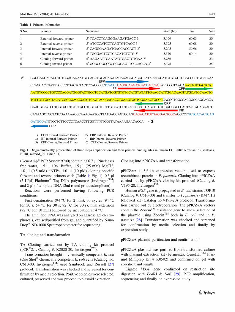
Table 1 Primers information
S.No. Primers Sequence Start (bp) Tm Size
1 External forward primer 50-TCACCTCAGGGAAGATGACC-30 3,199 60.05 20
2 External reverse primer 50-ATCCCATCCTCAGTGTCAGC-30 3,595 60.08 20
3 Internal forward primer 50-CAGGGAAGATGACCACCACT-30 3,205 59.96 20
4 Internal reverse primer 50-TGCGACTCCTCACATCTCTG-30 3,570 60.14 20
5 Cloning forward primer 50-AAGAATTCAATAGTGACTCTGAA-30 3,236 – 23
6 Cloning reverse primer 50-GCGCGGCCGCGCGCAGTTCCCACCA-30 3,395 – 25
GGGGAGCACAGCTGTGGAGAGAATGCCAGCTGCACAAATACAGAGGGAGGCTATACCTGCATGTGTGCTGGACGCCTGTCTGAA
CCAGGACTGATTTGCCCTGACTCTACTCCACCCCCTCACCTCAGGGAAGATGACCACCACTATTCCGTAAGAAATAGTGACTCTG
AATGTCCCCTGTCCCACGATGGGTACTGCCTCCATGATGGTGTGTGCATGTATATTGAAGCATTGGACAAGTATGCATGCAACTG
TGTTGTTGGCTACATCGGGGAGCGATGTCAGTACCGAGACCTGAAGTGGTGGGAACTGCGCCACGCTGGCCACGGGCAGCAGCA
GAAGGTCATCGTGGTGGCTGTCTGCGTGGTGGTGCTTGTCATGCTGCTCCTCCTGAGCCTGTGGGGGGCCCACTACTACAGGACT
CAGAAGCTGCTATCGAAAAACCCAAAGAATCCTTATGAGGAGTCGAGCAGAGATGTGAGGAGTCGCAGGCCTGCTGACACTGAG
GATGGGATGTCCTCTTGCCCTCAACCTTGGTTTGTGGTTATAAAAGAACACCA
1) EFP External Forward Primer 2) ERP External Reverse Primer 3) IFP Internal Forward Primer 4) IRP Internal Reverse Primer 5) CFP Cloning Forward Primer 6) CRP Cloning Reverse Primer
EFP
IFP CFP
CRP
IRP
ERP
5 -
- 3
Fig. 1 Diagrammatically presentation of three steps amplification and their primers binding sites in human EGF mRNA variant 3 (GenBank,
NCBI, ref/NM_001178131.1)
Mol Biol Rep (2014) 41:1445–1451 1447
123

DNA sequencing
DNA purification
Purified DNA followed a Shrimp alkaline phosphatase
(SAP) [30] and exonuclease I treatment [31] to removed
excess primers and dNTPs for sequencing PCR. The
Exonuclease I digested single stranded DNA into free
dNTPs and SAP removed the phosphate groups from
dNTPs to deactivate.
Sequencing reaction
Sequencing reaction is carried using sequencing kit pro-
tocol as described in BigDye� Terminator v3.1 cycle
Sequencing Kit and proceeded to the Automated DNA
Sequencer (Applied Biosystems 3730 DNA Analyzer).
Sequencing cycle consisted of an asymmetric amplification
of one strand of the DNA product.
All DNA sequences confirmed both in forward and
reverse direction by using Basic Local Alignment Search
Tool (BLAST) http://www.ncbi.nlm.nih.gov/blast.cgi).
Culturing of Pichia transformants
Pichia pastoris transformants obtained by electroporation
and were then selected on YPD medium (1 % yeast extract;
2 % bacto peptone; 2 % dextrose) followed vector manual
(InvitrogenTM). Transformants strains expressing hEGF
were grown in BMMY medium (1 % yeast extract; 1 %
methanol; 2 % bacto peptone; 100 mM potassium phos-
phate buffer of pH 6.0; 1.34 % YNB (yeast nitrogen base);
4 9 10–5 % biotin) in a 6 litre glass bioreactor at 30 �C
and aeration (0.1–1.0 vvm). The pH was maintained at 5.0
throughout fermentation process.
Biomass was measured as cell wet weight. The super-
natant was stored at -70 �C until needed for assays.
Expression study
The transformants of P. pastoris containing the integration
of the hEGF vector (pPICZaA—hEGF) in the genome
analyzed for hEGF expression. The cultured supernatants
from both transformed and untransformed strains were
analyzed by electrophoresis on a Tricine-SDS polyacryl-
amide gel (PAGE) gel. Western-blot analysis of the protein
performed according to standard procedure [32].
Proteins separated on a 15 % Tricine-SDS-PAGE gel
stained with Coomassie Brilliant Blue R250 and photo-
graphed. Protein electrophoresis identified the presence of
band, at about 6 kDa in the transformants, this roughly
showed standard hEGF. For further confirmation blotted
the gel onto the nitrocellulose membrane (cat. no. LC2000,
InvitrogenTM) using kit procedure. The blot was then
developed with the rabbit-anti-hEGF sera and mouse-anti-
rabbit immunoglobulin-G conjugated alkaline phosphatase.
Secretions of hEGF protein from the P. pastoris transfor-
mants assayed with an indirect enzyme-linked Immuno-
sorbent assay (ELISA).
Results
Huh-7 is a human hepatoma cell line that has numerous
uses [18–21]. In the current study we isolated Human EGF
gene from Huh-7 cell line followed by cDNA formation,
three steps PCR amplification of human EGF gene. Gene
sequencing was performed to verify the amplicon. More-
over the gene was cloned in pPICZaA and over- expressed
in P. pastoris.
Human EGF gene is 100 kb long coding gene, tran-
scription for 5,474 bases mRNA (NCBI, GenBank, Human
EGF mRNA variant 3, ref/NM_001178131.1). Which then
translated into 1,207 amino acid preproprotein and modi-
fied eventually into 53 amino acid mature single chain
polypeptide protein.
Total mRNA was isolated from Huh-7 cell line, reverse
transcribed into cDNA with oligo dT primer. A short
sequence of 560 bases of hEGF gene including mature
protein region was taken and designed three sets of primers
using Primer 3 software (http://frodo.wi.mit.edu/primer3/)
(Table 1; Fig. 1).
First amplification was performed from cDNA with
designed external primers set from 3,199 to 3,595 bp
(ref|NM_001178131.1) (Table 1; Fig. 1) and results for
expected amplicon size 396 bp checked by gel electro-
phoresis (Fig. 2).
The first PCR product was further amplified from 3,205
to 3,570 bp (365 bp) with internal primers for more spec-
ificity (Table 1; Fig. 1). Obligated amplicon size (365 bp)
checked by gel electrophoresis (Fig. 3). DNA fragment
Fig. 2 Gel electrophoresis for first round amplification, lane 1 shows
expected amplicon (396 bp), lane 2 is 100 bp DNA marker and lane 3
is negative control
1448 Mol Biol Rep (2014) 41:1445–1451
123
excised from gel and was purified. The sequencing was
carried out and sequencing results showed 100 % sequence
homology with wild-type human EGF gene sequence
(ref|NM_001178131.1|).
Second round amplicon further abridged (3,236–3,395 bp)
with specially designed cloning primers (Table 1; Fig. 1)
inherent with two restriction enzyme sites EcoRI and NotI
(GAATTC & GCGGCCGC respectively) for restriction site
digestion and appropriate cloning, Kex2 site, stop codon, AA
and GC regions were also incorporated for further expression
study in P. pastoris. Third round PCR product (Fig. 4) eluted
from gel and quantified by NanoDrop� (Spectrophotometer)
(ND-1000). Purified fragment was sequenced and found
required results (EMBL/EBI JQ346088).
Third round PCR product ligated in TA plasmid vector
(pCR� 2.1) (Fig. 5) by ligation reaction and transferred to
TOP10 competent cells. Extraction made for TA plasmid
and ligation results assured by excised fragment analysis
done by restriction site digestion with EcoRI/NotI,
sequencing and also by PCR amplification (Figs. 6, 7).
TA clone sequencing reaction was carried out with M13
primers as discussed above to confirm the sequence. Fur-
ther, we verified the active gene sequence homology with
BLAST, as the respective sequence is available at Gen-
Bank under the accession number (EMBL/EBI JQ346088).
Human EGF gene was ligated in pPICZaA plasmid by
ligation reaction and transferred to TOP10 competent cells
and P. pastoris (Fig. 8). Extraction made for pPICZaA
plasmid and ligation results assured by excised fragment
analysis done by restriction site digestion with EcoRI/NotI,
sequencing, PCR amplification and finally confirmed by
expression study.
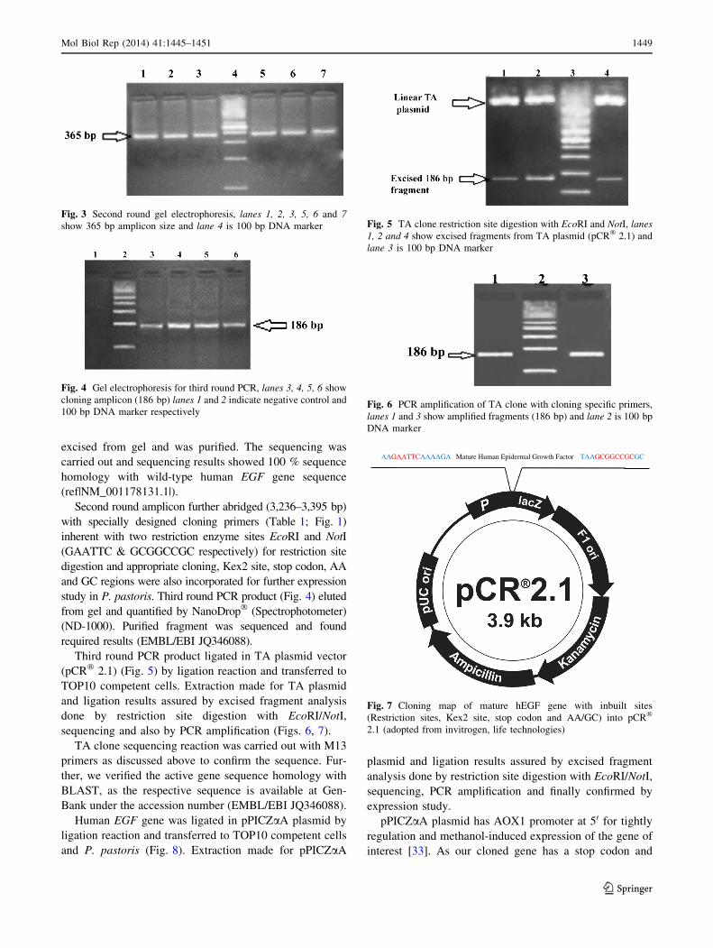
pPICZaA plasmid has AOX1 promoter at 50 for tightly
regulation and methanol-induced expression of the gene of
interest [33]. As our cloned gene has a stop codon and
Fig. 3 Second round gel electrophoresis, lanes 1, 2, 3, 5, 6 and 7
show 365 bp amplicon size and lane 4 is 100 bp DNA marker
Fig. 4 Gel electrophoresis for third round PCR, lanes 3, 4, 5, 6 show
cloning amplicon (186 bp) lanes 1 and 2 indicate negative control and
100 bp DNA marker respectively
Fig. 5 TA clone restriction site digestion with EcoRI and NotI, lanes
1, 2 and 4 show excised fragments from TA plasmid (pCR� 2.1) and
lane 3 is 100 bp DNA marker
Fig. 6 PCR amplification of TA clone with cloning specific primers,
lanes 1 and 3 show amplified fragments (186 bp) and lane 2 is 100 bp
DNA marker
AAGAATTCAAAAGA Mature Human Epidermal Growth Factor TAAGCGGCCGCGC
Fig. 7 Cloning map of mature hEGF gene with inbuilt sites
(Restriction sites, Kex2 site, stop codon and AA/GC) into pCR�
2.1 (adopted from invitrogen, life technologies)
Mol Biol Rep (2014) 41:1445–1451 1449
123
Kex2 site for proper cleavage so protein expressed without
the C-terminal peptide and has native N-terminus.
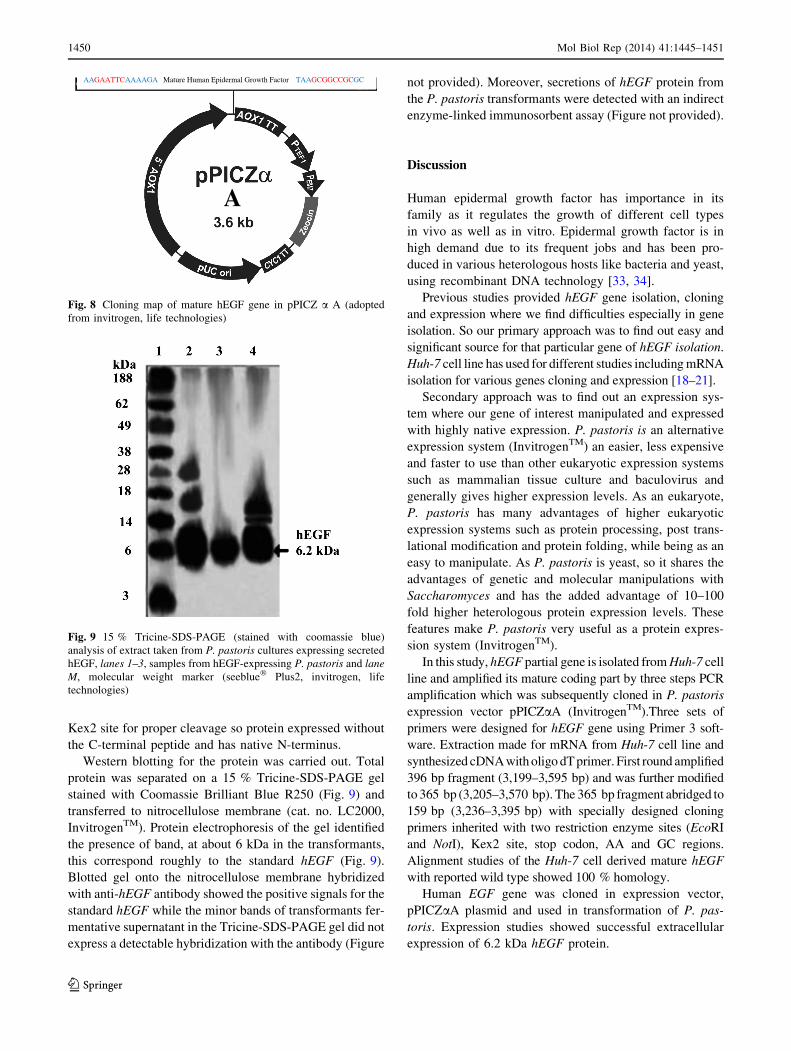
Western blotting for the protein was carried out. Total
protein was separated on a 15 % Tricine-SDS-PAGE gel
stained with Coomassie Brilliant Blue R250 (Fig. 9) and
transferred to nitrocellulose membrane (cat. no. LC2000,
InvitrogenTM). Protein electrophoresis of the gel identified
the presence of band, at about 6 kDa in the transformants,
this correspond roughly to the standard hEGF (Fig. 9).
Blotted gel onto the nitrocellulose membrane hybridized
with anti-hEGF antibody showed the positive signals for the
standard hEGF while the minor bands of transformants fer-
mentative supernatant in the Tricine-SDS-PAGE gel did not
express a detectable hybridization with the antibody (Figure
not provided). Moreover, secretions of hEGF protein from
the P. pastoris transformants were detected with an indirect
enzyme-linked immunosorbent assay (Figure not provided).
Discussion
Human epidermal growth factor has importance in its
family as it regulates the growth of different cell types
in vivo as well as in vitro. Epidermal growth factor is in
high demand due to its frequent jobs and has been pro-
duced in various heterologous hosts like bacteria and yeast,
using recombinant DNA technology [33, 34].
Previous studies provided hEGF gene isolation, cloning
and expression where we find difficulties especially in gene
isolation. So our primary approach was to find out easy and
significant source for that particular gene of hEGF isolation.
Huh-7 cell line has used for different studies including mRNA
isolation for various genes cloning and expression [18–21].
Secondary approach was to find out an expression sys-
tem where our gene of interest manipulated and expressed
with highly native expression. P. pastoris is an alternative
expression system (InvitrogenTM) an easier, less expensive
and faster to use than other eukaryotic expression systems
such as mammalian tissue culture and baculovirus and
generally gives higher expression levels. As an eukaryote,
P. pastoris has many advantages of higher eukaryotic
expression systems such as protein processing, post trans-
lational modification and protein folding, while being as an
easy to manipulate. As P. pastoris is yeast, so it shares the
advantages of genetic and molecular manipulations with
Saccharomyces and has the added advantage of 10–100
fold higher heterologous protein expression levels. These
features make P. pastoris very useful as a protein expres-
sion system (InvitrogenTM).
In this study, hEGF partial gene is isolated from Huh-7 cell
line and amplified its mature coding part by three steps PCR
amplification which was subsequently cloned in P. pastoris
expression vector pPICZaA (InvitrogenTM).Three sets of
primers were designed for hEGF gene using Primer 3 soft-
ware. Extraction made for mRNA from Huh-7 cell line and
synthesized cDNA with oligo dT primer. First round amplified
396 bp fragment (3,199–3,595 bp) and was further modified
to 365 bp (3,205–3,570 bp). The 365 bp fragment abridged to
159 bp (3,236–3,395 bp) with specially designed cloning
primers inherited with two restriction enzyme sites (EcoRI
and NotI), Kex2 site, stop codon, AA and GC regions.
Alignment studies of the Huh-7 cell derived mature hEGF
with reported wild type showed 100 % homology.
Human EGF gene was cloned in expression vector,
pPICZaA plasmid and used in transformation of P. pas-
toris. Expression studies showed successful extracellular
expression of 6.2 kDa hEGF protein.
AAGAATTCAAAAGA Mature Human Epidermal Growth Factor TAAGCGGCCGCGC
A
Fig. 8 Cloning map of mature hEGF gene in pPICZ a A (adopted
from invitrogen, life technologies)
Fig. 9 15 % Tricine-SDS-PAGE (stained with coomassie blue)
analysis of extract taken from P. pastoris cultures expressing secreted
hEGF, lanes 1–3, samples from hEGF-expressing P. pastoris and lane
M, molecular weight marker (seeblue� Plus2, invitrogen, life
technologies)
1450 Mol Biol Rep (2014) 41:1445–1451
123

To our knowledge it is the first report of hEGF partial
mature gene from Huh-7 cell line and its expression in
P. pastoris.
Acknowledgments We thank to Molecular Medicine Lab (CEMB)
for providing Huh-7 cell line. Thanks to Ahmad Usman Zafar, Na-
deem Ahmad, Muhammad Islam Khan and Tayyab Hussnain for
thoughtful discussions and critical reading of the manuscript.
Conflict of interest The authors declare that they have no com-
peting interests.
References
1. Cohen S, Carpenter G (1975) Human epidermal growth factor:
isolation and chemical and biological properties. PNAS 72:
1317–1321
2. Fraser JK, Zhu M, Wulur I, Alfonso Z (2008) Adipose-derived
stem cells. Methods Mol Biol 449:59–67
3. Mark JK, Mourad M, Ole M, Walter P, Yong YW, Ronald EB,
Peter JH, Robert MH, Jorg H (2006) An unusual mode of con-
certed evolution of the EGF-TM7 receptor chimera EMR2.
FASEB J 20(14):2582
4. Engler DA, Hauser MR, Cook JS, Niyogi SK (1991) Aromaticity
at position 37 in human epidermal growth factor is not obligatory
for activity. Mol Cell Biol 11(5):2425
5. Graeme IB, Noel MF, Michelle MS, Mary AW, Daniel C, Lailing
K, Mickey SU, Leslie BR, Ray SP (1986) Human epidermal
growth factor precursor: cDNA sequence, expression in vitro and
gene organization. Nucleic Acids Res 14(21):8427–8446
6. Carpenter G, Cohen S (1979) Epidermal growth factor. Annu Rev
Biochem 48:193–216
7. Anna MW (2005) Prospects for developmental biology and cell
therapy. Physiol Rev 85:635–678
8. Strutz F, Michael Z, Fuad NZ, Chang-Qing Y, Raghu K, Gerhard AM,
Eric GN (2002) Role of basic fibroblast growth factor-2 in epithelial
mesenchymal transformation. Kidney Int 61(5):1714–1728
9. Schuldiner MO, Joseph IE, Douglas AM, Nissim B (2000)
Effects of eight growth factors on the differentiation of cells
derived from human embryonic stem cells. Proc Natl Acad Sci
97(21):11307–11312
10. Robert MC, Anthony JW, Martin B, Annalisa P, Michael JT, Lain
DC, Harry G, Brian S (1987) The solution structure of human
epidermal growth factor. Nature 327:339–341
11. Umeoka K, Naoko S, Kenichi O, Tahara S, Reiko K, Shoichiro I,
Manabu N, Takeshi W, Yoshiyuki OAT (2001) Immuno histo-
chemical analysis of RCAS1 in human pituitary adenomas. Mod
Pathol 14(12):1232–1236
12. Adams JC (2001) Review cell-matrix contact structures. Cell Mol
Life Sci 58(3):371–392
13. Chung KF (2001) Cytokines in chronic obstructive pulmonary
disease. Eur Respir J 18(34):50–59
14. Philip JA, Michael BC, Cherie MC, Kenneth H (2009) Method of
using cytokine assay to diagnose, treat and evaluate inflammatory
and autoimmune diseases. Google Patents
15. Mieko K, Shigeaki T (1989) Topographic analysis of human epi-
dermal growth factor by monospecific antibodies and synthetic
peptides. Oxford Journals. Life Sciences. J Biochem 106(1):87–89
16. Beat MS, Anna F, Eric F, Albert J (1994) Liver expression of
epidermal growth factor RNA. J Biol Chem 269(31):9667–9670
17. John S, Cook E, Ian F, Sally P, Roger D, Michael AW, Mike D,
David MJ, John FP, Thakor P, Hilary L, Leslie DB (1982)
Chemical synthesis and cloning of a gene for human urogastrone.
Nucleic Acids Res 10(15):4467–4482
18. Patil MA, Zhang J, Ho C, Cheung ST, Fan ST, Chen XP (2006)
Hedgehog signaling in human hepatocellular carcinoma. Cancer
Biol Ther 5(1):111–117
19. Brett DL, Matthew JE, Andrew JS, Benno W, Timothy LT,
Christopher CL, Toshiaki M, Richard OH, Dennis RB, Jane AM,
Charles MR (2005) Complete replication of hepatitis c virus in
cell culture. Science 309(5734):623–626
20. Takimoto R, Kato J, Terui T, Takada K, Kuroiwa G, Wu J,
Ohnuma H, Takahari D, Kobune M, Sato Y, Takayama T,
Matsunaga T, Niitsu Y (2005) Augmentation of antitumor effects
of p53 gene therapy. Cancer Biol Ther 4(4):421–428
21. Jie L, Lilianna S, Ruben MB, Jan N, Liping H, Natascha N,
Steven D, Liegang L, Ulrich S, Andreas KN (2012) Comparative
analysis of phase I and II enzyme activities in 5 hepatic cell lines
identifies Huh-7 and HCC-T cells with the highest potential to
study drug metabolism. Arch Toxicol 86(1):87–95
22. Bao-Zhu Y, Mark JM, Catherine LK, Drazen BZ, Snorri ST,
Nicholas CP (1998) Cloning, characterization and chromo-
somal localization of a gene frequently deleted in human liver
cancer (DLC-1) homologous to Rat RhoGAP. Cancer Res
58:2196
23. Tanabe KK, Lemoine A, Finkelstein DM, Kawasaki H, Fujii T,
Chung RT, Lauwers GY, Kulu Y, Muzikansky A, Kuruppu D,
Lanuti M, Goodwin JM, Azoulay D, Fuchs BC (2008) Epidermal
growth factor gene functional polymorphism and the risk of
hepatocellular carcinoma in patients with cirrhosis. JAMA
299(1):53–60
24. Chomczynski P, Mackey K (1995) Modification of the trizol
reagent procedure for isolation of RNA from polysaccharide and
proteoglycan-rich sources. Biotechniques 19(6):942–945
25. Steve R, Helen JS (2000) Primer 3, http://frodo.wi.mit.edu/pri
mer3/ Bioinformatics methods and protocols methods in molec-
ular biology, 365–386
26. Benson DA, Karsch-Mizrachi I, Clark K, Lipman DJ, Ostell J,
Sayers EW (2012) GenBank. Nucleic Acids Res 40:D48–D53
27. Sambrook J, Russell DW (2006) Preparation and transformation
of competent E. coli using calcium chloride. Cold Spring Harb
Protoc. doi:10.1101/3932
28. Baron M, Reynes JP, Stassi D, Tiraby G (1992) A selectable
bifunctional b galactosidase: phleomycin-resistance fusion pro-
tein as a potential marker for eukaryotic cells. Gene 114:239–243
29. Plyler TR, Vallejos CE (2000) Method for cloning restriction
fragments containing the termini of BAC Inserts. Biotechniques
28(5):1012–1016
30. Kerri AK, Helen SH, Douglas RD, Constance LC, Budowle B
(2002) An improved method for post-PCR purification for
mtDNA sequence analysis. J Forensic Sci 47(4):811–818
31. Watanabe K, Emoto N, Sunohara M, Kawakami M, Kage H,
Nagase T, Ohishi N, Takai D (2010) Treatment of PCR products
with exonuclease I and heat-labile alkaline phosphatase improves
the visibility of combined bisulfate restriction analysis. Biochem
Biophys Res Commun 399(3):422–424
32. Frederick MA, Roger B, Robert EK, David DM, Seidman JG,
John AS, Kevin S (1999) Short protocols in molecular biology,
4th edn. Wiley, New York
33. Takanori O, Shunji S, Ken-Ichi M, Toru F, Koji Y, Makari Y,
Gakuzo T, Tetsuo M (1985) Synthesis and secretion of human
epidermal growth factor by Escherichia coli. Proc NatI Acad Sci
82(21):7212–7216
34. John MC, Graham HC, Mickhael JP, Mark ER (1991) Secretion
of human epidermal growth factor from S. cerevisiae using
synthetic leader sequences. Gene 106(2):267–271
Mol Biol Rep (2014) 41:1445–1451 1451
123