Embed Size (px)
Citation preview
BioOne sees sustainable scholarly publishing as an inherently collaborative enterprise connecting authors, nonprofit publishers, academic institutions, researchlibraries, and research funders in the common goal of maximizing access to critical research.
Exploring Two Two-Edged SwordsAuthor(s): David J. BrennerSource: Radiation Research, 178(1):7-16. 2012.Published By: Radiation Research SocietyDOI: http://dx.doi.org/10.1667/RR3085.1URL: http://www.bioone.org/doi/full/10.1667/RR3085.1
BioOne (www.bioone.org) is a nonprofit, online aggregation of core research in the biological, ecological, andenvironmental sciences. BioOne provides a sustainable online platform for over 170 journals and books publishedby nonprofit societies, associations, museums, institutions, and presses.
Your use of this PDF, the BioOne Web site, and all posted and associated content indicates your acceptance ofBioOne’s Terms of Use, available at www.bioone.org/page/terms_of_use.
Usage of BioOne content is strictly limited to personal, educational, and non-commercial use. Commercial inquiriesor rights and permissions requests should be directed to the individual publisher as copyright holder.

RADIATION RESEARCH 178, 7–16 (2012)0033-7587/12 $15.00�2012 by Radiation Research Society.All rights of reproduction in any form reserved.DOI: 10.1667/RR3085.1
2011 FAILLA AWARD LECTURE
Exploring Two Two-Edged Swords
David J. Brenner1
Center for Radiological Research, Columbia University Medical Center, New York, New York 10032
INTRODUCTION
Failla, Marie Curie, and Columbia University
It was a particular pleasure to receive the Radiation
Research Society Failla Award in Warsaw, Poland, the
birthplace of Marie Skłodowska Curie. Gioacchino Failla
was the first director of my own Institute, now called the
Columbia University Center for Radiological Research
(CRR). Starting in 1918, Failla was in charge of the
Columbia University Center for an astonishing 43 years,
before Harold Rossi and then Eric Hall took over the reins.
Between them, Rossi and Hall led the CRR for a further 49
years. Perhaps radiation is linked to scientific longevity, if
not to increased lifespan (1).
Failla was in fact one of Marie Curie’s graduate students,
and he received his doctorate from the Sorbonne in 1923.
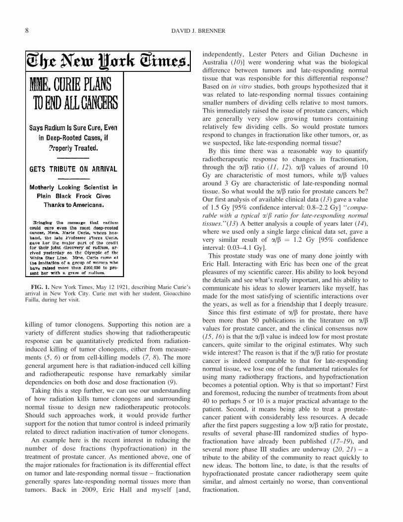
Two years earlier, Marie Curie had visited Failla in New
York City during her trip to the U.S. The New York Times
recorded her arrival (Fig. 1) with the headline ‘‘Mme. Curie
Plans to End All Cancers,’’ together with the memorable
subheading ‘‘Motherly looking scientist in plain black frock
gives thanks to Americans.’’
The Two Two-Edged Swords
Radiation’s two two-edged swords that have dominated
my own scientific thinking are:
1. Radiation can cure cancer versus radiation can induce
cancer.
2. Radiotherapy needs physics research versus radiother-
apy needs biological research.
The goal of this brief summary is to give some examples
of these contrasts, and try to draw some conclusions about
research directions, interleaved with some observations
about the scientists who have tried to push me in the right
directions over the years.
THE FIRST TWO-EDGED SWORD
Radiation Can Cure Cancer versus Radiation Can InduceCancer
1. Radiation Can Cure Cancer; But What is the DominantMechanism?
That radiotherapy can cure cancer was established early inthe twentieth century. Despite repeated claims for itsimminent demise in cancer therapy, radiotherapy remainsone of its staples, with more than half of all cancer patientsreceiving radiotherapy at some stage during their treatment.Despite more than a century of clinical radiotherapeuticexperience, however, there is still much debate about how itactually works, and how it should be optimally used.
In the first two decades of the twentieth century thegeneral view was that single large ‘‘castrating’’ doses wouldkill tumors more than surrounding normal tissues, and werethus the preferred modality (2). This prevailing viewchanged in the 1920s, in large part due to the work ofClaudius Regaud at the Institute Curie in Paris (3). Regaudand colleagues used the testis, a self-renewing tissue with aproliferating cell compartment, as a model for a growingtumor, and the overlying skin as a model of dose-limitingnormal tissue. Using these models in a variety of animalsthey demonstrated that fractionating the dose decreased skindamage but did not significantly reduce damage to the testis– the model for tumor. The implication was thatfractionation would thus increase the therapeutic advantagebetween tumor control and complications. Henri Coutard,took these concepts into the clinic at the Radium Institute inParis, where he clearly confirmed the radiotherapeuticadvantages of dose fractionation (4).
Since the 1920s fractionation has remained central to allradiotherapy, both because of the gains that it gives in termsof tumor control compared to late sequelae, but also becauseof the subsequent realization that fractionation is necessaryto deal with hypoxic tumor cells. By the 1980s theseconcepts had been quantified, through mechanistic modelssuch as the linear-quadratic formalism.
Central to these models is the basic idea that radiother-apeutic tumor control is related primarily to direct radiation
1Address for correspondence: Center for Radiological Research,Columbia University Medical Center, 630 West 168th Street, NewYork, NY 10032; e-mail: [email protected].
7
killing of tumor clonogens. Supporting this notion are a
variety of different studies showing that radiotherapeutic
response can be quantitatively predicted from radiation-
induced killing of tumor clonogens, either from measure-
ments (5, 6) or from cell-killing models (7, 8). The more
general argument here is that radiation-induced cell killing
and radiotherapeutic response have remarkably similar
dependencies on both dose and dose fractionation (9).
Taking this a step further, we can use our understanding
of how radiation kills tumor clonogens and surrounding
normal tissue to design new radiotherapeutic protocols.
Should such approaches work, it would provide further
support for the notion that tumor control is indeed primarily
related to direct radiation inactivation of tumor clonogens.
An example here is the recent interest in reducing the
number of dose fractions (hypofractionation) in the
treatment of prostate cancer. As mentioned above, one of
the major rationales for fractionation is its differential effect
on tumor and late-responding normal tissue – fractionation
generally spares late-responding normal tissues more than
tumors. Back in 2009, Eric Hall and myself [and,
independently, Lester Peters and Gilian Duchesne inAustralia (10)] were wondering what was the biologicaldifference between tumors and late-responding normaltissue that was responsible for this differential response?Based on in vitro studies, both groups hypothesized that itwas related to late-responding normal tissues containingsmaller numbers of dividing cells relative to most tumors.This immediately raised the issue of prostate cancers, whichare generally very slow growing tumors containingrelatively few dividing cells. So would prostate tumorsrespond to changes in fractionation like other tumors, or, aswe suspected, like late-responding normal tissue?
By this time there was a reasonable way to quantifyradiotherapeutic response to changes in fractionation,through the a/b ratio (11, 12). a/b values of around 10Gy are characteristic of most tumors, while a/b valuesaround 3 Gy are characteristic of late-responding normaltissue. So what would the a/b ratio for prostate cancers be?Our first analysis of available clinical data (13) gave a valueof 1.5 Gy [95% confidence interval: 0.8–2.2 Gy] ‘‘compa-rable with a typical a/b ratio for late-responding normaltissues.’’(13) A better analysis a couple of years later (14),where we used only a single large clinical data set, gave avery similar result of a/b ¼ 1.2 Gy [95% confidenceinterval: 0.03–4.1 Gy].
This prostate study was one of many done jointly withEric Hall. Interacting with Eric has been one of the greatpleasures of my scientific career. His ability to look beyondthe details and see what’s really important, and his ability tocommunicate his ideas to slower learners like myself, hasmade for the most satisfying of scientific interactions overthe years, as well as for a friendship that I deeply treasure.
Since this first estimate of a/b for prostate, there havebeen more than 50 publications in the literature on a/bvalues for prostate cancer, and the clinical consensus now(15, 16) is that the a/b value is indeed low for most prostatecancers, quite similar to the original estimates. Why suchwide interest? The reason is that if the a/b ratio for prostatecancer is indeed comparable to that for late-respondingnormal tissue, we lose one of the fundamental rationales forusing many radiotherapy fractions, and hypofractionationbecomes a potential option. Why is that so important? Firstand foremost, reducing the number of treatments from about40 to perhaps 5 or 10 is a major practical advantage to thepatient. Second, it means being able to treat a prostate-cancer patient with considerably less resources. A decadeafter the first papers suggesting a low a/b ratio for prostate,results of several phase-III randomized studies of hypo-fractionation have already been published (17–19), andseveral more phase III studies are underway (20, 21) – atribute to the ability of the community to react quickly tonew ideas. The bottom line, to date, is that the results ofhypofractionated prostate cancer radiotherapy seem quitesimilar, and almost certainly no worse, than conventionalfractionation.
FIG. 1. New York Times, May 12 1921, describing Marie Curie’sarrival in New York City. Curie met with her student, GioacchinoFailla, during her visit.
8 DAVID J. BRENNER
That prostate hypofractionation results are turning outreasonably as predicted is gratifying in its own right, but thesecond conclusion to be drawn here is that the consistencyof the clinical results with the predictions of standardmodels provides further confirmation that tumor controlmust indeed be primarily related to direct radiation-inducedkilling of tumor clonogens. Other mechanisms are no doubtinvolved (22), and may even dominate at ultra high dosesper fraction (23), but for conventional dose fractionation(;2 Gy/fraction) or even most hypofractionated protocols(2–5 Gy/fraction), it is likely that direct radiation-inducedkilling of tumor clonogens remains the dominant mecha-nism of tumor control.
2. Radiotherapy Can Induce Cancers: But How Does thisHappen at Very High Doses?
The 15-year relative survival rate for patients treated forbreast or prostate cancer is now about 75%, as compared,for example, to about 58% for breast just a decade ago. Asyounger patients are treated, and with longer life expectan-cy, radiotherapy-induced secondary malignancies are as-suming increasing importance. Ironically, the whole issue ofradiotherapy-induced secondary cancers is, to a significantextent, a testimony to the efficacy of modern dayradiotherapy – truly the price of success.
Back in 2000, and stimulated by our first interactions withour much missed late colleague Elaine Ron, we undertook alarge-scale tumor registry analysis, in which secondarycancers after prostate cancer radiotherapy were comparedwith secondary cancers after prostate cancer surgery (24).Prostate is probably unique in allowing such a directcomparison of secondary cancers after radiotherapy versussurgery – the advantage here being that all the studysubjects had prostate cancer, whereas most secondarycancer studies compare cancer risks with the generalpopulation. The study showed (24) that prostate radiother-apy-induced secondary cancers often occurred well outsidethe treatment volume – at least for treatments regimens thatwere common some decades ago. So studies of radiother-
apy-induced cancers should ideally include all sites, not just
those in the treatment volume – and of course the range of
studied times post exposure must be large – at least two
decades to estimate lifetime risks.
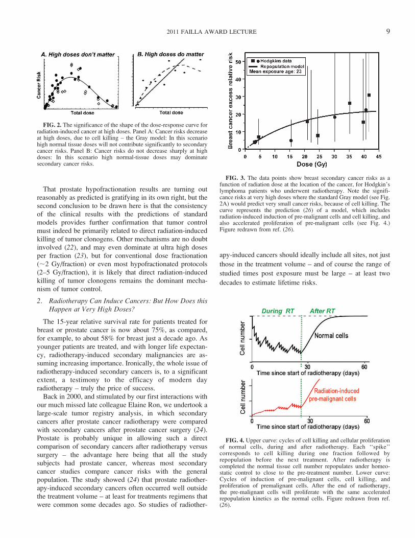
FIG. 2. The significance of the shape of the dose-response curve forradiation-induced cancer at high doses. Panel A: Cancer risks decreaseat high doses, due to cell killing – the Gray model: In this scenariohigh normal tissue doses will not contribute significantly to secondarycancer risks. Panel B: Cancer risks do not decrease sharply at highdoses: In this scenario high normal-tissue doses may dominatesecondary cancer risks.
FIG. 3. The data points show breast secondary cancer risks as afunction of radiation dose at the location of the cancer, for Hodgkin’slymphoma patients who underwent radiotherapy. Note the signifi-cance risks at very high doses where the standard Gray model (see Fig.2A) would predict very small cancer risks, because of cell killing. Thecurve represents the prediction (26) of a model, which includesradiation-induced induction of pre-malignant cells and cell killing, andalso accelerated proliferation of pre-malignant cells (see Fig. 4.)Figure redrawn from ref. (26).
FIG. 4. Upper curve: cycles of cell killing and cellular proliferationof normal cells, during and after radiotherapy. Each ‘‘spike’’corresponds to cell killing during one fraction followed byrepopulation before the next treatment. After radiotherapy iscompleted the normal tissue cell number repopulates under homeo-static control to close to the pre-treatment number. Lower curve:Cycles of induction of pre-malignant cells, cell killing, andproliferation of premalignant cells. After the end of radiotherapy,the pre-malignant cells will proliferate with the same acceleratedrepopulation kinetics as the normal cells. Figure redrawn from ref.(26).
2011 FAILLA AWARD LECTURE 9
Around 2000 was also the time when Intensity-Modulated
Radiation Therapy (IMRT) was becoming increasingly
popular; IMRT uses multiple pencil beams to produce
improved dose distributions around the tumor. Compared to
the older three-dimensional conformal radiotherapy (3D-
CRT), modern IMRT techniques minimize the amount of
normal tissue getting high doses. But IMRT does result in
larger volumes of normal tissue getting lower doses. So the
question arises: which is preferable in terms of secondary
cancers? Small volumes of normal tissue getting high doses
(3D-CRT), or larger volumes of normal tissue getting low
doses (IMRT)?
The answer, of course, depends on the shape of the dose-
response curve. As illustrated in Fig. 2A, if the dose-
response for radiation-induced cancer decreases sharply at
high doses, then high doses do not matter from the
perspective of radiation-induced cancer. This possible
bell-shaped dose-response is in fact the standard ‘‘Gray
model’’ (25), the one described in most text books, in which
radiation-induced cancer at high doses is a result of
competition between induced oncogenic transformation
(increases risk) and cell killing (which decreases the risk
at high doses). So in the context of IMRT, which essentially
removes very high doses and replaces them with lower
doses spread over larger volumes, if the standard Gray
model (Fig. 2A) is correct and high radiation doses produce
fewer secondary cancers, IMRT might result in more
secondary cancers. On the other hand, if cancer risks do not
decrease sharply at high doses (Fig. 2B), then IMRT would
be more likely to be advantageous from the perspective of
secondary cancers.
In fact recent epidemiology suggests that radiation-
induced cancer risks are generally not small at large doses.
An example is shown in Fig. 3 where secondary cancer risks
continue to increase up to at least 45 Gy – which is quite
inconsistent with the standard Gray model (Fig. 2A) where
cell killing would result in very small cancer risks at these
very high doses. Together with Rainer Sachs at UC
Berkeley, we wondered what could be responsible for these
significant secondary cancer risks at the large doses where
cell killing would be expected to eliminate most radiation-
induced pre-malignant cells. Finally we focused on
repopulation: it is well known that surviving normal cells
in heavily irradiated tissue proliferate rapidly under
homeostatic control in the time period up to a few months
after radiotherapy; so any radiation-produced pre-malignant
cells would also be expected to proliferate with just the
same kinetics, as illustrated in Fig. 4 (26). With what I
FIG. 5. Illustrating the increasing complexity and cost of accelerator-based radiotherapy systems that have been developed over the past 50years.
10 DAVID J. BRENNER
thought was brilliant insight, Sachs showed that, in a first
approximation, accelerated proliferation of pre-malignant
cells can exactly cancel out the effects of cell killing,
leaving a secondary cancer risk which will therefore not be
negligible at high doses (26).
My scientific collaborations over the years with Ray
Sachs have brought me enormous pleasure. Coming from a
mathematical physics background [see the Sachs-Wolfe
effect (27, 28)], Sachs has the analytic insights to develop
and use biophysical mathematical models in what I think is
the best way possible – investigating which are the
dominant mechanism for any given phenomenon. Figuring
out the dominant mechanism underlying a given phenom-
enon is often tough to do: like many scientists I too often get
sidetracked towards interesting but second-order effects -
and I am infinitely grateful to Sachs for setting me straight
on numerous occasions.
So to summarize, there is a third mechanism which is
critical for radiation-induced cancer at high doses. Beyond
induction and killing of pre-malignant cells, as described by
the standard Gray model, proliferation of premalignant cells
is an additional important player at high doses. The outcome
of this, in the context of IMRT, is that high doses generally
do matter in terms of secondary cancer risks (Fig 2B). This
being the case, it is likely that IMRT is reducing secondary
cancer risks relative to the older 3D CRT techniques. This
rather comforting conclusion does not, it should be said,
take into account the whole body dose from treatment-head
leakage radiation during IMRT: this is important because
IMRT takes a much longer time to deliver than conventional
3D CRT – a fixable problem that needs to be fixed (29).
The bottom line here is that because it takes two decades
or more to clinically assess secondary cancer risks from a
new radiation modality, we have no choice but to use
models. But we should use models which are (1)
mechanistic – the days of curve fitting are hopefully behind
us, and (2) validated as much as possible with what clinical
radiation epidemiology we have right now. Using the
standard Gray model, for example, would likely have led us
to quite erroneous conclusions about IMRT.
FIG. 6. The LAMPF linear accelerator, located on Mesita de LosAlamos in New Mexico. Protons were accelerated over LAMPF’shalf-mile length to an energy of 800 MeV, producing negative pions(at the far end of the photograph) for pion therapy.
FIG. 8. Taking advantage of the different DNA repair rates oftumors and late responding normal tissues. The left panel shows astandard 120 h/60 Gy low dose rate brachytherapy protocol, which weassume here to result in a tumor control probability of 80% and a latecomplication rate of 20%. If DNA repair rates are indeed faster intumors than in late-responding normal tissue – and there is evidence tosuggest this is the case – one can take advantage of this difference todesign protocols with the same overall dose and overall time, butwhich give much reduced late complications. Specifically thetemporally optimized protocol shown in the right panel features twoshort high-dose ‘‘spikes’’ at the beginning and the end of thetreatment, and would be predicted to produce much reduced latecomplications (43) for similar tumor control.
FIG. 7. John Dicello and his three Los Alamos postdocs: HowardAmols, Marco Zaider and David Brenner.
2011 FAILLA AWARD LECTURE 11

THE SECOND TWO-EDGED SWORD
Radiotherapy Needs Physics Research versusRadiotherapy Needs Biological Research1. Radiotherapy Needs Technology Research – But When
is Enough?
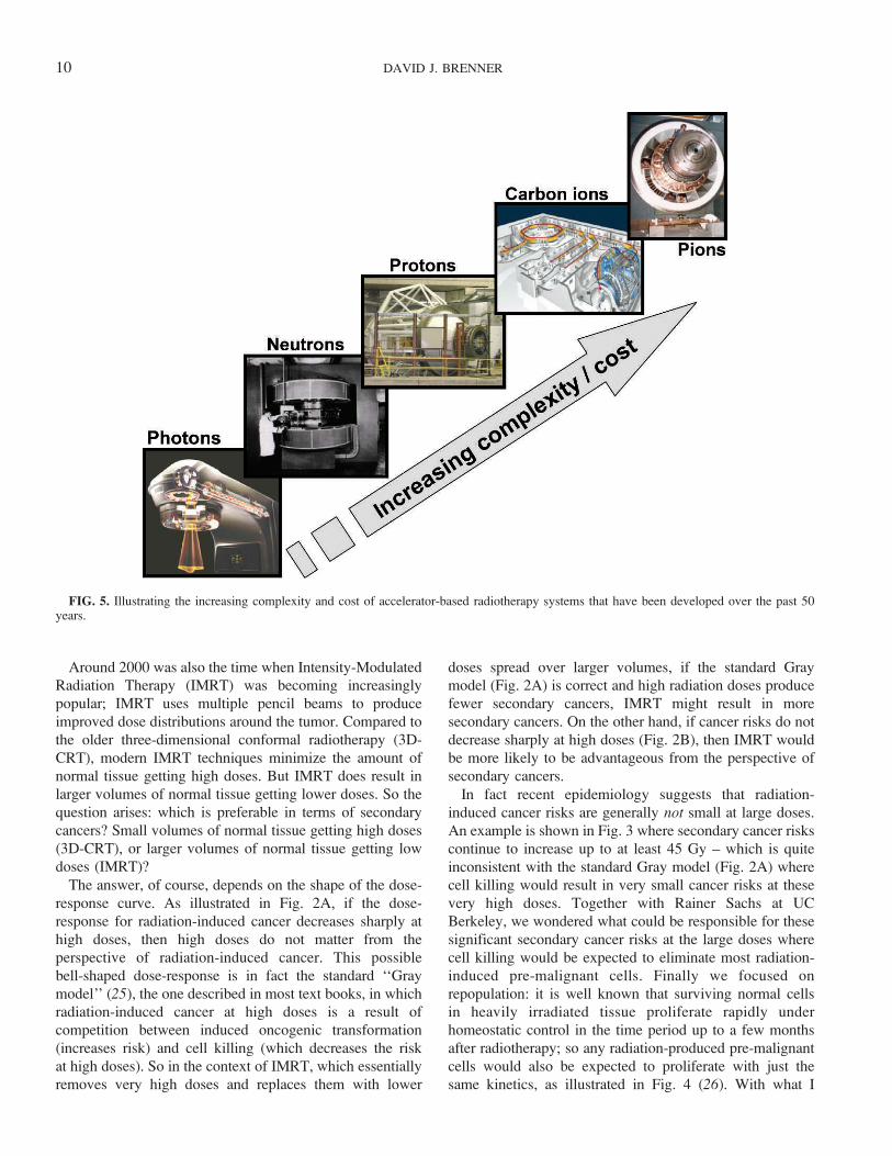
Since the 1940s, I think it is fair to say that most of theadvances in radiotherapy have been driven by newtechnologies. Clearly the advent of medical linacs afterWorld War II was a seminal event, arguably culminating inthe extraordinary proliferation of proton therapy machinestoday. Culminating may not be the right word here becausenegative pi meson (pion) radiotherapy, which had itsheyday in the 1980s, significant outstripped neutrons,protons, and even carbon ions, in terms of complexity andcost (Fig. 5). I was fortunate to have started my careerworking with pions, first for my graduate-school project atthe Rutherford Laboratory in the UK, and subsequently as apostdoc at the Los Alamos National Laboratory.
Pions were a physicist’s dream (30). The idea was that,like protons, pions stop at a controllable distance inside thebody, so giving an inherently better dose distribution thanphotons. But, unlike protons, when a negative pion stops, itis captured by a nearby atomic nucleus, which causes thatnucleus to fragment into low-energy particles – a pion ‘star’.These stars consist of short-ranged high-LET particles,which mostly deposit their energy locally, i.e., in the tumor.And being high-LET, these star particles are expected to beable to efficiently kill hypoxic cells. The physicists’ dream:better dose distributions and the hypoxic cell problem, allsolved with subatomic particles!
Pions were, sadly, an expensive failure. Expensive was a
big part of the story, because just to make a practical pion
beam, roughly 800 MeV protons are needed, almost four
times the energy of current-day proton machines. That
means a far bigger and far costlier machine than a proton
therapy machine. Figure 6 shows the LAMPF accelerator
used to make pions at Los Alamos – half a mile long!
FIG. 9. Breadboard version of the Rapid Automated Biodosimetry Tool [RABiT (51, 52)]. This device is designed to perform fully-automatedhigh-throughput estimates of past radiation exposure, based on fingerstick blood samples. The RABiT is designed to automate established assayssuch as micronucleus, c-H2AX and (in development) cytogenetic endpoints. Overall throughput is up to 30,000 samples per day, achievedthrough complete robotically-based automation of each assay, with in situ imaging in multi-well plates, together with innovations in high-speedimaging. In blinded studies, about 75% of RABiT dose estimates were within 0.5 Gy of the true dose, and 95% were within 1 Gy.
FIG. 10. The Columbia RABiT team. A truly interdisciplinary teamof physicists and biologists.
12 DAVID J. BRENNER
But space and cost were not the only reasons that pions
failed. Two more reasons were the paucity of randomized
trials (31), and the problem of a whole body dose of
neutrons emanating from the pion interactions (32). All
these problems – cost effectiveness, randomized trials, and
neutrons – resonate with similar issues facing contemporary
proton radiotherapy (33–35). Another striking similarity is
the considerable emphasis at the time directed towards
development of a radically smaller and cheaper accelerator
for pion therapy, the PIGMI (36); that never panned out for
pions, though high-field small-radius superconducting syn-
chrocyclotron are looking promising for proton radiother-
apy (37).
One of the great pleasures of working at the pion therapy
facility at Los Alamos was to be the tail end of the trio of
postdocs consecutively supervised by John Dicello (Fig. 7:
Howard Amols, Marco Zaider, and then myself). The two
smarter members of this trio both ended up in medical
physics, and it has been a pleasure to interact with them
over the years.
Coming back to the theme of this section, there is little
doubt that most of the gains in radiotherapy in the last half
century have been technology driven. Has technology
taken radiotherapy as far as it can? Probably so with the
‘‘big physics’’ approaches illustrated in Fig. 5. That being
said, (somewhat) smaller technology approaches, such as
the CyberKnife – a miniature X-ray machine mounted on
FIG. 11. ‘‘Like a dwarf on the shoulders of giants’’. German manuscript illustration, circa 1410. From the U.S. Library of Congress, Lessing J.Rosenwald Collection.
2011 FAILLA AWARD LECTURE 13

the end of a robotic arm - continue to drive the field
forward. But is it not time for radiobiologists to step up to
the plate?
2. Radiotherapy Needs Radiobiology – But We’ve beenSaying That for Fifty Years
When is radiobiology going to come through and really
make a positive difference to radiotherapy? Looking back
over the past half century, I think a skeptic could be
forgiven for saying the question should be ‘‘will it?’’ rather
than ‘‘when will it?’’ But I will make a couple of
suggestions of where I think ‘‘when’’ could practically be
‘‘pretty soon."
In the past half century there have been three major
themes as to where radiobiology can potentially help
radiotherapy: Molecular targeting, alternate fractionation,
and predictive assays. I will argue that molecular targeting
is still a long way from making a clinical impact, but that
perhaps there may be more immediate practical gains to be
made in alternate fractionation and in predictive assays.
Molecular Targeting
Included here are targeting hypoxic cells and targeting the
vasculature, and it is in these areas that most clinically-
oriented radiobiological research has been directed. To date
one would have to say that the resulting clinical impact has
been very limited – with the big gains typically just around
the corner. It does seems that there are some clinical gains to
be had - a recent meta-analysis (38) suggests that hypoxic
modification of radiotherapy (mainly hypoxic radiosensi-
tizers and hyperbaric oxygen) can improve some survival
rates by as much as 15%; but it seems that the enormous
cost associated with developing therapeutic drugs [typically
around a billion dollars over a decade (39)] may well be an
insuperable impediment to practical implementation of
molecular targeting in radiotherapy, unless the anticipated
gains are exceedingly large.
Alternate Fractionation
Changing fractionation schemes is far less expensive thandeveloping new drugs. The radiotherapy field has beenexperimenting with alternate fractionation schemes for morethan a century (9), sometimes for better (4) and sometimesfor worse (40). It was only in the 1970s, stemming largelyfrom the pioneering work of Rod Withers, Lester Peters andHoward Thames, that this enterprise got on to a moremechanistically-based path. Withers, Peters and Thamesshowed that fractionation schemes can be optimized to takeadvantage of, among other factors, accelerated repopulationand the different a/b ratios shown by tumors relative to late-responding normal tissues (41). Again recent meta-analysisof multiple trials has shown clear clinical benefits,particularly for younger patients, accruing from optimizedfractionation schemes (42), and here there are fewerimpediments (though certainly not none!) to wider scaleimplementation.
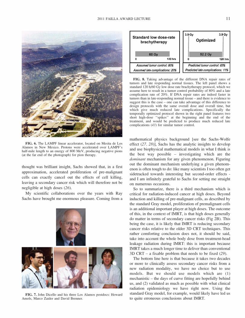
Withers, Peters and Thames correctly saw hyperfraction-ation as taking advantage of the differential a/b ratiobetween tumors and late-responding normal tissues; arethere other biological differences between tumors andnormal tissue that we can potentially take advantage of?In fact it has been strongly suspected for some times thatDNA repair rates are typically much faster in tumorscompared to late-responding normal tissue. Both fromanimal studies and from clinical studies, it appears thatcharacteristic DNA repair rates in tumors are around 1 h orless, whereas characteristic DNA repair times in late-responding tissues are of the order of 5 h or more (43). Canwe take advantage of this difference in repair rates to designoptimized fractionation schemes, analogous to takingadvantage of different a/b ratios?
In fact we probably can, and Fig. 8 shows a standard low-dose rate brachytherapy scheme (60 Gy in 120 h), next to a‘‘temporally optimized’’ scheme, which uses the same doseand the same overall time, but moves some of the dose tothe beginning and to the end of treatment. It is not hard to
FIG. 12. My own scientific giants.
14 DAVID J. BRENNER
show that one could significant improve the expectedclinical outcome with this temporally optimized scheme
(43), which could be relatively easily instituted in the clinic.
Predictive Assays
Predictive assays, both for tumor control and radiother-apy, have been intensively studied for several decades.
Lester Peters called them ‘‘the holy grail of radiobiology’’
(44). There is little doubt that if an effective predictive assayof normal-tissue radiosensitivity could be found, there
would be a significant therapeutic gain for radiosensitivepatients, and potentially for the remaining patient popula-
tion, perhaps even opening the door to individualized
radiotherapy dose prescriptions (45).
Why has no predictive assay worked? One answer is
technical, and is to do with assay variability. Clearly if an
assay result is not repeatable, i.e., has limited precision, thenits predictive power will be limited. Several groups have
quantified this observation (45, 46): for example Mackay
and Hendry (45) concluded that to be useful as a predictiveassay in the clinic, the assay variability needs to be at least
50% smaller (and ideally much smaller still) than theinherent variability in radiosensitivity among patients. This
is not a trivial requirement, and most cellular assays that
have been investigated (47) probably do not meet thiscriterion, in significant part because the assays are
performed manually in a laboratory setting.
It is of course well established that the results of cellularassays which involve a significant number of manually-
performed steps, such as pipetting, adding reagents,washing, microscope imaging etc, show considerable
intra-laboratory variability (48, 49) – and one way to
improve the precision, or repeatability, of cellular assays iswith complete automation (50).
In fact assay automation is something that my own group
has been working on for several years, in the context ofhigh-throughput radiation biodosimetry (51, 52) after a
large-scale radiological event. Here we have developed a
completely automated robotically-based biodosimetry sys-tem called the RABiT [Rapid Automated Biodosimetry
Tool, see Fig. 9)], which fully automates a number ofcellular assays such as micronucleus, c-H2AX, and (in
progress) other cytogenetic endpoints. Complete robotic
automation provides both high throughput and significantlyimproved repeatability. Given the availability of such tools,
I would suggest that it is worth revisiting cellular predictive
assays, given that we can now do much larger studies andwith much improved assay precision – the two factors that I
think doomed earlier predictive-assay research.
Discussing the RABiT system gives me an excuse togive my sincerest thanks to my own team at Columbia,
mostly pictured in Fig. 10, whose dedication to goodscience, and whose patience with my own foibles, is really
remarkable.
CONCLUSION
The Failla prize was a real honor, and reminded me ofNewton’s famous quote ‘‘If I have seen a little further it isby standing on the shoulders of giants.’’ But perhaps Ishould not have been too surprised to find out (53) thatNewton actually borrowed the quote from Bernard ofChartres, half a millennium earlier, who wrote ‘‘I am like adwarf on the shoulders of giants. If I can see more things ata great distance, it is not by virtue of any sharpness of sighton my part, but because I am carried high and raised up bytheir giant size.’’ Even more appropriate, I think, asillustrated in Fig. 11.
I have talked a little of my own scientific giants (Fig. 12),particularly my predecessors at Columbia University, Failla,Rossi, and Eric Hall, as well as my friend and teacher,Rainer Sachs at UC Berkeley. But I would like to end with athank you to my late colleague and friend, Elaine Ron.Elaine was the consummate radiation scientist, combiningscientific insight, compassion, and a complete intolerancefor bad science. She is very much missed.
REFERENCES
1. Cologne JB, Preston DL. Longevity of atomic-bomb survivors.Lancet 2000; 356:303–7.
2. Wintz H. Basic principles for successful Roentgen therapy ofcarcinoma. Radiology 1938; 30:35–42.
3. Regaud C, Ferroux R. Discordances des effets des rayons X, d’unepart dans les peau, d’autre part dans les testicule, par lefractionnement de la dose: Diminution de l’efficacite dans lepeau, maintien de l’efficacite dans le testicule. Compt Rend SocBiol 1927; 97:431–4.
4. Coutard H. The results and methods of treatment of cancer byradiation. Ann Surg 1937; 106:584–98.
5. Gerweck LE, Zaidi ST, Zietman A. Multivariate determinants ofradiocurability. I: Prediction of single fraction tumor control doses.Int J Radiat Oncol Biol Phys 1994; 29:57–66.
6. Fertil B, Malaise EP. Inherent cellular radiosensitivity as a basicconcept for human tumor radiotherapy. Int J Radiat Oncol BiolPhys 1981; 7:621–9.
7. Leborgne F, Leborgne JH, Fowler J, Zubizarreta E, Mezzera J.Accelerated hyperfractionated irradiation for advanced head andneck cancer: effect of shortening the median treatment duration by13 days. Head Neck 2001; 23:661–8.
8. Akagi Y, Hirokawa Y, Kagemoto M, Matsuura K, Ito A, Fujita K,et al. Optimum fractionation for high-dose-rate endoesophagealbrachytherapy following external irradiation of early stageesophageal cancer. Int J Radiat Oncol Biol Phys 1999; 43:525–30.
9. Thames HD, Hendry JH. Fractionation in Radiotherapy. Londonand New York: Taylor & Francis; 1987.
10. Duchesne GM, Peters LJ. What is the a/b ratio for prostate cancer?Rationale for hypofractionated high-dose-rate brachytherapy. Int JRadiat Oncol Biol Phys 1999; 44:747–8.
11. Thames HD, Jr., Withers HR, Peters LJ, Fletcher GH. Changes inearly and late radiation responses with altered dose fractionation:implications for dose-survival relationships. Int J Radiat OncolBiol Phys 1982; 8:219–26.
12. Barendsen GW. Dose fractionation, dose rate and iso-effectrelationships for normal tissue responses. Int J Radiat Oncol BiolPhys 1982; 8:1981–97.
13. Brenner DJ, Hall EJ. Fractionation and protraction for radiotherapy
2011 FAILLA AWARD LECTURE 15
of prostate carcinoma. Int J Radiat Oncol Biol Phys 1999;43:1095–101.
14. Brenner DJ, Martinez AA, Edmundson GK, Mitchell C, ThamesHD, Armour EP. Direct evidence that prostate tumors show highsensitivity to fractionation (low a/b ratio), similar to late-responding normal tissue. Int J Radiat Oncol Biol Phys 2002;52:6–13.
15. Proust-Lima C, Taylor JM, Secher S, Sandler H, Kestin L, PicklesT, et al. Confirmation of a low alpha/beta ratio for prostate cancertreated by external beam radiation therapy alone using a post-treatment repeated-measures model for PSA dynamics. Int J RadiatOncol Biol Phys 2011; 79:195–201.
16. Miralbell R, Roberts SA, Zubizarreta E, Hendry JH. Dose-fractionation sensitivity of prostate cancer deduced from radio-therapy outcomes of 5,969 patients in seven internationalinstitutional datasets: alpha/beta¼1.4 (0.9-2.2) Gy. Int J RadiatOncol Biol Phys 2012; 82:e17–24.
17. Yeoh EE, Botten RJ, Butters J, Di Matteo AC, Holloway RH,Fowler J. Hypofractionated versus conventionally fractionatedradiotherapy for prostate carcinoma: final results of phase IIIrandomized trial. Int J Radiat Oncol Biol Phys 2011; 81:1271–8.
18. Arcangeli S, Strigari L, Gomellini S, Saracino B, Petrongari MG,Pinnaro P, et al. Updated results and patterns of failure in arandomized hypofractionation trial for high-risk prostate cancer.Int J Radiat Oncol Biol Phys Epub 2012.
19. Pollack A, Walker G, Buyyounouski MK, Horowitz E, Price R,Feigenberg S, et al. Five year results of a randomized externalbeam radiotherapy hypofractionation trial for prostate cancer. Int JRadiat Oncol Biol Phys 2011; 81:S1.
20. Dearnaley D, Syndikus I, Sumo G, Bidmead M, Bloomfield D,Clark C, et al. Conventional versus hypofractionated high-doseintensity-modulated radiotherapy for prostate cancer: preliminarysafety results from the CHHiP randomised controlled trial. LancetOncol 2012; 13:43–54.
21. Kuban DA, Nogueras-Gonzalez GM, Hamblin L, Lee AK, Choi S,Frank SJ, et al. Preliminary report of a randomized dose escalationtrial for prostate cancer using hypofractionation. Int J Radiat OncolBiol Phys 2010; 78:S58–9.
22. Garcia-Barros M, Paris F, Cordon-Cardo C, Lyden D, Rafii S,Haimovitz-Friedman A, et al. Tumor response to radiotherapyregulated by endothelial cell apoptosis. Science 2003; 300:1155–9.
23. Park HJ, Griffin RJ, Hui S, Levitt SH, Song CW. Radiation-induced vascular damage in tumors: implications of vasculardamage in ablative hypofractionated radiotherapy (SBRT andSRS). Radiat Res 2012; 177:311–27.
24. Brenner DJ, Curtis RE, Hall EJ, Ron E. Second malignancies inprostate carcinoma patients after radiotherapy compared withsurgery. Cancer 2000; 88:398–406.
25. Gray LH, Radiation biology and cancer. In: Cellular RadiationBiology; A symposium considering radiation effects in the cell andpossible implications for cancer therapy, a collection of papers.Baltimore: William and Wilkins; 1965. p. 8–25.
26. Sachs RK, Brenner DJ. Solid tumor risks after high doses ofionizing radiation. Proc Natl Acad Sci USA 2005; 102:13040–5.
27. Sachs RK, Wolfe AM. Perturbations of a cosmological model andangular variations of the microwave background. Astrophys J1967; 147:73–90.
28. Stoeger WR, Ellis GF, Xu C. Observational cosmology. VI. Themicrowave background and the Sachs-Wolfe effect. Phys Rev DPart Fields 1994; 49:1845–53.
29. Hall EJ, Wuu CS. Radiation-induced second cancers: the impact of3D-CRT and IMRT. Int J Radiat Oncol Biol Phys 2003; 56:83–8.
30. Fowler PH, Perkins DH. The possibility of therapeutic applicationsof beams of negative pi-mesons. Nature 1961; 189:524–8.
31. Elwood JM. The design of clinical trials comparing pi-mesontherapy with conventional radiotherapy. J Can Assoc Radiol 1979;30:79–82.
32. Brenner DJ, Dicello JF, Zaider M. An interpretation of somebiological results obtained in range-modulated negative pionbeams. Int J Radiat Oncol Biol Phys 1982; 8:121–6.
33. Konski A, Speier W, Hanlon A, Beck JR, Pollack A. Is protonbeam therapy cost effective in the treatment of adenocarcinoma ofthe prostate? J Clin Oncol 2007; 25:3603–8.
34. Goitein M, Cox JD. Should randomized clinical trials be requiredfor proton radiotherapy? J Clin Oncol 2008; 26:175–6.
35. Brenner DJ, Hall EJ. Secondary neutrons in clinical protonradiotherapy: A charged issue. Radiother Oncol 2008; 86:165–70.
36. Knapp EA, Swenson DA. The Los Alamos Scientific Laboratoryion linear accelerator program. Int J Radiat Oncol Biol Phys 1977;3:383–5.
37. Schippers JM, Lomax AJ. Emerging technologies in protontherapy. Acta Oncol 2011; 50:838–50.
38. Overgaard J. Hypoxic modification of radiotherapy in squamouscell carcinoma of the head and neck–a systematic review and meta-analysis. Radiother Oncol 2011; 100:22–32.
39. DiMasi JA, Hansen RW, Grabowski HG. The price of innovation:New estimates of drug development costs. J Health Econ 2003;22:151–85.
40. Bates TD, Peters LJ. Dangers of the clinical use of the NSDformula for small fraction numbers. Br J Radiol 1975; 48:773.
41. Thames HD, Jr., Peters LJ, Withers HR, Fletcher GH. Acceleratedfractionation vs hyperfractionation: rationales for several treat-ments per day. Int J Radiat Oncol Biol Phys 1983; 9:127–38.
42. Baujat B, Bourhis J, Blanchard P, Overgaard J, Ang KK, SaundersM, et al. Hyperfractionated or accelerated radiotherapy for headand neck cancer. Cochrane Database Syst Rev 2010:CD002026.
43. Brenner DJ, Hall EJ, Huang Y, Sachs RK. Optimizing the timecourse of brachytherapy and other accelerated radiotherapeuticprotocols. Int J Radiat Oncol Biol Phys 1994; 29:893–901.
44. Peters LJ. The ESTRO Regaud lecture. Inherent radiosensitivity oftumor and normal tissue cells as a predictor of human tumorresponse. Radiother Oncol 1990; 17:177–90.
45. Mackay RI, Hendry JH. The modelled benefits of individualizingradiotherapy patients’ dose using cellular radiosensitivity assayswith inherent variability. Radiother Oncol 1999; 50:67–75.
46. Brock WA, Tucker SL, Geara FB, Turesson I, Wike J, Nyman J, etal. Fibroblast radiosensitivity versus acute and late normal skinresponses in patients treated for breast cancer. Int J Radiat OncolBiol Phys 1995; 32:1371–9.
47. West CM. Invited review: intrinsic radiosensitivity as a predictorof patient response to radiotherapy. Br J Radiol 1995; 68:827–37.
48. Radack KL, Pinney SM, Livingston GK. Sources of variability inthe human lymphocyte micronucleus assay: a population-basedstudy. Environ Mol Mutagen 1995; 26:26–36.
49. Bonassi S, Fenech M, Lando C, Lin YP, Ceppi M, Chang WP, etal. HUman MicroNucleus project: international database compar-ison for results with the cytokinesis-block micronucleus assay inhuman lymphocytes: I. Effect of laboratory protocol, scoringcriteria, and host factors on the frequency of micronuclei. EnvironMol Mutagen 2001; 37:31–45.
50. Sturgeon C, Hill R, Hortin GL, Thompson D. Taking a newbiomarker into routine use—a perspective from the routine clinicalbiochemistry laboratory. Proteomics Clin Appl 2010; 4:892–903.
51. Garty G, Chen Y, Salerno A, Turner H, Zhang J, Lyulko O, et al.The RABiT: a rapid automated biodosimetry tool for radiologicaltriage. Health Phys 2010; 98:209–17.
52. Garty G, Chen Y, Turner HC, Zhang J, Lyulko OV, Bertucci A, etal. The RABiT: a rapid automated biodosimetry tool forradiological triage. II. Technological developments. Int J RadiatBiol 2011; 87:776–90.
53. Merton RK. On the shoulders of giants. Chicago: University ofChicago Press; 1965.
16 DAVID J. BRENNER