Embed Size (px)
Citation preview

Cite this: RSC Advances, 2013, 3, 2553
Received 7th November 2012,Accepted 7th January 2013
Exploring the fluorescence switching phenomenon ofcurcumin encapsulated niosomes: in vitro real timemonitoring of curcumin release to cancer cells3
DOI: 10.1039/c2ra23382g
www.rsc.org/advances
Nagaprasad Puvvada,a Shashi Rajput,b B. N. Prashanth Kumar,b Mahitosh Mandalb
and Amita Pathak*a
This is the first report on the reversible fluorescence switching of
curcumin encapsulated niosomes exhibiting a strong blue color
excimer emission in the UV region and a green color luminescence
of the excited state monomer form of curcumin that undergoes
ESIPT in the visible region. The real time monitoring of the release
of curcumin from niosomes is feasible through the change of
fluorescence color from blue to green.
Currently, the design and development of a novel functionalnanocarrier and its fluorescence switching has created aprodigious curiosity in materials science research and contributedsignificantly to the understanding of intrinsic mechanisms inchemical and biological processes. In particular, the utilization ofnanocarriers in various applications such as sensing, therapy andimaging of organic fluorescent probes, plays a vital role inbiology.1,2 Curcumin (CU) is a natural yellow colored solid isolatedfrom the rhizome of the plant curcuma longa. It is used as acommon ingredient in foods, traditional medicines and cos-metics, and it is also implicated as an agent in various applicationssuch as antioxidants, anti-inflammatories, antitumoral, anti-HIVetc., and also as a fluorescent agent in the tracking of biologicalmechanisms at the molecular level.3–5 Liposome-based nanocar-riers in drug delivery systems have brought a tremendousparadigm change in cancer biology for clinical trials.2 However,the usage of such liposomes has been associated with severaldisadvantages for clinical trials due to the presence of chemicallyunstable phospholipids, oxidative degradation, etc.3 In addition,one of the major downsides of targeted liposomes is theirincapability to reach the destination site because of the highprobability of delivering to lysosomes with subsisting enzymes,including hydrolases and peptidases, which degrade the entitieswithin the liposome during the cell internalization process.6 Inorder to subdue the above problems, niosomes are the bestsubstitutes with enhanced stability and biocompatibility.7 In
general, niosomes are made up of non ionic surfactant vesiclesand gain structural rigidity as well as stealth behavior with theemployment of PEG or cholesterol.3 PEG is a hydrophilicbiocompatible polymer, which once coated over a nanocarrierfavors the active involvement of tagged targeting receptors andligands to interact with a tumor’s cell surface.8 Furthermore, PEGprevents cells adhesion, opsonization and uptake by thereticuloendothelial system.8 On the other hand, niosomes aremultilamellar vesicles formed by an oxyethylene–water shellcovered with polar head groups along with a bilayer group.9
Recently, Hong et al. designed and synthesized PEG functionalizedniosomes and used the resultant product as a delivery system indelivering an anticancer agent to cure cancer.10 In our study, weconstructed poly(ethylene glycol) methyl ether acrylate (PEGMEA-480) conjugated with cysteine (PEG-Cys) through the clickchemistry route for the synthesis of niosomes. PEG-Cys hascarboxyl and amine groups and these functional groups are pHsensitive for utilization in drug delivery applications.11,12
Previously, Radovic-Moreno et al. synthesized polymer nanoparti-cles consisting of functional groups similar to those used in ourstudy. The prepared nanoparticles were reported to be pHsensitive and their surface charge switchable for the delivery ofantibiotics to bacterial cells.13
Although there are various methods available for the identifica-tion of the release of entities from nanoparticles/vesicles withincells, among them fluorescence switching is a distinctivephenomenon, as it helps to construe the drug release profilefrom the niosomes into the external cellular environment.14–17
Recently, the design of fluorescence switching, based on theexcited state intramolecular proton transfer (ESIPT), has attractedconsiderable interest in photo physical processes. Various organicfluorescent probes, like 2-(29-hydroxyphenyl) benzoxazole and 2-(29-hydroxyphenyl) benzimidazole,18,19 exhibit ESIPT and result indual emissions of keto and enol tautomers in the excited state.Further, these molecules emit fluorescence with a large Stokesshift and are used in sensing cations or anions. Malinge et al. alsodiscovered that the naphthalimide derivative in encapsulatedsilica core shell nanoparticles exhibits fluorescence in the excimerform.20 In this context, we chose CU as a fluorescent probe
aDepartment of Chemistry, Kharagpur-721302, West Bengal, India.
E-mail: [email protected] of Medical Science and Technology, Indian Institute of Technology
Kharagpur, Kharagpur-721302, West Bengal, India
3 Electronic supplementary information (ESI) available. See DOI: 10.1039/c2ra23382g
RSC Advances
COMMUNICATION
This journal is � The Royal Society of Chemistry 2013 RSC Adv., 2013, 3, 2553–2557 | 2553
Dow
nloa
ded
by U
nive
rsity
of
Cal
gary
on
08 M
arch
201
3Pu
blis
hed
on 0
8 Ja
nuar
y 20
13 o
n ht
tp://
pubs
.rsc
.org
| do
i:10.
1039
/C2R
A23
382G
View Article OnlineView Journal | View Issue
because of its tendency to exhibit keto and enol tautomers.19 Inaddition, CU also act as a fluorescent probe in tracking niosomemovement within cells, which is also favourable for studying itsfluorescence switching properties, besides its anticancer activity.To date, few examples are available on the fluorescence switchingon/off route in the case of polymer assembled nanoparticles orvesicles, which impelled us to carry out this work on the photoswitching of vesicles.21,22 The above view instigated us tosynthesize CU encapsulated PEG functionalized niosomes throughthis approach.
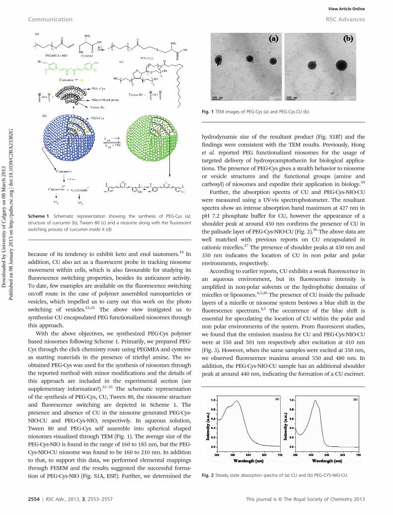
With the above objectives, we synthesized PEG-Cys polymerbased niosomes following Scheme 1. Primarily, we prepared PEG-Cys through the click chemistry route using PEGMEA and cysteineas starting materials in the presence of triethyl amine. The so-obtained PEG-Cys was used for the synthesis of niosomes throughthe reported method with minor modifications and the details ofthis approach are included in the experimental section (seesupplementary information3).23–25 The schematic representationof the synthesis of PEG-Cys, CU, Tween 80, the niosome structureand fluorescence switching are depicted in Scheme 1. Thepresence and absence of CU in the niosome generated PEG-Cys-NIO-CU and PEG-Cys-NIO, respectively. In aqueous solution,Tween 80 and PEG-Cys self assemble into spherical shapedniosomes visualized through TEM (Fig. 1). The average size of thePEG-Cys-NIO is found in the range of 160 to 185 nm, but the PEG-Cys-NIO-CU niosome was found to be 160 to 210 nm. In additionto that, to support this data, we performed elemental mappingsthrough FESEM and the results suggested the successful forma-tion of PEG-Cys-NIO (Fig. S1A, ESI3). Further, we determined the
hydrodynamic size of the resultant product (Fig. S1B3) and thefindings were consistent with the TEM results. Previously, Honget al. reported PEG functionalized niosomes for the usage oftargeted delivery of hydroxycamptothecin for biological applica-tions. The presence of PEG-Cys gives a stealth behavior to niosomeor vesicle structures and the functional groups (amine andcarboxyl) of niosomes and expedite their application in biology.10
Further, the absorption spectra of CU and PEG-Cys-NIO-CUwere measured using a UV-vis spectrophotometer. The resultantspectra show an intense absorption band maximum at 427 nm inpH 7.2 phosphate buffer for CU, however the appearance of ashoulder peak at around 450 nm confirms the presence of CU inthe palisade layer of PEG-Cys-NIO-CU (Fig. 2).26 The above data arewell matched with previous reports on CU encapsulated incationic micelles.27 The presence of shoulder peaks at 450 nm and350 nm indicates the location of CU in non polar and polarenvironments, respectively.
According to earlier reports, CU exhibits a weak fluorescence inan aqueous environment, but its fluorescence intensity isamplified in non-polar solvents or the hydrophobic domains ofmicelles or liposomes.4,5,26 The presence of CU inside the palisadelayers of a micelle or niosome system bestows a blue shift in thefluorescence spectrum.4,5 The occurrence of the blue shift isessential for speculating the location of CU within the polar andnon polar environments of the system. From fluorescent studies,we found that the emission maxima for CU and PEG-Cys-NIO-CUwere at 550 and 501 nm respectively after excitation at 410 nm(Fig. 3). However, when the same samples were excited at 350 nm,we observed fluorescence maxima around 550 and 480 nm. Inaddition, the PEG-Cys-NIO-CU sample has an additional shoulderpeak at around 440 nm, indicating the formation of a CU excimer.
Scheme 1 Schematic representation showing the synthesis of PEG-Cys (a);structure of curcumin (b), Tween 80 (c) and a niosome along with the fluorescentswitching process of curcumin inside it (d).
Fig. 1 TEM images of PEG-Cys (a) and PEG-Cys-CU (b).
Fig. 2 Steady state absorption spectra of (a) CU and (b) PEG-CYS-NIO-CU.
2554 | RSC Adv., 2013, 3, 2553–2557 This journal is � The Royal Society of Chemistry 2013
Communication RSC Advances
Dow
nloa
ded
by U
nive
rsity
of
Cal
gary
on
08 M
arch
201
3Pu
blis
hed
on 0
8 Ja
nuar
y 20
13 o
n ht
tp://
pubs
.rsc
.org
| do
i:10.
1039
/C2R
A23
382G
View Article Online

Similar kind of peaks were observed by Majhi et al.28 From theabove observation, it is clearly understood that CU fluorescenceemission maxima were not affected by the excitation wavelength(Fig. S2, ESI3). The observed blue shift from the emission maximaof PEG-Cys-NIO-CU and CU, clearly suggests the presence of CU,which is transferred from the more polar region to niosomescontaining less polar regions. The so-formed excimer fluorescenceemission maxima at a shorter wavelength and longer wavelengthregion appeared due to the presence of the excited normal (N*)form and the proton transfer tautomer (T*) form, respectively.29
Further, we calculated that the intensity ratio of the short-wavelength (N*) and the long-wavelength (T*) emission bands,(IN*/IT*), of this sample is around 1.28, which is found to be lessthan 2.1indicating that the environment is aprotic.30 In addition,the presence of intermolecular hydrogen bonds was confirmed bythe fact that the difference in the wave-number of the maxima ofthe N* (uN*) and the T* (uT*) bands was around 2100 cm21, whichis less than 2300 cm21.29,30 It was reported that curcumin readilyforms intermolecular hydrogen bonding, resulting in a blue shiftin the fluorescence spectrum.4 In our study, we observed anadditional blue shift compared to previous reports, which may bedue to the formation of intermolecular hydrogen bonds withniosome functional groups.31 Based on the above concept, weexplored the fluorescence switching mechanism of CU in aniosome, by sample excitation at 350 nm, which facilitates theformation of excimers (N* and T* forms) exhibiting blue emissionmaxima at 440 and 480 nm, respectively.32 At the same time, whenthe sample was excited at 410 nm, the monomeric form of CUinside the niosome underwent ESIPT, resulting in green emissionmaxima at 501 nm.4,5 However, the excitation dependent existenceof excimer and monomer forms of CU are still unclear. Adhikaryet al.5 reported the existence of a monomer form of CU thatundergoes ESIPT in a micelle system (cation, anion and neutralmicelle) at 408 nm excitation wavelength. These studies suggestthat the existence of a monomer form of CU in any micelle systemdefinitely undergoes proton transfer. Currently, our focus is on theutilization of the fluorescent switching niosome as a drug deliverycarrier and not the mechanism aspect of the niosome hydration of
the proton transfer dynamic. In particular, recent evidencesuggested that CU derivatives along with a N,N9-dimethyl groupshowed a considerable red shift for fluorescent switching.33 In thissense, a PEG-Cys moiety containing carboxylate and amine groupsmay help CU excimers to form inside the niosome. In addition, weperformed steady state anisotropy for CU and PEG-Cys-NIO-CU.34
From this study, we found that the steady state anisotropyconstant is around 0.1095, 0.2515 and 0.3449 for CU, PEG-Cys-NIO-CU (lex at 410 nm) and PEG-Cys-NIO-CU (lex at 350 nm),respectively. The abrupt increase in this anisotropy constantsuggests the presence of a more confined medium with motionalrestrictions on the CU molecules inside the niosome.35 Theprocess of fluorescence switching is clearly visualized throughfluorescence microscopy images and the corresponding solutionsare depicted as insets in Fig. 3. Interestingly, PEG-Cys-NIO-CUemitted blue and green color luminescence under semi confocalmicroscopy and also emitted luminescence maxima at 481 and501 nm, respectively. After confirming the visibility of thefluorescent switching of the PEG-Cys-CU sample, we furthersystematically investigated their ability to recognize the stabilityand CU release from a niosome for biological assays.
The stability of niosomes plays a pivotal role in their bio-imaging applications. The stability of PEG-Cys-NIO-CU wasevaluated using an absorption and fluorescence spectrophot-ometer after various time intervals (Fig. S3, ESI3). These resultsdisplayed insignificant photo degradation of the PEG-Cys-NIO-CUand also that the fluorescent switching is reversible. From Fig. S3,ESI,3 it can be concluded that the presence of CU inside theniosome did not affect the absorption maxima and its fluorescentswitching at varying time intervals. In addition, we found aninsignificant change in the hydrodynamic size of the PEG-Cys-NIO-CU sample using dynamic light scattering, indicating itsstability for up to 3 months (Fig. S1B (b) and S4, ESI3).
The pH sensitive functional groups (–COOH and –NH2) of PEG-Cys play a vital role in the controlled release of CU from niosomes.The amount of CU released at physiological pH is negligibleduring short intervals, but an acidic environment favors thedisorganization of the niosome structure resulting in a completeevacuation of CU to the outer aqueous environment (Fig. 4). This
Fig. 3 Fluorescence spectra of the PEG-Cys-CU sample with excitation wavelengthat (a) 350 nm and (b) 410 nm and corresponding fluorescence images of thesample solutions (shown as insets in same color).
Fig. 4 Variation in the amount of CU released from a PEG-Cys-CU sample over timeat pH 5 and 7.
This journal is � The Royal Society of Chemistry 2013 RSC Adv., 2013, 3, 2553–2557 | 2555
RSC Advances Communication
Dow
nloa
ded
by U
nive
rsity
of
Cal
gary
on
08 M
arch
201
3Pu
blis
hed
on 0
8 Ja
nuar
y 20
13 o
n ht
tp://
pubs
.rsc
.org
| do
i:10.
1039
/C2R
A23
382G
View Article Online

result was supported by DLS measurements for the sizedetermination of the niosome. At physiological pH no change inthe size of the niosome was observed, while at acidic pH (i.e., pH5) niosome size was reduced to around 27 nm, which indicatesthat its structure eventually diminished (Fig. S5, ESI3). We alsodetermined the surface charge of the free niosome and CUencapsulated niosome in varying pH of 5.5–7.4 (Fig. S6, ESI3). Inthis perspective, we prefer acidic environments for the employ-ment of niosomes in drug delivery applications. The acidic tumormicroenvironment and the surface negative charge of cells favorthe implementation of niosomes in drug delivery applications tocure cancer. The iso-electric points of PEG-Cys-NIO and PEG-Cys-NIO-CU were found to be 6.62 and 6.71, respectively. In the acidicmicroenvironment, the tumor and the cancer cell surface becomenegatively charged and are electrostatically attached to positivelycharged niosomes. A similar study of this mechanism is reportedin our previous paper.36
The cytotoxicity of CU, PEG-Cys-NIO and PEG-Cys-NIO-CU incultured MCF 7 cells was determined by MTT assay.36 The range ofconcentrations of CU used in the experiment was 1 to 50 mM. Theassay showed that 30% cell viability was achieved with 40 mM ofCU, while 25 mM of PEG-Cys-NIO-CU was needed to achieve thesame percentage of cell viability (Fig. 5). However, the PEG-Cys-NIO showed minimal cytotoxicity even at higher concentrations.The results were expressed as a percentage relative to the controlcell number. It could thus be understood that the PEG-Cys-NIO-CU significantly inhibits the growth of MCF 7 cells in a dose-dependent manner. Further, CU showed an IC50 (median growthinhibitory concentration value) of 27 mM, whereas CU-loadedniosomes showed 17 mM after 24 h of treatment, which waschosen for further experiments. The reason behind the reductionin the minimal inhibitory concentration of CU is that the presenceof a positive charge on the PEG-Cys-NIO-CU favors the adherenceand uptake by the negatively charged surface of the cell membraneresulting in the complete expulsion of entrapped CU in a cellularenvironment.
In this study we aim to advance the in vitro release profile of CUfrom PEG-Cys-NIO-CU through a cellular uptake study aftervarying time intervals (Fig. 6). At 15 min incubation, cells appear
blue due to the insignificant amount of drug release effect theniosomes. However, the cells gradually appear bluish green due tothe CU release at subsequent time points. Finally, cells arevisualized in green fluorescence after 6 h incubation due to thewhole exhaustive release of CU, while the blue fluorescencecompletely disappears. At the 24 h incubation point, greenfluorescence persists in the cells and appears to be more intensedue to the disappearance of the nuclear membrane and cellshrinkage. The cellular uptake and detailed mechanism behindthe change in cell fluorescence from blue to green is explained onthe basis of CU release and surface charge of the PEG-Cys-NIO-CU.The presence of the positive charge on the surface of the PEG-Cys-NIO-CU due to the lower pH leads to a gradual disintegration ofthe PEG-Cys-NIO-CU, thus resulting in a controlled release of CUfrom the PEG-Cys-NIO-CU.36 With increasing time intervals theacidity of the tumor microenvironment increases (i.e. the pHdecreases),12 thus increasing the positive charge on the PEG-Cys-NIO-CU, resulting in the collapse of the niosome structure therebyreleasing CU. The released CU eventually shifts the cellfluorescence from blue to green in an increasing manner athigher time points. In a nutshell, this study determined theincubation time period for the complete drug release along withcellular inhibition.
In summary, we report a facile method for the synthesis ofexcitation dependent reversible fluorescence switching CU loadedniosomes. This is the first report on the fluorescence switching ofcurcumin encapsulated niosomes that exhibited strong blue colorin UV region and green color luminescence in the visible region.The curcumin entities are released from the niosomes causing achange in fluorescence color from blue to green allowing for realtime monitoring.
Acknowledgements
We wish to thank Prof. N. D. Pradeep Singh and Prof. NilmoniSarkar for providing column and DLS facilities. The authorsare also grateful to Mr. Bibhas Roy for his generous help inhandling confocal imaging.
Fig. 5 Cell viability of (a) PEG-Cys-NIO, (b) PEG-Cys-NIO-CU and (c) CU samples forMCF 7 human breast cancer cells.
Fig. 6 Cellular drug release profiles of localized curcumin-loaded niosomes atvarious time intervals for an MCF 7 human breast cancer cell line.
2556 | RSC Adv., 2013, 3, 2553–2557 This journal is � The Royal Society of Chemistry 2013
Communication RSC Advances
Dow
nloa
ded
by U
nive
rsity
of
Cal
gary
on
08 M
arch
201
3Pu
blis
hed
on 0
8 Ja
nuar
y 20
13 o
n ht
tp://
pubs
.rsc
.org
| do
i:10.
1039
/C2R
A23
382G
View Article Online

References
1 N.-C. Fan, F.-Y. Cheng, J.-a. A. Ho and C.-S. Yeh, Angew. Chem.,Int. Ed., 2012, 51, 8806–8810.
2 S. B. Tiwari and M. M. Amiji, Curr. Drug Delivery, 2006, 3,219–232.
3 C. Ghatak, V. G. Rao, S. Mandal and N. Sarkar, Phys. Chem.Chem. Phys., 2012, 14, 8925–8935.
4 R. Adhikary, P. J. Carlson, T. W. Kee and J. W. Petrich, J. Phys.Chem. B, 2010, 114, 2997–3004.
5 R. Adhikary, P. Mukherjee, T. W. Kee and J. W. Petrich, J. Phys.Chem. B, 2009, 113, 5255–5261.
6 M. F. Francis, G. Dhara, F. M. Winnik and J. C. Leroux,Biomacromolecules, 2001, 2, 741–749.
7 K. M. Kazi, A. S. Mandal, N. Biswas, A. Guha, S. Chatterjee,M. Behera and K. Kuotsu, J. Adv. Pharm. Technol. Res., 2010, 1,374–380.
8 J. V. Georgieva, R. P. Brinkhuis, K. Stojanov, C. A. G. M. Weijers,H. Zuilhof, F. P. J. T. Rutjes, D. Hoekstra, J. C. M. van Hest andI. S. Zuhorn, Angew. Chem., Int. Ed., 2012, 51, 8339–8342.
9 S. Ghosh, A. K. Mandal, A. K. Das, T. Mondal andK. Bhattacharyya, Phys. Chem. Chem. Phys., 2012, 14, 9749–9757.
10 M. Hong, S. Zhu, Y. Jiang, G. Tang and Y. Pei, J. ControlledRelease, 2009, 133, 96–102.
11 A. F. Radovic-Moreno, T. K. Lu, V. A. Puscasu, C. J. Yoon,R. Langer and O. C. Farokhzad, ACS Nano, 2012, 6, 4279–4287.
12 M. J. Marın, F. Galindo, P. Thomas and D. A. Russell, Angew.Chem., Int. Ed., 2012, 51, 9657–9661.
13 A. F. Radovic-Moreno, T. K. Lu, V. A. Puscasu, C. J. Yoon,R. Langer and O. C. Farokhzad, ACS Nano, 2012, 6, 4279–4287.
14 A. C. Coleman, J. M. Beierle, M. C. A. Stuart, B. Macia, G. Caroli,J. T. Mika, D. J. van Dijken, J. Chen, W. R. Browne and B.L. Feringa, Nat. Nanotechnol., 2011, 6, 547–552.
15 E. H. Rego, L. Shao, J. J. Macklin, L. Winoto, G. A. Johansson,N. Kamps-Hughes, M. W. Davidson and M. G. Gustafsson, Proc.Natl. Acad. Sci. U. S. A., 2012, 109, E135–143.
16 D. Jiao, J. Geng, X. J. Loh, D. Das, T.-C. Lee and O. A. Scherman,Angew. Chem., Int. Ed., 2012, 51, 9633–9637.
17 J. Cusido, M. Battal, E. Deniz, I. Yildiz, S. Sortino and F.M. Raymo, Chem.–Eur. J., 2012, 18, 10399–10407.
18 Y. Xu, Q. Liu, B. Dou, B. Wright, J. Wang and Y. Pang, Adv.Healthcare Mater., 2012, 1, 485–492.
19 K. Abhinav and P. K. Panigrahi, Ann. Phys., 2010, 325,1198–1206.
20 J. Malinge, C. Allain, A. Brosseau and P. Audebert, Angew.Chem., Int. Ed., 2012, 51, 8534–8537.
21 R. M. Dickson, A. B. Cubitt, R. Y. Tsien and W. E. Moerner,Nature, 1997, 388, 355–358.
22 C. Li, Y. Zhang, J. Hu, J. Cheng and S. Liu, Angew. Chem., Int.Ed., 2010, 49, 5120–5124.
23 J. Dey and S. Shrivastava, Soft Matter, 2012, 8, 1305–1308.24 S. Mandal, V. G. Rao, C. Ghatak, R. Pramanik, S. Sarkar and
N. Sarkar, J. Phys. Chem. B, 2011, 115, 12108–12119.25 A. Luciani, J.-C. Olivier, O. Clement, N. Siauve, P.-Y. Brillet,
B. Bessoud, F. Gazeau, I. F. Uchegbu, E. Kahn, G. Frija and C.A. Cuenod, Radiology, 2004, 231, 135–142.
26 M. H. M. Leung and T. W. Kee, Langmuir, 2009, 25, 5773–5777.27 M. H. M. Leung, H. Colangelo and T. W. Kee, Langmuir, 2008,
24, 5672–5675.28 A. Majhi, G. M. Rahman, S. Panchal and J. Das, Bioorg. Med.
Chem., 2010, 18, 1591–1598.29 R. Das, G. Duportail, L. Richert, A. Klymchenko and Y. Mely,
Langmuir, 2012, 28, 7147–7159.30 A. S. Klymchenko and A. P. Demchenko, Phys. Chem. Chem.
Phys., 2003, 5, 461–468.31 A. Y. Li, J. Chem. Phys., 2007, 126, 154102.32 A. Majhi, G. M. Rahman, S. Panchal and J. Das, Bioorg. Med.
Chem., 2010, 18, 1591–1598.33 C. Ran, X. Xu, S. B. Raymond, B. J. Ferrara, K. Neal, B.
J. Bacskai, Z. Medarova and A. Moore, J. Am. Chem. Soc., 2009,131, 15257–15261.
34 N. Puvvada, D. Mandal, P. K. Panigrahi and A. Pathak,Toxicology Research, 2012, 1, 196–200.
35 A. Manna and S. Chakravorti, Photochem. Photobiol., 2012, 88,285–294.
36 P. Venkatesan, N. Puvvada, R. Dash, B. N. Prashanth Kumar,D. Sarkar, B. Azab, A. Pathak, S. C. Kundu, P. B. Fisher andM. Mandal, Biomaterials, 2011, 32, 3794–3806.
This journal is � The Royal Society of Chemistry 2013 RSC Adv., 2013, 3, 2553–2557 | 2557
RSC Advances Communication
Dow
nloa
ded
by U
nive
rsity
of
Cal
gary
on
08 M
arch
201
3Pu
blis
hed
on 0
8 Ja
nuar
y 20
13 o
n ht
tp://
pubs
.rsc
.org
| do
i:10.
1039
/C2R
A23
382G
View Article Online


![Fabrication of Curcumin Encapsulated Chitosan-PVA Silver ... · [18]. Polyvinyl alcohol (PVA), a water soluble synthetic polymer, having less toxicity, possess excellent wound dressing](https://img.dokumen.tips/doc/110x75/60a7a1ff0f83e13018683769/fabrication-of-curcumin-encapsulated-chitosan-pva-silver-18-polyvinyl-alcohol.jpg)