Embed Size (px)
Citation preview

ENHANCEMENT OF SOMATIC EMBRYOGENESIS IN
RECALCITRANT EAST AFRICAN HIGHLAND BANANAS
SADIK KASSIM
BSC AGRIC (HONS), MSC CROP SCIENCE, MAK
A DISSERTATION SUBMITTED TO
THE DIRECTORATE OF RESEARCH AND GRADUATE TRAINING
IN PARTIAL FULFILLMENT FOR THE AWARD OF DOCTOR OF PHILOSOPHY
IN PLANT BREEDING AND BIOTECHNOLOGY OF
MAKERERE UNIVERSITY
DECEMBER 2013
MAKERERE UNIVERSITY

i
DECLARATION
I, Sadik Kassim, certify that this study is original and has not been submitted for any other
degree award to any University before.
Signature………………………........ Date……………………………
Sadik Kassim
This dissertation has been examined with the approval of the following supervisors:
Signature…………………………… Date………………………………..
Dr. Settumba B. Mukasa (PhD)
Signature…………………………… Date………………………………..
Dr. Geoffrey Arinaitwe (PhD)
Doctoral Committee Members
Dr. Settumba Mukasa, Department of Agricultural Production, Makerere University, Uganda.
Dr. Geoffrey Arinaitwe, National Agricultural Research Organization (NARO), Uganda.
Dr. Denis Mpairwe, Department of Agricultural Production, Makerere University, Uganda.
Prof. P.R. Rubaihayo, Department of Agricultural Production, Makerere University, Uganda.
Dr. Andrew Kiggundu, National Agricultural Research Organization (NARO), Uganda.

ii
DEDICATION
To my late parents, Sheik Jafari Rumano and Safia Undo (Ms), and uncles, Sheik Kassim Dan
(deceased) and Sheik Ramathan Kassa for their love and care. Besides having had
biologically contributed the talent that has sustained my academic career, my late parents
inculcated in me values which have always made the „world‟ favorable to me. Indeed the last
words of both my departed parents at their sick beds were treasures: „we have nothing for you
to inherit but you will succeed through others provided you exhibit discipline, honesty, love
and commitment in your struggles‟.

iii
ACKNOWLEDGEMENT
God the Almighty made possible this humble achievement through a number of institutions
and persons whom I will live to remember with gratitude.
The study was funded by World Bank and the Government of Uganda through the
Millennium Science Initiative (MSI) program of the Uganda National Council for Science and
Technology (UNCST). I am absolutely indebted to Dr. Settumba B. Mukasa of the School of
Agricultural Sciences, Makerere University who was the principal investigator to the MSI
Banana Project under which the study was conducted.
Dr. Mukasa, and Dr. G. Arinaitwe of the National Banana Program of National Agricultural
Research Organization (NARO) were committal to supervising and mentoring me right from
the inception of the study to the end. I am equally grateful to other members of my Doctoral
Committee including Prof. Emeritus P.R. Rubaihayo, Dr. Denis Mpairwe and Dr. J. Ssebuliba
(former HoD) of the School of Agricultural Sciences, Makerere University and Dr. A.
Kiggundu, Head of the National Agricultural Biotechnology Centre, National Agricultural
Research Laboratories Institute (NARLI) for their critique and guidance especially during the
early stages of the study.
My appreciation also goes to all the lecturers at the School of Agricultural Sciences, Makerere
University who guided and/or supported me during the course of the study. To mention a few,
Associate Prof. P. Okori and Prof. P. Gibson voluntarily provided critical tips during the
development of this thesis while Dr. H. Talwana offered the microscope used for the
investigations.
Tissue culture experiments are normally laborious but require skilled, committed and reliable
technicians. In this regard I owe gratitude to all the technicians who assisted me including Mr.
David Bukenya, Mr. Charles Lugolobi and Mr. Moses Wafula at Makerere University Tissue
Culture Laboratory and Ms. Jackline Teko and Ms. Lillian Nangozi at NARLI.

iv
Dr. R. Edema of the School of Agricultural Sciences, Makerere University, Prof. E. Adipala
of Regional Universities Forum and Mr. Musa Onju Ismal, former Acting Chief
Administrative Officer Koboko District encouraged me to undertake the PhD study after I had
taken up an extension job at my home District, Koboko. Along the way, my student
colleagues especially Ms. Runyararo J. Rukarwa, Mr. Laban Turyagenda, Mr. Joshua
Mudingoto, Ms. Selmith Munyiri, Ms. Mayada Mahamoud, Mr. Anthony Pariyo and Mr.
Jimmy Obala counseled me whenever things got tougher. I sincerely thank them all for the
pull and push initiative towards my academic career development.
I am also greatly indebted to my family, relatives and friends for invaluable support and
enduring my absence both physically and emotionally during the study period. Lastly, I praise
God for this achievement especially provision of life, good health and protection, and pray
that He rewards abundantly all those who supported me and/or care about others.

v
TABLE OF CONTENTS
DECLARATION........................................................................................................................ i
DEDICATION ........................................................................................................................ ii
ACKNOWLEDGEMENTS .................................................................................................... iii
TABLE OF CONTENTS .......................................................................................................... v
LIST OF TABLES ................................................................................................................. ix
LIST OF FIGURES.................................................................................................................. xi
SUMMARY ............................................................................................................................ xii
CHAPTER ONE: INTRODUCTION ………………………………………………....1
1.1 East African highland banana among Musa spp. ……………………………………..1
1.1.1 Importance of AAA-EA banana in East Africa……………………………………….2
1.1.2 Constraints to production of AAA-EA banana ………………………………………..3
1.1.3 Constraints to improvement of AAA-EA banana ……………………………………..4
1.1.4 Somatic embryogenesis for improvement of AAA-EA banana ……….……………..5
1.2 Problem statement …………………………………………………………………….5
1.3 Justification of the study ………………………………………………………………6
1.4 Objectives ……………………………………………………………………………...8
1.5 Hypotheses …………………………………………………………………………….9
CHAPTER TWO: LITERATURE REVIEW ………………………………………...10
2.1 Introduction …………………………………………………………………………..10
2.2 Banana micropropagation …………………………………………………………….10
2.2.1 Rate of proliferation of banana in vitro ………………...…………………………….11
2.2.2 Somaclonal variation as a limiting factor to recoverable banana shoots in vitro……..12
2.2.2.1 Effect of explant source and technique on somaclonal variation …………………….13
2.2.2.2 The effect of number of subculture cycles on somaclonal variation in banana ……...13

vi
2.3 Somatic embryogenesis in banana……………………………………………………13
2.3.1 Role of hormones/growth regulators in somatic embryogenesis …………………….15
2.3.1.1 Efficacy of TDZ and 4-CPPU for embryogenic callus induction from scalp………...18
2.3.1.2 Efficacy of TDZ and 4-CPPU for embryogenic callus induction from male flowers...19
2.3.2 Inorganic salts and their role during in vitro culture …...…………………………… 20
2.3.2.1 Influence of salt formulation on callus induction from male flowers ………………. 23
CHAPTER THREE: GENERAL METHODOLOGY ……………………………….25
3.1 Genotypes and source of explants ……………………………………………………25
3.2 Experiments ………………………………………………………………………….25
3.3. Data Analysis ………………………………………………………………………...26
CHAPTER FOUR: IN VITRO PROLIFERATION AND SHOOT RECOVERY
AMONG THE EAST AFRICAN HIGHLAND BANANA …27
4.1 Introduction …………………………………………………………………………..27
4.2 Materials and methods ……………………………………………………………….27
4.2.1 Determining in vitro proliferation ability…...……………………………………….. 28
4.2.2 Determining recoverable shoots and derivation of predictive model for recoverable
shoots…………………………………………………………………………………29
4.3 Results ………………………………………………………………………………. 31
4.3.1 In vitro shoot and bud proliferation ………………………………………………….31
4.3.2 Recoverable shoots …………………………………………………………………..32
4.3.3 Predictive model for recoverable shoots ……………………………………………..35
4.4 Discussion ……………………………………………………………………………36
4.4.1 In vitro shoot and bud proliferation ………………………………………………….36
4.4.2 Recoverable shoots …………………………………………………………………..37
4.4.3 Predictive model for recoverable shoots ……………………………………………..38
4.5 Conclusion ………………………………………………………………………….39

vii
CHAPTER FIVE: EFFICACY OF TDZ AND 4-CPPU IN EMBRYOGENIC
CALLUS INDUCTION FROM SCALPS...……………………40
5.1 Introduction …………………………………………………………………………. 40
5.2 Materials and methods ……………………………………………………………… 40
5.2.1 Plant material used ………………………………………………………………….. 40
5.2.2 Generation of scalps ……………………………………………………………….....41
5.2.3 Callus induction and embryo development …………………………………………. 41
5.2.4 Data analysis …………………………………………………………………………42
5.3 Results ………………………………………………………………………………..42
5.3.1 Scalp formation ………………………………………………………………………42
5.3.2 Callus and embryo development ……………………………………………………..45
5.4 Discussion ……………………………………………………………………………49
5.4.1 Scalp formation ………………………………………………………………………49
5.4.2 Callus development and embryogenesis ……………………………………………. 50
5.5 Conclusion …………………………………………………………………………...52
CHAPTER SIX: EFFICACY OF TDZ AND 4-CPPU IN DIFFERENT SALT
FORMULATIONS IN EMBRYOGENIC CALLUS INDUCTION
FROM IMMATURE MALE FLOWERS ………………………53
6.1 Introduction …………………………………………………………………………..53
6.2 Materials and methods ……………………………………………………………….54
6.2.1 Plant material used……………………………………………………………………54
6.2.2 Effect of TDZ and 4-CPPU on callus induction ………………………..……………54
6.2.3 Callus induction on different basal salt mixture substituted on callus induction medium
supplemented with TDZ and 4-CPPU ……………………………………………….55
6.2.4 Data analysis …………………………………………………………………………55
6.3 Results ………………………………………………………………………………..56
6.3.1 Effect of TDZ and 4-CPPU on callus induction ………………………..……………56
6.3.2 Callus induction on different basal salt mixture substituted on callus induction medium
supplemented with TDZ and 4-CPPU ……………………………………………….68
6.4 Discussion ……………………………………………………………………………62

viii
6.4.1 Callus induction on MS medium supplemented with TDZ and CPPU ………………62
6.4.2 Callus induction on different basal salt mixture substituted on callus induction medium
supplemented with TDZ and CPPU ………………………………………………….64
6.5 Conclusion …………………………………………………………………………...67
CHAPTER SEVEN: GENERAL DISCUSSIONS …………………………………..68
CHAPTER EIGHT: CONCLUSIONS AND RECOMMENDATION …………….72
8.1 Conclusions …………………………………………………………………………..72
8.2 Recommendations ……………………………………………………………………73
REFERENCES ……………………………………………………………………….75
APPENDICES ……………………………………………………………………….88

ix
LIST OF TABLES
Table 1.1 Popular banana types grown in the East African region by genome and use……2
Table 4.1 Number of shoots and buds of AAA-EA banana cultivars, and Yangambi Km5 and
Sukali Ndizi cultivars at different subculture cycles on MS medium modified for
banana multiplication ……………………………………………………………32
Table 4.2 Number of recoverable shoots of AAA-EA banana cultivars, and Yangambi Km5
and Sukali Ndizi cultivars generated from one starting shoot tip through serial
multiplication (I – S4) on MS medium modified for banana multiplication …… 33
Table 4.3 Multiplication rate for total shoot buds and recovered shoots of AAA-EA banana
cultivars, and Yangambi Km5 and Sukali Ndizi cultivars after four subculture
cycles on MS medium modified for banana multiplication showing their
effectiveness in generating plantlets ………………………………………………34
Table 4.4 Predictive model for recovered shoots of AAA-EA banana cultivars, and Yangambi
Km5 and Sukali Ndizi generated on MS medium modified for banana
multiplication............................................................................................................36
Table 5.1 Cultivar shoot and multiple bud proliferation on different combinations of TDZ and
4- CPPU at first, second and third subculture cycles …………………………… 44
Table 5.2 Cultivar percentage ideal scalps on different combinations of TDZ and 4-CPPU at
the fourth subculture cycle ……………………………………………………….45
Table 5.3 Mean percentage callus formation on scalp cultures of AAA-EA banana cultivars
on different combinations of TDZ and CPPU by the fourth subculture cycle ........46
Table 5.4 Number of plantlets and their roots of various AAA-EA banana cultivars
regenerated out of twenty (20) embryogenic callus cultures induced by TDZ and 4-
CPPU growth regulators …………………………………………………………..48
Table 6.1 Percentage yellow callus (%YC) and embryogenic callus (%EC) formed from
immature male flowers of different banana cultivars on callus induction medium
supplemented with various combinations of TDZ and 4-CPPU ………………….56

x
Table 6.2 Percentage yellow callus (%YC) and embryogenic callus (%EC) formed from
immature male flowers of cv.s Mpologoma and Nakinyika on various basal salt
mixtures substituted on callus induction MA 1 medium containing MS vitamins
supplemented with 10µM TDZ+10µM 4-CPPU …………………………………59
Table 6.3 Mean number of immature male flower buds of cultivars Mpologoma and
Nakinyika that responded or differentiated into callus on various basal salt mixture
substituted on callus induction medium supplemented with 10µM TDZ+10µM 4-
CPPU …………………………………………………………………………… 60

xi
LIST OF FIGURES
Figure 3.1 Diagrammatic illustration of the general methodology …………………………..26
Figure 4.1 Characteristic shoot proliferation of banana cultivars on MS medium modified for
banana multiplication …………………………………………………………….35
Figure 5.1 Embryogenic callus formation on scalps of cultivar Nfuuka on 9µM TDZ + 9µM
4-CPPU at the third subculture cycle …………………………………………….46
Figure 5.2 Direct embryogenesis in cv. Nakabululu on 13µM TDZ+13µM 4-CPPU ………47
Figure 5.3 Differentiation of callus on scalps of cv. Nakinyika into somatic embryos and
shoots on transfer to hormone free MS medium …………………………………47
Figure 5.4 Different stages of plant regeneration through direct embryogenesis from scalps of
cv. Mbwazirume after transfer of embryogenic callus to MS medium with 5µM
BAP ………………………………………………………………………………48
Figure 5.5 A section of plants of cv.s Nakinyika and Mbwazirume regenerated through direct
embryogenesis induced by TDZ and 4-CPPU under acclimatization in soil……49
Figure 6.1 Different stages of callus development from immature male flower buds of cv.
Nfuuka on callus induction MA 1 medium supplemented with 15µM TDZ+15µM
4-CPPU………………………………………………………………………… 58
Figure 6.2 Different stages of callus development from immature male flower buds of cv.
Mplogoma on Gamborg B5 basal salt mixture substituted on callus induction MA
1 medium supplemented with 10µM TDZ+10µM 4-CPPU……………............. 61
Figure 6.3 Growth characteristics of immature male flower buds of cv.s Mplogoma and
Nakinyika on Chu N6 and SH salt mixtures substituted on callus induction MA 1
medium supplemented with 10µM TDZ+10µM 4-CPPU ………………………62
Figure 6.4 Plant development through organogenesis and somatic embryogenesis from
immature male flower buds of cv. Mplogoma on Gamborg B5 basal salt mixture
substituted on callus induction medium supplemented with 10µM TDZ+10µM 4-
CPPU …………………………………………………………………………....62

xii
ABSTRACT
Lack of appropriate in vitro protocols has limited advances in the use of tissue culture in rapid
multiplication of planting material and development of reliable genetic transformations for
AAA-EA banana. Embryogenic callus induction in AAA-EA banana has been difficult.
Conventionally auxins have been used in MS medium with or without purine-based
cytokinins for induction of embryogenesis. The low embryogenic response aside, the existing
protocols have been cultivar dependent. The main objective of this study was to enhance plant
regeneration through somatic embryogenesis in economically important AAA-EA banana
cultivars for efficient propagation and genetic improvement. The specific study areas
included: i) establishment of proliferation potential of AAA-EA banana cultivars in MS
medium, ii) enhancement of embryogenic callus induction from scalps of AAA-EA banana
cultivars through application of urea-type cytokinins, N-phenyl-N‟-1,2,3-thidiazol-5-ylurea
(TDZ) and N-(2-chloro-4-pyridyl)-N‟-phenylurea (4-CPPU), and iii) establishment of
appropriate basal salt mixture in presence of TDZ and 4-CPPU for embryogenic callus
induction from immature male flower buds of AAA-EA banana cultivars.
Seven AAA-EA banana cultivars namely Mpologoma, Musakala, Mbwazirume, Nfuuka,
Nakinyika, Nakabululu and Kabula from different clone sets were used. Yangambi Km5
(AAA) and Sukali Ndizi (AAB) were included for comparison in some of the experiments.
Proliferation potential and number of recoverable shoots from one shoot-tip explant of AAA-
EA banana cultivars on the MS derived medium for banana multiplication were determined
through normal shoot-tip culture. Various combinations of TDZ and 4-CPPU were tested for
embryogenic callus induction from scalps and immature male flower buds. By substituting
only the MS inorganic salts from the callus induction medium with the best TDZ and 4-CPPU
combination, salt formulations: Chu (N6), Eriksson, Gamborg B5, Nitsch, NLN, SH and
White were tested for embryogenic callus induction from immature male flower buds of
cultivars Mpologoma and Nakinyika.
The AAA-EA banana cultivars produced 3-4 new shoots in each subculture cycle and 57-169
recoverable shoots from one starting shoot-tip explant in 18 weeks. Non AAA-EA banana

xiii
cultivars, Sukali Ndizi and Yangambi Km5 produced 5 and 9 shoots from each subculture
cycle and 322 and 352 recoverable shoots, respectively in the same period. Scalps were
achieved on MS medium supplemented with TDZ and 4-CPPU in over 50% of the shoot-tip
cultures within 4 subculture cycles especially in the medium containing equal proportions of 9
– 13 µM of TDZ and 4-CPPU. Depending on cultivar and, TDZ and 4-CPPU combination,
embryogenic callus was formed directly on 2.5 – 20% of the scalp cultures on the auxin-free
scalp induction medium. Cultivars Nakinyika, Nakabululu, Mbwazirume and Nfuuka
developed regenerable embryogenic callus and recorded plant regeneration except
Mpologoma. All the cultivars recorded between 4.6 - 22.2% embryogenic response from
immature male flower buds on TDZ and 4-CPPU combinations. The callus induction medium
supplemented with 10µM TDZ+10µM 4-CPPU induced more embryogenic response. When
male flowers of cultivars Mpologoma and Nakinyika were cultured on the medium containing
10µM TDZ+10µM CPPU with the MS salts substituted by other salt formulations, they
recorded 11.4% and 8.3% embryogenic response, respectively, on Gamborg B5, which was
almost twice their response on pure MS medium.
Though the embryo to plant conversion rate was low, in both methods most of the studied
cultivars showed embryogenic responses which in some instances were greater than those
reported before. This suggests that a combination of TDZ and CPPU especially at 10 µM each
can enhance somatic embryogenesis in a range of AAA-EA banana cultivars and more in
interaction with Gamborg B5 salt formulation. Application and optimization of combined
TDZ and 4-CPPU especially in Gamborg B5 salt formulation for routine induction of
embryogenic callus and development of embryogenic cell suspensions, respectively, is thus
worthwhile. This will improve plant regeneration and consequently propagation and genetic
improvement of AAA-EA banana cultivars.

1
CHAPTER ONE
INTRODUCTION
1.1 East African highland banana among Musa spp.
Bananas (Musa spp.) belong to the genus Musa, order Zingiberales, family Musaceae. Based
on the basic chromosome number and morphological characteristics especially orientation and
arrangement of flowers in the inflorescence, the genus Musa is grouped into 5 sections:
Callimusa, Australimusa, Eumusa, Rhodochlamys and Incertae sedis (Constantine and
Rossel, 2001). The sections Callimusa and Australimusa contain species with a basic
chromosome number of 10 (2n = 20) while Eumusa and Rhodochlamys have species with a
basic chromosome number of 11 (2n = 22). The first four sections were proposed earlier
(Cheesman, 1948) while the last section, Incertae sedis was proposed later (Argent, 1976) and
has species with different unconfirmed basic chromosome numbers (Daniells et al., 2001).
Edible bananas belong to section Eumusa and derived from at least two wild species, Musa
acuminata Colla and Musa balbisiana Colla, the donors of A and B genome in bananas,
respectively (Simmonds and Shepherd, 1955). They comprise parthenocarpic seed sterile
diploid (AA) and autopolyploid (AAA, AAAA) clones of M. acuminata as well as
allopolyploid clones from crosses between M. acuminata and M. balbisiana (AAB, ABB,
AABB, AAAB, ABBB) (Roux et al., 2008). The primary center of diversity for M.
acuminata is Southeast Asia around Malaysia (Simmonds and Shepherd, 1955) whilst that for
M. balbisiana Southern Asia specifically India (Daniells et al., 2001). The allopolyploid
clones were produced by hybridization in areas where the two species overlapped (Stover and
Simmonds, 1987).
The East African highland banana (AAA-EA banana) is an autopolyploid triploid (AAA)
(Pillay et al., 2006). It evolved through mutation and chromosome restitution within the
highlands of East Africa (Karamura, 1998), now referred to as its secondary centre of genetic
diversity (Stover and Simmonds, 1987). Because of this, its genome is often denoted as AAA-
EA to differentiate it from other bananas with AAA genome composition such as Cavendish,

2
Gros Michel and Yangambi Km5 (Karamura, 1998). The AAA-EA group of bananas
comprises over 120 cultivars grouped into five clone sets namely Musakala, Nakabulubu,
Nakitembe, Nfuuka and Mbidde (Karamura and Pickersgill, 1999). The first four clone sets
are usually cooked and served in different food recipes while the last one is processed into
juice or beer (Karamura and Pickersgill, 1999).
1.1.1 Importance of AAA-EA banana in East Africa
Banana is the fourth most important food crop in the world after rice, wheat and maize in
terms of gross value of production estimated at 102 million metric tons harvested from 4.7
million hectares (FAOSTAT, 2012). Different banana genotypes dominate the various banana
growing regions of the world. The AAA-EA banana is grown in the Great Lakes region of
Eastern Africa covering parts of Uganda, Rwanda, Burundi, Tanzania, Kenya and Democratic
Republic of Congo (DRC) (Smale, 2006). It dominates other banana genotypes or varieties
grown in this region (Table 1.1).
Table 1.1 Popular banana types grown in the East African region by genome and use
Banana type/subgroup Common Name Genome Use (Purpose)
AAA-EA banana Matooke/Embidde AAA-EA Cooked/Juice/Beer
Cavendish Banana AAA Dessert
Gros Michel Bogoya AAA Dessert
Apple banana Sukali Ndizi AAB Dessert
Plantain Gonja AAB Roasted
Bluggoe Kivuuvu ABB Cooked
Yangambi Km5 Km5 AAA Juice/Beer
Pisang awak Kayinja ABB Juice/Beer
Ney Poovan Kisubi AB Juice/Beer
The AAA-EA banana is most popular in Uganda, Africa‟s first and world‟s second leading
producer of bananas after India (AATF, 2009). The cooking AAA-EA banana is locally
referred to as „matooke‟ and it is so prominent in the diet of the people that banana is ranked
3
the most important staple and traded food crop in the country (Aliguma and Karamura, 2006).
Each Ugandan consumes more than 400 Kg of banana, the highest per capita banana
consumption in the world (Clarke, 2003; Sergeant et al., 2004). Though mainly traded within
the country, undocumented huge volume of banana is informally exported especially to
neighboring Democratic Republic of South Sudan, Democratic Republic of Congo, Rwanda
and Kenya. Annually, Uganda produces 10.6 million metric tons of banana (AATF, 2009) on
over 1.6 million hectares or 37% of her arable land (FAO, 2005). About 75% of Ugandan
families are engaged in banana production which is a source of income for over 30% of the
Ugandan farmers (Oketch et al., 2004).
Being a major source of energy and having ability to produce fruit year-round, the AAA-EA
banana is reckoned as a sustainable food and income security crop in the East African region
(CGIAR, 2008). The government of Uganda particularly lists banana top among the potential
crops for achievement of „„prosperity for all‟‟. In this regard, in 2005, government designed a
special project codenamed Presidential Initiative on Banana Industrialization Development
(PIBID) to directly contribute to its value chain development and stimulate sustainable
production (Muranga, 2007). Before this direct political intervention towards the promotion
of the crop, a distinct program, the National Banana program was created under Uganda‟s
National Agricultural Research Organization (NARO) to work alongside member institutions
of the Consultative Group on International Agricultural Research (CGIAR) especially the
International Institute of Tropical Agriculture (IITA) and Bioversity International to improve
the productivity of the crop using diverse novel approaches (CGIAR, 2008).
1.1.2 Constraints to production of AAA-EA banana
Production of bananas generally is limited by scarcity of quality planting materials.
Conventionally, bananas are propagated by means of corms and suckers but these materials
are slow to multiply, bulky and often contaminated with pests and disease pathogens
(Vuylsteke, 2001). Bananas with AAA genome composition, including the AAA-EA banana,
particularly have low natural proliferation ability leading to inadequate production of suckers
to meet farmers‟ demand (Baiyeri and Aba, 2005).

4
Besides the low natural proliferation ability, the AAA-EA banana is susceptible to pests and
diseases; the most devastating pests being pests of roots and corms which can contaminate
planting material including Banana weevil (Cosmopolites sordidus) and plant parasitic
nematodes (Radopholus similis, Pratylechus coffeae, P. goodeyi and Helicotylechus
multicinctus (Tushemereirwe et al., 2004); and foliar diseases including Banana Xanthomonas
wilt (BXW) caused by Xanthomonas campestris pv. musacearum and Black Sigatoka caused
by the fungus Mycosphaerella fijiensis Morelet (Tushemereirwe et al., 2004). The AAA-EA
banana is also drastically affected by soil nutrient deficiency and drought (AATF, 2003).
These biotic and abiotic banana production constraints coupled with rampant poor agronomic
practices have caused significant decline in banana production in the East African region
during the last 20 years (Karamura et al., 2004; Nantale et al., 2008) especially in the
traditional banana growing areas of central Uganda. In Uganda banana yields declined from
15 to 20 tonnes per hectare in 1970s to 6 tones per hectare by 2000 and it is estimated that in
2005, the country lost US$ 35 million worth of bananas to Banana bacterial wilt (BBW) alone
(AATF, 2009).
1.1.3 Constraints to genetic improvement of AAA-EA banana
The above impediments to banana production could be addressed sustainably through
deployment of efficient regeneration protocols for mass production of disease-free banana
planting material and development of improved varieties. Use of clean planting materials and,
pest and disease resistant varieties is a vital component of an integrated pest management
(IPM) strategy for control of banana pests and diseases (AATF, 2009). Availability of banana
varieties that are drought tolerant and/or can thrive under poor soil conditions would increase
both productivity and production of the crop. However, the AAA-EA banana is not readily
responsive to regeneration protocols and yet its improvement through conventional breeding
methods is difficult due to its ploidy, limited genetic diversity and long generation time.
Besides, the AAA-EA bananas are characterized by high female and male sterility (Mukasa
and Rubaihayo, 1993; Nyine and Pillay, 2007), and low seed viability (Ssebuliba et al., 2006).

5
1.1.4 Somatic embryogenesis for improvement of AAA-EA banana
Somatic embryogenesis is of particular interest in crop improvement because an embryogenic
callus can be incubated in a liquid medium on a rotary shaker to give a homogeneous
embryogenic cell suspension (ECS) which can be used for genetic transformation and mass
propagation. It has been vigorously pursued as a convenient means for genetic improvement
in many crops which are naturally difficult to improve either due to their genome composition
or life cycle (Khaleda and Al-Forkan, 2006) such as banana, sweet potatoes, pineapple,
sugarcane, orchids and tree crops (Geo, 1985). However, the response of bananas particularly
AAA-EA banana cultivars to somatic embryogenesis has been low.
Plant cell differentiation and morphogenesis in vitro is highly dependent on culture medium
and conditions. The culture medium must contain all the chemical ingredients in proper
proportions that plant tissue needs for growth including mineral elements, organic compounds
such as vitamins, plant growth regulators and a carbon source (Kantharajah, 2001). A
continuous supply of optimal amounts of these nutrients and growth regulators are required
for induction and maintenance of callus culture. The presence of optimal proportions of
growth regulators especially auxins is necessary for embryo initiation, both for the induction
and maintenance of embryogenic cell suspension cultures (Gomez et al., 2000). Besides the
nutrient and growth regulator composition, callus induction and ECS maintenance is
influenced by pH and osmotic condition of the medium as well as the physical factors such as
light and temperature of the culture (Razdan, 1993).
1.2 Problem statement
Genetic improvement and mass propagation of EA- AAA banana using conventional methods
is difficult due to its biology and low suckering ability. Application of somatic embryogenic
techniques to facilitate its genetic improvement and mass propagation is limited by difficulties
in callus induction and ECS development. Embryogenic calli/ECS have been obtained in
different banana genome groups using auxins (2,4-D, IAA and NAA) either with or without
purine-based cytokinins. Most of the EA-AAA banana cultivars have however been
recalcitrant to these protocols.

6
Successful application of tissue culture techniques in crop breeding requires callus growth and
plant regeneration potential of each crop species or variety to be determined (Khaleda and Al-
Forkan, 2006). Embryogenic calli or embryogenic cell suspensions (ECS) have been
generated using mainly immature male flower (Cote et al., 1996; Grapin et al., 1996; Ghosh
et al., 2009; Mohandas et al., 2011) and scalp methods (Schoofs et al., 1998; Ramírez-
Villalobos and De García, 2008) in different banana genome groups including the AAA-EA
banana (Namanya et al., 2002; Sadik et al., 2007). However, the frequency of embryogenic
callus encountered on scalps of AAA-EA banana had been zero in most of the cultivars
(Strosse et al., 2003) while that from immature male flowers under standard in vitro callus
induction procedures and in the presence of 0.8-1.0 mgl-1
2,4-D ranges from 0.01 to 0.02%
though recently a frequency of 6.25% was recorded in cultivar Nakinyika following cold
stress pre-treatment of the immature flowers (Arinaitwe and colleagues unpublished results).
It has also been rare for two or more cultivars to show embryogenic response to the same
protocol (Tripathi et al., 2008a).
1.3 Justification of the study
Since propagation and improvement of AAA-EA banana using conventional methods are
difficult due to its biology, it is imperative to pursue in vitro techniques for its multiplication
and genetic improvement. Tissue culture techniques have been applied to enhance banana
breeding efficiency (Vuylsteke et al., 1997). Somatic embryogenic techniques particularly
have been used to facilitate genetic transformation as an avenue to bypass challenges of
polyploidy, low fertility, limited genetic variability and long generation time faced during
banana breeding (Meenakshi et al., 2007).
Several authors suggested protocols for transformation of banana through shoot tips (May et
al., 1995; Tripathi et al., 2003, 2008a) to avoid the challenges of generating callus and
establishing cell suspension cultures. Shoot tips are amenable to transformation regardless of
ploidy or the genotype of the banana (Tripathi et al., 2003, 2008a). But somatic embryogenic
techniques offer unparalleled means for genetic improvement of banana. Plants regenerated
from ECS frequently originate from a single cell and thus in case of transformation, it reduces
occurrence of chimeric plants, a problem commonly encountered when using shoot tips as the

7
target material (Strosse et al., 2003). Somatic embryogenic techniques also offer opportunities
for protoplast culture/fusion, production of synthetic seed and spontaneous or induced
mutagenesis to be applied. Besides facilitating genetic engineering through micro-injection,
electroporation or Agrobacterium techniques, protoplast culture enables transfer of organelles
such as mitochondria and/or chloroplasts between sexually incompatible parents and thus
creation of novel hybrids (Pelletier, 1993). Further more, though there has been scanty
information on in vitro proliferation potential of bananas especially AAA-EA banana
cultivars, somatic embryogenesis techniques have higher potential for mass propagation of
planting material than micropropagation through ordinary shoot-tip culture.
In vitro plant cell differentiation and morphogenesis is highly dependent on culture medium
(Khaleda and Al-Forkan, 2006). Conventionally, auxins especially 2,4-dichlorophenoxyacetic
acid (2,4-D), indole-3-acetic acid (IAA) and naphthalene acetic acid (NAA) have been used
either in combination with or without purine-based cytokinins for induction of embryogenesis
in banana (Sannasgala et al., 1991, Schoofs et al., 1998; Sadik, 2007). Urea-type cytokinins,
N-phenyl-N‟-1,2,3-thidiazol-5-ylurea (thidiazuron) (TDZ) and N-(2-chloro-4-pyridyl)-N‟-
phenylurea (forchlorfenuron) (4-CPPU) were reported to be highly active in regulating
morphogenesis in tissue culture of many plant species (Victor et al., 2004; Fiore et al., 2004;
Haruki et al., 2007). They were also reported to mimick both auxin and cytokinin effects on
growth and differentiation of cultured explants in different plant species (Murthy et al., 1998).
Despite their promising attributes for manipulation of morphogenesis or growth or cell
differentiation, they have not been widely tried in the AAA-EA banana. On the other hand
during the establishment of in vitro micro-propagation procedure of AAA-EA banana, only a
few salt formulations were evaluated (Talengera et al., 1994). Although Murashige and Skoog
(1962) (MS) medium was adopted for the routine micropropagation of AAA-EA banana
cultivars through shoot tip culture or direct organogenesis, there had been no justification that
it was also the best for induction of embryogenesis.
Given the limited success with the existing regeneration protocols, it was logical to study the
efficacy of TDZ and 4-CPPU with different salt formulations for embryogenic callus
induction in AAA-EA banana cultivars. It was prudent to try both scalps and immature male

8
flowers because among the explants that have been used in somatic embryogenesis studies in
banana , these two explants were reported to be the most responsive material for regenerable
callus/ECS induction and yet they have parallel advantages (Meenakshi et al., 2007). Other
explants that have been tested include immature zygotic embryos (Marroqin et al., 1993),
immature male flowers (Cote et al., 1996; Grapin et al., 1996; Namanya et al., 2002; Pérez
and Rosell, 2008, Ghosh et al., 2009; Mohandas et al., 2011), female flowers (Grapin et al.,
2000) and somatic tissues such as scalps (Schoofs, 1997; Schoofs et al., 1998; Strosse et al.,
2006; Sadik et al., 2007; Ramírez-Villalobos and De García, 2008), in vitro leaf bases and
corm slices (Novak et al., 1989). Whereas the immature male flower method is less complex
and takes a shorter time to induce callus, the scalp method remains a parallel alternative
method for banana regeneration, since some bananas including the cooking banana cultivars
such as „Endirira‟ (AAA-EA) do not produce male flowers on which the male bud method
depends (Schoofs et al., 1998). Use of scalps to generate ECS also allows one to obtain
explants from in vitro cultures or plants at any growth stage and thus in another way saves
time, since most banana genotypes including the AAA-EA banana take long to flower.The
purpose of this study, therefore, was to establish an efficient and consistent embryogenic
callus induction protocol for inherently recalcitrant AAA-EA banana to enhance genetic
improvement and propagule production.
1.4 Objectives
The main objective of this study was to enhance plant regeneration through somatic
embryogenesis in economically important AAA-EA banana cultivars for efficient propagation
and genetic improvement.
The specific objectives were to:
1. Establish the proliferation potential of AAA-EA banana cultivars in MS medium.
2. Enhance embryogenic callus induction from scalps of AAA-EA banana cultivars
through application of suitable concentrations of TDZ and 4-CPPU growth regulators.
3. Establish appropriate basal salt mixture in presence of TDZ and CPPU for
embryogenic callus induction from immature male flower buds of AAA-EA banana
cultivars.
9
1.5 Hypotheses
1. Number of shoot buds and recovered shoots produced by AAA-EA banana in MS
medium is cultivar dependent.
2. Percentage of embryogenic callus induced from scalps of AAA-EA banana is
improved by TDZ and 4-CPPU growth regulators.
3. Percentage of embryogenic callus induced by TDZ and CPPU on male flower buds of
AAA-EA banana cultivars is influenced by basal salt mixture.

10
CHAPTER TWO
LITERATURE REVIEW
2.1 Introduction
Production of AAA-EA banana is affected by scarcity of planting material, pests, diseases,
soil fertility and water stress among others. On the other hand, there are aseveral impediments
to its propagation and genetic improvement through conventional methods. This chapter is a
review of related work towards development of in vitro techniques for propagation and
improvement of AAA-EA banana. It discusses proliferation of banana in vitro and, efficacy of
urea-type cytokinins TDZ and 4-CPPU and various salt formulations for callus induction and
plant regeneration.
2.2 Banana micropropagation
Alternative means for multiplication of banana plants have been suggested including
macropropagation and in vitro micropropagation (tissue culture). Macro-propagation involves
field techniques where apical shoots of vigorously growing suckers are completely or partially
decapitated to stimulate further sucker proliferation in the field, and detached corm techniques
where corms preferably of maiden suckers are manipulated and placed in saw dust to produce
plantlets in a screenhouse (Baiyeri and Aba, 2005). Although macropropagation appears to be
simpler to conduct by farmers, it can propagate infected plants since low cost technologies
have not been developed to enable farmers detect disease pathogens from the generated
plantlets (Lorenzen, 2008).
On the other hand micro-propagation involves a collection of techniques used to grow plant
cells, tissues or organs in vitro, in an aseptic and controlled environment. It is commonly done
through shoot tip culture or somatic embryogenesis techniques (Strosse et al., 2003) and has a
number of advantages over other banana propagation methods to both banana growers and
breeders. It enables more rapid multiplication of disease-free planting material in a smaller
amount of space and has potential to yield several plantlets from a single isolated plant tissue
(Vuylsteke, 2001). Micropropagated banana plants also establish more quickly, grow more

11
vigorously, have shorter and more uniform production period, and produce higher yields than
propagules produced through other means (Vuylsteke, 2001). In fact, banana plants raised
through tissue culture produce more suckers in the field than those propagated conventionally
which in another way helps to overcome the problem of shortage of banana planting material
(Aish et al., 2003).
Micropropagation is a tool of particular interest to breeders of bananas and other vegetative
crops such pineapple and sugarcane (Geo, 1985). During plant breeding micropropagation can
be used for conservation of breeding stock, multiplication of novel hybrids bred through
conventional breeding methods or genetic transformation, regeneration of genetically
modified cells or cells after protoplast fusion and generation of somaclonal variants from
which selections can be made (Svetleva et al., 2003). Micropropagation especially through
somatic embryogenesis techniques is crucial for genetic improvement of banana.
2.2.1 Rate of proliferation of banana in vitro
Banana shoot proliferation in vitro is influenced by genotype/cultivar as well as growth
regulator type and concentration (Arinaitwe et al., 2000). In general genotypes with only A in
their genome produce fewer shoots than those with one or two Bs in their genome (Strosse et
al., 2003). Cultivars with the same genome composition may differ in proliferation capacity
(Arinaitwe et al., 2000) and shoot tips from different rhizomes of the same cultivar may
proliferate differently under in vitro conditions (Aish et al., 2004). AAA-EA banana cultivars
were reported to have low proliferation ability with small differences among them (Talengera
et al., 1994).
Usually cytokinins, particularly Benzylamino purine (BAP) has been used in MS medium for
induction of shoot proliferation in AAA-EA banana (Talengera et al., 1994). However,
information on number of shoots especially recoverable shoots that could be produced from
one starting shoot-tip explant after a given period and/or subculture cycle had been scanty.
Aish et al. (2004) reported an average of 124 plants from each shoot tip of cv. Basrai (AAA)
after five subculture cycles or months on MS basal medium supplemented with 5.0mgl-1
BAP.

12
Similar information on proliferation of AAA-EA banana cultivars could enhance development
of in vitro techniques for their multiplication.
2.2.2 Somaclonal variation as a limiting factor to recoverable banana shoots in vitro
Somaclonal variation is a phenotypic variation, either genetic or epigenetic displayed among
plants regenerated from tissue culture or soma clones (Larkin and Scowcroft, 1981) resulting
from both pre-existing variation within the explant and variation induced during the tissue
culture phase (Leela et al., 2003). It is manifested in different ways. Variation in karyotype,
isoenzyme pattern, precocity for bearing, ploidy level, growth, yield, quality, pigmentation,
disease resistance and tolerance to unfavorable soil conditions such as nutrient deficiency and
climatic conditions such as drought were reported in somaclones of different plant species
(Patil and Navale, 2000). Phenotypic variations such as rosy cluster of faint green leaves and
twisted leaves have been reported in bananas (El-Dougdoug et al., 2007).
Though somaclonal variants may have desirable attributes and in the long run lead to
selection of new varieties (Semal, 1986), they are condemned in commercial plantations
because they distort uniformity and may be inferior to the preferred variety. Therefore
prevention of their occurrence when multiplying bananas is critical. Occurrence and
frequency of somaclonal variation is influenced by genotype and culture conditions including
explant source, age of culture and concentration of growth regulators (Leela et al., 2003).
Since cultivar is usually fixed and the concentration of the adopted growth regulator can be
standardized within a protocol, there is little to worry about these two factors. In fact
practically there has been no evidence of the growth regulators routinely used in tissue culture
at optimum concentrations directly affecting or increasing incidence of somaclonal variation
in banana (Strosse et al., 2003). But explant source and subculture cycle were reported to
have substantial influence on somaclonal variation in banana (Strosse et al., 2003). Therefore
when selecting an explant and number of subculture cycles there should be considerable care
and knowledge to avoid wrong choices and prolonged subculturing of the primary explants.

13
2.2.2.1 Effect of explant source and technique on somaclonal variation
Explant source (Razdan, 1993) and regeneration technique used (Merkle et al., 1990) are
critical variable for somaclonal variation. Highly differentiated tissues such as roots, leaves
and stems are reported to produce more variations than meristemic tissues such as shoot tips
and axillary buds (Duncan, 1997). However, somaclonal variation was reported to be higher
in shoot tip derived cultures of cv. Grande Naine (5.3%) as compared to those derived through
somatic embryogenesis using other plant parts (Schukin et al., 1997). Similarly, Israeli et al.
(1996) reported frequency of off-types among Cavendish banana plants produced via shoot tip
culture to be higher than those obtained via somatic embryogenesis. Generally, Somatic
embryogenesis is unique from regeneration via organogenesis (Merkle et al., 1990). Somatic
embryos tend to be less subject to somaclonal variation than organogenesis presumably
because they do not tolerate it as it disrupts their ontogeny. Leela et al. (2003) suggested that
hypomethylation of DNA in somatic cells during somatic embryogenesis induces a state of
differentiation resembling that in the early zygotic embryos which leads to low somaclonal
variation. Although there are also reports that somaclonal variation in plants generated from
somatic embryogenic techniques is higher compared to the level observed in those produced
through classical shoot tip culture (Strosse et al., 2003), this review gives a clue to
minimizing somaclonal variation due to explant influence during multiplication of planting
material.
2.2.2.2 The effect of number of subculture cycles on somaclonal variation in banana
Culture age enhances variability among regenerated plants (Leela et al., 2003) because in
vitro culture conditions and rapid multiplication of a tissue affect genetic stability and
increase mutations which lead to somaclonal variation (Duncan, 1997). This scenario is also
observed in bananas (Reuveni and Israeli, 1990; Shepherd et al., 1996; Zhenxun and
Hongxian, 1997; Rodigues et al., 1998; Santos et al., 2004; El-Dougdoug et al., 2007;).
2.3 Somatic embryogenesis in banana
The technique of cloning isolated single cells in vitro under appropriate conditions into whole
plants is called somatic embryogenesis (Razdan, 1993). This potential of a cell to develop into
a whole plant lies mainly in cellular differentiation and is due to the fact that genes

14
responsible for plant cell differentiation are present within all individual cells (Razdan, 1993).
This implies that even inactive cells in differentiated tissues or organs are able to divide and
grow under adequate culture conditions because the differentiated cell undergoes
dedifferentiation during which the mature cell reverts to a meristematic state and forms
undifferentiated callus tissue and then re-differentiation whereby the dedifferentiated cell or
undifferentiated callus tissue forms a whole plant or plant organs (Razdan, 1993). Through
manipulation of the culture condition, somatic cells from callus or freshly isolated explants
can thus be induced to form somatic embryos.
The whole plant regeneration from callus tissue may occur either through organogenesis
(shoot-bud differentiation) or somatic embryogenesis where the cell or group of cells initiate a
development-pathway that leads to reproducible regeneration of non-zygotic embryos capable
of germinating to form complete plants (Razdan, 1993). Then again somatic embryogenesis
occurs in two ways: direct somatic embryogenesis and indirect somatic embryogenesis.
During direct somatic embryogenesis, somatic embryos form directly on the surface of
organized tissue such as a leaf or stem segment and the cells of the explant which develop into
embryos behave as if they are predetermined for embryogenic development but only waiting
for a suitable condition to allow its expression (Víctor, 2001). Whilst in indirect somatic
embryogenesis, embryos form indirectly via an intermediary step of callus or suspension
culture requiring more complex medium and other factors to induce dedifferentiation and
redifferentiation of the differentiated explant cells (Emons, 1994).
Although it is not clear which changes a somatic cell must undergo in order to become
embryogenically capable, non-embryogenic cells can be induced to an embryogenic state by a
variety of procedures including exposure to plant growth regulators, pH shock (including
change in pH of the culture medium), heat/ temperature shock or treatment with various
chemical substances, change in carbon source (from sucrose to glycerol), NH4+
concentration,
light quality and subculture duration (Víctor, 2001).

15
2.3.1 Role of hormones/growth regulators in somatic embryogenesis
Plant growth hormones are chemicals occurring naturally (endogenously) in plant tissues with
regulatory rather than a nutritional role in growth and development (George et al., 2008).
They are generally active at very low concentrations and exist in several classes including
auxins, cytokinins, gibberellins, ethylene and abscisic acid (Victor, 2001). They have
synthetic versions with a biological activity equals to or exceeds that of their natural form
(George et al., 2008). The synthetic versions of plant growth hormones or substances are
referred to as plant growth regulators and are added exogenous to in vitro cultures (George et
al., 2008). Though there are several classes of growth substances, plant differentiation in vitro
is majorly regulated by auxins and cytokinins (Raemakers et al., 1995).
Auxins are low-molecular weight organic substances, containing either an indole or an
aromatic ring (George et al., 2008). Besides indole-3-acetic acid (IAA), the most common
natural auxin in plants there other natural auxins including 4-chloro-IAA, indole-3-butyric
acid (IBA), phenylacetic acid (PAA) and precursors and metabolites of IAA such as indole-3-
pyruvic acid, tryptamine and tryptophol which also have auxin effect George et al. (2008).
The synthetic analogues of IAA include 2,4-dichlorophenoxyacetic acid (2,4-D), Naphthalene
acetic acid (NAA) and Pichloram (2,4,5-T) (Victor, 2001).
Both naturally occurring auxins and their synthetic counterparts, in spite of their different
structures, have similar biological properties and physiological effects (George et al., 2008).
According to the review of George et al. (2008) Forintance most of the IAA produced within
plants is conjugated to other compounds to form esters, amides or glycosyl esters which are
similar to synthetic auxins which can also be converted to conjugates with glucose (George et
al.,2008). Conjugation is a useful mechanism for storing IAA in cells and metabolizing excess
auxin and thus stabilizing the level of free auxin in the plant (Normanly et al., 2004). In
conjugated molecule the auxin is protected from oxidative breakdown and can be released
again through the action of enzymes (George et al., 2008). The metabolism of auxin as well as
the metabolism of any other hormone, consists of both biosynthetic and hormone-molecule
modifying reactions. Biosynthesis of auxin is more intensive in meristematic regions and
young growing organs such as rapidly growing leaves, apical buds, root tips, and developing
16
inflorescences and auxin is transported through the plant in special cells in vascular bundles
(George et al., 2008). The relative degree of activity of individual auxins in different growth
processes thus depends on the plant, organ, tissue, cell, age and physiological state of the
plant tissue (Davies, 2004) which on the other hand influence the rate of uptake and transport
of the applied auxin to the target tissue, the inactivation (oxidation and/or conjugation) of the
auxin within the explant, the level of the auxin in the explants and the sensitivity of the plant
tissue to auxin.
George et al. (2008) reviewed the role of auxins extensively. In general auxins promote,
mainly in combination with cytokinins, the growth of calli, cell suspensions and organs, and
also regulate the direction of morphogenesis. At the cellular level, auxins control basic
processes such as cell division and cell elongation. Since they are capable of initiating cell
division they are involved in the formation of meristems giving rise to either unorganised
tissue, or defined organs. In organized tissues, auxins are involved in the establishment and
maintenance of polarity and in whole plants their most marked effect is the maintenance of
apical dominance, mediation of tropisms, growth of stems, root development and conversion
of stems into flowers and consequently fruits (Daphne and Michael, 2005). Among the plant
growth regulators that mediate transition from somatic to embryogenic cells, auxins have
mainly been used to induce embryogenesis. In Angiosperm monocots, primary embryogenesis
was exclusively induced by auxin-supplemented media though a variety of growth regulators
induced somatic embryogenesis in dicot species (Raemakers et al., 1995). The auxins
commonly used for induction of embryogenesis are 2,4-D, NAA, IAA, IBA, Picloram and
Dicamba (Raemakers et al., 1995).
Whereas auxins readily induce embryogenesis, further development of the induced somatic
embryos is achieved by reducing or removing the auxin from the culture media because
continued presence of auxin in the media leads to differential change in expression of the
genes necessary to complete embryogenesis and instead the proembryogenic mass formed
contains other mRNAs and proteins which generally inhibit the continuation of
embryogenesis (George et al., 2008). The removal of the auxin results into inactivation of a
number of genes which allows embryogenesis to proceed. On the other hand polar transport of

17
auxin is essential for the establishment of bilateral symmetry during embryogenesis of
somatic and zygotic embryos (Fischer and Neuhaus, 1996). Reduction or removal of the auxin
particularly 2,4-D which usually increases the endogenous auxin level in the explant
(Michalczuk et al., 1992) from the culture medium allows establishment of a polar auxin
gradient which stimulates somatic embryogenesis. Therefore natural phenomena such as
conjugation of auxins with other compounds reduce the auxin levels in embryogenic callus to
a level which induces active embryogenesis. However different auxins regulate
embryogenesis in different ways. For instance ABA regulates genes involved in desiccation
and maturation phases of embryogenesis by regulating carbohydrate metabolism via
inhibition of a- amylase activity, stimulation of DNA synthesis and inhibition of peroxidase
activity which causes degradation of IAA leading to formation of pre-globular structures
(Rajasekaran et al., 1987b).
Similar to auxins there are two distinct groups of compounds with cytokinin effect: N6-
substituted adenine derivatives, the classical cytokinins and substituted urea compounds such
as diphenylurea (Victor, 2001). The natural N6-substituted adenine cytokinin, Zeatin is
produced in the root meristem and transported throughout the plant as Zeatin-riboside in the
phloem. Both zeatin and its synthetic analogues, Benyzladenine (BA), N6-benzylaminopurine
(BAP) and Kinetin stimulate DNA synthesis, cell division and a delayed response in
undifferentiated tissue leading to formation of shoot primordia and or adventitious shoots
(George et al.,2008). They also delay senescence or the aging of tissues and are responsible
for mediation of auxin transport throughout the plant. The cytokinin activity of substituted
urea compounds such as N-phenyl-N‟-1,2,3-thidiazol-5-ylurea (thidiazuron) (TDZ) has been
reported to be similar to that of the most active cytokinins of adenine-type (George et al.,
2008).
Cytokinins have not been used primarily to induce embryogenesis though they have
synergistic effect with auxins where a combination of the two can lead to callus formation
depending on the proportion (Victor, 2001). Since cytokinins stimulate cell division, they
could be important during the initial cell division phase of somatic embryogenesis (Danin et
al., 1993). However, though they have similar activity with adenine-based cytokinins,

18
ureatype cytokinins such as TDZ and N-(2-chloro-4-pyridyl)-N‟-phenylurea (forchlorfenuron)
(4-CPPU) were reported to promote growth of cytokinin-dependent callus cultures (George et
al., 2008). The unique influence of TDZ and 4-CPPU on morphogenesis in tissue culture was
reported by different authors (Victor et al., 2004; Fiore et al., 2004; Haruki et al., 2007) most
of which suggest that they could function both as auxin and cytokinin. Therefore as the
morphogenetic response of banana explants in culture is influenced by type besides the
concentration of growth regulators (Nirmalya and De Langhe, 1985) it was necessary to study
the effect of urea-type cytokinins on callus induction in banana.
2.3.1.1 Efficacy of TDZ and 4-CPPU for embryogenic callus induction from scalps
The type and concentration of growth regulators particularly of auxins are important factors to
consider in callus induction and generation of ECS. The most commonly used auxin for
induction of embryogenesis from scalps is 2,4-D (Schoofs et al., 1997; Schoofs et al., 1998;
Sadik et al., 2007). Schoofs et al. (1998) obtained ECSs from nodular callus generated by
inoculating scalps on a semi-solid half strength MS mineral salt mixture supplemented with 5
µM 2,4-D and 1 µM Zeatin. The embryogenic cell aggregates appeared within 6 weeks with
the cultivar Bluggoe (ABB) giving the best results. But some cultivars failed to produce any
embryogenic cell material. Sadik et al. (2007) initiated cell suspensions by inoculating scalps
directly in liquid medium comprising half strength MS mineral salts supplemented with 5µM
2,4-D and 1 µM Zeatin. Cell suspensions were seen 2-3 weeks depending on the cultivar but
there was poor embryogenic response in all the AAA-EA banana cultivars. This was possibly
because 2,4-D and Zeatin at those concentrations were not strong enough to induce somatic
embryogenesis in these inherently recalcitrant banana group. Earlier Sannasgala et al. (1992)
obtained embrygenic cell suspensions by inoculating scalps in liquid medium of MS
containing growth regulators 2,4-D, 2,4,5-Trichlorophenoxyacetic acid (2,4,5-T) and 6-
Benzyl-adenine (BA). Whitish globular structures with embryogenic characteristics were
released after the 3rd
and 4th
week but plant regeneration was poor.
Studies on effect of urea-type cytokinins on in vitro responses in various crops reveal better
responses than other cytokinins. TDZ and 4-CPPU were reported to be more effective for the
induction and morphogenesis of adventitious shoots than adenine-type cytokinin, BA in

19
Mesembryanthemum crystallinum (Haruki et al., 2007). However, TDZ showed more strength
in induction of axillary shoot proliferation in rose plants than CPPU (Singh and Syamal,
2001). In bananas, TDZ at relatively lower concentrations evoked higher degree of shoot
proliferation than adenine based cytokinins (Arinaitwe et al., 2000) while at relatively higher
concentrations above 8 µM, TDZ induces scalps in bananas (Sadik et al. (2007).
Fiore et al. (2004) carried callus induction, somatic embryogenesis and plant regeneration in
lemon (Citrus limon L.) and sweet orange (C. sinensis L.) on 16 different media, based on the
nutrients and vitamins of Murashige and Tucker medium (MT) supplemented with different
combinations of 2,4-D and 4-CPPU. Somatic embryos arose from callus at the surface of the
explants 3 - 5 months after culture initiation and the percentage of embryo formation in both
sweet orange and lemon were higher in the media containing 4-CPPU than those containing
2,4-D. These effects of TDZ and 4-CPPU on callus induction, somatic embryogenesis and
plant regeneration have however, not been widely tested in bananas especially AAA-EA
banana.
2.3.1.2 Efficacy of TDZ and 4-CPPU for embryogenic callus induction from immature
male flower buds
There was scanty information on effect of TDZ or 4-CPPU on callus induction and ECS
development from immature male buds of bananas. However, in other crops, urea-based
growth regulators especially TDZ induced a diverse array of culture responses ranging from
induction of callus to formation of somatic embryos (Murthy et al., 1998). TDZ exhibits the
unique property of mimicking both auxin and cytokinin effects on growth and differentiation
of cultured explants, although structurally it is different from either auxins or purine-based
cytokinins (Murthy et al., 1998).
When intact peanut (Arachis hypogaea L.) seeds were incubated on medium containing 10
µM TDZ and 50 µM BAP the observations revealed that the regenerants induced by TDZ
were somatic embryos while those induced by BAP were shoots (Victor et al., 2004). Ying-
Chun et al., 2000) induced callus of Phlaenopsis Nebulas from seed derived protocorms on ½
MS basal medium plus 0-1.0 mgl-1
(0-4.52 µM) TDZ and or 0-10 mgl-1
(0-45.24 µM) 2,4D.

20
They reported more callus on the medium supplemented with 0-1.0 mgl-1
(0-4.52 µM) TDZ
than with 0-10 mgl-1
(0-45.24 µM) 2,4D.
Synergetic effect between TDZ and auxins or other cytokinins has been evident. A
combination of 2,4-D and TDZ changed granular callus into a favorable friable form in
Oncidium (Fang et al., 2006). Tuong Huan et al. (2004) induced embryogenic calli from
protocorm of Cymbidium, a hybrid orchid, on media supplemented with NAA or 2,4-D alone
or in combination with TDZ. The medium containing the combination of 0.1 mgl−1
NAA and
0.01 mgl−1
TDZ formed more callus. However such interesting effects of TDZ or 4-CPPU on
callus induction had not been tried in AAA-EA banana.
TDZ and 4-CPPU have been shown to be efficacious in morphogenesis of many dicot plant
species however; information regarding their effect on in vitro regeneration of monocot
species especially bananas is limited. This implies that in investigating their effects on in vitro
regeneration of AAA-EA banana, it might be necessary to consider a wide range of their
concentrations. Shan et al. (2000) observed plant regeneration from calluses derived from
immature embryo culture of barley and wheat on a wide range of TDZ concentrations (0.045–
45 µM).
2.3.2 Inorganic salts and their role during in vitro culture
Nutrient media for plant tissue culture are designed to allow plant tissues to be maintained in
a totally artificial environment. Therefore most popular media formulations such as
Murashige and Skoog (MS) medium (Murashige and Skoog, 1962), White's medium (White,
1963), Gamborg's B5 medium (Gamborg et al., 1968) and Schenk and Hildebrandt (SH)
medium (Schenk and Hildebrandt, 1972) were formulated by analyzing nutrient components
in particular plant species. Murashige and Skoog (MS) medium was developed for culture of
tobacco and was formulated based on an analysis of the nutrient compounds present in the
tobacco tissue (Murashige and Skoog, 1962). It has comparatively high salt levels,
particularly of potassium (K) and Nitrogen (N). White's medium (White, 1963), was
developed originally for the culture of tomato roots and has low salt levels. Gamborg's B5
medium (Gamborg et al., 1968) was developed for soybean callus culture and contains a

21
much greater proportion of nitrate compared to ammonium ions. Schenk and Hildebrandt
(SH) medium was developed for the culture of callus of both monocots and dicots (Schenk
and Hildebrandt, 1972). Nitsch medium (Nitsch and Nitsch, 1969) was developed for anther
culture and contains lower salt concentrations than MS medium, but not as low as in White's
medium.
Salts as well as the forms in which they are suplied are very important (Gamborg et al., 1968).
Based on the quanties required by plants, mineral elements are grouped into two:
macroelements and microelements (Razdan, 1993). Macroelements consist of Nitrogen (N),
Potassium (K), Phosphorous (P), Calcium (Ca), Magnessium (Mg), and Sulphur (S). The
microelements commonly used in plant tissue culture are Iron (Fe), Manganese (Mn), Zinc
(Zn), Boron (Bo), Copper (Cu) and Molybdenum (Mo), Cobalt (Co) and Iodine (I).
Razdan (1993) and several other authors described the forms in which both macro- and
microelements are added to the media as well as their functions. In brief nitrogen is added to
plant nutrient media in oxidized nitrate ion (NO3-) form and/or reduced ammonium ion
(NH4+) form as inorganic salts. Both amount of nitrogen as well as the relative amounts of
NO3- and NH4
+ are very important though the level of NH4
+ is lower than that of NO3
- in most
media. Cultures of most species proliferate better on medium containing both nitrates and
ammonium inorganic nitrogen sources. But some species can grow on a medium with
ammonium alone provided one or more of the TCA cycle acids such as citrate, succinate and
malate are included in the medium. The use of both nitrogen forms helps to maintain the pH
of the medium and enhances plant response (Vinterhalter et al., 2007). Nitrogen may also be
added to medium in an organic form, as amino acids such as proline or glutamine or
hydrolysates such as casein hydrolysate. Since organic nitrogen is already reduced i.e. in the
form in which most nitrogen exists in the plant, it is taken up more readily than inorganic
nitrogen (Vinterhalter et al., 2007). Most inorganic nitrogen is converted to amino acids and
then to proteins and thus nitrogen is essential for plant growth. The amount of potassium
required by plants varies widely among different species but its concentration in media is
generally correlated with that of nitrate and it plays an important role in balancing negative
ions in plants as a major ion with a positive charge. Phosphorous is added to culture medium

22
as phosphate (PO4-) in sodium or potassium hydrogen phosphates and is an integral part of
nucleic acids and other structural compounds. Calcium is supplied mostly as calcium chloride
or calcium nitrate and is a co-factor of many enzymes and is particularly important in cell
wall synthesis. Magnesium is usually added as magnesium sulfate in concentrations similar to
that of calcium. In addition to balancing negative ions as a cation, it is an integral component
of the chlorophyll molecule and is critical for the functioning of enzymes. Sulfur is supplied
as the SO4- ion, generally with magnesium as the cation. It is a part of several amino acids and
has an important function in protein structure.
Although microelements are added in small quanties some play vital role in tissue culture. For
instance iron is added in a chelated form (Fe-EDTA) and is necessary for chlorophyll
synthesis and functions in many oxidation/reduction reactions. Hower, Fe in combination with
light synergistically promotes photodegradation of IAA (Dunlap and Robacker, 1988).
Manganese is usually added as manganese sulfate and is required for enzyme reactions,
particularly in respiratory and photosynthetic processes. Zinc is most commonly added to
media as sulfate salt in concentrations similar to that of manganese and is required in many
enzyme activities. Boron is supplied as boric acid in culture medium and is an essential
element in lignin biosynthesis and metabolism of phenolic acids. Copper is added to culture
medium as cupric sulfate in very low concentrations because at high amounts it can be toxic.
It is critical in many enzyme reactions including the cytochrome oxidase system.
Molybdenum is added as sodium molybdate in low concentrations in culture medium and
functions in the transformation of nitrate to ammonium. Cobalt, Iodine and other elements
including Nickel (Ni), Aluminum (Al) and Silicon (Si) have been added to some media
formulations but have not been found to be essential for most plant species.
A basal salt formulation or media is complex consisting of many elements or components
with some having divergent functions. Since there are considerable differences in
requirements for different plant species and genotypes or cultivars, it is important to test
several ideal media for particular plant species and genotypes for possible adoption of the
most responsive one wholesome or optimizing a few components in it (Kantharajah, 2001) for

23
the particular crop. The focus of this study therefore was to test different salt formulation for
callus induction in AAA-EA banana.
2.3.2.1 Influence of salt formulation on callus induction
The influence of culture medium on callus/ECS induction in bananas was studied by several
authors using both scalps (Schoofs et al., 1998; Nahamya, 2000; Sadik et al., 2007) and
immature male flowers (Cote et al., 1996; Ganapathi et al., 1999; Gomez et al., 2000).
Though the goal of most of these studies was to test effect of auxins and cytokinins, they to
some extent used some salt formulations and vitamins.
Schoofs et al. (1998) obtained scalps for induction of embryogenesis from several banana
cultivars with low proliferation rate by maintaining proliferating meristems on MS mineral
salt mixture supplemented with 1 µM IAA and 100 µM BAP and sub-culturing them at 4-8
weeks, depending on the cultivar. But too many sub-culture cycles (up to 9 depending on the
cultivar) were required. In similar studies Sadik et al. (2007) using an MS derived medium
described by Talengera et al. (1994) supplemented with various concentrations of BAP and
TDZ, generated scalps from AAA-EA banana cultivars but there response was relatively low.
Several other studies were carried out on callus induction and ECS development from
immature male buds of various banana genotypes. But only a few salt formulations were used.
Cote et al. (1996) achieved embryo development in cv. Grande Naine (AAA) by plating the
suspension on a medium of SH, vitamins of MS supplemented with 1.1 µM NAA, 0.2 µM
zeatin, 0.5 µM kinetin, and 0.7 µM 2ip. In another study, Gomez et al. (2000) achieved
embryo formation in hybrid cultivar FHIA – 18 (AAAB) using a medium of SH, vitamins of
MS supplemented with 0.2 mgl-1
NAA, 0.05 mgl-1
kinetin and 0.05 mgl-1
zeatin. Secondary
multiplication and maturation of the somatic embryos were achieved on basic MS medium
supplemented with 0.3 mgl-1
BAP, 1 mgl-1
IAA; and 0.5 mgl-1
BAP and 2 mgl-1
IAA
respectively. Ganapathi et al. (1999) also established embryogenic cultures using young male
flowers on full strength MS medium and White‟s medium supplemented with 0.22 µM BA
and 1.14 µM IAA. They found MS medium to be better than the White‟s medium in terms of

24
embryogenic callus induction. In general these studies clearly indicate that not many salt
formulations were tested in bananas especially the AAA-EA bananas.

25
CHAPTER THREE
GENERAL METHODOLOGY
3.1 Genotypes and source of explants
The study was conducted in the Plant Tissue Culture Laboratory at Makerere University
Agricultural Research Institute, Kabanyolo (MUARIK). Seven AAA-EA banana cultivars
namely Mpologoma, Musakala, Mbwazirume, Nfuuka, Nakinyika, Nakabululu and Kabula
from different clone sets as described by Karamura and Pickersgill (1999) were used.
Yangambi Km5, an AAA banana, with origin from West Africa (Pillay et al., 2006), and
Sukali Ndizi, an exotic banana with genome AAB (Pillay et al., 2003) were included for
comparison in some of the experiments. The explants were obtained from the banana
germplasm field at MUARIK and the banana fields of the National Banana Program at
National Agricultural Research Laboratories (NARL), Kawanda.
3.2 Experiments
First in vitro shoot proliferation potential and number of recoverable shoots on the adopted
MS derived banana multiplication medium was determined through normal shoot-tip culture.
Then both scalp and immature male flower bud methods were pursued for somatic
embryogenesis as illustrated diagrammatically below. In the scalp method, various
combinations of TDZ and 4-CPPU were tested for scalp and callus induction. Immature male
flower buds were excised and cultured on callus induction medium supplemented with
different combinations of TDZ and CPPU alone or in presence of Indoleacetic acid (IAA) and
Naphthalene acetic acid (NAA). Different salt formulations including Chu (N6), Eriksson,
Gamborg B5, Nitsch, NLN, SH and White were also tested for embryogenic callus induction
through immature male flowers by substituting only MS inorganic salts (MS vitamins and
other medium components retained) from the callus induction medium with the best TDZ and
4-CPPU combination.

26
Figure 3.1 Diagrammatic illustration of the general methodology: From banana plants where
shoot-tip explants were obtained to normal shoot culture or through scalp, callus
and embryo induction to plant development or from banana plants where
immature male flower explants were obtained through callus, embryo induction to
plant development.
Shoot-tip explants used for determination of shoot proliferation potential and generation of
scalps (Figure 3.1) were collected from young suckers of 40-100 cm height. Male flowers
were collected 1 to 3 weeks after flowering from the same fields.
3.3 Data Analysis
The data on differentiated shoots, buds and total shoots as well as that on rate of scalp
formation were subjected to ANOVA using GenStat 13th
Edition (GenStat, 2010) and the
means separated using the least significant difference (LSD) test at 5% level of significance.
Data on callus formation was summarized in percentages using Excel computer program.
Callus directly formed on the scalp induction media was summarized as percentage of
explants or cultures with callus on the scalps being formed. Induced callus was summarized as
a percentage of scalps that generated callus to the total number of scalps initiated on callus
induction medium. Callus response from immature male flowers was presented as percentage
of male flower explants that produced yellow callus to the total number of male flowers
initiated on callus induction medium. The methodology including data analysis for each
experiment is described in more detail in the respective chapters four, five and six.

27
CHAPTER FOUR
IN VITRO PROLIFERATION AND SHOOT RECOVERY AMONG THE EAST
AFRICAN HIGHLAND BANANA
4.1 Introduction
In vitro micropropagation is an alternative mean of producting banana planting material.
Micropropagation of banana, especially through shoot tip culture has of recent become a
routine technology in many banana growing areas of the world including East Africa (AATF,
2009). A couple of commercial tissue culture laboratories have been established and the
farmer adoption rate of tissue culture plants in the region has rapidly increased (AATF, 2003).
Consequently, enormous research efforts have targeted enhancing the existing banana
micropropagation protocols and adapting them to different AAA-EA banana cultivars
(Makara et al, 2010). However, there had been scanty information on proliferation potential of
AAA-EA banana cultivars in vitro. Under natural field conditions, AAA-EA bananas differ
greatly in suckering ability (Karamura and Pickersgill, 1999). Hitherto, there had been no
empirical data to compare in vitro proliferation of banana cultivars that naturally have low
suckering ability vis-a-vis those with high suckering ability. Information on in vitro
proliferation of AAA-EA banana cultivars would enhance development of micropropagation
protocols for their multiplication and aid producers of tissue culture plants in planning,
especially in deciding which cultivars would be economic to multiply. The objective of this
study therefore, was to determine proliferation potential and the number of recoverable or
weanable plantlets that can be generated from a single isolated shoot tip of AAA-EA banana
cultivars with different suckering ability in the field.
4.2 Materials and Methods
Seven AAA-EA banana cultivars from different clone sets (Mpologoma and Musakala from
Musakala clone set, Mbwazirume from Nakitembe clone set, Nfuuka and Nakinyika from
Nfuuka clone set, Nakabululu and Kabula from Nakabululu and Mbidde clone sets,
respectively) were studied. Non AAA-EA bananas, Yangambi Km5 (AAA) and Sukali Ndizi
(AAB) were included for comparison. Cultivars in Musakala clone set have low suckering

28
ability while those in Nakitembe and Nakabululu clone sets have higher suckering ability
(Karamura and Pickersgill, 1999). Other clone sets have moderate suckering ability.
4.2.1 Determining in vitro proliferation ability
Fifty shoot-tip explants of each cultivar were obtained from young suckers of 40-100 cm
height uprooted from a banana mother garden at Makerere University Agricultural Research
Institute, Kabanyolo (MUARIK). The suckers were trimmed to about 2-3 cm for both
diameter and height. The explants were sterilized by immersing them for 20 minutes in 15 %
v/v sodium hypochloride (a commercial bleach Jik containing 3.85% NAOCL was used) to
which 0.2 % v/v of Tween 20 was added, and then in 75 % ethanol for 10 minutes before
rinsing three times with distilled water. After sterilisation, the explants were further reduced
to uniform size of 2 cm diameter and height, and carefully bisected longitudinally. The two
halves of each shoot tip were initiated in a culture bottle containing 30 ml of the Murashige
and Skoog (MS) medium modified for banana multiplication as described by Talengera et al.
(1994).
The explants in culture bottles were placed in CRD in a growth room maintained at 26 ± 2°C,
in a light cycle of 16 hr with a photosynthetic photon flux (PPF) of about 60 µE/m2s1
(Strosse
et al., 2003). Initially, the cultures were transferred to fresh multiplication medium each week
for two weeks. At transfer 1 (T1) the dead or necrotic parts were cut off and each explant
split again into two parts while at transfer 2 (T2) any isolatable shoot meristem was cut and
cultured separately. Following T2, sub-culturing was done every 4 weeks for 4 cycles (S1–S4)
before transferring the cultures to rooting medium. After 4 weeks on rooting medium, the
cultures were weaned into steam sterilised soil substrate.
At each subculturing cycle, proliferating shoots and buds were counted and recorded before a
random shoot from each bottle was transferred to the next subculture cycle. Other growth
features such as shoot morphology and clustering were also observed. The number of
differentiated shoots and buds were noted, and the total computed as the total shoot
proliferation (TSP) potential per cycle. The data on differentiated shoots, buds and total

29
shoots were subjected to ANOVA using GenStat 13th
Edition (GenStat, 2010) and the means
separated using least significant differences (LSD) at 5% level of significance.
4.2.2 Determining recoverable shoots and derivation of predictive model for
recoverable shoots
As the shoots multiplied and increased in number with culture cycle, a manageable proportion
of twenty to thirty shoots were randomly picked and serially sub-cultured to determine the
number of plantlets that could be generated from a shoot tip in a given time period. The
number of recoverable shoots (RS) from an explant at end of a cycle was calculated as:
RS =N*P/S. Where,
P = number of plantlets produced at the end of each cycle
S = number of shoots sub-cultured at each cycle to produce P plantlets
N = cumulative number of shoots cultured at that cycle
After linearising the data on recovered shoots and realizing a non-linear recoverable shoot
growth pattern, the data were transformed into logarithmic scale to create new variables:
y = lnY,
where, Y is the number of recovered shoots and y is the transformed data. A least squares
method was then used to fit the transformed data or new variables to a model using GenStat
13th
Edition (GenStat, 2010):
yij = [(bk + bj)(xi – 1) + bq(xi – 1)2 + c].
Where,
yij = is the regression equation of cultivar j
bk = average linear regression coefficient, is the average linear rate of growth in
number of recoverable shoots for the cultivars
bj = difference of the specific linear regression coefficient for cultivar j from bk

30
bk + bj = individual linear regression coefficient for cultivar j, is the linear rate of
growth in number of recoverable shoots produced from one shoot-tip explant
of cultivar j per week or per unit change in time measured in weeks
xi = time in weeks to produce a given number of recoverable shoots from one shoot
tip explant
bq = average quadratic regression coefficient, is the average quadratic rate of
growth in number of recoverable shoots for the cultivars
c = intercept point of the regression line, is the number of explants at T1 when the
time (x) is 1week (or the number of explants at initiation multiplied by 4)
The number of recoverable shoots produced from one shoot tip after a given time could, thus
be predicted by converting the prediction for transformed data or transformed number of
recoverable shoots (y) into untransformed data or actual number of recoverable shoots (Y) as
follows:
Yij = eyij
Yij = e[(bk + bj)(xi – 1) + bq(xi – 1)² + c
or Yij = Exp[(bk + bj)(xi – 1) + bq(xi – 1)2
+ c].
Where,
Yij = number of recoverable shoots produced from one shoot tip explant of cultivar j
after a given time in weeks
e = 2.718282, is the base of the “natural logarithms‟‟
The assumptions for this prediction model are that: recoverable shoots of bananas multiply in
a linear function followed by a quadratic function; there is no contamination, the error in the
transformed equation is normally distributed with mean 0 and other assumptions for
regression hold.
The means of the multiplication rates for total shoots and recovered shoots of the cultivars
were computed to determine the proportion of total shoots recoverable or effectiveness to
generate plantlets from shoot buds. The linear regression coefficients (the rates of growth in

31
number of recoverable shoots) of the cultivars were separated using least significant
differences (LSD) at 5% level of significance.
4.3 Results
4.3.1 In vitro shoot and bud proliferation
The number of differentiated shoots and buds produced by the different cultivars in vitro is
given in Table 4.1. Significantly (P < 0.05) higher numbers of shoots were produced by
Yangambi Km5 and Sukali Ndizi than the AAA-EA banana cultivars). All through the
subculture cycles, Yangambi Km5 produced significantly (P < 0.05) the highest number of
shoots among the studied cultivars, followed by Sukali Ndizi which also produced
significantly (P < 0.05) higher number of shoots than the AAA-EA banana cultivars. The
cultivars with high number of buds in general showed higher total number of shoots or
proliferation ability.
The proliferation trend of the cultivars showed a linear increase and decrease in differentiated
shoots and buds, respectively over the successive subculture cycles. The increase in
differentiated shoots accounted for 92% of the sum of squares due to cycles, while the
decrease in buds accounted for 89% of the sum of squares due to cycles (ANOVA table not
shown). In general the AAA-EA banana cultivars produced between 3 and 4 total shoots
while Sukali Ndizi and Yangambi Km5 produced 5 and 9 total shoots, respectively, at each
subculture cycle. Among the AAA-EA banana cultivars Kabula had higher total shoots
though its shoots were stunted and resulted into fewer recovered shoots.

32
Table 4.1. Number of shoots and buds of AAA-EA banana cultivars, and Yangambi Km5 and
Sukali Ndizi cultivars at different subculture cycles on MS medium modified for
banana multiplication
Cultivar Subculture 1 Subculture 2 Subculture 3 Subculture 4
Shoots Buds Shoots Buds Shoots Buds Shoots Buds
Mpologoma 2.4 1.6 2.5 2.0 2.5 0.8 3.6 1.4
Musakala 1.5 1.6 2.2 2.1 2.7 0.7 4.0 0.8
Mbwazirume 1.9 1.5 2.2 1.9 2.5 0.7 2.7 0.6
Nfuuka 2.3 1.5 2.6 1.0 1.9 1.2 2.9 1.0
Nakinyika 1.8 2.4 2.4 2.0 2.3 0.9 2.9 0.7
Nakabululu 1.3 2.8 2.0 1.1 1.9 0.5 3.5 1.0
Kabula 1.8 1.3 2.4 1.9 3.0 3.3 4.4 1.5
Yangambi Km5 4.2 5.2 4.3 5.1 6.5 2.8 6.4 1.8
Sukali Ndizi 3.9 1.5 3.8 1.7 3.4 0.8 4.3 1.5
LSD(0.05) 0.80 1.10 0.71 1.11 0.78 0.78 0.93 0.66
4.3.2 Recoverable shoots
Differences in number of recoverable shoots among the cultivars were recorded as early as
transfer 2 at two weeks after the initiation of the cultures (Table 4.2). By the 4th
subculture
cycle, the number of shoots recovered from one starting shoot tip ranged from 57 (for Kabula)
to 169 (for Nakinyika) among the AAA-EA banana cultivars. Cv. Mbwazirume performed
second to Nakinyika with 154 shoots while the other AAA-EA banana cultivars produced a
moderate number of shoots. Sukali Ndizi and Yangambi Km5 produced significantly higher
number of recoverable shoots (322 and 352, respectively) compared to the AAA-EA banana
cultivars.
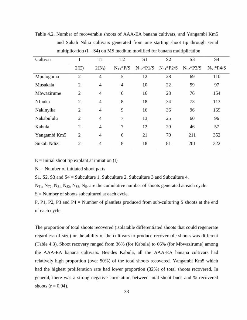
33
Table 4.2. Number of recoverable shoots of AAA-EA banana cultivars, and Yangambi Km5
and Sukali Ndizi cultivars generated from one starting shoot tip through serial
multiplication (I – S4) on MS medium modified for banana multiplication
Cultivar I T1 T2 S1 S2 S3 S4
2(E) 2(NI) NT1*P/S NT2*P1/S NS1*P2/S NS2*P3/S NS3*P4/S
Mpologoma 2 4 5 12 28 69 110
Musakala 2 4 4 10 22 59 97
Mbwazirume 2 4 6 16 28 76 154
Nfuuka 2 4 8 18 34 73 113
Nakinyika 2 4 9 16 36 96 169
Nakabululu 2 4 7 13 25 60 96
Kabula 2 4 7 12 20 46 57
Yangambi Km5 2 4 6 21 70 211 352
Sukali Ndizi 2 4 8 18 81 201 322
E = Initial shoot tip explant at initiation (I)
NI = Number of initiated shoot parts
S1, S2, S3 and S4 = Subculture 1, Subculture 2, Subculture 3 and Subculture 4.
NT1, NT2, NS1, NS2, NS3, NS4 are the cumulative number of shoots generated at each cycle.
S = Number of shoots subcultured at each cycle.
P, P1, P2, P3 and P4 = Number of plantlets produced from sub-culturing S shoots at the end
of each cycle.
The proportion of total shoots recovered (isolatable differentiated shoots that could regenerate
regardless of size) or the ability of the cultivars to produce recoverable shoots was different
(Table 4.3). Shoot recovery ranged from 36% (for Kabula) to 66% (for Mbwazirume) among
the AAA-EA banana cultivars. Besides Kabula, all the AAA-EA banana cultivars had
relatively high proportion (over 50%) of the total shoots recovered. Yangambi Km5 which
had the highest proliferation rate had lower proportion (32%) of total shoots recovered. In
general, there was a strong negative correlation between total shoot buds and % recovered
shoots (r = 0.94).

34
Table 4.3. Multiplication rate for total shoot buds and recovered shoots of different cultivars
after four subculture cycles on MS medium modified for banana multiplication
showing their effectiveness in generating plantlets
Cultivar Total shoot buds Recovered shoots Recovered shoots (%)
Mpologoma 4.14 2.20 53
Musakala 3.83 2.26 59
Mbwazirume 3.45 2.29 66
Nfuuka 3.54 1.96 55
Nakinyika 3.80 2.11 56
Nakabululu 3.48 1.95 56
Kabula 4.83 1.73 36
Yangambi KM5 9.04 2.88 32
Sukali Ndizi 5.18 2.71 52
LSD (0.05) 0.617
The morphology and shoot growth characteristics of the cultivars studied were conspicuously
different (Figure 4.1). For instance Yangambi Km5 and Nakinyika cultures had clustered
shoots accompanied with many leafy buds while Sukali Ndizi shoots were giant in stature
with moderate number of buds. The cultures of cv. Mbwazirume and other AAA-EA banana
cultivars had dominant and pointed shoots.

35
Figure 4.1 Characteristic shoot proliferation of banana cultivars on MS medium modified for
banana multiplication: A) Yangambi Km5, B) Sukali Ndizi, C) Nakinyika, and D)
Mbwazirume at subculture 1 on MS medium modified for banana multiplication.
4.3.3 Predictive model for recoverable shoots
The rates of multiplication of recoverable shoots produced from one shoot tip explant of the
cultivars were significantly different as exhibited by the linear regression coefficients (Table
4.4). The AAA-EA banana cultivars had lower multiplication rate of recoverable shoots as
compared to Yangambi Km5 and Sukali Ndizi. Among the AAA-EA banana cultivars
Nakinyika, Mbwazirume and Nfuuka had higher multiplication rates followed by
Mpologoma, Nakabululu, and Musakala. Kabula had the lowest multiplication rate of
recovered shoots. All the regression lines (not shown) for recoverable shoots had high
coefficients of determination (R2
> 90) and thus were used to derive predictive models for the
respective cultivars (Table 4.4). Since the AAA-EA banana cultivars had distinctly lower
regression coefficients, their LSD was calculated without including the control cultivars
which had higher and similar regression coefficients.

36
Table 4.4. Predictive model for recovered shoots of selected AAA-EA banana cultivars,
Yangambi Km5 and Sukali Ndizi generated on MS medium modified for banana
multiplication
Cultivar Genotype Predictive model Regression
coefficient
Nakinyika AAA-EA Y = e[0.289(x-1) – 0.00386(x-1)² +1.39]
0.289
Mbwazirume AAA-EA Y = e[0.276(x-1) – 0.00386(x-1)² +1.39]
0.276
Nfuuka AAA-EA Y = e[0.270(x-1) – 0.00386(x-1)² +1.39]
0.270
Mpologoma AAA-EA Y = e[0.261(x-1) – 0.00386(x-1)² +1.39]
0.261
Nakabululu AAA-EA Y = e[0.253(x-1) – 0.00386(x-1)² +1.39]
0.253
Musakala AAA-EA Y = e[0.248(x-1) – 0.00386(x-1)² +1.39]
0.248
Kabula AAA-EA Y = e[0.227(x-1) – 0.00386(x-1)² +1.39]
0.227
LSD (0.05) 0.0354
Yangambi Km5 AAA Y = e[0.352(x-1) – 0.00463(x-1)² +1.39]
0.352
Sukali Ndizi AAB Y = e[0.350(x-1) – 0.00463(x-1)² +1.39]
0.350
y = transformed number of recoverable shoots, Y = number of recoverable shoots,
e = 2.718282 and x = time in weeks.
4.4 Discussion
4.4.1 In vitro shoot and bud proliferation
The low in-vitro proliferation ability of AAA-EA banana cultivars and lack of significant
differences in proliferation among them was reported earlier (Talengera et al., 1994) and thus
was not surprising. Besides, this banana group has been reported to be less responsive to
regeneration protocols (Sadik et al., 2007) compared to other genotype groups. But the
outstanding proliferation rates of Yangambi Km5 (with an AAA genome) and Sukali Ndizi
(with an AAB genome) may be of interest to researchers. Conventionally cultivars having
only A in their genome have been reported to produce fewer shoots and buds compared to
those cultivars having one or two Bs in their genome (Strosse et al., 2003). It is also reported
that the origin of the A genome among the AAA-EA bananas is different from that of other
commonly grown banana cultivars with A genome (Simmonds and Shepherd, 1955). This

37
could suggest that the A genome of Musa acuminata Colla in the AAA-EA banana is less
responsive to in vitro proliferation using the available culture media formulations and
protocols.
Although cv. Nakabululu did not perform so well after producing a significantly higher
number of buds in subculture 1, it was generally observed that high bud proliferation
especially during the early subculture cycles influenced subsequent proliferation of the
cultures of all the cultivars and consequently their overall proliferation ability (Table 1; Table
2). Buds are precursors for shoot development and since they are not fully differentiated, it
was likely that they continued to divide when exposed to a fresh medium during sub
culturing. This implies that their decrease in number with subculture cycle indicated an
inherent overall decline in proliferation rate with increase in subculture cycle or age of the
culture. Formation of meristems and their subsequent development is controlled by
endogeneous auxins within the apixes whose decline with age triggers differention of buds
into shoots and terminates further bud development (George et al., 2008). However, as the
mean number of shoots was still increasing by the fourth subculture cycle, the process of sub-
culturing could be continued to produce numerous plants from a single explant. But abnormal
plants especially somaclonal variants might arise and/or increase to unacceptable numbers due
to onset of mutations. Considerable increase in frequency of variants has been reported in
bananas when maintained in tissue culture for over five months or 5 subculture cycles
(Rodigues et al., 1998; El-Dougdoug et al., 2007). The age of the culture and or number of
subculture cycles affect genetic stability due to spontaneous mutations which accumulate with
time (Duncan, 1997; Leela et al., 2003).
4.4.2 Recoverable shoots
There was a wide variation in ability of the cultivars to produce recoverable shoots.
Interestingly, the cultivars with high proliferation rate displayed less efficiency to produce
recoverable shoots (Table 4.3). This was associated with the number and morphology or
growth characteristics of their shoot cultures. For instance cv. Kabula produced numerous
stunted shoots which were difficult to isolate and subculture while cv. Mbwazirume had
moderate number of dominant and pointed shoots (Figure 4.1) which were easy to separate

38
and subculture. Yangambi Km5 produced numerous tiny differentiated shoots that were
difficult to recover and subculture compared to Sukali Ndizi with relatively fewer dominant
shoots. Since the explants were of uniform size, the variation in number of shoots among the
cultivars was probably due to differences in their response to culture conditions. Differences
in cultivar proliferation rate have been attributed to differences in growth regulator uptake,
translocation rate to meristematic regions, and metabolic processes among genotypes
(Blakesley, 1991; Youmbi et al., 2006).
The above observations imply that in vitro proliferation ability and particularly efficiency to
produce recoverable shoots of the different AAA-EA banana cultivars could be improved by
varying the culture conditions during the subsequent subculture cycles. Cultivars with high
proportion of buds to differentiated shoots and/or numerous tiny shoots require enhanced
shoot elongation and dominance. This could be achieved by reducing the amount of cytokinin
and other growth inducing substances in the medium in the subsequent subculture cycles
while adding gibberellins especially GA3 which enhances growth and elongation of dwarfed
or stunted banana plantlets (Damasco et el., 1996; Sanna et al., 2008), a phenomenon which
has also been demonstrated in common bean (Veltcheva and Svetleva, 2005) and cotton
(Divya et al., 2008).
On the other hand, those cultivars with low proportion of buds to differentiated shoots require
enhanced bud proliferation which could be achieved by increasing the amount of cytokinin
and adding other growth inducing substances such as abscisic acid in the subsequent
subculture cycles (Kantharajah, 2001). Application of more active cytokinins with carry-over
effect such as Thidiazuron (TDZ) sustained bud proliferation in bananas for some time
(Makara et al., 2010). Cytokinins produce delayed response in undifferentiated tissue (Mineo,
1990) which favors formation of shoot buds (Razdan, 1993).
4.4.3 Predictive model for recoverable shoots
In general the study suggested that in vitro banana proliferation and ability to recover
weanable shoots from shoot buds of AAA-EA bananas do not depend on the cultivar‟s
suckering ability in vivo. The number of recoverable shoots in vitro was influenced by the
cultivar‟s shoot growth characteristics. Thus a model for estimating number of plants that

39
could be produced from a shoot-tip explant after a given time would be useful to those
involved in micropropagation of AAA-EA banana cultivars. This study derived a regression
model that could be used to predict the number of recoverable plantlets from one shoot-tip
explant of AAA-EA banana cultivars after a given time period. For example through
calculation using the model, the number of shoots that could be recovered from a shoot-tip
explant of cv. Nakinyika after 18 weeks (Y) = e[0.289(x-1) – 0.00386(x-1)² +1.39]
= 178. The number
recorded from the experiment was 169 (Table 4.2). For cv. Mbwazirume (Y) = e[0.276(x-1) –
0.00386(x-1)² +1.39] = 142. The one obtained from the experiment was 154 (Table 4.2). The
difference was attributed to changes in shoot growth characteristics of each cultivar. For any
given AAA-EA cultivar, the linear regression coeffients above would be used depending on
its clone set. Regression models have been used to predict plant multiplication rates (Mendes
et al., 1999) and thus the model could be used to give a fair estimate. But the underlining
principle while developing the prediction model was that bananas may be safely sub-cultured
up to the 4-6th
subculture cycle corresponding to 4.5 - 6.5 months. Thus care must be taken
when predicting beyond this period as there may be increased changes in proliferation or
growth pattern depending on cultivar.
4.5 Conclusion
In vitro shoot proliferation in East African highland bananas on the widely adopted MS
medium modified for banana multiplication varies among the cultivars. The cultivars that
inherently displayed high shoot bud proliferation recorded low % recovered shoots (which
limited their potential to produce much more shoots) suggesting that in vitro proliferation is
cultivar dependent while the recovery of shoots from shoot buds of AAA-EA bananas is
cultivar independent. Based on shoot culture growth characteristics, the number of
recoverable shoots from one shoot-tip explant in a given time can be predicted using a
regression model. Using the predictive model, the number of recoverable shoots from one
shoot-tip explant for instance of cv. Mpologoma at 5.5 months (5 subculture cycles) and 6.5
months (6 subculture cycles) would be 176 and 245, respectively. This implies that in practice
one would require 7 starting shoot-tip explants to subculture for 5.5 months to generate
enough planting material to plant one hectare at spacing of 9 by 9 metres.

40
CHAPTER FIVE
EFFICACY OF TDZ AND 4-CPPU FOR EMBRYOGENIC CALLUS INDUCTION
FROM SCALPS OF RECALCITRANT EAST AFRICAN HIGHLAND BANANA
5.1 Introduction
In vitro plant cell differentiation and morphogenesis is highly dependent on culture medium
(Khaleda and Al-Forkan, 2006). Optimum proportion and/or concentration of growth
regulators especially auxins is necessary for successful induction and maintenance of
embryogenic callus or cell suspension cultures (Gomez et al., 2000). N-phenyl-N'-1,2,3-
thidiazol-5-ylurea (thidiazuron) (TDZ) and N-(2-chloro-4-pyridyl)-N'-phenylurea
(forchlorfenuron) (4-CPPU), both substituted phenylurea compounds, are reported to be
highly active in regulating morphogenesis in the tissue culture of many plant species (Victor
et al., 2004; Fiore et al., 2004; Haruki et al., 2007). They are known to exhibit a unique
property of mimicking both cytokinin and auxin effects on growth and differentiation of
cultured explants (Murthy et al., 1998). At low concentrations TDZ and 4-CPPU stimulate
shoot proliferation, simulating cytokinin activity while at high concentrations they induce
callus, typical of an auxin activity (Vinayak et al., 2009). Their effect seems to be distinctive
for each respective crop variety, suggesting that they could be suitable for determining variety
specific regeneration protocols (Vinayak et al., 2009). The aim of this study, therefore, was to
test the efficacy of a combination of TDZ and 4-CPPU growth regulators for enhancing
embryogenic callus induction in recalcitrant AAA-EA bananas through scalps.
5.2 Materials and Methods
5.2.1 Plant material
Five AAA-EA banana cultivars namely Mbwazirume, Mpologoma, Nakabululu, Nakinyika
and Nfuuka from different clone sets were selected for this study based on farmers‟ variety
preference. Shoot-tip explants were extracted from young suckers 40-100 cm tall from a field
at Makerere University Agricultural Research Institute, Kabanyolo (MUARIK). They were
sterilized and cultured on the Murashige and Skoog (MS) medium modified for banana

41
multiplication (Talengera et al., 1994) up to transfer 2 to produce adequate clean shoots for
generating scalps.
5.2.2 Generation of scalps
In vitro derived shoot-tips from transfer 2 were put on the MS medium modified with various
concentrations of TDZ and 4-CPPU growth regulators to generate scalps as designated below:
TDZ and 4-CPPU combination Designation
5µM TDZ + 5µM 4-CPPU M1
7µM TDZ + 7µM 4-CPPU M2
9µM TDZ + 9µM 4-CPPU M3
11µM TDZ + 11µM 4-CPPU M4
13µMTDZ + 13µM 4-CPPU M5
26µM TDZ M6
26µM 4-CPPU M7
These rates were selected based on different studies on effect of TDZ in banana (Sadik et al.,
2007) and other crops (Ying-Chun et al., 2000).
Fifty shoot-tips of each cultivar were used in each hormone treatment and placed in a
completely randomized design (CRD) in a dark growth room maintained at 26 ± 2°C. The
cultures were serially subcultured every four weeks until scalps were formed. The rate of
scalp formation in the various media was determined by counting the number of shoots and
buds in each subculture cycle until scalps were formed. Callus directly formed on the scalps
being generated was also recorded. Other growth features or characteristics such as corm-like
growth when observed were noted.
5.2.3 Callus induction and embryo development
The scalps were excised under dissecting microscope and cultured on callus induction
medium derived by combining 5µM 2,4-D (2,4-Dichlorophenoxyacetic acid) (Schoofs et al.,
1997) with TDZ and 4-CPPU singly and in combination at 5µM, 10µM and 15µM as

42
hormone treatment. Fifty scalps of each cultivar were used in each treatment. All the cultures
were placed in a CRD in a dark growth room maintained at 26 ± 2°C. They were observed
every two weeks for four months to observe embryogenic responses. Any embryogenic callus
achieved was transferred to the MS banana multiplication medium supplemented with 5µM
benzyl amino purine (BAP). Embryo to plant development was observed for at least 28 days.
The plantlets regenerated together with the number of normal roots (with hair roots) they
possessed were recorded.
5.2.4 Data analysis
The data on rate of scalp formation was subjected to analysis of variance (ANOVA) using
GenStat 13th
Edition (GenStat, 2010). The means were separated using least significant
differences (LSD) at 5% level of significance. Comparisons were done on number of shoots
and multiple buds across cultivars, subculture cycles and growth regulator combination.
Callus directly formed on the scalp induction media was summarized as percentage of
explants or cultures with callus on the scalps being formed. Induced callus was summarized as
a percentage of scalps that generated callus to the total number of scalps initiated on callus
induction medium.
5.3 Results
5.3.1 Scalp formation
Scalp formation in all the cultivars varied across hormone treatment and subculture cycle.
There were significant differences in mean shoot and bud proliferation on the different
combinations of TDZ and 4-CPPU (Table 5.1). At subculture 1, medium M5 (13µM
TDZ+13µM 4-CPPU) induced significantly (P < 0.05) more multiple buds with fewer shoots
in cultivars Nakinyika, Nakabululu and Nfuuka, which favors scalp formation. Medium M1
(5µM TDZ+5µM 4-CPPU) induced significantly (P < 0.05) higher number of multiple buds
in cv. Mpologoma but it induced high shoot proliferation which does not favor scalp
formation. However, at subculture 2, treatment M1 (5µM TDZ+5µM 4-CPPU) induced
significantly (P < 0.05) higher number of multiple buds in cultivars Mbwazirume, Nakinyika

43
and Nakabululu while cv. Nfuuka continued to produce significantly (P < 0.05) higher
numbers of multiple buds in Treatment M5 (13µM TDZ+13µM 4-CPPU).
There was significant increase in mean multiple bud proliferation from subculture 1 to
subculture 3 in all the cultivars but at varying levels depending on hormone treatment (Table
5.1). In cycle 3, cv. Nakinyika had significantly (P < 0.05) the highest mean multiple bud
proliferation on medium M6 (26 µM TDZ), followed by Nakabululu on M3 (9µM TDZ+9µM
4-CPPU) and Mbwazirume on M5 (13µM TDZ+13µM 4-CPPU). Cultivars Mpologoma and
Nfuuka produced the same number of multiple buds on medium M4 (11µM TDZ+11µM 4-
CPPU) and M7 (26 µM 4-CPPU), respectively.
Scalps were achieved in all TDZ and 4-CPPU combinations (Table 5.2). With exception of
cultivars Mpologoma and Mbwazirume, scalps were formed in over 50% of the cultures of the
various cultivars by the fourth subculture cycle. Mpologoma produced fewer (≤ 50%) scalps
in most of the media while Mbwazirume produced fewer scalps (37.5%) only in medium M7
(26 µM 4-CPPU). Cultivars Nakinyika and Nakabululu produced high number of scalps (≥
70%) in all the media or treatments.

44
Table 5.1. Cultivar shoot and multiple bud proliferation on different combinations of TDZ
and 4-CPPU (M1, M2, M3, M4, M5, M6 and M7) at first, second and third
subculture cycles
Cycle Cultivar Organ Mean number of shoots and multiple buds LSD
(0.05) M1 M2 M3 M4 M5 M6 M7
1 Mbwazirume Shoots 1.5 1.0 1.3 1.4 1.5 1.6 1.1 0.244
M. buds 3.9 1.6 2.8 2.9 3.2 2.7 2.4 0.626
Mpologoma Shoots 1.4 1.3 0.8 1.5 1.4 1.1 1.4 0.244
M. buds 1.3 2.0 1.4 1.7 1.4 0.9 0.8 0.626
Nakabululu Shoots 1.3 1.7 1.1 1.3 1.2 2.4 1.5 0.244
M. buds 3.0 3.6 5.3 4.2 6.5 2.1 4.5 0.626
Nakinyika Shoots 2.0 1.4 1.1 0.9 0.8 2.4 1.4 0.244
M. buds 3.2 2.0 4.0 4.3 7.8 2.4 4.2 0.626
Nfuuka Shoots 1.9 1.4 0.9 0.7 0.3 1.1 2.2 0.244
M. buds 1.5 3.2 3.1 4.0 4.3 2.9 1.9 0.626
2 Mbwazirume Shoots 0.3 0.8 0.6 0.7 0.4 0.6 0.6 0.244
M. buds 5.7 4.2 4.2 3.6 3.7 2.1 2.1 0.626
Mpologoma Shoots 0.7 0.6 0.5 0.6 0.0 0.6 1.4 0.244
M. buds 2.3 3.1 2.7 2.1 2.6 2.0 1.0 0.626
Nakabululu Shoots 0.7 1.0 1.1 0.8 1.2 1.6 1.8 0.244
M. buds 4.6 2.2 3.6 2.2 4.0 3.1 3.9 0.626
Nakinyika Shoots 0.3 0.3 0.3 0.5 0.4 1.8 1.3 0.244
M. buds 5.3 2.0 3.5 2.9 4.5 2.2 3.7 0.626
Nfuuka Shoots 0.7 1.0 0.5 0.4 0.4 1.0 1.3 0.244
M. buds 3.3 3.9 3.2 4.5 5.2 3.6 2.4 0.626
3 Mbwazirume Shoots 1.7 2.3 0.7 1.0 0.6 0.9 2.0 0.244
M. buds 5.8 6.2 4.4 8.8 9.1 8.6 6.9 0.626
Mpologoma Shoots 2.4 2.2 0.7 1.5 1.7 1.4 0.6 0.244
M. buds 7.2 5.0 6.8 8.3 4.9 4.5 6.1 0.626
Nakabululu Shoots 1.4 0.8 0.7 1.6 1.4 0.8 1.4 0.244
M. buds 8.6 6.6 9.3 8.5 8.6 8.5 8.4 0.626
Nakinyika Shoots 1.1 1.5 0.2 0.6 0.7 0.7 2.5 0.244
M. buds 8.3 6.9 8.5 8.4 8.2 9.4 9.8 0.626
Nfuuka Shoots 1.4 1.3 1.4 1.1 0.3 1.0 1.0 0.244
M. buds 8.2 7.8 7.6 7.7 7.3 7.2 8.3 0.626
Cycle LSD(0.05) = 0.160 for shoot and 0.410 for multiple buds
Cultivar LSD(0.05) = 0.206 for shoot and 0.529 for multiple buds
M. buds= multiple buds.

45
Table 5.2. Cultivar percentage ideal scalps on the different combinations of TDZ and 4-CPPU
at the fourth subculture cycle
Cultivar Percentage ideal scalps under different media treatment
M1 M2 M3 M4 M5 M6 M7
Mbwazirume 75.0 60.0 60.0 70.0 70.0 66.7 37.5
Mpologoma 47.1 40.0 50.0 70.0 60.0 50.0 31.3
Nakabulu 70.6 75.0 82.4 72.2 71.4 75.0 87.5
Nakinyika 88.2 60.0 70.0 70.0 70.0 100.0 100.0
Nfuuka 76.5 82.4 73.3 94.1 69.2 50.0 50.0
5.3.2 Callus and embryo development
As the scalps formed on the media containing various combinations of TDZ and 4-CPPU,
compact yellowish, pinkish and cream callus tissue developed on them (Figure 5.1). The
percentage of scalp cultures that had callus on them varied with hormone treatment and
cultivar (Table 5.3). By cycle 4 (achieved through serial transfer an explant from each of the
primary explants), all the cultivars produced callus on their scalp cultures even on the medium
containing the lowest concentration of TDZ and 4-CPPU (5µM TDZ + 5µM 4-CPPU).
Cultivar Nakinyika had the highest percentage of cultures with callus (20%) on medium M5
(13µM TDZ + 13µM 4-CPPU) and M6 (26 µM TDZ), Mpologoma (20%) on M6 (26 µM
TDZ), Nfuuka (17.5%) on M1 (5µM TDZ + 5µM 4-CPPU), Nakabululu (17.5%) on M7 (26
µM 4-CPPU) and Mbwazirume (12.5%) on M6 (26 µM TDZ). All the cultivars performed
well on MS supplemented with 26 µM TDZ with up to 12.5 - 20% of their scalp cultures
having callus on them by cycle 4 (Table 5.3).

46
Figure 5.1 Embryogenic callus formation on scalps of cultivar Nfuuka on 9µM TDZ + 9µM
4-CPPU at the third subculture cycle: (A) Ideal scalp, (B) Callus initiation, (C)
Embryo development (D) Secondary embryo development.
Table 5.3. Mean percentage callus formation on scalp cultures of AAA-EA banana cultivars
on different combinations of TDZ and CPPU by the fourth subculture cycle
Cultivar Mean percentage callus under different media treatment
M1 M2 M3 M4 M5 M6 M7
Mbwazirume 10.0 10.0 2.5 5.0 10.0 12.5 2.5
Mpologoma 2.5 5.0 2.5 10.0 17.5 20.0 2.5
Nakabululu 2.5 5.0 10.0 10.0 10.0 15.0 17.5
Nakinyika 7.5 5.0 7.5 7.5 20.0 20.0 17.5
Nfuuka 17.5 12.5 7.5 10.0 5.0 15.0 2.5
Treatment LSD (0.05) = 6.905; Cultivar LSD (0.05) = 5.836
As the callus on scalps aged and became necrotic by cycle 4, some embryos developed on it
and differentiated directly into shoot (Figure 5.2). This was observed in cv. Nakabululu on
13µM TDZ+13µM 4-CPPU. When the callus from the different cultivars was removed and
cultured on a hormone free medium, it differentiated into somatic embryos and shoots (Figure
5.3) regardless of initial hormone treatment implying that it was all homogeneously
embryogenic.

47
Figure 5.2 Direct embryogenesis in cv. Nakabululu on 13µM TDZ+13µM 4-CPPU: (A)
Callus on scalp, (B) Embryo initiation on callus, (C – D) embryo to shoot
development.
Figure 5.3 Differentiation of callus on scalps of cv. Nakinyika into somatic embryos and
shoots on transfer to hormone free MS medium: (A) Callus on scalp (B) Embryo
development, (C) Callus differentiation into shoot (E) Scalp tissue differentiation
into shoot.
When the callus from the different cultivars was removed and transferred to MS medium
containing 5µM BAP, it only regenerated into plantlets (Figure 5.4) in a few cultures of
cultivars Nakinyika, Nakabululu, Nfuuka and Mbwazirume (Table 5.4). Of the plantlets
regenerated 5 out of 12 and 4 out of 5 plantlets of cultivars Nakinyika and Mbwazirume,
respectively, developed normal roots (Table 5.4) and were weaned (Figure 5.5). But when the
scalps were excised and cultured on a medium with 5µM 2,4-D to generate callus they all
died.

48
Figure 5.4 Different stages of plant regeneration through direct embryogenesis from scalps of
cv. Mbwazirume after transfer of embryogenic callus to MS medium with 5µM
BAP: (A) Embryogenic callus development, (B) Somatic embryo development, (C
– D) embryo to plant development, (E) Root development on the plantlets under
the same medium.
Table 5.4 Number of plantlets and their roots of various AAA-EA banana cultivars
regenerated out of twenty (20) embryogenic callus cultures induced by TDZ and
4-CPPU growth regulators
Cultivar Number of plantlets
regenerated
Number of plantlets with
roots
Mean root number
Mbwazirume 5 4 6.0
Mpologoma 0 0 0
Nakabulu 10 1 2.0
Nakinyika 12 5 5.4
Nfuuka 2 0 0

49
Figure 5.5 Plants of cvs Nakinyika and Mbwazirume regenerated through direct
embryogenesis induced by TDZ and 4-CPPU after transfer of the embryogenic
callus to MS medium with 5µM BAP under acclimatization in soil.
5.4 Discussion
5.4.1 Scalp formation
Cultivars Nakinyika and Nakabululu produced more multiple buds on treatment M5 (13µM
TDZ+13µM 4-CPPU) at subculture 1 but at subculture 2, they produced more multiple buds
on treatment M1 (5µM TDZ+5µM 4-CPPU). This was probably due to changes in effect of
the growth regulators with subculture cycle especially TDZ which was reported to have carry-
over effect (Makara et al., 2010). Concentrations of TDZ and 4-CPPU above 7 µM appear to
suppress shoot apical dominance and favor multiple bud proliferation. However, in cv.
Mbwazirume, mean bud proliferation on 5µM TDZ + 5µM 4-CPPU was significantly higher
than on 7µM TDZ + 7µM 4-CPPU suggesting that combined TDZ and 4-CPPU at low
concentration permits vigorous proliferation of both shoots and buds depending on cultivar.
TDZ is active in inducing adventitious and axillary shoot proliferation (Ashraf et al., 2004;
Youmbi et al., 2006) which are precursors for scalp formation. Similarly, 4-CPPU induced
multiple axillary shoot proliferation in Impatiens walleriana L. (Subotić et al., 2008).

50
Multiple bud proliferation was significantly (P < 0.05) higher on 13µM TDZ+13µM 4-CPPU
than on 26 µM TDZ and 4-CPPU when used individually. The interaction of TDZ and 4-
CPPU to induce more multiple bud proliferation when used in combination at lower
concentration (13 µM) compared to when used singly at concentration of 26 µM suggests
both synergistic and additive effects and implies that high levels of each of them can be
avoided. Achievement of plant regeneration at lower concentrations of combined TDZ and 4-
CPPU could be advantageous in reducing somaclonal variation. In bananas, somaclonal
variations of different types have occurred due to high concentrations of growth hormones
such as 6-benzylaminopurine and dicamba (Leela et al., 2003).
5.4.2 Callus development and embryogenesis
Embryogenic callus was directly formed on the scalp induction medium containing TDZ and
4-CPPU. Commonly, embryogenesis is induced after inoculating the ideal scalps onto
medium containing 2,4-D or other auxins (Schoofs et al., 1998; Sadik et al., 2007; Ramírez-
Villalobos and De García. 2008). The formation of embryogenic callus under TDZ and 4-
CPPU treatments as described in this study conforms to reports that at high concentrations,
these urea-type cytokinins exhibit an auxin activity and thus could induce embryogenesis
(Vinayak et al., 2009). Chhabra et al. (2008) suggested that at high concentrations, TDZ
imposes stress on the explant and could redirect its development from shoot organogenesis to
somatic embryogenesis. At concentrations higher than 2μM, TDZ induced direct somatic
embryogenesis on cotyledonary node explants of recalcitrant lentil (Chhabra et al., 2008).
Earlier, TDZ was reported to have induced higher somatic embryogenesis in protocalli than
even 2,4-D (Lehminger-Mertens and Jacobsen, 1989).
Although TDZ and 4-CPPU exhibit an auxin activity and induce somatic embryos in cultures
grown on media devoid of auxins, the mechanism of these urea-type cytokinins to induce
embryogenic callus in bananas are of interest as earlier reports suggested that somatic
embryogenesis could be induced in dicot species on cytokinin-supplemented media
(Raemakers et al., 1995) but auxins solely mediated the transition from somatic to
embryogenic cells in angiosperm monocots (Víctor, 2001). TDZ induced somatic
embryogenesis in rice but after a short treatment with 2,4-D (Aparna and Rashid, 2004). This

51
implies further research on whether auxins solely mediate transition from somatic to
embryogenic cells in angiosperm monocots is also necessary.
Cultivar Nfuuka formed callus on medium M1 with low levels of TDZ and 4-CPPU. Relating
to the name „Nfuuka‟ „„meaning ever changing‟‟in one of the dialects in Central Uganda, the
response of cv. Nfuka suggested that it is mutatic and may on all occasions respond to a wider
range of treatments than other AAA-EA banana cultivars. However, the increase in callus
formation on low levels of TDZ and 4-CPPU with subculture cycle was attributed to carry-
over effect of urea-type cytokinins especially TDZ (Makara et al., 2010).
The differentiation of callus on scalps generated on TDZ and 4-CPPU into somatic embryos
and shoots is in agreement with other reports. Razdan (1993) reported that a whole plant
regeneration from callus tissue may occur either through organogenesis (shoot-bud
differentiation) or somatic embryogenesis where the cell or group of cells initiate a
development-pathway that leads to reproducible regeneration of non-zygotic embryos capable
of germinating to form complete plants. There was more embryo development when the
callus was removed and cultured on a hormone free medium probably due to reduction in
auxin activity resulting from the high concentrations of TDZ and 4-CPPU. Though the
process of somatic embryogenesis is often initiated in media containing high levels of auxins,
later reduction in auxin concentration usually stimulates further embryo development since
lower auxin concentrations induce pro-embryogenic cells to divide and develop into embryos
(Sharp et al., 1980).
The scalps formed on media containing TDZ and 4-CPPU died when transferred on to
medium containing 5μM 2,4-D possibly due to excessive cumulative auxin activity effect
(George et al., 2008). Since urea-type cytokinins especially TDZ have carry-over effect
(Makara et al., 2010), their auxin activity together with that of 2,4-D could have been too high
for the stimulation of the cells in the scalp tissue to dedifferentiate and form callus but rather
killed them. However, somatic embryos were produced from stem explants of Golden Pothos
(Epipremnum aureum) on MS medium supplemented with 0.5 mgl-1
(2.26 μM) 2,4-D and 2
mgl-1
(8.07 μM) CPPU, and 0.5 mgl-1
(2.26 μM) 2,4-D and 2.0 mgl-1
(9.08 μM) TDZ (Zhang

52
et al., 2004). This suggests that lower concentration of 2,4-D is required for scalps generated
on media containing TDZ.
Among the explants that have been used in somatic embryogenesis in bananas such as
immature zygotic embryos, immature male flowers, female flowers, scalps, leaf bases and
corm slices, scalps were reported to be among the most responsive material for initiating
regenerable embryogenic cell suspension cultures (Meenakshi et al., 2007). One impediment
to using the scalp method to generate callus in AAA-EA bananas has been the long time
required to achieve ideal scalps. Up to 9 subculture cycles on a medium containing 100 µM
BAP (Schoofs et al., 1998) or 6 subculture cycles on a medium containing 10 µM TDZ (Sadik
et al., 2007) were required. Recently Ramírez-Villalobos and De García (2008) reduced the
production time of embryogenic scalps from tetraploid somaclonal variant CIEN-BTA-03 of
the Cavendish subgroup from 8-14 months (reported for the other tetraploids of this subgroup
(Strosse et al. 2006)) to 6 months by growing shoot apexes in 25 mgl-1
BA plus 0.217 mgl-1
IAA but the mean percentage of explants with scalps was only 38%. Though tetraploid
bananas generally take longer to develop than triploids, in this study, scalps of AAA-EA
bananas were achieved in over 50% of the scalp cultures within 4 subculture cycles on TDZ
and 4-CPPU. The influence of TDZ on proliferation in AAA-EA bananas had been reported
(Arinaitwe et al., 2000; Sadik et al., 2007; Makara et al., 2010). This is the first report on
combined effect of TDZ and CPPU on scalps and regenerable embryogenic callus induction
in AAA-EA bananas.
5.5 Conclusion
Embryogenic callus was formed on 2.5 – 20% of the scalp cultures depending on cultivar and
interaction of TDZ and 4-CPPU combination. Cultivars Nakinyika, Nakabululu, Mbwazirume
and Nfuuka developed regenerable embryogenic callus and recorded plant regeneration
through direct embryogenesis. Early plant regeneration at lower concentrations of combined
TDZ and 4-CPPU could reduce occurrence of somaclonal variation. These results thus
suggest that the percentage of embryogenic callus induced from scalps of AAA-EA banana
cultivars is increased by TDZ and 4-CPPU growth regulators.

53
CHAPTER SIX
EFFICACY OF TDZ AND 4-CPPU IN DIFFERENT SALT FORMULATIONS IN
EMBRYOGENIC CALLUS INDUCTION FROM IMMATURE MALE FLOWERS OF
AAA-EA BANANA
6.1 Introduction
Culture medium nutrient components and growth regulator combination greatly influence
plant cell differentiation and morphogenesis in vitro (Kantharajah, 2001). Auxins, particularly
2,4-dichlorophenoxyacetic acid (2,4-D) have been widely used in combination with
cytokinins such as Benzyl adenine (BA) (Sannasgala et al., 1992), Zeatin (Schoofs et al.,
1998; Ramírez-Villalobos and De García, 2008) and Benzyl amino purine (BAP) (Sadik et
al., 2007) for initiation and maintenance of embryogenic cell suspension (ECS) in banana.
Whereas urea-type cytokinins, N-phenyl-N‟-1,2,3-thidiazol-5-ylurea (thidiazuron) (TDZ) and
N-(2-chloro-4-pyridyl)-N‟-phenylurea (forchlorfenuron) (4-CPPU) have desirable attributes
for embryogenic callus induction from plant explants (Murthy et al., 1998; Haruki et al.,
2007), they have not been tried for induction of somatic embryogenesis in banana especially
through immature male flower buds. On the other hand a number of salt formulations were
described by different authors including MS (Murashige and Skoog, 1962), Chu N6 (Chu,
1975), Eriksson (Eriksson, 1965), Gamborg B5 (Gamborg et al., 1968), Nitsch (Nitsch and
Nitsch, 1969), NLN (Lichter, 1982), SH (Schenk and Hilderbrandt, 1972) and White (White,
1963) for use in tissue culture. During the establishment of in vitro micro-propagation
procedure for the AAA-EA banana, only a few salt formulations were evaluated (Talengera et
al., 1994). Although MS medium was adopted for routine micropropagation of the AAA-EA
banana through shoot tip culture, there has been no justification that its salt mixture is also the
best for callus induction or ECS development. The aim of this study, therefore, was to test
efficacy of TDZ and 4-CPPU, and different salt formulations for embryogenic callus
induction in AAA-EA banana cultivars through immature male flowers.

54
6.2 Materials and Methods
6.2.1 Plant material
Five AAA-EA banana cultivars namely Mbwazirume, Mpologoma, Nakabululu, Nakinyika
and Nfuuka from different clone sets were used in this study. Sukali Ndizi (AAB) which is
responsive to regeneration protocols was included as a check. Male flowers were collected 1
to 3 weeks after flowering from the field banana germplasm at Makerere University
Agricultural Research Institute, Kabanyolo (MUARIK) and the banana fields of the National
Banana Program at National Agricultural Research Laboratories (NARL), Kawanda. They
were reduced to 0.8 – 2 cm within 6 hours after collection and the reduced immature male
flowers were surface sterilized in 70% ethyl alcohol for 2 minutes before they were initiated
within two hours.
6.2.2 Effect of TDZ and 4-CPPU on callus induction
Using a scalpel with fine blades, immature flower bud clusters were excised under dissection
microscope from the 18th
- 8th
position and cultured on callus induction medium containing
5µM 2,4-D supplemented with different combinations of TDZ and CPPU alone or in presence
of IAA and NAA as designated below:
TDZ and 4-CPPU combination Designation
5µM TDZ+5µM 4-CPPU H1
10µM TDZ+10µM 4-CPPU H2
15µM TDZ+15µM 4-CPPU H3
5µM TDZ+5µM 4-CPPU+1mgl-1
IAA+1mgl-1
NAA H4
10µM TDZ+10µM 4-CPPU+1 mgl-1
IAA+1mgl-1
NAA H5
In each treatment fifty male flowers of each cultivar were initiated on separate petridishes and
placed in a completely randomized design (CRD) in a dark growth room maintained at 26 ±
2°C. The cultures were observed every two weeks for 8 months. Yellow callus (YC) and
embryogenic callus (EC) developing from the flower buds was recorded. Any organ-like
structure developing on the callus or alongside embryos was noted.

55
6.2.3 Callus induction on different basal salt mixture substituted on callus induction
medium supplemented with TDZ and 4-CPPU
By substituting only MS inorganic salts (MS vitamins and other medium components
retained) from the callus induction medium supplemented with 10µM TDZ+10µM 4-CPPU
(the most efficacious combination in pure MS medium), salt formulations of Chu (N6),
Eriksson, Gamborg B5, Nitsch, NLN, SH and White were tested for embryogenic callus
induction in AAA-EA banana cultivars. Briefly immature male flower buds of two cultivars
Mpologoma and Nakinyika (selected based on their earlier contrasting responses and
availability of male flowers) were prepared as described above and cultured on semisolid
callus induction media containing the above salt formulations, full strength MS vitamins,
30gl-1
sugar, 5µM 2,4-D and 10µM TDZ+10µM 4-CPPU at pH 5.7. Fifty male flowers of
each cultivar were used in each salt mixture treatment and placed in a CRD in a dark growth
room maintained at 26 ± 2°C. The cultures were observed every two weeks for at least 8
months. The number of flower buds on each petridish that responded (grew or differentiated
into callus) was recorded. Yellow callus, embryogenic callus and organ-like structures
developing on the callus or alongside embryos were recorded. Any callus with embryos
achieved from both experiments was transferred onto the MS derived banana multiplication
medium described by Talengera et al. (1994) but hormone free and embryo to plant
development observed for two months.
6.2.4 Data analysis
The data on callus formation were summarized in percentages using Excel computer program.
Yellow callus response was presented as percentage of male flower explants that produced
yellow callus to the total number of male flowers initiated on callus induction medium.
Embryogenic callus response was expressed as proportion of callus cultures with embryos.
The data on number of immature male flower bud clusters that responded on the various basal
salt mixtures was subjected to ANOVA using GenStat 13th
Edition (GenStat, 2010). The
means were separated using least significant differences (LSD) at 5% level of significance.
Comparisons were done on number of flower bud clusters that responded across cultivars and
salt formulations.

56
6.3 Results
6.3.1 Effect of TDZ and 4-CPPU on callus induction
The response of the immature male flower buds of the different cultivars on the callus
induction medium containing various combinations of TDZ and 4-CPPU did not vary during
the first two months except some cultures of cv. Mpologoma on medium H1 (5µM
TDZ+5µM 4-CPPU) dried without forming yellow callus. From 3-8 months, there were
differences in response among the cultivars on the different growth regulator combinations
(Table 6.1). Between 3-4 months the cultures of Mpologoma and Mbwazirume showed
embryogenic response on H2 (10µM TDZ+10µM 4-CPPU) (formed a white translucent callus
with somatic embryos on its surface) of up to 4.62% and 5.72% respectively. The cultures of
other cultivars had not yet formed embryogenic callus. Between 5-6 months, Nakabululu and
Nakinyika showed embryogenic response of 4.76% on H3 (15µM TDZ+15µM 4-CPPU) and
5.26% on H5 (10µM TDZ+10µM 4-CPPU+10 mgl1-
IAA+10mgl1-
NAA) respectively. From
7-8 months, Nakabululu showed 22.22% embryogenic response on H2 (10µM TDZ+10µM 4-
CPPU) while Mpologoma and Nfuuka had 2.38% and 9.09% embryogenic response
respectively on H3 (15µM TDZ+15µM 4-CPPU). Most of the callus was white and
translucent and formed on primary yellow callus (Figure 6.1). About 5% of the embryogenic
calli had friable tissue. Most of the flower buds of Sukali Ndizi did not differentiate into
callus instead grew excessively into floral structures. A few flower buds of AAA-EA banana
cultivars also grew without differentiating into callus particulars on low TDZ and 4-CPPU
concentrations.

57
Table 6.1. Percentage yellow callus (%YC) and embryogenic callus (%EC) formed from
immature male flowers of different banana cultivars on callus induction medium
supplemented with various combinations of TDZ and 4-CPPU
Cultivar
Hormone
treatment
Period in months taken to induce embryos
3-4 months
%YC %EC
5-6 months
%YC %EC
7-8 months
%YC %EC
Mpologoma H1 66.7 0.0 23.3 0.0 3.3 0.0
H2 100.0 4.6 76.5 0.0 0.0 0.0
H3 100.0 0.0 42.9 0.0 23.8 2.4
H4 100.0 0.0 0.0 0.0 0.0 0.0
H5 96.8 3.2 32.3 0.0 0.0 0.0
Mbwazirume H1 67.8 1.7 65.5 1.7 0.0 0.0
H2 97.1 5.7 97.1 5.7 61.3 0.0
H3 100.0 0.0 100.0 0.0 4.4 0.0
H4 100.0 0.0 0.0 0.0 0.0 0.0
H5 100.0 0.0 0.0 0.0 0.0 0.0
Nfuuka H1 100.0 0.0 100.0 0.0 100.0 0.0
H2 100.0 0.0 92.9 0.0 42.9 0.0
H3 72.7 0.0 72.7 0.0 54.6 9.1
H4 0.0 0.0 0.0 0.0 0.0 0.0
H5 100.0 0.0 0.0 0.0 0.0 0.0
Nakabululu H1 100.0 0.0 70.0 0.0 70.0 0.0
H2 80.0 0.0 77.8 0.0 44.4 22.2
H3 100.0 0.0 95.2 4.8 90.5 9.5
H4 100.0 0.0 100.0 0.0 18.2 0.0
H5 0.0 0.0 0.0 0.0 0.0 0.0
Nakinyika H1 100.0 0.0 100.0 0.0 0.0 0.0
H2 100.0 0.0 69.2 0.0 0.0 0.0
H3 100.0 0.0 100.0 0.0 67.7 0.0
H4 34.2 0.0 0.0 0.0 0.0 0.0
H5 79.0 0.0 63.2 5.3 63.2 5.3
Sukali Ndzi H1 100.0 0.0 83.3 0.0 0.0 0.0
H2 100.0 0.0 0.0 0.0 0.0 0.0
H3 100.0 0.0 100.0 0.0 66.7 0.0
H4 100.0 0.0 90.9 0.0 0.0 0.0
H5 100.0 0.0 100.0 0.0 0.0 0.0

58
Figure 6.1 Different stages of callus development from immature male flower buds of cv.
Nfuuka on callus induction medium supplemented with 15µM TDZ+15µM 4-
CPPU: A) white translucent callus on primary yellow callus, B) white callus, C)
Callus with primary embryos, D) Embryogenic callus with friable tissue.
6.3.2 Callus induction on different basal salt mixture substituted on callus induction
medium supplemented with TDZ and 4-CPPU
In the first two months there were no differences in response of the cultivars on the various
salt mixtures except 5.88% of the cultures of cultivar Mpologoma formed callus with somatic
embryos on Gamborg B5 basal salt mixture which increased in 3-4th
months to 11.43% (Table
6.2). Between 3-4months, 8.33% and 3.33% of cultures of cultivar Nakinyika formed
embryos on Gamborg B5 and White basal salt mixtures respectively. The cultures of both
cultivars on Nitsch, NLN and SH salt mixtures dried within 4 months without forming any
somatic embryos. Those on Chu N6 and Eriksson salt mixtures dried in 6 months while those
on Gamborg B5 and White salt mixtures dried after 8 months. On pure MS medium cultures
of cultivar Mpologoma had 4.62% embryogenic response between 3-4 months and dried in 6
months while cultures of cv. Nakinyika dried after the same period without showing any
embryogenic response.

59
Table 6.2. Percentage yellow callus (%YC) and embryogenic callus (%EC) formed from
immature male flowers of cv.s Mpologoma and Nakinyika on various basal salt
mixtures supplemented with 10µM TDZ+10µM 4-CPPU
Cultivar
Basal salt
mixture
Period in months taken to induce embryos
3-4 months
%YC %EC
5-6 months
%YC %EC
7-8 months
%YC %EC
Mpologoma Chu N6 100.0 0.0 83.9 0.0 0.0 0.0
Nakinyika Chu N6 82.6 0.0 82.6 0.0 0.0 0.0
Mpologoma Eriksson 90.0 0.0 90.0 0.0 0.0 0.0
Nakinyika Eriksson 100.0 0.0 84.4 0.0 0.0 0.0
Mpologoma Gamborg B5 100.0 11.4 97.1 11.4 97.1 11.4
Nakinyika Gamborg B5 86.2 8.3 86.2 8.3 86.2 8.3
Mpologoma Nitsch 100.0 0.0 0.0 0.0 0.0 0.0
Nakinyika Nitsch 97.1 0.0 0.0 0.0 0.0 0.0
Mpologoma NLN 94.3 0.0 0.0 0.0 0.0 0.0
Nakinyika NLN 91.4 0.0 0.0 0.0 0.0 0.0
Mpologoma SH 97.5 0.0 0.0 0.0 0.0 0.0
Nakinyika SH 53.9 0.0 0.0 0.0 0.0 0.0
Mpologoma White 87.2 0.0 87.2 0.0 87.2 0.0
Nakinyika White 100.0 3.3 100.0 3.3 100.0 3.3
Mpologoma MS 100.0 4.6 76.5 0.0 0.0 0.0
Nakinyika MS 100.0 0.0 69.2 0.0 0.0 0.0
The cultures on the various salt formulations responded differently (Table 6.3). There was
significantly (P < 0.05) higher survival and differentiation of the flower buds into callus on
Chu N6 and White basal salt mixtures than on other salt mixtures except flower bud cultures
of cultivar Nakinyika had also higher survival in MS medium. The survival and
differentiation of the flower buds of both cultivars on Nitsch was significantly (P < 0.05) low.
The response of the cultures of both cultivars on Eriksson (ER) and SH was moderate.

60
Cultures of cultivar Nakinyika had significantly (P < 0.05) lower survival than those of
Mpologoma on Gamborg B5 (Gamb.), Nitsch and NLN basal salt mixtures.
Table 6.3. Mean number of male flower buds that differentiated into callus on various salt
mixtures substituted on callus induction medium with 10µM TDZ+10µM 4-CPPU
Cultivar Basal salt mixture LSD
(0.05) Chu N6 ER Gamb. Nitsch NLN SH White MS
Mpologoma 7.26 5.53 6.28 3.35 6.54 5.84 7.94 5.91 1.219
Nakinyika 7.26 5.09 4.59 3.39 4.75 6.41 7.94 7.94
LSD (0.05) 0.610
The morphology and growth characteristics of the cultures also varied among the basal salt
mixture. The flower buds of both cultivars grew normally on Gamborg B5 and White salt
mixtures and differentiated into callus some of which had visible embryos (Figure 6.2). The
embryos survived for over 6 months though some of the callus cultures had blackening at the
base. Organ-like structures also grew on some of the calli.

61
Figure 6.2 Different stages of callus development from immature male flower buds of cv.
Mplogoma on Gamborg B5 basal salt mixture substituted on callus induction
medium supplemented with 10µM TDZ+10µM 4-CPPU: A) flower bud
differentiation into callus, B) white callus, C) development of embryos together
with organ like structures, D) development of secondary embryos, E)
Embryogenic callus with friable tissue, F) Callus with somatic embryos at
different stages.
The cultures on Chu N6 and Eriksson grew vigorous but most of them had water cells
especially in cv. Nakinyika (Figure 6.3). A few flower buds differentiated into yellow callus
and grew normally until they dried due to age. The cultures on Nitsch, NLN and SH salt
mixtures had slow growth characterized with blackening at the base of the initiated flower
buds. Most of the flower buds on these three salt formulations dried at an early stage without
showing any sign of embryogenic response (Figure 6.3). When the callus of the various
cultivars on the different salt formulations substituted on the callus induction medium
supplemented with TDZ and 4-CPPU was removed and cultured on a hormone free MS
medium, it regenerated into shoots through both organogenesis and somatic embryogenesis
(Figure 6.4) regardless of salt formulation confirming embryogenic competence.
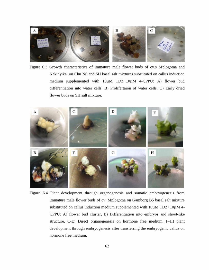
62
Figure 6.3 Growth characteristics of immature male flower buds of cv.s Mplogoma and
Nakinyika on Chu N6 and SH basal salt mixtures substituted on callus induction
medium supplemented with 10µM TDZ+10µM 4-CPPU: A) flower bud
differentiation into water cells, B) Prolifertaion of water cells, C) Early dried
flower buds on SH salt mixture.
Figure 6.4 Plant development through organogenesis and somatic embryogenesis from
immature male flower buds of cv. Mplogoma on Gamborg B5 basal salt mixture
substituted on callus induction medium supplemented with 10µM TDZ+10µM 4-
CPPU: A) flower bud cluster, B) Differentiation into embryos and shoot-like
structure, C-E) Direct organogenesis on hormone free medium, F-H) plant
development through embryogenesis after transferring the embryogenic callus on
hormone free medium.

63
6.4 Discussion
6.4.1 Callus induction on callus induction medium supplemented with TDZ and 4-
CPPU
Embryogenic callus formation among the cultivars on the callus induction medium
supplemented with various combinations of TDZ and 4-CPPU varied. However all the AAA-
EA banana cultivars studied showed embryogenic response implying that a combination of
TDZ and 4-CPPU with 2,4-D is efficacious for callus induction from male flower buds of
AAA-EA banana cultivars. Favorability of combinations of urea-type cytokinins with auxins
for induction of embryogenesis was reported in other banana genotypes. Divakaran et al.
(2011) generated embryogenic callus from bract explants of diploid (2n = 22) banana
cultivars on Murashige and Skoog medium supplemented with TDZ (0.045–9.00 μM). When
they transferred the embryogenic callus on MS basal medium supplemented with biotin and or
glutamine it produced somatic embryos and consequently regenerated normal plants with little
somaclonal variation and survival rate in soil of 90%. In other crops, combined effect of TDZ
and 2,4-D induced somatic embryogenesis in non-responsive caryopses of rice (Aparna and
Rashid, 2004). Tuong Huan et al. (2004) achieved more embryogenic calli induction from
protocorm of Cymbidium, a hybrid orchid, on medium containing a combination of NAA and
TDZ than on NAA alone. A Combination of 2,4-D and TDZ also changed granular callus into
a favorable friable form in Oncidium (Fang et al., 2006). Fiore et al. (2004) reported higher
percentage of embryo formation in both sweet orange (C. sinensis L.) and lemon (Citrus
limon L.) on 4-CPPU when they used media supplemented with different combinations of 2,4-
D and 4-CPPU to induce callus and somatic embryogenesis.
All the cultivars (except Nfuuka) formed more somatic embryos on the medium supplemented
with 10µM of each of TDZ and 4-CPPU than on the medium supplemented with 5µM or
15µM of each of them. The low response of the cultures on the callus induction medium
supplemented with 15µM of each of TDZ and 4-CPPU was attributed to excessive auxin
effect. At high concentrations urea-type cytokinins behave typical like auxins and thus the
auxin activity from them added to that of 2,4-D could have been too high to induce
embryogenic response. This implies cv. Nfuuka which responded best on the medium

64
supplemented with 15µM of each of TDZ and CPPU require higher auxin activity to stimulate
embryogenesis in it.
Instead of differentiating into callus on the medium supplemented with 5µM of each of TDZ
and 4-CPPU some of the flower buds proliferated into new floral tissue. Hernandez and
Rosell-Garcia (2008) reported inflorescence proliferation in dwarf Cavendish male flower on
medium supplemented with 5µM TDZ. At low concentrations urea-type cytokinins exhibit
cytokinin activity which favors tissue proliferation. It means at 5µM, TDZ and 4-CPPU added
none or minimal auxin activity to that of 2,4-D which was not sufficient to induce
embryogenic response. It further implies that cv. Sukali Ndizi (AAB) whose cultures had
excessive proliferation on all the treatments require much higher auxin supply and thus
needed higher TDZ and 4-CPPU amounts to stimulate embryogenesis. Banana genotypes with
AAB appear to require higher auxin concentration to induce embryogenesis probably due to
their heterogenomic hybrid nature especially the presence of the B genome which is usually
associated with higher tolerance to stress. Embryogenic callus and somatic embryos were
generated from male flower buds of banana cv. Rasthali (AAB) on MS medium supplemented
with 18.10 μM 2,4-D, 5.37 μM NAA and 5.71 μM IAA (Sukhada et al., 2010).
6.4.2 Callus induction on different basal salt mixture substituted on callus induction
medium supplemented with TDZ and CPPU
Under the same MS vitamins and hormone treatment embryogenic response was observed on
Gamborg B5, MS and Whites basal salt mixtures but none on Chu N6, NLN, Nitsch and SH
salt formulations. Other authors reported on callus induction and ECS development from
immature male buds of different banana genotypes on some of these salt formulations. Cote et
al. (1996) achieved embryo development in Musa AAA cv. Grande Naine (AAA) by plating
its cell suspension on a medium of SH with MS vitamins. Similarly Gomez et al. (2000)
achieved embryo formation in hybrid cultivar FHIA–18 (AAAB) using a medium of SH with
MS vitamins. When Ganapathi et al. (1999) initiated young banana male flowers on full
strength MS medium and White‟s medium, they found MS medium to be better than the
White‟s medium in terms of embryogenic callus induction. But in this study no cultivar

65
responded on SH while cultivar Nakinyika showed embryogenic response on white basal salt
mixture but not on MS medium.
Notwithstanding cultivar differences, the performance of the various basal salt mixtures was
probably influenced by their interaction with TDZ and CPPU in the media. For instance urea-
type cytokinins especially TDZ in synergy with Ca2+
stimulates ethylene production (Wing-
Kin and Shang, 1986) which most likely caused the cultures on the basal salt mixtures with
high calcium content especially MS and NLN to dry early (Table 6.2). Gamborg B5 basal salt
mixture has low calcium content compared to the other salt mixtures; perhaps that was why
both its callus and embryos survived for longer time than cultures on the other salt mixtures
(Table 6.2).
On the other hand both cultivars Mpologoma and Nakinyika formed more embryos on
Gamborg B5 basal salt mixture than on other salt mixtures including on MS medium one of
the most commonly used media for plant tissue culture. Compared to the other salt
formulations, Gamborg B-5 basal salt mixture has also the lowest total nitrogen content of
which 93% is in nitrate form and only 7% in ammonical form. Since banana cultures have a
high demand for nitrogen (Purseglove, 1972; Stover and Simmond, 1987) and mostly prefer
ammonical nitrogen (Marchal, 1990) the limited amount of nitrogen in it possibly created a
stress which favored embryo formation. It was probably because of the same reason that the
White basal salt mixture which has also lower total nitrogen imposed stress and induced
embryogenic response in cv. Nakinyika.
The trend in survival and response of the flower buds on each petridish was not clear.
However the survival of flower buds in the culture medium was linked with their
embryogenic response. The cultures of both Mpologoma and Nakinyika on Gamborg B5 salt
mixture survived for the same period. But cultivar Mpologoma whose flower buds had
significantly (P < 0.05) higher survival rate showed higher embryogenic response. The higher
survival of the flower buds of cv. Mpologoma on the callus induction medium possibly
increased their chances for embryogenic response. It implies that individual flower bud

66
survival and duration of survival of the cultures on the various salt formulations determined
embryogenic response.
When the callus of the various cultivars from the different salt formulations substituted on
callus induction medium supplemented with TDZ and CPPU was transferred into hormone
free MS derived medium, it developed into shoot through both organogenesis and somatic
embryogenesis (Figure 6.4). An explant can regenerate into an entire plant either through
organogenesis whereby shoots and roots simply form sequentially or somatic embryogenesis
whereby a cell or group of cells from the explant initiate and develop viable embryos (Victor,
2001). The review by Victor (2001) reveals that both organogenesis and somatic
embryogenesis can occur in the same explant but originating from particular tissue layers or
cells within the explant. Besides differences in physiological state of the flower buds or
tissues and cells within them, the occurrence of organogenesis and somatic embryogenesis
together in this study was attributed to the presence of TDZ and CPPU in the media and their
interaction with the various salt mixtures. Urea-based growth regulators especially TDZ was
reported to induce a diverse array of culture responses including induction of callus,
formation of somatic embryos and other structures (Murthy et al., 1998). TDZ induced both
direct shoot organogenesis and somatic embryogenesis on cotyledonary node explants of
lentil (Lens culinaris Medik.) (Chhabra et al., 2008). This is possibly because of its unique
property of mimicking both auxin and cytokinin effects on growth and differentiation of
cultured explants (Murthy et al., 1998).
Previously auxins particularly 2,4-D was used in combination with purine-based cytokinins
such as BA, BAP and Zeatin for induction of embryogenesis in AAA-EA banana. Besides
low embryogenic responses it was rare for more than two cultivars to show embryogenic
response on a single treatment though embryogenic response in banana was reported to be
genotype dependent (Youssef et al., 2010). In this study where the callus induction medium
was supplemented with urea-type cytokinins TDZ and CPPU, all the studied AAA-EA banana
cultivars produced regenerable embryogenic callus especially on the callus induction medium
supplemented with 10µM TDZ+10µM CPPU. When cultivars Mpologoma and Nakinyika
were cultured on the same medium containing 10µM TDZ+10µM CPPU but the MS salts

67
substituted with the other salt formulations, 11.4% and 8.3% of their cultures respectively had
embryos on Gamborg B5 salt formulation almost twice their response on pure MS medium.
Since the male flowers were collected randomly from the same fields and therefore, there
were no instances of differences in embryogenic response due to differences in developmental
stage of the flower buds (Youssef et al., 2010), these responses were rated to those
remarkably reported in other genotypes such as by Ghosh et al. (2009). Recently when Ghosh
et al. (2009) established embryogenic cell suspension culture of Cavendish cultivar Robusta
(AAA) using immature male flowers they reported embryogenic callus response of 10.3%.
Though some cultures proliferated instead of forming regenerable embryogenic callus, given
the limited availability of immature flowers and/or long time required to wait for banana
plants to flower, the induced floral tissue could be used as a source of flower buds for more
callus induction. Perez-Hernandez and Rosell-Garcia (2008) found newly induced floral
tissues from banana inflorescence cultured on 5 µM TDZ to be embryogenically competent.
6.5 Conclusion
The callus induction medium supplemented with a combination of TDZ and CPPU induced
various levels of embryogenic response from immature male flower buds of AAA-EA banana
cultivars Mbwazirume, Mpologoma, Nakabululu, Nakinyika and Nfuuka. The callus
induction medium supplemented with 10µM TDZ+10µM CPPU induced more embryogenic
response. Embryogenic response on Gamborg B5 salt mixture substituted on the callus
induction medium supplemented with 10µM TDZ+10µM CPPU doubled especially in cv.
Mpologoma. The results thus suggest that the percentage of embryogenic callus induced by
TDZ and 4-CPPU on immature male flower buds of AAA-EA cultivars is influenced by basal
salt mixture and in the presence of these two growth regulators, Gamborg B5 salt formulation
is more appropriate for callus induction followed by White and MS salts depending on
cultivar.

68
CHAPTER SEVEN
GENERAL DISCUSSIONS
This study determined proliferation potential and number of recoverable or weanable plantlets
that could be generated from a single isolated shoot tip of AAA-EA banana cultivars with
different suckering ability in the field on the adopted MS derived medium for banana
multiplication. Understanding the trend in in vitro proliferation of the AAA-EA banana
cultivars would enhance development of micropropagation protocols for their multiplication
and aid producers of tissue culture plants in planning especially by estimating number of
shoot-tip explants to initiate in culture to produce a given number of plantlets. It also pursued
somatic embryogenic techniques which have higher potential for mass propagation of planting
material and possible use of the tissues for genetic transformation. Specifically urea-type
cytokinins TDZ and 4-CPPU, and different salt formulations were tested for embryogenic
callus induction in AAA-EA banana cultivars through scalps and immature male flowers.
The AAA-EA banana cultivars produced 3-4 new shoots in each subculture cycle and 57-169
recoverable shoots from one starting shoot-tip explant in 18 weeks. The study thus revealed a
variation in in vitro shoot proliferation among the studied AAA-EA banana cultivars but the
trend was different from that of their suckering ability in in vivo. Since differences in cultivar
proliferation rate have been attributed to differences in growth regulator uptake, translocation
rate to meristematic regions, and metabolic processes among genotypes (Blakesley, 1991;
Youmbi et al., 2006), the inconsistence in proliferation in vitro and suckering in vivo implied
that though they belong to the same genome group, AAA-EA banana cultivars differ in
growth regulator uptake, translocation and metabolic processes. The ability of the cultivars to
produce recoverable shoots was influenced by the number and morphology or growth
characteristics of their shoot cultures suggesting that both proliferation and efficiency to
produce recoverable shoots of AAA-EA banana cultivars could be improved by varying
culture conditions or adjusting the amount of cytokinin and other growth inducing substances
in the medium during the subsequent subculture cycles. As the multiplication rate of
recoverable shoot depended on the cultivars‟ shoot growth characteristics, the number of

69
recoverable shoots from one shoot-tip explant of each cultivar in a given time could be
predicted using a regression model and thus a producer of tissue culture plants could be able
to project the number of plants to produce from a given number of shoot tip explants.
The results from somatic embryogenic techniques which have higher potential for
micropropagation and are more preferred in case of genetic transformation were promising
both from the scalp and immature male flower methods. One impediment to using the scalp
method to generate callus in AAA-EA banana had been the long time required to achieve
ideal scalps. On TDZ and 4-CPPU combination, scalps of AAA-EA bananas were achieved in
over 50% of the shoot-tip cultures within 4 subculture cycles. Besides long time required
generating scalps there has been low embryogenic response on the existing protocols. On the
TDZ and 4-CPPU treatment, embryogenic callus was formed on 2.5 – 20% of the scalp
cultures depending on cultivar. Cultivars Nakinyika, Nakabululu, Mbwazirume and Nfuuka
developed regenerable embryogenic callus and recorded plant regeneration through direct
embryogenesis. The plantlets of cultivars Nakinyika and Mbwazirume developed normal
roots and were weaned onto soil. Though the embryo to plant conversion rate was low, these
embryogenic callus responses were within the range and in some instances higher than those
reported even in other genotypes using the scalp method. For instance Xu et al. (2005)
reported a 10% embryogenic response for Williams and 0.1 to 6.2% for other Cavendish-type
bananas.
Though embryogenic response from male flower buds is often influenced by genotype and the
development stage of the buds (Youssef et al., 2010), the immature male flower bud cultures
of most of the same cultivars (Mpologoma, Mbwazirume, Nakabululu, Nakinyika and
Nfuuka) on callus induction medium supplemented with TDZ and 4-CPPU showed 4.6 -
22.2% embryogenic response especially on the medium with 10µM TDZ+10µM CPPU
combination. These embryogenic callus responses were also comparable and in some
instances higher than those reported even in other genotypes using the immature male flower
method. For example Escalant et al. (1994) reported a range of 0-7% embryogenic response
in different banana and plantain genotypes. Navarro et al. (1997) reported 2 - 6%
embryogenic callus response for Grand Naine (AAA) while Strosse et al. (2003) reported a

70
mean percentage of ideal embryogenic callus of 8% for the same cultivar. Recently Ghosh et
al. (2009) established embryogenic cell suspension culture (ECS) of Cavendish banana
cultivar Robusta (AAA) using immature male flowers and achieved percentage embryogenic
callus response of 10.3. This implies application of TDZ and 4-CPPU overcomes
recalcitrance in AAA-EA banana cultivars and makes them responsive to somatic
embryogenesis to levels comparable to that of other banana genotypes such Cavendish.
When cultivars Mpologoma and Nakinyika were cultured on the same medium containing
10µM TDZ+10µM 4-CPPU but the MS salts substituted with the other salt formulations, their
cultures recorded 11.4% and 8.3% embryogenic response respectively on Gamborg B5 salt
mixture which was almost twice their response on pure MS medium. The outstandingly
higher performance of both cultivars on Gamborg B5 basal salt mixture than on other salt
mixtures including on MS medium one of the most commonly used media for plant tissue
culture suggest that TDZ and CPPU interacts with salt components and interacts more
favorable with Gamborg B5 salts for induction of embryogenesis.
From the scalp method the embryogenic callus response of the different cultivars varied from
2.5 – 20% on medium with equal proportions of 9 – 13 µM TDZ and 4-CPPU. The range of
embryogenic callus response for the same cultivars on almost the same proportions of TDZ
and 4-CPPU using immature male flower method was 4.6 - 22.2%. Though there were
differences in response of some of the cultivars using the two methods the range for the two
methods was similar. This is consistent with observation by Strosse et al. (2006) that both
methods could yield similar results.
Considering all the 3 experiments and not contradicting the discussions in chapter 4, the trend
in the regeneration ability of the AAA-EA banana cultivars used in this study appears to be
linked to either their seed fertility or suckering ability in the field. For instance among the
cultivars that responded fairly well, cv. Nakabululu belongs to Nakabululu clone set while
cultivars Nfuuka and Nakinyika belong to Nfuuka clone set. These two clone sets were
reported to be more female fertile than cultivars in Musakala and Nakitembe clone sets
(Ssebuliba et al., 2006). The ability of cv. Mbwazirume to regenerate plantlets with normal

71
roots yet it belongs to Nakitembe clone set which is less seed fertile suggests that cultivar
regeneration ability may also be associated with its suckering ability in the field. According to
Karamura and Pickersgill (1999), cultivars in Nakitembe and Nakabulu clone sets have higher
suckering ability than those from other clone sets. This further means that cultivar
Mpologoma performed poorly because it belongs to Musakala clone set which is less female
fertile (Ssebuliba et al., 2002) and also produces fewer suckers in the field (Karamura and
Pickersgill, 1999). However this observation in banana requires further research to elucidate.
72
CHAPTER EIGHT
CONCLUSIONS AND RECOMMENDATION
8.1 Conclusions
In vitro proliferation of AAA-EA banana on the widely adopted MS medium modified for
banana multiplication varied from 3-4 new shoots in each subculture cycle and between 57
and 169 recoverable shoots from one starting shoot-tip explant in 18 weeks. The results
suggest that the number of shoot buds produced by AAA-EA banana in MS medium is
cultivar dependent however; the number of recoverable shoots is influenced by the cultivar‟s
shoot growth characteristics and is independent of its proliferation ability. Since the
multiplication rate of recoverable shoot depended on the cultivars‟ shoot growth
characteristics, the number of recoverable shoots from one shoot-tip explant of each cultivar
in a given time could be predicted using a regression model.
Scalps of AAA-EA bananas were achieved on over 50% of the shoot-tip cultures within 4
subculture cycles and embryogenic callus was directly formed on 2.5 – 20% of the scalp
cultures on the auxin-free scalp induction medium with TDZ and 4-CPPU depending on
cultivar. Most of cultivars showed better responses on the medium supplemented with TDZ
and 4-CPPU at equal proportions of 9 - 13µM. Cultivars Nakinyika, Nakabululu,
Mbwazirume and Nfuuka developed regenerable embryogenic callus and recorded plant
regeneration through direct embryogenesis. This implies that media supplemented with TDZ
and 4-CPPU combination reduce the time taken to induce scalps and increase the percentage
of embryogenic callus induced from scalps of EA-banana cultivars.
The callus induction medium supplemented with TDZ and 4-CPPU induced various levels of
embryogenic response from immature male flower buds of AAA-EA banana cultivars
Mbwazirume, Mpologoma, Nakabululu, Nakinyika and Nfuuka. Most of the cultivars showed
embryogenic response on the medium with 10µM TDZ+10µM CPPU ranging from 4.6 -
22.2%. Embryogenic response on Gamborg B5 salt mixture substituted on the callus
induction medium supplemented with 10µM TDZ+10µM CPPU was higher than on other salt

73
formulation. The results thus suggest that the percentage of embryogenic callus induced by
TDZ and 4-CPPU on immature male flower buds of AAA-EA cultivars is influenced by basal
salt mixture and in the presence of these two growth regulators, Gamborg B5 salt formulation
is more appropriate for callus induction followed by White and MS salts depending on
cultivar.
Previously auxins particularly 2,4-D was used in MS medium in combination with purine-
based cytokinins such as BA, BAP and Zeatin for induction of embryogenesis in AAA-EA
banana. Besides the long time required especially for generating scalps there has been low
embryogenic response on the existing protocols. It has also been rare for more than two
cultivars to show embryogenic response on a same treatment. In this study, using urea-type
cytokinins, TDZ and 4-CPPU, ideal scalps were achieved within 4 subculture cycles. In both
the scalp and immature male flower methods, most of the studied cultivars showed
embryogenic responses on the same treatment which in some instances were higher than those
reported before in AAA-EA banana cultivars and even other banana genotypes. Earlier plant
regeneration at lower concentrations of combined TDZ and CPPU could reduce occurrence of
somaclonal variation. This implies that application and optimization of combined TDZ and 4-
CPPU especially in Gamborg B5 salt formulation for routine induction of embryogenic callus
and development of embryogenic cell suspensions, respectively, will improve plant
regeneration and consequently propagation and genetic improvement of AAA-EA banana
cultivars.
8.2 Recommendations
The study demonstrated the potential of in vitro approach for production of banana planting
materials. Depending on the AAA-EA banana cultivar‟s shoot culture growth characteristics,
producers of tissue culture plants through shoot-tip culture can project the number of
recoverable shoots from one shoot-tip explant in a given time using a regression model. The
model for instance for cv. Mpologoma is Y = e[0.261(x-1) – 0.00386(x-1)² +1.39]
; where Y = number of
recoverable shoots, e = 2.718282 and x = time in weeks. This prediction model can give a fair
estimate up to the 4th
- 6th
subculture cycle corresponding to 4.5 - 6.5 months. Care must be
taken when predicting beyond this period as changes may occur in proliferation or growth

74
pattern depending on cultivar. The model may be applied to other cultivars that have similar
shoot culture growth characteristics to that of AAA-EA bananas. As the ability of a cultivar to
produce recoverable shoots is influenced by the number and morphology or growth
characteristics of its shoot culture, further studies are recommended on how to improve both
proliferation and efficiency to produce recoverable shoots of AAA-EA banana cultivars.
The scalp induction medium and callus induction medium supplemented with equal
proportions of 9 - 13 µM TDZ and 4-CPPU and 10µM TDZ+10µM CPPU respectively
induced embryogenic responses from scalps and immature male flower buds respectively
from most the studied cultivars. Embryogenic response on Gamborg B5 salt mixture
substituted on the callus induction medium supplemented with 10µM TDZ+10µM CPPU was
higher than on other salt formulations. Though the embryo to plant conversion rate was low,
application and optimization of combined TDZ and 4-CPPU especially in Gamborg B5 salt
formulation for routine induction of embryogenic callus and development of embryogenic cell
suspensions respectively is worthwhile.
The overall response of the studied AAA-EA banana cultivars from different clone sets
suggests a linkage between their regeneration ability and their seed fertility or suckering
ability in the field. It is therefore recommended that further studies are done to elucidate
whether the regeneration ability of the AAA-EA banana cultivars is linked to their seed
fertility or suckering ability in the field. This will provide a vital insight to the basis for
recalcitrancy in this banana group.

75
REFERENCES
AATF, 2003. African Agricultural Technology Foundation. Report of Small Group Meeting
(SGM) on Improved Production of Bananas and Plantains in sub-Saharan Africa, 22nd
August 2003, Hotel Equatoria Kampala, Uganda. Available from: http://www.aatf-
africa.org/UserFiles/File/sgm-bananas.pdf (Accessed 23 July 2011).
AATF, 2009. Feasibility study on technologies for improving Banana for resistance against
Bacterial wilt in Sub-Saharan Africa. Nairobi, Kenya: African Agricultural Technology
Foundation, ISBN 9966-775-06-4.
Aish, M., Iqba, H., Muhammad, F.A. and Hamid, R. 2003. Enhanced rate of multiplication of
tissue cultured raised banana (Musa) plants in the field. Pakistan Journal of Biological
Sciences, 6: 58–588.
Aliguma, A. and Karamura, E. 2006. Community coping mechanisms in response to BBW
effects in Mukono District. International Network for Improvement of Banana and
Plantain of the International Plant Genetic Resources Institute (IPGRI-INIBAP).
Aparna, G. and Rashid, A. 2004. TDZ-induced somatic embryogenesis in non-responsive
caryopses of rice using a short treatment with 2,4-D. Plant Cell, Tissue and Organ
Culture, 76: 29–33.
Argent, G.C.G. 1976. The wild bananas of Papua New Guinea. Notes from the Royal
Botanical Garden, Edinburgh, 35: 77-114.
Arinaitwe, G., Rubaihayo, P.R. and Magambo, M.J.S. 2000. Proliferation rate effects of
cytokinins on banana (Musa spp.) cultivars. Scientia Horticulturae, 86: 13-21.
Ashraf, H.F., Yehia, H.E., Osama, M.E. and Madkour, M.A. 2004. A highly efficient
regeneration system via somatic embryogenesis from immature embryos of Egyptian
wheat cultivars (Triticum aestivum L.) using different growth regulators. Arab Journal
of Biotechnology, 7: 229-238.
Baiyeri, K.P. and Aba, S.C. 2005. Response of Musa species to Macropropagation. I: Genetic
and initiation media effects on number, quality and survival of plantlets at pre-nursery
and early nursery stages. African Journal of Biotechnology, 4: 223-228.

76
Becker, D.K., Dugdale, B., Smith, M.K., Harding, R.M and Dale, J.L. 2000. Genetic
transformation of Cavendish banana (Musa spp. AAA group) cv. Grand Nain via
micro-projectile bombardment. Plant Cell Reports, 19: 229-234.
Blakesley, D. 1991. Uptake and metabolism of 6-benzyladenine in shoot proliferation of
Musa and Rhododendron. Plant Cell, Tissue and Organ Culture, 25: 69-74.
CGIAR, 2008. http://www.cgiar.org/monthlystory/september2008.html
Cheesman, E.E. 1948. The classification of the bananas. Kew Bulletin. 3: 11-28, 145-157,
323-328.
Chhabra, G., Chaudhary, D., Varma, M., Sainger, M. and Jaiwal P.K. 2008. TDZ-induced
direct shoot organogenesis and somatic embryogenesis on cotyledonary node explants
of lentil (Lens culinaris Medik.). Physiology and Molecular Biology of Plants, 14: 348-
353.
Chu, C.C., et al. 1975. Establishment of an efficient medium for anther culture of rice through
comparative experiments on the nitrogen sources. Scienta Sinica, 18: 659-668.
Clarke, T. 2003. Banana lab opens in Uganda: Genetic modification of clonal crop could soon
follow. Nature News, August 22. http://www.bioedonline.org/news/news.cfm?art=430.
Constantine, D. and Rossel, G. 2001. Musaceae: An annotated list of the species of Ensete,
Musa and Musella. http://www.users.globalnet.co.uk/drc/index.htm.
Cote, F.X., Domergne, R., Monmarson, S., Schwendiman, J., Teisson, C. and Escalant, J.V.
1996. Embryogenic cell suspensions from male flower of Musa AAA cv. Grand Nain.
Physiology Plant, 97: 285-290.
Damasco, O.P., Smith, M.K. and Adkins, S.W. 1996. Gibberellic acid detection of dwarf
offtypes in micropropagated Cavendish bananas. Australian Journal of Experimental
Agriculture, 36: 237-241.
Daniells, J., Jenny, C., Karamura, D. and Tomekpe, K. 2001. Musalogue: A catalogue of
Musa germplasm. Diversity in the genus Musa. Arnaud, E. and Sharrock, S. (Eds.)
International Network for the Improvement of Banana and Plantain, Montpellier,
France.
Daphne, J.O. and Michael, T.M. 2005. Hormones, Signals and Target Cells in Plant
Development. Cambridge University Press. Page 158.
77
Davies, P.J. 2004. Regulatory factors in hormone action: Level, location and signal
transduction. pp. 16-35 in: Davies P.J. (ed.) Plant Hormones, Kluwer Academic
Publishers, Dordrecht.
Divakaran, S.P. and Nair, A.S. 2011. Somatic embryogenesis from bract cultures in diploid
Musa acuminata cultivars from South India. Scientia Horticulturae, 131 (1): 99-102.
Divya, K., Swathi Anuradha, T., Jami, S.K. and Kirti, P.B. 2008. Efficient regeneration from
hypocotyl explants in three cotton cultivars. Biologia Plantarum, 52: 201-208.
Duncan, R.R. 1997. Tissue culture-induced variation and Crop improvement. Advanced
Agronomy, 58: 201-240.
Dunlap, J.R. and Robacker, K.M., 1988. Nutrient salts promote Light-induced degradation of
Indole-3-acetic acid in tissue culture media. Plant Physiology, 88: 0379-0382.
El-Dougdoug, K.A., El-Harthi, H.M.S., Korkar, H.M. and Taha, R.M. 2007. Detection of
somaclonal variations in banana tissue culture using isozyme and DNA fingerprint
analysis. Journal of Applied Sciences Research, 3: 622-627.
Ericksson, T. 1965. Studies on the growth requirements and measurements of cell cultures of
Haplopappus gracilis. Physiologia Plantarum, 18: 976-993.
FAO, 2005. FAO STAT Agriculture Data. www.fao.org.
FAO, 2012. FAOSTAT Agriculture Data. www.fao.org.
Fiore, S., Fabio D. P., Francesco, C. and Maurizio, S. 2004. Effect of 2, 4-D and 4-CPPU on
somatic embryogenesis from stigma and style transverse thin cell layers of Citrus.
Plant Cell, Tissue and Organ Culture, 68: 57-63.
Fischer, C. and Neuhaus, G. 1996. Influence of auxin on the establishment of bilateral
symmetry in monocots. Plant Journal, 9: 659-669.
Gamborg, O.L., Miller, R.A. and Ojima, K.1968. Nutrient requirement of suspension cultures
of soybean root cells. Experimental Cell Research, 50: 151-158.
Ganapathi, T.R., Suprasanna, P., Bapat, V.A., Kulkarni, V.M., Rao, P.S. 1999. Somatic
embryogenesis and plant regeneration from male flower buds in banana. Current
Science, 79: 1229-1231.
GenStat, 2010. GenStat procedure Library Release PL21.3, version 13.3.0.5165.
Geo, J.B., 1985. The application of tissue culture to plant improvement and propagation in the
Ornamental Horticultural Industry. In Tissue Cultue as a plant production system for

78
horticultural crops. Edited by Zimmerman R.H., Martinus Nijhoff Publishers,
Lanncaster Griesbach R.J. and Lawson R.H. Robert Pp 15-33.
George, E. F. et al. (eds.), 2008. Plant Propagation by Tissue Culture, 3rd
Edition, Springer,
175–204.
Ghosh, A., Ganapathi, T.R., Nath, P. and Bapat, V.A. 2009. Establishment of embryogenic
cell suspension cultures and Agrobacterium-mediated transformation in an important
Cavendish banana cv. Robusta (AAA). Plant Cell, Tissue and Organ Culture, 97 (2):
131-139.
Gomez, R.K., Gilliard, T., Barrnco, L.A. and Reyes, M. 2000. Somatic embrogenesis in liquid
media. Maturation and enhancement of germination of the hybrid cultivar FHIA – 18
(AAAB). Info Musa. The International Magazine on Banana and Plantain, 9 (1): 12-
16.
Grapin, A., Schwendiman, J., Teisson, C. 1996. Somatic embryogenesis in plantain bananas.
In vitro Cellular Developmental Biology-Plant, 32: 66-71.
Haruki, S., Sakae, A., Makiko, U., Yuko, M. and Akihiro, N. 2007. Effect of urea-type
cytokinins on the adventitious shoots regeneration from cotyledonary node explant in
the common ice plant, Mesembryanthemum crystallinum. Plant Production Science,
10: 47-56.
Israeli, Y., Ben-Bassat, D., Reuveni, O. 1996. Selection of stable banana clones which do not
produce dwarf somaclonal variants during in vitro culture. Scientia Horticulturae, 67:
197-205.
Jalil, M., Khalid, N., Othman, R.Y. 2003. Plant regeneration from embryogenic suspension
cultures of Musa acuminata cv. Mas (AA). Plant Cell Tissue and Organ Culture, 75:
209-214.
Kantharajah, A.S. 2001. Introduction to Plant tissue culture. University of Western Sydney,
Hawkersbury (Richmond and Blacktown).
Karamura, D. and Pickersgill, B. 1999. A classification of the clones of East African Highland
bananas (Musa) found in Uganda. Plant Genetic Resources Newsletter, 119: 1-6.
Karamura, D., Mgenzi, B., Karamura, E. and Sharrock, S. 2004. Exploiting IK for the
management and maintenance of Musa Biodiversity on farm. African Crop Science
Journal, 12(1): 71-78.
79
Karamura, D.A. 1998. Numerical Taxonomic Studies of the East African Highland Bananas
(Musa AAA-East Africa) in Uganda. A thesis submitted for the degree of Doctor of
Philosophy, Department of Agricultural Botany, The University of Reading. January,
1998.
Khaleda, L. and Al-Forkan, M. 2006. Genotypic variability in callus induction and plant
regeneration through somatic embryogenesis of five deepwater rice (Oryza sativa L.)
cultivars of Bangladesh. African Journal of Biotechnology 5 (16): 1435-1440.
Larkin, P. J. and Scowcroft, W. R. 1981. Somaclonal variation – a novel source of variability
from cell cultures for plant improvement. Theoretical and Applied Genetics, 60: 197-
214.
Leela, S., Jaya, R.S. and Bollama, K.T. 2003. Analyzing somaclonal variation in
micropropagated bananas (Musa spp). In Vitro Cellular and Development Biology -
Plant, 39: 551-556.
Lehminger-Mertens, R. and Jacobson, H.J. 1989. Plant regeneration from pea protoplasts via
somatic embryogenesis. Plant Cell Report, 8: 379-382.
Lichter, R. 1982. Induction of haploid plants from isolated pollen of Brassica napus. Z.
Pflanzenphysiology, 105: 427-434.
Lorenzen, J. 2008. Growing bananas from “seeds”. R4D Review. Available from:
http://r4dreview.org/2008/09/growing-bananas-from seeds (Accessed: 23 July 2011).
Makara, A.M., Rubaihayo, P.R. and Magambo, M.J.S. 2010. Carry-over effect of Thidiazuron
on banana in vitro proliferation at different culture cycles and light incubation
conditions. African Journal of Biotechnology, 9: 3079-3085.
Marchal, J. 1990. Nutritional physiology of banana plants in in vitro culture and during the
hardening-off period. In: Fruits, 1990. pp. 119-122.
May, G.D., Rownak, A., Mason, H., Wiecko, A., Novak, F. J., Arntzen, C.J. 1995. Generation
of transgenic banana (Musa acuminata) plants via Agrobacterium-mediated
transformation. Bio/Technology, 13: 486-492.
Meenakshi S., Suprasanna, P., Bapat, V.A., Kulkarni, U.G. and Shinde, B.N. 2007.
Developing somatic embryogenic culture system and plant regeneration in banana.
National Symposium on Plant Biotechnology held at Dehra Dun, during October 12-
14, 2006. BARG Newsletter Issue no. 285, October 2007.

80
Mendes, B.M.J., Filippi, S.B., Demétrio, C.G.B. and Rodriguez, A.P.M. 1999. A statistical
approach to study the dynamics of micropropagation rates, using banana (Musa spp.) as
an example. Plant Cell Reports, 18: 967-971.
Merkle S. A., Parrot, W.A. and Williams, E.G. 1990. Application of somatic embryogenesis
and embryo cloning. Pages 67-102 in Plant tissue culture: application and limitations
(Bhojwani, S.S, ed.). Amsterdam, Neetherlands, Elsevier.
Michalczuk, L., Ribnicky, D.M., Cooke, T.J. and Cohen, J.D. 1992. Regulation of indole-3-
acetic acid biosynthetic path-ways in carrot cell-cultures. Plant Physiology, 100: 1346-
1353.
Mineo, L. 1990. Plant tissue culture techniques. Pages 151-174, in Tested studies for
laboratory teaching. Volume 11. (C.A. Goldman, Editor). Proceedings of the Eleventh
Workshop/Conference of the Association for Biology Laboratory Education (ABLE),
195 Pp.
Mohandas, S., Sowmya, H.D., Manjula, R., Pratibha, K.Y., Meenakshi, S. and Ajay, K.M.
2011. Development of highly regenerative embryogenic cell suspensions of 'Nanjangud
Rasbale' (syn. 'Rasthali', Musa, AAB, Silk subgroup) and transformants with AMP
gene. Acta Horticulturae, 897: 245-254.
Mukasa, S.B. and Rubaihayo P.R. 1993. Male fertility in Uganda banana germplasm. African
Crop Science Journal, 1: 87-93.
Muranga, 2007. Models of Knowledge based transfer- The case of the Presidential Initiative
on Banana Industrial Development (PIBID): http://www.east-
sym.org/documents/P2_Muranga_ModelsOfKnowledge.pdf. (Accessed on 3rd
June
2012)
Murashige, T. and Skoog, F. 1962. A revised medium for rapid growth and bioassays with
tobacco tissue cultures. Physiologia Planarum, 15: 473-497.
Murthy, B.N.S., Murch, S.J. and Saxena, P.K. 1998. Thidiazuron: A potent regulator of in
vitro plant morphogenesis. In Vitro Cellular and Developmental Biology – Plant, 34:
267-275.
Namanya, P., Magambo, M. S., Mulumba, G. and Tushemereirwe W. K. 2002. Regeneration
of plants in East African highland bananas. Proceedings of INIBAP Workshop. July
22nd
– 26th
, 2002, Kampala Uganda.

81
Nantale, G., Kakudidi, E.K., Karamura, D.A., Karamura, E. and Soka, G. 2008. Scientific
basis for banana cultivar proportions on-farm in East Africa. African Crop Science
Journal, 16: 41-49.
Navarro, C., Rosa Ma, E. and Mayo, A. 1997. Plant Cell, Tissue and Organ Culture, 51: 17-
25.
Nirmalya, B. and De Langhe, E. 1985. A tissue culture technique for rapid clonal propagation
and storage under minimal growth conditions of Musa (Banana and plantain). Plant
Cell Reports, 4: 351-354.
Nitsch, J.P. and Nitsch, C. 1969. Haploid plants from pollen grains. Science, 165, 85.
Nookaraju, A., Barreto, M.S., Karibasappa, G.S. and Agrawa, D.C. 2007. Synergistic effect of
CPPU and benzyladenine on embryo rescue in six stenospermocarpic cultivars of
grapevine. Vitis, 46: 188-191.
Normanly, J., Slovin, J.P. and Cohen, J.D. 2004. Auxin biosynthesis and metabolism. pp. 36-
62 in Davies P.J. (ed): Plant Hormones: Biosynthesis, Signal Transduction, Action.
Springer-/Kluwer Acad. Publ., New York, Dordrecht.
Novak, F.J., Afza, R., Van Duren, M., Parea-Dallos, M., Conger, B.V and Xialang, T. 1989.
Somatic embryogenesis and plant regeneration in suspension cultures of dessert (AA
and AAA) and cooking (AAB) bananas (Musa spp). Bio/Technology, 7: 154-159.
Nyine, M. and Pillay, M. 2007. Banana nectar as a medium for testing pollen viability and
germination in Musa. African Journal of Biotechnology, 6: 1175-1180.
Okech, S., Gold, C.S., Abele, S., Nankinga, C.M., Wetala, P.M., van Asten, P., Nambuye, A.
and Ragama, P. 2004. Agronomic, pests and economic factors influencing
sustainability of banana-coffee systems of Western Uganda and potential for
improvement. Uganda Journal of Agricultural Science, 9: 432-444.
Panis, B., Wauwe, A.V. and Swennen, R. 1993. Plant regeneration through direct somatic
embryogenesis from protoplast of banana (Musa spp). Plant cell Reports, 12: 403-407.
Patil, P.V. and Navale, P.A. 2000. Somaclonal variation in fruit crops. A review. Journal of
Maharashra Agriculture University, 25: 8-14.
Payne, R.W., Murray, D.A., Harding, S.A., Baird, D.B. and Soutar, D.M. 2009. GenStat for
Windows (12th
Edition) Introduction. VSN International, Hernel Hempstead.

82
Pelletier, G., 1993. Plant breeding: Principles and prospects. Edited by M.D Hayward, N.O.
Bosemark and I. Romagosa. Chapman & Hall, London.
Perez-Hernandez J.B. and Rosell-Garcia, P. 2008. Inflorescence proliferation for somatic
embryogenesis induction and suspension-derived plant regeneration from banana (Musa
AAA, cv. „Dwarf Cavendish‟) male flowers. Plant Cell Reports, 27: 965-971.
Pillay, M. Ogundiwin, E., Tenkouano, A. and Dolezel, J. 2006. Ploidy and genome
composition of Musa germplasm at the International Institute of Tropical Agriculture
(IITA). African Journal of Biotechnology, 5: 1224-1232.
Pillay, M., Hartman, J., Dimkpa, C. and Makumbi, D. 2003. Establishing the genome of
'Sukali Ndizi'. African Crop Science Journal, 11: 119-124.
Purseglove, J.W. 1972. Tropical crops, Monocotyledons, Vol.2. Longmans, London.
Raemakers, C.J.J.M., Jacobsen, E. and Visser, R.G.F. 1995. Secondary somatic
embryogenesis and applications in plant breeding. Euphytica, 81: 93-107.
Rajasekaran, K., Hein, M.B., Davis, G.C., Carnes, M.G. and Vasil, I.K. 1987b. Endogenous
growth regulators in leaves and tissue cultures of Pennisetum purpureum Schum.
Journal of Plant Physiology, 130: 12-25.
Ramírez-Villalobos, M. and García, E. 2008. Obtainment of embryogenic cell suspensions
from scalps of the banana CIEN-BTA-03 (Musa sp., AAAA) and regeneration of the
plants. Electronic Journal of Biotechnology ISSN: 0717-3458.
Razdan, M.K. 1993. An introduction to Plant Tissue Culture. Intercept, Hampchire, UK. 398
pp.
Reuveni, O. and Israeli, Y., 1990. Measures to reduce somaclonal variation in in vitro
propagated bananas, Acta Horticulturae, 275: 307-313.
Rodrigues, P.H.V., Tulmann, A., Casseiri, P. and Mendes, B.M.J. 1998. Influence of the
number of subcultures on somaclonal variation in micropropagated Naico (Musa spp.
AAA group). Acta Horticulturae, 490: 469-473.
Roux, N., Baurens, F.C., Doležel, J., Hribová, E., Heslop-Harrison, J.S., Town, C., Sasaki, T.,
Matsumoto, T., Aert R., Remy, S., Souza, M. and Lagoda, P. 2008. Genomics of
banana and plantain (Musa spp.), Major staple crops in the tropics. Pp. 83-111 in
Genomics of Tropical Crop Plants (Moore P and Ming R eds.). Plant Genetics and
Genomics: Crops and Models Vol 1. Springer, New York.

83
Sadik, K., Rubaihayo, P.R., Magambo, M.J.S. and Pillay, M. 2007. Generation of cell
suspensions of East African highland bananas through scalps. African Journal of
Biotechnology, 6: 1352-1357.
Sanna, E.E., Mostafa, A.M. and Saleh, M.M.S. 2008. Effect of Gibberellic Acid and Male
Bud Removal on Yield and Fruit Quality of Banana Plants. Research Journal of
Agriculture and Biological Sciences, 4: 289-292.
Sannasgala, K., Dumrotier, F. and De Langhe. 1991. Feasibility of somatic embryo
development in Musa cv. Bluggoe. Tropical Agricultural Research 3.
Santos, C.C.C. and Rodrigues, P. H. V., 2004. Somaclonal variation event on
micropropagated Pacovan banana seedling (Musa spp. AAB group). Bragantia, 63,
no.2, p.201-205. ISSN 0006-8705.
Schenk, R.U. and Hildebrandt, A.C. 1972. Medium and techniques for induction and growth
of monocotyledonous and dicotyledonous plant cell cultures. Canadian Journal of
Botany, 50: 199-204.
Schoofs, H., 1997. The orgin of embryogenic cells in Musa, PhD thesis, KU Leuven,
Belgium.
Schoofs, H., Panis, B. and Swennen, R. 1998. Competence of scalps for somatic
embryogenesis in Musa. Acta Horticulturae, 490: 475-483.
Semal, J., (ed) 1986. Somaclonal variations and Crop Improvement. Proceedings of a seminar
in the CEC Programme of Coordination of Research on Plant Protein Improvement, 3rd
– 5th September 1985, Gembloux, Belgium.
Sergeant, A., Rutabingwa, D., Karamura, E., Muranga, F., Pedraza, F., Ssemwanga, J.,
Christy, R.D., Tushemereirwe, W. and Kalema, W.S. 2004. A market-driven
transformation strategy for the banana industry in Uganda. A public–private sector
partnership.
Shan, X., Li, D. and Qu, R. 2000.Thidiazuron promotes in vitro regeneration of Wheat and
Barley. In Vitro Cellular and Development Biology – Plant, 36: 207-210.
Sharp, W.R., Sondahl, M.R., Caldas, L.S. and Maraffa, S.B. 1980. The physiology of in vitro
asexual embryogenesis. Horticultural Reviews, 2: 268-310.

84
Shchukin, A., Ben-Bassat, D. Israel, Y., 1997. Plant Regeneration via Somatic embryogenesis
in Grande Naine banana and its effect on Somaclonal variation. Acta Horticulturae,
447: 317-318.
Shepherd, K., Souza F. V. D. and Da Silva, K. M., 1996. Mitotic instability in banana
varieties. IV. BAP concentration and effects of number of subcultures, Fruit, 51: 211-
216.
Simmonds, N.W. and Shepherd, K. 1955. The taxonomy and origins of the cultivated
bananas. Journal of the Linnean Society of London (Botany), 55: 302-312.
Singh, H. P., 2002. Indian Bananas – issues and strategies. Global conference on Banana and
Plantain, October 28 – 31, Bangalore, India. Abstracts, 2002: 1 - 2.
Singh, S.K. and Syamal, M.M. 2001. A short pre-culture soak in thidiazuron or
forchlorfenuron improves axillary shoot proliferation in rose micropropagation.
Scientia Horticulturae, 91: 169 -177.
Smale, M. 2006. Assessing the impact of crop genetic improvement in sub-Saharan Africa:
Research context and highlights. In: Melinda, S., Edmeades, S., and De Groote (Eds.).
Promising Crop biotechnologies for smallholder farmers in East Africa: Bananas and
Maize. Genetic Resources Policies Briefs 19-2006.
Smale, M. and De Groote, H. 2003. Diagnostic research to enable adoption of transgenic crop
varieties by smallholder farmers in Sub-Saharan Africa. African Journal of
Biotechnology, 2 (12): 586-595.
Smith, M.K. 1988. A review of factors influencing the genetic stability of micropropagated
bananas. Fruits, 43: 219-223.
Ssebuliba, R., Talengera, D., Makumbi, D., Namanya, P., Tenkouano, A., Tushemereirwe, W.
and Pillay, M. 2006. Reproductive efficiency and breeding potential of East African
highland (Musa AAA–EA) bananas. Field Crops Research, 95: 250–55.
Stover, R. H., and N. W. Simmonds. 1987. Bananas. 3rd ed. London: Longman.
Strosse, H., Domergue, R., Panis, B., Escalant, J.V. and Cote, F. 2003. Banana and Plantain
embryogenic cell suspensions (A. Vezina and C. Picq, eds). INIBAP Technical
guidelines 8. The International Network for the improvement of Banana and Plantain,
Montpellier, France.

85
Strosse, H., Schoofs, H., Panis, B., Andre, E., Reyniers, K. and Swennen, R. 2006.
Development of embryogenic cell suspensions from shoot meristematic tissue in
bananas and plantains (Musa spp.). Plant Science, 170: 104-112.
Subotić, A., Jevremović, S., Cingel, A. and Milošević, S. 2008. Effect of urea – type
cytokinins on axillary shoots regeneration of Impatiens walleriana L. Biotechnology,
Diagnosis Press, ISSN 1310-2818, 22 (3).
Sukhada, M., Prathibha, K.Y., Soumya, H.D. and Ranganath, R.M. 2010. The origin and
development of secondary somatic embryos in banana cv. Nanjangud Rasbale (Silk gp.
Rasthali, AAB). Acta Horticulturae, 865: 349-352.
Svetleva, D., Velcheva, M. and Bhowmik, G. 2003. Biotechnology as a useful tool in
common bean (Phaseolus vulgaris L.) improvement, A review. Euphytica 131: 189-
200.
Talengera, D., Magambo, M.J.S. and Rubaihayo, P.R. 1994. Testing for a suitable culture
medium for micropropagation of east African Highland Bananas. African Crop Science
Journal, 2: 17-21.
Tripathi, L., Tripathi, J.N. and Tushemereirwe, W.K. 2008a. Rapid and efficient production of
transgenic East African highland Banana (Musa spp.) using intercalary meristematic
tissues. African Journal of Biotechnology, 7: 1438-1445.
Tripathi, L., Tripathi, J.N., Oso, R.T., Hughes, J.A. and Keese. P. 2003. Regeneration and
transient gene expression of African Musa species with diverse genomic constitution
and ploidy levels. Tropical Agriculture, 80: 182-187.
Tuong Huan Le Van, Takamura, T. and Tanaka, M. 2004. Callus formation and plant
regeneration from callus through somatic embryo structures in Cymbidium orchid. Plant
Science, 166: 1443-1449.
Tushemereirwe, W.K., Kangire, A., Kubiriba, J., Nakyanzi, M. and Gold, C.S., 2004.
Diseases threatening banana biodiversity in Uganda. African Crop Science Journal, 2:
19-26.
Veltcheva, M.R. and Svetleva D.L. 2005. In vitro Regeneration of Phaseolus Vulgaris L. via
organogenesis from petiole explants. Journal of Central European Agriculture, 6: 53-
58.

86
Victor, J.M.R., Murch, S.J., KrishnaRaj, S. and Saxena, P.K. 2004. Somatic embryogenesis
and organogenesis in peanut: The role of thidiazuron and N6-benzylaminopurine in the
induction of plant morphogenesis. Plant Growth Regulation, 28: 9-15.
Víctor, M.J. 2001. Regulation of in vitro somatic embryogenesis with emphasis on the role of
endogenous hormones. Revista Brasileira De Fisiologia Vegetal, 13: 196-223.
Vinayak, V., Dhawan, A.K. and Gupta, V.K. 2009. Efficacy of non-purine and purine
cytokinins on shoot regeneration in vitro in sugarcane. Indian Journal of
Biotechnology, 8: 227-231.
Vinterhalter, B., Ninković, S., Zdravković- Korać, S., Subotić, A. and Vinterhalter, D. 2007.
Effect of Nitrogen salts on the growth of Ceratonia siliqua L. shoot cultures. Archives
of Biological Sciences, Belgrade, 59: 217-222.
Visser, C., Qureshi, J.A., Gill, R. and Saxena, P.K. (1992). Morphoregulatory role of
thidiazuron. Plant Physiology, 99: 1704-1707.
Vuylsteke, D. 2001. Strategies for Utilization of Genetic Variation in Plantain Improvement.
PhD Thesis. K.U. Leuven, Belgium.
Vuylsteke, D., Ortiz, R., Shaun, B.F. and Crouch, J.H. 1997. Plantain Improvement. Plant
breeding reviews, 14: 267-320.
White, P. R. 1963. The cultivation of Animal and Plant cells, Ronald Press, New York.
Wing-Kin, Y. and Shang, F.A.Y. 1986. Effect of Thidiazuron, a Cytokinin-Active Urea
Derivative, in Cytokinin-Dependent Ethylene Production Systems. Plant Physiology,
80: 515-519.
Xu, C.X., Panis, B., Strosse, H., Li, H.P., Xiao, H.G., Fan, H.Z., Swennen. R. 2005.
Establishment of embryogenic cell suspensions and plant regeneration of the dessert
banana Williams (Musa AAA group). Journal of Horticultural Science and
Biotechnology, 80: 523-528.
Ying-Chun, C., Chen, C. and Wei-chin, C. 2000. A reliable protocol for plant regeneration
from callus culture of Phalaenopsis. In Vitro Cellular and Developmental Biology –
Plant, 36: 420-423.
Youmbi, E., Ella, B. and Tomekpe, K. 2006. Effect of Thidiazuron on in vitro proliferation
capacities of some banana (Musa spp.) cultivars with weak multiplication potential.
Akdeniz Üniversitesi Ziraat Fakültesi Dergisi, 19: 255-259.

87
Youssef, M.A., Andrew James, C., Mayo-Mosqueda, A., Ku-Cauich, J.R., Grijalva-Arango,
R., Escobedo-GM, R.M. 2010. Influence of genotype and age of explant source on the
capacity for somatic embryogenesis of two Cavendish banana cultivars (Musa
acuminata Colla, AAA). African Journal of Biotechnology, 9: 2216-2223.
Zhang, Q., Chen, J. and Henny, R.J. 2004. Direct somatic embryogenesis and plant
regeneration from leaf, petiole, and stem explants of Golden Pothos. Plant Cell
Reports, 23: 587-595.
Zhenxun, W. and Hongxian, L., 1997. Chromosome aberration in banana micro-propagation.
Acta Genetica Sinica, 24: 550-560.

88
APPEDICES
Appedix I
Murashige and Skoog (MS) based Banana micropropagation medium with modification by
Talengera et al. (1994).
No. Element/Compound
Group A
(Major Elements)
Use 100 ml per
litre of Medium
Stock Dissolve in 1000 ml of DDH2O
1 HN4NO3 Weigh 16.5gm
2 KHO3 Weigh 19.0gm
3 CaCL2.2H2O Weigh 4.4gm
4 MgSO4.7H2O Weigh 3.7gm
5 KH2PO4 Weigh 1.7gm
Group B
(Minor Elements)
Use 5 ml per litre
of Medium
Stock Dissolve in 500 ml of DDH2O
1 H2BO3 Weigh 0.620 gm
2 MnSO4.4H2O Weigh 2.230 gm
3 ZnSO.4H2O Weigh 0.860 gm
4 KI Weigh 0.0830 gm
5 NaMoO4.2H2O Weigh 0.025 gm
6 CuSO4.5H2O Weigh 0.0025 gm
7 CoCl2.6H2O Weigh 0.0025 gm
Group C
(Vitamins Elements)
Use 5 ml per litre
of Medium
Stock Dissolve in 500 ml of DDH2O
1 Nicotinic acid Weigh 0.050 gm
2 Pyridoxine-HCL Weigh 0.050 gm
3 Thiamine-HCL Weigh 0.010 gm
4 Glycine Weigh 0.02 gm
Group D (Iron)
Use 5 ml per litre
of Medium
Stock Dissolve In100 ml of DDH2O
1 Na-EDTA (Dissolve 0.746 gm pre-heat to dissolve with 7)
2 FeSO4.7H2O (Dissolve 0.557 gm)
Group E (Myo-inositol)
Use 10 ml per
litre of Medium
1 My-inositol Weigh 10 gm per litre of DDH2O
Group F (Ascobic Acid)
1 Ascobic acid Weigh 2 gm per 500 mls DDH2O
Group H (Phytohormones) Use 5 ml per litre

89
of Medium
1 BAP 0.1 gm per 100 mls of DDH2O
Group G (Carbon source)
Use 30 gm per
litre of Medium
1 Sucrose Weigh 30 gm in 1000 mls of medium
Adjust PH
1 PH Use 0.1M of HCL or 0.1M of NaOH to adjust PH to 5.8
Gelling Agent
1
Phytagel, Gelrite or
Agar Add 2.3 gm of Phytagel or 2 gm of Gelrite or 7.0 gm of Agar,
heat to dissolve,
dispense to culture vessels, autoclave at 121oC for 15 min
Key: DD = Double Distilled
Water

90
Appendix II
The callus induction medium that was supplemented with TDZ and 4-CPPU for inducing
embryogenic callus from male flowers
Compound Concentration
MS salts Full strength
MS vitamins Full strength
IAA 5.7 µM (1mg/l)
2,4-D 5.0 µM (1.105mg/l)
Biotin 4.09 µM (1mg/l)
NAA 5.4 µM (1mg/l)
Ascorbic acid 20 mg/l
Sucrose 30g/l
Gel-rite 2.0g/l
PH 5.7