Embed Size (px)
Citation preview

Experimental/VCA
Monday January 26, 2015
7:00am - 8:30am
Grand Ballroom AB
7:00 AM - 7:04 AM Lentivirally-delivered gene therapies protect against the late adverse effects of radiotherapy in
free flaps
The Institute of Cancer Research & The Royal Marsden Hospital, London, , United Kingdom
Aadil A. Khan, MD, MRCS, MPH; Martin McLaughlin; Joan Kyula; Michelle Wilkinson; Paul
A Harris; Kevin J Harrington; The Institute of Cancer Research & The Royal Marsden Hospital
INTRODUCTION AND AIMS
Adjuvant radiotherapy can be harmful to autologous, free flaps, leading to late adverse effects
(LAEs) characterized by fat necrosis, volume loss and contracture that often require salvage
reconstruction. This study aims to radioprotect free flaps using a virally-delivered gene therapy
strategy, without compromising oncological efficacy.
MATERIAL AND METHODS
Using a rodent, superficial inferior epigastric artery (SIEA) free flap irradiation model, we
characterized the LAE phenotype that developed after irradiation with 50 Gy/3 fractions. Flap
outcomes were measured using clinical, imaging, histological and molecular end-points. LAE
severity was scored using the Radiation Therapy Oncology Group (RTOG) adverse effects
scoring system.
Separate lentiviral vectors encoding the superoxide dismutase 2 (LVSOD2) gene and a small
hairpin RNA (shRNA) against connective tissue growth factor (CTGF) (LVshCTGF) were
generated. Vectors were used to infect SIEA flaps ex vivo prior to irradiation (50 Gy/3 fx) 1
month post-operatively. Flap outcomes post-irradiation were quantified using the same endpoints
described previously.

A tumor recurrence model was developed to investigate the oncological safety of free flap
radioprotection. After flap infection with vector, syngeneic breast cancer cells were engrafted
directly into the flap. Established tumors were irradiated (20 Gy/5 fractions) and tumor volume
measurements performed.
RESULTS Irradiated free flaps developed a LAE phenotype characterized by significant skin paddle
contracture (p< 0.05), volume loss (p< 0.001), fat necrosis (p< 0.05), fibrosis (p< 0.05), SOD2
depletion and CTGF over-expression compared to non-irradiated flaps.
LVSOD2-infected flaps irradiated with 50 Gy/3fx showed significantly less skin paddle
contracture (p< 0.05), volume loss (p< 0.05) and acute/late toxicity severity (p< 0.05) compared
to control flaps irradiated with 50 Gy/3 fx. .
Flaps infected with LVshCTGF did not experience a reduction in acute toxicities but did exhibit
a significant improvement in skin paddle surface area (p < 0.05), (but not volume loss) compared
to LVSOD2 (relative skin paddle surface area: LVSOD2 = 48% v. LVshCTGF = 71%).
Tumor recurrence experiments demonstrated that tumors growing in LVSOD2-infected flaps
responded better to radiotherapy compared to those growing in PBS-sham flaps (median
survival: no RT (PBS flap) = 10 days, 20 Gy/5 fx (PBS flap) = 27 days, 20 Gy/5 fx (LVSOD
flap) = n/a; p =0.0005).
CONCLUSION(S)
LVSOD2 and LVshCTGF appear to have different radioprotective qualities. LVSOD2 resulted
in better retention of flap volume post-irradiation, whereas, LVshCTGF appeared to selectively
protect against cutaneous LAEs. Flap radioprotection did not result in the radioprotection of
tumor recurrences.
7:04 AM - 7:08 AM The Stromal Vascular Fraction of Autologous Fat Graft Induces Proliferation of Mammary
Progenitor Cells in Healthy and Cancer-Containing Breast Tissue
Imran Ratanshi, Winnipeg, MB, Canada
Imran Ratanshi, MD, MSc1; Sumanta Chatterjee, PhD
2; Laliberte Michael, MD, FRCSC
1; Sarah
Blelloch, BSc1; Blair Peters, MD
1; Afshin Raouf, PhD
2; Janice Safneck, MD, FRCPC
1; Edward
W. Buchel, MD, FACS1; (1)University of Manitoba, (2)Manitoba Institute of Cell Biology
PURPOSE: Autologous fat grafting is widely used for managing contour asymmetries in breast
reconstruction. Adipose tissue contains a rich population of both multipotent mesenchymal stem
cells and vascular progenitor cells (collectively referred to as adipose-derived stem cells, or
ASCs). In preparing lipoaspirate for grafting, centrifugation results in three distinct layers: (i) an
oily supranate, (ii) a middle fat layer, and (iii) an infranate layer. This infranate layer of
lipoaspirate is referred to as the stromal vascular fraction (SVF) and contains enriched

populations of ASCs. Despite widespread clinical use, interactions between the SVF of
lipoaspirate and breast parenchyma remain poorly understood. Our study investigated the
potential proliferative effect of SVF on (i) healthy breast tissue and (ii) cancer-adjacent breast
tissue.
MATERIALS AND METHODS: Healthy breast tissue and abdominal lipoaspirate were
obtained from patients undergoing elective reduction mammoplasty and liposuction. Cancer-
adjacent breast tissue (defined as breast tissue greater than 3cm from the primary breast cancer)
and lipoaspirate were obtained from patients undergoing mastectomy and abdominally based
primary free flap reconstruction. Samples of SVF and breast parenchymal cells were then
isolated from both sources using established cell digestion protocols. The presence of ASCs
within the SVF was identified using established multi-lineage differentiation, colony-forming
unit-fibroblast (CFU-f), and cell surface marker assays. Lastly, SVF cells were co-cultured with
(i) healthy breast tissue cells and (ii) cancer-adjacent breast tissue cells using a three-dimensional
matrix called Matrigel™. Control cultures consisted of breast cells from (i) healthy breast tissue
and (ii) cancer-adjacent breast tissue in the absence of SVF. After 14 days, the total cell numbers
and mammary progenitor cell populations from each culture group were quantified using colony
forming cell (CFC) assays.
RESULTS: Differentiation, CFU-f, and surface marker assays demonstrated the presence of
ASCs in all SVF samples. Co-cultures of cancer-adjacent breast cells with SVF showed a 9-fold
expansion of breast progenitor cells (control = 3-fold) compared to a 5.5-fold expansion in co-
cultures of healthy breast cells (control = 2-fold) with SVF based on CFC assays.
CONCLUSIONS: Our study demonstrates a marked proliferative effect of SVF on cancer-
adjacent breast cells compared to cells derived from healthy breast tissue. This may mimic the
clinical scenario of SVF-enriched fat grafting in patients who have undergone a lumpectomy for
breast cancer treatment.
7:08 AM - 7:12 AM Vascularized, Tissue-Engineered Skin Equivalents With and Without Dermal Appendages
Weill Cornell Medical College, New York, NY, USA
Rachel C. Hooper, MD1; Adam Jacoby, BA
2; Ope Asanbe, MD
2; Hector Osoria, BA
2; Alice
Harper, BA2; Jason A. Spector, MD, FACS
2; (1)Weill Cornell Medical Center, (2)Weill Cornell
Medical College
Introduction: Reconstruction of large areas of skin loss is challenging, especially with limited
sites for autologous tissue harvest. Despite widespread use, contemporary xenografts, allografts
and autografts are avascular and rely upon non-guided host cellular invasion for
neovascularization and incorporation. This takes several weeks, especially in patients with
significant co-morbidities. Furthermore, none of these products have dermal appendages. Here,
we fabricate full-thickness skin equivalents with a hierarchical vascular supply consisting of an
interweaving network of microvessels in circuit with macrovessels, also with and without dermal
appendages.

Methods: “U-shaped,” 1.5 mm diameter Pluronic F127 macrofibers, bridged by dense, 3D
networks of 100-500 µm Pluronic F127 microfibers were sacrificed in type I collagen with
encapsulated human foreskin fibroblasts (HFF1) at a density of 1 x10⁶ cells/mL. Twenty-four
hours following fiber sacrifice, 5 x10⁶
cells/mL of human aortic smooth muscle cells (HASMC)
were seeded into the patent intraluminal space. The following day, 5 x10⁶ cells/mL of human
umbilical vein endothelial cells (HUVEC) were seeded. Lastly, 1 x10⁶ cells/mL of human
epidermal keratinocytes (HEK) were topically seeded onto scaffolds. Hair follicles from adult
rats were implanted into the surface of a subset of scaffolds. Scaffolds underwent daily media
changes and were fixed and processed for histology after 7 or 14 days of culture.
Results: Macrochannels were successfully lined with HUVEC and HASMC, generating
anatomically appropriate neointimal and neomedial layers by day 7 and maintained by day
14. Proliferation of HFF1 was evident after 7 days and increased after 14 days. HEK
proliferated and increased in thickness in a time-dependent manner, leading to the formation of a
stratified “epidermal-like” layer along the construct surface. Immunohistochemical analysis
revealed CD31+ HUVEC along the luminal surface of the macrochannel and the one cell layer
thick microvessel linings, fibroblast specific-1-expressing fibroblasts within the collagen bulk,
and involucrin-expressing keratinocytes along the scaffold surface after both 7 and 14
days. Scaffolds with implanted rat hair follicles demonstrated appendages with intact inner root
sheaths and hair shafts, with surrounding viable cells by day 7.
Conclusions: We successfully fabricated vascularized tissue-engineered skin equivalents with
and without hair follicles. We successfully incorporated an inherent vascular network that
recapitulates the hierarchical organization of an arteriole, venule, and capillary bed that is
suitable for microanastomosis and immediate perfusion. With a built-in vascular network, vital
epidermal (HEK) and dermal (HFF, collagen) components, and dermal appendages, these
constructs hold tremendous promise for the future of tissue-engineered, full-thickness skin
equivalents.
7:12 AM - 7:18 AM
Discussion
7:18 AM - 7:22 AM Periosteal Prefabrication of Pre-vascularized Artificial Bone Scaffolds
AO Research Institute Davos, Davos, , Switzerland
Fabian Duttenhoefer, MD, DDS; University Hospital Freiburg; Goetz A. Giessler, MD, PhD;
Kassel Clinic
Background: Over the last decades, bone tissue engineering with tailor-made scaffolds fit to
defect dimensions developed into a promising alternative to overcome the drawbacks of
autologous bone grafts. Still, early neovascularization, crucial for successful implantation,
presents the major challenge of these constructs.

Purpose: The aim of this study was to evaluate the neovascularization potential and bone
forming capacity of an autologous artificial space (bioreactor) between the medial femur condyle
and its periosteum that facilitates the design of a defined vascular pedicle flap (descending
genicular vessels and superomedial genicular vessels) on bioresorbable scaffold structures.
Material and Methods: In four skeletally mature swiss alpine sheep the bioreactor, composed
of a PEEK construct (side walls), a vascular pedicle flap (roof) and the deperiosted compacta
(floor), was locked into position by two 2.4 standard screws. The sheep were separated in two
groups: In one group (n=2) the created rectangular space was filled with a bioresorbable β-
tricalcium phosphate scaffold, whereas the bioreactor of the other group (n=2) was left empty
(negative control). µCT scans were performed immediately and 12 weeks postoperatively.
Fluorochrome labeling was performed four (Calcein Green) and eight (Xylenol Orange) weeks
postoperatively to document new bone formation. Animals were euthanized 12 weeks
postoperatively and surgery sites are examined histologically (Giemsa Eosin, pre-mortal intra-
vascular injection of Indian Ink to visualize vessels).
Results: Fluorochrome Labelling displayed moderate early bone formation after 4 weeks with
already in-growth into the bioreactor. After 8 weeks pronounced new bone formation into the
bioreactor could be shown. After 12 weeks Eosin-Giemsa staining showed degradation and bony
replacement of the scaffold material, bone in-growth into scaffold voids and extended neo-
vascularization into the scaffold originating from the periosteal flap. µCT data showed that after
12 weeks of implantation 45% of the ROI contains new bone like tissue composed of new bone
and ossified scaffold material. The overall volume increase of bone like tissue in the ROI is +9%
and the calcification level increases by 30%.
Conclusion: Results indicate that the bioreactor concept strongly promotes new bone formation,
ossification and extended vascularization of the scaffold material. The newly formed bone is
highly calcified and thus of ideal quality for transplantation purposes.
Figure 1: A) Bone box with periosteal flap and B) histological image after 12 weeks of
implantation showing new bone formation (*bone in-growth, #bony degradation & scaffold
replacement) and neovascularization (arrows).

7:22 AM - 7:26 AM A Novel, In-Vitro, 3D, vascularized Platform to Study the Role of Perictye Function in Large
Vessel Vasculogenesis
Weill Cornell Medical Center, New York, NY, USA
Rachel C. Hooper, MD; Ope A. Asanbe, MD; Adam Jacoby, BA; Hector Osoria, BS; Tarek
Elshazly, MD, candidate; Peipei Zhang; Jaime L. Bernstein, BS; Bella M. Vishnevsky, BS;
Yuliya Olifer, BS; Alaa A. Alibrahim; Benjamin P. Cohen, BS; Dan Cheung, BS; Jason A.
Spector, MD, FACS; Weill Cornell Medical Center
Introduction: Mesenchymal in origin, pericytes are thought to have the ability to differentiate
into smooth muscle cells. In previous work we demonstrated that human aortic smooth muscle
cells (HASMC) co-cultured with human umbilical vein endothelial cells (HUVEC) within
microchannels resulted in the formation of anatomically correct neointimal and neomedial layers.
Here, we co-culture fibroblasts, pericytes and endothelial cells within microchannels to observe
the spatial relationship among the cells as well the ability of endothelial cells to mediate pericyte
to vascular smooth muscle cell transition, forming a medial layer in this “large vessel” model.
Methods: “U-shaped” Pluronic F127 fibers, 1.5 mm in diameter, were sacrificed in type I
collagen. Twenty four hours following fiber sacrifice, 5 x10⁶
cells/mL of human foreskin
fibroblasts (HFF) and 5 x10⁵ cells/mL of human placental pericytes (HPLP) were seeded into the
microchannel. The following day, 5 x10⁶ cells/mL of HUVEC were seeded. For comparison, a
subset of microchannels was seeded with 5 x10⁶ cells/mL of HASMC and 5 x10
6 cells/mL of
HUVEC, 24 hours apart. All constructs underwent daily media changes and were fixed and
processed for histology after 7 and 14 days of culture.
Results: Microchannels seeded with HFF/HPLP/HUVEC formed an intact endoluminal lining
with increasing thickness over time. α- SMA- and desmin-expressing HPLP were interspersed
throughout the endoluminal lining. CD31 and vWF expressing endothelial cells were noted along
the luminal surface after 7 days and throughout the endoluminal lining after 14 days. Adherans
junctions among the HUVEC were present along the luminal surface in close proximity to
HPLP, contributing to vessel wall stability. Collagen IV deposition within the lining increased in
a time-dependent manner.
Conclusions: Here, we demonstrate that the co-culture of pericytes and endothelial cells in a
large vessel in-vitro model promotes the formation of an intact endoluminal lining, perhaps
related to endothelial cell secretion of paracrine factors that promote a pericyte to vascular
smooth muscle cell transition or perhaps through maintenance of the pericyte phenotype
itself. As such, the role of the pericyte may be expanded from association with capillary-sized
vessels to include association with arterioles and larger.
7:26 AM - 7:30 AM
Discussion
7:30 AM - 7:34 AM Recipient autologous adipose-derived stem cells prolonged allotransplant survival in a miniature
swine hind-limb model
Kaohsiung Chang Gung Memorial Hospital and Chang Gung University, Kaohsiung, , Taiwan
Kuo Yur-Ren, MD, PHD; Chien-Chang Chen, MD; Yen-Chou Chen, MD; Chin-Kuei Hsieh,
MS; Kaohsiung Chang Gung Memorial Hospital and Chang Gung University College of
Medicine
Purpose: Vascularized composite allo-transplantation (VCA) has tremendous potential
application for reconstructive surgery. In this study, we investigated whether the recipient
adipose-derived stem cells (ASCs) in additional to irradiation and short-term immunosuppressant
(FK-506) as an immunosuppressant could prolong allotransplant survival by a swine hind-limb
VCA model. We also trace the engraftment of recipient cells into allotransplant tissue.
Materials and Methods: The outbred miniature swine underwent heterotopic hind-limb transplant
as a VCA model (female swine as donor and male swine as recipient). Group 1: VCA without
treatment as a control group. Group 2: VCA followed by recipient autologous ASC infusions
(given on days 0, +7, +14, +21). Group 3: VCA with Tacrolimus (FK-506) (day 0~+28 days).
Group 4: VCA combined with irradiation, FK-506 (day 0~+28), and autologous ASC infusions
(day 0, +1, +7, +14, +21). Tissue samples were biopsied, and flow cytometry was performed to
quantify T cell populations. ELISAs were used to measure the levels of TGF-β, IL-10, and IFN-
γ.
Results: Result revealed multiple injection of recipient ASCs combined with irradiation and
FK506 could significantly prolong allo-transplanted hind-limb survival. Histological
examination of the ASC-IR-FK506 group displayed the lowest degree of rejection in allo-skin
and muscle tissues. The percentage of CD4+/CD25+/foxp3+ regulatory T-cells in the circulating
blood revealed significant increase in the ASC-IR-FK506 group at 4 weeks post-transplant
compared to the other groups. Analysis of recipient peripheral blood serum revealed that TGF-βl
was no apparently increased at 2 weeks in animals treated with ASCs-FK506-I/R group, as
compared to that in controls. However, the TGF-βl was significantly increased at 15 weeks post-
transplant. The PCR analysis of donor tissues showed Y-chromosome positive expression cells
(recipient cells) apparently existed in donor allo-skin and allo-muscular tissues. This indicated
recipient cells could engraft and replace donor cells in allotransplant tissue.
Conclusion: Recipient ASCs in additional to irradiation and short-term FK-506 prolong
allotransplant of swine hind-limb survival. The recipient cells could exist in allotransplant tissue.
7:34 AM - 7:38 AM Eyelid Transplantation: Lessons from a Total Face Transplant and the Importance of Blink
Michael Sosin, Baltimore, MD, USA
Michael Sosin, MD1; Gerhard S. Mundinger, MD
2; Amir H. Dorafshar, MBChB
3; Mark Fisher,
MD4; Branko Bojovic
2; Michael R. Christy
2; Nicholas T. Iliff, MD
5; Eduardo D. Rodriguez,
MD, DDS1; (1)New York University Langone Medical Center, (2)R Adams Cowley Shock

Trauma Center, (3)Johns Hopkins Hospital, (4)North Shore – Long Island Jewish Hospital
System, (5)Johns Hopkins Bayview Medical Center
Purpose
Despite inclusion of periorbital structures in facial transplants, critical assessment of post-
transplant short and long-term periorbital function has not been reported. The purpose of this
manuscript is to report recovery of ocular and periorbital functional with critical appraisal of
post-transplant blink in the setting of revisional surgery.
Methodology/Design
Prospective ocular and periorbital functional assessments were completed at multiple time points
in a patient undergoing facial transplantation and subsequent revision surgeries. Function was
evaluated using clinical ocular examinations, visual acuity assessments, photography, and video
at various intervals from preoperative baseline to 13.5 months post-transplant. During this
period, revisional surgeries involving periorbital structures were performed at 6 and 9-months
post-transplant.
Results
Pre-transplant, volitional blink was 100% in both eyes. Involuntary blink was 40% on the right
and 90% on the left, with occasional full closure. Following face transplantation, voluntary blink
was preserved (Figure 1), partial skin sensation was present, and involuntary blink improved to
70% in the right eye and 100% in the left eye. Following revision surgery, visual acuity,
voluntary, and involuntary blink were impaired. By 7.5-months post-revision, improvement
comparable to the pre-transplantation assessment was observed (Table 1 and Video 1).
Conclusion
Adherence to principles of blink preservation is critical in periorbital transplantation. Involuntary
blink is essential to preserving vision, and can be improved post-transplantation. Revisional
surgery may temporarily impair advances made with initial allotransplantation. A comprehensive
understanding of ocular biomechanics and function is invaluable to the reconstructive surgeon
performing facial transplantation involving periorbital structures, and post-transplant revision
surgeries.
Figure 1. Volitional blink (a) pre transplant, (b) 5-months posttransplant, and (c) 13.5-months
following face transplant surgery (7.5-months following the first revision surgery).

Table 1. Critical ophthalmologic and periorbital assessment at 4 distinct time points.

Video: (available for presentation)
(Part 1) Blink reflex assessment, slowed to capture involuntary blink 6-months post transplant
and 1-week prior to first revision surgery. Blink is approximately 90% in the patient's right eye
and 100% in the left eye.
(Part 2) Blink reflex assessment, slowed to capture involuntary blink 1-week following initial
revision surgery (Le Fort III advancement). Findings demonstrate impaired blink reflex
bilaterally, approximately 10% in the right eye and 60% in the left eye.
(Part 3) Blink reflex assessment, slowed to capture involuntary blink 7.5-months following initial
revision surgery (13.5-months following the face transplant). Marked improvement from the
previous video, showing 40% of blink in the right eye and 90% in the left eye.
7:38 AM - 7:42 AM Skin-specific Immunobiology of Vascularized Composite Allografts in Tolerance and Acute
Rejection
Massachusetts General Hospital, Boston, MA, USA
David A. Leonard, MD; Harrison Powell; Alexander Albritton; Christopher Mallard; Curtis L
Cetrulo, Jr; Josef M. Kurtz; Massachusetts General Hospital, Harvard Medical School
Purpose

We have previously reported induction of vascularized composite allograft (VCA) tolerance
across major histocompatibility (MHC) barriers by establishment of durable hematopoietic
mixed chimerism in MGH Miniature Swine. Tissue-specific mechanisms appear to play a critical
role in VCA outcome. The purpose of this study was to characterize the skin immune system in
VCA tolerance and acute rejection.
Methods
Two MGH miniature swine underwent VCA and chimerism-mediated tolerance induction.
Control animals (n=2) received VCA alone. VCA and host skin biopsies were performed
regularly from day 14 to 250 for chimeric recipients, and on alternate days from transplant until
complete rejection in controls. Biopsies were incubated in Dispase II to separate epidermis from
dermis, then further digested in trypsin and collagenase D respectively. For FACS, dermal and
epidermal cell suspensions were analyzed for lineage (CD3, CD4, CD8, g/d T cells, MHC Class
2, Langerin) and donor/host hematopoietic origin. Langerhans cells were FACS sorted and
plated as stimulators to peripheral blood leukocyte responders in mixed lymphocyte reaction
(MLR).
Results
In mixed chimeras, host-derived T cells were identified in VCA dermis two weeks post-
transplant (CD4+ 20-30%, CD8+ 5-6%, g/d 20-60%) and host-derived Langerhans’ cells in VCA
epidermis (5-15%). No signs of rejection were observed. Infiltration of host dermis with donor T
cells was also observed but donor-derived Langerhans’ cells were not detected in host epidermis
until day 150. Over time, skin chimerism levels equilibrated with peripheral blood. In controls,
donor-derived T cells and Langerhans’ cells in the VCA were rapidly replaced with recipient
cells as rejection progressed. Donor-derived cells reached the limited of detection by day 8, and
rejection was complete by day 8. In the absence of mixed chimerism, neither donor-derived T
cells nor Langerhans’ cells were identified in the host skin of control animals at any point.
In MLR, Langerhans cells were potent stimulators of naïve donor-type, host-type and 3rd
party
responders. However, no proliferation was observed in responders from the chimeric VCA
recipient, demonstrating tolerance of both host- and donor-derived lymphocytes in these animals.
Conclusions
We have demonstrated the establishment of T cell and Langerhans’ cell chimerism in tolerant
VCAs. The infiltration of host-type T cells without evidence of rejection suggests that tolerance
is induced rapidly in this model. The establishment of durable cutaneous chimerism in tolerant
VCAs is consistent with functional integration of donor and recipient derived T cells and
Langerhans’ cells and maintenance of homeostasis in the skin immune system.

7:42 AM - 7:48 AM
Discussion
7:48 AM - 7:52 AM Effects of thymus on donor-specific chimerism: Comparison of osseomyocutaneous sternum
composite tissue allotransplantation model with and without thymus
University of Illinois at Chicago, Chicago, IL, USA
Mehmet Bozkurt, MD1; Fatih Zor, MD
2; Huseyin Karagoz, MD, PhD
3; Halil Safak Uygur, MD
1;
Joanna Cwykel3; Maria Siemionow, MD, PhD, DSc
3; (1)Cleveland Clinic, (2)Gulhane Military
Medical Academy, College of Medicine, (3)University of Illinois at Chicago, College of
Medicine
Donor-specific chimerism is believed to provide donor specific tolerance of allograft, so many
studies aim to increase chimerism levels following allotransplantation. Thymus plays a major
role in development of self-tolerance and have a crucial role for induction of acquired tolerance
to exogenous antigens. However, the role of thymus on donor-specific chimerism following
vascularized bone marrow transplantation is still unclear.
The aim of this study is to compare the effects of thymus on donor-specific chimerism by the
application of osseomyocutaneous sternum composite tissue allotransplantation models with and
without thymus transplantation.
Materials and Methods
Five composite osseomyocutaneous (sternum, pectoralis muscles and skin) (group 1) and five
osseomyothymocutaneous (sternum, thymus, pectoralis muscles and skin) (group 2) heterotopic
allotransplantations were performed between 10 Lewis-Brown Norway (LBN, RT11+n
) donors
and 10 Lewis rats (RT1l) recipients. Immunosuppressive treatment with cyclosporine A was used
during the study.
Standard two-color flow cytometry was used to evaluate the presence of donor-specific
chimerism for MHC class I (RT1n) antigen in the peripheral blood of Lewis rat recipients during
follow-up period at day 7, 21, 63, 100, and the end point of the experiment. PCR analyses of
donor-specific chimerism in lymphoid organs (lymph nodes, skin, spleen and thymus) were
performed at the end of the study.
Results
During follow-up period significant increase in CD4 positive T cell population of was observed
in Group 1 and Group 2, however chimerism level in Group 2 (2.52% for RT1n/CD4 and 1.18%
for RT1n/CD8) was almost two times higher than Group 1 (0.9% for RT1
n/CD4 and 0.65% for
RT1n/CD8).

Mean chimerism level of B lymphocyte population and monocyte/granulocyte/ dendritic cells
subsets were detected at the level below 1% in both Group 1 and Group 2.
Chimerism was also confirmed with PCR in liver and spleen in all animals in Group 2. In Group
1 only one animal had LBN-derived DNA in the liver, two in the spleen. Only one animal in
each of the groups showed chimerism in the recipient rat thymus.
Conclusions
Our results showed that inclusion of thymus causes a significant increase in chimerism levels
following vascularized composite allotransplantation. This study indicates a potential use of
thymus transplantation for induction of stable and high level mixed hematopoietic chimerism and
subsequent donor specific tolerance.
7:52 AM - 7:56 AM Dynamic Skeletal Changes of an Osteomyocutaneous Facial Allograft Five Years Following
Transplantation
Cleveland Clinic, Cleveland, OH, USA
Bahar Bassiri Gharb, MD, PhD; Gaby Doumit, MD, MSc; Antonio Rampazzo, MD, PhD;
Francis Papay, MD; Maria Siemionow, MD, PhD, DSc; Risal Djohan, MD; Cleveland Clinic
Background-More than 30 face transplantations have been performed worldwide, most
including part of the facial skeletal framework. The aim of this study was to evaluate if the
skeletal component of a facial allograft undergoes changes following transplantation under the
modified circulatory pattern and effects of the immunosuppressive regimen.
Materials and Methods-Pre and postoperative CT scans of the facial bones, CT angiogram
(CTA) of the neck vessels and bone mineral densitometry (BMD) were evaluated. The pre and
postoperative CT images were overlapped to assess skeletal changes and the changes were
expressed both in a numeric and color-coded scale (Medical Modeling 3D Systems). The values
of the serum calcium, phosphate, vitamin D, alkaline Phosphatase, thyroid and parathyroid
hormones, TSH, FSH, LH, estradiol, total protein and albumin, serum creatinine and creatine
clearance were reviewed.
Results-At 5 years follow up the patient was 51 year-old, clinically asymptomatic and presented
good stability of the Le Fort III skeletal component of the facial allograft. Five years CT images
revealed fibrous union of all of skeletal fixation sites except the right zygomatic arch. There was
increased bone resorption at the osteotomy sites, left infraorbital rim and left maxillary buttress
and anterior maxilla. Patchy areas of bone deposition were detected at the level of septum and
alveolar bones. CTA showed segmental absence at the origin of the left external carotid artery,
good opacification of the rest of the external carotid arteries and its branches likely due to
retrograde flow and attenuated origin of the left lingual artery with good distal opacification.
BMD evidenced osteopenia of the spine. The patient presented mild hypoalbuminemia (3.4 g/dL)
and perimenopausal hormonal levels. All of the remaining laboratory values were within normal
limits.

Conclusions-This is the longest follow-up reported for a facial allograft with an important bony
component. Despite the patient presented multiple risk factors for bone resorption, facial
allograft osteopenia was only discovered at the level of the left infraorbital rim and anterior
maxilla. These findings could be explained with the occlusion of the left external carotid system
and retrograde revascularization. Bilateral arterial repair is recommended in the event of full-face
allotransplantation in order to maximize the normal physiology of the skeletal component of the
allograft.
7:56 AM - 8:00 AM Presensitization of the VCA Donor with Bone Marrow Transplant of the Recipient Origin
Facilitates Chimerism Induction and Extends Vascularized Composite Allograft Survival
Cleveland Clinic, Cleveland, IL, USA
Maria Siemionow, MD, PhD, DSc1; Joanna Cwykiel, MSc
1; Antonio Rampazzo, MD
2; Bahar
Bassiri Gharb, MD2; Maria Madajka, PhD
2; Mehmet Bozkurt, MD
2; Serdar Nasir, MD
2;
Aleksandra Klimczak, PhD2; (1)University of Illinois at Chicago, (2)Cleveland Clinic
Introduction: One of the successfully tested approaches is conditioning of the recipient with
donor bone marrow cells (BMC) at the time of vascularized composite allotransplantation
(VCA). Rodent models confirmed that infusion of donor-derived BMC at the time of
allotransplantation induces donor-specific tolerance via establishment of mixed hematopoietic
chimerism. We propose a new approach for induction of tolerance via conditioning of the VCA
donor with BMC transplant of the recipient origin.
Methods: The ACI donors of the VCA (groin flaps) were conditioned with 80x106 of PKH-26
pre-stained Lewis (recipient) BMC at 24 and 72hours prior to VCA transplantation. A total of 50
VCA were performed between ACI donors and Lewis recipients. Animals were divided into:
groups I and II- donors were preconditioned with the recipient BMC at 24 or 72hours and was
followed by VCA under 7 day protocol of anti-αβ-TCR/Cyclosporine A. In groups III and IV,
following donor presensitization at 24 or 72hours, the VCA was transplanted to the Lewis
recipients without immunosuppression. In group V, VCA was performed without donor
conditioning. In group VI, VCA was performed without donor preconditioning and without
immunosuppression of the recipient. Engraftment of recipient-origin cells into different tissue
compartments of the donor (Flow cytometry, PCR, immunofluorescence), induction of donor-
specific chimerism and tolerance and VCA survival were assessed. .
Results: Migratory pathways of Lewis BM PKH-26+ cells into different ACI lymphoid and non-
lymphoid organs were confirmed by PCR and immunofluorescent analysis. Lewis-derived BMC
were detected in the peripheral blood, lymph nodes, spleen, thymus, and liver of the ACI donor
at 24 and 72 hours following BMC transplantation. Lewis BMC were detected in ACI thymus at
72 hours after BMC infusion. Skin and lungs were negative for presence of BMC of Lewis origin
at both time points.
Groups III, IV and VI rejected VCA transplants, at an average of 8, 14 and 10 days. In groups I,
II and V, the mean survival time was 80, 64 and 30 days respectively. In groups I and II, donor-
specific chimerism assessed in the peripheral blood, decreased from 8.8% and 11.4%, on day 7,
to 3.7% and 4.7% when the VCA flaps manifested grade-3 rejection.

Conclusions: Donor preconditioning with BMC of recipient origin is a novel approach, which
results in development of both the recipient and donor-specific chimerism, leading to extension
of the VCA survival without side effects of immunosuppression or myeloablation and without
risk of GVHD development.
8:00 AM - 8:06 AM
Discussion
8:06 AM - 8:10 AM The Vascularized Bone Marrow Is Temporarily Important for Achieving Donor-Specific
Tolerance via Engraftment in Immediate VCA
Chang Gung Memorial Hospital & Chang Gung University, Taipei, , Taiwan
Nidal F. AL Deek, MSc, MD; Hui-Yun Cheng, PhD; Chih-Jen Wen, PhD; Cheng-Hung Lin,
MD; Ling-Yi Shih, MSc; Fu-Chan Wei, MD, FACS; Chang Gung Memorial Hospital & Chang
Gung Medcial College & University
Introduction: Induction and maintenance of donor-specific tolerance without myeloablative
conditioning and long-term immunosuppression was a challenge to vascularized composite
allotransplantation (VCA). Few animal trials have ever investigated the success of immediate
VCA without cytoreductive and myloablative therapies.
We hypothesized that vascularized bone marrow (VBM) of VCA may induce donor-specific
tolerance under short-duration immunosuppressant treatment.
Materials and Methods: Wild type and luciferase transgeneic Lewis rats were used as VCA
donors while Brown-Norway (BN) rats were used as VCA recipients. A novel model comprised
of combined groin flap with whole femur osteomyocutaneous flap was transplanted under the
following regiment: Antilymphocyte serum (ALS) at day -3, +1; cyclosporine (CsA 16mg/kg) at
day 0-7; and rapamycin at day 8-28. The femur bone in VCA was removed from tolerant group
at day 90 for some recipients with survived VCA. Different hematopoietic cell lineages in
recipients' peripheral blood were assessed by flow cytometry. IVIS bioluminescence imaging
system was used to non-invasively assess engraftment and trafficking of donor cells with
luciferase expression. Secondary Lewis and F344 antigen challenge in the form of skin grafting
was performed on BN recipients with long-term survived VCA .
Results: Overall graft survival rate, with rejection-free, was 71.4 %. No fibrosis or rejection in
the bony component of the flap in all tolerant animals was detected at day 90. All tolerant
animals demonstrated donor-specific tolerance with acceptance of Lewis skin graft and rejection
of F344 skin graft at day 100. The femur bone was successfully removed without compromising
flap survival and without inducing rejection up to day 200. Donor skin tolerance was maintained
after bone removal, as confirmed by skin graft challenge. Peripheral lymphocyte panel of the
tolerant recipients showed higher level of CD4 T cells and lower level of B cells compared to the

rejection counterparts. Donor cells engraftment was detected via IVIS bioluminance imaging
system.
Conclusion: The VBM is capable under short-term immunosuppressant treatment to achieve
tolerance via donor cells engraftment. Once engrafted donor cells are well-established, the
vascularized bone marrow may become expendable. Non-invasive imaging systems are
promising and may have an increasing role in the future.
8:10 AM - 8:14 AM Optimizing Reconstruction with Periorbital Transplantation: Clinical Indications and Anatomic
Considerations
Michael Sosin, Baltimore, MD, USA
Michael Sosin, MD1; Gerhard S. Mundinger
2; Nicholas T. Iliff, MD
3; Joani M. Christensen
4;
Amir H. Dorafshar, MBChB5; Michael R. Christy
4; Branko Bojovic
4; Eduardo D. Rodriguez,
MD, DDS6; (1)MedStar Georgetown University Hospital, (2)R Adams Cowely Shock Trauma
Center, (3)Johns Hopkins Bayview Medical Center, (4)R Adams Cowley Shock Trauma Center,
(5)Johns Hopkins Hospital, (6)New York University Langone Medical Center
Purpose
Reconstruction of composite tissue defects of the periorbital subunit is challenging since the
goals of effective reconstruction may vary from one individual to another. Multiple facial
transplants involving the transfer of periorbital contents as part of a larger (total) facial allograft
have yielded encouraging results. The development of technical and anatomic feasibility toward
transplanting isolated periorbital tissue is gaining momentum. However, it remains unclear
which type of injury is suitable for application of periorbital subunit allotransplantation. The
purpose of this manuscript is to explore the indications and anatomic feasibility of periorbital
transplantation, emphasizing the importance of evaluating patient's preoperative vision and
mechanical eyelid function by reviewing our institutions repository of facial injury.
Methodology/Design
Institutional review board approval was obtained at the R Adams Cowley Shock Trauma Center
for a retrospective chart review conducted on patients with periorbital defects, including
candidates for facial allotransplantation. Patient history and images were critically evaluated to
assess potential candidacy for an isolated periorbital VCA, or total face (incorporating the
periorbital subunit) VCA. Patient's facial defects, visual acuity, and periorbital function were
critically reviewed to identify indications for periorbital transplantation and to design an
anatomically feasible periorbital allograft tailored for specific patient defects (Figure 1a, 1b).
Allograft harvest dissections were performed at the Maryland State Anatomy Board (Baltimore,
MD).
Results

A total of 7 facial or periorbital transplant candidates representing 6 different etiological disease
conditions were selected as suitable indications for periorbital transplantation. Other disease
conditions not captured by our patient population warranting consideration were reviewed.
Autoimmune disorders, warranted consideration but was ultimately excluded from the
indications for periorbital transplantation. Cadaveric allograft harvest was successfully
completed in 4 hemifaces and 1 full face. Isolated periorbital subunits were harvested based on a
bipedicle vascular supply via the facial vessels and superficial temporal vessels (Figure 1c). The
zygomatic and buccal branches of the facial nerve were preserved in 100% of hemifaces.
Conclusion
Transplantation of isolated periorbital structures or full face transplantation including periorbital
structures is technically feasible. The goal of periorbital transplantation is to re-establish the
protective mechanisms of the eye, to prevent deterioration of visual acuity, and optimize
aesthetic outcomes. Indications for periorbital transplantations include: ballistic trauma, animal
attack, thermal burn, chemical (acid or alkali) burn, and neoplasm.
Figure 1. (a) Preoperative basal cell carcinoma of the left supraorbital region. (b) Candidate for
isolated periorbital allotransplantation following resection. (c) Allograft for isolated periorbital
transplantation.
8:14 AM - 8:18 AM Pushing the Boundary of Solid Organ and Vascularized Composite Allotransplantation: A
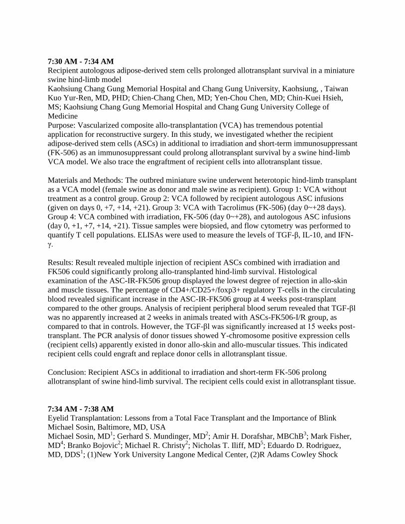
Clinical Chest wall, Thymus, and Heart Transplant Simulation
Johns Hopkins Hospital, Baltimore, MD, USA
Michael Sosin, MD1; Gianluca Torregrossa, MD
2; Amir H. Dorafshar, MBChB
1; (1)Johns
Hopkins Hospital, (2)Mount Sinai Hospital
Purpose

The integration of vascularized composite allotransplantation (VCA) into the field of clinical
cardiac transplantation may serve as a potential solution or at the very least may improve
multiple challenges to cardiac surgery by expanding the donor pool, prolonging allograft
survival, reconstructing chest wall deformity, and decreasing immunosuppressive requirements.
Recently, our group has described a proof-of-concept cadaveric model of harvesting the chest
wall, thymus and heart as an en bloc donor allograft.1 The purpose of this study is to design a
proof-of-concept cadaveric model to describe technical aspects of allograft harvest and recipient
inset of two different techniques termed, ‘en bloc' and ‘two stage.' Moreover, each technique is
critically evaluated based upon advantages, disadvantages, potential risks, and ability to be
integrated into a clinical practice.
Methodology/Design
Following institutional review board approval, 4 cadavers (2 male and 2 female) were obtained
and identified as donor or recipient. Two different techniques of combined solid organ and VCA
entitled: ‘en bloc' and ‘two stage' were designed to assess for technical feasibility, degree of
difficulty, and timing. The ‘en bloc' combined solid organ and VCA technique incorporates the
skin, sternum, costae, intercostal muscles, thymus, and heart as a single transferrable allograft.
(Figure 1) The ‘two stage' combined solid organ and VCA technique incorporates two
transferrable allografts: (1) heart and (2) skin, sternum, costae, intercostal muscles, and thymus
as two distinct entities that are anastomosed into one allograft during the recipient inset.
Results
A total of 2 mock chest wall, thymus, and heart transplant procedures were performed in
cadaveric donor and recipients. The ‘en bloc' and ‘two stage' procedures were both successfully
performed. The ‘en bloc' and ‘two stage' procedures were both successfully performed in the
presence of a multidisciplinary team of cardiac surgeons and plastic and reconstructive surgeons
present for both procedures (Video 1). Table 1 and 2 summarizes the differences of both
techniques.
Conclusion
Our cadaveric surgical simulation demonstrates both, the ‘en bloc' and the ‘two stage' chest wall,
thymus, and heart VCA are technically feasible models for clinical application that are associated
with their respective advantages and disadvantages.

Figure 1. (a) En-bloc allograft, (b) recipient, and (c) inset.
8:18 AM - 8:22 AM Prevalence and Distribution of Potential Vascularized Composite Allotransplant Donors,
Implications for Optimizing the Donor-Recipient Match in a UNOS-Based Allocation System
Institute for Plastic Surgery, Southern Illinois University, Springfield, IL, USA
Shaun D. Mendenhall, MD; Mauricio De la Garza; Tim Daugherty; Jennifer Koechle; Emma
Hoffmann; Steven Verhulst; Michael Neumeister; Bradford West; Southern Illinois University
School of Medicine
BACKGROUND: Vascularized composite allotransplantation (VCA) is the transplantation of
multiple tissue types as a functional unit such as a hand, face, or abdominal wall. There have
been close to 100 hand and 29 face transplants to date. The 5-year allograft survival rate for

hand transplants is > 90% compared to 75% for kidney transplants. UNOS recently elected to
classify VCAs as "organs" and began oversight of VCA allocation on July 3rd
, 2014. Little is
known about the prevalence and distribution of organ donors who could also be candidates for
VCA donation in this new allocation system.
METHODS: A custom dataset of donor characteristics was obtained from UNOS of all brain-
dead donors from 2008-2013. To identify the prevalence of potential VCA donors, inclusion and
exclusion criteria used for VCA were applied to the dataset. Frequency analyses were then
performed of characteristics important for VCA matching.
RESULTS: The dataset began with 42,414 brain-dead donors and after applying the inclusion
and exclusion criteria, decreased to 17,460 (41.2%). The number of potential VCA donors per
UNOS region ranged from 85-527/yr (Fig. 1). The majority of potential VCA donors were blood
type O, CMV+, Whites, with the least common profile being blood type AB, CMV-, Asians (Fig.
2).
CONCLUSIONS: VCA is in the early stages of standard of care as evidenced by UNOS
oversight and increasing acceptance by the medical community. Analysis of the UNOS donor
database reveals a large potential donor pool. Understanding the characteristics of previous
organ donors can guide VCA teams in optimizing the donor-recipient match in this new field of
transplantation and thus maximize patient outcomes following transplantation.

8:22 AM - 8:28 AM
Discussion