Embed Size (px)
Citation preview
Experimental and Toxicologic Pathology 68 (2016) 47–53
Chronic lead poisoning magnifies bone detrimental effects in anovariectomized rat model of postmenopausal osteoporosis
Ching Ming Leea, Antonela Romina Terrizzia, Clarisa Bozzinia, Adriana Emilce Piñeirob,María Inés Contia, María Pilar Martíneza,*aDepartment of Physiology, Faculty of Dentistry, University of Buenos Aires, ArgentinabDepartment of Toxicology and Legal Chemistry, Faculty of Pharmacy and Biochemistry, University of Buenos Aires, Argentina
A R T I C L E I N F O
Article history:Received 20 April 2015Received in revised form 11 August 2015Accepted 17 September 2015
Keywords:Lead poisoningBone biomechanicsOvariectomy
A B S T R A C T
Lead (Pb) is a persistent environmental contaminant that is mainly stored in bones being an importantsource of endogenous lead exposure during periods of increased bone resorption as occurs in menopause.As no evidence exists of which bone biomechanical properties are impaired in those elderly women whohad been exposed to Pb during their lifetime, the aim of the present study is to discern whether chroniclead poisoning magnifies the deterioration of bone biology that occurs in later stages of life. Weinvestigated the effect of Pb in the femora of ovariectomized (OVX) female Wistar rats who had beenintoxicated with 1000 ppm of Pb acetate in drinking water for 8 months. Structural properties weredetermined using a three-point bending mechanical test, and geometrical and material properties wereevaluated after obtaining the load/deformation curve. Areal Bone Mineral Density (BMD) was estimatedusing a bone densitometer. Femoral histomorphometry was carried out on slices dyed with H&E(Hematoxylin and Eosin). Pb and OVX decreased all structural properties with a higher effect when bothtreatments were applied together. Medullar and cortical area of femurs under OVX increased, allowingthe bone to accommodate its architecture, which was not observed under Pb intoxication. Pb and OVXsignificantly decreased BMD, showing lead treated ovariectomized rats (PbOVX) animals the lowest BMDlevels. Trabecular bone volume per total volume (BV/TV%) was decreased in OVX and PbOVX animals in54% compared to the control animals (p < 0.001). Pb femurs also showed 28% less trabeculae than thecontrol (p < 0.05). We demonstrated that Pb intoxication magnifies the impairment in bonebiomechanics of OVX rats with a consequent enhancement of the risk of fracture. These results enablethe discussion of the detrimental effects of lead intoxication in bone biology in elderly women.
ã 2015 Elsevier GmbH. All rights reserved.
Contents lists available at ScienceDirect
Experimental and Toxicologic Pathology
journal homepage: www.else vie r .de /e t p
1. Introduction
Lead (Pb) is a persistent air pollutant which can be released intothe environment via numerous routes, mainly by industrialactivities. During infancy and childhood, lead is deposited intrabecular bone because it is the most active site of remodeling;whereas, in adulthood lead is deposited in both trabecular andcortical bone (Aufderheide and Wittmers, 1992). We previouslyreported that chronic intoxication with Pb impaired growthparameters and induced negative effects on bone structuralproperties (Conti et al., 2012). Monir et al. (2010) demonstrated
* Corresponding author at: MT Alvear 2142, 3rd floor “A”, Argentina.Fax: +54 1145083958.
E-mail addresses: [email protected], [email protected](M.P. Martínez).
http://dx.doi.org/10.1016/j.etp.2015.09.0070940-2993/ã 2015 Elsevier GmbH. All rights reserved.
that lead exposure in adult bone of female mice decreased bonemineral density and collagen maturity, altered mineral/matrixratios and increased bone marrow area and bone turnover,resulting in a trend toward weaker bones. As Pb in bone has ahalf-life in the order of years to decades, skeletal stores may be animportant source of endogenous lead exposure during periods ofincreased bone resorption, as occurs in menopause. Changes inhormonal status related to menopause cause bone tissue loss and adecrease in bone mineral density (Meema and Meema, 1976),affecting bone tissue material properties at multiple scales,resulting in low bone mass and micro-architectural deterioration(Ettinger et al., 1998). Previously reported studies have shown asignificant association between older women with higher bloodlead levels and an increased risk of osteoporosis with a consequentsusceptibility to bone fractures (Khalil et al., 2008). Nash et al.(2004) demonstrated that lead stored in bone could significantlyincrease blood lead levels in perimenopausal women due to
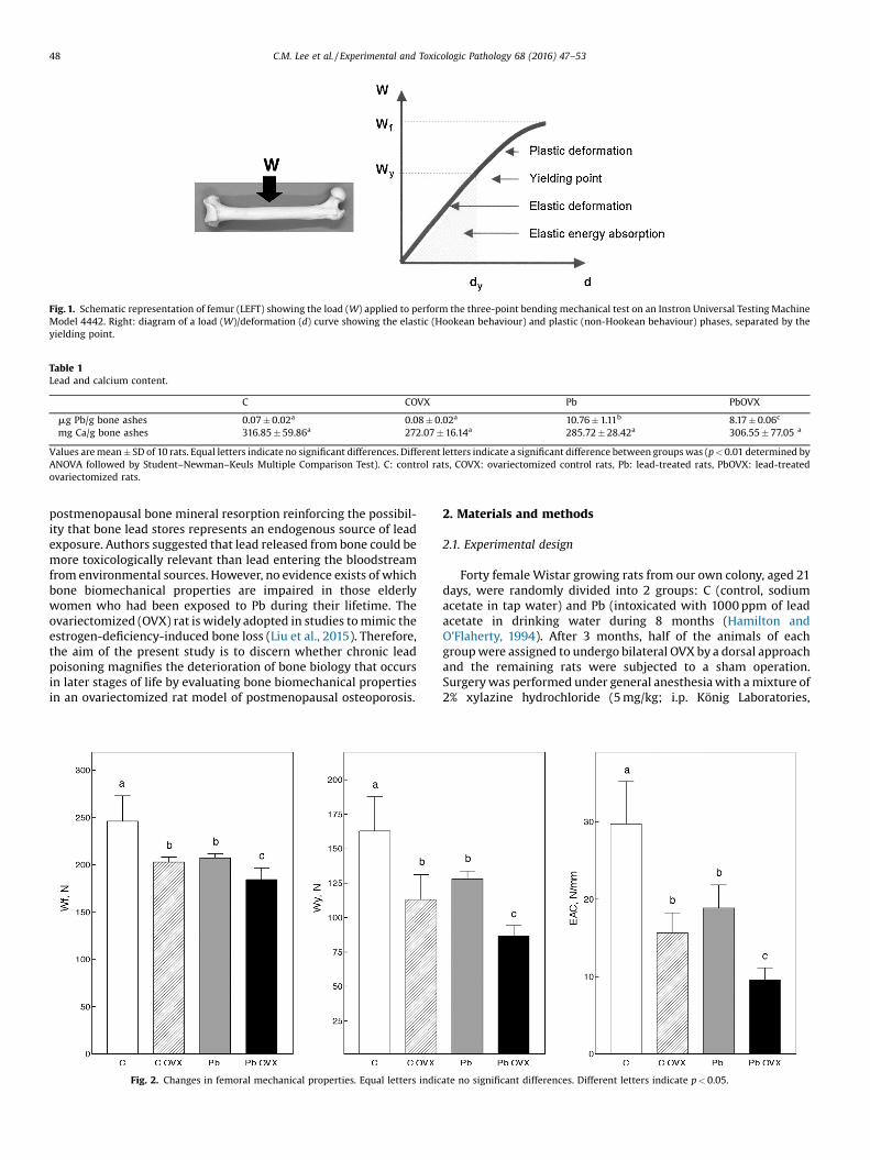
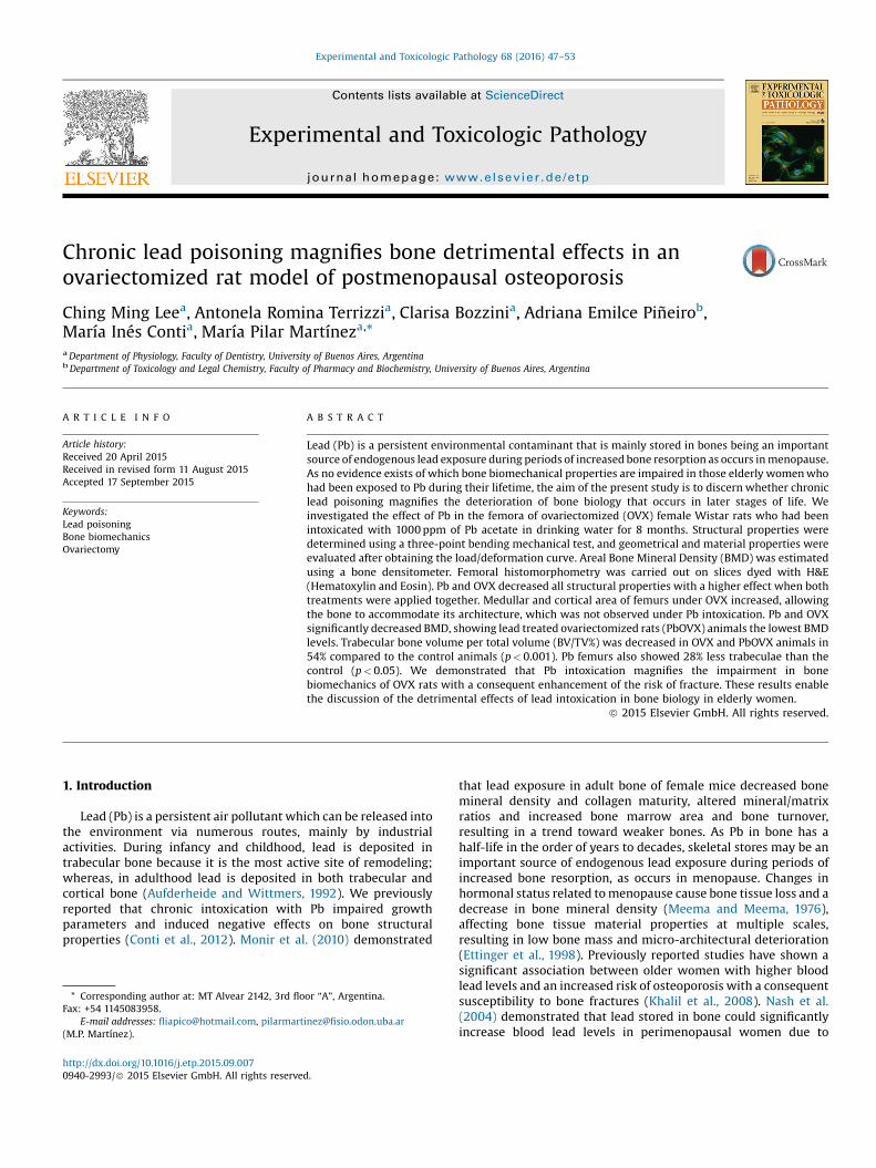
Fig. 1. Schematic representation of femur (LEFT) showing the load (W) applied to perform the three-point bending mechanical test on an Instron Universal Testing MachineModel 4442. Right: diagram of a load (W)/deformation (d) curve showing the elastic (Hookean behaviour) and plastic (non-Hookean behaviour) phases, separated by theyielding point.
Table 1Lead and calcium content.
C COVX Pb PbOVX
mg Pb/g bone ashes 0.07 � 0.02a 0.08 � 0.02a 10.76 � 1.11b 8.17 � 0.06c
mg Ca/g bone ashes 316.85 � 59.86a 272.07 � 16.14a 285.72 � 28.42a 306.55 � 77.05 a
Values are mean � SD of 10 rats. Equal letters indicate no significant differences. Different letters indicate a significant difference between groups was (p < 0.01 determined byANOVA followed by Student–Newman–Keuls Multiple Comparison Test). C: control rats, COVX: ovariectomized control rats, Pb: lead-treated rats, PbOVX: lead-treatedovariectomized rats.
48 C.M. Lee et al. / Experimental and Toxicologic Pathology 68 (2016) 47–53
postmenopausal bone mineral resorption reinforcing the possibil-ity that bone lead stores represents an endogenous source of leadexposure. Authors suggested that lead released from bone could bemore toxicologically relevant than lead entering the bloodstreamfrom environmental sources. However, no evidence exists of whichbone biomechanical properties are impaired in those elderlywomen who had been exposed to Pb during their lifetime. Theovariectomized (OVX) rat is widely adopted in studies to mimic theestrogen-deficiency-induced bone loss (Liu et al., 2015). Therefore,the aim of the present study is to discern whether chronic leadpoisoning magnifies the deterioration of bone biology that occursin later stages of life by evaluating bone biomechanical propertiesin an ovariectomized rat model of postmenopausal osteoporosis.
Fig. 2. Changes in femoral mechanical properties. Equal letters indic
2. Materials and methods
2.1. Experimental design
Forty female Wistar growing rats from our own colony, aged 21days, were randomly divided into 2 groups: C (control, sodiumacetate in tap water) and Pb (intoxicated with 1000 ppm of leadacetate in drinking water during 8 months (Hamilton andO’Flaherty, 1994). After 3 months, half of the animals of eachgroup were assigned to undergo bilateral OVX by a dorsal approachand the remaining rats were subjected to a sham operation.Surgery was performed under general anesthesia with a mixture of2% xylazine hydrochloride (5 mg/kg; i.p. König Laboratories,
ate no significant differences. Different letters indicate p < 0.05.
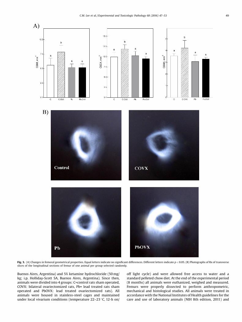
Fig. 3. (A) Changes in femoral geometrical properties. Equal letters indicate no significant differences. Different letters indicate p < 0.05. (B) Photographs of Rx of transverseslices of the longitudinal sections of femur of one animal per group selected randomly.
C.M. Lee et al. / Experimental and Toxicologic Pathology 68 (2016) 47–53 49
Buenos Aires, Argentina) and 5% ketamine hydrochloride (50 mg/kg; i.p. Holliday-Scott SA, Buenos Aires, Argentina). Since then,animals were divided into 4 groups: C=control rats sham operated,COVX: bilateral ovariectomized rats, Pb= lead treated rats shamoperated and PbOVX: lead treated ovariectomized rats). Allanimals were housed in stainless-steel cages and maintainedunder local vivarium conditions (temperature 22–23 �C, 12-h on/
off light cycle) and were allowed free access to water and astandard pelleted chow diet. At the end of the experimental period(8 months) all animals were euthanized, weighed and measured.Femurs were properly dissected to perform anthropometric,mechanical and histological studies. All animals were treated inaccordance with the National Institutes of Health guidelines for thecare and use of laboratory animals (NIH 8th edition, 2011) and
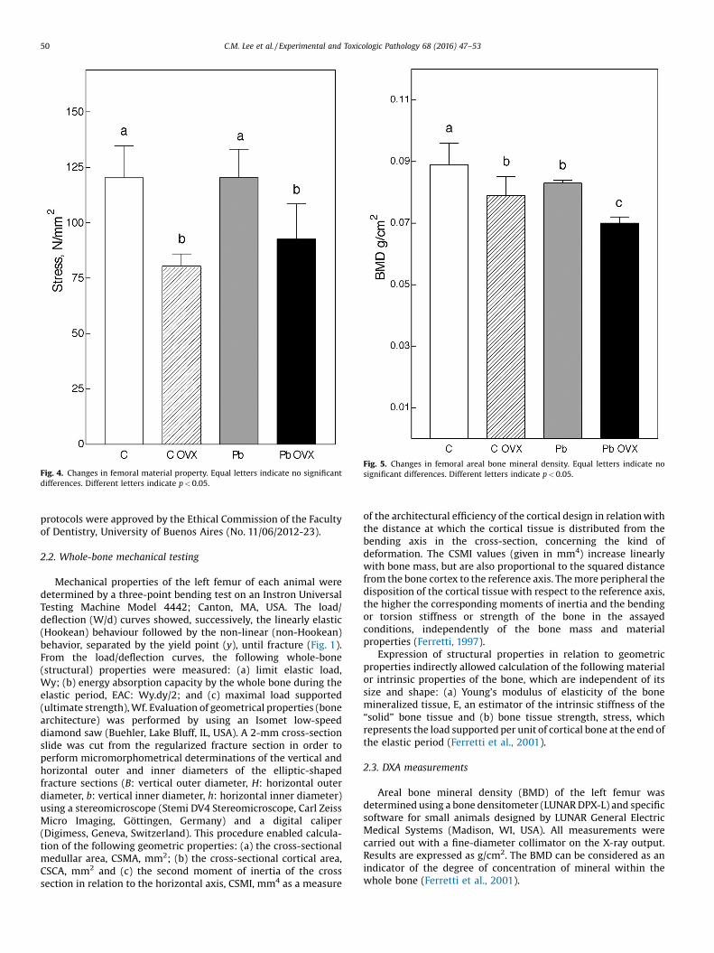
Fig. 4. Changes in femoral material property. Equal letters indicate no significantdifferences. Different letters indicate p < 0.05.
Fig. 5. Changes in femoral areal bone mineral density. Equal letters indicate nosignificant differences. Different letters indicate p < 0.05.
50 C.M. Lee et al. / Experimental and Toxicologic Pathology 68 (2016) 47–53
protocols were approved by the Ethical Commission of the Facultyof Dentistry, University of Buenos Aires (No. 11/06/2012-23).
2.2. Whole-bone mechanical testing
Mechanical properties of the left femur of each animal weredetermined by a three-point bending test on an Instron UniversalTesting Machine Model 4442; Canton, MA, USA. The load/deflection (W/d) curves showed, successively, the linearly elastic(Hookean) behaviour followed by the non-linear (non-Hookean)behavior, separated by the yield point (y), until fracture (Fig. 1).From the load/deflection curves, the following whole-bone(structural) properties were measured: (a) limit elastic load,Wy; (b) energy absorption capacity by the whole bone during theelastic period, EAC: Wy.dy/2; and (c) maximal load supported(ultimate strength), Wf. Evaluation of geometrical properties (bonearchitecture) was performed by using an Isomet low-speeddiamond saw (Buehler, Lake Bluff, IL, USA). A 2-mm cross-sectionslide was cut from the regularized fracture section in order toperform micromorphometrical determinations of the vertical andhorizontal outer and inner diameters of the elliptic-shapedfracture sections (B: vertical outer diameter, H: horizontal outerdiameter, b: vertical inner diameter, h: horizontal inner diameter)using a stereomicroscope (Stemi DV4 Stereomicroscope, Carl ZeissMicro Imaging, Göttingen, Germany) and a digital caliper(Digimess, Geneva, Switzerland). This procedure enabled calcula-tion of the following geometric properties: (a) the cross-sectionalmedullar area, CSMA, mm2; (b) the cross-sectional cortical area,CSCA, mm2 and (c) the second moment of inertia of the crosssection in relation to the horizontal axis, CSMI, mm4 as a measure
of the architectural efficiency of the cortical design in relation withthe distance at which the cortical tissue is distributed from thebending axis in the cross-section, concerning the kind ofdeformation. The CSMI values (given in mm4) increase linearlywith bone mass, but are also proportional to the squared distancefrom the bone cortex to the reference axis. The more peripheral thedisposition of the cortical tissue with respect to the reference axis,the higher the corresponding moments of inertia and the bendingor torsion stiffness or strength of the bone in the assayedconditions, independently of the bone mass and materialproperties (Ferretti, 1997).
Expression of structural properties in relation to geometricproperties indirectly allowed calculation of the following materialor intrinsic properties of the bone, which are independent of itssize and shape: (a) Young’s modulus of elasticity of the bonemineralized tissue, E, an estimator of the intrinsic stiffness of the“solid” bone tissue and (b) bone tissue strength, stress, whichrepresents the load supported per unit of cortical bone at the end ofthe elastic period (Ferretti et al., 2001).
2.3. DXA measurements
Areal bone mineral density (BMD) of the left femur wasdetermined using a bone densitometer (LUNAR DPX-L) and specificsoftware for small animals designed by LUNAR General ElectricMedical Systems (Madison, WI, USA). All measurements werecarried out with a fine-diameter collimator on the X-ray output.Results are expressed as g/cm2. The BMD can be considered as anindicator of the degree of concentration of mineral within thewhole bone (Ferretti et al., 2001).
C.M. Lee et al. / Experimental and Toxicologic Pathology 68 (2016) 47–53 51
2.4. Histological analyses
For histomorphometric studies femurs were resected and fixedin 10% buffered formaldehyde solution for 48 h, decalcified inethylendiaminotetracetic acid (EDTA, Sigma) pH 7.4 for 25 daysand then embedded in paraffin to perform sections following thelongitudinal axis. The sections were stained with Hematoxylin andEosin (H&E) for histological analysis of subchondral trabecularbone volume, (BV/TV%, Parfitt et al., 1987). The histomorphometricdeterminations were performed on digital microphotographs(40x) of the sections, using Image Pro Plus 4.5 software (MediaCybernetics, Inc., Warrendale, PA, USA).
2.5. Bone ash determinations
After mechanical testing, femurs were desiccated for bone ashdetermination in a muffle furnace at 600 �C for 18 h. Pb and calcium(Ca) content in these ashes was determined by a Varian SpectrAA-10 Plus atomic absorption spectrophotometer (Varian1) equippedwith a deuterium lamp for background correction and hollow-cathode lamps for each of the elements studied.
2.6. Statistical analysis
Data were analyzed by one-way analysis of variance (ANOVA),followed by Student–Newman–Keuls Multiple Comparison Test.Analyses were performed using the Software package Instat andPrism V.3 (GraphPad Software Inc., San Diego, USA). A p-value lessthan 0.05 was considered statistical significant.
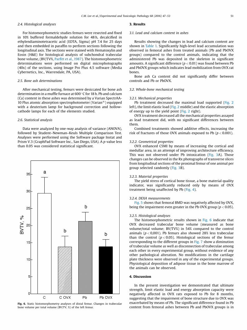
Fig. 6. Static histomorphometry analyses of distal femur. Changes in trabecularbone volume per total volume (BV/TV, %) of the left femur.
3. Results
3.1. Lead and calcium content in ashes
Results showing the changes in lead and calcium content areshown in Table 1. Significantly high-level lead accumulation wasobserved in femoral ashes from treated animals (Pb and PbOVXgroups) compared to the control animals, indicating that theadministered Pb was deposited in the skeleton in significantamounts. A significant difference (p < 0.01) was found between Pband PbOVX groups which indicates lead mobilization from OVX ratbones.
Bone ash Ca content did not significantly differ betweencontrols and Pb or PbOVX.
3.2. Whole-bone mechanical testing
3.2.1. Mechanical propertiesPb treatment decreased the maximal load supported (Fig. 2
left), the limit elastic load (Fig. 2 middle) and the elastic absorptionof energy up to the yield point (Fig. 2 right).
OVX treatment decreased all the mechanical properties assayedas lead treatment did, with no significant differences betweenthem.
Combined treatments showed additive effects, increasing therisk of fractures of those OVX animals exposed to Pb (p < 0.001).
3.2.2. Geometrical propertiesOVX enhanced CSMI by means of increasing the cortical and
medullar area, in an attempt of improving architecture efficiency.This was not observed under Pb intoxication (Fig. 3A). Thosechanges can be observed in the Rx photographs of transverse slicesfrom longitudinal sections of the proximal femur of one animal pergroup selected randomly (Fig. 3B).
3.2.3. Material propertiesThe yield stress of cortical bone tissue, a bone material quality
indicator, was significantly reduced only by means of OVXtreatment being unaffected by Pb (Fig. 4).
3.2.4. DEXA measurementsFig. 5 shows that femoral BMD was negatively affected by OVX,
being the impairment even greater in the Pb OVX group (p < 0.05).
3.2.5. Histological analysesThe histomorphometric results shown in Fig. 6 indicate that
OVX decreased trabecular bone volume (measured as bonevolume/total volume; BV/TV%) in 54% compared to the controlanimals (p < 0,001). Pb femurs also showed 28% less trabeculaethan the control (p < 0.05). Histological sections of the femurcorresponding to the different groups in Fig. 7 show a diminutionof trabecular volume as well as disconnection of trabeculae amongeach other in every experimental group, without evidence of anyother pathological alteration. No modifications in the cartilageplate thickness were observed in any of the experimental groups.Physiological deposition of adipose tissue in the bone marrow ofthe animals can be observed.
4. Discussion
In the present investigation we demonstrated that ultimatestrength, limit elastic load and energy absorption capacity werenegatively affected in OVX rats exposed to Pb for 8 months,suggesting that the impairment of bone structure due to OVX wasexacerbated by means of Pb. The significant difference found in Pbcontent from femoral ashes between Pb and PbOVX groups is in

Fig. 7. Femur histomorphometric analyses showing the effects of chronic lead poisoning in an ovariectomized rat model of postmenopausal osteoporosis. (C) Photograph oftransverse slice of the longitudinal section of femur (40�) of one randomly selected animal per control group. A high-resolution version of this slide for use with the VirtualMicroscope is available as eSlide: VM01012. C OVX: Photograph of transverse slice of the longitudinal section of femur (40�) of one randomly selected animal per controlovariectomized group (bilateral ovariectomy after 3 months from the beginning of the experimental period). A high-resolution version of this slide for use with the VirtualMicroscope is available as eSlide: VM01013. Pb: photograph of transverse slice of the longitudinal section of femur (40�) of one randomly selected animal per lead intoxicatedgroup (1000 ppm of lead acetate in drinking water during 8 months). A high-resolution version of this slide for use with the Virtual Microscope is available as eSlide: VM01014.PbOVX: Photograph of transverse slice of the longitudinal section of femur (40�) of one randomly selected animal per ovariectomized lead intoxicated group (1000 ppm of leadacetate in drinking water during 8 months and with bilateral ovariectomy after 3 months from the beginning of the experimental period). A high-resolution version of this slidefor use with the Virtual Microscope is available as eSlide: VM01015.
52 C.M. Lee et al. / Experimental and Toxicologic Pathology 68 (2016) 47–53
agreement with previously reported studies indicating that leadwas mobilized from the skeleton during conditions of high boneturnover, such as menopause (Silbergeld et al., 1988). Themechanical properties of bone strongly depend on the intrinsicmechanical quality of its constitutive substance (material proper-ties) and the amount and spatial distribution of the mineralisedtissue (geometrical properties) (Ferretti, 1997). Lead poisoningmay affect either of both. When we analysed bone tissue strength(stress), we found that this property decreased by means of OVX.This reduction was not observed in femurs of the lead intoxicatedgroups. Femurs of OVX animals enhanced its moment of inertia(CSMI) by resorption on the endosteal surface and apposition onthe periosteal surface in order to try to compensate theimpairment in the material properties. It is known that boneswith material distribution further away from the centre aresignificantly stronger (Cole and Van der Meulen, 2011). Interest-ingly, those femurs of the PbOVX group could not perform theadaptation mentioned above. It seems that Pb does not allowarchitectural accommodation which may be due to an alteredosteoclastic activity widely reported in the literature (Pounds et al.,1991).
The ability of bone to bear loads does not only depend onmaterial and geometrical properties, but also on bone total mass. Inthis experiment, BMD was decreased in femurs of the OVX group
and an additive effect due to Pb in OVX animals was observed. Ithas been established that small changes in BMD induce largechanges in bone strength and that sites with similar BMD butdifferent architecture, show differences in strength and stiffness(Cole and Van der Meulen, 2011). These findings would partiallyexplain the increased impairment in structural properties of theOVX animals exposed to Pb reported in the present investigation.
The manifestation of lead intoxication in bone is the result ofcomplex interplay between many effects, involving cellular andchemical processes in the bone matrix, and also systemic andendocrine effects. It has been established that Pb decreasescalcium absorption and replaces calcium hydroxyapatite to formPb phosphate, resulting in a decrease of Ca in bone (Hongke et al.,2014). In our study, Ca content in femur ashes did not showchanges between experimental groups, in accordance with otherstudies (Martínez et al., 2011; Monir et al., 2010). It would seemthat Ca content is not directly responsible for the impairment inbone structural properties, but also other factors as alteration incollagen fibers or microstructural crystal arrangements may beinvolved (Yerramshetty and Akkus, 2008). Further analyses ofmicro and nanoscale characteristics of the bone are necessary tofully elucidate the femoral material properties in this experimentalmodel.
C.M. Lee et al. / Experimental and Toxicologic Pathology 68 (2016) 47–53 53
In summary, we demonstrated that lead intoxication magnifiedthe impairment in bone biomechanics of OVX rats with aconsequent enhancement of the risk of fracture. What appearsto be happening, at least concerning mechanical properties, is thatPb would restrict the physiological architectural accommodationof long bones to compensate changes in material properties. It alsoseems to enhance the decreased bone mass in those bones alreadyaffected by OVX. The above results enable the discussion of thedetrimental effects of lead intoxication in bone biology in elderlywomen.
Conflict of interest
All authors have no conflicts of interest.
Funding information
This work was supported by research grants from University ofBuenos Aires (UBACyT 20020110100014).
Acknowledgments
The authors acknowledge the collaboration of physiologylaboratory technician Graciela M. Champin, Elsa Lingua from theDepartment of Physiology and Ricardo Orzuza from the Depart-ment of Biochemistry, School of Dentistry, University of BuenosAires.
References
Aufderheide AC, Wittmers Jr. LE. Selected aspects of the spatial distribution of leadin bone. Neurotoxicology 1992;13:809–19.
Cole J, Van der Meulen M. Whole bone mechanics and bone quality. Clin. Orthop.Relat. Res. 2011;469:2139–49.
Conti MI, Terrizzi AR, Lee CM, Mandalunis PM, Bozzini C, Piñeiro AE, Martinez MP.Effects of lead exposure on growth and bone biology in growing rats exposed tosimulated high altitude. Bull. Environ. Contam. Toxicol. 2012;88(6):1033–7.
Ettinger B, Pressman A, Sklarin P, Bauer DC, Cauley JA, Cummings SR. Associationsbetween low levels of serum estradiol, bone density, and fractures amongelderly women: the study of osteoporotic fractures. J. Clin. Endocrinol. Metab.1998;83:2239–43.
Ferretti JL, Cointry GR, Capozza RF, Capiglioni R, Chiappe MA. Analysis ofbiomechanical effects on bone and on the bone muscle interactions in smallanimal models. J. Musculoskelet. Neuron Interact. 2001;1:263–74.
Ferretti JL. Biomechanical properties of bone. Osteoporosis and Bone Densitometry.Berlin: Springer Verlag; 1997 p. 143–161.
Hamilton JD, O’Flaherty EJ. Effects of lead exposure on skeletal development in rats.Fundam. Appl. Toxicol. 1994;22(4):594–604.
Hongke L, Guiping Y, Zhongqiong Y, Shujun D, Renyong J, Jiao X, Xu S, Li L, Cheng L.Effects of subchronic exposure to lead acetate and cadmium chloride on rat’sbone: Ca and Pi contents, bone density, and histopathological evaluation. Int. J.Clin. Exp. Pathol. 2014;7:640–7.
Khalil N, Cauley JA, Wilson JW, Talbott EO, Morrow L, Hochberg MC, Hillier TA,Muldoon SB, Cummings SR. Relationship of blood lead levels to incidentnonspine fractures and falls in older women: the study of osteoporoticfractures. J. Bone Miner. Res. 2008;23:1417–25.
Liu XL, Li CL, Lu WW, Cai WX, Zheng LW. Skeletal site-specific response toovariectomy in a rat model: change in bone density and microarchitecture. Clin.Oral Implants Res. 2015;26(4):392–8.
Martínez MP, Bozzini C, Olivera MI, Dmytrenko G, Conti MI. Aluminum bone toxicityin immature rats exposed to simulated high altitude. J. Bone Miner. Metab.2011;29(5):526–34.
Meema S, Meema HE. Menopausal bone loss and estrogen replacement. Isr. J. Med.Sci. 1976;12:601–6.
Monir AU, Gundberg CM, Yagerman SE, van der Meulen MCH, Budell WC, Boskey AL,Dowd TL. The effect of lead on bone mineral properties from female adult C57/BL6 mice. Bone 2010;47:888–94.
Nash D, Magder LS, Sherwin R, Rubin RJ, Silbergeld EK. Bone density-relatedpredictors of blood lead level among periand postmenopausal women in theUnited States, The Third National Health and Nutrition Examination Survey,1988–1994. Am. J. Epidemiol. 2004;160:901–11.
Parfitt AM, Drezner MK, Glorieux FH, Kanis JA, Malluche H, Meunier PJ, et al. Bonehistomorphometry: standardization of nomenclature, symbols, and units:Report of the ASBMR Histomorphometry Nomenclature Committee. J. BoneMiner. Res. 1987;2:595–610.
Pounds JG, Long GJ, Rosent JF. Cellular and molecular toxicity of lead in bone.Environ. Health Perspect. 1991;91:17–32.
Silbergeld EK, Schwartz J, Mahaffey K. Lead and osteoporosis: mobilization of leadfrom bone in postmenopausal women. Environ. Res. 1988;47:79–94.
Yerramshetty JS, Akkus O. The associations between mineral crystallinity and themechanical properties of human cortical bone. Bone 2008;42:476–82.