Embed Size (px)
Citation preview
REVIEW
Expanded roles of the Fanconi anemiapathway in preserving genomic stability
Younghoon Kee and Alan D. D’Andrea1
Department of Radiation Oncology and Pediatric Oncology, Dana-Farber Cancer Institute, Harvard Medical School, Boston,Massachusetts 02115, USA
Studying rare human genetic diseases often leads to abetter understanding of normal cellular functions. Fan-coni anemia (FA), for example, has elucidated a novelDNA repair mechanism required for maintaining geno-mic stability and preventing cancer. The FA pathway, anessential tumor-suppressive pathway, is required for pro-tecting the human genome from a specific type of DNAdamage; namely, DNA interstrand cross-links (ICLs). Inthis review, we discuss the recent progress in the studyof the FA pathway, such as the identification of newFANCM-binding partners and the identification ofRAD51C and FAN1 (Fanconi-associated nuclease 1) asnew FA pathway-related proteins. We also focus on therole of the FA pathway as a potential regulator of DNArepair choices in response to double-strand breaks, and itsnovel functions during the mitotic phase of the cell cycle.
The basic Fanconi anemia (FA) pathway
FA is a genomic instability syndrome characterized bybone marrow failure, developmental abnormalities, andincreased incidence of cancers (D’Andrea and Grompe2003; Moldovan and D’Andrea 2009). At the cellularlevel, FA cells display increased chromosomal aberra-tions, particularly radials, and hypersensitivity to DNAinterstrand cross-link (ICL) agents. DNA ICLs are amongthe most deleterious DNA lesions, since they block DNAreplication and transcription. DNA ICLs can be caused byendogenous sources such as nitrous acid and aldehydes,or exogenous agents such as Cisplatin and its derivatives.Because of its essential functions in preserving genomicstability, the FA pathway provides a unique model forstudying eukaryotic DNA repair and DNA damage re-sponses, particularly against DNA ICLs.
FA is a genetically heterogeneous disease, caused bymutations in at least 13 distinct genes (FANCA, FANCB,FANCC, FANCD1, FANCD2, FANCE, FANCF, FANCG,FANCI, FANCJ, FANCL, FANCM, and FANCN). All 13
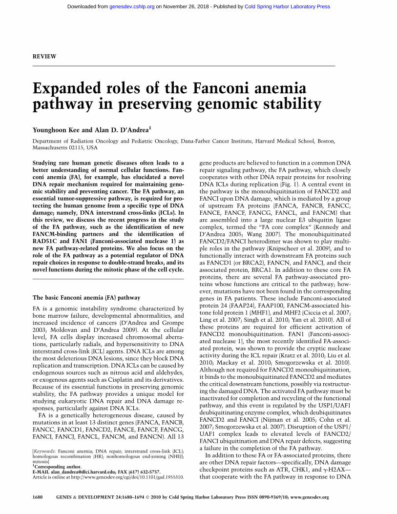
gene products are believed to function in a common DNArepair signaling pathway, the FA pathway, which closelycooperates with other DNA repair proteins for resolvingDNA ICLs during replication (Fig. 1). A central event inthe pathway is the monoubiquitination of FANCD2 andFANCI upon DNA damage, which is mediated by a groupof upstream FA proteins (FANCA, FANCB, FANCC,FANCE, FANCF, FANCG, FANCL, and FANCM) thatare assembled into a large nuclear E3 ubiquitin ligasecomplex, termed the ‘‘FA core complex’’ (Kennedy andD’Andrea 2005; Wang 2007). The monoubiquitinatedFANCD2/FANCI heterodimer was shown to play multi-ple roles in the pathway (Knipscheer et al. 2009), and tofunctionally interact with downstream FA proteins suchas FANCD1 (or BRCA2), FANCN, and FANCJ, and theirassociated protein, BRCA1. In addition to these core FAproteins, there are several FA pathway-associated pro-teins whose functions are critical to the pathway; how-ever, mutations have not been found in the correspondinggenes in FA patients. These include Fanconi-associatedprotein 24 (FAAP24), FAAP100, FANCM-associated his-tone fold protein 1 (MHF1), and MHF2 (Ciccia et al. 2007;Ling et al. 2007; Singh et al. 2010; Yan et al. 2010). All ofthese proteins are required for efficient activation ofFANCD2 monoubiquitination. FAN1 (Fanconi-associ-ated nuclease 1), the most recently identified FA-associ-ated protein, was shown to provide the cryptic nucleaseactivity during the ICL repair (Kratz et al. 2010; Liu et al.2010; Mackay et al. 2010; Smogorzewska et al. 2010).Although not required for FANCD2 monoubiquitination,it binds to the monoubiquitinated FANCD2 and mediatesthe critical downstream functions, possibly via restructur-ing the damaged DNA. The activated FA pathway must beinactivated for completion and recycling of the functionalpathway, and this event is regulated by the USP1/UAF1deubiquitinating enzyme complex, which deubiquitinatesFANCD2 and FANCI (Nijman et al. 2005; Cohn et al.2007; Smogorzewska et al. 2007). Disruption of the USP1/UAF1 complex leads to elevated levels of FANCD2/FANCI ubiquitination and DNA repair defects, suggestinga failure in the completion of the FA pathway.
In addition to these FA or FA-associated proteins, thereare other DNA repair factors—specifically, DNA damagecheckpoint proteins such as ATR, CHK1, and g-H2AX—that cooperate with the FA pathway in response to DNA
[Keywords: Fanconi anemia; DNA repair; interstrand cross-link (ICL);homologous recombination (HR); nonhomologous end-joining (NHEJ);mitosis]1Corresponding author.E-MAIL [email protected]; FAX (617) 632-5757.Article is online at http://www.genesdev.org/cgi/doi/10.1101/gad.1955310.
1680 GENES & DEVELOPMENT 24:1680–1694 � 2010 by Cold Spring Harbor Laboratory Press ISSN 0890-9369/10; www.genesdev.org
Cold Spring Harbor Laboratory Press on November 26, 2018 - Published by genesdev.cshlp.orgDownloaded from

damage (Andreassen et al. 2004; Wang 2007; Moldovanand D’Andrea 2009). These checkpoint proteins preservegenomic stability by arresting the cell cycle after DNAdamage. Cells deficient in ATR kinase, a master regulatorof S-phase checkpoint, are impaired in damage-induciblemonoubiquitination of FANCD2, and are hypersensi-tive to ICLs (Andreassen et al. 2004). Many of the FAproteins are direct substrates of ATR or its effectorkinase, CHK1—including FANCD2, FANCI, FANCA,and FANCE (Pichierri and Rosselli 2004; Smogorzewskaet al. 2007; Wang et al. 2007; Ishiai et al. 2008; Collinset al. 2009)—and these phosphorylation events are re-quired for a functional FA pathway and ICL repair. TheFANCM and FAAP24 complex is also required upstreamfor efficient ATR-mediated checkpoint control (Colliset al. 2008; Luke-Glaser et al. 2010; Schwab et al. 2010).
Recently, FANCJ was also shown to regulate the ATR-dependent checkpoint (Gong et al. 2010). Taken together,these studies suggest that the FA pathway is closelyinterconnected with other DNA damage response signals.The precise molecular function of individual FA proteinsin repairing DNA and communicating with other DNAdamage responses remains largely unknown. However,recent studies have provided unique insights to the field.These findings may potentially bring venues for noveltherapeutic approaches for treating both FA patients andFA pathway-deficient tumors.
FANCM plays multiple distinct roles during ICL repair
While facilitating DNA cross-link repair, the FA pathwaymust coordinate other DNA damage-responsive events to
Figure 1. A schematic model for the FA pathway.(A) Activation of the FA pathway. DNA ICL isdirectly recognized by FANCM–FAAP24–MHF pro-tein complex. This complex recruits the FA corecomplex by direct interaction between FANCM andFANCF. The recruited FA core complex, containinga PHD E3 ubiquitin ligase domain in the FANCLsubunit, subsequently monoubiquitinates its twosubstrates, FANCD2 and FANCI, on chromatin.The monoubiquitinated FANCD2–FANCI becomesan active form, recruits newly identified nucleaseFAN1 or interacts with a series of DNA repair pro-teins (including BRCA1, PALB2 ½FANCN�, BRCA2,and FANCJ ½BACH1/BRIP1�) at the damaged sites,and facilitates downstream repair pathways. RAD51C,a newly identified FA-like protein, may have a func-tional interaction with FANCD2 at this step.FANCD2–FANCI probably also recruits other nu-cleases and TLS polymerases to process the ICL (notshown). The new players in the FA pathway de-scribed in the text are highlighted in dashed lines.(B) The FA pathway cross-talks with ATR–CHK1checkpoint proteins. ATR and its effector kinase,CHK1, are required for damage-inducible activationof the FA pathway. ATR–CHK1 phosphorylates (redarrows) multiple FA and FA-associated proteins,including FANCA, FANCE, FANCD2, FANCI, andBRCA1. FANCM is also phosphorylated upon DNAdamage by an unknown kinase. In turn, the stabilityand activity of ATR–CHK1 are promoted (blackarrows) by the FANCM–FAAP24 heterodimer andFANCJ by independent mechanisms. FANCM alsomediates the formation of the BRAFT complex,which contains the FA core complex members andthe BLM complex containing Topo IIIa and RMI1/2(not shown).
Expanded roles of the FA pathway
GENES & DEVELOPMENT 1681
Cold Spring Harbor Laboratory Press on November 26, 2018 - Published by genesdev.cshlp.orgDownloaded from

stabilize stalled replication forks, to convey signals toDNA checkpoint pathways, and to facilitate recovery ofreplication forks. These events are largely coordinated byFANCM, a unique member of the FA pathway that playsdistinct roles in checkpoint activation, chromatin remod-eling, and ICL repair. FANCM contains a DEAH-typeATPase-dependent helicase domain, and it is one of theonly two FA proteins with recognizable enzymatic activ-ities, together with the helicase activity of FANCJ (Whitby2010). FANCM is evolutionarily well conserved, as it isthe only FA protein that has apparent orthologs in yeast(MPH1 in Saccharomyces cerevisiae, and Fml1 in Schizo-saccharomyces pombe) and archaebacteria (Hef) (Meeteiet al. 2005; Sun et al. 2008). Also, FANCM is one of onlytwo proteins in the human genome known to contain botha helicase/ATPase domain and an endonuclease domain ina single polypeptide, the other being ERCC4 (XPF) (Aliet al. 2009). The identity of FANCM as a bona fide FAprotein is still in question, since the only known FANCM-deficient patient cell line, EUFA867, harbors an additionalmutation in FANCA, and the full correction of the FAphenotype requires ectopic expression of both FANCMand FANCA (Singh et al. 2009). However, FANCM clearlyplays critical roles in the FA pathway, and functionscooperatively with other FA core members. For instance,in DT40 cells, a double knockout of FANCM and FANCCexhibits ICL hypersensitivity similar to individual geneknockouts, indicating that FANCM and FANCC areepistatic (Mosedale et al. 2005). One role of FANCM inthe FA pathway is to sense damaged DNA and recruit theFA core complex. FANCM was originally identified asa subunit of the FA core complex (Meetei et al. 2005), butits cellular distribution is distinct from the other FA coremembers. FANCM forms a heterodimeric complex withFAAP24, a protein sharing high similarity to the C ter-minus of FANCM and also containing an endonuclease-like domain (Ciccia et al. 2007). In vitro, the FANCM/FAAP24 complex binds directly to DNA structures thatresemble stalled replication forks. Also, the FANCM/FAAP24 complex binds constitutively to chromatin,whereas other FA core proteins become enriched in thechromatin fraction only after DNA damage (Meetei et al.2005; Mosedale et al. 2005; Kim et al. 2008). Furthermore,depletion of FANCM results in impaired localization ofthe FA core proteins. Taken together, these results suggesta model in which the FANCM/FAAP24 complex serves asa sensor that recognizes the DNA lesion and subsequentlyrecruits the FA core complex to induce monoubiquitina-tion of FANCD2/FANCI. However, cells from the FANCMknockout mice are still capable of inducing residualmonoubiquitinated FANCD2 (Bakker et al. 2009). Accord-ingly, there may be a FANCM-independent recruitingmechanism for the FA core complex, or there may bea basal-level interaction between the FA core complex andFANCD2/I in the absence of FANCM/FAAP24.
FANCM also has unique features that are not shared byother FA core complex members. FANCM knockout micedisplay typical FA phenotypes (e.g., hypersensitivity toICL, chromosomal breakage, and increased G2/M arrest),but also exhibit additional atypical phenotypes such as
a low female ratio and increased cancer-related death(Bakker et al. 2009). FANCM-deficient cells are also hy-persensitive to camptothecin, a topoisomerae I (Topo I)inhibitor that induces replication fork arrests, followedby DSB generation (Singh et al. 2009).
Unlike other FA subtypes, FANCM-deficient cells ex-hibit elevated frequency of sister chromatid exchange(SCE). Bloom’s syndrome, a rare genetic disorder causedby a single mutation in the RecQ family helicase BLM,also exhibits elevated SCE, among other phenotypes thatresemble FA, such as reduced fertility, genomic instability,and increased hematological cancers (Liu and West 2008).Interestingly, the overlapping phenotypes of FA and BLMcould be linked molecularly to the ICL-induced assemblyof a supercomplex, termed BRAFT (BLM, RPA ½replicationprotein A�, FA, and Topo IIIa), that contains the membersof the FA core complex, BLM, and its associated proteins(Meetei et al. 2003). A recent study provided a directdemonstration that FANCM serves as an adapter betweenthe FA proteins and the BLM complexes (Deans and West2009), and it was demonstrated that the elevated fre-quency of SCE is due to the failed assembly of the BRAFTthrough FANCM. Together, FANCM plays a critical rolein mediating the cross-talk between the FA proteins andBLM complex, particularly in response to ICLs, and thiscould be the fundamental link for the overlapping clinicalphenotypes between the two genetic disorders.
One of the FA-independent functions of FANCM is itsability to activate DNA damage checkpoints. FANCMand FAAP24, but not other FA proteins, were shown toassociate physically with a complex containing ATR–HCLK (HCLK2 is a stabilizer and signaling mediator ofATR), and thus are required for proper activation of theATR-mediated S-phase checkpoints in response to repli-cation stress (Collis et al. 2008). Depletion of FANCM–FAAP24 leads to uninhibited cell cycle progressionthrough G2/M phases. This function is dependent on itsATPase activity, which is dispensable for its monoubi-quitination of FANCD2/FANCI, further suggesting theexistence of at least two separate functions. One mech-anism for ATR activation is the ability of FANCM toretain TOPBP1, a stimulator of ATR kinase activity, inthe chromatin (Schwab et al. 2010). Although this check-point-activating function is not involved directly withthe FA pathway, FANCM-mediated ATR activation mayprovide an indirect mechanism for further activating theFA pathway (see Fig. 1B). Also, more recent studiesindicate that FANCM/FAAP24 can activate a specificICL checkpoint response by enhancing RPA binding atthe site of the ICL (AD D’Andrea, unpubl.).
In addition to checkpoint signaling, FANCM has a role inremodeling replication forks. In vitro, FANCM binds DNAstructures that mimic replication forks and Holliday junc-tions, and moves the junction points through its branchmigration activity in an ATPase-dependent manner (Gariet al. 2008b). This activity is also not coupled to the FApathway, since the ATPase activity is dispensable forFANCD2/I monoubiquitination (Xue et al. 2008). A sub-sequent report showed that the fork migration activity leadsto fork reversal during replication blockage, and suggested
Kee and D’Andrea
1682 GENES & DEVELOPMENT
Cold Spring Harbor Laboratory Press on November 26, 2018 - Published by genesdev.cshlp.orgDownloaded from

that this might be a mechanism for stabilizing forks duringICL repair, and limiting promiscuous recombination events(Gari et al. 2008a). Indeed, the ATPase activity was shown tobe required for resistance to ICL agents (Xue et al. 2008). Invivo, FANCM-deficient cells showed uncontrolled replica-tion fork movement and impaired fork restart during re-covery from DNA damage (Luke-Glaser et al. 2010; Schwabet al. 2010), in line with FANCM’s a role in remodeling forksduring DNA repair. Taken together, FANCM may preventundesired DNA synthesis during replication stress and DNArepairby coordinating checkpointactivation and remodelingDNA fork structures, while activating the FA pathway tofacilitate ICL repair. FANCM-deficient cells are hypersensi-tive to various agents that inhibit replication fork progres-sion (e.g., hydroxyurea ½HU�, aphidicolin ½APH�, and camp-tothecin) (Schwab et al. 2010) that are not seen in otherFA-deficient cells, further suggesting that FANCM has widerroles in protecting stalled forks from mutagenic recombina-tion and collapse.
Recent studies further demonstrate the complexity ofFANCM function and regulation. These studies identifiedtwo histone fold proteins, named MHF1 and MHF2, asbinding partners of FANCM (Singh et al. 2010; Yan et al.2010). Both MHF1 and MHF2 appear to have coevolvedwith FANCM, as their orthologs exist from yeast tohuman (Yan et al. 2010). The new FANCM-associated pro-teins cooperate in both the FA pathway-dependent andindependent functions of FANCM, as they are required forFANCD2 monoubiquitination, cross-linker repair, andbranch migration-mediated fork reversal. The main func-tion of MHF1–2 proteins appears to be stabilizing FANCMat the sites of ICL lesion (Yan et al. 2010). In addition, theepistatic relationship between the MHF and FANCMorthologs was observed in DT40 and yeast, suggestingtheir cooperation in a common pathway (Yan et al. 2010).Both MHF1 and MHF2 were shown previously to be iden-tical to centromere-binding factor CENP-S and CENP-X,suggesting that they may also act to redirect FANCM topromote centromere stability (Yan et al. 2010)
Collectively, FANCM exerts multiple roles in preserv-ing genome stability in response to DNA damage. Dam-aged DNA, especially ICL-modified DNA, can block theprogression of replication forks. FANCM initially acti-vates the S-phase checkpoint via its association with theATR/CHK1/HCLK signaling complex to inhibit DNAreplication. Next, FANCM recruits the FA core complexto the damaged chromatin and induces monoubiquitina-tion of FANCD2/FANCI, thereby facilitating the process-ing of ICL repair, as illustrated in Figure 1. Finally,FANCM stabilizes the stalled replication forks by inhib-iting reversal of the fork progression and limiting un-wanted crossovers between sister chromatids.
Broader roles of the FA pathway during ICL repair
Repairing double-strand breaks (DSBs)—homologousrecombination (HR) vs. nonhomologousend-joining (NHEJ)
The FA pathway plays a fundamental role in coordinatingthe repair of DNA DSBs. DSBs can be generated in
numerous ways. Various genotoxic agents generate DSBs,including ionizing radiation (IR), chemicals that blockDNA replication fork progression (such as HU), orbyproducts of cellular metabolism (such as reactiveoxygen species (ROS)�. DSBs can also be generated as anintermediate of normal cellular processes during immu-noglobulin class switching in immune cells. Failure toproperly repair DSBs leads to chromosome transloca-tions, genomic instability, and cell death or transforma-tion. Cells have evolved two major specialized mecha-nisms to cope with these highly deleterious lesions;namely, HR and NHEJ.
Through HR, DNA sequences are exchanged betweentwo similar or identical DNA strands. NHEJ is an al-ternative pathway in which two broken DSBs are sealeddirectly, without the need of homologous templates.Cells have evolved specialized proteins that facilitateeither HR or NHEJ for DSB repair. Figure 2 describesthe simple mechanistic models for HR and NHEJ, bothpathways requiring multiple-step reactions. Because HRuses genetically identical complementary DNA strandsas templates for repair, it is error-free, while NHEJ can beerror-prone, since DNA end ligation can occur withoutpreserving the correct frame of codons, resulting in loss ofgenetic information. Despite the higher risk of genomicinstability, mammalian cells depend primarily on NHEJ,unlike in yeast, where HR is the primary choice forrepairing DSBs.
DSB as an intermediate step in ICL repair
In addition to the above-mentioned ‘‘direct’’ DSB-gener-ating means, a DSB can also be generated as an interme-diate of the ICL-repairing process (Fig. 3; Kennedy andD’Andrea 2005; Moldovan and D’Andrea 2009). In thecurrent model, ICL hinders the progression of replicationfork, which will activate checkpoints and monoubiquiti-nation of FANCD2/FANCI, while the fork is stabilized byrepair proteins. The cross-links are thought to be unhookedinitially by serial activities of at least two endonucleases(XPF–ERCC1 and MUS81–EME1) (Niedernhofer et al. 2004;Hanada et al. 2006), and the lesion is subsequently bypassedby translesion synthesis (TLS) polymerases, which might berecruited by FANCD2 (Knipscheer et al. 2009; Moldovanand D’Andrea 2009). These processes would generate aDSB, which must be resolved properly for resumption ofreplication fork progression. The legitimate choice forDSB repair at this point would be HR, mainly because ofavailability of homologous templates. This hypothesisis strongly backed by reports that ablation of HR genes,but not NHEJ genes, leads to growth sensitivity towardICL-inducing agents in various organisms (Cole 1973;Grossmann et al. 2001). Consistent with this notion isthat Ku deletion chicken DT40 mutant cells are resistantto ICL agents compared with wild-type cells (Takata et al.1998), and one interpretation could be that elevated HRwould be beneficial for cells in repairing ICL. Below, wesummarize and discuss the evidence that indicates HR isdefective in FA cells, and how the FA pathway regulatesresolution of the DSB intermediates during ICL repair.
Expanded roles of the FA pathway
GENES & DEVELOPMENT 1683
Cold Spring Harbor Laboratory Press on November 26, 2018 - Published by genesdev.cshlp.orgDownloaded from

Evidence for the FA pathway promoting HR
Numerous studies have suggested that the FA pathwaypromotes HR repair. In DT40 cells, FANCG and FANCD2knockout was shown to be defective in HR-mediated DSBrepair and gene conversion at the IgG locus (Yamamotoet al. 2005). FANCC knockout DT40 cells are defective inHR and it is shown to be epistatic with XRCC2, a RAD51paralog, in sensitivity to ICL-inducing agents, suggestingthat FANCC functions with XRCC in repairing DNAcross-links (Niedzwiedz et al. 2004). Most HR assays inmammalian cells were performed using a reporter-basedsystem. In this system, site-specific DSBs are introducedby the restriction enzyme I-Sce1, resulting in the HR-mediated generation of a functional GFP ORF. Repair byHR is scored as a positive GFP signal (Richardson et al.1999). Using this system, it was shown that patient-derived FANCA-, FANCG-, and FANCD2-deficient cellsare defective, albeit to a mild degree, in HR repair(Nakanishi et al. 2005). Also, siRNA-mediated depletionof FANCI and FANCD2 resulted in reduced HR activities(Smogorzewska et al. 2007). Using an ICL-containingplasmid reporter system, it was further shown that de-pletion of FA genes, in addition to other DNA repaircomponents, reduced HR activities (Zhang et al. 2007).
Consistent with these reports that the FA pathway pro-motes HR activities, a study using knockout mice did notshow an epistatic relationship between FA and NHEJ inrepairing DSBs, as FANCD2�/�/Prkdcsc/sc double-mutantmice (Prkd has reduced DNS-dependent protein kinase½DNA-PK� activity) are more sensitive to IR than Prkdcsc/sc
single-mutant mice (Houghtaling et al. 2005). USP1, thedeubiquitinating enzyme required for deubiquitinationof FANCD2/FANCI and completion of the FA path-way (Oestergaard et al. 2007; Smogorzewska et al. 2007;Kim et al. 2009), was also required for efficient HRactivity, shown in a mouse knockout system (Kim et al.2009), consistent with the reports that the FA promotesHR.
How the FA pathway promotes HR is unclear, butmost evidence suggests that the monoubiquitination ofFANCD2 is the critical regulatory event for promotingHR activity. The modified FANCD2 has been shown torecruit several DNA repair factors involved in HR (suchas BRCA1, BRCA2, RAD51, and NBS1) to the damagedchromatin to facilitate repair (Garcia-Higuera et al. 2001;Nakanishi et al. 2002; Hussain et al. 2004; Wang et al.2004; Zhi et al. 2009). Thus, deregulation of FANCD2monoubiquitination in FA or USP1-deficient cells coulddirectly result in the failure of recruitment of the DNA
Figure 2. A schematic for DSB repair by HR andNHEJ pathways. DSBs can be repaired by either HRor NHEJ. New players (RAD51C and POLN/HEL308) in the HR pathway are displayed in theleft side of the figure, and are described in detailbelow. For initiation of HR, DSB ends must beresected to expose 39 overhangs of ssDNA by theexonuclease activity of CtIP. The exposed ssDNA israpidly coated with RPA. RPA is then replaced byRAD51, the step facilitated by BRCA1, PALB2, andBRCA1. A mediator protein, RAD52, also helpsRAD51 loading (not shown). The resulting ssDNA–RAD51 presynaptic filaments are capable of invad-ing the homologous region in the nearby duplexDNA, forming a triplex DNA called a D-loop. DNApolymerases further extend DNA synthesis (possiblyby combined or redundant activities of POLh, POLd,and POLN), and the recombination intermediatesare finally resolved to complete the repair (notshown). RAD51C, one of the five RAD51 paralogsfound in human cells, appears to promote loading ofRAD51 (required for RAD51 foci formation) at anearly step of HR. RAD51C—by forming a complexwith another paralog, XRCC3—may also act toresolve Holliday junctions at the later step of HR(Liu et al. 2007). NHEJ directly seals two DSB endsand does not generally require DSB end resection.Binding of Ku70–80 heterodimer (the regulatorysubunits of DNA-PK) at DSB ends recruits DNA-PKcs. The activated DNA-PKcs recruits DNA ligaseIV (LIG4), which subsequently joins two brokenDNA ends. NHEJ can occur without homology, such
as ligation between two blunt ends or ends with overhangs that can be processed by resection or fill-in. Recent studies suggested thatthe MRE11 nuclease may function in end processing (Zha et al. 2009) A minor form of NHEJ, microhomology-directed NHEJ, is notdescribed here to keep simplicity. In addition to these ‘‘core’’ NHEJ proteins, other factors, such as MRE11 of the MRN complex,regulate certain types of NHEJ (Deng et al. 2009; Zha et al. 2009).
Kee and D’Andrea
1684 GENES & DEVELOPMENT
Cold Spring Harbor Laboratory Press on November 26, 2018 - Published by genesdev.cshlp.orgDownloaded from

repair factors to the DSB lesions, leading to defectiveinitiation of the HR process.
Another line of evidence suggests that FANCD2 mayalso functionally cooperate with other DNA-metabolizingenzymes, possibly in the later step of HR. It was shownthat DNA polymerase POLN and its associated helicase,HEL308, are required for HR activities and resistance toICL-inducing agents (Moldovan et al. 2010). Notably, anepistatic relationship between POLN and FANCD2/FANCI in HR was observed, and, consistently, aHEL308 and FANCD2 double knockout was similarlysensitive to ICL-inducing agents compared with singleknockouts in Caenorhabditis elegans (Muzzini et al.2008). Interestingly, C. elegans helq-1, an ortholog ofHEL308, was shown to promote disassembly of post-synaptic RAD51 filament, a step required for completionof HR (Ward et al. 2010). Collectively, these resultssuggest a cooperative role of POLN/HEL308 and the FApathway in promoting HR and ICL repair. In addition toPOLN, the DNA synthesis step during HR appears to bemediated by other polymerases, as POLh and POLd wereshown to be capable of synthesizing DNA from D-loop(displacement loop) recombination intermediates in vitro(McIlwraith et al. 2005; Li et al. 2009). These suggest that
the multiple polymerases may participate cooperativelyor redundantly in the completion of HR.
Last, it is tempting to speculate that there may bean indirect mechanism that contributes to decreasedHR activity in FA cells. For instance, FA cells mayhave increased p53 activity, which in turn suppressesHR (Mekeel et al. 1997). Also, it cannot be ruled outthat the ‘‘upstream’’ FA core complex (FANCA, FANCB,FANCC, FANCE, FANCF, FANCG, FANCL, and FANCM)proteins may have additional roles linked to the regu-lation of HR activity, but separate from their role inFANCD2/FANCI monoubiquitination.
BRCA1, BRCA2, PALB2 (partner and localizerof BRCA2), and FANCJ
More direct evidence for the participation of the FApathway in HR came from the identification of BRCA2as a FA subtype, FANCD1 (Howlett et al. 2002). BRCA2 isa critical factor required for initiating HR by directlybinding and facilitating RAD51 loading onto ssDNA(Moynahan et al. 2001b; Litman et al. 2005). Subse-quently, PALB2, a factor that binds and regulates locali-zation of BRCA2 (Xia et al. 2006), was identified as a FA
Figure 3. A simplified scheme for ICL repair. Pro-gression of replication forks is blocked by ICL. Thestalled replication forks can trigger multiple surveil-lance mechanisms, one of them being monobiquiti-nation of the FANCD2/I heterodimer. The initialevent is thought to be the incising of ICLs by serialor combined activities of XPF–ERCC1 and MUS81–EME1. Potentially, the newly identified FAN1 mightact on this step. These nucleases cut one side of thedamaged DNA, unhooking the ICL and leavinga gap. The gap is subsequently bypassed by TLSpolymerases, probably REV1, followed by removalof the monoadducts and repairing the gap. DSBs,a byproduct of the ICL repair process, are subse-quently repaired by HR (see Fig. 2). ActivatedFANCD2/FANCI (brown circle) were shown to berequired at multiple steps, including the nucleolyticincision and the TLS-mediated bypass (Knipscheeret al. 2009). Whether FANCD2/FANCI also func-tions directly in the HR process is unknown.
Expanded roles of the FA pathway
GENES & DEVELOPMENT 1685
Cold Spring Harbor Laboratory Press on November 26, 2018 - Published by genesdev.cshlp.orgDownloaded from

subtype, FANCN (Xia et al. 2007). BRCA1, a breast andovarian cancer susceptibility gene, is not a FA gene per se,but it is closely linked to the FA pathway, as BRCA1-deficienct cells also display cross-linker sensitivity, chro-mosomal breakage, and an HR defect (Moynahan et al.2001a; Kennedy and D’Andrea 2005). A series of recentstudies demonstrated critical interplays among BRCA1,BRCA2, and PALB2 in coordinating the HR process (Syet al. 2009; Zhang et al. 2009a,b). It was shown thatPALB2 also binds directly to BRCA1, thus bridging theinteraction between BRCA1 and BRCA2. Importantly,BRCA1 mutations in patients that disrupt its interactionwith PALB2 result in impaired HR and DNA damageresponse. Collectively, a model can be assembled inwhich BRCA1 acts to recruit PALB2, which in turnrecruits BRCA2 onto ssDNA. FANCJ (also known asBACH1 ½BRCA1-associated C-terminal helicase� or BRIP1½BRCA1-interacting protein C-terminal helicase 1�) is alsomutated in hereditary breast cancer, and is required forHR (Bridge et al. 2005; Levitus et al. 2005; Levran et al.2005; Litman et al. 2005). Genetic analyses in C. eleganssuggested that dog-1, a FANCJ ortholog, is epistatic withFANCD2 in ICL repair, suggesting that FANCJ func-tions cooperatively with FANCD2, although FANCJ wassuggested to work downstream from RAD51, unlikeFANCD2 (Youds et al. 2008). Interestingly, it was alsoshown that FANCJ is not epistatic with BRC-2, the C.elegans BRCA1, in ICL repair, suggesting that FANCJparticipates in two distinct pathways. This observation isconsistent with reports that the FANCJ–BRCA1 interac-tion is not required for HR and ICL repair (Bridge et al.2005; Peng et al. 2007). How FANCJ functions mechanis-tically in HR is unclear, although it was suggested tofunction in limiting promiscuous recombination fila-ment intermediates (Sommers et al. 2009).
A well-known feature that distinguishes BRCA1,BRCA2 (FANCD1), PALB2 (FANCN), and BACH1(FANCJ) from other FA proteins is that these proteinsare not required for monoubiquitination of FANCD2. Inaddition, these factors are more strictly required for HRactivity compared with the rest of the FA proteins. Thefact that germline mutations in these genes increase therisk of breast and ovarian cancers (Wooster and Weber2003; Turnbull and Rahman 2008) suggests that HR-mediated DNA repair is a critical tumor-suppressivemechanism lacking in these and other cancers.
Altogether, there seem to be multiple mechanisms bywhich the FA pathway promotes the HR pathway. Pre-cisely how these multiple events converge in a coordi-nated manner for promotion of HR is an important futuretopic. For instance, how can one link the HR-deficientphenotypes seen in FA cells (e.g., FANCA) to the ‘‘down-stream’’ FANCD1/BRCA2 phenotypes? Is the monoubi-quitinated FANCD2 a critical link between the ‘‘up-stream’’ FA proteins and downstream factors such asBRCA2 or PALB2? At least one report showed thatBRCA2 interacts physically with FANCG, and that theyare epistatic in cross-linker sensitivity (Wilson et al.2008). But why are the upstream FA mutant cells onlymodestly deficient in HR compared with the cells mu-
tated in core HR genes? It is possible that the FA proteinspreferentially regulate a ‘‘sub-HR’’ pathway that is dedi-cated to resolving ICL damage. Along these lines, BRCA2may be engaged in a wider range of DSB repairs, includingthe ICL repair and other repairs.
Familial mutations of RAD51C identifiedin FA-like patients
Identification of the 13 FA genes has provided hints thata major function of the FA pathway is to regulate HRactivities during ICL repair. There are still some FApatients with unassigned FA subtypes, and identificationof additional FA genes will provide further insights intothe molecular functions of the FA pathway. Two recentstudies indeed provide such insights. One study reportedthe identification of RAD51C—a member of the RAD51-like family that is essential for RAD51-mediated HR—asa causative gene biallelically mutated in a family with‘‘FA-like’’ phenotypes (Vaz et al. 2010). An accompanyingstudy further reported identification of monoallelic (het-erozygous) germline mutations in RAD51C genes result-ing in an increased incidence of breast and ovariancancers (Meindl et al. 2010). These two reports reaffirmthe close genetic relationship between FA and breastcancer susceptibility (Levy-Lahad 2010). Among the threeaffected children from the family with the RAD51Chomozygous mutation, two siblings died at a very youngage, with multiple congenital abnormalities and elevatedchromosomal breakages in lymphocytes upon exposureto ICL, strongly suggesting FA. The only surviving patientexhibited many characteristic FA phenotypes, such asICL-induced chromosomal breakage, congenital abnor-malities (short stature), and pronounced cell cycle arrestat G2/M, and the cellular phenotypes were rescued byexogenous RAD51C cDNA expression (Vaz et al. 2010).These results suggest that RAD51C could be the 14th FAcomplementation group (FANCO). However, the assign-ment was tentative, since there was only a single familyreported so far, and the only surviving patient, at the ageof 10, did not show the hematological abnormalitiesand/or cancers commonly observed in FA patients. How-ever, as Vaz et al. (2010) noted, the age of onset of thesephenotypes among FA patients varies. Therefore, a ‘‘pro-visional’’ assignment of FANCO was given, subject toconfirmation with additional families with the mutation(C Mathew, pers comm.).
These reports put RAD51C in the list of cancer sus-ceptibility genes that are classified as classic FA-associ-ated HR genes, including BRCA1, BRCA2 (FANCD1),PALB2 (FANCN), and BACH1 (FANCJ) (Turnbull andRahman 2008; Levy-Lahad 2010; Meindl et al. 2010).Whether RAD51C has direct functional relationshipswith FA proteins remains unclear. RAD51C appears tohave multiple roles in HR, as it is required for RAD51foci, suggestive of an upstream function, and for resolvingHolliday junction intermediates at a later step of HR(Takata et al. 2001; French et al. 2002; Liu et al. 2004,2007; Rodrigue et al. 2006). The latter function remainscontroversial, given the recent identification of GEN1 as
Kee and D’Andrea
1686 GENES & DEVELOPMENT
Cold Spring Harbor Laboratory Press on November 26, 2018 - Published by genesdev.cshlp.orgDownloaded from

the true resolvase responsible for resolution of Hollidayjunction (Ip et al. 2008). RAD51C contains a functionalNLS (nuclear localization signal) at the C terminus, and itwas suggested to play a role in nuclear transport ofRAD51 following DNA damage (French et al. 2002;Gildemeister et al. 2009). In agreement with this, over-expression of RAD51 partially rescued the Cisplatinsensitivity of RAD51C mutant DT40 cells, suggestingthat at least partial function of RAD51C is to promoteRAD51 activity during HR (Takata et al. 2001). HowRAD51C promotes RAD51 foci formation and HR ac-tivity is unknown, although one report indicates thatbinding of RAD51C to RAD18, a RING domain E3ubiquitin ligase involved in PCNA monoubiquitination,is required for RAD51 foci and HR activity (Huang et al.2009).
As different FA proteins are involved in these steps aswell, RAD51C may communicate with multiple FA pro-teins at different steps for promoting HR. Also, as thereare other known paralogs of RAD51 (RAD51B, RAD51D,XRCC2, and XRCC3) that are all critical for HR activity,ICL resistance, and genomic stability (Takata et al. 2000;Thompson and Schild 2001; Rodrigue et al. 2006), it ispossible that mutation of these genes might be responsi-ble for other unassigned FA subtypes. Altogether, thesereports further emphasize the importance of the FApathway in regulating HR.
Choice between HR and NHEJ
How cells choose one pathway or the other during DSBrepair remains poorly understood. The key determinantfor the selectivity between the two pathways appears tobe the availability of sister chromatids. When DSBs occurduring replication fork arrest, the easy accessibility ofa homologous template makes HR the best choice. Thus,HR is believed to act primarily on ‘‘replication-associated’’DSBs. However, random DNA-damaging events, such aschemical modification of DNA (e.g., caused by IR), mayoccur even when chromatin is in a compact stage, such asduring G1 phase of the cell cycle. NHEJ may be an easierand efficient choice in the latter case, where homologousDNA is not necessarily accessible. Accordingly, it isreasonable for cells to depend heavily on HR during S orG2 phases, when the homologous template or sisterchromatids are present nearby, while using NHEJ duringG1 phases. An experiment using the DT40 systemsupported this hypothesis by showing that ku70 mutantsare extremely sensitive to IR when cells are synchronizedin G1, and recover in S/G2 phases, while rad54 mutantsare selectively sensitive in S/G2 phases (Takata et al.1998). There may be multiple mechanisms to regulatedifferential usage during the cell cycle.
Although one decisive factor for differential usage maybe the availability of sister chromatids, increasing evi-dence suggests that there may be competition betweenthe two pathways (Pierce et al. 2001; Karathanasis andWilson 2002; Branzei and Foiani 2008; Yun and Hiom2009). A major mechanism appears to be governed byCDK1-mediated phosphorylation of CtIP (Sae2 in yeast)
in S phase. This event activates the nuclease in DNA endresection, thus facilitating ssDNA formation and theinitiation of HR (Sartori et al. 2007; Huertas et al. 2008).CtIP phosphorylation in S phase was also shown to beessential for BRCA1 recruitment and HR activities (Yuand Chen 2004; Yun and Hiom 2009). Thus, recognitionof DSB ends by CtIP may be a decision point at which theHR pathway is preferred over NHEJ. Although it is notknown whether an absence of CtIP phosphorylation leadsto increased NHEJ activity in S phase, NHEJ activity doesappear to persist throughout the cell cycle (Takata et al.1998; Kim et al. 2005), further supporting the hypothesisof the existence of interpathway competition. However,certain mechanisms might also exist to actively down-regulate NHEJ activity during S phases (Lee et al. 1997;Chen et al. 2005). A series of recent studies led byNussenzweig and coworkers (Cao et al. 2009; Buntinget al. 2010) further supports this ‘‘competition’’ hypoth-esis by demonstrating that the interplay between BRCA1and 53BP1 may mediate a pathway choice between HRand NHEJ. 53BP1 is an ATM substrate involved in DNAdamage checkpoints, and it is required for facilitatingcertain types of NHEJ processes (DiTullio et al. 2002;Difilippantonio et al. 2008). These studies elegantlyshowed that an underlying mechanism for elevatedgenomic instability in BRCA1-deficient cells is the over-use of NHEJ for DSB repair. A critical NHEJ-promotingevent is the inhibition of CtIP-mediated DNA end re-section by 53BP1. Deletion of 53BP1 restored RAD51 fociand genomic stability in BRCA1-deficient cells. Deletionof LIG4, the end-joining ligase that functions down-stream in NHEJ, did not rescue the phenotype, while itstill inhibited end-joining, reaffirming the notion that53BP1 binding is the determinant of the HR inhibitoryevent. Interestingly, in a more recent independent study,a 53BP1 mutation was found to be a suppressor ofproliferation-deficient and ICL-sensitive phenotypes ofBRCA1-null cells in a transposon-mediated geneticscreen (Bouwman et al. 2010). It was shown that 53BP1depletion restores HR activity (RAD51 foci), decreasesthe amount of chromosome breakages in BRCA1-nullcells, and abrogates the ATM-mediated DNA damagecheckpoint. The decreased DNA checkpoint activity maybe due to restored HR and efficient DNA repair activi-ties. Furthermore, Bouwman et al. (2010) found a strongcorrelation between reduced levels of 53BP1 and moreaggressive triple-negative (estrogen, progesterone, andHER2 receptor-negative) breast tumors. This suggeststhat having a secondary mutation in 53BP1 might bea compensatory mechanism for the BRCA1-deficienttumors to be drug-resistant.
Although the above studies focus only on BRCA1-deficient settings, the role of 53BP1 in promoting NHEJmay be more widespread (Difilippantonio et al. 2008;Dimitrova et al. 2008). Together, these studies suggestthat the displacement of 53BP1 by BRCA1 provides a keyDNA repair pathway choice, favoring HR. Interestingly,BRCA1 binding to DSB ends also provides a mechanismfor inhibiting the exonuclease activity of MRE11, a sub-unit of MRN complex (Paull et al. 2001). The MRE11
Expanded roles of the FA pathway
GENES & DEVELOPMENT 1687
Cold Spring Harbor Laboratory Press on November 26, 2018 - Published by genesdev.cshlp.orgDownloaded from
exonulcase activity was shown to be required for both HRand NHEJ (Zha et al. 2009). Thus, BRCA1 may also pro-mote HR by specifically inhibiting the MRE11-mediatedDSB end processing event required for NHEJ. Thesefindings further suggest that there may be broader mech-anisms that regulate the dynamic competition betweenHR and NHEJ factors at the site of DSB lesions. Veryrecent studies indicate that FA proteins may have similarfunctions at the site of DSBs during ICL repair.
The FA pathway suppresses NHEJ and promotes HR
While multiple mechanisms exist to ensure selectiveactivation of HR in S phases, how this might be achievedduring ICL repair is unknown. Since FA-deficient cells areonly mildly sensitive to DSB-inducing agents (e.g., IR andHU), the FA pathway is specialized, at least in part, in theregulation of HR activities, specifically during ICL repair.As described above, monoubiquitinated FANCD2-medi-ated recruitment of HR factors (e.g., BRCA1 and BRCA2)may be one mechanism of promoting HR during ICLrepair. Whether the FA pathway also suppresses NHEJhas not been explored until recently. A series of recentstudies—using a combination of C. elegans, chickenDT40, and mammalian cell studies—has provided pro-vocative evidence that the FA pathway is indeed involvedin the active suppression of the NHEJ pathway (Adamoet al. 2010; Pace et al. 2010). In the former study, the FAphenotypes (e.g., ICL sensitivity, chromosomal break-ages, and developmental abnormality) of fcd-2 wormsand mammalian FA-deficient cells (FANCD2, FANCA,and FANCC) were all, at least in part, rescued by in-hibition of the NHEJ pathway, while, in the latter study,ICL sensitivity and HR defect of the FANCC knockoutDT40 cells were partially rescued by additional Ku70knockout. (The discrepancy here is that the latter studydid not observe the suppressive effects by knocking outDNA-PK catalytic subunit ½DNA-PKcs� or Lig4, otherdownstream NHEJ factors.) These results suggest that theFA phenotypes arise not simply due to a weakness in HR,but also to a failure in NHEJ suppression. Although theunderlying molecular mechanism remains to be identi-fied, several different scenarios are possible. Loss of theFA pathway may result in impaired recruitment of NHEJ-suppressive factors such as PARP-1 and RAD18 (Saberiet al. 2007), leading to a heightened NHEJ activity. Al-ternatively, loss of the FA pathway may result in impairedrecruitment of early HR factors at DSB sites that might becoupled to the impaired displacement of NHEJ factorsfrom the lesion. Indeed, inappropriate recruitment ofDNA-PKcs at the site of DSBs was observed in theFANCD2-deficient cells (Adamo et al. 2010). FANCD2might also have a cryptic role in blocking the recruitmentof NHEJ factors to the site of DSBs, as purified FANCD2was shown to possess an intrinsic exonuclease activity invitro (Pace et al. 2010) that might generate specific DNAstructures that exclude binding of NHEJ factors. Poly-ubiquitination and degradation of Ku80 on chromatin isa possible mechanism for its release from chromatin(Postow et al. 2008), and the FA pathway may be required.
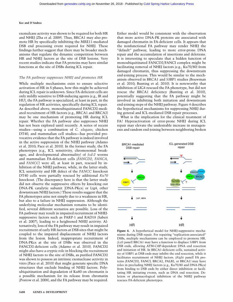
Either model would be consistent with the observationthat more active DNA-PK proteins are associated withdamaged chromatin in FA-deficient cells. It appears thatthe nonfunctional FA pathway may render NHEJ the‘‘default’’ pathway, leading to more error-prone DNArepair and the accumulation of insertions and deletions.It is interesting to speculate that a hidden function ofmonoubiquitinated FANCD2/FANCI complex might befacilitating removal of NHEJ factors (e.g., Ku70/80) fromdamaged chromatin, thus suppressing the downstreamend-joining process. This would be similar to the mech-anism observed in BRCA1 and 53BP1 studies (Bouwmanet al. 2010; Bunting et al. 2010). It is noteworthy thatinhibition of LIG4 rescued the FA phenotype, but did notrescue the BRCA1 deficiency (Bunting et al. 2010),potentially suggesting that the FA pathway might beinvolved in inhibiting both initiation and downstreamend-joining steps of the NHEJ pathway. Figure 4 describesthe hypothetical mechanisms for suppressing NHEJ dur-ing general and ICL-mediated DSB repair processes.
What is the implication for the clinical treatment ofFA? Hyperactivation of error-prone NHEJ during ICLrepair may elevate the undesirable increase in mutagen-esis and random end-joining between neighboring broken
Figure 4. A hypothetical model for NHEJ-suppressive mecha-nisms during DSB repair. For repairing ‘‘replication-associated’’DSBs, multiple mechanisms can be employed to promote HR.(Left panel) BRCA1 may have a function to displace 53BP1 fromDSB ends, allowing ATM-CtIP-dependent DNA end resectionand initiation of HR. In BRCA1-deficient cells, sustained activ-ity of 53BP1 at DSB ends may inhibit the end resection, while itfacilitates recruitment of NHEJ factors. (Right panel) FA pro-teins (FANCD2, FANCI, BRCA2, PALB2, or BRCA1) may haveroles in precluding NHEJ factors (e.g., Ku70/80 and DNA-PKcs)from binding to DSB ends by either direct inhibition or facili-tating HR initiating events, such as DNA end resection. De-letion or pharmacological inhibition of the NHEJ pathwayrescues FA-deficient phenotypes.
Kee and D’Andrea
1688 GENES & DEVELOPMENT
Cold Spring Harbor Laboratory Press on November 26, 2018 - Published by genesdev.cshlp.orgDownloaded from

chromosomes (Newell et al. 2004). Likewise, improperusage of NHEJ during the hematopoietic program, whereHR plays a more critical role, may result in genomicinstability, leading to bone marrow failure. These studiesalso suggest that usage of DNA-PK inhibitors may bebeneficial for treating both FA patients and FA-associatedtumors, although there may also be a risk of enhancedtoxicity for FA patients treated with the drugs. Furtherinvestigation of the potential therapeutic approach mayhave a significant impact on the treatment of FA patients.
Identification of FAN1
Although the key molecular event in the FA pathway isthe generation of monoubiquitinated FANCD2/FANCI,the exact molecular functions of the modified FANCD2/FANCI proteins remain enigmatic. Four recent studies(Liu et al. 2010; Kratz et al. 2010; Mackay et al. 2010;Smogorzewska et al. 2010) describe at least one novelfunction of monoubiquitinated FANCD2. They showedthat FAN1 (previously known as KIAA1018), a novelnuclease with a N terminus UBZ (ubiquitin zinc finger)domain and a C terminus nuclease domain, associateswith monoubiquitinated FANCD2. The initial identifi-cation of FAN1 as a FA-like factor came from differentapproaches, as it was identified from a genome-wideshRNA screen searching for ICL sensitizer (Smogorzewskaet al. 2010), as a binding partner of FANCD2 (Liu et al.2010), as a binding partner of MLH1 mismatch repairprotein (Kratz et al. 2010), and through a bioinformaticsearch for the UBZ and nuclease domain-containing pro-teins (Mackay et al. 2010). The groups collectively foundthat FAN1 (1) possesses a nuclease activity that is re-quired for cellular resistance against ICL agents, (2) isrecruited to damaged DNA via its UBZ domain andmonoubiquitinated FANCD2, and (3) associates ratherspecifically with monoubiquitinated FANCD2. As dis-cussed throughout this review, one important function ofmonoubiquitinated FANCD2 is the promotion of HR.Indeed, depletion of FAN1 resulted in reduced HR effi-ciency in human cells (Mackay et al. 2010), although theformation of RAD51 or RPA foci was not affected,suggesting that FAN1 might have a role downstream inthe HR process.
Collectively, a model can be assembled that FAN1 isrecruited to the damaged sites via specific interactionbetween the UBZ domain of FAN1 and monoubiquiti-nated FANCD2, followed by the nucleolytic cleavage ofDNA (potentially near ICL), which is necessary for sub-sequent ICL repair. Exactly at which step FAN1 acts topromote ICL repair remains unknown. However, as theWalter laboratory (Knipscheer et al. 2009) demonstratedusing Xenopus extracts, monoubiquitinated FANCD2 isrequired for the nucleolytic incision and unhooking of theICL. An intriguing question is whether FAN1 representsthe cryptic nuclease activity observed in the in vitrosystem. However, as there are several other nucleasesproposed to act during ICL (such as the MUS81–EME1,XPF–ERCC1, SLX4 complex, and the FANCD2 nucleaseitself) (Niedernhofer et al. 2005; Knipscheer et al. 2009;
Moldovan and D’Andrea 2009; Pace et al. 2010), the exactfunctions of each nuclease in promoting ICL repair is yetto be elucidated.
Expanded roles of the FA pathway in mitosis
DNA damage often occurs spontaneously during DNAsynthesis, resulting in nucleotide misincorporations, in-sertions, and deletions (Branzei and Foiani 2008). ManyDNA repair events are believed to predominate during Sphase. HR repair can also occur in G2 phase, and it maytherefore prevent the passage of unrepaired mutations todaughter cells in mitosis (Branzei and Foiani 2008).Whether DNA repair machineries remain active duringmitosis is unclear, and the higher-ordered chromatincompaction of mitosis may prevent access of the repairmachineries. To date, most studies of FA proteins havefocused on their role in regulating DNA repair mecha-nisms during S phases.
A series of recent studies has provided exciting newevidence that the FA pathway has roles during mitosis.Specifically, the pathway can serve as a surveillancemechanism to monitor unrepaired DNA (Chan et al.2009; Naim and Rosselli 2009a). Fragile sites, regions inthe chromosome that represent incomplete DNA repli-cation, such as gaps, are known to be caused spontane-ously or by treating cells with drugs causing replicativestress (e.g., APH). FA-deficient cells exhibit increasedchromosomal breaks at fragile sites, and this high inci-dence may be linked to high cancer rates in FA patients(Porfirio et al. 1991; Fundia et al. 1994; Howlett et al.2005). These early studies suggested that the FA pathwayhas a role in regulating the generation and frequency ofunstable regions in the chromatin. However, the mech-anistic link between the FA pathway and the abnormalchromosomal regions was unclear. Interestingly, theHickson and Rosselli groups (Chan et al. 2009; Naim andRosselli 2009a) have demonstrated that APH treatmentin cells induces the increased staining of FANCD2 fociin fragile sites within the compact mitotic chromosomes.Furthermore, the lack of FANCD2 staining in FA-deficient cells (which also suggested that they are likelyto be monoubiquitinated forms of FANCD2) stronglycorrelated with generation of elevated micronuclei indaughter cells. Interestingly, the location of FANCD2(and FANCI) spots reside in the extremities of mitoticDNA structures, termed ultrafine DNA bridges (UFB)(Chan et al. 2007), which are shown to connect the com-mon fragile sites between sister chromosomes. Thus, ahypothesis is that the presence of FANCD2/FANCI onthe fragile sites may be a surveillance mechanism tomark the incomplete replication that escaped the S or G2checkpoint, and certain mechanisms might exist duringmitosis to repair such lesions (Chan and Hickson 2009).
The molecular function of FANCD2/FANCI in theunstable region remains unclear, but it appears to berelated, at least partially, to the recruitment and stabili-zation of BLM protein on the bridges (Naim and Rosselli2009a). Accordingly, FA-deficient cells have decreasedBLM localization on the bridges. BLM is a RecQ helicase
Expanded roles of the FA pathway
GENES & DEVELOPMENT 1689
Cold Spring Harbor Laboratory Press on November 26, 2018 - Published by genesdev.cshlp.orgDownloaded from
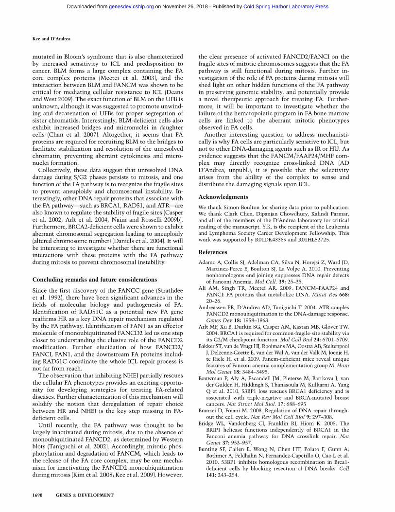
mutated in Bloom’s syndrome that is also characterizedby increased sensitivity to ICL and predisposition tocancer. BLM forms a large complex containing the FAcore complex proteins (Meetei et al. 2003), and theinteraction between BLM and FANCM was shown to becritical for mediating cellular resistance to ICL (Deansand West 2009). The exact function of BLM on the UFB isunknown, although it was suggested to promote unwind-ing and decatenation of UFBs for proper segregation ofsister chromatids. Interestingly, BLM-deficient cells alsoexhibit increased bridges and micronuclei in daughtercells (Chan et al. 2007). Altogether, it seems that FAproteins are required for recruiting BLM to the bridges tofacilitate stabilization and resolution of the unresolvedchromatin, preventing aberrant cytokinesis and micro-nuclei formation.
Collectively, these data suggest that unresolved DNAdamage during S/G2 phases persists to mitosis, and onefunction of the FA pathway is to recognize the fragile sitesto prevent aneuploidy and chromosomal instability. In-terestingly, other DNA repair proteins that associate withthe FA pathway—such as BRCA1, RAD51, and ATR—arealso known to regulate the stability of fragile sites (Casperet al. 2002; Arlt et al. 2004; Naim and Rosselli 2009b).Furthermore, BRCA2-deficient cells were shown to exhibitaberrant chromosomal segregation leading to aneuploidy(altered chromosome number) (Daniels et al. 2004). It willbe interesting to investigate whether there are functionalinteractions with these proteins with the FA pathwayduring mitosis to prevent chromosomal instability.
Concluding remarks and future considerations
Since the first discovery of the FANCC gene (Strathdeeet al. 1992), there have been significant advances in thefields of molecular biology and pathogenesis of FA.Identification of RAD51C as a potential new FA genereaffirms HR as a key DNA repair mechanism regulatedby the FA pathway. Identification of FAN1 as an effectormolecule of monoubiquitinated FANCD2 led us one stepcloser to understanding the elusive role of the FANCD2modification. Further elucidation of how FANCD2/FANCI, FAN1, and the downstream FA proteins includ-ing RAD51C coordinate the whole ICL repair process isnot far from reach.
The observation that inhibiting NHEJ partially rescuesthe cellular FA phenotypes provides an exciting opportu-nity for developing strategies for treating FA-relateddiseases. Further characterization of this mechanism willsolidify the notion that deregulation of repair choicebetween HR and NHEJ is the key step missing in FA-deficient cells.
Until recently, the FA pathway was thought to belargely inactivated during mitosis, due to the absence ofmonoubiquitinated FANCD2, as determined by Westernblots (Taniguchi et al. 2002). Accordingly, mitotic phos-phorylation and degradation of FANCM, which leads tothe release of the FA core complex, may be one mecha-nism for inactivating the FANCD2 monoubiquitinationduring mitosis (Kim et al. 2008; Kee et al. 2009). However,
the clear presence of activated FANCD2/FANCI on thefragile sites of mitotic chromosomes suggests that the FApathway is still functional during mitosis. Further in-vestigation of the role of FA proteins during mitosis willshed light on other hidden functions of the FA pathwayin preserving genomic stability, and potentially providea novel therapeutic approach for treating FA. Further-more, it will be important to investigate whether thefailure of the hematopoietic program in FA bone marrowcells are linked to the aberrant mitotic phenotypesobserved in FA cells.
Another interesting question to address mechanisti-cally is why FA cells are particularly sensitive to ICL, butnot to other DNA-damaging agents such as IR or HU. Asevidence suggests that the FANCM/FAAP24/MHF com-plex may directly recognize cross-linked DNA (ADD’Andrea, unpubl.), it is possible that the selectivityarises from the ability of the complex to sense anddistribute the damaging signals upon ICL.
Acknowledgments
We thank Simon Boulton for sharing data prior to publication.We thank Clark Chen, Dipanjan Chowdhury, Kalindi Parmar,and all of the members of the D’Andrea laboratory for criticalreading of the manuscript. Y.K. is the recipient of the Leukemiaand Lymphoma Society Career Development Fellowship. Thiswork was supported by R01DK43389 and R01HL52725.
References
Adamo A, Collis SJ, Adelman CA, Silva N, Horejsi Z, Ward JD,Martinez-Perez E, Boulton SJ, La Volpe A. 2010. Preventingnonhomologous end joining suppresses DNA repair defectsof Fanconi Anemia. Mol Cell. 39: 25–35.
Ali AM, Singh TR, Meetei AR. 2009. FANCM–FAAP24 andFANCJ: FA proteins that metabolize DNA. Mutat Res 668:20–26.
Andreassen PR, D’Andrea AD, Taniguchi T. 2004. ATR couplesFANCD2 monoubiquitination to the DNA-damage response.Genes Dev 18: 1958–1963.
Arlt MF, Xu B, Durkin SG, Casper AM, Kastan MB, Glover TW.2004. BRCA1 is required for common-fragile-site stability viaits G2/M checkpoint function. Mol Cell Biol 24: 6701–6709.
Bakker ST, van de Vrugt HJ, Rooimans MA, Oostra AB, SteltenpoolJ, Delzenne-Goette E, van der Wal A, van der Valk M, Joenje H,te Riele H, et al. 2009. Fancm-deficient mice reveal uniquefeatures of Fanconi anemia complementation group M. Hum
Mol Genet 18: 3484–3495.Bouwman P, Aly A, Escandell JM, Pieterse M, Bartkova J, van
der Gulden H, Hiddingh S, Thanasoula M, Kulkarni A, YangQ et al. 2010. 53BP1 loss rescues BRCA1 deficiency and isassociated with triple-negative and BRCA-mutated breastcancers. Nat Struct Mol Biol. 17: 688–695
Branzei D, Foiani M. 2008. Regulation of DNA repair through-out the cell cycle. Nat Rev Mol Cell Biol 9: 297–308.
Bridge WL, Vandenberg CJ, Franklin RJ, Hiom K. 2005. TheBRIP1 helicase functions independently of BRCA1 in theFanconi anemia pathway for DNA crosslink repair. Nat
Genet 37: 953–957.Bunting SF, Callen E, Wong N, Chen HT, Polato F, Gunn A,
Bothmer A, Feldhahn N, Fernandez-Capetillo O, Cao L et al.2010. 53BP1 inhibits homologous recombination in Brca1-deficient cells by blocking resection of DNA breaks. Cell
141: 243–254.
Kee and D’Andrea
1690 GENES & DEVELOPMENT
Cold Spring Harbor Laboratory Press on November 26, 2018 - Published by genesdev.cshlp.orgDownloaded from

Cao L, Xu X, Bunting SF, Liu J, Wang RH, Cao LL, Wu JJ, PengTN, Chen J, Nussenzweig A, et al. 2009. A selective re-quirement for 53BP1 in the biological response to genomicinstability induced by Brca1 deficiency. Mol Cell 35: 534–541.
Casper AM, Nghiem P, Arlt MF, Glover TW. 2002. ATRregulates fragile site stability. Cell 111: 779–789.
Chan KL, Hickson ID. 2009. On the origins of ultra-fineanaphase bridges. Cell Cycle 8: 3065–3066.
Chan KL, North PS, Hickson ID. 2007. BLM is required forfaithful chromosome segregation and its localization definesa class of ultrafine anaphase bridges. EMBO J 26: 3397–3409.
Chan KL, Palmai-Pallag T, Ying S, Hickson ID. 2009. Replica-tion stress induces sister-chromatid bridging at fragile siteloci in mitosis. Nat Cell Biol 11: 753–760.
Chen BP, Chan DW, Kobayashi J, Burma S, Asaithamby A,Morotomi-Yano K, Botvinick E, Qin J, Chen DJ. 2005. Cellcycle dependence of DNA-dependent protein kinase phos-phorylation in response to DNA double strand breaks. J Biol
Chem 280: 14709–14715.Ciccia A, Ling C, Coulthard R, Yan Z, Xue Y, Meetei AR,
Laghmani el H, Joenje H, McDonald N, de Winter JP et al.2007. Identification of FAAP24, a Fanconi anemia corecomplex protein that interacts with FANCM. Mol Cell 25:331–343.
Cohn MA, Kowal P, Yang K, Haas W, Huang TT, Gygi SP,D’Andrea AD. 2007. A UAF1-containing multisubunit pro-tein complex regulates the Fanconi anemia pathway. MolCell 28: 786–797.
Cole RS. 1973. Repair of DNA containing interstrand crosslinksin Escherichia coli: Sequential excision and recombination.Proc Natl Acad Sci 70: 1064–1068.
Collins NB, Wilson JB, Bush T, Thomashevski A, Roberts KJ,Jones NJ, Kupfer GM. 2009. ATR-dependent phosphorylationof FANCA on serine 1449 after DNA damage is importantfor FA pathway function. Blood 113: 2181–2190.
Collis SJ, Ciccia A, Deans AJ, Horejsi Z, Martin JS, Maslen SL,Skehel JM, Elledge SJ, West SC, Boulton SJ. 2008. FANCMand FAAP24 function in ATR-mediated checkpoint signalingindependently of the Fanconi anemia core complex. Mol Cell
32: 313–324.D’Andrea AD, Grompe M. 2003. The Fanconi anaemia/BRCA
pathway. Nat Rev Cancer 3: 23–34.Daniels MJ, Wang Y, Lee M, Venkitaraman AR. 2004. Abnormal
cytokinesis in cells deficient in the breast cancer suscepti-bility protein BRCA2. Science 306: 876–879.
Deans AJ, West SC. 2009. FANCM connects the genome in-stability disorders Bloom’s Syndrome and Fanconi Anemia.Mol Cell 36: 943–953.
Deng Y, Guo X, Ferguson DO, Chang S. 2009. Multiple roles forMRE11 at uncapped telomeres. Nature 460: 914–918.
Difilippantonio S, Gapud E, Wong N, Huang CY, Mahowald G,Chen HT, Kruhlak MJ, Callen E, Livak F, Nussenzweig MC,et al. 2008. 53BP1 facilitates long-range DNA end-joiningduring V(D)J recombination. Nature 456: 529–533.
Dimitrova N, Chen YC, Spector DL, de Lange T. 2008. 53BP1promotes non-homologous end joining of telomeres by in-creasing chromatin mobility. Nature 456: 524–528.
DiTullio RA Jr, Mochan TA, Venere M, Bartkova J, Sehested M,Bartek J, Halazonetis TD. 2002. 53BP1 functions in an ATM-dependent checkpoint pathway that is constitutively acti-vated in human cancer. Nat Cell Biol 4: 998–1002.
French CA, Masson JY, Griffin CS, O’Regan P, West SC, ThackerJ. 2002. Role of mammalian RAD51L2 (RAD51C) in re-combination and genetic stability. J Biol Chem 277: 19322–19330.
Fundia A, Gorla N, Larripa I. 1994. Spontaneous chromosomeaberrations in Fanconi’s anemia patients are located at fragilesites and acute myeloid leukemia breakpoints. Hereditas
120: 47–50.Garcia-Higuera I, Taniguchi T, Ganesan S, Meyn MS, Timmers
C, Hejna J, Grompe M, D’Andrea AD. 2001. Interaction ofthe Fanconi anemia proteins and BRCA1 in a commonpathway. Mol Cell 7: 249–262.
Gari K, Decaillet C, Delannoy M, Wu L, Constantinou A. 2008a.Remodeling of DNA replication structures by the branchpoint translocase FANCM. Proc Natl Acad Sci 105: 16107–16112.
Gari K, Decaillet C, Stasiak AZ, Stasiak A, Constantinou A.2008b. The Fanconi anemia protein FANCM can promotebranch migration of Holliday junctions and replication forks.Mol Cell 29: 141–148.
Gildemeister OS, Sage JM, Knight KL. 2009. Cellular redistri-bution of Rad51 in response to DNA damage: Novel role forRad51C. J Biol Chem 284: 31945–31952.
Gong Z, Kim JE, Leung CC, Glover JN, Chen J. 2010. BACH1/FANCJ acts with TopBP1 and participates early in DNAreplication checkpoint control. Mol Cell 37: 438–446.
Grossmann KF, Ward AM, Matkovic ME, Folias AE, Moses RE.2001. S. cerevisiae has three pathways for DNA interstrandcrosslink repair. Mutat Res 487: 73–83.
Hanada K, Budzowska M, Modesti M, Maas A, Wyman C, EssersJ, Kanaar R. 2006. The structure-specific endonucleaseMus81–Eme1 promotes conversion of interstrand DNA cross-links into double-strands breaks. EMBO J 25: 4921–4932.
Houghtaling S, Newell A, Akkari Y, Taniguchi T, Olson S,Grompe M. 2005. Fancd2 functions in a double strand breakrepair pathway that is distinct from non-homologous endjoining. Hum Mol Genet 14: 3027–3033.
Howlett NG, Taniguchi T, Olson S, Cox B, Waisfisz Q, De Die-Smulders C, Persky N, Grompe M, Joenje H, Pals G, et al.2002. Biallelic inactivation of BRCA2 in Fanconi anemia.Science 297: 606–609.
Howlett NG, Taniguchi T, Durkin SG, D’Andrea AD, GloverTW. 2005. The Fanconi anemia pathway is required for theDNA replication stress response and for the regulation ofcommon fragile site stability. Hum Mol Genet 14: 693–701.
Huang J, Huen MS, Kim H, Leung CC, Glover JN, Yu X, Chen J.2009. RAD18 transmits DNA damage signalling to elicithomologous recombination repair. Nat Cell Biol 11: 592–603.
Huertas P, Cortes-Ledesma F, Sartori AA, Aguilera A, JacksonSP. 2008. CDK targets Sae2 to control DNA-end resectionand homologous recombination. Nature 455: 689–692.
Hussain S, Wilson JB, Medhurst AL, Hejna J, Witt E, Ananth S,Davies A, Masson JY, Moses R, West SC, et al. 2004. Directinteraction of FANCD2 with BRCA2 in DNA damage re-sponse pathways. Hum Mol Genet 13: 1241–1248.
Ip SC, Rass U, Blanco MG, Flynn HR, Skehel JM, West SC. 2008.Identification of Holliday junction resolvases from humansand yeast. Nature 456: 357–361.
Ishiai M, Kitao H, Smogorzewska A, Tomida J, Kinomura A,Uchida E, Saberi A, Kinoshita E, Kinoshita-Kikuta E, KoikeT, et al. 2008. FANCI phosphorylation functions as a molec-ular switch to turn on the Fanconi anemia pathway. Nat
Struct Mol Biol 15: 1138–1146.Karathanasis E, Wilson TE. 2002. Enhancement of Saccharomyces
cerevisiae end-joining efficiency by cell growth stage but notby impairment of recombination. Genetics 161: 1015–1027.
Kee Y, Kim JM, D’Andrea AD. 2009. Regulated degradation ofFANCM in the Fanconi anemia pathway during mitosis.Genes Dev 23: 555–560.
Expanded roles of the FA pathway
GENES & DEVELOPMENT 1691
Cold Spring Harbor Laboratory Press on November 26, 2018 - Published by genesdev.cshlp.orgDownloaded from

Kennedy RD, D’Andrea AD. 2005. The Fanconi Anemia/BRCA pathway: New faces in the crowd. Genes Dev 19:2925–2940.
Kim JS, Krasieva TB, Kurumizaka H, Chen DJ, Taylor AM,Yokomori K. 2005. Independent and sequential recruitmentof NHEJ and HR factors to DNA damage sites in mammaliancells. J Cell Biol 170: 341–347.
Kim JM, Kee Y, Gurtan A, D’Andrea AD. 2008. Cell cycle-dependent chromatin loading of the Fanconi anemia corecomplex by FANCM/FAAP24. Blood 111: 5215–5222.
Kim JM, Parmar K, Huang M, Weinstock DM, Ruit CA, KutokJL, D’Andrea AD. 2009. Inactivation of murine Usp1 resultsin genomic instability and a Fanconi anemia phenotype. DevCell 16: 314–320.
Knipscheer P, Raschle M, Smogorzewska A, Enoiu M, Ho TV,Scharer OD, Elledge SJ, Walter JC. 2009. The Fanconi anemiapathway promotes replication-dependent DNA interstrandcross-link repair. Science 326: 1698–1701.
Kratz K, Schopf B, Kaden S, Sendoel A, Eberhard R, Lademann C,Cannavo E, Sartori AA, Hengartner MO, Jiricny J. 2010.Deficiency of FANCD2-associated nuclease KIAA1018/FAN1 sensitizes cells to interstrand crosslinking agents. Cell
142: 77–88.Lee SE, Mitchell RA, Cheng A, Hendrickson EA. 1997. Evidence
for DNA-PK-dependent and -independent DNA double-strand break repair pathways in mammalian cells as a func-tion of the cell cycle. Mol Cell Biol 17: 1425–1433.
Levitus M, Waisfisz Q, Godthelp BC, de Vries Y, Hussain S,Wiegant WW, Elghalbzouri-Maghrani E, Steltenpool J,Rooimans MA, Pals G, et al. 2005. The DNA helicaseBRIP1 is defective in Fanconi anemia complementationgroup J. Nat Genet 37: 934–935.
Levran O, Attwooll C, Henry RT, Milton KL, Neveling K, Rio P,Batish SD, Kalb R, Velleuer E, Barral S, et al. 2005. TheBRCA1-interacting helicase BRIP1 is deficient in Fanconianemia. Nat Genet 37: 931–933.
Levy-Lahad E. 2010. Fanconi anemia and breast cancer suscep-tibility meet again. Nat Genet 42: 368–369.
Li X, Stith CM, Burgers PM, Heyer WD. 2009. PCNA is requiredfor initiation of recombination-associated DNA synthesis byDNA polymerase d. Mol Cell 36: 704–713.
Ling C, Ishiai M, Ali AM, Medhurst AL, Neveling K, Kalb R, YanZ, Xue Y, Oostra AB, Auerbach AD, et al. 2007. FAAP100 isessential for activation of the Fanconi anemia-associatedDNA damage response pathway. EMBO J 26: 2104–2114.
Litman R, Peng M, Jin Z, Zhang F, Zhang J, Powell S, AndreassenPR, Cantor SB. 2005. BACH1 is critical for homologous re-combination and appears to be the Fanconi anemia geneproduct FANCJ. Cancer Cell 8: 255–265.
Liu Y, West SC. 2008. More complexity to the Bloom’s syn-drome complex. Genes Dev 22: 2737–2742.
Liu Y, Masson JY, Shah R, O’Regan P, West SC. 2004. RAD51Cis required for Holliday junction processing in mammaliancells. Science 303: 243–246.
Liu Y, Tarsounas M, O’Regan P, West SC. 2007. Role of RAD51Cand XRCC3 in genetic recombination and DNA repair. J Biol
Chem 282: 1973–1979.Liu T, Ghosal G, Yuan J, Chen J, Huang J. 2010. FAN1 acts with
FANC1–FANCD2 to promote DNA interstrand cross-linksrepair. Science (in press).
Luke-Glaser S, Luke B, Grossi S, Constantinou A. 2010.FANCM regulates DNA chain elongation and is stabilizedby S-phase checkpoint signalling. EMBO J 29: 795–805.
Mackay C, Declais AC, Lundin C, Agostinho A, Deans AJ,Macartney TJ, Hofmann K, Gartner A, West SC, Helleday T,et al. 2010. Identification of KIAA1018/FAN1, a DNA repair
nuclease recruited to DNA damage by monoubiquitinatedFANCD2. Cell 142: 65–76.
McIlwraith MJ, Vaisman A, Liu Y, Fanning E, Woodgate R, WestSC. 2005. Human DNA polymerase eta promotes DNAsynthesis from strand invasion intermediates of homologousrecombination. Mol Cell 20: 783–792.
Meetei AR, Sechi S, Wallisch M, Yang D, Young MK, Joenje H,Hoatlin ME, Wang W. 2003. A multiprotein nuclear complexconnects Fanconi anemia and Bloom syndrome. Mol CellBiol 23: 3417–3426.
Meetei AR, Medhurst AL, Ling C, Xue Y, Singh TR, Bier P,Steltenpool J, Stone S, Dokal I, Mathew CG, et al. 2005. Ahuman ortholog of archaeal DNA repair protein Hef isdefective in Fanconi anemia complementation group M.Nat Genet 37: 958–963.
Meindl A, Hellebrand H, Wiek C, Erven V, Wappenschmidt B,Niederacher D, Freund M, Lichtner P, Hartmann L, Schaal Het al. 2010. Germline mutations in breast and ovarian cancerpedigrees establish RAD51C as a human cancer susceptibil-ity gene. Nat Genet. 42: 410–414
Mekeel KL, Tang W, Kachnic LA, Luo CM, DeFrank JS, PowellSN. 1997. Inactivation of p53 results in high rates ofhomologous recombination. Oncogene 14: 1847–1857.
Moldovan GL, D’Andrea AD. 2009. How the fanconi anemiapathway guards the genome. Annu Rev Genet 43: 223–249.
Moldovan GL, Madhavan MV, Mirchandani KD, McCaffrey RM,Vinciguerra P, D’Andrea AD. 2010. DNA polymerase POLNparticipates in cross-link repair and homologous recombina-tion. Mol Cell Biol 30: 1088–1096.
Mosedale G, Niedzwiedz W, Alpi A, Perrina F, Pereira-Leal JB,Johnson M, Langevin F, Pace P, Patel KJ. 2005. The vertebrateHef ortholog is a component of the Fanconi anemia tumor-suppressor pathway. Nat Struct Mol Biol 12: 763–771.
Moynahan ME, Cui TY, Jasin M. 2001a. Homology-directed dnarepair, mitomycin-c resistance, and chromosome stability isrestored with correction of a Brca1 mutation. Cancer Res 61:4842–4850.
Moynahan ME, Pierce AJ, Jasin M. 2001b. BRCA2 is required forhomology-directed repair of chromosomal breaks. Mol Cell7: 263–272.
Muzzini DM, Plevani P, Boulton SJ, Cassata G, Marini F. 2008.Caenorhabditis elegans POLQ-1 and HEL-308 function intwo distinct DNA interstrand cross-link repair pathways.DNA Repair (Amst) 7: 941–950.
Naim V, Rosselli F. 2009a. The FANC pathway and BLMcollaborate during mitosis to prevent micro-nucleation andchromosome abnormalities. Nat Cell Biol 11: 761–768.
Naim V, Rosselli F. 2009b. The FANC pathway and mitosis: Areplication legacy. Cell Cycle 8: 2907–2911.
Nakanishi K, Taniguchi T, Ranganathan V, New HV, MoreauLA, Stotsky M, Mathew CG, Kastan MB, Weaver DT,D’Andrea AD. 2002. Interaction of FANCD2 and NBS1 inthe DNA damage response. Nat Cell Biol 4: 913–920.
Nakanishi K, Yang YG, Pierce AJ, Taniguchi T, Digweed M,D’Andrea AD, Wang ZQ, Jasin M. 2005. Human Fanconianemia monoubiquitination pathway promotes homologousDNA repair. Proc Natl Acad Sci 102: 1110–1115.
Newell AE, Akkari YM, Torimaru Y, Rosenthal A, Reifsteck CA,Cox B, Grompe M, Olson SB. 2004. Interstrand crosslink-in-duced radials form between non-homologous chromosomes, butare absent in sex chromosomes. DNA Repair (Amst) 3: 535–542.
Niedernhofer LJ, Odijk H, Budzowska M, van Drunen E, MaasA, Theil AF, de Wit J, Jaspers NG, Beverloo HB, HoeijmakersJH, et al. 2004. The structure-specific endonuclease Ercc1–Xpfis required to resolve DNA interstrand cross-link-induceddouble-strand breaks. Mol Cell Biol 24: 5776–5787.
Kee and D’Andrea
1692 GENES & DEVELOPMENT
Cold Spring Harbor Laboratory Press on November 26, 2018 - Published by genesdev.cshlp.orgDownloaded from

Niedernhofer LJ, Lalai AS, Hoeijmakers JH. 2005. Fanconianemia (cross)linked to DNA repair. Cell 123: 1191–1198.
Niedzwiedz W, Mosedale G, Johnson M, Ong CY, Pace P, PatelKJ. 2004. The Fanconi anaemia gene FANCC promoteshomologous recombination and error-prone DNA repair.Mol Cell 15: 607–620.
Nijman SM, Huang TT, Dirac AM, Brummelkamp TR, KerkhovenRM, D’Andrea AD, Bernards R. 2005. The deubiquitinatingenzyme USP1 regulates the Fanconi anemia pathway. Mol Cell17: 331–339.
Oestergaard VH, Langevin F, Kuiken HJ, Pace P, Niedzwiedz W,Simpson LJ, Ohzeki M, Takata M, Sale JE, Patel KJ. 2007.Deubiquitination of FANCD2 is required for DNA crosslinkrepair. Mol Cell 28: 798–809.
Pace P, Mosedale G, Hodskinson M, Rosado IV, SivasubramaniamM, Patel KJ. 2010. Ku70 corrupts DNA repair in the absence ofthe Fanconi anemia pathway. Science.329: 219–223.
Paull TT, Cortez D, Bowers B, Elledge SJ, Gellert M. 2001.Direct DNA binding by Brca1. Proc Natl Acad Sci 98: 6086–6091.
Peng M, Litman R, Xie J, Sharma S, Brosh RM Jr, Cantor SB.2007. The FANCJ/MutLa interaction is required for correc-tion of the cross-link response in FA-J cells. EMBO J 26:3238–3249.
Pichierri P, Rosselli F. 2004. The DNA crosslink-induced S-phasecheckpoint depends on ATR–CHK1 and ATR–NBS1–FANCD2 pathways. EMBO J 23: 1178–1187.
Pierce AJ, Hu P, Han M, Ellis N, Jasin M. 2001. Ku DNA end-binding protein modulates homologous repair of double-strand breaks in mammalian cells. Genes Dev 15: 3237–3242.
Porfirio B, Smeets D, Beckers L, Caporossi D, Tedeschi B,Vernole P, Joenje H, Nicoletti B, Dallapiccola B. 1991. Fragilesites and chromosome instability: The distribution of breaksinduced by cis-diamine-dichloro-platinum (II) in Fanconianemia lymphocyte cultures. Hum Genet 86: 256–260.
Postow L, Ghenoiu C, Woo EM, Krutchinsky AN, Chait BT,Funabiki H. 2008. Ku80 removal from DNA through doublestrand break-induced ubiquitylation. J Cell Biol 182: 467–479.
Richardson C, Elliott B, Jasin M. 1999. Chromosomal double-strand breaks introduced in mammalian cells by expressionof I-Sce I endonuclease. Methods Mol Biol 113: 453–463.
Rodrigue A, Lafrance M, Gauthier MC, McDonald D, HendzelM, West SC, Jasin M, Masson JY. 2006. Interplay betweenhuman DNA repair proteins at a unique double-strand breakin vivo. EMBO J 25: 222–231.
Saberi A, Hochegger H, Szuts D, Lan L, Yasui A, Sale JE,Taniguchi Y, Murakawa Y, Zeng W, Yokomori K, et al.2007. RAD18 and poly(ADP-ribose) polymerase indepen-dently suppress the access of nonhomologous end joiningto double-strand breaks and facilitate homologous recombi-nation-mediated repair. Mol Cell Biol 27: 2562–2571.
Sartori AA, Lukas C, Coates J, Mistrik M, Fu S, Bartek J, Baer R,Lukas J, Jackson SP. 2007. Human CtIP promotes DNA endresection. Nature 450: 509–514.
Schwab RA, Blackford AN, Niedzwiedz W. 2010. ATR activa-tion and replication fork restart are defective in FANCM-deficient cells. EMBO J 29: 806–818.
Singh TR, Bakker ST, Agarwal S, Jansen M, Grassman E,Godthelp BC, Ali AM, Du CH, Rooimans MA, Fan Q, et al.2009. Impaired FANCD2 monoubiquitination and hypersen-sitivity to camptothecin uniquely characterize Fanconi ane-mia complementation group M. Blood 114: 174–180.
Singh TR, Saro D, Ali AM, Zheng XF, Du CH, Killen MW,Sachpatzidis A, Wahengbam K, Pierce AJ, Xiong Y, et al.2010. MHF1–MHF2, a histone-fold-containing protein com-
plex, participates in the Fanconi anemia pathway viaFANCM. Mol Cell 37: 879–886.
Smogorzewska A, Matsuoka S, Vinciguerra P, McDonald ER III,Hurov KE, Luo J, Ballif BA, Gygi SP, Hofmann K, D’AndreaAD, et al. 2007. Identification of the FANCI protein, a mono-ubiquitinated FANCD2 paralog required for DNA repair.Cell 129: 289–301.
Smogorzewska A, Desetty R, Saito TT, Schlabach M, Lach FP,Sowa ME, Clark AB, Kunkel TA, Harper JW, Colaiacovo MP,et al. 2010. A genetic screen identifies FAN1, a FanconiAnemia-associated nuclease necessary for DNA interstrandcrosslink repair. Mol Cell 39: 36–47.
Sommers JA, Rawtani N, Gupta R, Bugreev DV, Mazin AV,Cantor SB, Brosh RM Jr. 2009. FANCJ uses its motor ATPaseto destabilize protein–DNA complexes, unwind triplexes,and inhibit RAD51 strand exchange. J Biol Chem 284: 7505–7517.
Strathdee CA, Gavish H, Shannon WR, Buchwald M. 1992.Cloning of cDNAs for Fanconi’s anaemia by functionalcomplementation. Nature 358: 434.
Sun W, Nandi S, Osman F, Ahn JS, Jakovleska J, Lorenz A,Whitby MC. 2008. The FANCM ortholog Fml1 promotesrecombination at stalled replication forks and limits crossingover during DNA double-strand break repair. Mol Cell 32:118–128.
Sy SM, Huen MS, Chen J. 2009. PALB2 is an integral componentof the BRCA complex required for homologous recombina-tion repair. Proc Natl Acad Sci 106: 7155–7160.
Takata M, Sasaki MS, Sonoda E, Morrison C, Hashimoto M,Utsumi H, Yamaguchi-Iwai Y, Shinohara A, Takeda S. 1998.Homologous recombination and non-homologous end-joining pathways of DNA double-strand break repair haveoverlapping roles in the maintenance of chromosomal in-tegrity in vertebrate cells. EMBO J 17: 5497–5508.
Takata M, Sasaki MS, Sonoda E, Fukushima T, Morrison C,Albala JS, Swagemakers SM, Kanaar R, Thompson LH,Takeda S. 2000. The Rad51 paralog Rad51B promotes homol-ogous recombinational repair. Mol Cell Biol 20: 6476–6482.
Takata M, Sasaki MS, Tachiiri S, Fukushima T, Sonoda E, SchildD, Thompson LH, Takeda S. 2001. Chromosome instabilityand defective recombinational repair in knockout mutants ofthe five Rad51 paralogs. Mol Cell Biol 21: 2858–2866.
Taniguchi T, Garcia-Higuera I, Andreassen PR, Gregory RC,Grompe M, D’Andrea AD. 2002. S-phase-specific interactionof the Fanconi anemia protein, FANCD2, with BRCA1 andRAD51. Blood 100: 2414–2420.
Thompson LH, Schild D. 2001. Homologous recombinationalrepair of DNA ensures mammalian chromosome stability.Mutat Res 477: 131–153.
Turnbull C, Rahman N. 2008. Genetic predisposition to breastcancer: Past, present, and future. Annu Rev Genomics Hum
Genet 9: 321–345.Vaz F, Hanenberg H, Schuster B, Barker K, Wiek C, Erven V,
Neveling K, Endt D, Kesterton I, Autore F et al. 2010.Mutation of the RAD51C gene in a Fanconi anemia-likedisorder. Nat Genet 42: 406–409.
Wang W. 2007. Emergence of a DNA-damage response networkconsisting of Fanconi anaemia and BRCA proteins. Nat Rev
Genet 8: 735–748.Wang X, Andreassen PR, D’Andrea AD. 2004. Functional in-
teraction of monoubiquitinated FANCD2 and BRCA2/FANCD1 in chromatin. Mol Cell Biol 24: 5850–5862.
Wang X, Kennedy RD, Ray K, Stuckert P, Ellenberger T,D’Andrea AD. 2007. Chk1-mediated phosphorylation ofFANCE is required for the Fanconi anemia/BRCA pathway.Mol Cell Biol 27: 3098–3108.
Expanded roles of the FA pathway
GENES & DEVELOPMENT 1693
Cold Spring Harbor Laboratory Press on November 26, 2018 - Published by genesdev.cshlp.orgDownloaded from

Ward JD, Muzzini DM, Petalcorin MI, Martinez-Perez E,Martin JS, Plevani P, Cassata G, Marini F, Boulton SJ.2010. Overlapping mechanisms promote postsynapticRAD-51 filament disassembly during meiotic double-strandbreak repair. Mol Cell 37: 259–272.
Whitby MC. 2010. The FANCM family of DNA helicases/translocases. DNA Repair (Amst) 9: 224–236.
Wilson JB, Yamamoto K, Marriott AS, Hussain S, Sung P,Hoatlin ME, Mathew CG, Takata M, Thompson LH, KupferGM, et al. 2008. FANCG promotes formation of a newlyidentified protein complex containing BRCA2, FANCD2 andXRCC3. Oncogene 27: 3641–3652.
Wooster R, Weber BL. 2003. Breast and ovarian cancer. N EnglJ Med 348: 2339–2347.
Xia B, Sheng Q, Nakanishi K, Ohashi A, Wu J, Christ N, Liu X,Jasin M, Couch FJ, Livingston DM. 2006. Control of BRCA2cellular and clinical functions by a nuclear partner, PALB2.Mol Cell 22: 719–729.
Xia B, Dorsman JC, Ameziane N, de Vries Y, Rooimans MA,Sheng Q, Pals G, Errami A, Gluckman E, Llera J, et al. 2007.Fanconi anemia is associated with a defect in the BRCA2partner PALB2. Nat Genet 39: 159–161.
Xue Y, Li Y, Guo R, Ling C, Wang W. 2008. FANCM of theFanconi anemia core complex is required for both monoubi-quitination and DNA repair. Hum Mol Genet 17: 1641–1652.
Yamamoto K, Hirano S, Ishiai M, Morishima K, Kitao H,Namikoshi K, Kimura M, Matsushita N, Arakawa H,Buerstedde JM, et al. 2005. Fanconi anemia protein FANCD2promotes immunoglobulin gene conversion and DNA repairthrough a mechanism related to homologous recombination.Mol Cell Biol 25: 34–43.
Yan Z, Delannoy M, Ling C, Daee D, Osman F, Muniandy PA,Shen X, Oostra AB, Du H, Steltenpool J, et al. 2010. Ahistone-fold complex and FANCM form a conserved DNA-remodeling complex to maintain genome stability. Mol Cell37: 865–878.
Youds JL, Barber LJ, Ward JD, Collis SJ, O’Neil NJ, Boulton SJ,Rose AM. 2008. DOG-1 is the Caenorhabditis elegans
BRIP1/FANCJ homologue and functions in interstrandcross-link repair. Mol Cell Biol 28: 1470–1479.
Yu X, Chen J. 2004. DNA damage-induced cell cycle checkpointcontrol requires CtIP, a phosphorylation-dependent bindingpartner of BRCA1 C-terminal domains. Mol Cell Biol 24:9478–9486.
Yun MH, Hiom K. 2009. CtIP-BRCA1 modulates the choice ofDNA double-strand-break repair pathway throughout thecell cycle. Nature 459: 460–463.
Zha S, Boboila C, Alt FW. 2009. Mre11: Roles in DNA repairbeyond homologous recombination. Nat Struct Mol Biol 16:798–800.
Zhang N, Liu X, Li L, Legerski R. 2007. Double-strand breaksinduce homologous recombinational repair of interstrandcross-links via cooperation of MSH2, ERCC1–XPF, REV3,and the Fanconi anemia pathway. DNA Repair (Amst) 6:1670–1678.
Zhang F, Fan Q, Ren K, Andreassen PR. 2009a. PALB2 function-ally connects the breast cancer susceptibility proteinsBRCA1 and BRCA2. Mol Cancer Res 7: 1110–1118.
Zhang F, Ma J, Wu J, Ye L, Cai H, Xia B, Yu X. 2009b. PALB2links BRCA1 and BRCA2 in the DNA-damage response. Curr
Biol 19: 524–529.Zhi G, Wilson JB, Chen X, Krause DS, Xiao Y, Jones NJ, Kupfer
GM. 2009. Fanconi anemia complementation groupFANCD2 protein serine 331 phosphorylation is importantfor fanconi anemia pathway function and BRCA2 interac-tion. Cancer Res 69: 8775–8783.
Kee and D’Andrea
1694 GENES & DEVELOPMENT
Cold Spring Harbor Laboratory Press on November 26, 2018 - Published by genesdev.cshlp.orgDownloaded from

10.1101/gad.1955310Access the most recent version at doi: 24:2010, Genes Dev.
Younghoon Kee and Alan D. D'Andrea stabilityExpanded roles of the Fanconi anemia pathway in preserving genomic
References
http://genesdev.cshlp.org/content/24/16/1680.full.html#ref-list-1
This article cites 132 articles, 44 of which can be accessed free at:
License
ServiceEmail Alerting
click here.right corner of the article or
Receive free email alerts when new articles cite this article - sign up in the box at the top
Copyright © 2010 by Cold Spring Harbor Laboratory Press
Cold Spring Harbor Laboratory Press on November 26, 2018 - Published by genesdev.cshlp.orgDownloaded from