Embed Size (px)
Citation preview
1
EXCRETION AND OSMOREGULATION
Introduction :
In our body various metabolic activities are carried out and continuously
numbers of by-products are formed. Some of by-products are harmful, if they remain
within the body. It should be eliminated out of the body. Body regularly throws these
toxic, harmful, unwanted substances out of the body. e.g. nitrogenous waste.
In higher animals including man Homeostasis is maintained by two process;
i.e. excretion and osmoregulation.
Homeostasis:
The regulation or maintenance of a constant body fluid or internal environment
is called homeostasis. The temperature, amount of water and glucose concentration
are at almost constant in homeostasis.
Excretion :
Removal of unwanted metabolic waste products from the body is referred as
excretion.
Waste products are formed due to catabolism of glucose, amino acids, glycerol
and fatty acids. Waste products formed are CO2 ,H2O,bile pigments, nitrogenous
wastes ,excess of inorganic salts, hormones and vitamins. Nitrogenous wastes are
derived from excess or unusual proteins in the food and from breakdown of damaged
dead cells. Ammonia, urea, uric acids are major forms of nitrogenous waste excreted
by the animals. Skin, lungs and liver acts as accessory excretory organ. But Kidney
plays a major role as an excretory organ in all vertebrates.
To eliminate these nitrogenous wastes, excretory system is developed in the
animal body.
Modes of Excretion :
On the basis of types of nitrogenous waste to be removed from the body,
animals show three modes of excretion;
1) Ammnotelism :
Phenomenon of formation of excretory product in the form of ammonia is
called ammonotelism.
Organisms which excrete ammonia as main excretory product are called
ammonotelic.
Ammonia is highly toxic nitrogenous waste produced at the end of protein
metabolism in liver as primary nitrogenous waste.

2
Due to its small molecular size and high water solubility, needs large amount
of water to removed from body.
Its conc.in the body is kept very low and harmful to surrounding tissues.
It has to be eliminated out as soon as it is formed.
About 300-500 ml of water is required for elimination of 1 g of ammonia.
Examples: - Aquatic invertebrates like sponges, hydra, bony fishes, tadpole
larva of frog, salamander.
2) Ureotelism:-
Elimination of nitrogenous waste in the form of urea is called ureotelism.
As ammonia is very toxic, it must be converted to less toxic form.
In liver, ammonia combine with CO2 to form urea by ornithine cycle and
formation requires expenditure of energy.
Urea can be excreted at lower rate.
Urea is soluble in water and stored in dissolved form called urine.
About 50ml of water is required for elimination of 1 g of urea.
Examples: - Terrestrial animals like frog ,turtle, toads ,mammals, marine fishes.
3) Uricotelism:-
Elimination of nitrogenous waste in the form of uric acid is called uricotelism.
Uric acid is least toxic nitrogenous waste and can be retained in body for
longer period.
Synthesis of uric acid from ammonia in liver by inosinic pathway.
Uric acid is eliminated in the form of solid pallets.
Such type of excretion is found in those animals which have to converse the
water.
Example:- Land snails, terrestrial insects, reptiles, birds etc.
Gout: In humans and other mammals small quantity of uric acid is formed in the
body by breakdown of purine and pyrimidine nitrogen bases of nucleic acid. In some
persons due to defective metabolism, excess uric acid gets deposited in joints of
bones which cause painful arthritis called gout.
Human Excretory system:- The excretory system is a passive biological system that removes excess,

3
Unnecessary materials from the body fluids of an organism.
The human excretory system consists of the following parts:
A pair of kidneys
A pair of ureters
A urinary bladder
A urethra
Kidneys:
Kidneys are dark red,bean shaped structures located on either sides of the
backbone and protected by the last two ribs.
Each human adult kidney has a length of 10-12 cm, a width of 5-7 cm, 4 cm
thick and weighs around 120-170g.
Right kidney is slightly lower than left.
The kidneys have an inner concave structure.
They have peritoneal covering only an anterior surface.So they described as
retroperitoneal.
4
At the centre of which there is a notch called hilum. Through this,human
excretory system enter the kidney.
Inside the kidney, there are two zones, an outer cortex and an inner medulla.
Blood vessels, Lymph vessels, nerves and ureter enter or leave from hilus
renalis of kidney.
Functions of Kidney:
1.Regulation of fluid balance: The kidney controls osmotic pressure of extra
cellular body fluids by regulating the amount of water lost from body.
2.Regulation of electrolyte concentrations: The concentration of electrolytes
like Sodium, Potassium, Chloride ,Bicarbonates etc in blood also regulated.
It is performed by selective tubular reasbsorption process in proximal tubule.
3.Maintenance of acid-base balance.
4.Removal of other substances like mineral salts, iodides, drugs, arsenic and
bacteria are recovered of the blood by kidney only.
5.Kidney secretes rennin which is an enzyme but acts as hormone which
changes the plasma protein.
6.Kidney secretes erythropoietin which stimulates the formation of RBC.
Blood supply to kidney:
Renal Vein: The renal veins are veins that drain the kidney. They connect the kidney to the
inferior vena cava. Because the inferior vena cava is on the right half of the
body, the left renal vein is generally the longer of the two.
Renal Artery:
The renal arteries normally arise off the abdominal aorta and supplies blood
to the kidney. Renal artery divides into capillaries which carry blood to the
glomerulus of the uriniferous tubule . Renal arteries carry a large portion of
the total blood flow to the kidneys. Up to a third of the total cardiac output
can pass through the renal arteries to be filtered by the kidneys.
o L.S. Of kidney:
Histological, each kidney is composed of about one million nephrons.
L.S. of kidney shows two distinct regions-

5
1.Renal Cortex:
It is outer dark part of kidney. It consists of Bowman’s capsule and
nephrons.
2.Renal Medulla:
Medulla is made up of conical modularly pyramids (6-20).Between the
pyramids cortex extends as renal column of bertini. Each pyramid has a wide
base attached to the cortex and narrow apex toward an inner space called renal
papillae.
3.Column of Bertini :
It is a part of the cortex continued inside medulla between pyramids.
4.Renal Pelvis :
The large funnel shaped space of the calyx is continued into pelvis situated
near the hilus.The ureter is connected to the pelvis.This hollow region of the
kidney is filled with its secretion.i e urine.The edge of the pelvis contains cup
like extensions called major and minor calyces.Each minor calyx receives
urine from collecting ducts join to form duct of Bellini towards papilla of
pyramid.
Ureters:
6
These are one pair. Ureters are thin and muscular tube of about 40 cm long. These
arise from hilum of kidney and open into urinary bladder. Upper portion is renal
pelvis and lower is ureter proper.
Urinary bladder:
It is a single,large,thin muscular elastic bag located in abdominal cavity.It is a pear
shaped structure lined by transitional epithelium that allows expansion.The
collection of urine in bladder and discharging time from urethra is called micturition.
Internally, the bladder has a triangular area called trigone in which three openings
are opened.Two are openings of ureters ,one is opening of urethra. A urinary bladder
can collect urine temperorly( 500ml to 1 litre ).
Urethra:
It is a short canal. Its length is 20 cm long in male and 4cm in female. It is called
urinogenital duct in male as it serves for passage of urine and semen.
Nephrons:
Kidney produces urine by its microscopic functional units called nephron.
A nephron is essentially a long coiled duct in which the coiling takes a
definite course.
A nephron along with the collecting tubule is also called as a uriniferous
tubule.
There are about 1 to 1.2 millions nephron in each kidney.
Total length of each nephron is about 3 cm and 20-60 µm in diameter in
mammals.
Structure of a Nephron (Uriniferous tubule):
Nephrons are the structural and functional units of kidney. It is a thin walled,
coiled, duct line by single layer of epithelial cells. Its proximal end is blind while the
distal end opens into collecting duct. Proximal and distal ends of nephrone both lie
in the cortex while middle region lies in the medulla of the kidney. Nephron
differentiated into: Malpighian body & Renal tubule.

7
Figure:- Ultrastructure of Nephrone
1) Malpighian body:
It has a cup like Bowman’s capsule and a network of blood capillaries called
glomerulus. Bowman’s capsule is a small double walled cup. Outer layer is parietal
and inner layer is visceral. The space enclosed by the two layers is called urinary
space. Visceral cells form passage of fluid to filtrate in to Bowman’s capsule.
Glomerulus is a blood capillary network in Bowman’s capsule. Entering is afferent
renal arteriole and exists as efferent arteriole. Afferent lumen is wider than arteriole.
The capillaries have small pores of about 100A0diameter. There is an intimate
connection between Glomerulus and Bowman’s capsule. Two together are referred
as Malpighian body/ Pygmalion/ Renal corpuscles.
2 )Renal tubule: It is coiled tubular located behind the Bowman’s capsule. It has
following parts:
Proximal convoluted tubule (PCT):
Internally it is lined with brush bordered cuboidal epithelium. It increases the surface
area for absorption. Mitochondria provide the energy for active absorption. It is
located in cortex and responsible for reabsorption.
8
Henle’s loop:
It is middle U-shaped. It lies in the medulla. It has thin descending limb and a thick
ascending limb. Both limbs are supplied with parallel capillary system called vasa
recta. These supply nutrient and carry reabsorbed water away.
Distal convoluted tubule (DCT):
It is posterior part and located in cortex. It maintains the concentration of urine.
Collecting duct (CD):
The DCT opens in to collecting tubule. It is present in the medulla region. The
collecting tubule joins to form large Ducts of Bellini and to Renal pelvis. These ducts
drain all the urine towards the pelvis.
About 85% nephrons lie in cortex (cortical nephrons) they don’t have vasa rectae.
About 15% of total nephrons lie in medulla (juxtamedullary nephrons).These are
highly supplied with vasa rectae.
Mechanism of Urine Formation:
Urine formation is complete in three steps:
1) Ultrafiltration (Glomerular filtration) : It is the first process of urine formation. It takes place in the glomerulus.The
dissolved substances are filtered out in to the Bowman’s capsule due to the pressure
of blood.
The afferent arteriole enters the glomerulus and exist the form of efferent arteriole.
The useful and harmful substances are filtered in Bowman’s capsule. These are
glucose, amino acids, vitamins and harmful substances---nitrogenous wastes (like
uric acid, ammonia, creatine, large amount of salts) etc. These are the low molecular
weight substances. Large molecules like protein, fats, and carbohydrates are not
filtered.
The diameter of afferent arteriole is wider than diameter of efferent arteriole.
So that more blood enters into the glomerulus and less blood volume exists. Which
create hydrostatic pressure of blood in capillaries and force tend to move fluid out
of the glomerulus. It is called ultra filtration.
The force is called effective filtration pressure (EFP).
EFP is produced by
a) The glomerular hydrostatic pressure is the blood pressure in the glomerular
capillaries which is about 55 mmHg.
b) The osmotic pressure of blood which is 30mmHg due to presence of plasma
proteins. It opposes the capillary hydrostatic pressure.

9
c) The hydrostatic pressure of glomerular capsule is caused by filtrate that
reaches into the Bowman’s capsule. It is about 15 mm of Hg.
The net filtration pressure is =
Capillary hydrostatic pressure – ( osmotic pressure + filtrate hydrostatic
pressure)
55-(30+15)=10 mmHg
About 180 liters of fluid are filtered from plasma but only about 1.5 litre of
urine is produced every day.
Fig. Malpighian body
2) Selective Reabsorption :
About 99% of filtrate is reabsorbed. This process includes two process
depending upon concentration gradient. I) Passive transport or osmosis- along the
concentration gradient. II) Active transport- against the concentration gradient by
using ATP molecules.
As the filtrate moves through renal tubule, it comes in contact with blood found
in peritubular capillaries. So, exchange occurs between blood and filtrate are alter.
High threshold substances are completely reabsorbed for eg. Glucose and amino
acids. Low threshold substances are uric acid and urea.
Other substances reabsorbed by active transport include amino acids, sodium,
calcium, potassium, and ions. Potassium and chloride ions are reabsorbed in DTC.
Water is reabsorbed by osmosis in PCT,DCT and descending limb of loop of
Henle, everywhere except in ascending limb of loop of Henle. It is called obligatory
of water.
PCT pumps out glucose, amino acid and ions like potassium, calcium and
chloride ion are absorbed by diffusion.

10
Fig : showing selective absorption and tubular secretion
3) Tubular Secretion :
It is the final step in urine formation. It takes place in distal convoluted and
collecting. When tubular flows through the distal convoluted tubule(DCT) unwanted
substances present in the blood such as uric acid, hippuric acid ,creatine, ammonia,
K+ and H+ are secreted by the blood in to the tubular fluid by the process of active
transport. At the same time, Na, Cl, and Ca are moved from the urine into blood to
regulate the concentration of ions in plasma. Water also reabsorbed or secreted in
the DCT according to the need of water by the body. The regulation of water is
controlled by antidiuretic hormone (ADH) released from posterior pituitary gland.
Aldosteron from adrenal gland maintains sodium ion concentration and calcium ion
concentration is maintained by calcitonin and parathormone.
Composition of Urine:
Composition of urine produced depends upon water intake, diet, environmental
temperature, mental state and physiological state of the person. About 1.2 to 1.5
liters of urine per day is produced. Urine is transparent, pale yellow in colour, aquous
fluid which is usually acidic in nature. The pale yellow colour of the urine is mainly
dependent upon the presence of a pigment urochrome.
o Isotonic urine is the concentration of water in urine= concentration of water
in blood plasma.
o Hypotonic urine is the concentration of water in urine > concentration of
water in blood plasma.
o Hypertonic urine is the concentration of water in urine< concentration of
water in blood plasma.
11
The normal specific gravity of urine varies between 1.003 and 1.040. The
odour of the normal urine is slightly aromatic and is due to the presence of
large number of volatile organic substances particularly the bad smelling
substance – urinod. When allowed to stand for some time, the urine smells of
ammonia due to the bacterial decomposition of urea to ammonia.
Urea is the main nitrogenous constituent of human urine. Besides
urea, it contains other nitrogenous substances like ammonia, uric acid,
creatinine and hippuric acid. Sodium chloride is the principal mineral salt in
urine. Small amount of inorganic salts like chlorides, sulphates and
phosphates of potassium, calcium and magnesium are also present. Non-
nitrogenous organic substances include small amounts of vitamin C, oxalic
acid and phenolic substances. Glucose is normally negligible in
amount. Proteins, bile salts, bile pigments, glucose and ketone bodies occur
in urine in various pathological conditions.
Role of kidney in Osmoregulation:
Osmoregulation is the process which regulates the concentration and osmotic
pressure of blood by regulating the water contents of blood plasma. It is an important
process as excessive loss of water may cause dehydration whereas excess of water
intake may dilute the body fluids.
Role of ADH (anti diuretic hormone):
i) ADH is secreted from the pituitary gland.
ii) It changes the permeability of DCT and collecting duct and regulate the
absorption of water from the filtrate.
iii) More secretion or no secretion of ADH depends upon the need of more or
less water of the body.
iv) Therefore, when body needs more water ADH is secreted and more water
is absorbed from the filtrate and hypertonic urine with less water is
excreted.
v) Similarly when there is excess water in the body, no ADH is secreted and
hypotonic urine with more water is excreted. Thus human kidneys
maintain the delicate balance of water volume of body fluids.
.
Kidney failure:
Kidney failure, also known as renal failure or renal insufficiency, is a
medical condition in which the kidneys fail to adequately filter waste products from
the blood.The two main forms are acute kidney injury, which is often reversible with

12
adequate treatment, and chronic kidney disease, which is often not reversible. In
both cases, there is usually an underlying cause.
Kidney failure is mainly determined by a decrease in glomerular filtration
rate which is the rate at which blood is filtered in the glomeruli of the kidney. The
condition is detected by a decrease in or absence of urine production or
determination of waste products (creatinine or urea) in the blood. Depending on the
cause, hematuria (blood loss in the urine) and proteinuria (protein loss in the urine)
may be noted.
In kidney failure, there may be problems with increased fluid in the
body increased acid levels, raised levels of potassium, decreased levels of
calcium, increased levels of phosphate, and in later stages anemia. Bone health may
also be affected. Long-term kidney problems are associated with an increased risk
of cardiovascular disease.
Types of kidney failure:
Kidney failure can be divided into two categories: acute kidney injury or chronic
kidney disease. The type of renal failure is differentiated by the trend in the
serum creatinine; other factors that may help differentiate acute kidney injury from
chronic kidney disease include anemia and the kidney size on sonography as chronic
kidney disease generally leads to anemia and small kidney size.
Acute kidney injury :
Acute kidney injury (AKI), previously called acute renal failure (ARF), is a
rapidly progressive loss of renal function, generally characterized
by oliguria (decreased urine production, quantified as less than 400 mL per day in
adults, less than 0.5 mL/kg/h in children or less than 1 mL/kg/h in infants); and fluid
and electrolyte imbalance. AKI can result from a variety of causes, generally
classified as prerenal, intrinsic, and post renal. The underlying cause must be
identified and treated to arrest the progress, and dialysis may be necessary to bridge
the time gap required for treating these fundamental causes.
Causes of Acute Kidney Failure
Acute kidney failure can occur for many reasons. Among the most common reasons
are:
acute tubular necrosis (ATN).
severe or sudden dehydration.
toxic kidney injury from poisons or certain medications.

13
autoimmune kidney diseases, such as acute nephritic syndrome and interstitial
nephritis urinary tract obstruction.
Chronic kidney disease :
Chronic kidney disease (CKD) can also develop slowly and initially show
few symptoms.CKD can be the long term consequence of irreversible acute disease
or part of a disease progression.
Causes of chronic kidney disease
High blood pressure (hypertension) and diabetes are the most common
causes of kidney disease. The evidence indicates that high blood pressure causes just
over a quarter of all cases of kidney failure. Diabetes has been established as the
cause of around a quarter of all cases.
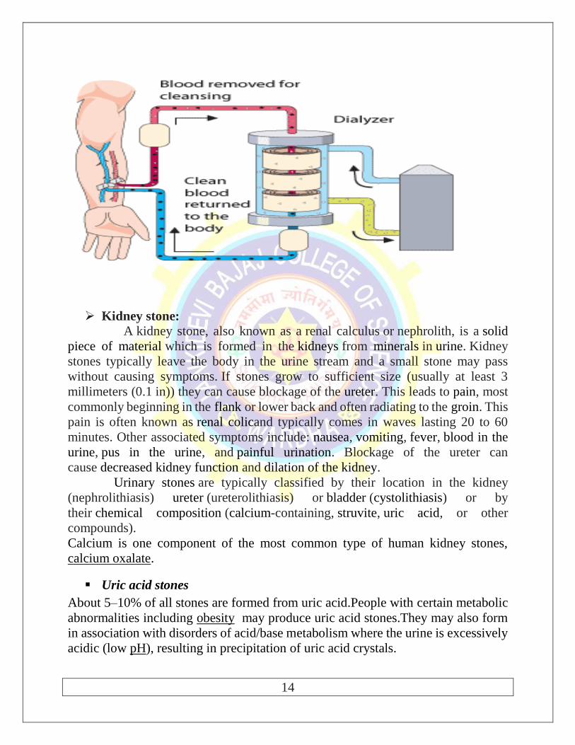
Dialysis:
In medicine, dialysis (from Greek “dialusis” ,meaning dissolution, “dia”
,meaning through and “lysis” , meaning loosening) is primarily used toprovide an
artificial replacement for lost kidney function in people with renal failure. Dialysis
may be used for those with an acute disturbance in kidney function or for those with
progressive but chronically worsening kidney function- a state known as chronic
kidney disease. The latter form may develop over months or years , but in contrast
to acute kidney injury is not usually reversible and dialysis is regarded as a “holding
measure” until a renal transplant can be performed or sometimes as the only
supportive measure in those for whom a transplant would be inappropriate.
The kidneys have an important role in maintaining health. When healthy,
the kidneys maintain the body's internal equilibrium of water and minerals (sodium,
potassium, chloride, calcium, phosphorus, magnesium, sulphate). The
acidic metabolism end-products that the body cannot get rid of via respiration are
also excreted through the kidneys. The kidneys also function as a part of
the endocrine system, producing erythropoietin, calcitriol and renin. Erythropoietin
is involved in the production of red blood cells and calcitriol plays a role in bone
formation. Dialysis is an imperfect treatment to replace kidney function because it
does not correct the compromised endocrine functions of the kidney. Dialysis
treatments replace some of these functions through diffusion (waste removal) and
ultrafiltration (fluid removal).
14
Kidney stone:
A kidney stone, also known as a renal calculus or nephrolith, is a solid
piece of material which is formed in the kidneys from minerals in urine. Kidney
stones typically leave the body in the urine stream and a small stone may pass
without causing symptoms. If stones grow to sufficient size (usually at least 3
millimeters (0.1 in)) they can cause blockage of the ureter. This leads to pain, most
commonly beginning in the flank or lower back and often radiating to the groin. This
pain is often known as renal colicand typically comes in waves lasting 20 to 60
minutes. Other associated symptoms include: nausea, vomiting, fever, blood in the
urine, pus in the urine, and painful urination. Blockage of the ureter can
cause decreased kidney function and dilation of the kidney.
Urinary stones are typically classified by their location in the kidney
(nephrolithiasis) ureter (ureterolithiasis) or bladder (cystolithiasis) or by
their chemical composition (calcium-containing, struvite, uric acid, or other
compounds).
Calcium is one component of the most common type of human kidney stones,
calcium oxalate.
Uric acid stones
About 5–10% of all stones are formed from uric acid.People with certain metabolic
abnormalities including obesity may produce uric acid stones.They may also form
in association with disorders of acid/base metabolism where the urine is excessively
acidic (low pH), resulting in precipitation of uric acid crystals.

15
Struvite stones
About 10–15% of urinary calculi are composed of struvite (ammonium magnesium
phosphate, NH4MgPO4·6H2O). Struvite stones (also known as "infection stones",
urease or triple-phosphate stones)form most often in the presence of infection by
urea-splitting bacteria. Using the enzyme urease, these organisms metabolize urea
into ammonia and carbon dioxide. This alkalinizes the urine resulting in favorable
conditions for the formation of struvite stones.
Renal calculi: Stone or insoluble mass of crystallised salts (oxalates,
etc.) formed within the kidney.
Glomerulonephritis: Inflammation of glomeruli of kidney.
Kidney transplantation:
Kidney transplantation is the organ transplant of a kidney into a patient with end-
stage renal disease. Kidney transplantation is typically classified as deceased-donor
(formerly known as cadaveric) or living-donor transplantation depending on the
source of the donor organ. Living-donor renal transplants are further characterized
as genetically related (living-related) or non-related (living-unrelated) transplants,
depending on whether a biological relationship exists between the donor and
recipient.
Regulation of Kidney function:
The functioning of the kidneys is efficiently monitored and regulated by
hormonal feedback mechanisms involving the hypothalamus, JGA and the heart.
Osmoreceptors in the body are activated by changes in blood volume,
body fluid volume and ionic concentration. An excessive loss of fluid from the body
can activate these receptors which stimulate the hypothalamus to release antidiuretic
hormone (ADH) or vasopressin from the neurohypophysis. ADH increases
permeability of renal tubules for absorption. An increase in body fluid volume can
suppress the osmoreceptors and suppress the ADH secretion. ADH can also affect
the kidney function by its constrictor effects on blood vessels. This causes an
increase in blood pressure. An increase in blood pressure can increase the glomerular
blood flow.
The JGA plays a complex regulatory role. A fall in glomerular blood
flow/glomerular blood pressure/GFR can activate the JG cells to release renin which
converts angiotensinogen in blood to angiotensin I and further to angiotensin II.
Angiotensin II is vasoconstrictor, increases the glomerular blood pressure and
thereby GFR. Angiotensin II also activates the adrenal cortex to release Aldosterone.
Aldosterone causes reabsorption of Na+ and water from the distal parts of the tubule.

16
This also leads to an increase in blood pressure and GFR. This complex mechanism
is generally known as the Renin-Angiotensin mechanism.
An increase in blood flow to the atria of the heart can cause the release
of Atrial Natriuretic Factor (ANF). ANF can cause vasodilation (dilation of
blood vessels) and thereby decrease the blood pressure, acts as a check on the renin-
angiotensin mechanism.
The atria of the heart have been shown to produce ANF hormone. It is
responsible for lowering blood volume and blood pressure by promoting salt and
water excretion in the urine.
Uremia :
Uremia was the term for the contamination of the blood with urine. It is the
presence of an excessive amount of urea in blood. The term uremia is now used for
the illness accompanying kidney failure.Normal value of urea in blood is 0.01-
0.03%, but it rises above0.05% then it is called uremia.
Nephritis or Bright’s disease :
It is characterised by inflammation of both kidneys.In nephritis the disorder
involved such as haematuria,protenuria, hypertension,oedema ,oligouria.
Childrens from age 6-16yrs when infected with Streptococcal
pharyngitis,they suffer from Streptococcal glomerulornephritis.
Accessory excretory organs:
In addition to the urinary system, the skin, lungs and liver of vertebrates are
accessory excretory organs.
(1) Skin: Human skin is thick, impermeable and shows presence of two types
of skin glands: sweat gland and sebaceous glands.
Human skin possesses glands for secreting two fluids on its
surface, namely sweat from the sweat glands and sebum from sebaceous
glands. Sweat is a watery fluid containing in solution primarily contains
sodium-chloride, lactic acid, urea, amino acids and glucose. It helps in
excreting mainly water and sodium chloride, and a small amount of urea
and lactic acid. It helps in thermoregulation. Sebum is a wax-like secretion
which helps to excrete some lipids such as waxes, sterols, other
hydrocarbons and fatty acids on the skin. It mixes with the sweat on the
surface of the skin making it softer and lubricating the hair.
(2) Lungs: Lungs are the main respiratory organs of vertebrates. CO2 and
water are produced during the process of oxidation of glucose.Water is

17
used for metabolic process.Lungs help to eliminate the entire volume of
carbon dioxide produced in the body and excess water is thrown out in the
form of water vapour during expiration. When lungs fail to eliminate
enough carbon dioxidee the kidneys attempt to compensate. They change
some of the carbon dioxide into sodium bicarbonate, which becomes part
of the blood buffer system.


![Osmoregulation and Excretion [Important words are in bold]](https://img.dokumen.tips/doc/110x75/56649e895503460f94b8df70/osmoregulation-and-excretion-important-words-are-in-bold.jpg)