Embed Size (px)
Citation preview

Excitation of the Heart

Intro Muscle cells of the
myocardium are excitable: with electrical stimulation they will contract
Leads to contraction of heart
Leads to pumping of blood
Does not require stimulation from CNS

Sinoatrial Node SA node aka “the
pacemaker” Found within the wall of
the right atrium Where electrical signals
are initiated Sets HR Controlled by the
autonomic nervous system

STOP!
I know what you're going to say: “If the heart stimulates itself, why is it controlled by the autonomic nervous system?”
Because we don't need to tell our heart to beat it is an automatic process therefore it is grouped under the autonomic nervous system
All process in the body are either autonomic or somatic
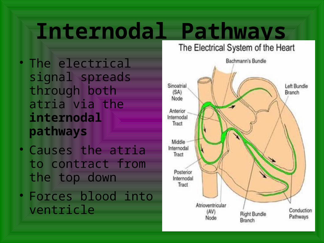
Internodal Pathways The electrical signal
spreads through both atria via the internodal pathways
Causes the atria to contract from the top down
Forces blood into ventricle

Atrioventricular Node
AV node Located at the bottom of the right atria Passes the electrical signal from the atria to the
ventricles Also passes signal into a region of specialized
tissue that runs down the ventricular septum: the bundle of His Splits to form the right and left bundle branches

Purkinje Fibres From the bundle of
His the branches pass the signal on to the Purkinje fibres
Purkinje fibres pass the electrical signal to the ventricles

Coronary Circulation Remember that the heart is a working muscle
that needs a constant supply of oxygen as well as fuel and nutrients
Blood is supplied to the heart through two main arteries: the right and left coronary arteries
Branch off of the aorta and divide multiple times, supplying all regions of the myocardium with oxygenated blood


Cardiac Cycle Defined as the series
of events that occurs through one heart beat
Diastole: phase of relaxation Heart fills with blood
Systole: phase of contraction Heart contracts and
ejects blood

Pressure During the cardiac cycle there are dramatic
changes in pressure Pressure propels the blood through the circulation
Systolic blood pressure: pressure observed in the arteries during the contraction phase
Diastolic blood pressure: pressure observed in the arteries during relaxation of heart
Normal bp is 120/80

The Vascular System and Blood
Vascular system is formed by a network of vessels that transport blood throughout the body
As you follow the path of blood through the body away from the heart, the vessels branch out and get smaller
Main categories of vessels: Arteries, arterioles, capillaries, venules, and veins

Arteries Carry blood away
from heart Thick, muscular walls
that are very elastic Ability to stretch and
recoil is important in assisting the movement of blood during diastole

Arterioles Smaller than arteries Surrounded by rings of smooth muscle that can
contract or relax Controlled by the nervous system
Nervous system can control the distribution of blood flow to different organs using arterioles


Capillaries Smallest vessel Walls are very thin – one cell thick Location of exchange of gases and nutrients Interesting fact: if you were to line up all of the
capillaries from one person, they would form a line of more than 40,000 km long.

Veins Return blood to the heart Become larger as they move away from the
capillaries Venules --> veins --> vena cava Carry deoxygenated blood (except the
pulmonary veins)

Blood Main role is to transport oxygen, carbon dioxide
and nutrients Two main components: plasma and blood
cells Plasma: fluid component
Composed mostly of water Makes up about 55% of blood Within you will find nutrients, proteins, ions, and
gases

Blood Cells Red blood cells – most abundant blood cell
Transport O2 and CO2 Contain a specialized protein called hemoglobin
which can bind O2 and CO2 White blood cells – less than 1% of blood
Play an important role in protecting the body from disease

Platelets Incomplete cells –
fragments Important in the
regulation of blood clotting
In your notes...
Refer to pages 115 and 116 to explain the skeletal muscle pump and the thoracic pump