Embed Size (px)
Citation preview

Nano Res
1
Excellent photothermal conversion of core/shell
CdSe/Bi2Se3 quantum dots
GZ. Jia1,2, WK. Lou1, F. Cheng3, XL. Wang4, JH. Yao4, N. Dai5, HQ. Lin6, and K.Chang1 ()
Nano Res., Just Accepted Manuscript • DOI: 10.1007/s12274-014-0629-2
http://www.thenanoresearch.com on November 7 2014
© Tsinghua University Press 2014
Just Accepted
This is a “Just Accepted” manuscript, which has been examined by the peer-review process and has been
accepted for publication. A “Just Accepted” manuscript is published online shortly after its acceptance,
which is prior to technical editing and formatting and author proofing. Tsinghua University Press (TUP)
provides “Just Accepted” as an optional and free service which allows authors to make their results available
to the research community as soon as possible after acceptance. After a manuscript has been technically
edited and formatted, it will be removed from the “Just Accepted” Web site and published as an ASAP
article. Please note that technical editing may introduce minor changes to the manuscript text and/or
graphics which may affect the content, and all legal disclaimers that apply to the journal pertain. In no event
shall TUP be held responsible for errors or consequences arising from the use of any information contained
in these “Just Accepted” manuscripts. To cite this manuscript please use its Digital Object Identifier (DOI®),
which is identical for all formats of publication.
Nano Research
DOI 10.1007/s12274-014-0629-2
Excellent photothermal conversion of core/shell
CdSe/Bi2Se3 quantum dots
GZ. Jia1,2, WK. Lou1, F. Cheng3, XL. Wang3, JH.
Yao3, N. Dai5, HQ. Lin6, and K.Chang1*
1 Institute of Semiconductors, PR China
2 Tianjin Chengjian University, PR China
3 Changsha University of Science and Technology,
PR China
4 Nankai University, PR China
5 Inst Tech Phys, Nat Lab Infrared Phys, PR China
6 Beijing Computational Science Research Center,
PR China
The water-dispersed CdSe/Bi2Se3 core/shell quantum dots were
synthesized by a cation exchange reaction. To give the mechanism of
core/shell quantum dots with excellent photothermal conversion
efficiency and near-infrared photostability.

Excellent photothermal conversion of core/shell
CdSe/Bi2Se3 quantum dots
GZ. Jia1,2, WK. Lou1, F. Cheng3, XL. Wang4, JH. Yao4, N. Dai5, HQ. Lin6, and K.Chang1 ()
Received: day month year
Revised: day month year
Accepted: day month year
(automatically inserted by
the publisher)
© Tsinghua University Press
and Springer-Verlag Berlin
Heidelberg 2014
KEYWORDS
cation exchange,
quantum dots,
photothermal,
type-II heterostructure,
CdSe/Bi2Se3
ABSTRACT
The water-dispersed CdSe/Bi2Se3 core/shell QDs with a photothermal
conversion coefficient of 27.09% were synthesized by a cation exchange
reaction. The microstructrue and crystal structure of the QDs, which were
confirmed by TEM and XRD, showed that cation exchange partly occurred
inside the CdSe QDs. Two main mechanisms can result in the excellent
photothermal conversion: radiative recombination of carriers inhibited due to
forming the type-II semiconductor heterostructure and large surface-to-volume
ratio of QDs. The photothermal conversion experiments results indicate that
CdSe/Bi2Se3 QDs showed higher photothermal conversion efficiency and
excellent NIR photostability.
Introduction
Photothermal effect has attracted intensive interests
in recent years due to its potential application in
nanoscale heat sources[1], biological imaging[2, 3],
spectroscopy[4], drug delivery[5], nanocatalysis[6],
and photothermal cancer therapy[7-21]. Most of
recent works focused on photothermal effects in
noble metal nanoparticles caused by surface plasmon
polariton (SPP). The strong interaction between light
and noble metal nanparticles can change electron
transient processes in atoms and moleculars in
Nano Research
DOI (automatically inserted by the publisher)
Research Article
Address correspondence to K. Chang. [email protected]
| www.editorialmanager.com/nare/default.asp
2 Nano Res.
biological systems through SPPs[7, 10, 13, 15, 18, 19,
21]. Optically excited noble metal nanoparticles can
be used as nanoscale heat sources through
dissipation of absorbed light into thermal energy.
Plasmon enhanced metal nanoparticles could be used
in photothermal cancer therapy.
Here, we suggest a new direction of photothermal
effect in semiconductor nanostructures based on
band engineering. We demonstrate that CdSe/Bi2Se3
core/shell quantum dots (QDs) can be developed as
photothermal ablation (PTA) to replace the
traditional therapeutic approaches to treat and
control cancers, which can effectively avoid harming
healthy cells and destroying the immune system[22].
This mainly based on that CdSe/Bi2Se3 QDs have a
large absorption coefficient and high photothermal
conversion efficiency in near-infrared (NIR,
λ=700-1100nm) wavelengths. The near infrared
irradiation leads to relatively low scattering and
absorption, and several centimeters penetration in
biological tissues[7, 18]. In addition, Bi and Se belong
to the promising bio-friendly elementary and Bi2Se3
is with low cytotoxicity[23-25]. This decides Bi2Se3
becoming promising photothermal agents and
potential application in cancers therapy by
photothermal technology. Recently Bi2X3 (X=Se,
Te, …), known as topological insualtors, have
attracted enormous attention in condensed matter
physics because of their unique electronic properties.
Topological insulators are a class of quantum
materials possessing metallic surface states and
insulating bulk crystals[26-31]. Enhanced
thermoelectric performance in topological insulators
has attracted intensive interests since the helical edge
and surface states can provide us ballistic channels
for electrons[32]. The figure of merit (ZT) is expected
to be larger than 1 in these materials.
Due to the unique advantages of PTA therapy,
various photothermal agents have appeared as the
NIR photothermal agents for cancer therapy, such as
the organic compounds[14], carbon-based materials
[11, 20, 33], noble metal nanostructures[7, 34-36], and
semiconductor compounds[12, 16, 18, 37]. Although
the noble metal nanostructures have appeared as the
most studied agents with large optical extinction
coefficients and good photothermal conversion
efficiency because of their tunable surface plasmon
resonance (SPR) properties in the NIR wavelength,
some substantial shortcomings impose limitations in
their wide therapeutic applications, for example, too
large size to increase bloodstream circulation time[38,
39], lacking good photothermal stability[17], and
expensive raw material, etc. The investigations have
shown that few-layer Bi2Se3 can significantly enhance
the contribution of exotic surface states due to large
surface-to-volume ratios[40]. Low-dimensional
semiconductor nanostructures can effectively
suppress the bulk effect and discover some novel
physical properties. Layered or layered-like Bi2Se3
nanostructures have been fabricated by various
methods for potential applications in spintronic
devices. More recently, Bi2Se3 nanoplates as a new
photothermal coupling agent for PTA of cancer cells
can be utilized for enhanced X-ray computed
tomography imaging of tumor tissue in vivo[22].
However, the considerably large Bi2Se3 nanoplates
with an average diameter of about 90 nm can hold
back the further bioapplications. Generally, the
nanoparticles size between 10 and 50 nm is more
suitable for intravenous injection to increase
effectively bloodstream circulation time[17]. To the
best of our knowledge, there is no synthesis method
developed to explore proper size Bi2Se3
nanomaterials with favorable biocompatibility for
application in biomedical fields.
In this communication, we report the first
example of water-dispersed CdSe/Bi2Se3 core/shell
QDs which are quickly synthesized in the cation
exchange method assisted by ultrasonic irradiation,
as shown in Fig. 1(a). As-prepared CdSe/Bi2Se3 QDs
not only possess high photothermal conversion
efficiency in 808nm wavelength but also excellent
photostability and favorable biocompatibility due to
the QDs with thio-stabilized surface. These features
are attributed to type-II QDs heterostructure and
large surface-to-volume ratio of QDs.
Experimental
Synthesis of CdSe QDs: Selenium powder,
CdCl2·2.5H2O, Na2SO3, Bi(NO3)3·5H2O, thioglycollic

www.theNanoResearch.com∣www.Springer.com/journal/12274 | Nano Research
3 Nano Res.
acid (TGA), all the reagents are AR, ultrapure water,
microwave system(MAS-I) was used for the synthesis
of CdSe QDs, ultrasound system(KQ2200DE) for the
synthesis of Bi2Se3. CdSe QDs were synthesized
according to previously published articles. Briefly, Se
powder (0.0632 g) , Na2SO3 (0.3025 g) and 40 mL
water were added into a 100 mL flask. The mixture
was stirred vigorously at 80℃ under nitrogen
atmosphere. After 3 h, the transparent Na2SeSO3
solution was obtained. CdCl2·2.5H2O (0.2740 g) was
dissolved into 100 mL water absolutely, then 5 drops
of TGA was added into CdCl2 solution dropwise. The
solution changed milk white quickly when TGA was
added. 10 mL 1 M NaOH solution was prepared to
adjust the pH value of CdCl2 solution to about 8.
When the NaOH was added into CdCl2 solution, the
white precipitate would disappeared and the
solution would changed transparent again at pH=7.
Nitrogen gas was used to deaerate oxygen for at least
30 min and the CdCl2 precursor was obtained. Under
N2 protection, the Na2SeSO3 solution was injected
into the CdCl2 precursor and mixed absolutely. The
mixture was transferred into the microwave system.
CdSe QDs solution was prepared after 5 minutes’
microwave irradiation of 1000W at 100 ℃.
Cation exchange reaction: Bi(NO3)3·5H2O was added
into the QDs solution and transferred into ultrasound
system. When the solid Bi(NO3)3·5H2O was dropped
into the solution, it changed dark brown from white.
After 5 minutes’ ultrasonic irradiation, the solid was
dissolved and the color of QDs changed dark red. No
precipitate was observed in the solution. 30 mL
acetone was added into the solution and precipitate
was appeared. After centrifuging and washing three
times, the precipitate was collected and dried. Black
powder was obtained.
Determination instruments: UV-Vis absorption
spectra were obtained using a Perkin Lambda
UV-Vis-near-infrared spectrophotometer.
Transmission electron microscopy (TEM) and
high-resolution (HR) TEM images were recorded on
a JEOL microscope operated at 200kV, respectively.
All optical measurements were performed at room
temperature under ambient condition.
Photothermal conversion: For measuring the
photothermal conversion performance of CdSe/Bi2Se3
core/shell QDs, 808nm NIR laser was delivered
through a quartz cuvette containing aqueous
dispersion (1.0 mL) of hydrophilic the sample with
the same QDs concentrations, and the light source
was an external adjustable power 808 nm
semiconductor laser device. The output power was
independently calibrated using an optical power
meter and was found to be 1.6 W for a spot size of
~0.6cm2. A thermocouple with an accuracy of ±0.1℃
was inserted into the aqueous dispersion of the QDs
perpendicular to the path of the laser. The
temperature was recorded one time per 10s.
Results and discussion
CdSe nanocrystals were chose as the reaction body
due to mature preparation technology and the high
degree of control over size and shape that has been
achieved. The water-dispersed CdSe QDs were first
synthesized via microwave irradiation. The size and
shape of CdSe QDs can be controlled by changing the
experiment conditions. CdSe/Bi2Se3 core/shell QDs
can be further formed by the ultrasonic wave-assisted
cation exchange reactions. The Bi3+ ions react with Cd
atoms to yield CdSe/Bi2Se3 QDs by the cation
exchange reaction. Ion exchange reactions depend
sensitively on the size and shape of the nanocrystals
[41, 42]. The QDs with a large surface-to-volume ratio
can be favor the cation exchange and lower phase
transition temperatures due to lower activation
energies for the diffusion of atoms and ions in the
QDs. The conversion to Bi2Se3 is favored for the small
size nanocrystals because of low activation energies
for the diffusion of atoms and ions[42]. Bi atom is
slightly larger than the Cd atom, which kinetically
hindered the cation exchange reaction at ambient
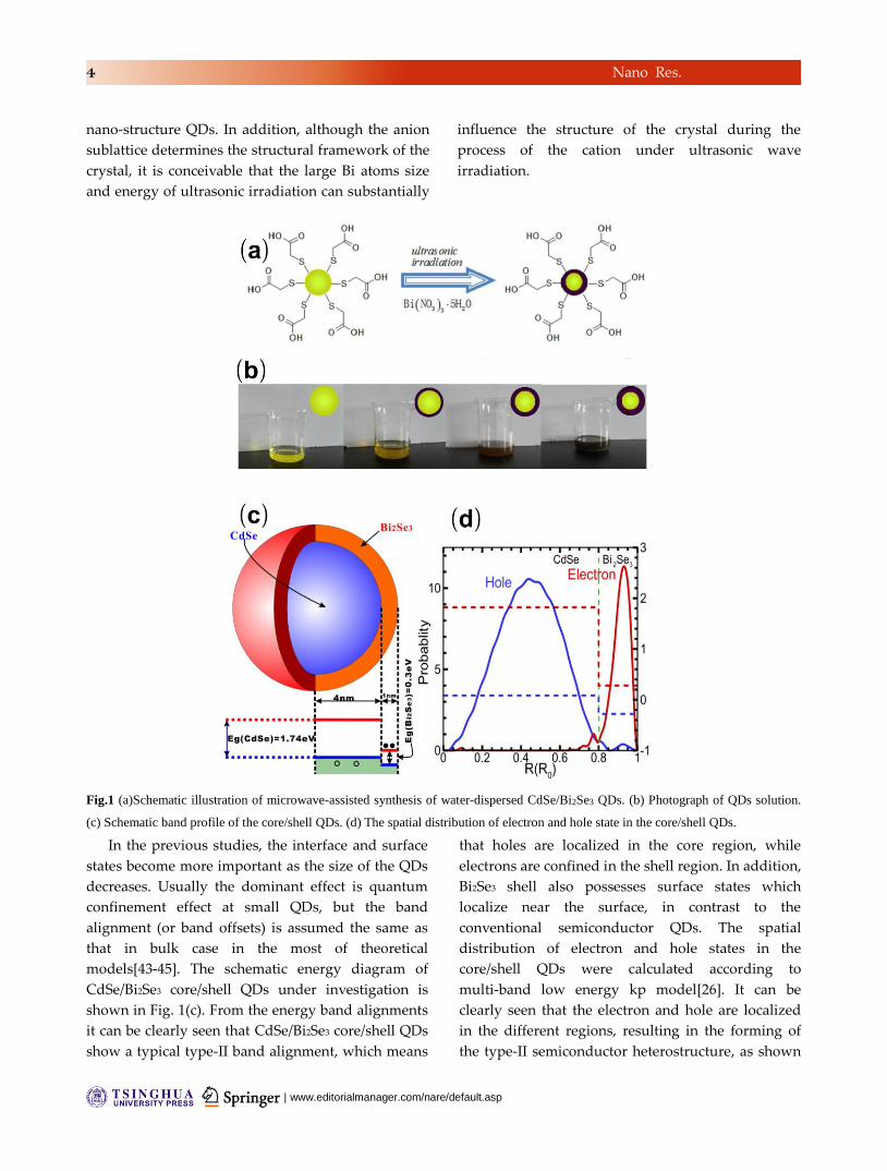
temperature. As shown in Fig. 1(b), the solution color
gradually change from yellow-green to black with
increasing of the concentration of Bi(NO3)3·5H2O
under ultrasonic wave irradiation, indicating cation
exchange reaction to happen and form the Bi2Se3
shell. For the large size CdSe QDs, we found the
cation exchange to be virtually prohibited under
similar irradiation power and time. This further
proves that the reaction energy barrier is much lower
in small sized crystals than in larger systems, even in
| www.editorialmanager.com/nare/default.asp
4 Nano Res.
nano-structure QDs. In addition, although the anion
sublattice determines the structural framework of the
crystal, it is conceivable that the large Bi atoms size
and energy of ultrasonic irradiation can substantially
influence the structure of the crystal during the
process of the cation under ultrasonic wave
irradiation.
Fig.1 (a)Schematic illustration of microwave-assisted synthesis of water-dispersed CdSe/Bi2Se3 QDs. (b) Photograph of QDs solution.
(c) Schematic band profile of the core/shell QDs. (d) The spatial distribution of electron and hole state in the core/shell QDs.
In the previous studies, the interface and surface
states become more important as the size of the QDs
decreases. Usually the dominant effect is quantum
confinement effect at small QDs, but the band
alignment (or band offsets) is assumed the same as
that in bulk case in the most of theoretical
models[43-45]. The schematic energy diagram of
CdSe/Bi2Se3 core/shell QDs under investigation is
shown in Fig. 1(c). From the energy band alignments
it can be clearly seen that CdSe/Bi2Se3 core/shell QDs
show a typical type-II band alignment, which means
that holes are localized in the core region, while
electrons are confined in the shell region. In addition,
Bi2Se3 shell also possesses surface states which
localize near the surface, in contrast to the
conventional semiconductor QDs. The spatial
distribution of electron and hole states in the
core/shell QDs were calculated according to
multi-band low energy kp model[26]. It can be
clearly seen that the electron and hole are localized
in the different regions, resulting in the forming of
the type-II semiconductor heterostructure, as shown

www.theNanoResearch.com∣www.Springer.com/journal/12274 | Nano Research
5 Nano Res.
in Fig. 1(d). The electron and hole pairs are excited in
Bi2Se3 shell by 808nm laser, the photo-excited holes
will relaxed into the core region. The spatial
separation between electron and hole states results in
very small recombination rate, i.e., a long lifetime of
electron-hole pair[46]. Notice that the bottom of the
conduction band of Bi2Se3 is slightly higher than the
top of the valence band of CdSe, therefore almost all
energy of electron-hole pairs gained from laser will
be released to the crystal system, leading to increase
of the crystal temperature. The electron and hole
pairs are excited in Bi2Se3 shell by 808nm laser, the
photo-excited holes will relaxed into the core region.
Notice that the bottom of the conduction band of
Bi2Se3 is slightly higher than the top of the valence
band of CdSe, therefore almost all energy of
electron-hole pairs gained from laser will be released
to the crystal system, leading to increase the crystal
temperature.
Fig. 2 (a) Typical TEM image of CdSe QDs sample. (b) TEM image of CdSe/Bi2Se3 QDs, showing the high quality crystalline structure.
(c) The energy-dispersive X-ray spectroscopy (EDS) of CdSe QDs and CdSe/Bi2Se3 core/shell QDs. (d)Powder X-ray diffraction (XRD)
patterns of the same samples.
Since the Bi2Se3 shell layer is formed by CdSe alloy
with random substitutional Cd atom at the surface of
CdSe nanocrystal, the photoexcited electron could be
trapped and spatially separated by the electron and
hole states in core or shell regions, which leads to
increasing of electron-hole pair lifetime. Recent
experiments show that Bi2Se3 ultrathin films can
open considerably large bandgap and allow greater
access the exotic surface states[47-49]. But, it is a very
difficult now to directly probe topological surface
states in the ultrathin Bi2Se3 shell layer of the
core/shell QDs by the Angle resolved photoemission
spectroscopy (ARPES) and transport measurement.
Therefore, one cannot determine the role of the
surface states in the process of photothermal
conversion.
| www.editorialmanager.com/nare/default.asp
6 Nano Res.
The characteristics of synthesized samples are
analyzed by transmission electron microscopy (TEM).
The high-resolution TEM (HRTEM) analysis on
samples of CdSe QDs and Bi2Se3 are shown in Fig.
2(a). The HRTEM image is taken along the [0001]
crystallinegraphic direction, clearly revealing
crystalline lattice fringes. The as-grown CdSe QDs
were mostly having an average radius of about 5nm.
The growth kinetics could not be finely controlled at
the low temperature reaction condition, which can
result in a relatively broad size distribution of QDs.
This broad size distribution should not be crucial for
photothermal effect in such core/shell QDs, while the
key factor is the spatial separation of photo-excited
electrons and holes caused by the type-II band
alignment. After cation exchange reaction (Fig. 2(b)),
the HRTEM clearly shows that the parent lattice is
finely kept and an interface between the core and
shells is not obviously observed, which indicates that
cation exchange reaction does not disturb the
crystalline shape of the parent QDs. The
energy-dispersive spectroscopy (EDS) of samples
before and after cation exchange, as shown in Fig. 2(c)
confirms the presence of Cd, Se, and Bi elements,
indicating that Bi ion has successfully exchange with
Cd ion. The nanocrystals were further characterized
by X-ray powder diffraction (XRD) (see Fig. 2(d)). All
of the diffraction peaks from the samples (a) to (g)
can be readily indexed to zinc blende structure. The
three strong peaks with values of 23.78, 39.38, and
46.48 corresponding to the (111), (220), and (311)
planes, respectively. We do not observe any
characteristic diffraction peak corresponding to the
Bi2Se3 by Gausses fitting the XRD (111) diffraction
peak, which confirm the formation of Bi2Se3 shell
surrounding the CdSe core.
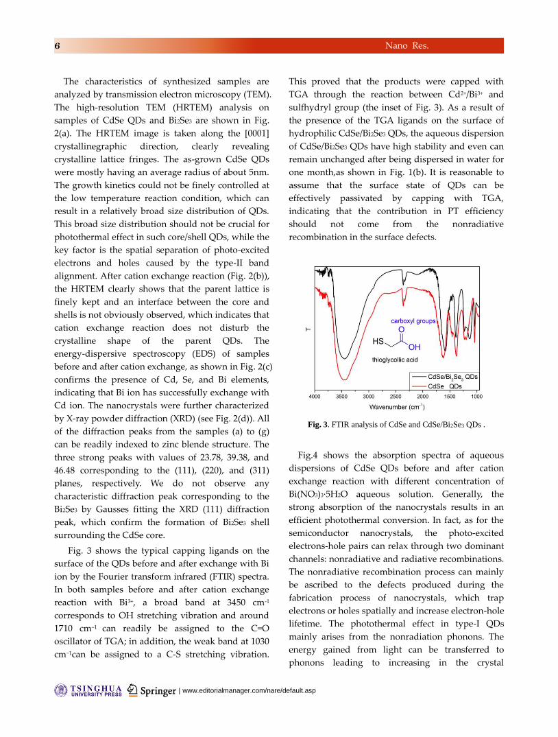
Fig. 3 shows the typical capping ligands on the
surface of the QDs before and after exchange with Bi
ion by the Fourier transform infrared (FTIR) spectra.
In both samples before and after cation exchange
reaction with Bi3+, a broad band at 3450 cm-1
corresponds to OH stretching vibration and around
1710 cm−1 can readily be assigned to the C=O
oscillator of TGA; in addition, the weak band at 1030
cm−1can be assigned to a C-S stretching vibration.
This proved that the products were capped with
TGA through the reaction between Cd2+/Bi3+ and
sulfhydryl group (the inset of Fig. 3). As a result of
the presence of the TGA ligands on the surface of
hydrophilic CdSe/Bi2Se3 QDs, the aqueous dispersion
of CdSe/Bi2Se3 QDs have high stability and even can
remain unchanged after being dispersed in water for
one month,as shown in Fig. 1(b). It is reasonable to
assume that the surface state of QDs can be
effectively passivated by capping with TGA,
indicating that the contribution in PT efficiency
should not come from the nonradiative
recombination in the surface defects.
Fig. 3. FTIR analysis of CdSe and CdSe/Bi2Se3 QDs .
Fig.4 shows the absorption spectra of aqueous
dispersions of CdSe QDs before and after cation
exchange reaction with different concentration of
Bi(NO3)3·5H2O aqueous solution. Generally, the
strong absorption of the nanocrystals results in an
efficient photothermal conversion. In fact, as for the
semiconductor nanocrystals, the photo-excited
electrons-hole pairs can relax through two dominant
channels: nonradiative and radiative recombinations.
The nonradiative recombination process can mainly
be ascribed to the defects produced during the
fabrication process of nanocrystals, which trap
electrons or holes spatially and increase electron-hole
lifetime. The photothermal effect in type-I QDs
mainly arises from the nonradiation phonons. The
energy gained from light can be transferred to
phonons leading to increasing in the crystal

www.theNanoResearch.com∣www.Springer.com/journal/12274 | Nano Research
7 Nano Res.
temperature. Usually, the lifetime of photo-excited
electron-hole pairs in type-I QDs is quite short. This
rapid recombination process strongly limits the
photothermal effect. As the molar weight of
Bi(NO3)3·5H2O was gradually increased, the
absorbance intensity of the 808 nm band increased
and redshifted. This indicates that the cation
exchange process has happened from surface to core
in CdSe QDs. The shift in absorbance edges were
attributed to the increase of the molar weight of Bi3+
in the shell region of the QDs and transformation of
heterostructure type.
Fig. 4. UV-VIS-NIR absorption spectra of aqueous dispersions
of CdSe QDs before and after cation exchange reaction with
different molar weight of Bi(NO3)3: (a)0 g, (b)0.00485g,
(c)0.0097g, (d)0.01455g, (e)0.0194, (e)0.02425g, (f)0.0291 and
(g)0.03395g.
The differential thickness of Bi2Se3 shell QDs
were synthesized by reaction between CdSe QDs
with various concentration of Bi(NO3)3·5H2O under
irradiation of the same ultrasonic power and time.
The photothermal effect of samples was
investigated by monitoring the temperature of 1mL
aqueous solutions of various Bi2Se3 shell thickness
irradiated by a NIR laser (808 nm, 1.6W), as shown
in the Fig. 5(a). The blank experiment demonstrates
that the temperature of pure water is increased by
less than 3°C. With the addition of the molar ratio
between Bi and Cd, the temperature of the aqueous
dispersion can rapidly increase by 50°C in 5min.
The difference of the temperature change slightly
for the different Bi2Se3 shell thickness due to faster
heat loss at higher temperature[18], although the
aqueous dispersion containing the same
concentration QDs with larger molar Bi2Se3 ratio
can absorb more efficiently photons of 808 nm laser
and then has higher NIR photothermal conversion
capability.
As illustrated in Fig. 5(b), the temperature
increases rapidly with increasing of Bi3+
concentration, goes up dramatically with the
increase of Bi2Se3 shell thickness, and then saturates
with further increase of Bi2Se3 shell thickness. This
phenomenon should be attributed to a fast heat loss
at relatively high temperature. The temperature
change is basically stable, even if the Bi2Se3 shell
thickness increase with continuing of the cation
exchange reaction. The photothermal conversion
efficiency indicates that the dominant photothermal
conversion processes closely related to the process of
cation exchange, i.e., the thickness of Bi2Se3 shell. Bi
atoms can appear at the surface of QDs by
substituting for Cd atoms under the low
concentration Bi3+ reaction condition. The Bi2Se3 shell
can gradually be formed with increasing of Bi3+
concentration. As the thickness of Bi2Se3 shell region
increases, the QDs form type-II heterostructures.
The lifetime of electron-hole pairs could be
enchanced significantly due to bandgap reduced
from the direct bandgap to indirect bandgap,
therefore the most energy gained from light will be
converted into phonon system, leading to increasing
of crystal temperature.
The ideal photothermal agent should be with
excellent photothermal conversion efficiency and
photostability for biological applications, the
photothermal conversion efficiency was calculated
according to Roper's report and independent of the
shape of the nanocystals aqueous dispersion[18, 37].
The temperature decrease of the solution was
monitored to determine the heat transfer time
constant from the dispersion system to the room
temperature. The heat conversion efficiency are
higher than 20% for sample (d) (e) (g) (f), which
should be attributed to the strong NIR absorption

| www.editorialmanager.com/nare/default.asp
8 Nano Res.
and effective nonradiative electron relaxation
dynamics (see Fig. 5(c)). To investigate the NIR
photostability of CdSe/Bi2Se3 QDs, three cycles of
laser ON/OFF with NIR light were used. The same
elevated temperature for the different cycles
indicated that as-prepared nanoparticles show
excellent photostability, no significant decrease for
the temperature elevation was observed for
CdSe/Bi2Se3 nanoparticles solution (see Fig. 5(d)).
This indicates that CdSe/Bi2Se3 nanoparticles are
with perfect photothermal conversion ability and
excellent photostability, and can act as photothermal
agents to effectively destroy the cancer cells.
Fig. 5. (a) Photothermal effect of the irradiation of the aqueous dispersion of QDs with the different thickness Bi2Se3 shell using the NIR
laser shining (808 nm, 1.6 W), in which the irradiation lasted for 5 min, and then the laser was shut off. (b)Plot of temperature change
over a period of 5min versus the different Bi3+ concentration during the cation exchange reaction process. (c)Time constant for heat
transfer from the system is determined to be τs=315s by applying the linear time data from the cooling period (after 300s) versus
negative natural logarithm of driving force temperature, which is obtained from the cooling stage of panel (a). (d)Temperature elevation
of the typical CdSe/Bi2Se3 QDs for sample (e) and (f)over three laser ON/OFF cycles of 808 nm NIR laser irradiation.
In order to clearly understand the mechanism of
PT conversion in type-II core/shell structure QDs,
carriers dynamic can be analyzed as follows. For
type-I semiconductor QDs, the dominant PT
conversion channels are defect-assistent
recombination and phonon-mediated relaxation
processes of electrons to the conduction band bottom
before radiative recombination. As for the core/shell
www.theNanoResearch.com∣www.Springer.com/journal/12274 | Nano Research
9 Nano Res.
semiconductor QDs, carrier lifetime and optical
transition rate decisively depend on the energy band
alignments of semiconductor heterostructures. There
are two important features suggesting the type-II
band alignment. First, one can see clearly that
additional Bi2Se3 shell growth leads to redshift of the
absorption bandedge and beyond the band edge of
bulk CdSe (1.75eV), (see Fig. 4) which is important
evidence suggesting the type-II band alignment with
the increasing of Bi2Se3 shell thickness[50]. In
addition, it can be clearly seen that photothermal
conversion exhibits an abrupt increase as Bi2Se3
thickness increases (see Fig. 5(a)). These two features
can be ascribed to the significant increase of carrier
lifetimes due to the spatial separation of the carriers,
i.e., the type-II band alignment[46, 51]. The excess
energy of photo-excited electron-hole pairs is much
larger than the narrow bandgap, therefore the excess
energy will heat the crystal of QDs through phonon
emission. Especially, almost all energy of
electron-hole pairs gained from light will be
converted into phonon system, since the conduction
band bottom of Bi2Se3 shell is only slightly higher
than the valence band top of CdSe core. Usually, for
the photo-excited electron-hole pairs in the narrow
band gap semiconductors, the excess kinetic energy
can create an effective temperature which could be
much higher than the lattice temperature. In the
type-II QDs, spatially separate carriers are localized
in the two different regions (the core or shell regions)
due to the type-II band alignment(Fig.1(d)) [47,
52-54]. The Auger and radiative decay lifetime can be
very long due to the spatially separated electron-hole
pairs[55]. The spatial separation can effectively
inhibit radiative recombination of carrier and
increase the lifetime, which results in the
temperature equilibrium between carriers and
crystal, i.e., high efficient photothermal conversion.
Conclusion
In summary, water-dispersed CdSe/Bi2Se3 core/shell
QDs with a photothermal conversion coefficient of
27.09% were synthesized by a cation exchange
reaction. CdSe/Bi2Se3 core/shell QDs confirmed by
TEM and XRD, showed that cation exchange
occurred inside the CdSe QDs. The radiative
recombination of carriers can be inhibited due to
forming the type-II semiconductor heterostructure.
The photothermal conversion experiments results
indicate that the CdSe/Bi2Se3 nanoparticles show
higher photothermal conversion efficiency and
excellent NIR photostability.
Acknowledgements
This work has been partly supported by the National
Key Basic Research Program of China
(2012CB934201), the National Basic Research
Program of China (973 Program) under Grant No.
2011CB922204 and 2012CB934304, and the National
Natural Science Foundation of China (11147024,
11247025, 10934007,11304306,11374002, 61290303).
References
[1] Zhang, W.; Li, Q.; Qiu, M., A plasmon ruler based on
nanoscale photothermal effect. Opt. Express 2013, 21,
172-181.
[2] Ting, L.; Jiguang, T.; Zhaolong, C.; Ying, L.; Jiao, L.; Si,
L.; Huihui, L.; Jinhua, Z.; Xingsheng, Y., Anti-TROP2
conjugated hollow gold nanospheres as a novel
nanostructure for targeted photothermal destruction of
cervical cancer cells. Nanotechnology 2014, 25, 345103
(11 pp.)-345103 (11 pp.).
[3] Jinyeong, Y.; Hun, K.; Suho, R.; Sungwook, S.; Hyun
Ok, K.; Hyun; Hyo-Il, J.; Chulmin, J., Photothermal
spectral-domain optical coherence reflectometry for
direct measurement of hemoglobin concentration of
erythrocytes. Biosens. Bioelectron. 2014, 57, 59-64.
[4] Strzalkowski, K.; Zakrzewski, J.; Malinski, M.,
Determination of the Exciton Binding Energy Using
Photothermal and Photoluminescence Spectroscopy. Int.
J. Thermophys. 2013, 34, 691-700.
[5] Wang, Z.; Chen, Z.; Liu, Z.; Shi, P.; Dong, K.; Ju, E.; Ren,
J.; Qu, X., A multi-stimuli responsive gold
nanocage-hyaluronic platform for targeted
photothermal and chemotherapy. Biomaterials 2014, 35,
9678-88.
[6] Byeon, J. H.; Kim, Y.-W., Au-TiO2 Nanoscale
Heterodimers Synthesis from an Ambient Spark
Discharge for Efficient Photocatalytic and Photothermal
| www.editorialmanager.com/nare/default.asp
10 Nano Res.
Activity. ACS Appl. Mater. Interfaces 2014, 6, 763-767.
[7] Chen, J.; Glaus, C.; Laforest, R.; Zhang, Q.; Yang, M.;
Gidding, M.; Welch, M. J.; Xia, Y., Gold Nanocages as
Photothermal Transducers for Cancer Treatment. Small
2010, 6, 811-817.
[8] Chu, M.; Pan, X.; Zhang, D.; Wu, Q.; Peng, J.; Hai, W.,
The therapeutic efficacy of CdTe and CdSe quantum dots
for photothermal cancer therapy. Biomaterials 2012, 33,
7071-7083.
[9] Cole, J. R.; Mirin, N. A.; Knight, M. W.; Goodrich, G. P.;
Halas, N. J., Photothermal Efficiencies of Nanoshells and
Nanorods for Clinical Therapeutic Applications. J. Phys.
Chem. C. 2009, 113, 12090-12094.
[10] Dickerson, E. B.; Dreaden, E. C.; Huang, X.; El-Sayed,
I. H.; Chu, H.; Pushpanketh, S.; McDonald, J. F.; El-Sayed,
M. A., Gold nanorod assisted near-infrared plasmonic
photothermal therapy (PPTT) of squamous cell
carcinoma in mice. Cancer Lett. 2008, 269, 57-66.
[11] Fisher, J. W.; Sarkar, S.; Buchanan, C. F.; Szot, C. S.;
Whitney, J.; Hatcher, H. C.; Torti, S. V.; Rylander, C. G.;
Rylander, M. N., Photothermal Response of Human and
Murine Cancer Cells to Multiwalled Carbon Nanotubes
after Laser Irradiation. Cancer Res. 2010, 70, 9855-9864.
[12] Hessel, C. M.; Pattani, V. P.; Rasch, M.; Panthani, M.
G.; Koo, B.; Tunnell, J. W.; Korgel, B. A., Copper Selenide
Nanocrystals for Photothermal Therapy. Nano Lett. 2011,
11, 2560-2566.
[13] Huang, X.; Tang, S.; Liu, B.; Ren, B.; Zheng, N.,
Enhancing the Photothermal Stability of Plasmonic
Metal Nanoplates by a Core-Shell Architecture. Adv.
Mater. 2011, 23, 3420-+.
[14] Jaemoon, Y.; Jihye, C.; Doyeon, B.; Eunjung, K.;
Eun-Kyung, L.; Huiyul, P.; Jin-Suck, S.; Kwangyeol, L.;
Kyung-Hwa, Y.; Eun-Kyung, K.; Yong-Min, H.; Seungjoo,
H., Convertible Organic Nanoparticles for Near-Infrared
Photothermal Ablation of Cancer Cells. Angew. Chem.
Int. Ed. 2011, 50, 441-4.
[15] Jain, P. K.; Huang, X.; El-Sayed, I. H.; El-Sayed, M. A.,
Noble Metals on the Nanoscale: Optical and
Photothermal Properties and Some Applications in
Imaging, Sensing, Biology, and Medicine. Acc. Chem. Res.
2008, 41, 1578-1586.
[16] Lambert, T. N.; Andrews, N. L.; Gerung, H.; Boyle, T.
J.; Oliver, J. M.; Wilson, B. S.; Han, S. M., Water-soluble
germanium(0) nanocrystals: Cell recognition and
near-infrared photothermal conversion properties.
Small 2007, 3, 691-699.
[17] Tang, S.; Huang, X.; Zheng, N., Silica coating
improves the efficacy of Pd nanosheets for
photothermal therapy of cancer cells using near infrared
laser. Chem. Commun. 2011, 47, 3948-3950.
[18] Tian, Q.; Jiang, F.; Zou, R.; Liu, Q.; Chen, Z.; Zhu, M.;
Yang, S.; Wang, J.; Wang, J.; Hu, J., Hydrophilic Cu9S5
Nanocrystals: A Photothermal Agent with a 25.7% Heat
Conversion Efficiency for Photothermal Ablation of
Cancer Cells in Vivo. Acs Nano. 2011, 5, 9761-9771.
[19] Tian, Q.; Tang, M.; Sun, Y.; Zou, R.; Chen, Z.; Zhu, M.;
Yang, S.; Wang, J.; Wang, J.; Hu, J., Hydrophilic
Flower-Like CuS Superstructures as an Efficient 980 nm
Laser-Driven Photothermal Agent for Ablation of Cancer
Cells. Adv. Mater. 2011, 23, 3542-+.
[20] Yang, K.; Zhang, S.; Zhang, G.; Sun, X.; Lee, S.-T.; Liu,
Z., Graphene in Mice: Ultrahigh In Vivo Tumor Uptake
and Efficient Photothermal Therapy. Nano. Lett. 2010,
10, 3318-3323.
[21] Lim, D.-K.; Barhoumi, A.; Wylie, R. G.; Reznor, G.;
Langer, R. S.; Kohane, D. S., Enhanced Photothermal
Effect of Plasmonic Nanoparticles Coated with Reduced
Graphene Oxide. Nano Lett. 2013, 13, 4075-4079.
[22] Li, J.; Jiang, F.; Yang, B.; Song, X.-R.; Liu, Y.; Yang,
H.-H.; Cao, D.-R.; Shi, W.-R.; Chen, G.-N., Topological
insulator bismuth selenide as a theranostic platform for
simultaneous cancer imaging and therapy. Sci. Rep. 2013,
3.
[23] Ai, K.; Liu, Y.; Liu, J.; Yuan, Q.; He, Y.; Lu, L.,
Large-Scale Synthesis of Bi2S3 Nanodots as a Contrast
Agent for In Vivo X-ray Computed Tomography Imaging.
Adv. Mater. 2011, 23, 4886-4891.
[24] Kinsella, J. M.; Jimenez, R. E.; Karmali, P. P.; Rush, A.
M.; Kotamraju, V. R.; Gianneschi, N. C.; Ruoslahti, E.;
Stupack, D.; Sailor, M. J., X-Ray Computed Tomography
Imaging of Breast Cancer by using Targeted
Peptide-Labeled Bismuth Sulfide Nanoparticles. Angew.
Chem. Int. Ed. 2011, 50, 12308-12311.

www.theNanoResearch.com∣www.Springer.com/journal/12274 | Nano Research
11 Nano Res.
[25] Rabin, O.; Perez, J. M.; Grimm, J.; Wojtkiewicz, G.;
Weissleder, R., An X-ray computed tomography imaging
agent based on long-circulating bismuth sulphide
nanoparticles. Nat. Mater. 2006, 5, 118-122.
[26] Chang, K.; Lou, W. K., Helical Quantum States in
HgTe Quantum Dots with Inverted Band Structures. Phys.
Rev. Lett. 2011, 106, 4.
[27] Kim, N.; Lee, P.; Kim, Y.; Kim, J. S.; Kim, Y.; Noh, D. Y.;
Yu, S. U.; Chung, J.; Kim, K. S., Persistent Topological
Surface State at the Interface of Bi2Se3 Film Grown on
Patterned Graphene. Acs Nano 2014, 8, 1154-1160.
[28] Liu, H.; Jiang, H.; Sun, Q.-F.; Xie, X. C., Dephasing
Effect on Backscattering of Helical Surface States in 3D
Topological Insulators. Phys. Rev. Lett. 2014, 113,
046805-046805.
[29] Moore, J. E., The birth of topological insulators.
Nature 2010, 464, 194-198.
[30] Reijnders, A. A.; Tian, Y.; Sandilands, L. J.; Pohl, G.;
Kivlichan, I. D.; Zhao, S. Y. F.; Jia, S.; Charles, M. E.; Cava,
R. J.; Alidoust, N.; Xu, S.; Neupane, M.; Hasan, M. Z.;
Wang, X.; Cheong, S. W.; Burch, K. S., Optical evidence of
surface state suppression in Bi-based topological
insulators. Phys. Rev. B. 2014, 89.
[31] Wang, L.-L.; Huang, M.; Thimmaiah, S.; Alam, A.;
Bud'ko, S. L.; Kaminski, A.; Lograsso, T. A.; Canfield, P.;
Johnson, D. D., Native defects in tetradymite Bi2(TexSe3-x)
topological insulators. Phys. Rev. B. 2013, 87.
[32] Ghaemi, P.; Mong, R. S. K.; Moore, J. E., In-Plane
Transport and Enhanced Thermoelectric Performance in
Thin Films of the Topological Insulators Bi2Te3 and Bi2Se3.
Phys. Rev. Lett. 2010, 105.
[33] Ghosh, S.; Dutta, S.; Gomes, E.; Carroll, D.;
D'Agostino, R., Jr.; Olson, J.; Guthold, M.; Gmeiner, W. H.,
Increased Heating Efficiency and Selective Thermal
Ablation of Malignant Tissue with DNA-Encased
Multiwalled Carbon Nanotubes. Acs Nano 2009, 3,
2667-2673.
[34] Chen, J.; Yang, M.; Zhang, Q.; Cho, E. C.; Cobley, C.
M.; Kim, C.; Glaus, C.; Wang, L. V.; Welch, M. J.; Xia, Y.,
Gold Nanocages: A Novel Class of Multifunctional
Nanomaterials for Theranostic Applications. Adv. Funct.
Mater. 2010, 20, 3684-3694.
[35] Chen, H.; Shao, L.; Ming, T.; Sun, Z.; Zhao, C.; Yang,
B.; Wang, J., Understanding the Photothermal
Conversion Efficiency of Gold Nanocrystals. Small 2010,
6, 2272-2280.
[36] Kim, D.; Jeong, Y. Y.; Jon, S., A Drug-Loaded
Aptamer-Gold Nanoparticle Bioconjugate for Combined
CT Imaging and Therapy of Prostate Cancer. Acs Nano
2010, 4, 3689-3696.
[37] Roper, D. K.; Ahn, W.; Hoepfner, M., Microscale
heat transfer transduced by surface plasmon resonant
gold nanoparticles. J. Phys. Chem. C. 2007, 111,
3636-3641.
[38] Alkilany, A. M.; Nagaria, P. K.; Hexel, C. R.; Shaw, T.
J.; Murphy, C. J.; Wyatt, M. D., Cellular Uptake and
Cytotoxicity of Gold Nanorods: Molecular Origin of
Cytotoxicity and Surface Effects. Small 2009, 5, 701-708.
[39] Loo, C.; Lin, A.; Hirsch, L.; Lee, M. H.; Barton, J.;
Halas, N. J.; West, J.; Drezek, R., Nanoshell-enabled
photonics-based imaging and therapy of cancer. Technol.
Cancer Res. T. 2004, 3, 33-40.
[40] Li, H.; Cao, J.; Zheng, W.; Chen, Y.; Wu, D.; Dang, W.;
Wang, K.; Peng, H.; Liu, Z., Controlled Synthesis of
Topological Insulator Nanoplate Arrays on Mica. J. Am.
Chem. Soc. 2012, 134, 6132-6135.
[41] Chia-Chun, C.; Herhold, A. B.; Johnson, C. S.;
Alivisatos, A. P., Size dependence of structural
metastability in semiconductor nanocrystals. Science
1997, 276, 398-401.
[42] Son, D. H.; Hughes, S. M.; Yin, Y. D.; Alivisatos, A. P.,
Cation exchange reactions-in ionic nanocrystals. Science
2004, 306, 1009-1012.
[43] Ivanov, S. A.; Piryatinski, A.; Nanda, J.; Tretiak, S.;
Zavadil, K. R.; Wallace, W. O.; Werder, D.; Klimov, V. I.,
Type-II core/shell CdS/ZnSe nanocrystals: Synthesis,
electronic structures, and spectroscopic properties. J.
Am. Chem. Soc. 2007, 129, 11708-11719.
[44] Chen, C. Y.; Cheng, C. T.; Lai, C. W.; Hu, Y. H.; Chou,
P. T.; Chou, Y. H.; Chiu, H. T., Type-II CdSe/CdTe/ZnTe
(core-shell-shell) quantum dots with cascade band edges:
The separation of electron (at CdSe) and hole (at ZnTe)
by the CdTe layer. Small 2005, 1, 1215-1220.
[45] Allione, M.; Ballester, A.; Li, H.; Comin, A.; Movilla, J.
| www.editorialmanager.com/nare/default.asp
12 Nano Res.
L.; Climente, J. I.; Manna, L.; Moreels, I.,
Two-Photon-Induced Blue Shift of Core and Shell Optical
Transitions in Colloidal CdSe/CdS Quasi-Type II Quantum
Rods. Acs Nano 2013, 7, 2443-2452.
[46] Zhu, H.; Song, N.; Lian, T., Wave Function
Engineering for Ultrafast Charge Separation and Slow
Charge Recombination in Type-II Core/Shell Quantum
Dots. J. Am. Chem. Soc. 2011, 133.
[47] Balet, L. P.; Ivanov, S. A.; Piryatinski, A.; Achermann,
M.; Klimov, V. I., Inverted core/shell nanocrystals
continuously tunable between type-I and type-II
localization regimes. Nano Lett. 2004, 4, 1485-1488.
[48] Zhang, Y.; He, K.; Chang, C.-Z.; Song, C.-L.; Wang,
L.-L.; Chen, X.; Jia, J.-F.; Fang, Z.; Dai, X.; Shan, W.-Y.;
Shen, S.-Q.; Niu, Q.; Qi, X.-L.; Zhang, S.-C.; Ma, X.-C.; Xue,
Q.-K., Crossover of the three-dimensional topological
insulator Bi2Se3 to the two-dimensional limit. Nat. Phys.
2010, 6, 584-588.
[49] Vargas, A.; Basak, S.; Liu, F.; Wang, B.; Panaitescu, E.;
Lin, H.; Markiewicz, R.; Bansil, A.; Kar, S., The Changing
Colors of a Quantum-Confined Topological Insulator. Acs
Nano 2014, 8, 1222-1230.
[50] Smith, A. M.; Mohs, A. M.; Nie, S., Tuning the
optical and electronic properties of colloidal
nanocrystals by lattice strain. Nat. Nanotech. 2009, 4,
56-63.
[51] Kim, S.; Fisher, B.; Eisler, H. J.; Bawendi, M., Type-II
quantum dots: CdTe/CdSe(core/shell) and
CdSe/ZnTe(core/shell) heterostructures. J. Am. Chem.
Soc. 2003, 125, 11466-11467.
[52] Bang, J.; Park, J.; Lee, J. H.; Won, N.; Nam, J.; Lim, J.;
Chang, B. Y.; Lee, H. J.; Chon, B.; Shin, J.; Park, J. B.; Choi,
J. H.; Cho, K.; Park, S. M.; Joo, T.; Kim, S., ZnTe/ZnSe
(Core/Shell) Type-II Quantum Dots: Their Optical and
Photovoltaic Properties. Chem. Mater. 2010, 22,
233-240.
[53] Chang, K.; Xia, J. B., Spatially separated excitons in
quantum-dot quantum well structures. Phys. Rev. B.
1998, 57, 9780-9786.
[54] Nemchinov, A.; Kirsanova, M.; Hewa-Kasakarage, N.
N.; Zamkov, M., Synthesis and characterization of type-II
ZnSe/CdS core/shell nanocrystals. J. Phys. Chem. C. 2008,
112, 9301-9307.
[55] Oron, D.; Kazes, M.; Banin, U., Multiexcitons in
type-II colloidal semiconductor quantum dots. Phys. Rev.
B. 2007, 75, 7.
Electronic Supplementary Material: Details of the
macroscopic model for calculating photothermal
efficiencies and heat transfer time constant are
available in the online version of this article at
http://dx.doi.org/10.1007/s12274-***-****-*
(automatically inserted by the publisher).
www.theNanoResearch.com∣www.Springer.com/journal/12274 | Nano Research
Nano Res.
Electronic Supplementary Material
Excellent photothermal conversion of core/shell
CdSe/Bi2Se3 quantum dots
GZ. Jia1,2, WK. Lou1, F. Cheng3, XL. Wang3, JH. Yao3, N. Dai5, HQ. Lin6, and K.Chang1 ()
Supporting information to DOI 10.1007/s12274-****-****-* (automatically inserted by the publisher)
Calculating Photothermal Efficiencies and Heat Transfer Time Constant
In order to clearly understand the photothermal conversion process, we further analysis the photothermal
conversion of nanopariticales solution and determine the system heat transfer time constant and the
photothermal conversion efficiency based on the macroscopic model. Similar to the ones previously
published[1-3], the energy balance can be expressed as
,i Np i Np Surr Lossi
dTmC Q Q Q
dt (1)
where m and NpC are the mass and heat capacity of water and T is the solution temperature. The
photothermal energy from the nanocrystals QNp can be written as
808(1 10 )A
IQ I (2)
where I is the laser power, 808A is the absorbance at the excitation wavelength of laser, and is the
photothermal conversation efficiency. The heat lost to the surroundings by the cuvette walls was given
as
| www.editorialmanager.com/nare/default.asp
Nano Res.
( )Loss SurrQ hA T T (3)
where h his heat transfer coefficient, A is the surface area of the container, T and SurrT is ambient
temperature of the surroundings. The temperature profile after the laser is turned on/turn off can be obtained
by solution of the equation (1). Therefore, the photothermal conversion efficiency can be determined as
808
( )
(1 10 )Max Surr Dis
A
hS T T Q
I
(4)
The system heat transfer time constant is determined during the cooling process of solution after the laser
was turned off. The heat transfer time constant is importantly reflect the heat energy releasing characteristic
of nanoparticals, which can be given by applying the linear time data from the cooling period
vs negative natural logarithm of driving force temperature.
,i Np i
i
mC
hS
(5)
References
[1] Chen, H.; Shao, L.; Ming, T.; Sun, Z.; Zhao, C.; Yang, B.; Wang, J., Understanding the Photothermal Conversion
Efficiency of Gold Nanocrystals. Small 2010, 6, 2272-2280.
[2] Tian, Q.; Jiang, F.; Zou, R.; Liu, Q.; Chen, Z.; Zhu, M.; Yang, S.; Wang, J.; Wang, J.; Hu, J., Hydrophilic Cu9S5
Nanocrystals: A Photothermal Agent with a 25.7% Heat Conversion Efficiency for Photothermal Ablation of Cancer
Cells in Vivo. Acs Nano 2011, 5, 9761-9771.
[3] Roper, D. K.; Ahn, W.; Hoepfner, M., Microscale heat transfer transduced by surface plasmon resonant gold
nanoparticles. Journal of Physical Chemistry C 2007, 111, 3636-3641.
Address correspondence to K.Chang, [email protected]