Embed Size (px)
Citation preview

Tutorial Module 5Tutorial Module 5
Examples of Morphologic Types of Examples of Morphologic Types of i i i i li i i i lPneumonia in Domestic AnimalsPneumonia in Domestic Animals
Alfonso LópezAlfonso LópezAtlantic Veterinary CollegeAtlantic Veterinary College
U i it f P i Ed d I l dU i it f P i Ed d I l dUniversity of Prince Edward IslandUniversity of Prince Edward IslandCanadaCanada
©2009©2009
July 25July 25

Examples of Diseases that Cause Examples of Diseases that Cause Bronchopneumonia in Domestic AnimalsBronchopneumonia in Domestic Animals
Suppurative BronchopneumoniaSuppurative BronchopneumoniaSuppurative BronchopneumoniaSuppurative Bronchopneumonia Fibrinous BronchopneumoniaFibrinous BronchopneumoniaFibrinous BronchopneumoniaFibrinous Bronchopneumonia

Enzootic Pneumonia is a clinico-epidemiological
Enzootic Pneumonia of calvesEnzootic Pneumonia of calves
term that corresponds with a sub-acute to chronic suppurative bronchopneumonia in young calves raised in confinement.
Enzootic Pneumonia is a multifactorial disease predisposed by environmental factors (temperature, humidity, poor air circulation, crowding), stress, immune status viral infections (PI-3 Adenovirusimmune status, viral infections (PI 3, Adenovirus, IBR, BRSV, etc.). Bacteria such as Pasteurella multocida (A, D), Hemophilus somnus , Actinobacilluspyogenes, Mycoplasmas (M. bovis, M. dispar) are often isolated from the lungoften isolated from the lung.
Enzootic pneumonia has a high morbidity but low mortality since the pneumonic process often involves
l ll ti f th lonly a small portion of the lung.
Neonatal diarrhea can also predispose calves to Enzootic pneumonia. The dehydration presumably impairs the respiratory defense mechanisms predisposing calves to pneumonia.

Enzootic Pneumonia of calvesEnzootic Pneumonia of calves
The classic lesion in Enzootic pneumonia is a suppurative bronchopneumonia in which purulent exudate can be expressedpurulent exudate can be expressed from the bronchi (See insert).
Microbiology is not always rewarding. In some cases, the viral infection that predisposed to pneumonia is long gone. In a farm with high incidence of enzooticwith high incidence of enzootic pneumonia, it is imperative to eliminate problems such as excessive crowding, poor
til ti h i t tventilation, changes in temperature, etc.
Texture of an affected lung would
.
be firmer than normal and on cut surfaces a purulent exudate could be easily expressed from airways.
Note the cranioventral consolidation of lungs. In this particular calf, the lesion of enzootic pneumonia was extensive enough to cause death.

Enzootic Pneumonia of CalvesEnzootic Pneumonia of Calves This was an incidental finding. Note consolidation only affecting 10% of the lung parenchyma (arrow)
Although the distribution of lung lesions in Enzootic pnuemonia of calves is cranioventral, the magnitude of lung involvement varies considerably from case to case. It is considered that over 60% of lung parenchyma needs to g p ybe involved to incriminate suppurative bronchopneumonia as the cause of death.
Cut sectionCut section showing purulent exudate showing purulent exudate (arrow). The (arrow). The mucoidmucoid appearance of the appearance of the exudate suggests exudate suggests chronicitychronicity (goblet cell (goblet cell
hyperplasia)hyperplasia)hyperplasia).hyperplasia).

Sequels of Enzootic Pneumonia of CalvesSequels of Enzootic Pneumonia of Calves
BronchiectasisBronchiectasisBronchiectasisBronchiectasis
AbscessesAbscessesAbscessesAbscesses
Pleural adhesions Pleural adhesions Pleural adhesions Pleural adhesions Dilated bronchi (ruptured walls) filled with Dilated bronchi (ruptured walls) filled with mucopurulent exudatemucopurulent exudateDilated bronchi (ruptured walls) filled with Dilated bronchi (ruptured walls) filled with mucopurulent exudatemucopurulent exudate
Pleural adhesionsPleural adhesions
Abscesses affecting the Abscesses affecting the cranioventralcranioventral lunglungAbscesses affecting the Abscesses affecting the cranioventralcranioventral lunglung

B i P i M h i i i (Shi i
Shipping FeverShipping Fever
Bovine Pneumonic Mannheimiosis (Shipping Fever) is the number one cause of feedlot mortality in North America.
Shipping Fever is caused by Mannheimia (Pasteurella) haemolytica A1 a commensalbacterium present in the bovine nasal flora that is effectively destroyed by the normal bovine lung.
When the defense mechanisms are i d b i l i f ti (PI 3 BHV 1compromised by viral infections (PI-3, BHV-1,
BRSV viruses) or stress, inhaled M. haemolyticacolonizes the lung causing a severe fibrinous bronchopneumonia and toxemia.
•BRSV, PI-3 and IBR
bronchopneumonia and toxemia.
A powerful cytotoxin for ruminant leukocytes is produced by M. haemolytica A1 which further
BRSV, PI 3 and IBR •Stress•Dehydration
compromises pulmonary defense mechanisms.

ShipShipShipShip
LungLung
ping ping ping ping Feve
FeveFeveFeve
Rib cageRib cagerrrr
Lungs from a steer that died acutely with
Fibrinous implies severe injury that causes extensive damage to pulmonary tissue with leakage of fibrin into the alveoli and pleura. Shipping fever is an archetypical example of fibrinous bronchopneumoniaLungs from a steer that died acutely with
respiratory signs. Note a thick layer of fibrin over the lungs and on the parietal pleura lining the rib cage.
example of fibrinous bronchopneumonia.
Yellow fluid is typically present in the thoracic of cattle dying with Shipping Fever (asterisk).

Shipping FeverShipping Fever
Note the Note the cranioventralcranioventral lung lung covered with a covered with a thick layer of thick layer of
fibrinfibrinfibrin. fibrin. Consolidation Consolidation
involves over 60% involves over 60% of the total lung of the total lung parenchyma. parenchyma.
Affected lung has Affected lung has a hard texture on a hard texture on
palpationpalpation
Consolidated lung in Shipping Fever typically yields pure cultures of M. haemolytica. In outbreaks of Shipping Fever, it is important to determine the primary cause that render the lungs
palpation.palpation.
susceptible to M. haemolytica such as a virus, stress, management, etc.
Reducing these predisposing factors also reduce the incidence of the disease. Mannheimia haemolytica vaccines have limited value in field conditions but viral vaccination and good management practices aimed in reducing stress have a beneficial effect on the overall incidence of this disease in cattle. Shipping fever should not be confused with Hemorrhagic Septicemia which is a devastating disease in Africa and Asia caused by Pasteurella multocida serotypes B and E. Hemorrhagic Septicemia does not occur in the Americas.

Typical marbled appearance of Typical marbled appearance of lung on cut surfacelung on cut surface
Shipping Fever (Lung cut surface)
Some types of bacteria causing fibrinous bronchopneumonia such as Mannheimiabronchopneumonia such as Mannheimia haemolytica A1 produce powerful toxins that induce lung necrosis and irregular areas of cuagulative necrosis. There is also thrombosis of the lymphatic vessels
Shipping FeverShipping Feveralso thrombosis of the lymphatic vessels causing notable distention of the interlobular. These two changes give the affected lung a typical mosaic or marbled appearance.marbled appearance.
Distended interlobular septa Distended interlobular septa pp(arrows) and areas of coagulation (arrows) and areas of coagulation
necrosis (asterisks)necrosis (asterisks)

Bovine Histophilosis Complex(Histophilus somni)*
Septicemia Septicemia
(TME)(TME)Reproductive Reproductive (infertility)*(infertility)*
Heart Heart
((myocarditismyocarditis))
EarEar
(otitis)(otitis)RESPIRATORYRESPIRATORY
Suppurative Suppurative bronchopneumoniabronchopneumonia
Fibrinous bronchopneumonia
* Normal vaginal flora
* Previously called Haemophilus somnus

Respiratory Respiratory HistophilosisHistophilosis
The respiratory form of hemophilosis
11
can cause:
1. Suppurative bronchopneumonia in steers or in calves as part of the Enzootic pneumonia
2. Fibrinous bronchopneumonia similar to Shipping Fever.
Histophilus somni is part of the normal vaginal flora but it is also involved in respiratory Histophilosis in cattle
22Histophilosis in cattle.
Remember that H. somni is also involved in septicemia and SNC lesions (TME), reproductive (infertility endometritis abortion)reproductive (infertility, endometritis, abortion), myocarditis and otitis.
Sometimes the lung lesions caused by H. somni cannot be differentiated from thosesomni cannot be differentiated from those caused by M. haemolytica. It is also common to isolate both of these bacteria from the same lung.

Mycoplasma Mycoplasma bovisbovis PneumoniaPneumonia
Chronic, locally extensive suppurative bronchopneumoniaChronic, locally extensive suppurative bronchopneumonia
Mycoplasma bovis is involved in bovine bronchopneumonia, arthritis, otitis and p , ,mastitis. Grossly, the pneumonic lung show characteristic lumpy areas of “caseous” necrosis which may resemble but are not true abscesses.
I k h t thi ki H th ld
Consolidation involving 60-70% lung.
I know what you are thinking: How on earth could I know if a bronchopneumonia is caused by Pasteurella multocida, Histophilus somni or Mycoplasma bovis. Is this going to be asked in th ? W ll t ll Wh ? B it ithe exam? Well, not really. Why? Because it is not possible by gross examination alone to determine the precise etiology of a bovine bronchopneumonia. That is precisely why we l d l l f b t i l fialways send lung samples for bacteriology, fix
tissues of histopathology and in some cases, stain tissues with immunoperoxidase.

Ovine Enzootic Ovine Enzootic PneumoinaPneumoina
• Clinical term / Suppurative bronchopneumoniaSuppurative bronchopneumonia
• Multifactorial etiology: PI-3, RSV, Adenovirus, M. haemolytica, Mycoplasmas
• Farm environment
• Immune statusSource unknown • Immune status
•High morbidity
•Low mortality
Sou ce u o
•Low mortality
•BALT hyperplasia
Note Note cranioventralcranioventral consolidation affecting consolidation affecting around 15% of lung parenchyma. around 15% of lung parenchyma.
Chronic, locally extensive suppurative bronchopneumoniaChronic, locally extensive suppurative bronchopneumonia

Ovine Ovine MannhemiosisMannhemiosis
(Mannheimia haemolytica(Mannheimia haemolytica))(Mannheimia haemolytica(Mannheimia haemolytica))
P iP i S i iS i iPneumonicPneumonic SepticemicSepticemic
• M. haemolytica biotype T (Bib t i h l ti )
• M. haemolytica biotype A(Biberstenia haemolytica)
• Stress
F lminating
• Similar pathogenesis to Shipping fever in cattle
PI 3 RSV Adeno ir s Chlam dia • Fulminating
• Sepsis
• DIC
• PI-3, RSV, Adenovirus, Chlamydia
• Stress
The port of entry is aerogenous • DIC
•• Hemorrhages, edemaHemorrhages, edema
• The port of entry is aerogenous
•• Fibrinous bronchopneumoniaFibrinous bronchopneumonia

Ovine Pneumonia (Ovine Pneumonia (Mannheimia haemolytica)Mannheimia haemolytica)
Pneumonic Mannheimiosis / SheepSheep.
Note cranioventral consolidation of lung involving 30-40% of lung parenchyma There are someparenchyma. There are some tags of fibrin on the pleural surface.
It is no wonder why someSubcute extensive fibrinous bronchopneumoniaSubcute extensive fibrinous bronchopneumonia It is no wonder why some people use the term “hepatization” to describe the fibrinous consolidation of a lung. You probably agree that the
Subcute, extensive fibrinous bronchopneumoniaSubcute, extensive fibrinous bronchopneumonia
You probably agree that the consolidated lung here looks pretty much like liver tissue.
The pathogenesis of Ovine Mannheimiosis is similar toThe pathogenesis of Ovine Mannheimiosis is similar to Bovine Shipping Fever where M. hamemolytica colonize the lung after the pulmonary defense mechanisms have been impaired by stress, viral infections, etc.

Cli i l t
Porcine Enzootic PneumoniaPorcine Enzootic Pneumonia
• Clinical term
•• Suppurative Suppurative bronchopneumoniabronchopneumonia
• Mycoplasma hyopneumoniae (primaryMycoplasma hyopneumoniae (primary pathogen)
• Predisposing factors: crowding, excessive ammonia, temperature, stressexcessive ammonia, temperature, stress
• High morbidity but low mortality
S d i f ti• Secondary infections:
• P. multocida• Archanobacterium pyogenesy• Mycoplasma hyorrhinis
• Affected pigs show slow growth
• The severity of lesions vary considerable

Porcine Enzootic PneumoniaPorcine Enzootic Pneumonia
Enzootic pneumonia is a disease of pigs caused by Mycoplasma hyopneumoniae but infection is predisposed by environmental factors such as humidity, temperature, crowding, andsuch as humidity, temperature, crowding, and stress.
The immune status of piglets also plays a i ifi t l i th d it fsignificant role in the occurrence and severity of
this disease.
The severity of lung lesions in Mycoplasma hyopneumoniae infection is often exacerbated by opportunistic bacteria such as Pasteurella multocida, Haemophilus suis, Archanobacteriumpyogenes as well as by other less pathogenic mycoplasmas such as M. hyorrhinis.
Similar to Enzootic Pneumonia of calves, Enzootic Pneumonia of pigs has a high morbidityEnzootic Pneumonia of pigs has a high morbidity rate and a low mortality since it only usually involves small portions of the lung.

Porcine Enzootic PneumoniaPorcine Enzootic Pneumonia
Chronic, suppurative bronchopneumoniaChronic, suppurative bronchopneumonia
Note cranioventral consolidation of the lung Note cranioventral consolidation of the lung ginvolving 40% of the lung parenchyma.
ginvolving 40% of the lung parenchyma.
Bacteriology: Mycoplasma hyopneumoniae is a rather fastidious organism to grow and only a few laboratories in the world are capable of isolationa few laboratories in the world are capable of isolation.

Porcine Porcine PleuropneumoniaPleuropneumonia
•• ((ActinobacillusActinobacillus pleuropneumoniaepleuropneumoniae))
• Primary pathogen
• High mortality in naïve farmsHigh mortality in naïve farms
•• Fibrinous bronchopneumoniaFibrinous bronchopneumonia
•• CranioventralCranioventral•• CranioventralCranioventral
•• DorsocaudalDorsocaudal
Coagulative necrosis in lung• Coagulative necrosis in lung
• Sepsis, shock
• Sequestrum

Porcine Porcine PleuropneumoniaPleuropneumonia
Actinobacillus pleuropneumoniae is a primary pathogen, in other words, it can cause pnuemonia without the need of other co-factors such as a viral infection or stress.
The port of entry for Actinobacilluspleuropneumoniae is aerogenous.
In acute cases, porcine pleuropneumoniaappears as a sudden death and some affected pigs show hemorrhagic fluid oozing from the nostrils (arrows).
The fundamental lesion in porcine pleuropneumonia is a fibrinous bronchopneumonia as seen in the next slide.-
IMPORTANT NOTE IMPORTANT NOTE This disease is the exception to the rule. Fibrinous consolidation as in other forms of fibrinous bronchopneumonia is not always cranioventral in porcine pleuropneumonia.

Note Note massive amounts of fibrin massive amounts of fibrin on the on the pleural surface involving pleural surface involving over over 80% of lung 80% of lung parenchyma (red arrow)parenchyma (red arrow)
Note a single large, unilateral, focal lesion Note a single large, unilateral, focal lesion in the caudal diaphragmatic lung lobe (blue in the caudal diaphragmatic lung lobe (blue parenchyma (red arrow).parenchyma (red arrow).
Few adhesions Few adhesions between the between the visceral visceral (lung) and parietal (ribs) (lung) and parietal (ribs) pleuraspleuras are also are also visiblevisible ((arrowsarrows))
p g g (p g g (arrow). arrow).
ActinobacillusActinobacillus pleuropneumoniaepleuropneumoniae frequently frequently produces largeproduces large "lumps""lumps" in the lung as seenin the lung as seen visible visible ((arrowsarrows).).
A unique characteristic of this infection in A unique characteristic of this infection in pigs is that the distribution of fibrinous pigs is that the distribution of fibrinous pneumonia is not alwayspneumonia is not always cranioventralcranioventral ItIt
produces large produces large lumpslumps in the lung as seen in the lung as seen in this picture. in this picture.
If the pig survives the acute phase of If the pig survives the acute phase of pneumonia is not always pneumonia is not always cranioventralcranioventral. It . It may affect the dorsal or caudal lung may affect the dorsal or caudal lung ((asterisksasterisks).).
pneumonia, the focal pneumonic lesions pneumonia, the focal pneumonic lesions becomes a large becomes a large sequestrumsequestrum..

Aspiration Pneumonia DogAspiration Pneumonia DogAcute, extensive suppurative Acute, extensive suppurative bronchopneumoniabronchopneumonia
Aspiration of ingested, regurgitated or vomited food occurs in all species. Monogastric animals generally die acutely because of shock due to the acid pH of the stomach contents while polygastric animals maythe stomach contents, while polygastric animals may survive and develop chronic pneumonia.
Aspiration typically results in a bronchopneumonia but is not always bilateral or symmetrical particularly ifis not always bilateral or symmetrical, particularly if aspiration occurs during recumbence or surgery.
The lesions, depending on severity and composition of aspirated material vary from suppurative to fibrinousaspirated material, vary from suppurative to fibrinous to gangrenous.
Aspiration pneumonia is common in dogs with parvovirus and cats with panleukopeniaparvovirus and cats with panleukopenia.
It is also frequent in animals with neurological problems involving laryngeal muscles (e.g., rabies). Sometimes histology is required to confirm food
Cranioventral consolidation. Sometimes, histology is required to confirm food particles in the lung.

Subcute, extensive fibrinous bronchopneumoniaSubcute, extensive fibrinous bronchopneumonia
These lungs are from a horse that died 24 hours after receiving a “milk-shake” by the owner prior to a race. “Milk shakes” are a mixture of Gatorade bicarbonate and other salts used to enhance athletic performance Improper
Aspiration Pneumonia HorseAspiration Pneumonia Horse
shakes are a mixture of Gatorade, bicarbonate and other salts used to enhance athletic performance. Improper tubing by the owner lead to intratracheal accidental administration.
Note cranioventral consolidation affecting 60-70% of the lungs. A large portion of the cranial lobes are covered with a thick layer of fibrin. The entire lungs were also edematous hence the rib imprints on the pleural surface./

Aspiration Pneumonia CatAspiration Pneumonia Cat
Acute locally extensive suppurativeAcute locally extensive suppurativeThese lungs are from a cat that died during anesthesia for routine spaying. Although first suspected to be an aspiration pneumonia during
Acute, locally extensive suppurative Acute, locally extensive suppurative bronchopeumoniabronchopeumonia
aspiration pneumonia during anesthesia, microscopic examination of the lung revealed a subacutebronchopneumonia with many macrophages and neutrophils. Since macrophages are only detected in large numbers about 48 hours after the acute inflammatory process, it wasacute inflammatory process, it was concluded that this cat went to surgery with an undetected bacterial pneumonia which eventually culminated in respiratory failure andculminated in respiratory failure and death.
Aspiration pneumonia is sporadically seen in animals with neurological signs and swallowing problems.
Unilateral cranioventral consolidation (C)

Note cranioventral consolidation of the lung (C). This puppy had a parvovirus infection and the bronchopneumonia was suspected to be either the result of aspiration of vomit or secondary to impaired defense mechanisms cause by the diarrhea or by the immunosuppressive effect of virus (leucopoenia)
CC
CC
SubacuteSubacute, diffuse, suppurative bronchopneumonia, diffuse, suppurative bronchopneumonia

Enough of bronchopneumonia? Enough of bronchopneumonia?
Lets move to some examples of lung diseases Lets move to some examples of lung diseases characterized by interstitial pneumonia.characterized by interstitial pneumonia.
Examples of Diseases that Cause Examples of Diseases that Cause Interstitial Pneumonia in Domestic Interstitial Pneumonia in Domestic
llAnimalsAnimals

Common Viral Infections Causing Interstitial Common Viral Infections Causing Interstitial Pneumonia in Domestic Animals in North AmericaPneumonia in Domestic Animals in North America
•• CattleCattle:: Infectious Bovine Rhinotracheitis (IBR), Bovine Respiratory Infectious Bovine Rhinotracheitis (IBR), Bovine Respiratory SyncytialSyncytialVirus (BRSV) and ParainfluenzaVirus (BRSV) and Parainfluenza--3 virus.3 virus.
•• Small Ruminants:Small Ruminants: Ovine Adenovirus, Respiratory Ovine Adenovirus, Respiratory SyncytialSyncytial Virus, Lymphoid Virus, Lymphoid Interstitial Pneumonia (Interstitial Pneumonia (MaediMaedi), ), CaprineCaprine Arthritis Encephalitis (CAE).Arthritis Encephalitis (CAE).
HH•• Horses:Horses: Equine Influenza, Equine Virus Equine Influenza, Equine Virus RhinopneumonitisRhinopneumonitis (EVR), Adenovirus, (EVR), Adenovirus, Equine Viral Equine Viral ArteritisArteritis..
•• Pigs:Pigs: Swine Influenza (SISwine Influenza (SI), ), Porcine CircovirusPorcine Circovirus--2 (PC2 (PC--2), Porcine Respiratory and 2), Porcine Respiratory and Reproductive Syndrome (PRRS).Reproductive Syndrome (PRRS).
•• DogsDogs: Canine Distemper, Canine Parainfluenza, Canine : Canine Distemper, Canine Parainfluenza, Canine Herpes.Herpes.
•• Cats:Cats: Feline Calicivirus, Feline Rhinotracheitis, Feline Calicivirus, Feline Rhinotracheitis, ChlamydialChlamydial infections infections ((rickettsiarickettsia).).

Viral Pneumonia / HorseViral Pneumonia / Horse
Note prominent rib imprints on lung surface. The texture of this lung was elastic and edematousedematous.
Viral infections are the most common cause of interstitial pneumonia in horses. Confirmation of the diagnosis typically requiresof the diagnosis typically requires histopathology and viral detection in the lung i.e., virus isolation, PCR or immunoperoxidase.
Most respiratory viral infections such as equineMost respiratory viral infections such as equine influenza, equine viral rhinopneumonitis, and adenovirus have mild clinical signs and transient interstitial pneumonia (flu-like disease) Its significance is largely bydisease). Its significance is largely by predisposing horses to secondary bacterial pneumonia.
Subacute, diffuse, severe, interstitial pneumonia

Viral Pneumonia / PigViral Pneumonia / Pig
Subacute, diffuse, severe, interstitial pneumonia.
Note prominent rib imprints on lung surfaces. The lungs failed to collapse when the thorax was opened and the lung texture was notably elastic.
PRRS, PC-2 and swine influenza are the most common etiologies in porcine interstitial pneumonia. Confirmation of the diagnosis would require histopathology or viral detection in the lung i.e., virus isolation, PCR or immuno-peroxidase.

A t diff i t titi l
Porcine Reproductive and Respiratory Syndrome (PRRS)Porcine Reproductive and Respiratory Syndrome (PRRS)
Acute, severe, diffuse, interstitial pneumonia.
Note the meaty and hyperemic appearance of the lung of a young pig that died with respiratory signs.
Lungs failed to collapse and costal imprints were evident on the visceral pleura. No exudate is present in the bronchi or pleura.
Final diagnosis requires laboratory tests such as PCR, virus isolation or immunohistochemistry
ImmunoperoxidaseImmunoperoxidase showingshowing positive positive staining for PPRS antigen in staining for PPRS antigen in
h ( )h ( )macrophages (arrow).macrophages (arrow).

Canine Distemper, a worldwide, highly contagious disease of young Canidae(dogs, wolves, foxes, coyotes) is caused Subacute severe purulent conjunctivitis
Canine DistemperCanine Distemper
( g , , , y )by a Morbillivirus of the family paramyxo-viridae. Distemper virus is antigenicallyrelated to the human Measles virus and Rinderpest virus in cattle (not present on
Subacute, severe purulent conjunctivitis
p ( pthe American Continent).
The clinical signs and lesions of distemper are multisystemic including p y gconjunctivitis, rhinitis, bronchointerstitialpneumonia, enteritis (transient), skin macula (abdomen), parakeratosis of skin (paws), enteritis, and encephalitis. The (p ) pCNS lesions are perhaps the most life threatening.
The prevalence of Canine Distemper has p pbeen markedly reduced by effective vaccination.
Superimposed bacterial and protozoalp p p(Toxoplasma gondii) infections of the lungs are quite common in canine distemper.
Purulent conjunctivitis *Purulent conjunctivitis *

Canine DistemperCanine Distemper
Note hyperemia and discrete rib imprints typical of interstitial pneumonia.
Like other respiratory viral infections, distemper causes necrosis of the bronchiolar epithelium. Pulmonary lesions are ep e u u o a y es o s a ereversible and transient; however, secondary bacterial infections by Bordetella bronchiseptica, Streptococcus spp., and E. coli are p pp ,common. These bacteria cause severe suppurative broncho-pneumonia and death.
IntracytoplasmicIntracytoplasmic inclusion inclusion Inclusion bodies in Inclusion bodies in
Some dogs survive the respiratory form of distemper but often succumb to the encephalitic form of this disease. y py p
bodies in bronchial bodies in bronchial epithelium (arrows). H&E. epithelium (arrows). H&E. Bar 10µm.Bar 10µm.
bronchial epithelium bronchial epithelium (arrows). (arrows). ImmunopeImmunope‐‐roxidaseroxidase. Bar 10µm. Bar 10µm

Acute multifocal vesicular glossitis
Feline CalicivirusFeline Calicivirus
Acute, multifocal vesicular glossitis.
Feline Calicivirus infections affects domestic and wild Felidae and it has an importantand wild Felidae and it has an important clinical significance in cats. The most notable lesions include rhinitis, ulcerative stomatitis(ulcers and inflammation of the oral cavity), glossitis (vesicles ulcers and inflammation inN t th t l i lN t th t l i l glossitis (vesicles, ulcers and inflammation in tongue), conjunctivitis rhinitis and interstitial pneumonia.
Note the two large vesicles Note the two large vesicles on the tongue (arrows)on the tongue (arrows)
Source uknown
Exudate in conjunctiva and nostrils.
Source uknown
Clinical signs and severity of lesions vary from subclinical to fatal depending on the Calicivirus. Since interstitial pneumonia and respiratory lesions are nonspecific for felinerespiratory lesions are nonspecific for feline Calicivirus infection, laboratory tests are always required for a definitive diagnosis.

Lymphoid Interstitial Pneumonia (LIP) is also
Lymphoid Interstitial PneumoniaLymphoid Interstitial Pneumonia
Lymphoid Interstitial Pneumonia (LIP) is also known as Ovine Progressive Pneumonia (OPP) or MAEDI
Note rib imprints in lung (arrows) and p g ( )foam in the trachea. The pale color of the lungs is due to chronicity of lesion that reduces the blood to tissue ratio.
LIP is an important slow viral disease of sheep in most countries, except Australia and New Zealand. The etiology is a non-oncogenic retrovirus. Transmission gpresumably occurs through infected colostrum.
Like many other retroviral diseases, LIP is y ,characterized by progressive emaciation and dyspnea.
Histologically, alveolar walls are markedly g y, ythickened due to a massive infiltration of lymphocytes.
Chronic, severe, interstitial pneumonia Ovine.

Interstitial Pneumonia is also caused by Interstitial Pneumonia is also caused by Allergic or ChemicalAllergic or Chemical‐‐Toxic Damage to the AirToxic Damage to the Air‐‐
Blood BarrierBlood BarrierBlood BarrierBlood Barrier

Cattle
Atypical Interstitial Pneumonia is an archaic term that must be abandoned. What use to be atypical is now typical. The so-called "atypical interstitial pneumonia" of cattle comprises several distinct conditions characterized grossly by diffuse interstitial pneumonia:
1. Extrinsic allergic alveolitis (hypersensitivity pneumonitis or farmer's lung) is caused by inhalation of fungal spores Saccharopolyspora (Micropolyspora) faeni from moldy hay Abresponse deposition of Ag/Ab complexes in the blood air barrier C'/PMN mediated injury to pneumocytes type-I interstitial pneumonia. It is most commonly seen in cattle fed silage.
2. Bovine Pulmonary Edema and Emphysema (BPEE; fog fever). Ingestion of pasture (foggage) containing large amounts of L-tryptophan metabolized 3-methylindole (3-MI) toxic injury to pneumocytes type 1 interstitial pneumonia with severe edema and emphysema . BPEE is most commonly seen in grazing cattle. y g g
3. Reinfection syndrome. It is a hypersensitivity reaction to reinfection with larvae of Dictyocaulus viviparus. Pathogenesis of the lesions similar to extrinsic allergic alveolitis. Most commonly seen in calves recently moved to pasture. y y p
4. Bovine Respiratory Syncytial Virus. It has only been recently described. It is an acute fatal pneumonitis in feedlot cattle due to BRSV and presumably to hypersensitivity reaction against this virus. A similar condition occurs in children with the human strain of RSV. g
5. Other respiratory syndromes with interstitial pneumonia. Milk allergy (type I hypersensitivity in dairy cows), pit (manure) gases (H2S); "silo filler disease" (NO2), etc.

Extrinsic Allergic Extrinsic Allergic AlveolitisAlveolitis
Farmer’s LungFarmer’s Lung
• Moldy hay
• Inhalation of fungal spores
• Antibody response to spores
• Type III Hypersensitivity
• Ab-Ag complex deposition in air-blood barrier
• Activation of Complement (C’)• Activation of Complement (C )
• PMN-mediated injury to pneumonocytes type I
• Interstitial pneumonia
Inhalations of fungal spores of Sacharopolyspora rectivirgula (Formerly Micropolyspora faeni ) in moldy hay g p p y p g ( y p y p ) y ycan result in type III hypersensitivity. Deposition of antigen-antibody complexes in alveoli activates the complement (C') and results in chemotaxis of polymorphonuclear leukocytes. Activation of C' and neutrophils result in a leukocyte-mediated injury to pneumocytes type I resulting in interstitial pneumonia

Extrinsic Allergic Extrinsic Allergic AlveolitisAlveolitis
Farmer’s Lung or Hypersensitivity Farmer’s Lung or Hypersensitivity PnuemonitisPnuemonitis
Lung parenchyma appears "meaty" withLung parenchyma appears "meaty" with distention of interlobular septa due to the edema (arrows).
As in all interstitial pneumonias lungs failed toAs in all interstitial pneumonias, lungs failed to collapse when the thorax was opened. The texture was notably elastic and the lungs were remarkably heavy. Based on gross and microscopic findings these lungs weremicroscopic findings, these lungs were diagnosed with an interstitial pneumonia compatible with extrinsic allergic alveolitis.
Confirmation of the diagnosis would requireConfirmation of the diagnosis would require serology showing high titers for Sacharo-polyspora rectivirgula (formerly Micropolysporafaeni ).
Chronic, severe, interstitial pneumonia / Bovine.

• Pasture with L-tryptophan Bovine Pulmonary Edema and EmphysemaBovine Pulmonary Edema and Emphysema
“Fog Fever”“Fog Fever”• L-tryptophan to 3-methyl-
indole (3M-I) in rumen.
• 3M-I in circulation -> lung3M I in circulation > lung
• Toxic injury to pneumocytes-I
• Leakage of fluid (edema)Leakage of fluid (edema)
• Gasping for air (emphysema)Experimentally reproduced by injections of 3M-I.
Bovine Pulmonary Edema and Emphysema (BPEE; Fog Fever). This form of "atypical interstitial pneumonia" results from the ingestion of chemical pneumotoxicants. This disease occurs 2-3 weeks after cattle have been put on pasture (foggage) containing large amounts of L-occurs 2 3 weeks after cattle have been put on pasture (foggage) containing large amounts of Ltryptophan. This amino acid is metabolized in the rumen into 3-methylindole and, via the blood, reaches the lung where Clara cells in the bronchioles further metabolize it into a highly pneumotoxic substance. Injury to type I pneumocytes change the permeability coefficient of the air-blood barrier which allows plasma fluid and protein to leak into alveoli causing severe edema. Emphysema develops because of violent gasping of air in animals that are suffocating (asphyxia) due to the severe pulmonary edema.

Bovine Pulmonary Edema and EmphysemaBovine Pulmonary Edema and Emphysema
“Fog Fever”“Fog Fever”
Source unknown
This is the type of lesion that you see in lungs of steers that developed dyspnea a few weeks after being introduced to a new pasture Animalsbeing introduced to a new pasture. Animals generally die and when the thorax is opened during the postmortem examination, the lungs fail to collapse. Costal imprints are found on the pleural surface The lungs are typically heavypleural surface. The lungs are typically heavy and edematous, and have elastic and crepitous texture because of extensive interstitial emphysema.
Note an edema characterized by distention of interlobular septa (arrows) and some bubbles of gas (asterisks).
Detection of 3-MI is not done routinely for diagnostic purposes and diagnosis always requires histopathology and a good clinical history indicating that the animals develophistory indicating that the animals develop respiratory distress days after exposure to lush pasture.
Acute, diffuse, severe, interstitial pneumonia; pulmonary edema and
emphysema

Examples of Diseases that Cause Examples of Diseases that Cause Granulomatous Pneumonia in Granulomatous Pneumonia in
Domestic AnimalsDomestic Animals
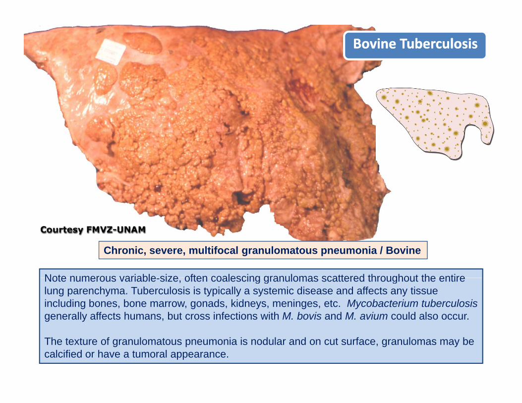
Bovine TuberculosisBovine Tuberculosis
Note numerous variable size often coalescing granulomas scattered throughout the entire
Chronic, severe, multifocal granulomatous pneumonia / Bovine
Note numerous variable-size, often coalescing granulomas scattered throughout the entire lung parenchyma. Tuberculosis is typically a systemic disease and affects any tissue including bones, bone marrow, gonads, kidneys, meninges, etc. Mycobacterium tuberculosisgenerally affects humans, but cross infections with M. bovis and M. avium could also occur.
The texture of granulomatous pneumonia is nodular and on cut surface, granulomas may be calcified or have a tumoral appearance.

Equine TuberculosisEquine Tuberculosis
Chronic severe multifocal granulomatous pneumonia
Note numerous focal to coalescing granulomas i l h
Chronic, severe, multifocal granulomatous pneumonia. Equine Lung / Cut surface.
in lung parenchyma.
Mycobacterium tuberculosis generally affects humans, but cross infections with M. bovis and M i ld l Pi b ff t dM. avium could also occur. Pigs can be affected by all three species. The incidence of human and animal tuberculosis has notably increased in many countries.
Remember that typical granulomatous lesions seen in cows and sheep (caseous necrosis and calcification) with tuberculosis are not seen in h t d d I th th i lhorses, cats and dogs. In these other animals, tubercles have the appearance of tumoral growths (sarcomatous).
Hi t th l d i l t i (A id f t)Histopathology and special stain (Acid fast) or bacteriologic culture are required for confirmation.

Granulomatous PneumoniaGranulomatous Pneumonia
Tuberculosis. Histopathology
Note typical granuloma formed by a necrotic center (asterisks) surrounded by macrophagescenter (asterisks) surrounded by macrophages and then by an external band of fibrous connective tissue (double arrows) infiltrated by lymphocytes and plasma cells (not seen at this magnification). There are few normal alveoli (a)magnification). There are few normal alveoli (a) around the granulomas.
In most granulomatous pneumonias, the etiologic agent can be detected with special stains. age ca be de ec ed spec a s a s
Large numbers of acid-fast bacilli (red) in affected tissue.

Note several large white nodules (asterisks) in
RhodococcusRhodococcus equi equi PneumoniaPneumonia
Note several large white nodules (asterisks) in the lung of this horse. This is a disease of world wide importance caused by a telluric organism. Infection in foals has three important forms: Respiratory intestinal and skeletal TheRespiratory, intestinal and skeletal. The respiratory form is characterized by a chronic cough and weight loss.
Histopathology: Pyogranulomas formed by macrophages, neutrophils, giant cells and scant fibrous tissue. R. equi shares some pathogenic similarities with myocobacterial organismssimilarities with myocobacterial organisms. Both organisms are resistant to phagocytosis and are typically found in the cytoplasm of macrophages or giant cells.
The nodules are hard in texture and on a cut surface reveal a caseous appearance (See next slide).
Chronic, severe, locally extensive, pyogranulomatous bronchopneumonia

Note large granulomatous ceseous (cheese
RhodococcusRhodococcus equi equi PneumoniaPneumoniaEquine lung on cut surface.
Note large, granulomatous, ceseous (cheese-like) nodules formed by necrotic material and not by pus, therefore should not be referred to as abscesses.
Microscopically, granulomas have many macrophages containing gram-positive coccobacilli. The final diagnosis is made by culturing Rhodococcus equiculturing Rhodococcus equi.
R. equi also produces ulcerative enterocolitis in foals and it has been reported to cause lymphadenitis in pigslymphadenitis in pigs.
Rhodococcus equi has recently been reported in men and women with Human Immunodeficiency Virus (HIV) infection PleaseImmunodeficiency Virus (HIV) infection. Please be very cautious with this serious and fatal disease. If abstinence is not desired, do not play Russian roulette with your life - be sure you always practice safe sexyou always practice safe sex.
Chronic, severe, locally extensive, pyogranulomatous bronchopneumonia

Granulomatous Pneumonia Canine Granulomatous Pneumonia Canine BlastomycosisBlastomycosis
Granulomatous pneumonia, severe, Granulomatous pneumonia, severe, multifocalmultifocal
Gross: Note small nodules in all areas of the lung. These lesions are typical of granulomatous pneumonia Sometimes hstopathology is required to differentiate granulomatous pneumonia
pp
pneumonia. Sometimes hstopathology is required to differentiate granulomatous pneumonia from neoplastic nodules in the lung.
Histopathology: Giant cell containing several PAS-positive Blastomyces dermatitides (arrows).

Granulomatous Lesions Feline Infectious Peritonitis Granulomatous Lesions Feline Infectious Peritonitis
LungLung
SpleenSpleen
LungLung
LiverLiver
C f /
Note numerous small granulomas in lung liver and spleen Granulomatous lesions in FIP
Chronic, multifocal, severe, granulomatous pneumonia-hepatitis-splenitis / cat
Note numerous small granulomas in lung, liver and spleen. Granulomatous lesions in FIP result from deposition of antigen antibody complexes in arteries (type III hypersensitivity).
For a close-up of lung and liver lesions go to next slide.

Feline Infectious Peritonitis (FIP)Feline Infectious Peritonitis (FIP)
Note many granulomas in the lung (arrows) and in the liver (arrowheads)
Feline Infectious Peritonitis occurs in the "wet/effusive form" characterized by abdominal and pleuralabdominal and pleural effusions (accumulation of fluid), and in the "dry/non-effusive form" in which effusion is not present.
Ch i ltif l l t i h titie us o s ot p ese t
In both wet and dry forms of FIP pyogranulomas are typically present in most internal organs
Chronic, multifocal, granulomatous pneumonia-hepatitis
In both wet and dry forms of FIP, pyogranulomas are typically present in most internal organs.
The "dry/non-effusive forms cause Pyogranulomatous encephalitis and endophthalmitis. It is presumed that cats with strong cell-mediated immunity suffer only a transient infection. Cats with partial cell mediated immunity develop the dry form and the cats with poor cell mediatedwith partial cell-mediated immunity develop the dry-form and the cats with poor cell-mediated immunity develop the most severe, wet-form of FIP. There is still much to learn about the pathogenesis of FIP.

Granulomatous Pneumonia Granulomatous Pneumonia / / MülleriusMüllerius capillariacapillaria / Ovine/ Ovine..
Multifocal, chronic, severe, granulomatous pneumonia Multifocal, chronic, severe, granulomatous pneumonia
Numerous parasitic larvae
Numerous, subNumerous, sub--pleural granulomas distributed throughout the pleural granulomas distributed throughout the lungs (lungs (black arrowsblack arrows). This parasitic disease is commonly found ). This parasitic disease is commonly found in sheep and goats The lungworm requires intermediate hostin sheep and goats The lungworm requires intermediate host
in the lung (white arrows). Also, note moderate thickening of alveolar septa largely due to infiltration of
in sheep and goats. The lungworm requires intermediate host in sheep and goats. The lungworm requires intermediate host (snail). The greenish appearance of some granulomas is due to (snail). The greenish appearance of some granulomas is due to eosinophilseosinophils. Nodules become hard (calcified) and . Nodules become hard (calcified) and microscomicrosco--picallypically are small granulomas containing larvae and eggs.are small granulomas containing larvae and eggs.
lymphocytes seen in this section as dark round cells.

FETAL PNEUMONIAFETAL PNEUMONIAand and
ABORTIONABORTIONABORTIONABORTION

Fetal lung
The fetus is suspended (floats) in amniotic fluid during gestation. Fetal lung does have air, but
Fetal lung
g g g ,instead is filled with a locally produced fluid known as “fetal lung fluid” (FLF). This fluid has high viscosity and serves as a barrier to prevent aspiration of amniotic fluid during normal "fetal p grespiratory movements” with the glottis is closed.
Since fetal lung does not contain Since fetal lung does not contain air it sinks when placed in formalinair it sinks when placed in formalinSince fetal lung does not contain Since fetal lung does not contain air it sinks when placed in formalinair it sinks when placed in formalinair, it sinks when placed in formalin air, it sinks when placed in formalin or water. At the time of birth, lung or water. At the time of birth, lung fluid is rapidly absorbed by fluid is rapidly absorbed by lymphaticslymphatics, and , and filled filled with air with air (aeration) Aerated lung floats(aeration) Aerated lung floats
air, it sinks when placed in formalin air, it sinks when placed in formalin or water. At the time of birth, lung or water. At the time of birth, lung fluid is rapidly absorbed by fluid is rapidly absorbed by lymphaticslymphatics, and , and filled filled with air with air (aeration) Aerated lung floats(aeration) Aerated lung floats(aeration). Aerated lung floats (aeration). Aerated lung floats when placed in fluid. Nonwhen placed in fluid. Non--aerated aerated lung does not float.lung does not float.
(aeration). Aerated lung floats (aeration). Aerated lung floats when placed in fluid. Nonwhen placed in fluid. Non--aerated aerated lung does not float.lung does not float.

Although fetal pneumonia is commonly
Abortion and Fetal PneumoniaAbortion and Fetal Pneumonia
seen in aborted fetuses, etiological diagnosis of abortion is only reached in approximately 30% of cases. This low diagnostic success rate is, in part, because of the status of the dam, which may be responsible for abortion, is rarely investigated. There are many maternal conditions such as acidosis, fever, toxemia, hypoxia, anemia, etc. that may cause abortion, yet the fetus would have no lesions. In contrast, the approach in human abortions is always to do an exhaustive examination of the mother (endocrine and serologic tests).Pneumonia is difficult to evaluate grossly in the fetus.
Pneumonia is a common lesion inPneumonia is a common lesion in aborted fetuses and the port of entry could be via inhalation of amniotic fluid or hematogenous through the umbilical veins Aspiration of amniotic fluidveins. Aspiration of amniotic fluid contaminated with bacteria from placentitis typically results in fetal pneumonia.

Under abnormal conditions ("fetal distress)”
MeconiumMeconium Aspiration SyndromeMeconium Aspiration Syndrome
Under abnormal conditions ( fetal distress) hypoxemic fetuses’ defecate in utero and pass meconium into amniotic fluid. During fetal hypoxia there is increase fetal intestinal peristalsis and relaxation of the anal sphincter, causing the
Meconium is the first intestinal content formed when material present in the amniotic fluid is normally swallowed and mixes with intestinal secretions including bile, hence the yellow color.
release of meconium into the amniotic fluid.
Meconium-contaminated amniotic fluid may be subsequently aspirated when fetuses "gasp for i " i t th i l t d d t tt t tsecretions including bile, hence the yellow color. air" in utero as their last and desperate attempt to
correct fetal hypoxia.
This diagram illustrates how meconium is passed into amniotic fluid and then aspirated into the lung.
1. Meconium in intestine (normal)
2. Meconium defecated into amniotic fluid following intrauterine hypoxia
3. Meconium in the oral cavity an hnasopharynx.
4. Meconium finally aspirated into the lungs.

Ovine Abortion / Fetal anoxia Ovine Abortion / Fetal anoxia Meconium stained skin
Note meconiumNote meconium‐‐stained skinstained skinThis is a dramatic example of intrauterine expulsion of meconium and thus diagnostic of severe fetal anoxia. Subsequent examination of the respiratory
Note meconiumNote meconium stained skinstained skin
Subsequent examination of the respiratory tract of this near-term fetus revealed meconium in the trachea and bronchi. Meconium aspiration and fetal pneumonia were microscopicallywere microscopicallycorroborated.
Note bronchiole containing gleukocytes and meconium (yellow material). The microscopic features of inhalation of amniotic fluid contaminated with bacteria and meconium are those of suppurative broncho-pneumonia, however, lesions in p , ,fetuses are not restricted to the cranioventral lung.

Some images were acquired from veterinary colleges of Some images were acquired from veterinary colleges of Canada, United States and Mexico and the names ofCanada, United States and Mexico and the names ofCanada, United States and Mexico and the names of Canada, United States and Mexico and the names of pathologists who contributed with some slides are known. pathologists who contributed with some slides are known. Their valuable contribution is sincerely acknowledged.Their valuable contribution is sincerely acknowledged.
I would like to thank Adriana Lopez, University of Western I would like to thank Adriana Lopez, University of Western Ontario, and Eileen Kinch, University of Prince Edward Ontario, and Eileen Kinch, University of Prince Edward Island for editorial assistance; Dr. María Forzán, Atlantic Island for editorial assistance; Dr. María Forzán, Atlantic Veterinary College, for critically reviewing these modules.Veterinary College, for critically reviewing these modules.

Module 5Module 5Module 5Module 5
Examples of Diseases that Cause Examples of Diseases that Cause Bronchopneumonia in Domestic AnimalsBronchopneumonia in Domestic AnimalsBronchopneumonia in Domestic AnimalsBronchopneumonia in Domestic Animals
If you have any comments, criticisms or suggestions about this tutorial If you have any comments, criticisms or suggestions about this tutorial
module please let me know. module please let me know.
If you find any errors or typos please let me know too If you find any errors or typos please let me know too
[email protected]@upei.ca