Embed Size (px)
Citation preview

EXAMINATION OF THE GI TRACTTRACT
Aim: Optimal use of endoscopic and/or radiologic imaging techniques to reach maximal information without causing less
trouble for the patient


Essential role of GI tract
• Absorption of nutritients
• Excretion
• Maintaining coordinated passage• Maintaining coordinated passage

Approach to the patient
• To take thorough history
• Physical examination
• Laboratory investigations (blood,stool,Helicobacter pylori,etc)• Laboratory investigations (blood,stool,Helicobacter pylori,etc)
• Ultrasound
• Endoscopy
• X-ray (plain film)
• CT, MRI, angiography, isotope scan,PET, selective enterography, capsullar video endoscopy

HISTORYcomplain:when, where, what is it like?
• ABDOMINAL PAIN• ALTERATION IN BOWEL HABIT• Difficulties in swallowing, NAUSEA, • Difficulties in swallowing, NAUSEA,
VOMITUS• BLEEDING: hemathemesis, melena,
hematochezia• GENERAL MEDICAL HISTORY• FAMILY HISTORY

Main principles
• Immediate distinction between urgent problem and nonacute disorder
• Determination of the temporal evolution of symptomes and to be able
• Determination of the temporal evolution of symptomes and to be able to differentiate between organic and psychosomatic alterations
• Consider all information to avoid unnecessary tests in the workup


Problems of swallowing
• 1. determination the nature of dysphagia: difficulty in swallowing liquids, solids or both. Careful oropharingeal examination is needed
• 2. oesophageal (barium or with water soluble contrast media) X-ray studies
• 2. oesophageal (barium or with water soluble contrast media) X-ray studies
• 3. oesophagoscopy: to obtain biopsy specimen from a lesion causing filling defect on X-Ray, the diagnosis of peptic oesophagitis or Barett’s oesophagus are based only on endoscopy; sclerotherapy of oesophageal and gastric variciescould be performed exclusively by this tool
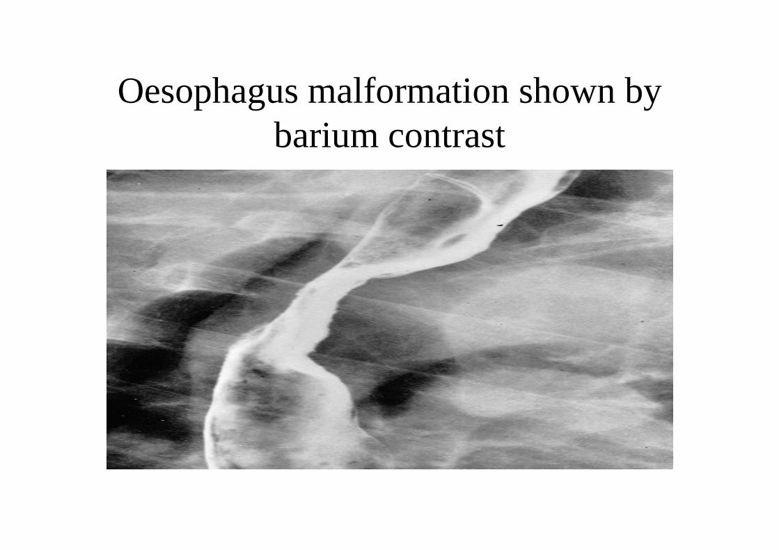
Oesophagus malformation shown by barium contrast

ABDOMINAL PAIN
• Acute /chronic
• Characterization: sharp, cramping, burning
• Intensity: acceptable-intolerable• Intensity: acceptable-intolerable
• Localization: upper/lower; localized/diffuse, radiating
• Relationship to meals: on fasting,while eating, shortly after/or 30-90 min later

Common causes of abdominal pain
• Gastrointestinal causes:Acute and chronic cholecystitis, Appendicitis, bowel
obstruction, constipation, diverticular disease, gastroenteritis, IBD, irritable bowel, pancreatitis, gastroenteritis, IBD, irritable bowel, pancreatitis, peptic ulcer and reflux disease, tumours
• Extraintestinal causes: Abdominal aortic aneurysm, Dysmenorrhea, Epididymitis, Incomplete abortion, Nephrolithiasis, Ovarian cyst, Pelvic inflammatory disease, Pregnancy, ÚTI, Pb intoxication, Porphyria

Alteration in bowel habit
• Diarrhoea and/or constipation: consider intestinal or extraintestinal cause
• Evaluate with the related symptomes (weight loss, fever, bloating….)fever, bloating….)
• Stool:determination of frequency, consistency, colour, if any blood is present, presence of mucus and pus,
• Nocturnal diarrhoea consider always as organic

Factors and disorders associated with constipation
• General: lack of physical activity (eg.hospitalization), not enough fiber and fluid intake
• Systemic: side effect of drugs ( antacids, analgesics, psychopharmacologic agents,iron ), Endocrine disorders ( hypothyroidism, hyperparathyroidism), reflectory in hypothyroidism, hyperparathyroidism), reflectory in nephrolithiasis, and in pancreatitisMetabolic disorders ( hyper Ca, uraemia, hypoK)
• Colonic: Obstruction, IBS, Diverticular disease, Painful anorectal disorders
• Neurogenic: Hirschprung’s disease, CNS disorders, Diabetic neuropathy, Psychogenic cause

Common causes of diarrhoea
Infection: viral, bacterial, parasite infestation (e.g. amoeba)
Diet, food and lactose intolerance, much alcohol, reaction to certain medications or laxative abuse.reaction to certain medications or laxative abuse.
GI diseases that cause diarrhoeaCrohn's disease, ulcerative colitis, irritable bowel syndrome, gastrointestinal tumours, and malabsorption syndromes such as coeliac disease.
• Extraintestinal diseases: hyperthyroidism, functional- psychogenic, systemic mastocytosis

Physical examination 1.
• Inspection: colour: paleness or jaundice, nutritional condition, contour: enourmously large masses, ascites, fistulas in the perianal large masses, ascites, fistulas in the perianal region, forced movement of the bowel may be seen
• Auscultation: absence of bowel sounds in evolving ileus or an obstructing process, or succusion splash-in gastric outlet obstruction

Physical examination 2.
• Palpation: detecting tenderness and massesRebound tenderness (direct or referred) after removal of the
examining hand is a clue to localized or generalized peritonitis, abdominal emergencies.
Rectal digital examination: masses intrinsic to the rectum, abnormalities in the pelvis might result palpational finding on the punch of the Douglas, presence or absence of fresh bright red bloody or maroon stool.
• Percussion: to assess: free air beneath the diaphragm; liver and spleen size; retention in the urinary bladder

Laboratory tests
• Total blood count, ESR, liver enzymes, bilirubin, amylase-lipase
• Tumour-markers (CEA,α FP, CA19.9)• For malabsorption: stool fat painted by sudan, • For malabsorption: stool fat painted by sudan,
starch by iodine reaction, muscle remnants by light microscopic evaluation
• D-xylose absorption test to separate mucosal disease from pancreatic insufficiency
• For Lactose intolerance: Hydrogen breath test• For Helicobacter Pylori: 13Carbon urea breath test

Ultrasound (US)+doppler
• Usefull in delineation of abdominal massess• To determine wall tickness (stomach…)• To find stones and backward dilatation of ducts ( bile, and
ureter, pyelon) and cysts of the pancreas and the kidneyureter, pyelon) and cysts of the pancreas and the kidney• To determine TNM stadium of tumours• To assess blood flow in deep veins• To find and monitor aortic aneurysma• For monitoring and follow up (cure/relaps) in
oncohaematology• Help in perfoming US guided needle biopsies

ENDOSCOPY
• THIS PROCEDURE ALLOWS FOR DIRECT INSPECTION OF THE MUCOSA AND THE LESION ITSELF
• Permit to identify and differentiate between peptic and neoplastic ulcerating lesions; to find the bleeding siteand neoplastic ulcerating lesions; to find the bleeding siteand to perfom cauterization
• PERMITTING DETECTION OF: -CANCERS AND POLYPS (that may be missed by barium X-ray studies),-inflammatory changes of the mucosa,
• to diagnose HP, performing biopsies and polypectomy

Endoscopy ( flexible fiberoptic instrument) forms
• Oesophago-gastro-duodenoscopy• Colonoscopy• Sigmoidoscopy (rigid): for identifying the lower 25 cm of
the colonthe colon• Recto,- anoscopy (rigid)• ERCP• Selective CT enterography, MRI• Capsullar video endoscopyRadiologic (X-ray) examination may be only preferred whenthere are contraindications to safe endoscopy

Contraindications to endoscopy
Absolute ContraindicationsRelative
Contraindications
Patients who are unable to cooperate
Patients with large Zenker's diverticulum
Patients who are moribund
Patients with suspected perforated viscus
Dysphagic patient who has not had an esophagogram recently
Patients who are hemodynamically unstable

Upper gastrointestinal endoscopy:permits evaluation of the esophagus, stomach, duodenum;
ERCP: permits inspection and canulation of the papilla Vateri, and visualise the architecture of pancreas, ductal dilatations, the branching of the bile ducts, to perform papillotomy and stent implantationimplantation
Colonoscopy: visualises the entire colon, the terminal ileum and the colonic polyps can be removed,biopsy might be performed from lesions for histological evalutation

Indications for endoscopy of the esophagus
Diagnostic
Dysphagia and odynophagia, suspected gastroesophageal reflux disease
Cancer surveillance (eg, Barrett's esophagus)
Abnormal results of barium esophagogram
Therapeutic
Dilatation of strictures, both benign and malignant
Sclerotherapy and banding of varices
Stent placement in palliative therapy of malignant stricture
Laser therapy of esophageal and gastroesophageal malignancies
Removal of foreign body

ERCP


colonoscopy

X-ray studies
• First choice for determining free air (sign of perforated viscus) on plain film
• First choice to confirm ileus (nivous in the small or large intestine) on plain film
• Best in assessing motility and alterations of small intestine relatively inaccessible to fiberoptic instruments.
• When endoscopy is contraindicated: severe ulcerative colitis: barium enema with air contrast techniques,
• For visualisation of fistulas• With absorbeable contrast media: to find cause of recurrent
pneumonias and aspiration


Chron’s disease. Barium enema

Other tools for visualization
• CT,MRI: TNM staging
• PET(positron emission tomography) in case of questionof question
• Promt mesenteric angiography: in suspicion of vascular disease: mesenteric ischaemia and bleeding from the small intestine
• Isotope scan

Case presentation of a 21yrs old ♂
The only symptome is fever38 C for 3 months
• Physical examinaton: normal• abd. US normal• CRP: 27,58, ESR: 22 mm/h• Sources of infection by ORL consultation were disclosed • Thyroid function, immun panel tests: negative• viral serology: CMV, EBV IgG positive• viral serology: CMV, EBV IgG positive• Echocardiograpy: normal, • After a month thorough follow up he said that he missed to tell that he found
the stool bloody- he did not think that it might be imnportant( psychosomatic alteration is to be suspected)
• Panendoscopy: duodenal ulcer• Repeated US ( 2 months apart) revelaed inhomogenity in the spleen, therefore • Abdominal CT had been performed before colonoscopy: the ileum seemed to
be tickened by this method:

Selectiv enterography revealed Chron’s disease.


BLEEDING DIVERTICULUM ANGIOGRAPHY

Izotop-tagged red blood cell
Case presentation
Complain:melaena
Gastroscopy: negativeGastroscopy: negative
Colonoscopy: negative, but from the coecum (valv Bauchin) black stool appeared meaning that the bleeding is originated from the small bowel

But, in the present case
Szelektív angiográfia:
This is a positive finding
But, in the present case extravasation was not seen

In the first part of the jejunum appr. 10-12 cm long pathological vessel structure was seen in the arterial phase . AV malformation? Therefore capsullar enterographia had been indicated. According to that result the patient was operated
CT ENTEROGRAPHY
( not shown)
been indicated. According to that result the patient was operated

angiodysplasia

Reasons of small bowel bleeding are
Angiodysplasia and arterial malformatio ( 30-40% ),
Otheres less frequently:Otheres less frequently:
NSAID enteropathy, Erosive jejunitis-ileitis,Diverticular ( Meckel ),Crohn’s diseaseIntussusception, tumors: leiomyoma, carcinoid, lymphoma, adenocc., Ischaemiás enteropathy, Aortoenteral fistula

50 % the bleeding source remain unknown after the upper and lower ract endoscopy had been performed
Push – enteroscopy or capsular endoscopy is indicated Push – enteroscopy or capsular endoscopy is indicated in these cases when others,like CT - MR enterography, radioizotop scan and angiography failed to find the bleeding source.
After thorough examination still 5% of bleedings remain obscure

JEJUNUM ANGIODYSPLASIA
Seen on capsullar enterography