Embed Size (px)
Citation preview
EXAMEN NEUROLOGIQUE DU PATIENT DE REANIMATION
[email protected] Université de Versailles
Institut Pasteur Hôpital Raymond Poincaré
Garches - France
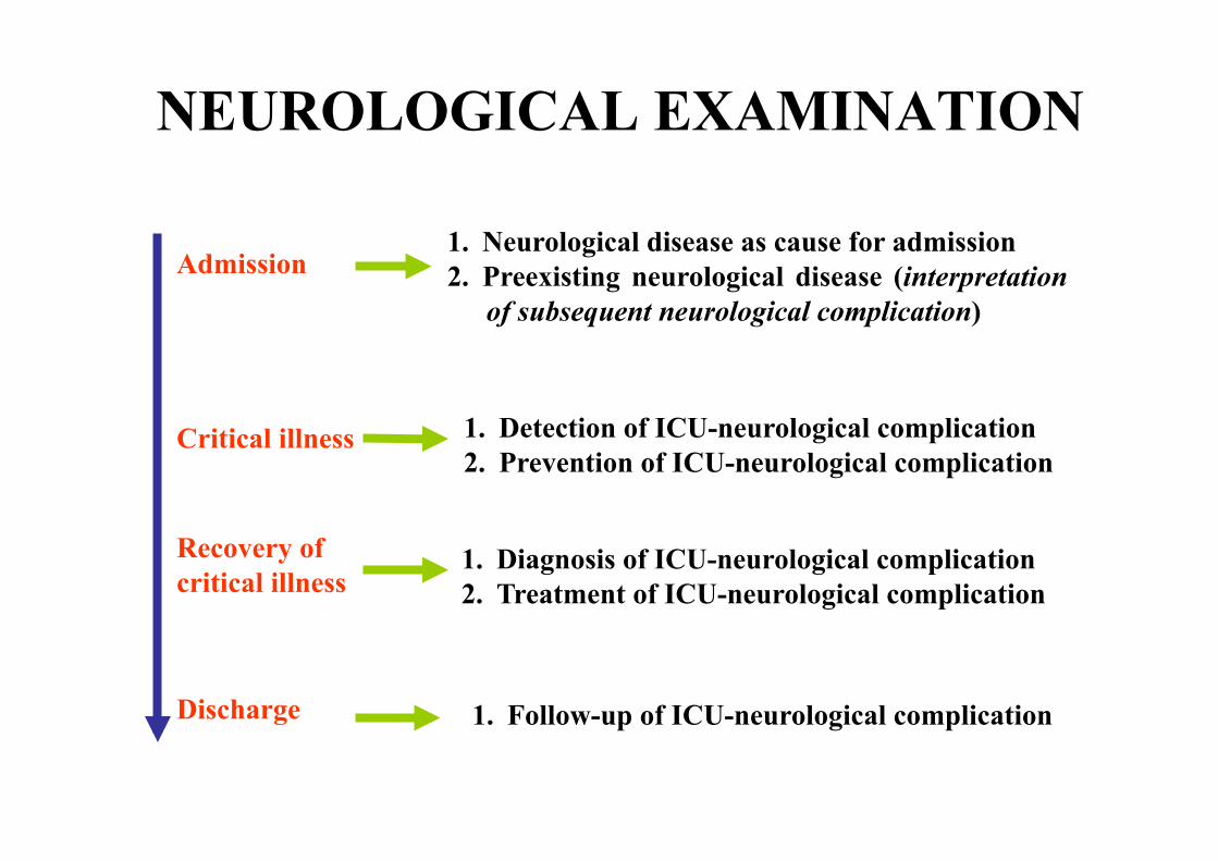
NEUROLOGICAL EXAMINATION
Admission 1. Neurological disease as cause for admission 2. Preexisting neurological disease (interpretation
of subsequent neurological complication)
Critical illness 1. Detection of ICU-neurological complication 2. Prevention of ICU-neurological complication
Recovery of critical illness
1. Diagnosis of ICU-neurological complication 2. Treatment of ICU-neurological complication
Discharge 1. Follow-up of ICU-neurological complication

LE SYSTÈME NERVEUX CENTRAL
1. COMA 2. SYNDROME CONFUSIONNEL 3. EPILEPSIE 4. EXAMEN NEUROLOGIQUE DU PATIENT
SEDATE 5. COMPORTEMENT DE MALADIE 6. TROUBLES COGNITIFS 7. SYNDROMES MEDULLAIRES
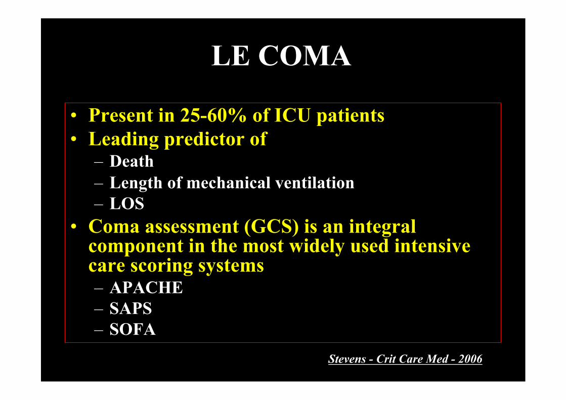
LE COMA
• Present in 25-60% of ICU patients • Leading predictor of
– Death – Length of mechanical ventilation – LOS
• Coma assessment (GCS) is an integral component in the most widely used intensive care scoring systems – APACHE – SAPS – SOFA
Stevens - Crit Care Med - 2006
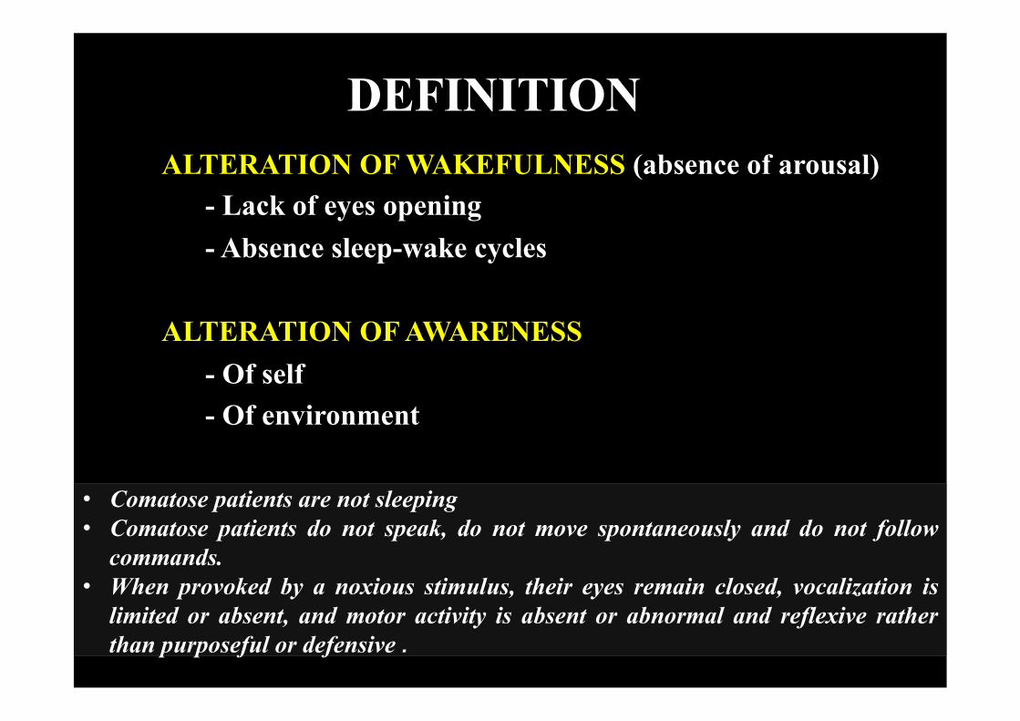
• Comatose patients are not sleeping • Comatose patients do not speak, do not move spontaneously and do not follow
commands. • When provoked by a noxious stimulus, their eyes remain closed, vocalization is
limited or absent, and motor activity is absent or abnormal and reflexive rather than purposeful or defensive .
ALTERATION OF WAKEFULNESS (absence of arousal) - Lack of eyes opening - Absence sleep-wake cycles
ALTERATION OF AWARENESS
- Of self - Of environment
DEFINITION

NON-ORGANIC
Presence of avoidance - Blinking to threat - Do not let hand fall on his face
If any doubt, do an EEG

COMATOSE PATIENT
BRAINSTEM RESPONSES 1. Eyes spontaneous movement 2. Eyes position 3. Oculocephalogyre response 4. Oculovestibular response 5. Pupillar size 6. Pupillar light reflex 7. Corneal reflex 8. Grimace 9. Cough reflex 10. Oculocardiac response 11. Respiratory pattern
FOCAL SIGNS Comparison between right and left body 1. Motor responses to order or painful stimulation 2. Limbs tone 3. Tendon reflexes 4. Plantar reflex
Verbal response Eyes response Motor response
SCALE
MYOCLONUS 1. Limbs 2. Lids

MOTOR RESPONSE
External rotation of lower limb + babinski
decortication
« décérebration »

COMATOSE PATIENT EYES POSITION
Wijdicks E.F.M. Plum and Posner
Skew deviation (Cajal nuclei)
Hemispheric /pons
T h a l a m u s /mesenceph
B i l a t e r a l hemispheric

COMATOSE PATIENT
A: roving B: periodic alternating gaze (bi H., cereb.) C: ping pong (bi H., cereb.)
D: convergence nystagmus (mesenceph.) E: bobbing (pons) F: dipping (bi H.)
Wijdicks E.F.M.

PUPILS
Adapted from Plum and Posner's Diagnosis of Stupor and Coma

No localisation value Intracranial hypertension
SIXTH NERVE PALSY

THIRD NERVE PALSY

Aneurysm I.C. or A.S.
Cavernous sinus thrombosis
Uncal herniation
THIRD NERVE PALSY

HERNIATION

CLAUDE BERNARD HORNER
Sympathetic nervous system
Dissection Catheter
Cancer
Spine
Medulla (Wallenberg)
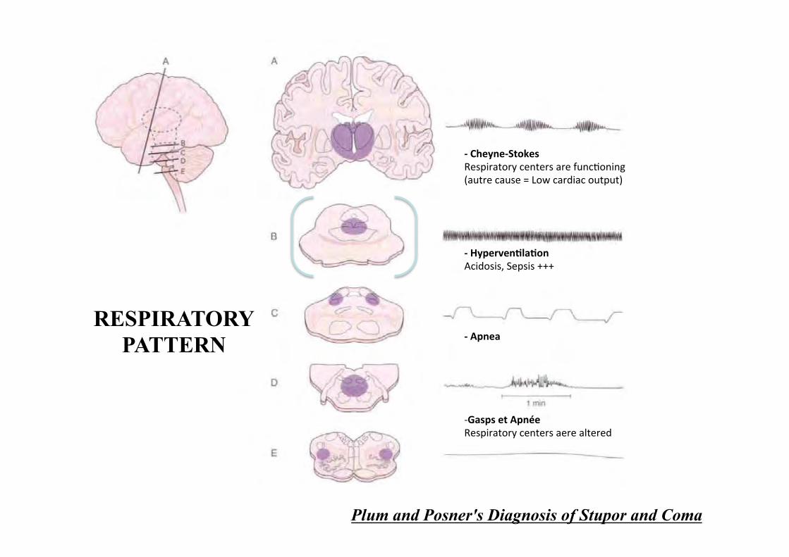
Plum and Posner's Diagnosis of Stupor and Coma
-‐ Cheyne-‐Stokes Respiratory centers are func0oning (autre cause = Low cardiac output)
-‐ Hyperven=la=on Acidosis, Sepsis +++
-‐ Apnea
-‐ Gasps et Apnée Respiratory centers aere altered
RESPIRATORY PATTERN
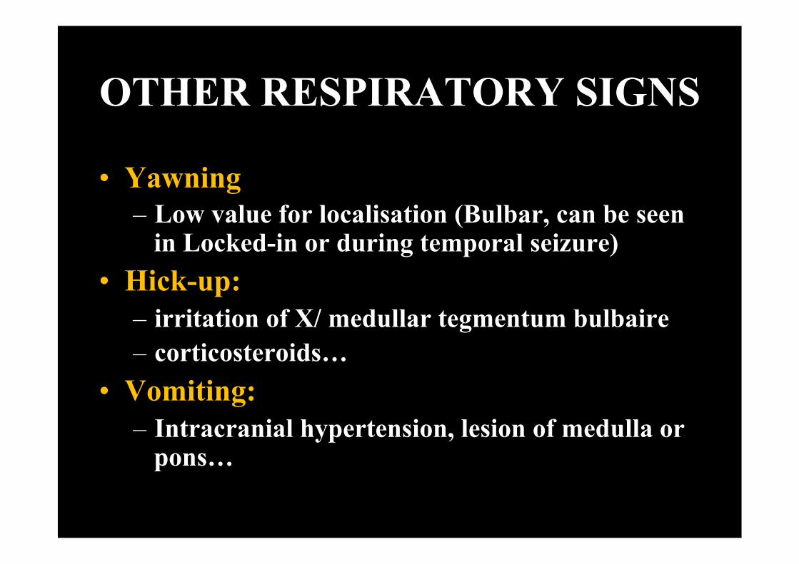
OTHER RESPIRATORY SIGNS
• Yawning – Low value for localisation (Bulbar, can be seen
in Locked-in or during temporal seizure) • Hick-up:
– irritation of X/ medullar tegmentum bulbaire – corticosteroids…
• Vomiting: – Intracranial hypertension, lesion of medulla or
pons…

VITAL SIGNS • HYPERTENSION
– Adrenergic storm +++ – Intracranial hypertension – toxic (amphétamin, cocaine) – hypertensive encephalopathy (PRES)
• HYPOTENSION – shock, (can be the cause of the coma +++) – Neurogenic myocardiac dysfunction (ou « Tako-Tsubo »)
• RESPIRATORY DISTRESS – Can be cause of the coma +++ – Medulla syndrome – Neurogenic pulmonary edema
• HYPERTHERMIA – Infection,: méningitis/encéphalitis/abscesses, paludism +++ – Heat stroke

FOUR & GLASGOW COMA SCALES
Wijdicks et al – Ann Neurol - 2005
Patients with the lowest GCS score could be further distinguished using the FOUR score.
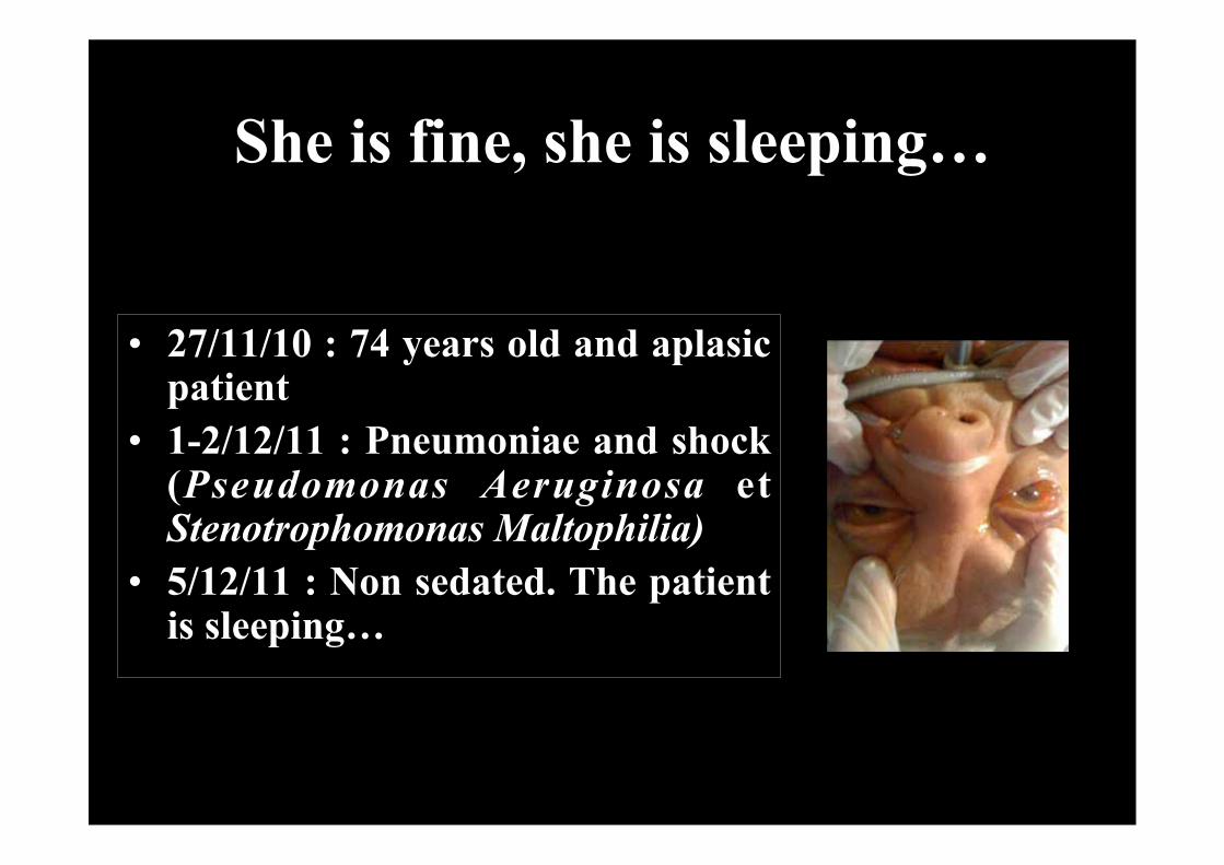
She is fine, she is sleeping…
• 27/11/10 : 74 years old and aplasic patient
• 1-2/12/11 : Pneumoniae and shock (Pseudomonas Aeruginosa et Stenotrophomonas Maltophilia)
• 5/12/11 : Non sedated. The patient is sleeping…

She is fine, she is sleeping…

Delirium / Confusion DSM IV : Diagnostic Statistical Manual (of American Psychiatric Association) A. Trouble de conscience : diminution de la réactivité à l’environnement,
difficultés attentionnelles, à se concentrer
B. Atteinte d’une ou plusieurs fonctions cognitives : – Trouble du langage – Troubles de mémoire – Désorientation temporelle et spatiale – Troubles du jugement et du raisonnement
C. Apparition brutale ou rapidement progressive (mn, heures ou jours),
fluctuation des symptômes au cours du temps D. Le trouble est lié à un ou plusieurs des éléments suivants :
– Pathologie médicale – Prise médicamenteuse / intoxication – Sevrage Romain Sonneville

INCIDENCE Auteur, année Population Réa, n Critère (échelle) Fréq.
Dubois, ICM 2001 Med-chir, n=216 Delirium (ICDSC) 19%
Ely, CCM 2001 Med, n=48 Delirium (CAM-ICU)
60%
Ely, Crit care 2003 Med non ventilés, n=261 Delirium (CAM-ICU)
48%
Woods, ICM 2004 Med, n=143 Agitation (MAAS) 16%
Ely, JAMA 2004 Med et USIC, n=224 Delirium (CAM-ICU)
82%
Jaber, Chest 2005 Med-chir, n=211 Agitation (Ramsay) 52%
Ely, ICM 2007 Chir-Trauma, n=100 Delirium (CAM-ICU)
70%
Ely, JAMA 2007 Med-chir, n=106 Delirium (CAM-ICU)
80%
Ouimet, ICM 2007 Med-chir, n=820 Delirium (ICDSC) 32%

DISTRIBUTION
Ely et al – JAMA - 2004

DELIRIUM TYPES
Hyperactive delirium: agitation, restlessness, emotional lability Hypoactive delirium: withdrawal, flat affect, lethargy and decreased responsiveness

DELIRIUM SCALES
Sedation/Agitation : - RAMSAY - Richmond Agitation Sedation Scale (RASS) - Adaptation to the Intensive Care Unit Environment
(ATICE) Delirium :
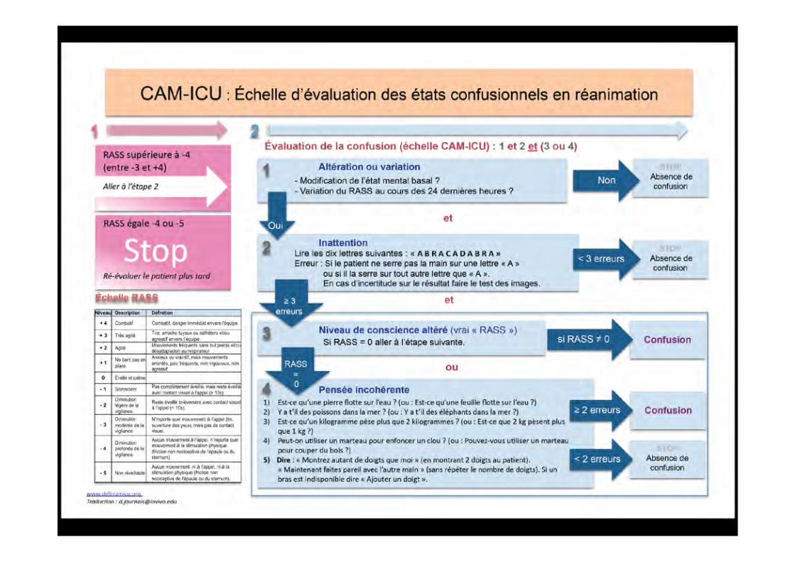
- Confusion Assessment Method for the ICU (CAM-ICU) - Intensive Care Delirium Screening Check-list (ICDSC)
MUST BE COMPLETD BY 1. A FULL NEUROLOGICAL EXAMINATION (focal sign, neck stiffness…) 2. EEG

Intensive Care Delirium Screening Checklist (ICDSC) 8 items, côtés 0 ou 1
Réalisée une fois par 8 heures Si total > ou = 4 : DELIRIUM 1. Conscience altérée (Si A ou B, ne pas poursuivre évaluation)
A. Pas de réponse à une stimulation : ne pas coter B. Réponse à une stimulation intense et répétée (voix, douleur) : idem C. Réponse à une stimulation modérée : 1 D. Patient normalement éveillé : 0 E. Réponse exagérée à la stimulation : 1
2. Inattention (0 ou1) 3. Désorientation (0 ou 1) 4. Hallucinations (0 ou 1) 5. Agitation ou ralentissement psycho-moteur (0 ou 1) 6. Discours ou humeur inappropriés (0 ou 1) 7. Troubles du cycle veille-sommeil (0 ou 1) 8. Fluctuations des symptômes (0 ou 1)
Bergeron ICM, 2001
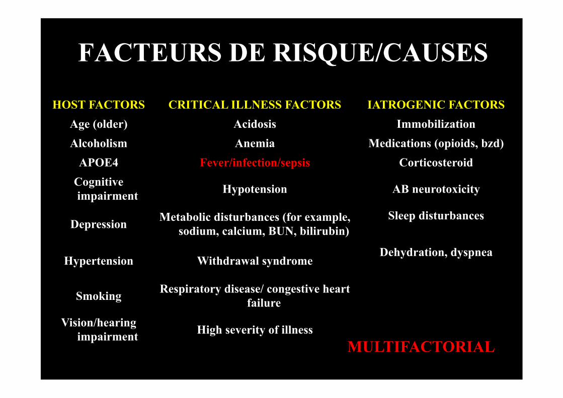
FACTEURS DE RISQUE/CAUSES
HOST FACTORS CRITICAL ILLNESS FACTORS IATROGENIC FACTORS Age (older) Acidosis Immobilization Alcoholism Anemia Medications (opioids, bzd)
APOE4 Fever/infection/sepsis Corticosteroid Cognitive impairment Hypotension AB neurotoxicity
Depression Metabolic disturbances (for example, sodium, calcium, BUN, bilirubin)
Sleep disturbances
Hypertension Withdrawal syndrome Dehydration, dyspnea
Smoking Respiratory disease/ congestive heart failure
Vision/hearing impairment High severity of illness
MULTIFACTORIAL

A 55 years old man was hospitalised for a septic shock. Blood culture were positive to Staphylococcus aureus and CSF analysis showed an aseptic meningitis. At admission, neurological examination was normal as well as MRI of the brain and spine. Transoesophageal cardiac ultrasound only showed a thrombus in the left atria. He also underwent surgery for a septic arthritis of the right knee. A right jugular catheter could not be put. A goitre was also noticed. Vasopressor and mechanical ventilation were discontinued the 3rd and 5th days from admission. The patient was treated with methicillin and heparin. Day 8, the patient developed agitation and delirium. Neurological examination showed a weakness of the right arm and slight right central facial palsy as well a ptosis and miosis of the right eye. The right arm was also oedematous. The brain CT scan and CSF analysis were normal. EEG showed a slow cortical activity. Biochemical screening showed a moderate renal insufficiency. Blood culture were negative.
CASE REPORT 2

Case reprot 2
1. Left pre-rolandic lesion 2. Thrombosis of right jugular vein 3. Orbenin overdose

CAS CLINIQUE
Mme X…, âgée de 53 ans, traitée par CS et I- pour un LED, est hospitalisée pour une insuffisance rénale aiguë en rapport avec une Mat traité par EP compliqué d’un hématome de la cuisse. Survenue d’un état d’agitation et délirant: « on me vole mon enfant, les médecins me vole mon enfant… »

CRISE D’EPILEPSIE
Population Monitoring Seizure CS NCS Wijdicks
1996 Liver transplant
(n=630) Clinical 28 (4%) 28 (4%) -
Navarro 2010
Heart transplant (n=166) Clinical 8 (4.8%) 8 (4.8%) -
Oddo 2009
Critically ill (n=201)
cEEG 45 (22%) 10 (48%) 35 (78%)
Claassen 2004
Altered consciousness (n=570) cEEG 110 (19%) 9 (1%) 101 (18%)
* CS: convulsive seizure ** NCS: non convulsive seizure
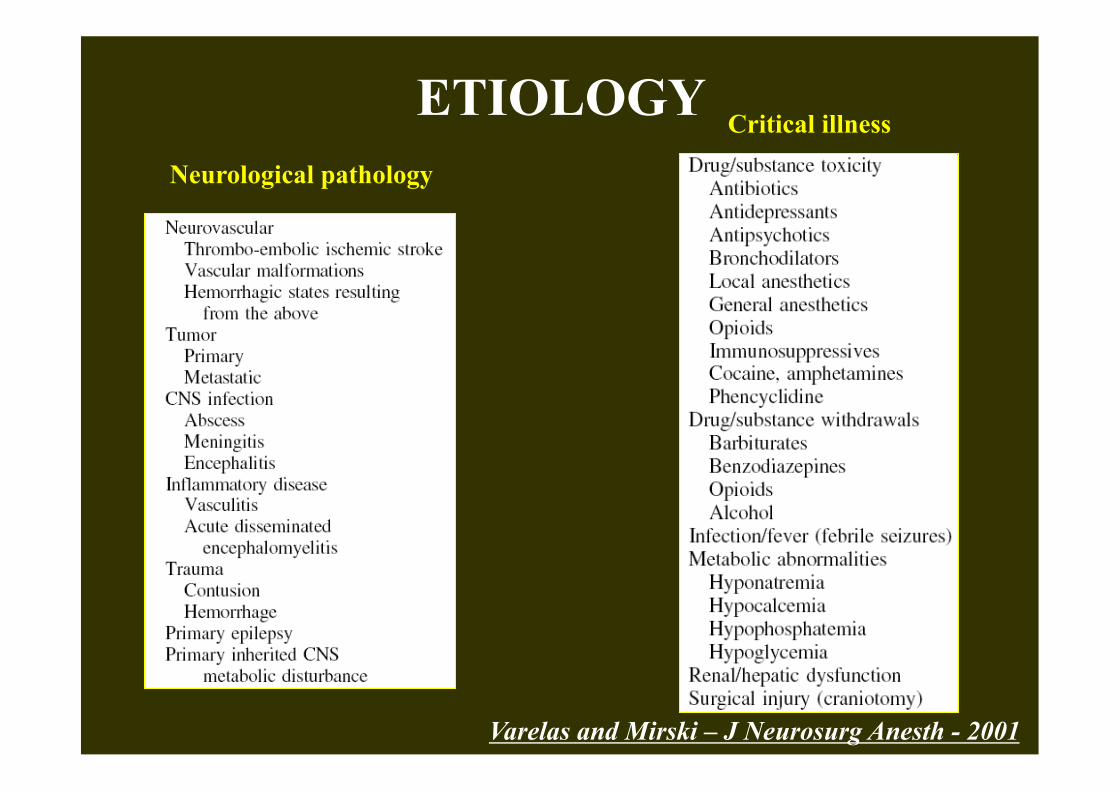
ETIOLOGY Neurological pathology
Critical illness
Varelas and Mirski – J Neurosurg Anesth - 2001
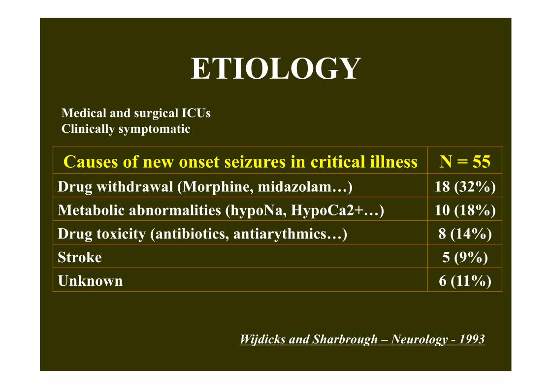
ETIOLOGY
Wijdicks and Sharbrough – Neurology - 1993
Causes of new onset seizures in critical illness N = 55 Drug withdrawal (Morphine, midazolam…) 18 (32%) Metabolic abnormalities (hypoNa, HypoCa2+…) 10 (18%) Drug toxicity (antibiotics, antiarythmics…) 8 (14%) Stroke 5 (9%) Unknown 6 (11%)
Medical and surgical ICUs Clinically symptomatic

WHEN DOING AN EEG? • Clinical suspicion of seizure
1. Abnormal limbs movement 2. Abnormal ocular movement 3. Facial myoclonia 4. Unexplained coma 5. Unexplained delirium
• Diagnosis of coma (if doubt) • Diagnosis of brain death • Diagnosis of metabolic encephalopathy • Monitoring (TBI, SHA, Hypothermia….)

SEIZURE
• Seizure versus SE (emergency) • CSE (emergency) versus NCSE • Subtle SE (emergency) versus other NCSE
Melerkorp and Holtkamp – Lancet Neurology - 2007

DYSFONCTION NEUROLOGIQUE AIGUË
CHEZ LE PATIENT SEDATE

ERREUR(S)? A 52 years old and alcoholic man was hospitalised in ICU for an hypoxemic community-acquired pneumonia, with b l o o d c u l t u r e s p o s i t i v e t o S . pneumoniae. At admission, neurological examination was normal and there was no neck stiffness. One day later, he required mechanical ventilation and developed agitation, which was treated with Haloperidol. Because he became more and more agitated, he was heavily sedated. Agitation was ascribed to alcohol withdrawal. A week later, while the patient was sedated, bilateral larged fixed pupils were observed. CT scan showed diffuse brain oedema and lumbar puncture meningitis.

ERREURS
1. Absence d’interprétation clinique de l’agitation. 2. Absence de monitoring de la sédation. 3. Considérer que l’examen neurologique chez un patient sédaté est ininterprétable . 4. Rapporter l’’agitation uniquement à un sevrage éthylique? 5. Ne pas avoir fait de PL lorsque le patient a été sédaté.

SEVRAGE ETHYLIQUE By definition, the patient must have two
or more of the following after cessation or reduction of alcohol use that has been heavy or prolonged:
1. autonomic hyperactivity (sweating, tachycardia);
2. increased hand tremor; 3. insomnia; 4. nausea or vomiting; 5. transient visual, tactile, or auditory
hallucinations or illusions; 6. psychomotor agitation; anxiety and
tonic-clonic seizures.
DTS is characterised by a fluctuating disturbance of consciousness and change in cognition occurring over a short period of time. It is accompanied by a further exacerbation of autonomic symptoms (sweating, nausea, palpitations and tremor) and an exacerbation of psychological symptoms including anxiety. Treatment: BZD
Clinical Institute Withdrawal Assessment Scale for Alcohol,

EFFECTS OF SEDATION
• Severe septic or critically ill patients often require sedation
• Sedation is a risk factor for delirium • How to detect acute brain dysfunction in
sedated critically ill patients?

DESIGN
[12-24h] Every day Discontinuation of sedation
1st N.E N.E Coma/Delirium
Within 3 days
Reproducibility of neurological examination was satisfactory
Sharshar et al – Crit Care Med – 2011
Non brain injured critical ill patients

NEUROLOGICAL EXAMINATION 12-24H OF SEDATION Fitting set Validation set Number of patients 72 72 Midazolam (mg/kg) 0.9 (0.6 to 1.8) 1.3 (0.8 to 2.0) Subfentanyl (µg/kg) 2.0. (0.8 to 4.0) 2.0 (0.7 to 4.6) sedation to inclusion (hours) 12 (12-24) 12 (12-24) ATICE (from 0 to 20) 9 (9 to 10) 9 (9 to 10) RASS Not tested -4 (-4 to -2) Blinking to strong light (%) 31 (43) 28 (39) Absent eye movement (%) 66 (93) 67 (93) Myosis (%) 45 (63) 38 (54) Pupillary light response (%) 51 (71) 58 (82) Corneal reflex (%) 65 (90) 66 (92) Oculocephalic response (%) 32 (47) 33 (46) Cough response (%) 36 (51) 60 (83) Grimacing (%) 41 (57) 48 (69)

Multiple logistic model
28-DAYS MORTALITY
Sharshar et al – Crit Care Med - 2011
Fitting set Validation set
OR (95%CI) P OR (95%CI) P SAPS-II at inclusion 1.06 (1.02 to 1.09) 0.003 1.03 (1.00 to 1.07) 0.051 Absent cough response 7.80 (2.00 to 30.4) 0.003 5.44 (1.35 to 22.0) 0.017 C-index (SE) 0.836 (0.055) 0.743 (0.067)
RESPONSES ASSESSED BETWEEN THE 12Th AND 24th H OF SEDATION

ALTERED MENTAL STATUS (after discontinuation of sedation)
Fitting set Validation set
Criteria Confusion or coma Delirium or coma OR (95%CI) P OR (95%CI) P
SAPS-II at inclusion 1.04 (1.00 to 1.07) 0.058 1.03 (0.99 to 1.08) 0.10
Absent oculocephalic response 4.49 (1.34 to 15.1) 0.015 5.64 (1.63 to 19.5) 0.006
RESPONSES ASSESSED BETWEEN THE 12Th AND 24th hours of sedation
Multiple logistic model
Sharshar et al – Crit Care Med - 2011

NEUROLOGICAL EXAMINATION
1. Feasible, reproducible and interpretable 2. Enables to detect focal neurological sign 3. Enables to estimate critical illness severity (cough
reflex) 4. Enables to identify patient at risk to develop
de l i r ium a f ter sedat ion d i scont inuat ion (Oculocephalogyre response) – Titration of sedation?
Sharshar et al – Crit Care Med - 2011
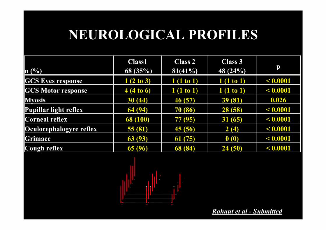
NEUROLOGICAL PROFILES
n (%)
Class1 68 (35%)
Class 2 81(41%)
Class 3 48 (24%) p
GCS Eyes response 1 (2 to 3) 1 (1 to 1) 1 (1 to 1) < 0.0001 GCS Motor response 4 (4 to 6) 1 (1 to 1) 1 (1 to 1) < 0.0001 Myosis 30 (44) 46 (57) 39 (81) 0.026 Pupillar light reflex 64 (94) 70 (86) 28 (58) < 0.0001 Corneal reflex 68 (100) 77 (95) 31 (65) < 0.0001 Oculocephalogyre reflex 55 (81) 45 (56) 2 (4) < 0.0001 Grimace 63 (93) 61 (75) 0 (0) < 0.0001 Cough reflex 65 (96) 68 (84) 24 (50) < 0.0001
Rohaut et al - Submitted
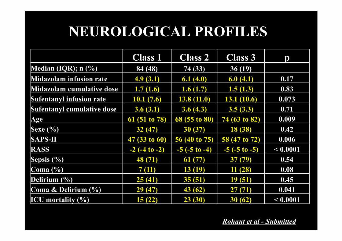
NEUROLOGICAL PROFILES
Class 1 Class 2 Class 3 p Median (IQR); n (%) 84 (48) 74 (33) 36 (19) Midazolam infusion rate 4.9 (3.1) 6.1 (4.0) 6.0 (4.1) 0.17 Midazolam cumulative dose 1.7 (1.6) 1.6 (1.7) 1.5 (1.3) 0.83 Sufentanyl infusion rate 10.1 (7.6) 13.8 (11.0) 13.1 (10.6) 0.073 Sufentanyl cumulative dose 3.6 (3.1) 3.6 (4.3) 3.5 (3.3) 0.71 Age 61 (51 to 78) 68 (55 to 80) 74 (63 to 82) 0.009 Sexe (%) 32 (47) 30 (37) 18 (38) 0.42 SAPS-II 47 (33 to 60) 56 (40 to 75) 58 (47 to 72) 0.006 RASS -2 (-4 to -2) -5 (-5 to -4) -5 (-5 to -5) < 0.0001 Sepsis (%) 48 (71) 61 (77) 37 (79) 0.54 Coma (%) 7 (11) 13 (19) 11 (28) 0.08 Delirium (%) 25 (41) 35 (51) 19 (51) 0.45 Coma & Delirium (%) 29 (47) 43 (62) 27 (71) 0.041 ICU mortality (%) 15 (22) 23 (30) 30 (62) < 0.0001
Rohaut et al - Submitted
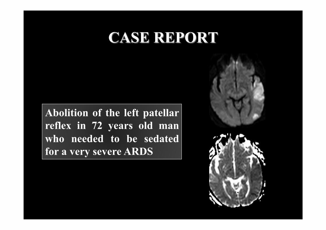
CASE REPORT
Abolition of the left patellar reflex in 72 years old man who needed to be sedated for a very severe ARDS

LONG-TERM COGNITIVE DECLINE

MID
-TE
RM
CO
GN
ITIV
E D
EC
LIN
E
Girard et al – Crit Care Med - 2010
77 Medical ICU patients Memory Attention Concentration
Hippocampus Frontal cortex

Pandharipande et al – NEJM - 2013

COMPORTEMENT DE MALADIE REPONSE A L’AGRESSION
ENCEPHALOPATHIE

CONCEPT OF SICKNESS BEHAVIOR
• Sickness behavior is a major, highly preserved and adaptive component of response to stress.
• The sickness behavior includes stereotypical changes: anxiety, immobility, somnolence, starving, autonomic and neuroendocrine changes etc…
• It involves the amygdala and hippocampus and is mediating by specific structures: circumventricular organs and vagal nerve .
Dantzer et al – Nat Neurosci Rev-2008

PHYSIOLOGICAL BRAIN SIGNALLING
Dantzer et al – Nat Rev Sci - 2008
Circumventricular organs (no BBB)
Sickness behavior Adapted autonomic and neuroendocine response
Vagal nerve
Delirium +

ANXIETY & FEAR (patients with GBS)
N (%)
Sharshar et al – BMJopen access-2012
0
20
40
60
80
100
Dyspnea Pain Weakness Uncertainty
No MV MV

PANICU PROJECT: TO ASSESS THE PROGNOSIS VALUE OF ANXIETY AT ADMISSION IN ICU

Sickness behavior
Delirium
Coma
Sedation
Neuroendcrine system Hippocampus Limbic system
Psychologic disorders (Anxiety, depression,
PTSD)
Frontal cortex
Hippocampus
Brainstem
Acute brain dysfunction Brain structures Outcomes
Death
Cognitive impairment
(Memory and executive functions
-
+

Non sedated
EXAMINATION Normal
Focal sign
Delirium Agitation
Coma
Myoclonus
Brain imaging
EEG - Biochemistry - Drugs Brain imaging
EEG - Biochemistry - Drugs Brain imaging
Biochemistry - Drugs Brain imaging EEG
LUMBAR PUNCTURE IF ANY DOUBT
UNCONTROLLED SEPSIS?
Sedated
Discontinuation of sedation
Brainstem reflexes
Sedation necessary
Monitoring
Antagonist?

DYSFONCTIONS CEREBRALES AIGUËS
Fever
Medical history (Alcohol, Epilepsia…)
Circonstances (CO…) Glycemia
Imaging,
± AB ± CSF
Neck stiffness
Focal sign Trauma
Imaging
Seizure
Imaging
± AB ± CSF
Imaging
± CSF
EEG, Imaging
± CSF
EEG SI DOUTE OU ABSENCE DE CAUSE / BILAN STANDARD

SPINAL CORD SYNDROMES
Syringomyelic
Complete transection
Brown Séquard Anterior spinal artery
Posterior spinal artery
Subacute combined degeneration
ALS
Poliomyelitis

LE SYSTÈME NERVEUX PERIPHERIQUE

PERIPHERAL NERVOUS SYSTEM Neurono -
neuropathy Neuropathy Myasthenic
syndrome Myopathy
Symmetry Bilateral ± symmetrical
Variable MM: asymmetrical
PN: symmetrical PRN: symmetrical
Bilateral and symmetrical
Bilateral and symmetrical
Proximal vs distal
Proximal or distal
MM: distal PN: distal
PRN: proximal
Proximal ++
Proximal ++
Topography
Limbs, Bulbar,
respiratory
MM: ≥ 1 nerve PN: limbs ± respiratory
PRN: limbs, trunk, bulbar, facial,
respiratory
Variable Limbs, facial,
bulbar, trunk or respiratory Ptosis (often
unilateral) and diplopia
Variable
Limbs, facial, bulbar, trunk,
respiratory
MM: mononeuropathy multiplex; PN: polyneuropathy; polyradiculoneuropathy

PERIPHERAL NERVOUS SYSTEM
Neurono- Neuropathy
Neuropathy Myasthenic syndrome Myopathy
Tone flaccidity flaccidity flaccidity flaccidity
Tendon reflexes Lost or
pyramidal signs (ALS)
Lost or decreased Preserved Preserved
I d i o - m u s c u l a r response Preserved Preserved Preserved Absent
Atrophy Pronounced Pronounced No Variable
Other motor signs Fasiculation - Fatigability Fluctuation
Myalgia Myotonia
Other neurological signs Cramps
± sensory loss ± dysautonomia
No sensory loss (except L.
Eaton) No sensory loss

MRC SUM SCORE
Kleyweg et al. - Muscle Nerve - 1991 0
60
48
36
Pare
sis
Nor
mal
Se
vere

SEMIOLOGY
• Weakness – Bilateral et symmetric – Four limbs – Essentially proximal – Sparing the face
• ± sensory deficit • ± Areflexia • ± Amyotrophic
RIGHT LEFT
SHOULDER ELBOW
WRIST HIP
KNEE ANKLE
0,0 0,5 1,0 1,5 2,0 2,5 3,0 3,5 4,0 4,5 5,0
NM score
De Jonghe et al JAMA 2002
CSF: not helpful CK: normal, slightly or highly increased (Status asthmaticus)

ICU-ACQUIRED PARESIS
Frequent and severe complication associated with 1. Increased mortality 2. Prolonged weaning and reintubation 3. Increased length of stay in ICU 4. Disability
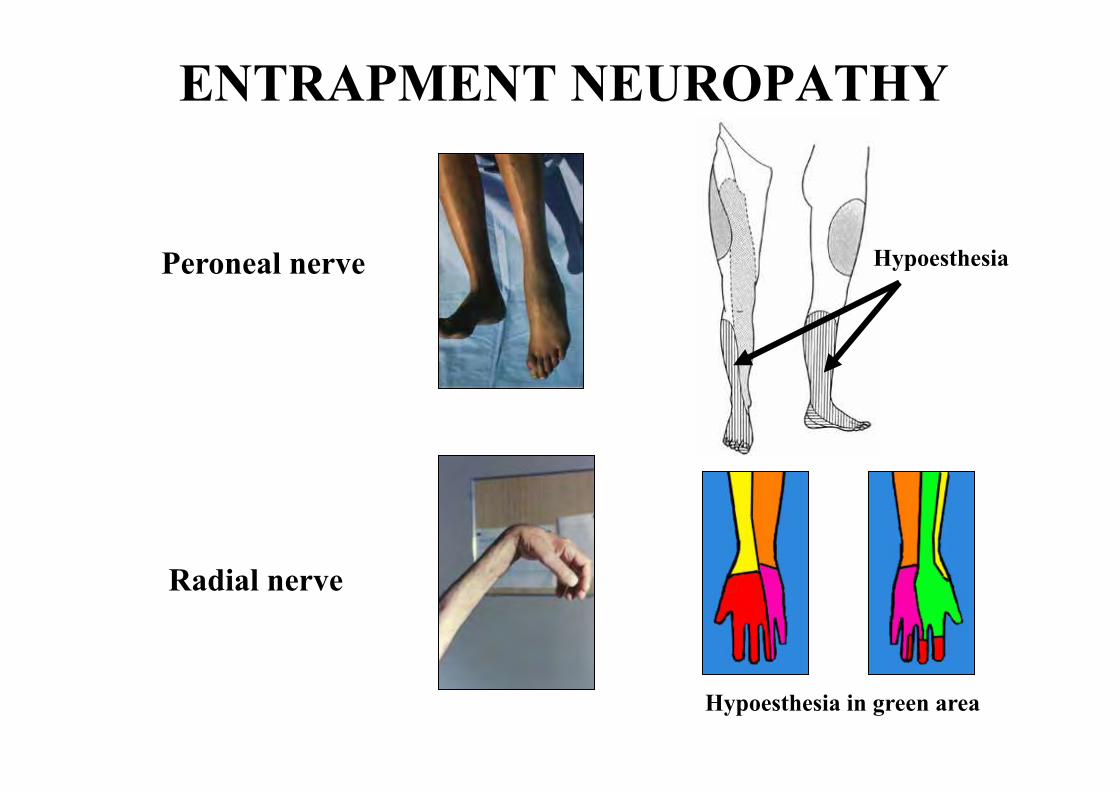
ENTRAPMENT NEUROPATHY
Peroneal nerve
Radial nerve
Hypoesthesia in green area
Hypoesthesia

University of Versailles Raymond Poincaré
Service de Réanimation Pr Djillali Annane
MERCI
Institut Pasteur Fabrice Chrétien
Human Histopathology and Animal Models

MID
-TE
RM
CO
GN
ITIV
E D
EC
LIN
E
Saczynski et al – NEMJ - 2012
225 Post-operative patients

Iwashyna et at – JAMA - 2010

Shah et al- AJRCCM - 2013
Swallowing dysfunction?

Eikermann et al-ICM-2006 ; Dhand - Resp Care - 2006
ELECTROPHYSIOLOGY
Motor and sensory NCS (nerve)
Needle EMG (muscle)
Repeated stimulation (NMJ)
Direct muscle stimulation (excitability)
Supramaximal nerve stimulation (muscle strength and fatigue)
±
+
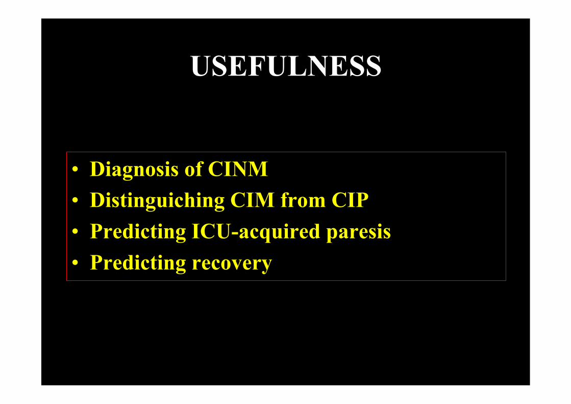
USEFULNESS
• Diagnosis of CINM • Distinguiching CIM from CIP • Predicting ICU-acquired paresis • Predicting recovery

ANATOMY
Plum and Posner
A. Diffuse hemispheric lesion
B. Diencephalic lesion C. Mesencephalic and diencephalic lesion D. Mesencephalic and pons lesion E. Pons lesion

APPROACH
1. Diagnosis 2. Severity 3. Causes 4. Prognosis
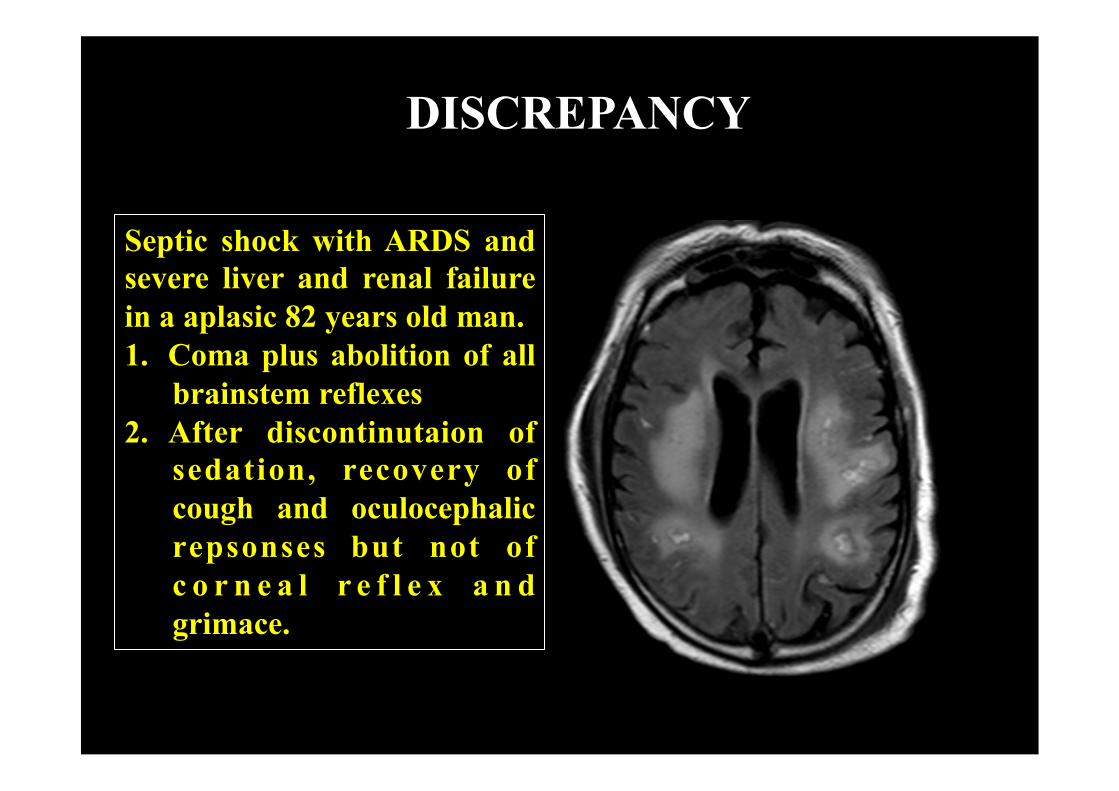
Septic shock with ARDS and severe liver and renal failure in a aplasic 82 years old man. 1. Coma plus abolition of all
brainstem reflexes 2. After discontinutaion of
sedation, recovery of cough and oculocephalic repsonses but not of c o r n e a l r e f l e x a n d grimace.
DISCREPANCY
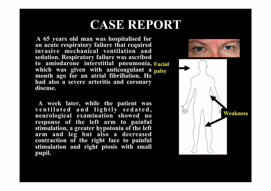
CASE REPORT A 65 years old man was hospitalised for an acute respiratory failure that required invasive mechanical ventilation and sedation. Respiratory failure was ascribed to amiodarone interstitial pneumonia, which was given with anticoagulant a month ago for an atrial fibrillation. He had also a severe arteritis and coronary disease.
A week later, while the patient was v e n t i l a t e d a n d l i g h t l y s e d a t e d , neurological examination showed no response of the left arm to painful stimulation, a greater hypotonia of the left arm and leg but also a decreased contraction of the right face to painful stimulation and right ptosis with small pupil.
Weakness
Facial palsy

CASE REPORT
Three days after discontinuation of sedation, the patient developed agitation and delirium. General and neurological examination was not changed. EEG was normal. Agitation and delirium was ascribed to discontinuation of sedation. Three days later, patient complained of pain of the legs and ankles that turned to be due to bacterial and ischemic myositis.
DELIRIUM WITHOUT (new) FOCAL NEUROLOGICAL SIGN IN A RECENTLY NON SEDATED PATIENT

CASE REPORT
Tendon reflex became brisk in 72 years old man heavi ly sedated and paralyzed for a very severe ARDS (Spinal cord MRI: normal)

CASE REPORT
Presence of corneo-pterygoidien reflex in 34 years old man who needed to be sedated for a very severe ARDS complicating a CO poisoning
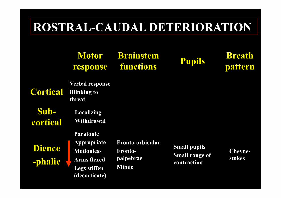
Motor response
Brainstem functions Pupils Breath
pattern
Cortical Verbal response Blinking to threat
Sub-cortical
Localizing Withdrawal
Dience -phalic
Paratonic Appropriate Motionless Arms flexed Legs stiffen (decorticate)
Fronto-orbicular Fronto-palpebrae Mimic
Small pupils Small range of contraction
Cheyne-stokes
ROSTRAL-CAUDAL DETERIORATION

Motor response
Brainstem functions Pupils Breath
pattern
Midbrain Motionless Arms and legs extended (decerebrate)
Light pupillar reflex OCR, OVR (vertical)
Unilateral fixed large (3rd nerve) Midposition and fixed (Tegmentum) Bilateral fixed large (tectum)
Pons Motionless and flaccid or lower limbs flexion
Corneal reflex OCR, OVR (horizontal) Corneal pterygoid reflex
pinpoint Eupneic or ataxic
Medulla Motionless and flaccid or lower limbs flexion
Cough reflex Oculo-cardiac reflex
Ataxic
ROSTRAL-CAUDAL DETERIORATION

Continuum : process posteriorly spreading from the area postrema (deprived from BBB, neuro-inflammatory signaling)
HYPOTHESES Continuum? Reversibility?

Sensitivity to sedation
Neuro-degeneration
- No difference in term of sedatives dose - Difference in SAPS-II - Neuro-inflammatory process starting at the level of the area postrema (posterio localisation, no BBB)
- Difference in age
HYPOTHESES
- effect of sedation reduction on profil C or D?
- Animal studies
- Aging? Animal studies?
Neuro-inflammation

NEUROLOGICAL ASSESSMENT
BRAINSTEM RESPONSES 1. Eyes spontaneous movement 2. Eyes posi=on 3. Oculocephalogyre response 4. Pupillar size 5. Pupillar light reflex 6. Corneal reflex 7. Grimace 8. Cough reflex
FOCAL SIGNS Comparison between right and leY body 1. Motor responses to order or painful s=mula=on 2. Limbs tone 3. Tendon reflexes 4. Plantar reflex
Verbal response Eyes response Motor response
GLASGOW COMA SCALE

OR death 95% CI
Age 1.38 1.16-1.65
Shock 3.87 1.96-7.65
Coma 20.22 9.42-54.09
CPR 13.4 5.18-34.63
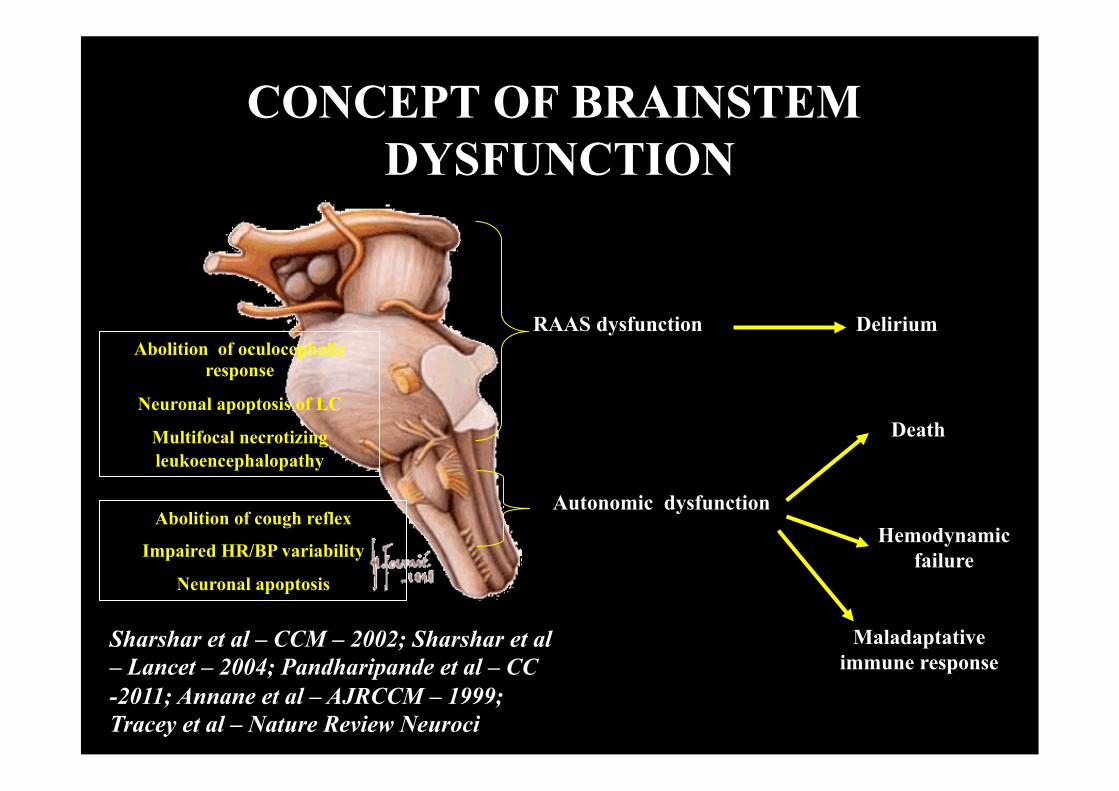
Abolition of oculocephalic response
Neuronal apoptosis of LC
Multifocal necrotizing leukoencephalopathy
Death
Abolition of cough reflex
Impaired HR/BP variability
Neuronal apoptosis
RAAS dysfunction Delirium
Maladaptative immune response
Autonomic dysfunction Hemodynamic
failure
CONCEPT OF BRAINSTEM DYSFUNCTION
Sharshar et al – CCM – 2002; Sharshar et al – Lancet – 2004; Pandharipande et al – CC -2011; Annane et al – AJRCCM – 1999; Tracey et al – Nature Review Neuroci

DIFFERENTIAL DIAGNOSIS OF PARAPLEGIA
Areflexic and flaccid paraplegia Think about injury of the spinal cord, conus medullaris syndrome or a cauda equina syndrome, especially if there is a sensory level, urinary retention or pelvic hypoesthesia
Doubt: usefulness of MRI, electrophysiological testing
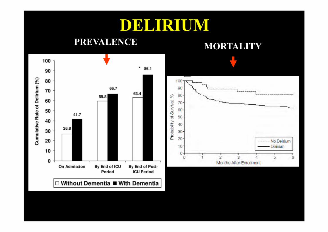
DELIRIUM PREVALENCE MORTALITY
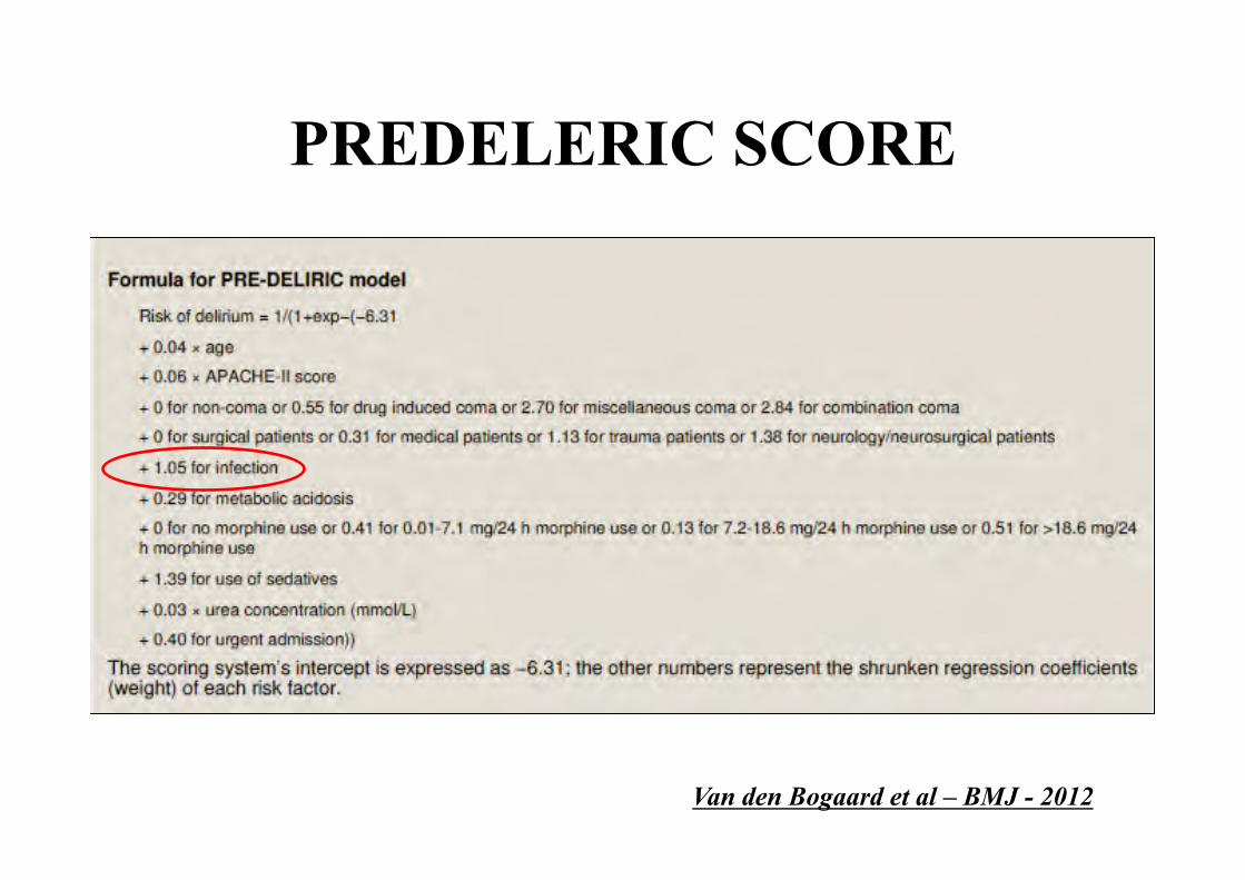
PREDELERIC SCORE
Van den Bogaard et al – BMJ - 2012

• Standard EEG in 110 septic patients - Septic shock: 45 (41%); sedation: 46 (42%) • EEG within the 72h from admission
ELECTROENCEPHALOGRAM
Characteristics n=110
Dominant frequency – n (%) alpha 21 (19) delta 36 (33)
theta 53 (48) Amplitude – n (%)
low-voltage 71 (65)
Absence of reactivity – n (%) 27 (25) Electrographic seizure – n (%) 17 (15) Triphasic waves – n (%) 7 (6) PEDs – n (%) 21 (19) Synek’s classification ≥ 3 – n (%) 34 (31) Young’s classification > 1 – n (%) 41 (37) Azabou et al - Submitted

ME
CH
AN
ISM
S O
F L
ON
G-T
ER
M
CO
GN
ITIV
E D
EC
LIN
E IN
SE
PSIS

DIAGNOSTICS DIFFERENTIELS

Droite Gauche
Rotation de la tête Mouvement
des yeux
Cible
Faisceau longitudinal médial
Muscle droit externe
Muscle droit interne
OCULO-CEPHAOLOGYRE
RESPONSE

OPHTALMOPLEGIE INTERNUCLAIRE ANTERIEURE