Embed Size (px)
Citation preview

1
Exam #2
Name: ____________________________________________________________
13 pages, 64 questions, 75 minutes.
True or False (T/F) ?
1._______Photoelectric effect is the primary mechanism for contrast in projection radiography and CT.
2. _______Like the Fourier Transform, the Radon Transform has an exact inverse.
3._______In CT and projection radiography, higher frequencies penetrate the body more, and in ultrasound, higher
frequencies penetrate the body less.
4._______Undersampling the projection data in CT results in aliasing that manifests as streaking artifacts around
small bright objects.
5. _______Nuclear medicine utilizes ionizing electromagnetic radiation, like CT and x-ray.
6. _______The direction of photon propagation distinguishes the x-rays detected in projection radiography and CT
from the gamma rays detected in nuclear medicine.
7. _______Planar scintigraphy is the only direct nuclear medicine imaging technique.
8._______All nuclear medicine techniques detect the distribution of radiotracers introduced into the body.
9._______Nuclear medicine techniques form an image by detecting the varying intensity of gamma rays.
10._______Nuclear medicine techniques depend on the attenuation of gamma rays to create contrast in the image.
11.______Collimation is used to prevent blurring from Compton Scatter when PET imaging.
12.______The projection slice theorem provided the basis for us to develop reconstruction algorithms given
projection data.
13.______The acoustical impedances of most soft tissues in the body match each other very closely.
14. ______Ultrasound is a functional imaging modality because it can form an image quickly.
15. ______Ultrasound is characterized by sound wave frequencies greater than 20kHz.
16.______In order to image deeper in the body, it is sometimes necessary to increase the frequency of theultrasound machine.
17.______SPECT has no corresponding projection mode imaging technique (as CT has projection radiography).
2
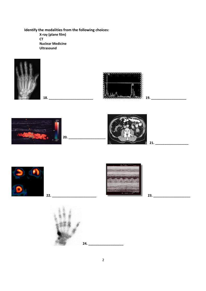
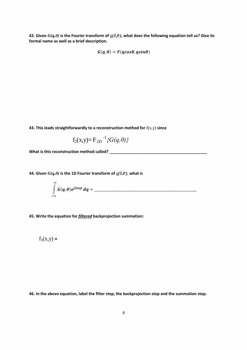
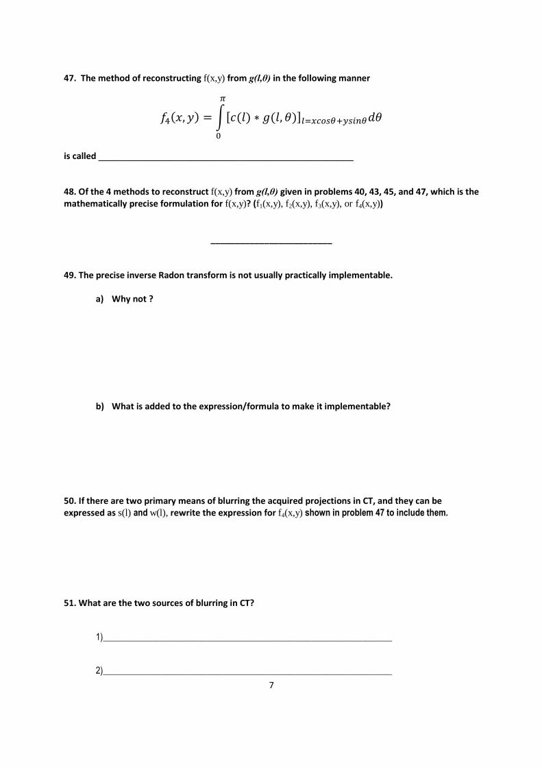
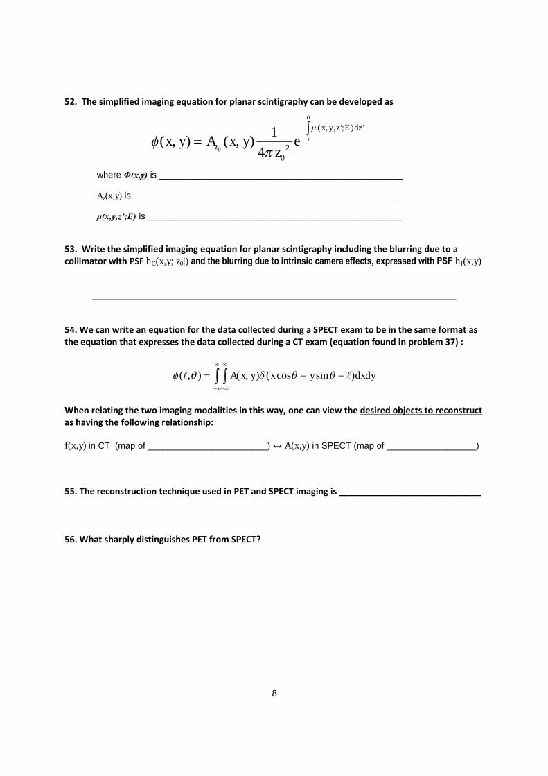
Identify the modalities from the following choices:X-ray (plane film)CTNuclear MedicineUltrasound
24. ___________________
20. ____________________
21. __________________
19. ___________________
22. ________________________
18. ________________________
23. ____________________

3
Fill in the blank / Short answer.
25. How would you categorize a wave in which the particle motion is parallel to the direction of
propagation? ______________________________
26. How would you categorize a wave in which the particle motion is perpendicular to the direction
of propagation? ______________________________
27. How would you categorize a wave which cannot propagate in a vacuum?_____________________
28. How would you categorize a wave which can propagate in a vacuum?________________________
29. With increasing frequency, the ability to resolve objects with ultrasound improves or decreases?
______________________
30. Assuming integer increments, how many unique “views” (projections) of your object f(x,y) do you
have when projection imaging? __________
31. Our formulation of reconstruction methods was based on 1st generation CT technology. What
physical assumption did this enforce?______________________________________________________
32. This represents a “standard attenuation equation” through a homogenous slab:
ି
a) With regard to CT imaging, what is
B ? ___________________________________________________________________________
A ?___________________________________________________________________________
μ ?___________________________________________________________________________
d ? ___________________________________________________________________________
b) With regard to Ultrasound imaging, what is
B ? ___________________________________________________________________________
A ?___________________________________________________________________________
μ ?___________________________________________________________________________
d ? ___________________________________________________________________________
33. In CT, what are we (ideally) mapping? __________________________________________________
34. In Ultrasound, what are we (ideally) mapping?__________________________________________

4
35. Given f(x,y): and the following standard frame of reference:
A black circle with intensity = 0 with 2 identical squares centered along a 45 degree line.
Sketch the following: Include (approximate) relative intensities between the three sketches and keepconsistent axis limits in order to show the relative locations of features.
l
a) g(l,0)
l
b) g(l,45)
l
c) g(l,90)
x [space units]y[s
pace
units
]
θ=0o

5
36. Given g(l,θ)=cos(√ω0l )δ(θ-45o), what is f(x,y) ? (Show any necessary sketches or work)
37. Given ( , ) ( , ) ( cos sin )g f x y x y dxdy
For all l and θ constant; g(l,θ) is a______________________________________
For all l and all θ ; g(l,θ) is ___________________________________________
38. What is (,) = +) (, called ? _______________________________________
39. Sketch b45(x,y) given
f(x,y) = b45(x,y) =
40. Write the equation for straight backprojection summation:
f1(x,y) =
41. In the above equation, label the “backprojection” step and the “summation” step.
6
42. Given G(q,θ) is the Fourier transform of g(l,θ), what does the following equation tell us? Give itsformal name as well as a brief description.
(,) = (,)
43. This leads straightforwardly to a reconstruction method for f(x,y) since
f2(x,y)=F2D-1{G(q,θ)}
What is this reconstruction method called? ____________________________________________
44. Given G(q,θ) is the 1D Fourier transform of g(l,θ), what is
න (,)ஶ
ିஶ
= ______________________________________________________________
45. Write the equation for filtered backprojection summation:
f3(x,y) =
46. In the above equation, label the filter step, the backprojection step and the summation step.
7
47. The method of reconstructing f(x,y) from g(l,θ) in the following manner
ସ݂(ݕ,ݔ) = න [ (ܿ )݈ ∗ ݃( ߠୀ௫௦ఏା௬௦ఏ݀[(ߠ݈,
గ
is called _______________________________________________
48. Of the 4 methods to reconstruct f(x,y) from g(l,θ) given in problems 40, 43, 45, and 47, which is themathematically precise formulation for f(x,y)? (f1(x,y), f2(x,y), f3(x,y), or f4(x,y))
_________________________
49. The precise inverse Radon transform is not usually practically implementable.
a) Why not ?
b) What is added to the expression/formula to make it implementable?
50. If there are two primary means of blurring the acquired projections in CT, and they can beexpressed as s(l) and w(l), rewrite the expression for f4(x,y) shown in problem 47 to include them.
51. What are the two sources of blurring in CT?
1)_________________________________________________________________
2)_________________________________________________________________
8
52. The simplified imaging equation for planar scintigraphy can be developed as
where Φ(x,y) is _________________________________________________
Az(x,y) is _____________________________________________________
μ(x,y,z’;E) is ___________________________________________________
53. Write the simplified imaging equation for planar scintigraphy including the blurring due to acollimator with PSF hC(x,y;|z0|) and the blurring due to intrinsic camera effects, expressed with PSF hI(x,y)
_________________________________________________________________________________
54. We can write an equation for the data collected during a SPECT exam to be in the same format asthe equation that expresses the data collected during a CT exam (equation found in problem 37) :
dxdyyxyxA )sincos(),(),(
When relating the two imaging modalities in this way, one can view the desired objects to reconstructas having the following relationship:
f(x,y) in CT (map of ________________________) ↔ A(x,y) in SPECT (map of __________________)
55. The reconstruction technique used in PET and SPECT imaging is _____________________________
56. What sharply distinguishes PET from SPECT?
0
0
( , , '; ) '
20
1( , ) ( , )
4z
x y z E dz
zx y A x y ez
9
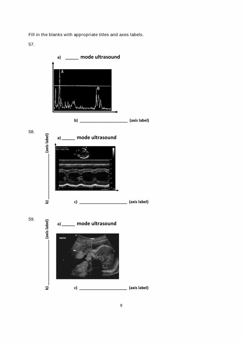
Fill in the blanks with appropriate titles and axes labels.
57.
58.
59.
a) ______ mode ultrasound
b) ______________________ (axis label)
a) ______ mode ultrasound
c) ______________________ (axis label)b)
___
___
___
___
___
___
___
(axi
sla
be
l)
a) ______ mode ultrasound
c) ______________________ (axis label)b)
___
___
___
___
___
___
___
(axi
sla
bel
)

10
60. Consider a an ultrasound transducer with aperture S(r)=rect(r/D), where r = 22 yx , designed
to propagate waves in the z direction.
a) What shape is this transducer in the x-y plane?_______________________________________
b) The region where the z distance from the transducer is less than D2/λ is called the
__________________________________________________________
c) The region considered the “far field” of the transducer, where the distance from thetransducer is greater than D2/λ, is called the
___________________________________________________________
d) In this (far field) region, the more accurate field model, or diffraction formulation, of thetransducer is related to S(r) in what way?
_____________________________________________________________________
e) If the transducer aperture is changed from the original geometry specified above to
D
rrectrS
2)(
What happens to the beamwidth in the region where z< D2/λ? ________________________
What happens to the beamwidth in the region where z> D2/λ? ________________________
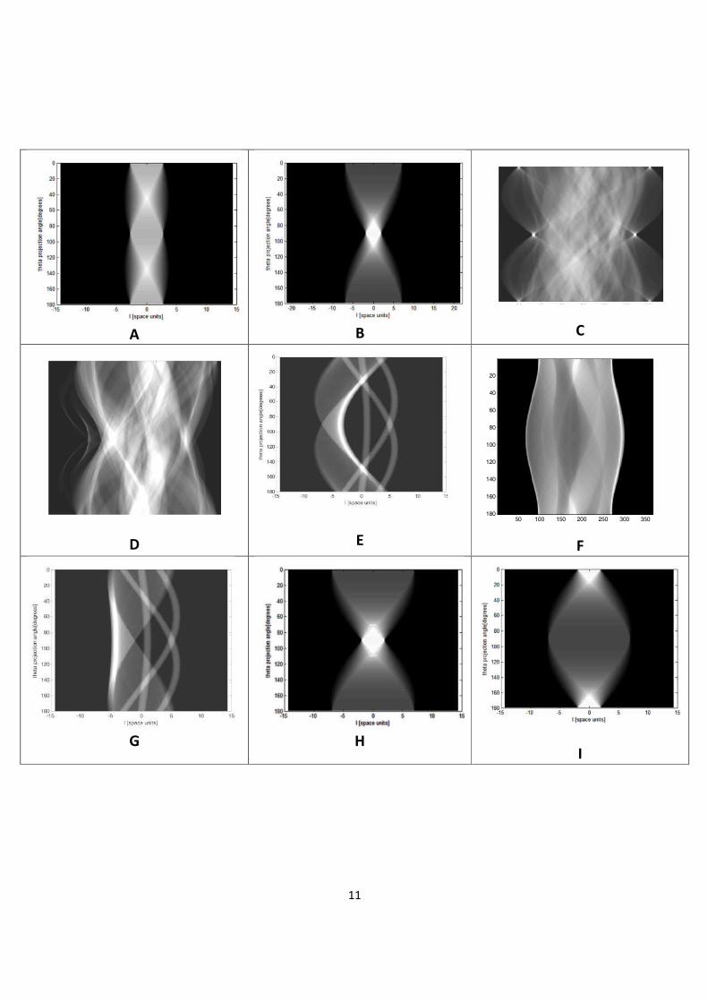
Given the following standard projection angle frame of reference
Match the following objects to their corresponding Radon Transform (sinogram) on thelast page.
61. _______________ 62. _______________63. _______________
x [space units]
y[s
pace
units]
-10 -5 0 5 10
-10
-8
-6
-4
-2
0
2
4
6
8
10x [space units]
y[s
pace
units]
-10 -5 0 5 10
-10
-8
-6
-4
-2
0
2
4
6
8
10
x [space units]y[s
pace
units
]
θ=0o
11
A B C
D E F
G HI
50 100 150 200 250 300 350
20
40
60
80
100
120
140
160
180

12
64. Suppose a 4 MHz acoustic pulse travels from a transducer through 1.3 cm of fat, then encounters
an interface with kidney at normal incidence.
Given:
R = reflectivity =
ti
ti
i
r
ZZ
ZZ
I
I
coscos
coscos
12
12
μa= attenuation factor [cm-1] =67.8
fa [dBcm-1MHz-1]Characteristic impedance, Z
[kg m-2s-1] · 106 Velocity [m/s]
Fat 0.63 1.35 1450
Kidney 1.0 1.62 1560
a) At what time interval after the transmitted pulse will the first reflected pulse (the echo) arrive
back at the transducer?
b) Taking only attenuation into account, what is the fraction (decimal or percentage) of the incident
waveform that encounters the fat/kidney interface? *please read carefully*
fat kidney
1.3cm
fat

13
c) Taking only reflection characteristics at the fat/kidney interface into account, what is the fraction
(decimal or percentage) of the incident waveform that is transmitted into the kidney?
d) Taking both attenuation and reflection losses into account, what will be the amplitude loss in
decibels of the returning waveform?
e) What change in your answer would you see if the frequency of the transducer was changed to
10MHz and why ? (no numbers necessary – just words)