Embed Size (px)
Citation preview

m y c o l o g i c a l r e s e a r c h 1 1 3 ( 2 0 0 9 ) 1 3 2 2 – 1 3 3 4
j ourna l homepage : www.e lsev ier . com/ loca te /mycres
Evolutionary relationships between aquatic anamorphsand teleomorphs: Tricladium and Varicosporium
Jinx CAMPBELLa,*, Ludmila MARVANOVAb, Vladislav GULISc
aDepartment of Coastal Sciences, University of Southern Mississippi, Ocean Springs, MS 39564, USAbCzech Collection of Microorganisms, Institute of Experimental Biology, Faculty of Science, Masaryk University, Tvrdeho 14,
602 00 Brno, Czech RepubliccDepartment of Biology, Coastal Carolina University, Conway, SC 29528-6054, USA
a r t i c l e i n f o
Article history:
Received 5 May 2009
Received in revised form
28 August 2009
Accepted 3 September 2009
Available online 10 September 2009
Corresponding Editor:
Marc Stadler
Keywords:
Aquatic hyphomycetes
Fontanospora
Molecular taxonomy
Morphology
Variocladium
* Corresponding author. Tel.: þ1 228 818 88E-mail address: [email protected]
0953-7562/$ – see front matter ª 2009 The Bdoi:10.1016/j.mycres.2009.09.003
a b s t r a c t
Tricladium, with 21 accepted species, is the largest genus of aquatic hyphomycetes. It encom-
passes species with dematiaceous as well as mucedinaceous colonies. Conidiogenesis is thal-
loblastic; conidiogenous cells proliferate percurrently or sympodially. Conidia have typically
two alternate primary lateral branches. Fontanospora and Variocladium are segregates of Tricla-
dium, differing by conidial branching. Varicosporium comprises nine species, one not well
known. Conidiogenesis is blastic or thalloblastic, conidiogenous cells proliferate sympodially
or are determinate; conidiaregularly produce primary andsecondary branches andoften frag-
ment into part conidia. Molecular analyses on the 28S rDNA of 86 isolates, including 16 species
of Tricladium, five species of Varicosporium, two species of Fontanospora and one species of Var-
iocladium, place these hyphomycetes within Helotiales. Tricladium is polyphyletic and placed in
six clades; Varicosporium is polyphyleticandplacedin threeclades; Fontanospora ispolyphyletic
within a single clade. Variocladium is placed with poor supportas a sister taxon to Varicosporium
giganteum, Hymenoscyphus scutula and Torrendiella eucalypti.
ª 2009 The British Mycological Society. Published by Elsevier Ltd. All rights reserved.
Introduction seven to Sordariomycetes, eight to Dothideomycetes and one to
Previously published results, based on teleomorph–anamorph
relationships and on molecular analyses of various genes of
rDNA, place the ascomycetous aquatic hyphomycetes in the
subphylum Pezizomycotina in five classes, viz Dothideomycetes,
Leotiomycetes, Orbiliomycetes, Pezizomycetes and Sordariomycetes,
and the basidiomycetous aquatic hyphomycetes in two clas-
ses of Basidiomycota, viz Urediniomycetes and Hymenomycetes
(Marvanova 2002, 2007). In the proposed new classification
of fungi (Hibbett et al. 2007) they appear in the subkingdom
Dicarya; the aquatic ascomycetous members (66 taxa) are dis-
tributed in the same classes as mentioned above. Most of
them (42) show affinity to Leotiomycetes, eight to Orbiliomycetes,
78; fax: þ1 228 872 4264.
ritish Mycological Society
Pezizomycetes.
This contribution continues our study of phylogenetic re-
lationships of aquatic hyphomycetes. Previously (Campbell
et al. 2006), using molecular analyses of 28S rDNA, we have
shown that species of Lemonniera, Margaritispora and Gonio-
pila support the hypothesis of convergent evolution of the
asexual propagules in aquatic hyphomycetes (Ingold 1975),
very probably due to their specific adaptation to the life in
a lotic water environment. Species of Lemonniera, a morpho-
logically relatively homogeneous holoanamorphic taxon
with phialidic conidiogenesis, appeared to belong to two
quite distinct clades (Leotiomycetes and Dothideomycetes) of
ascomycetes.
. Published by Elsevier Ltd. All rights reserved.

Evolutionary relationships of Tricladium and Varicosporium 1323
This time we have chosen two morphologically heteroge-
neous genera with thalloblastic (Hennebert & Sutton 1994) or
blastic conidiogenesis: Tricladium (with some segregates) and
Varicosporium. Conidia of both are ‘‘open branching systems’’
in the sense of Kendrick (2003), and can be understood as mod-
ified hyphae with the function of propagules (Descals 1985).
The inter-generic differences are not quite clear; there are spe-
cies whose classification in one or the other genera is debatable
(e.g. Tricladium indicum, Tricladium terrestre, Varicosporium tricla-
diiforme, and the anamorph of Hymenoscyphus varicosporoides).
Tricladium, with 21 recognized species, is the largest ana-
morph genus among aquatic hyphomycetes. It is based on Tri-
cladium splendens (Ingold 1942). The generic characters are
branched conidia, typically consisting of an axis and two
broadly divergent primary alternate branches, developing in
acropetal succession. Conidiogenesis is thalloblastic, i.e. the co-
nidial axis during its development is integrated with the coni-
diogenous cell. The species are delineated on: (1) the degree
of conidiophore branching (from simple to profusely branched);
(2) the conidiogenous cell proliferation (percurrent, sympodial
or absent); (3) the shape of the conidial axis and branches (par-
allel-walled, tapering distally, nearly straight, curved or genicu-
late, apices rounded or acute, branch insertion constricted to
varying extent or unconstricted); and (4) the presence of a para-
basal axial appendage before conidial secession. Colonies may
be mucedinaceous or dematiaceous. Some species of Tricladium
produce occasional secondary branches in culture, but it was
described as a specific character for T. terrestre as this character
also occurs in specimens in nature (Park 1974).
Tricladium is morphologically heterogeneous. With an in-
creasing number of species, some also with more than two
primaries and with secondary branches, it became difficult
to define clearly its scope and to delimit them against similar
taxa. To reduce the morphological heterogeneity there were
several attempts to segregate some species from Tricladium
into new genera. Iqbal (1974) erected Scorpiosporium, based
on Scorpiosporium minutum, where he also included Tricladium
angulatum. The main distinguishing characters were the ‘scor-
pioid’ (geniculate) conidial axis and unconstricted branch in-
sertion. However, these features are not always linked (cf.
also Ando & Kawamoto 1985); there are species with genicu-
late axis and constricted branch insertion (e.g. Tricladium pat-
ulum) and, moreover, the proliferations of the conidiogenous
cells are percurrent in S. minutum, but sympodial in T. angula-
tum. Therefore Marvanova & Descals (1996) recombined S.
minutum in Tricladium.
Dyko (1978) segregated Tricladium eccentricum into a new ge-
nus, Fontanospora, together with a new species Fontanospora
alternibrachiata. The main distinguishing features from Tricla-
dium are the median constriction of the conidial axis and
two branches, inserted closely to each other, one below and
one above this constriction.
Variocladium was published by Descals et al. (1998) to ac-
commodate two similar species, Tricladium giganteum and
Scorpiosporium rangiferinum, with large conidia of variable
branching pattern (alternate and opposite primaries, and
also secondaries appearing in situ) and acute distal ends. The
conidiogenous cells are percurrently proliferating in the for-
mer and determinate in the latter. A spermatial state resem-
bling Phialocephala dimorphospora was reported in
Variocladium giganteum by Willoughby & Minshall (1975) as
well as in Variocladium rangiferinum (Descals & Webster 1982).
Three Tricladium species have known teleomorphs, all clas-
sified in Helotiales: Hymenoscyphus splendens (Helotiaceae) is the
teleomorph of T. splendens (Abdullah et al. 1981), Hydrocina chae-
tocladia (Hyaloscyphaceae) is the teleomorph of Tricladium chaeto-
cladium (Webster et al. 1991), and Cudoniella indica (Helotiaceae)
was described as the teleomorph of Tricladium indicum isolate
from South Africa (Webster et al. 1995), but the conspecificity
of Webster’s isolate with the type of this species described
from Himalaya (India) was doubted by Sivichai et al. (2003, see
below). Phialidic andromorphs have been described in T. chae-
tocladium, Tricladium curvisporum, Tricladium minutum, Tricla-
dium obesum, Tricladium robustum, T. splendens and T. terrestre.
Varicosporium is based on Varicosporium elodeae (Kegel 1906).
Nine species are accepted at present, one of them not well
known and by some authors classified in Tricladium (Sivichai
et al. 2003). Varicosporium is characterized by branched conidia
with rarely nearly straight, but often variously curved conidial
axis and by regularly formed, diverging primary and second-
ary (in some species also of higher order) conidial branches.
Unlike in Tricladium, the conidia have a strong tendency to
fragment into simpler part conidia, often similar to those of
a Tricladium. Conidiogenesis in V. elodeae is blastic, whereas
it is thalloblastic in the rest of Varicosporium species. There is
no species of Varicosporium with percurrent proliferation of
conidiogenous cells known at present. In V. elodeae the coni-
diogenous cells do not elongate after having produced the first
conidium and the subsequent conidia arise retrogressively (or
randomly) down the conidiophore. In the rest of the species
there are sympodial elongations or no growth of the conidiog-
enous cells after release of the first conidium. The species are
distinguished by the curvature of the conidial elements
(nearly straight, arcuate, helical or geniculate) by the mor-
phology of conidial elements (parallel-walled, subulate,
slightly clavate), by the shapes of apices (rounded or acute)
and by branch insertion (constricted or unconstricted).
The only published teleomorph in Varicosporium is H. vari-
cosporoides, but the classification of the anamorph in Varicospo-
rium was doubted by Sivichai et al. (2003) and Boonyuen et al.
(2006). Marvanova (unpubl.) has obtained minute apothecia
of a leotiomycetan discomycete in a mating experiment with
two isolates of V. giganteum from Canada. A phialidic andro-
morph is known in this species (Crane 1968) and was discov-
ered in Varicosporium scoparium (Marvanova unpubl.).
The aims of this study were: 1) to test the hypothesis that
the morphologically defined taxa are supported by molecular
analyses of LSU rDNA nucleotide sequences; 2) to establish
phylogenetic relationships of the anamorphic species to asco-
mycete clades; 3) to establish the relationships of the pleo-
morphic aquatic taxa with their terrestrial relatives.
Materials and methods
Collection, isolation and characterisation
Collections were made from streams in Europe and North
America. A loopful of fresh foam was streaked onto a micro-
scope slide coated with a thin layer of malt extract agar

1324 J. Campbell et al.
(0.1–2 % MEA plus 100 mg L�1 chloramphenicol or penicillin/
streptomycin [Sigma, St. Louis, MO]) and kept at 10–15 �C.
When isolated from submerged plant litter (USA isolates),
material was first incubated in sterile distilled water in Petri
dishes to induce sporulation and then suspended conidia
were transferred onto 0.1 % MEA with 200 mg L�1 of strepto-
mycin and 200 000 units/L of penicillin G. After 24–48 h germi-
nating conidia were transferred to MEA plates supplemented
with antibiotics and incubated at 10–18 �C (Marvanova & Bar-
locher 1998; Marvanova & Gulis 2000). Cultures were checked
for bacterial and fungal contaminants, subcultured to MEA
plates and incubated at 15 �C. All isolates were monoconidial.
DNA extraction, sequencing and cladistic analysis
Mycelia were harvested directly from MEA plates, incubated
with 200 units of lyticase at 30 �C for 4 h, and ground at hourly
intervals during incubation using a micropestle. RNaseA
(40 mg) was added and incubated for 20 min at 65 �C and ge-
nomic DNA extracted using Qiagen’s DNeasy Plant Mini Kit
(Qiagen. 2004) following the procedure by Raja et al. (2003).
The 50 end of the 28S ribosomal gene was amplified with
Taq PCR Master Mix Kit (Qiagen. 2002a) using fungal primers
LROR (Bunyard et al. 1994) and LR6 (Vilgalys & Hester 1990).
The PCR products were cleaned up with Qiaquick PCR Purifi-
cation Kit (Qiagen. 2002b) and sequenced directly. Sequences
(Table 1) were aligned with published sequence data (Table 2)
using Clustal X (Thompson et al. 1997), and then refined man-
ually in Se-Al (Rambaut 1996).
The rationale for the taxon sampling followed Campbell
et al. (2006). Additionally, the sequences derived in this study
were entered into BLAST, NCBI (Altschul et al. 1997) to help
identify sequence similarity to other taxa and to determine
which orders and families should be included in the phyloge-
netic analyses.
Bayesian Metropolis coupled Markov chain Monte Carlo
(B-MCMCMC) analyses were performed with the general
time reversible model of substitution model (Rodriguez et al.
1990) with invariant sites and gamma distribution
(GTRþ IþG) using MrBayes 3.0 (Huelsenbeck & Ronquist
2001): searches were conducted for a total of 2 000 000 genera-
tions with phylogenetic trees sampled every 100 generations.
Three independent B-MCMCMC analyses were conducted to
verify likelihood convergence and burn-in parameter. Out of
20 001 resulting trees from each analysis, the initial 3111 trees
were identified as burn-in prior to the convergence of likeli-
hoods and were excluded from post-run analyses. The major-
ity rule consensus tree from 16 890 trees was generated with
average branch lengths (Fig 1).
Maximum parsimony (MP) analyses were performed using
PAUP*4.0b 10 (Swofford 2002). A two-step search approach
was employed in an attempt to avoid local optima (Olmstead
et al. 1993). Step-one consisted of 100 heuristic replicates with
random starting trees, random stepwise addition and tree-bi-
section–reconnection branch swapping with MulTrees off and
a maximum of two trees saved per replicate. All of the shortest
trees from these initial runs were saved and then used as
starting trees for the second step, which consisted of searches
with MulTrees on and the maximum number of trees set to
10 000. Gaps were treated as missing data.
Nodal support was assessed by non-parametric bootstrap-
ping and Bayesian posterior probabilities. Bootstrap values
(Felsenstein 1985) were calculated from 1000 replications us-
ing a heuristic search on 100 replicates with random starting
trees, random stepwise addition and MulTrees off. Posterior
probabilities were calculated from the majority rule consen-
sus tree of the 20 000 trees from the Bayesian analyses, with
the initial 3111 trees excluded as burn in prior to the conver-
gence of likelihoods.
Results and discussion
Initial analyses were performed on w250 species with repre-
sentatives from 17 orders of ascomycetes, and with basidio-
mycetes as outgroup taxa (data not shown). All species of
Tricladium (16), Fontanospora (2), Variocladium (1) and Varicospo-
rium (5) treated in this study are placed among the Leotiomy-
cetes. These placements were also supported in the BLAST
searches. Further analyses were run with 86 taxa, including
our 45 isolates; members of the Pezizomycetes were used as
outgroup taxa.
The Bayesian tree (Fig 1) indicates that Tricladium is poly-
phyletic and placed in six clades; Varicosporium is polyphyletic
and placed in three clades; Fontanospora is polyphyletic within
a single clade; Variocladium is placed with poor support as a sis-
ter taxon to Varicosporium giganteum, Hymenoscyphus scutula
and Torrendiella eucalypti.
The first group (Figs 1, 2) includes Tricladium species with
dark colonies (Tricladium clade 1). These five species (Tricla-
dium castaneicola, Tricladium indicum, Tricladium obesum, Tricla-
dium splendens and Tricladium terrestre) form a relatively
homogeneous group from the point of colony color and of co-
nidial morphology. They all have dark colonies; conidiophores
in culture are mostly lateral, relatively short, often un-
branched (except in T. splendens, where they typically consist
of a stipe with acrotonous branches); conidiogenous cell pro-
liferation is percurrent (in T. splendens the type of the genus,
and in T. obesum), sympodial (in T. castaneicola and T. terrestre)
or they are determinate (in T. indicum); conidia are medium
sized to large (span ca. 100–200 (400) mm, smaller in T. obesum),
and the conidial elements are mostly 5–11 mm wide (except in
T. castaneicola); secondary branches are occasional or regularly
present (in T. terrestre), branch insertion is constricted to vary-
ing extent; phialidic andromorph is known in T. obesum (Mar-
vanova 2004), T. splendens and T. terrestre (Descals & Webster
1982). However, four Tricladium species with dark colonies ap-
pear in at least three other clades (see below).
The two strains of T. splendens treated here were also se-
quenced by Baschien et al. (2006, Fig 3). Neighbor joining anal-
yses on the 18S rDNA placed T. splendens in a well supported
clade with the helotialean aquatic anamorphs Anguillospora
crassa, Anguillospora furtiva, Anguillospora fustiformis, Anguillo-
spora mediocris and Geniculospora grandis. The same grouping
was found with another isolate of T. splendens by Belliveau &
Barlocher (2005, Fig 2) using maximum parsimony and
weighted parsimony on the 18S rDNA. Baschien et al. (2006,
Fig 15, ITS1–5.8S–ITS2 rDNA sequences, maximum likelihood)
found a close relationship to Zalerion varium (which according
to Bills et al. (1999) may be a member of Helotiales), and to
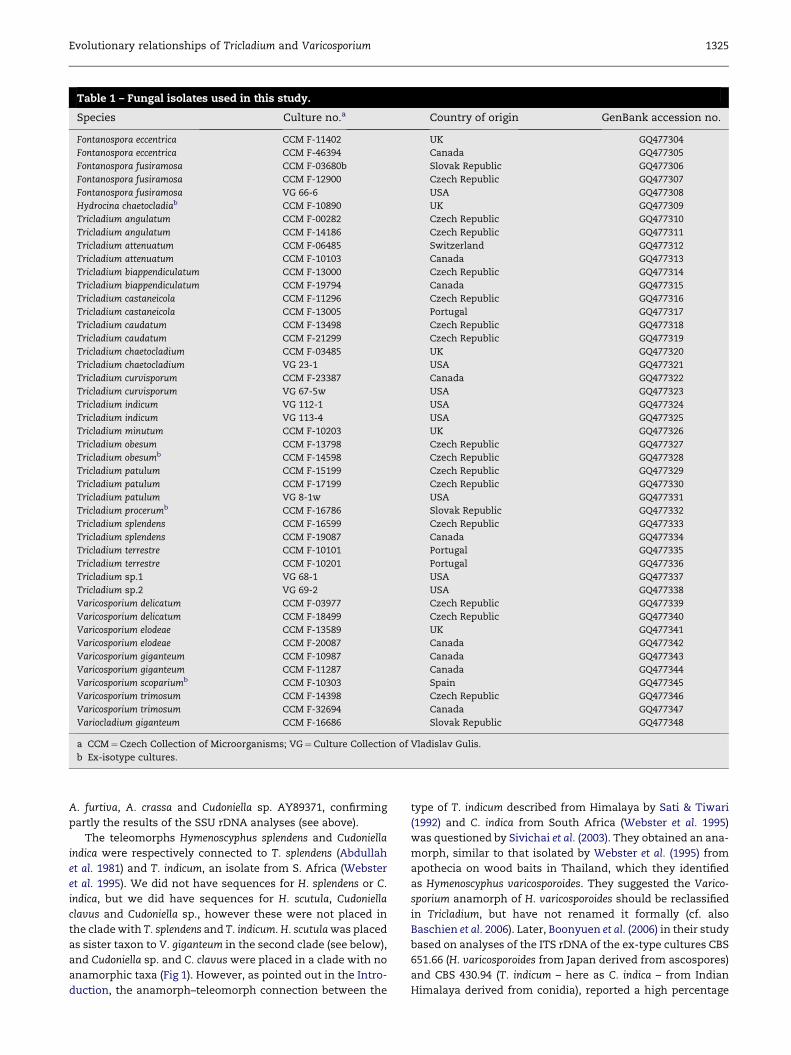
Table 1 – Fungal isolates used in this study.
Species Culture no.a Country of origin GenBank accession no.
Fontanospora eccentrica CCM F-11402 UK GQ477304
Fontanospora eccentrica CCM F-46394 Canada GQ477305
Fontanospora fusiramosa CCM F-03680b Slovak Republic GQ477306
Fontanospora fusiramosa CCM F-12900 Czech Republic GQ477307
Fontanospora fusiramosa VG 66-6 USA GQ477308
Hydrocina chaetocladiab CCM F-10890 UK GQ477309
Tricladium angulatum CCM F-00282 Czech Republic GQ477310
Tricladium angulatum CCM F-14186 Czech Republic GQ477311
Tricladium attenuatum CCM F-06485 Switzerland GQ477312
Tricladium attenuatum CCM F-10103 Canada GQ477313
Tricladium biappendiculatum CCM F-13000 Czech Republic GQ477314
Tricladium biappendiculatum CCM F-19794 Canada GQ477315
Tricladium castaneicola CCM F-11296 Czech Republic GQ477316
Tricladium castaneicola CCM F-13005 Portugal GQ477317
Tricladium caudatum CCM F-13498 Czech Republic GQ477318
Tricladium caudatum CCM F-21299 Czech Republic GQ477319
Tricladium chaetocladium CCM F-03485 UK GQ477320
Tricladium chaetocladium VG 23-1 USA GQ477321
Tricladium curvisporum CCM F-23387 Canada GQ477322
Tricladium curvisporum VG 67-5w USA GQ477323
Tricladium indicum VG 112-1 USA GQ477324
Tricladium indicum VG 113-4 USA GQ477325
Tricladium minutum CCM F-10203 UK GQ477326
Tricladium obesum CCM F-13798 Czech Republic GQ477327
Tricladium obesumb CCM F-14598 Czech Republic GQ477328
Tricladium patulum CCM F-15199 Czech Republic GQ477329
Tricladium patulum CCM F-17199 Czech Republic GQ477330
Tricladium patulum VG 8-1w USA GQ477331
Tricladium procerumb CCM F-16786 Slovak Republic GQ477332
Tricladium splendens CCM F-16599 Czech Republic GQ477333
Tricladium splendens CCM F-19087 Canada GQ477334
Tricladium terrestre CCM F-10101 Portugal GQ477335
Tricladium terrestre CCM F-10201 Portugal GQ477336
Tricladium sp.1 VG 68-1 USA GQ477337
Tricladium sp.2 VG 69-2 USA GQ477338
Varicosporium delicatum CCM F-03977 Czech Republic GQ477339
Varicosporium delicatum CCM F-18499 Czech Republic GQ477340
Varicosporium elodeae CCM F-13589 UK GQ477341
Varicosporium elodeae CCM F-20087 Canada GQ477342
Varicosporium giganteum CCM F-10987 Canada GQ477343
Varicosporium giganteum CCM F-11287 Canada GQ477344
Varicosporium scopariumb CCM F-10303 Spain GQ477345
Varicosporium trimosum CCM F-14398 Czech Republic GQ477346
Varicosporium trimosum CCM F-32694 Canada GQ477347
Variocladium giganteum CCM F-16686 Slovak Republic GQ477348
a CCM¼Czech Collection of Microorganisms; VG¼Culture Collection of Vladislav Gulis.
b Ex-isotype cultures.
Evolutionary relationships of Tricladium and Varicosporium 1325
A. furtiva, A. crassa and Cudoniella sp. AY89371, confirming
partly the results of the SSU rDNA analyses (see above).
The teleomorphs Hymenoscyphus splendens and Cudoniella
indica were respectively connected to T. splendens (Abdullah
et al. 1981) and T. indicum, an isolate from S. Africa (Webster
et al. 1995). We did not have sequences for H. splendens or C.
indica, but we did have sequences for H. scutula, Cudoniella
clavus and Cudoniella sp., however these were not placed in
the clade with T. splendens and T. indicum. H. scutula was placed
as sister taxon to V. giganteum in the second clade (see below),
and Cudoniella sp. and C. clavus were placed in a clade with no
anamorphic taxa (Fig 1). However, as pointed out in the Intro-
duction, the anamorph–teleomorph connection between the
type of T. indicum described from Himalaya by Sati & Tiwari
(1992) and C. indica from South Africa (Webster et al. 1995)
was questioned by Sivichai et al. (2003). They obtained an ana-
morph, similar to that isolated by Webster et al. (1995) from
apothecia on wood baits in Thailand, which they identified
as Hymenoscyphus varicosporoides. They suggested the Varico-
sporium anamorph of H. varicosporoides should be reclassified
in Tricladium, but have not renamed it formally (cf. also
Baschien et al. 2006). Later, Boonyuen et al. (2006) in their study
based on analyses of the ITS rDNA of the ex-type cultures CBS
651.66 (H. varicosporoides from Japan derived from ascospores)
and CBS 430.94 (T. indicum – here as C. indica – from Indian
Himalaya derived from conidia), reported a high percentage

Table 2 – Sequences obtained from GenBank.
Species GenBank accession no.
Baeomyces placophyllus AF356658
Brasiliomyces trina AB022350
Bulgaria inquinans DQ470960
Chlorencoelia sp. AY789351
Cudoniella clavus DQ470944
Cudoniella sp. AY789375
Cyathicula coronata AF222491
Cystotheca wrightii AB022355
Dermea acerina DQ247801
Dibaeis baeomyces AF279385
Erysiphe betae AB079684
Fabrella tsugae AF356694
Gyromitra esculenta FJ176906
Helicodendron conglomeratum AY856900
Helvella lacunosa U42681
Heyderia abietis AY789289
Hyaloscypha daedaleae AY789415
Hydrocina chaetocladia AY789412
Hymenoscyphus scutula AY789431
Hymenoscyphus sp. AF430278
Icmadophila ericetorum DQ883694
Lachnum cf. bicolor AY544674
Lachnum virgineum AY544646
Lambertella tubulosa AY616237
Leveillula taurica AB022387
Microsphaera pulchra AB022389
Morchella esculenta AF279398
Neofabraea malicorticis AY544662
Ombrophila violacea AY789365
Ostropa barbara AY584642
Pezicula carpinea DQ470967
Phyllactinia moricola AB022401
Podosphaera longiseta AB022423
Potebniamyces pyri DQ470949
Rhytisma acerinum AF356696
Siphula ceratites DQ986775
Spirosphaera floriformis AY616238
Torrendiella eucalypti DQ195800
Typhulochaeta japonica AB022415
Uncinula septata AB183532
1326 J. Campbell et al.
(99.5 %) of bp similarity between these specimens (see their
Table 2), and suggested this indicated that H. varicosporoides
had a Tricladium anamorph.
Moreover, there is a third large-spored (holoanamorphic)
species of Tricladium, Tricladium marylandicum, described
from USA (Crane 1968) whose relationships with T. indicum
and H. varicosporoides are also unresolved. All these three
taxa are characterized by dark colonies and large conidia
with 1–4 primary and (in culture) also secondary branches.
Multi-gene molecular analyses employing all these species
may help solve this problem.
This first group also contains Lambertella tubulosa, Helico-
dendron conglomeratum and Spirosphaera floriformis. They also
have dark colonies, but with their helicoform conidia are mor-
phologically and ecologically quite different. They are called
aero-aquatic hyphomycetes and unlike aquatic hyphomy-
cetes, they thrive in standing or slow-moving waters, with
low dissolved oxygen, and their conidia have air-trapping
shapes, resistant against submerging, and adapted to dis-
persal on the water surface. Spirosphaera appears to be
polyphyletic however, as Voglmayr (2004, partial nuc 28S
and ITS1–5.8S–ITS2 rDNA) and Baschien et al. (2006, the
same gene regions) placed Spirosphaera cupreorufescens in
a well supported clade with Anguillospora longissima in the
Dothideomycetes.
The second group (Figs 1, 3, 4) contains Tricladium p.p.-Var-
icosporium p.p.-Fontanospora. It consists of two subclades:
The first subclade (Figs 1, 3) contains Tricladium attenuatum,
Tricladium biappendiculatum, Tricladium minutum, Tricladium
patulum, Varicosporium elodeae, Varicosporium trimosum, Fonta-
nospora eccentrica, Fontanospora fusiramosa. If we exclude T. min-
utum, which is on a poorly supported branch within this
subclade, then the unifying features here are pale colonies, co-
nidial branch insertion constricted to various extent, rela-
tively narrow (usually up to 4 mm wide) conidial elements,
absence of spermatial synanamorph and unknown teleo-
morph. Except for T. biappendiculatum, the conidiophore usu-
ally consists of a stipe and a fertile head which, if fully
developed, is a multibranched structure with numerous coni-
dia in lateral as well as terminal position. However, the
branching pattern of the conidia is different: in Fontanospora
there are two nearly opposite laterals inserted near the middle
of a bent axis; V. elodeae and Varicosporium trimosum have con-
idia with primary and secondary branches (in the latter the
conidia are relatively simple, consisting of an axis bearing
one primary and one secondary branch, both inserted at right
angle to their parent structures); the two Tricladium species
have typical conidia for this genus differing by geniculate
axis and blunt conidial apices in T. patulum against nearly
straight axis and acute conidial apices in T. attenuatum. T. biap-
pendiculatum also has acute conidial ends, but its conidio-
phores are short, often simple, and conidiogenous cell
proliferation is percurrent.
T. minutum is on a poorly supported branch within the
above subclade. This may support its former segregation in
Scorpiosporium by Iqbal (1974), but in morphology, there are
no characters diagnostic enough on the generic level to justify
its separation. T. minutum has brown colonies, percurrent con-
idiogenous cells, small to medium large conidia with genicu-
late axis, and typically two primary branches with
unconstricted branch insertion. A phialidic andromorph was
seen in one of the British isolates by E. Descals (Marvanova
& Descals 1996).
V. elodeae was sequenced by Baschien et al. (2006, Fig 3, 18S
rDNA, neighbor joining, maximum likelihood and parsimony
analyses) and was placed in a poorly supported clade with
T. patulum, which is congruent with our results. Both strains
of V. elodeae sequenced by Baschien et al. (2006), as well as
those sequenced in our study, produce extracellular green pig-
ments in agar cultures, and the conidia have parallel-walled
axis and branches, typical for isolates from water.
The second subclade differs from the species of the first
subclade by having dark colonies. V. giganteum has large con-
idia (spanning over 200 mm), with geniculate axis produced on
sympodial conidiogenous cells and primary and secondary
branches with unconstricted insertion. Variocladium giganteum
is placed with poor support as a sister taxon to the species
within the second subclade. It is relatively isolated from other
Tricladium species, indicating that its segregation might have
been justified. It is unique in the large dimensions of conidia

Fig 1 – B-MCMCMC majority rule consensus tree generated with average branch lengths from 16 890 trees inferred from
Bayesian analyses on the 28S rDNA data. Maximum parsimony bootstrap supports and Bayesian posterior probability values
greater than 50 % are given, respectively, at the corresponding nodes.
Evolutionary relationships of Tricladium and Varicosporium 1327
and variable branching pattern (alternate or opposite). Colo-
nies are dark, conidiogenous cells percurrent and conidial
branch insertion is unconstricted.
The third group (Figs 1, 5) contains Tricladium chaetocladium
(including its teleomorph Hydrocina chaetocladia), Tricladium
curvisporum, Tricladium sp.1 and sp.2 and Varicosporium delica-
tum. In our study, these species form a poorly supported clade
within Helotiales. There are only two morphological characters
which the four Tricladium species have in common: a sigmoid
conidial axis and a phialidic andromorph in two of them. Oth-
erwise they are culturally and morphologically quite different.
T. chaetocladium, represented by two isolates from ascospores
of the teleomorph and two from the anamorphic state, has
V. delicatum as a sister taxon. However, their morphological
Fig 2 – Group 1. Conidia from Tricladium clade 1. (A) Tricladium terrestre CCM F-10101. (B–C) Tricladium indicum VG 112-1.
(D) Tricladium castaneicola CCM F-11296. (E) T. castaneicola CCM F-10605. (F) Tricladium splendens CCM F-16599. (G) T. splendens
CCM F-19087. (H–J) Tricladium obesum CCM F-14598. (K–L) T. obesum CCM F-13798. Scale bar a (A–C) [ 100 mm, scale bar b
(D–L) [ 50 mm.

Fig 3 – Group 2, subclade 1. Conidia from Varicosporium clade 1. (A–B) Varicosporium elodeae CCM F-20087. (C) Varicosporium
trimosum CCM F-14398. (D) V. trimosum CCM F-32694. Conidia from Fontanospora clade 1. (E–F) Fontanospora fusiramosa VG 66-6.
(G) F. fusiramosa CCM F-03680b. (H) Fontanospora eccentrica CCM F-11402. (I) F. eccentrica CCM F-46394. Conidia from Tricladium
clade 2. (J) Tricladium attenuatum CCM F-06485. (K) T. attenuatum CCM F-10103. (L) Tricladium patulum CCM F-17199. (M–N)
Tricladium biappendiculatum CCM F-13000. (O–R) Tricladium minutum CCM F-10203. Scale bar [ 50 mm.
Evolutionary relationships of Tricladium and Varicosporium 1329

Fig 4 – Group 2, subclade 2. Conidia from Varicosporium clade 1. (A–B) Varicosporium giganteum CCM F-11287. Conidium from
Variocladium clade 1. (C) Variocladium giganteum CCM F-16686. Scale bar [ 100 mm.
1330 J. Campbell et al.
similarity is low and can be seen only in the relatively large
conidia with constricted or subconstricted branch insertion.
Another small subclade linked to T. chaetocladium, consists of
two isolates of T. curvisporum and two problematic taxa Tricla-
dium sp.1 VG 68-1 and Tricladium sp.2 VG 69-2, both isolated
from sedges submerged in fresh water and representing unde-
scribed species. Tricladium sp.1 has conidia somewhat similar
to those of T. angulatum or T. minutum, but it has black colo-
nies, the conidial elements are wider, the conidial axis is sig-
moid and axis and branches have conspicuously acute
apices; elongation of conidiogenous cell is sympodial. Tricla-
dium sp.2 is a similar isolate with dark colonies and sympodial
elongation of conidiogenous cells, but conidia are much more
delicate with 0–2(3) primary branches and an occasional sec-
ondary branch produced after release. Conidia may bear
some resemblance to part conidia of V. delicatum.
H. chaetocladia (HME 4375, ex-isotype, teleomorph of T. chae-
tocladium) was found to be placed within a poorly supported,
so called ‘‘bsa’’ clade (Wang et al. 2005, LSU, SSU, 5.8S rDNA)
containing ecologically similar saprotrophic fungi, mostly
with bright apothecia, preferring aquatic or at least humid
habitats. In their parsimony analysis the closest neighbors
are two species of Mitrula and two of Vibrissea (Vibrisseaceae),
but in their maximum likelihood tree topology Hydrocina ap-
pears closest only to Vibrissea spp. (Wang et al. 2005). In a later
study, however, (Wang et al. 2006a, parsimony analysis of LSU,
SSU and 5.8S rDNA regions) they place this fungus into Hyalo-
scyphaceae, but with no bootstrap support. Finally, in an exten-
sive study based on SSU, LSU and 5.8S rDNA sequences of 99
taxa (Wang et al. 2006b), it is placed in the so-called Mitrula
clade among Helotiaceae, again without bootstrap support
(cf. also Raja et al. 2008, LSU, maximum parsimony).
T. chaetocladium in the study of Belliveau & Barlocher (2005,
SSU rDNA) formed a poorly supported clade with T. angulatum
and with Monilinia fructicola (Sclerotiniaceae) a fruit parasite on
Rosaceae. The classification in Hyaloscyphaceae, suggested
tentatively by Webster et al. (1991) remains questionable.
The only Hyaloscypha hit in our blast search grouped with Tri-
cladium procerum.
The remaining species treated in our study form rather
small clades within Helotiales. The fourth group (Figs 1, 6) con-
sists of T. angulatum and Varicosporium scoparium, both of
which have pale colonies and sympodial conidiogenous cell
proliferation, but otherwise there is very little morphological
similarity between them. V. scoparium is a rare species, which
differs from other Varicosporium species by ‘‘scopiform’’
(broom-like) conidia. This clade also contains the teleomorphs
Chlorencoelia sp., Heyderia abietis and Fabrella tsugae, all placed
in Dermateaceae, but this alignment is only poorly supported.
The only other aquatic hyphomycetes having teleomorphs
in that family are Anguillospora crassa, Casaresia sphagnorum
and Filosporella sp. (Marvanova 2007); none of them are mor-
phologically similar to Tricladium or Varicosporium.
Our results corroborate the distant placement of T. angula-
tum and T. splendens, the type of the genus, revealed by Belli-
veau & Barlocher (2005) and by Baschien et al. (2006).
Although in T. splendens the teleomorph was described in
Hymenoscyphus, Helotiaceae (Abdullah et al. 1981), T. angulatum
seems to have affinity to Hyaloscyphaceae (Belliveau & Bar-
locher 2005, SSU rDNA; Baschien et al. 2006, SSU, ITS, rDNA).
Both taxa differ considerably also in phenotypic characters:
T. splendens has dark colonies, percurrent conidiogenous cells
and constricted conidial branch insertion, whereas T. angula-
tum has pale colonies, polyblastic sympodial conidiogenesis
and unconstricted conidial branch insertion.
The fifth group (Figs 1, 6) contains T. procerum, forming
a small, but highly supported clade with members of two fam-
ilies of Helotiales: Hyaloscypha daedaleae and Hymenoscyphus sp.
(Hyaloscyphaceae and Helotiaceae, respectively). It does not
seem to be related to other species of Tricladium. T. procerum
seems rare, known so far only from the type locality in the Slo-
vak Republic and from a few streams in the Czech Republic, all

Fig 5 – Group 3. Conidia from Tricladium clade 3. (A) Hydrocina chaetocladia conidial state, CCM F-10890. (B) Tricladium chae-
tocladium CCM F-03485. (C) Tricladium sp. 1, VG 68-1. (D) Tricladium sp. 2, VG 69-2. (E) Tricladium curvisporum CCM F-23387.
Conidia from Varicosporium clade 2. (F) Varicosporium delicatum CCM F-03977. (G) V. delicatum CCM F-18499. Scale bar [ 50 mm.
Fig 6 – Groups 4–6. Conidium from Tricladium clade 4. (A) Tricladium angulatum CCM F-00282. Conidium from Tricladium clade
5. (B) Tricladium procerum CCM F-16786. Conidia from Tricladium clade 6. (C–D) Tricladium caudatum CCM F-21299 (this culture
was isolated from a conidium without parabasal extension, but it appears in some conidia). Conidium from Varicosporium
clade 3. (E) Varicosporium scoparium CCM F-10303. Scale bar [ 50 mm.

1332 J. Campbell et al.
with soft water (Marvanova 1988). Affinity to Hyaloscyphaceae
based on rDNA sequence data (Sokolski et al. 2006, ITS1–
5.8S–1TS2) was found also in Dwayaangam colodena, an aquatic
hyphomycete collected in foam on streams in the Canadian
boreal forests and isolated from Picea mariana needles. Conidia
of D. colodena have dichotomous and trichotomous branches
on a stalk and are quite different from those of T. procerum
or T. angulatum, mentioned above as possible hyaloscypha-
ceaean members.
The sixth group (Figs 1, 6) contains Tricladium caudatum,
which appears with 88 % support in a small clade with Rhy-
tisma acerinum (Rhytismatales). The only other aquatic hypho-
mycete with such affinity is Tricladiopsis flagelliformis which
has quite different conidia. Tricladium caudatum is also the
only species in our group of taxa with affinity outside Helot-
iales. It is unique within this genus by typical in situ production
of an excentric caudal extension on conidia. CCM F-21299, iso-
lated from a conidium lacking this extension, produces exten-
sions only in a small percentage of conidia. The high similarity
of nucleotide sequences with isolate CCM F-13498, collected in
the same area and cultured from a conidium bearing this
extension supports the conspecificity of both isolates.
Conclusions
Using molecular analyses of the partial 28S rDNA, we were not
able to unequivocally support the classification of taxa based
on conidiogenesis or on morphological similarity of conidia.
The ambiguity in inter-generic classification of Tricladium
and Varicosporium evident on the morphological level is not re-
solved on the basis of the LSU rDNA gene analysis. Neither the
branching pattern nor the conidiogenesis seem to be diagnos-
tic for generic separation.
Molecular phylogenetic studies in aquatic hyphomycetes
have one limitation, which is not always fully recognized
and in some individual cases may lead to misinterpretations:
there are very few ex-type cultures (Marvanova 2007) and
therefore we have to rely upon our ability to correctly identify
specimens used for sequencing in accordance with the proto-
logue. This may be sometimes difficult due to the phenotypic
plasticity and genetic variability (Hebert et al. 2003), an insuf-
ficient protologue or a lack of experience in distinguishing
morphological characters.
The molecular taxonomy is still in the stage of accumulat-
ing knowledge and search for suitable genes for sequencing.
Presently, especially concerning the staurosporous and scole-
cosporous hyphomycetes, the amount of treated taxa and se-
quenced genes is small.
The inconsistencies between classifications based on mor-
phology and phylogenetic analysis are especially striking in
staurosporous anamorphs because our classification in this
group is, besides conidiogenesis, predominantly based on co-
nidial morphology. We face a disquieting problem: our visual
approach to classification of objects forces us to group those
with similar shapes together, but the phylogenetic analyses
sometimes link taxa with markedlydissimilarconidia. Some re-
cent results in other groups with conspicuous conidial configu-
ration, e.g. Spirosphaera cupreorufescens, an aero-aquatic
hyphomycete with complex conidia consisting of intertwined
branched filament and closely related to scolecosporous Anguil-
lospora longissima (Voglmayr 2004) or Aquaphila albicans with
sickle-shaped conidia and close affinity to some helicosporous
hyphomycetes (Tsui et al. 2007), raise a similar question.
Close phylogenetic relationships between aquatic hypho-
mycete taxa with branched and with unbranched conidia in-
dicate that conidial branching might have arisen repeatedly
and as the response to microhabitat conditions influencing
distribution of species within a stream. A stream is a hydro-
logically complex habitat and offers various niches in its
rapid and slow or deep and shallow parts. Branch number
as well as their arrangement might represent advantages or
disadvantages in leaf colonization within a particular
microhabitat.
The majority of anamorphic aquatic hyphomycetes treated
here show relationships to Helotiales, contributing so to the 23
species whose affinity to that order is already known (Marva-
nova unpubl.). Only one species is placed in Rhytismatales, an
order housing among others the leaf parasite Rhytisma aceri-
num. However, Helotiales in the contemporary interpretation
is considered paraphyletic (Hibbett et al. 2007) and not well re-
solved (Wang et al. 2006b). Its future refinement may link at
least some aquatic hyphomycetes with possible smaller
monophyletic lineages.
As in previous studies (Belliveau & Barlocher 2005;
Baschien et al. 2006), the teleomorphs described in some pleo-
morphic taxa on the basis of cultural studies, have not shown
clear relationships to terrestrial representatives of the same
genera. One of the reasons may be that not enough terrestrial
teleomorphs were included in the study. The LSU gene region
sequences are not frequently represented in GenBank.
Acknowledgements
This article is part of the project MSM00216222416 (LM). LM is
also grateful to C. Baschien for valuable comments during
preparation of the manuscript.
r e f e r e n c e s
Abdullah SK, Descals E, Webster J, 1981. Teleomorphs of threeaquatic hyphomycetes. Transactions of the British MycologicalSociety 77: 475–483.
Altschul SF, Madden TL, Schaffer AA, Zhang J, Zhang Z, Miller W,Lipman DJ, 1997. Gapped BLAST and PSI-BLAST: a new gen-eration of protein database search programs. Nucleic AcidsResearch 25: 3389–3402.
Ando K, Kawamoto I, 1985. New and interesting microfungi fromfallen leaves in Japan: two Tricladium species. Transactions ofthe Mycological Society of Japan 26: 39–47.
Baschien C, Marvanova L, Szewzyk U, 2006. Phylogeny of selectedaquatic hyphomycetes based on morphological and moleculardata. Nova Hedwigia 83: 311–352.
Belliveau MJ-R, Barlocher F, 2005. Molecular evidence confirmsmultiple origins of aquatic hyphomycetes. Mycological Research109: 1407–1412.
Bills GF, Platas G, Pelaez F, Masurekar P, 1999. Reclassification ofa pneumocandin-producing anamorph Glarea lozoyensis gen et
Evolutionary relationships of Tricladium and Varicosporium 1333
sp. nov., previously identified as Zalerion arboricola. MycologicalResearch 103: 179–192.
Boonyuen N, Sivichai S, Worapong J, Hywel-Jones N, 2006. Amolecular phylogenetic study of selected in goldian (sic) spe-cies. BRT Research Reports 2006: 56–63.
Bunyard BA, Nicholson MS, Royse DJ, 1994. A systematic assess-ment of Morchella using RFLP analysis of the 28S ribosomalRNA gene. Mycologia 86: 762–772.
Campbell J, Shearer C, Marvanova L, 2006. Evolutionary relation-ships among aquatic anamorphs and teleomorphs: Lemonniera,Margaritispora, andGoniopila. Mycological Research 110: 1025–1033.
Crane JL, 1968. Freshwater hyphomycetes of the Northern Appa-lachian Highland including New England and three CoastalPlain states. American Journal of Botany 55: 996–1002.
Descals E, 1985. Conidia as modified hyphae. Proceedings of theIndian Academy of Sciences (Plant Sciences) 94: 209–227.
Descals E, Webster J, 1982. Taxonomic studies on aquatic hy-phomycetes III. Some new species and a new combination.Transactions of the British Mycological Society 78: 405–437.
Descals E, Marvanova L, Webster J, 1998. New taxa and combi-nations of aquatic hyphomycetes. Canadian Journal of Botany76: 1647–1659.
Dyko BJ, 1978. New aquatic and waterborne hyphomycetes fromthe Southern Appalachian Mountains in the United States.Transactions of the British Mycological Society 70: 409–416.
Felsenstein J, 1985. Confidence limits on phylogenies: an ap-proach using the bootstrap. Evolution 39: 783–791.
Hebert PDN, Cywinska A, Ball SL, deWaard JR, 2003. Biologicalidentifications through DNA barcodes. Proceedings of the RoyalSociety of London 270: 313–321.
Hennebert GL, Sutton BC, 1994. Unitary parameters in conidio-genesis. In: Hawksworth DL (ed), Ascomycete Systematics. Prob-lems and Perspectives in the Nineties. Plenum Press, New Yorkand London, pp. 65–76.
Hibbett DS, Binder M, Bischoff JF, Blackwell M, Cannon PF,Eriksson OE, Huhndorf S, James T, Kirk PM, Lucking R, ThorstenLumbsch H, Lutzoni F, Matheny PB, McLaughlin DJ, Powell MJ,Redhead S, Schoch CL, Spatafora JW, Stalpers JA, Vilgalys R,Aime MC, Aptroot A, Bauer R, Begerow D, Benny GL,Castlebury LA, Crous PW, Dai Y-C, Gams W, Geiser DM,Griffith DM, Gueidan C, Hawksworth DL, Hestmark G, Hosaka K,Humber RA, Hyde KD, Ironside JE, Koljalg U, Kurtzman CP,Larsson K-H, Lichtwardt R, Longcore J, Miaxdlikowska J, Miller A,Moncalvo J-M, Mozley-Standridge S, Oberwinkler F, Parmasto E,Reeb V, Rogers JD, Roux C, Ryvarden L, Sampaio JP, Schussler A,Sugyiama J, Thorn RG, Tibell L, Untereiner W, Walker C, Wang Z,Weir A, Weiss M, White MM, Winka K, Yao Y-J, Zhang N, 2007.A higher-level phylogenetic classification of the Fungi.Mycological Research 111: 509–547.
Huelsenbeck JP, Ronquist F, 2001. MR BAYES: Bayesian inferenceof phylogenetic trees. Bioinformatics 17: 754–755.
Ingold CT, 1942. Aquatic hyphomycetes of decaying alder leaves.Transactions of the British Mycological Society 25: 339–417.
Ingold CT, 1975. Hooker Lecture 1974. Convergent evolution inaquatic fungi: the tetraradiate spore. Biological Journal of theLinnean Society 7: 1–25.
Iqbal SH, 1974. Scorpiosporium minutum gen. nov., sp. nov., anaquatic hyphomycete. Biologia (Lahore) 20: 17–21.
Kegel W, 1906. Varicosporium Elodeae, ein Wasserpilz mit auffal-lender Konidienbildung. Berichte der Deutschen BotanischenGesellschaft 24: 213–216.
Kendrick WB, 2003. Analysis of morphogenesis in hyphomycetes:new characters derived from considering some conidiophoresand conidia as condensed hyphal systems. Canadian Journal ofBotany 81: 75–100.
Marvanova L, 1988. New hyphomycetes from aquatic environ-ments in Czechoslovakia. Transactions of the British MycologicalSociety 90: 607–617.
Marvanova L, 2002. Aquatic hyphomycetes – emerging outlines oftheir classification in the fungal system. In: Third InternationalMeeting on ‘‘Plant Litter Processing in Freshwater’’, September7–12, 2002, Szentendre, Hungary. (Abstract), p. 11.
Marvanova L, 2004. New hyphomycete species from streams inthe Sumava National Park (Bohemian Forest, Czech Republic).Czech Mycology 56: 193–202.
Marvanova L, 2007. Aquatic hyphomycetes and their meiosporicrelatives: slow and laborious solving of a jig-saw puzzle. In:Ganguli BN, Deshmukh SK (eds), Fungi Multifaceted Microbes.Anamaya Publishers, New Delhi, pp. 128–152.
Marvanova L, Barlocher F, 1998. Hyphomycetes from Canadianstreams III. Arcispora bisagittaria anam. gen. et sp. nov.Mycologia 90: 531–536.
Marvanova L, Descals E, 1996. Hyphomycetes from streams: newtaxa and new combination. Mycotaxon 60: 455–469.
Marvanova L, Gulis V, 2000. Notes on aquatic hyphomycetes andstreamborne spora from Austria. Osterreichische Zeitschrift furPilzkunde 9: 125–140.
Olmstead RG, Bremer B, Scott KM, Palmer JD, 1993. A molec-ular systematic analysis of the Asteridae sensu lato basedon rbcL sequences. Annals of the Missouri Botanical Garden80: 700–722.
Park D, 1974. Tricladium terrestre sp. nov. Transactions of the BritishMycological Society 63: 179–183.
Qiagen, 2004. DNeasyTM Plant Mini Handbook. QIAGEN Inc., USA.Qiagen, 2002a. Taq PCR Handbook for Taq PCR Master Mix Kit.
QIAGEN Inc., USA.Qiagen, 2002b. QIAquick Spin Handbook for QIAquick PCR Purification
Kit. QIAGEN Inc., USA.Raja H, Campbell J, Shearer CA, 2003. Freshwater ascomycetes:
Cyanoannulus petersenii a new genus and species from sub-merged wood. Mycotaxon 88: 1–17.
Raja HA, Miller AN, Shearer CA, 2008. Freshwater ascomycetes:Aquapoterium pinicola, a new genus and species of Helotiales(Leotiomycetes) from Florida. Mycologia 100: 141–148.
Rambaut A, 1996. Se-Al: sequence alignment editor. vers. 1.0 alpha 1.0.University of Oxford, UK.
Rodriguez F, Oliver J, Marin A, Medina J, 1990. The general sto-chastic model of nucleotide substitution. Journal of TheoreticalBiology 142: 485–502.
Sati SC, Tiwari N, 1992. A new species of Tricladium from KumaunHimalaya, India. Mycological Research 96: 229–232.
Sivichai S, Jones EBG, Hywel-Jones NL, 2003. Lignicolous fresh-water Ascomycota from Thailand: Hymenoscyphus varicospor-oides and its Tricladium anamorph. Mycologia 95: 340–346.
Sokolski S, Piche Y, Laitung B, Berube JA, 2006. Streams in Quebecboreal and mixed-wood forests reveal a new aquatic hypho-mycete species, Dwayaangam colodena sp. nov. Mycologia 98:628–636.
Swofford DL, 2002. PAUP*. Phylogenetic Analysis Using Parsimony(*and Other Methods). ver. 4. Sinauer Associates, Sunderland,Massachusetts.
Thompson JD, Gibson TJ, Plewniak F, Jeanmougin F, Higgins DG,1997. The Clustal-X windows interface: flexible strategies formultiple sequence alignment aided by quality analysis tools.Nucleic Acids Research 25: 4876–4882.
Tsui KM, Sivichai S, Rossman AY, Berbee ML, 2007. Tubeufia asi-ana, the teleomorph of Aquaphila albicans in the Tubeufiaceae,Pleosporales, based on cultural and molecular data. Mycologia99: 884–894.
Vilgalys R, Hester M, 1990. Rapid genetic identification mapping ofenzymatically amplified ribosomal DNA from several Crypto-coccus species. Journal of Bacteriology 172: 4238–4246.
Voglmayr H, 2004. Spirosphaera cupreorufescens sp. nov., a rareaeroaquatic fungus. Studies in Mycology 50: 221–228.
Wang Z, Binder M, Hibbett DS, 2005. Life history and systematicsof the aquatic discomycete Mitrula (Helotiales, Ascomycota)

1334 J. Campbell et al.
based on cultural, morphological, and molecular studies.American Journal of Botany 92: 1565–1574.
Wang Z, Binder M, Schoch CL, Johnson PR, Spatafora JW,Hibbett DS, 2006a. Evolution of Helotialean fungi(Leotiomycetes, Pezizomycotina): a nuclear rDNA phylogeny.Molecular Phylogenetics and Evolution 41: 295–313.
Wang Z, Johnson PR, Takamatsu S, Spatafora JW, Hibbett DS,2006b. Toward a phylogenetic classification of theLeotiomycetes based on rDNA data. Mycologia 98:1065–1075.
Webster J, Eicker A, Spooner BM, 1995. Cudoniella indica sp. nov.(Ascomycetes, Leotiales), the teleomorph of Tricladium indicum,an aquatic fungus isolated from a South African river. NovaHedwigia 60: 493–498.
Webster J, Scheuer Ch, Om-Kalthoum Khattab S, 1991. Hydrocinachaetocladia gen. et sp. nov., the teleomorph of Tricladiumchaetocladium. Nova Hedwigia 52: 65–72.
Willoughby LG, Minshall GW, 1975. Further observations on Tri-cladium giganteum Iqbal. Transactions of the British MycologicalSociety 65: 77–82.


![3.3 Aquatic Resources - California State Water Resources ... · Section 3.3.5 [Aquatic Resources] Potential Impacts and Mitigation. 3.3.2.1 Aquatic Species Numerous aquatic species](https://img.dokumen.tips/doc/110x75/5fdf05efaeffa42ca171b579/33-aquatic-resources-california-state-water-resources-section-335-aquatic.jpg)