Embed Size (px)
Citation preview

Page 1/26
Parasitism-evoked horizontal gene transfer betweenplants as a novel trigger for specialized metabolismevolutionEiichiro Ono ( [email protected] )
Suntory Global Innovation Center (SIC) Ltd. https://orcid.org/0000-0003-0238-6480Kohki Shimizu
Osaka Prefecture UniversityJun Murata
Suntory Foundation for Life Sciences (SUNBOR) https://orcid.org/0000-0001-7867-330XAkira Shiraishi
Suntory Foundation for Life Sciences (SUNBOR) https://orcid.org/0000-0003-3456-3074Ryusuke Yokoyama
Tohoku University https://orcid.org/0000-0003-0326-0433Hiromi Toyonaga
Suntory Global Innovation Center (SIC) Ltd.Yuka Kutsumi
Suntory Global Innovation Center (SIC) Ltd.Shota Yamamoto
Osaka Prefecture UniversityYusuke Takagaki
Osaka Prefecture UniversityRika Takada
Osaka Prefecture UniversityManabu Horikawa
Suntory Foundation for Life Sciences (SUNBOR) https://orcid.org/0000-0003-3070-006XAtsushi Hoshino
National Institute for Basic Biology (NIBB) https://orcid.org/0000-0003-1926-5028Koh Aoki ( [email protected] )
Osaka Prefecture University https://orcid.org/0000-0002-2002-8233
Research Article
Keywords: Cuscuta, Horizontal gene transfer, Metabolic evolution, Parasitism, Sesumum

Page 2/26
Posted Date: September 9th, 2021
DOI: https://doi.org/10.21203/rs.3.rs-885568/v1
License: This work is licensed under a Creative Commons Attribution 4.0 International License. Read Full License

Page 3/26
AbstractRecent genomic studies of parasitic plants have revealed that there are numerous footprints indicative ofhorizontal gene transfer (HGT) to the parasites from their host plants. However, the molecularmechanisms and biological impacts of this phenomenon have remained largely unknown. Here, we madethe striking observation that two parasitic dodders, Cuscuta campestris and C. australis, have functionalhomologues of Si_CYP81Q1, which encodes piperitol/sesamin synthase (PSS) in the phylogeneticallyremote plant Sesamum indicum (sesame). The apparent lack of sequence similarity between the regions�anking PSS in Sesamum and Cuscuta spp. suggests the occurrence of HGT tightly associated with thePSS gene. Upon parasitism, C. campestris induced expression of the host Si_CYP81Q1 at the parasiticinterface and mature and intron-retained Si_CYP81Q1 mRNA was transferred to C. campestris, suggestingthat CYP81Q1 was translocated via RNA-mediated HGT. Thus, parasitism-evoked HGT might have had anunexpected role in the metabolic evolution of plants.
IntroductionCuscuta spp. (Convolvulaceae, Solanales), commonly known as dodders, are obligate parasitic plantswith a broad host range1,2. In a recent genome analysis, one species, C. australis, was estimated to beseparated by about 55 MYA from the phylogenetically related morning glory (Ipomoea nil.,Convolvulaceae, Solanales)3. Cuscuta plants are rootless and lea�ess, and thus have limited or nophotosynthetic ability4. To obtain biochemical resources for survival and proliferation, Cuscuta haveacquired the ability to form connections with autotrophic host plants through a specialized root-likestructure called the haustorium, which enables acquisition of sugars and minerals from the host. Notably,recent studies have shown that Cuscuta plants also take in genomic DNA and messenger RNA from hostplants5,6. Interestingly, the strictosidine synthase-like gene in C. australis showed a much higher sequencesimilarity with related genes in Brassicaceae than genes in close relatives of C. australis, suggesting HGTfrom Brassicaceae host plants to the parasites7. C. australis also acquired an acyltransferase gene fromFabaceae host plants8. These data suggest that the Cuscuta genome has acquired a substantial numberof genes during its evolution; however, whether such HGT-derived genes in Cuscuta are functional has notbeen clari�ed, and the mechanisms by which genetic molecules are delivered from host plants to Cuscutahave not been elucidated.
Despite their lack of photosynthetic ability, Cuscuta plants are known to produce a variety of bioactivecompounds. Cuscuta spp. are used in Asian traditional herbal medicine for anti-aging, anti-in�ammatory,hepatoprotective, pain reliever, and aphrodisiac purposes9,10. Previous studies demonstrated that Cuscutaspp. contain a variety of specialized bioactive metabolites including �avonoids, steroids, andalkaloids10,11, which might represent adaptive metabolic evolution to speci�c situations and might re�ectthe unique physiology of these plants.
Page 4/26
Specialized metabolites are lineage-speci�c and usually restricted to a phylogenetically close group,known as a chemotaxonomic group, due to the common biosynthetic origin of an ancestor e.g.,iso�avonoid in Fabaceae. However, some specialized metabolites are sporadically found inphylogenetically unrelated plants, forming metabolic ‘patchiness’ in chemotaxonomy. The sporadicoccurrence of the specialized metabolite can be explained by convergent evolution (CEV) based on theindependent occurrence of biosynthetic genes, which produce common metabolites in several plants. Forexample, a specialized metabolite derived from xanthine, caffeine, can be found not only in Camelliasinensis (tea) and Coffea arabica (coffee), which are both in the Asterid group, but also in Theobromacacao (cacao) and Citrus sinensis (orange), which are in a distinct clade, the Rosids. The discontinuouspresence of caffeine has been explained as CEV of caffeine synthase genes12. More recently, CEV of abiosynthetic gene cluster for momilactone biosynthesis was uncovered in different plant lineages13,indicating that CEV is the genetic basis for sporadically distributed metabolites in plants.
Sesamin is another example of a sporadically occurring specialized metabolite. Lignans are a large classof phenylpropanoid-dimeric metabolites found in plants. Sesamin is the major lignan in seeds ofSesamum spp. (Pedaliaceae, Lamiales) and is also found in phylogenetic relatives of Sesamum, e.g.Pedaliaceae and Paulowniaceae14. In sesame seeds, a cytochrome P450 monooxygenase of S. indicum,Si_CYP81Q1, also known as piperitol/sesamin synthase (PSS), forms two methylenedioxy bridges (MDB)on the two aromatic rings in the sequential conversion of pinoresinol to piperitol and then to sesamin15.The strikingly homologous CYP81Q genes are functionally conserved in phylogenetic relatives of S.indicum, which produce sesamin and its related metabolites15,16,17. This is an example of a typicalmetabolic radiation within a restricted taxonomic group sharing a common biosynthetic gene.
In addition to being found in Lamiales, sesamin is also discontinuously observed in phylogeneticallyunrelated plants such as Piper spp. (black pepper), Magnolia spp. (a basal angiosperm), and Ginkgo(Gymnosperm)14,18,19. Given the lineage speci�city of specialized metabolites, it is remarkable thatCuscuta spp. commonly contain sesamin and related metabolites such as cuscutoside (SupplementaryFig. 1), since thus far, sesamin has not been reported in Convolvulaceae other than Cuscuta9,14,20,21,22.Notably, sesamin is detected in Cuscuta palaestina, which is a parasite of host plants that do notaccumulate sesamin, suggesting that C. palaestina de novo produce sesamin in planta rather thanabsorbing it from host plants22. Thus, sesamin serves as an excellent example of the sporadicoccurrence of a specialized metabolite. Nevertheless, how Cuscuta acquired the ability to producesesamin has not yet been elucidated (Fig. 1a).
The PSS genes in Lamiales are the only genes known to encode proteins with sesamin synthase activity.There are at least three possible explanations for the sporadic occurrence of sesamin: 1) functionaldifferentiation or gene loss (FD/GL), 2) CEV of a sesamin synthase, and 3) HGT of a sesamin synthasegene. A previous report on FD of sesamin synthase showed that S. alatum CYP81Q3 produces pluviatilolinstead of sesamin, resulting in the exceptional absence of sesamin in S. alatum among the Sesamumspp.17. FD/GL is likely restricted to a relatively small taxonomic group due to its dependence on
Page 5/26
metabolic radiation coupled with a common biosynthetic gene. By contrast, CEV occurs in distant,unrelated phylogenies, free from metabolic radiation. As a result, CEV genes tend not to be similar due totheir distinct genetic origins. HGT provides an alternative explanation for the presence of sporadicmetabolites and likely occurs in parasitic plants such as Cuscuta, Striga, and Orobanche spp. via theunusual physical interaction these plants have with their hosts. In this case, HGT genes are expected tobe structurally similar to genes originally encoded in host plants. Identi�cation of sesamin synthasegenes from non-Lamiales plants would not only clarify the metabolic origins of sesamin but also shinenew light on the nature of metabolic evolution of specialized metabolites.
In this study, we attempt to unravel the origin in Cuscuta plants of sesamin and structurally relatedmetabolites with MDB by integrating comparative genomics, parasitism testing, and biochemicalapproaches. The results obtained using these different approaches support the unexpected �nding thatparasitism-mediated functional HGT is a possible driving force for sporadic metabolic evolution.
Results And Discussion
Identi�cation of Cuscuta CYP81Q-related genesWe analyzed lignans in C. campestris seeds and detected sesamin and related lignans (SupplementaryFig. 1). To elucidate the molecular basis of sesamin biosynthesis in Cuscuta plants, we investigatedgenome and transcriptome data of C. campestris by Basic Local Alignment Search Tool (BLAST) searchusing Si_CYP81Q1 (AB194714) as a query23. We found two CYP81Q1 homologs, Cc_CYP81Q110(Cc046292) in scaffold18 and Cc_CYP81Q111 (Cc015414) in scaffold84. Both of these genes arepredicted to have three introns, their putative amino acid sequences are 95% identical, and they share ca.75% identity with Si_CYP81Q1 (Fig. 1b, Table 1, Supplementary Table 1). We identi�ed Cc_CYP81AX6(Cc047366) as a third homolog of CYP81Q1 in the C. campestris genome; however, Cc_CYP81AX6 hasrelatively low amino acid identity to Si_CYP81Q1 (51%) and formed a distinct phylogenetic clustertogether with iso�avone hydroxylases from Fabaceae such as Ge_CYP81E124,25 and CYP81E-relatedgenes from I. nil. This cluster is distinct from that harboring Cc_CYP81Q110 and Cc_CYP81Q111. Bycontrast, no genes with striking similarity to Si_CYP81Q1 were observed in the I. nil genome ortranscriptome26.
We also found a single P450 gene, Ca_CYP81Q111 (C065N002E0.1), that has high structural similarity toSi_CYP81Q1 (75% amino acid identity) in another (+)-sesamin producing dodder, Cuscuta australis, whichis phylogenetically related to C. campestris and whose genome has been sequenced (Fig. 1b, Table 1,Supplementary Table 1)3,14. The size of the C. australis genome (c.a. 264 Mbp) is approximately half ofthat of C. campestris (c.a. 556 Mbp). This is consistent with the idea that the C. campestris genomedoubled in size due to either a recent whole genome duplication (WGD), estimated to have occurred in theC. campestris genome around 1.5 MYA after its divergence from C. australis or via a hybridization event

Page 6/26
between phylogenetic relatives2,3,21,23. Based on this, we concluded that Ca_CYP81Q111 is singlet in thegenome, whereas Cc_CYP81Q110 and Cc_CYP81Q111 are twin homeologs.
We next used a bioinformatics approach to try to construct the DNA sequences of Cuscuta CYP81Q-related genes using public NGS data of the following Cuscuta plants: C. californica, C. gronovii, and C.americana from subgenus Grammica; C. europaea and C. epithymum from subgenus Cuscuta; and C.re�exa, C. japonica, and C. monogyna from subgenus Monogynella, which is the most primitive group ofCuscuta and is similar to nonparasitic relatives1. Partial sequences including exon 2 of Cuscuta CYP81Qgenes were found by BLAST search in Grammica and Cuscuta, but not in the three species ofMonogynella (Supplementary Fig. 2a). The absence of CYP81Q-related genes was experimentallyexamined by PCR using primers designed for exon 2. No ampli�cation was observed from genomic DNAof C. japonica belonging to Monogynella as a template while a speci�c band was ampli�ed from that ofC. campestris belonging to Grammica (Supplementary Fig. 2b). Additionally, sesamin was not detected inseeds of C. japonica (Supplementary Fig. 1), consistent with the idea that there is no CYP81Q-relatedgene in this species. The three exon 2 sequences from subgenus Grammica clustered with those of C.australis and C. campestris, whereas the two sequences of subgenus Cuscuta formed a phylogeneticclade distinct from that of Grammica, roughly in line with the phylogeny of Cuscuta spp.
These results support the notions that 1) the CYP81Q-related gene is widely conserved among subgeneraGrammica and Cuscuta, 2) the origin of this gene likely ascends to a monophyletic ancestor locatedbetween subgenera Monogynella and Cuscuta, and 3) the gene has undergone nucleotide diversi�cationfrom the common ancestral CYP81Q orthologue along with speciation of the subgenera Cuscuta andGrammica.
In contrast to Cuscuta spp., we did not �nd any homologs of Si_CYP81Q1 in the public genomes ofGinkgo biloba, Magnolia ashei, or Piper nigrum in BLAST search, despite the fact that their phylogeneticrelatives produce sesamin. This result is consistent with previous notions regarding plant P450phylogeny, i.e. that proteins in the CYP81 family have not been conserved in basal angiosperms andgymnosperms27. Considering that these plants are phylogenetically very distant from Lamiales, theirsesamin synthase genes, which are as yet unidenti�ed, are thought to be independent in origin fromSi_CYP81Q1 and probably occurred through CEV (Fig. 1a).
It should be noted that Si_CYP81Q1 has a unique Ala (Ala308) residue that is crucial for catalysis of MDBformation in its distal I-helix, where among P450s, a conserved Thr residue (distal-Thr) is commonlylocated (Supplementary Fig. 3)16,28. Each of the three Cuscuta P450s, Cc_CYP81Q110, Cc_CYP81Q111,and Ca_CYP81Q111, also has an Ala residue (Ala313, Ala310, and Ala310, respectively), at the sitecorresponding to the distal-Thr residue (Supplementary Fig. 3). These data indicate that Cc_CYP81Q110,Cc_CYP81Q111, and Ca_CYP81Q111 satisfy the structural requirements to be considered putative MDB-forming enzymes and most likely catalyze the production of sesamin.
Functional evaluation of Cuscuta CYP81Q genes
Page 7/26
Structural conservation of CYP81Q1-related genes within Cuscuta spp. suggests that they have commonbiochemical functions in planta. To evaluate biochemical properties of the Cuscuta CYP81Q genes,recombinant proteins were co-expressed with a C. campestris cytochrome P450 reductase, Cc_CPR1(Cc043955), in a yeast system29. When fed with (+)-pinoresinol, Cc_CYP81Q110, Cc_CYP81Q111, orCa_CYP81Q111 formed two MDBs on the two aromatic rings of the (+)-pinoresinol and produced (+)-sesamin via (+)-piperitol (Fig. 2). The stereochemistry of the (+)-sesamin produced by the three CuscutaCYP81Q genes was identical to that generated by Si_CYP81Q1 using (+)-pinoresinol as a substrate.Moreover, they showed trace levels of MDB-forming activity for (+)-epipinoresinol and (-)-pinoresinol. Theresults of LC-MS analysis revealed that the product formed from (+)-epipinoresinol is likely to be apiperitol isomer, (+)-pluviatilol, which has a single MDB, and that the product from (-)-pinoresinol is (-)-sesamin, which has two MDBs. In contrast, the third CYP81 family protein found in C. campestris,Cc_CYP81AX6, did not show MDB-forming activity for any of the pinoresinol isomers tested in this study(Supplementary Fig. 4). Collectively, the data show that the three Cuscuta CYP81Q genes encodefunctional PSS and provide a molecular basis for the presence of sesamin in Cuscuta spp.
Open-source RNA-seq analyses showed that the two Cc_CYP81Q genes are coordinately expressed inseedlings and �ower bud clusters (Fig. 3a)30, whereas Ca_CYP81Q111 was expressed in buds, ovariesand seeds but not in germinating seedlings (Fig. 3b)3. Moreover, we detected the presence of (+)-sesaminin C. campestris in both fruits containing seeds and �ower bud clusters grown on Nicotiana tabacum as anon-sesamin producing parasitic host (Supplementary Fig. 1). This is consistent with the expressionpro�les of the two C. campestris PSS genes. By contrast, the third homolog in C. campestris,Cc_CYP81AX6, showed negligible expression in these organs. Collectively, these results show that thesesamin found in Cuscuta plants is de novo synthesized in reproductive organs and seeds, rather thanproduced by the host plant and transported to Cuscuta plants.
Structural comparison of CYP81Q-related genesThe high structural similarity and functional conservation between sesame and Cuscuta CYP81Q genesprompted us to estimate the time of divergence these P450 genes using the RelTime method31. The pointof divergence of sesame and Cuscuta CYP81Q1 genes was calculated to be 47.8 MYA using theproportional mode (constant rate) method calibrated to the time of divergence between sesame and olive(Olea europaea), both of which are Lamiales, which was calculated to be 81 MYA (Supplementary Table2, Supplementary Fig. 5). By contrast, the point of divergence of sesame and Cuscuta plants wasestimated to be 84 MYA16. This temporal discrepancy in the estimation of divergent time among theCuscuta CYP81Q genes as compared with lineage speciation suggests a recent origin for the PSS genes,i.e. after speciation of Cuscuta.
We next ampli�ed the genomic regions containing CYP81Q genes from S. indicum and C. campestrisusing genomic DNA as a template and speci�c PCR primer sets. Then, we empirically con�rmed thesequence of the PCR-ampli�ed fragments by sequencing and found that both Cc_CYP81Q110 andCc_CYP81Q111 consist of four exons separated by introns (three introns total), as predicted based on
Page 8/26
whole-genome sequence data. In contrast, Si_CYP81Q1 has two exons with a single intron15,33. The �rstand the third introns in the two Cc_CYP81Q genes were highly conserved (Fig. 1c). Since the overallgenomic structure of these two Cc_CYP81Q genes was also conserved in another CYP81Q-related gene,Ca_CYP81Q111 found in C. australis genome3, generation of the �rst and the third introns of CYP81Q1genes in Cuscuta plants might have occurred prior to speciation of C. campestris and C. australis.Importantly, the position of the second intron of the three Cuscuta CYP81Q genes was identical to that ofthe intron in Si_CYP81Q1. By contrast, Cc_CYP81AX6 had only a single intron and thus, showed adifferent genomic structure (a single intron and two exons). These common genomic signatures amongCYP81Q genes observed between distantly-related plants, i.e. Cuscuta and Sesamum, implies a sharedorigin for these cytochrome P450 genes.
To further evaluate genomic features of CYP81Q genes, we compared nucleotide sequence similarity ofintrons from sesame and dodder by CLUSTALW alignment (Table 1). The intron of Si_CYP81Q1 (170 bp)and intron 2 of the three Cuscuta CYP81Q genes (222–228 bp) shared approximately 40% nucleotidesequence identity, whereas the intron of Si_CYP81Q1 and intron 3 of Cc_CYP81Q111, which iscomparable in length to intron 2 (275 bp), shared only 25% identity, a level comparable to random. Theseresults highlight structural similarity between the intron of Si_CYP81Q1 and intron 2 of the three CuscutaCYP81Q genes. Structural comparison among intron 1 or intron 3 of Cuscuta CYP81Q genes revealedstructural divergence of Cc_CYP81Q110 from the highly homologous Cc_CYP81Q111 andCa_CYP81Q111, each of which was predicted to have a repetitive sequence in intron I (Table 1)34.Moreover, we detected DNA transposons (Stowaway and hAT) in introns of Cuscuta CYP81Q genes (Fig.1) 35,36. Cc_Sto1 and Cc_Sto2 were located in introns 1 and 3 of Cc_CYP81Q110, respectively, whereasCc_hAT1 was located in intron 1 of both Cc_CYP81Q111 and Ca_CYP81Q111 (Supplementary Fig. 6).Intriguingly, the characteristic footprint sequence of Stowaway was detected in introns 1 and 3 of bothCc_CYP81Q111 and Ca_CYP81Q111 at the locations corresponding to where Cc_Sto1 and Cc_Sto2 arelocated in Cc_CYP81Q110.
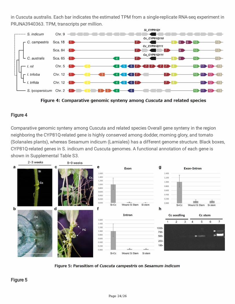
Comparison of gene synteny in CYP81Q-related generegionsWe next compared gene synteny in the regions surrounding the CYP81Q genes in S. indicum, C.campestris, and C. australis (Fig. 4). In the S. indicum genome, Si_CYP81Q1 (SIN_1025734) is located onchromosome 9, between ATPase (SIN_1025733) and an uncharacterized protein (SIN_1025735) next tothe FLOWERING LOCUS C expresser (FLX)-like gene (SIN_1025736). By contrast, Cc_CYP81Q110 andCc_CYP81Q111 were both located in reverse orientation as compared to their neighboring genes, in thevicinity of an E3-class ubiquitin ligase (E3UbL). Speci�cally, Cc_CYP81Q110 is between DUF4228(Cc046290) and a hypothetical protein (Cc046293), and Cc_CYP81Q111 is between DUF4228 (Cc015411:gene number 1, Fig. 4) and another hypothetical protein (Cc015416: gene number 9, Fig. 4,Supplementary Table 1). Moreover, these genomic features were also observed nearby the RAL50776gene in the C. australis genome. The observation that the genomic features in introns and the regionadjacent to Cc_CYP81Q111 were distinct from those of Cc_CYP81Q110 and Ca_CYP81Q111 (Figs. 1 and

Page 9/26
4) suggests that the twin homeologs in C. campestris were brought by hybridization (allopolyploidization)between two Cuscuta plants, both of which had the ability to produce sesamin, rather than occurringthrough WGD of an ancient C. campestris genome (autopolyploidization).
Although multiple E3UbL genes were observed between DUF4228 (LOC109177929: gene number 1, Fig.4) and a hypothetical protein (LOC109177844: gene number 9, Fig. 4) in chromosome 5 of the I. nilgenome, there were no cytochrome P450 genes in these regions of the three Ipomoea genomescorresponding to the Cuscuta PSS genes. This is notable since the arrangement of genes betweenDUF4228 and LOC109177844 in the I. nil genome is structurally analogous to those between Cc046290and Cc015411, and between Cc046293 and Cc015416, in the C. campestris genome.
Collectively, comparative genomics approaches based on gene synteny across species highlight theunique presence of CYP81Q genes in Cuscuta plants, supporting the idea that CYP81Q genes appeared inthe Cuscuta genome by HGT. We cannot exclude the alternative possibility that an ancestor of I. nil had aCYP81Q1 ortholog but lost it during speciation, although aside from Cuscuta, no other Convolvulaceaeplants are known to accumulate sesamin14,37.
Parasitism-mediated HGTThe observance of PSS genes with conserved genomic features and molecular functions in Sesamumand the distantly-related parasitic Cuscuta plants suggests that HGT of PSS occurred through parasitismbetween their ancestors. To help explore the feasibility of this model of HGT, we examined whether C.campestris is able to parasitize S. indicum by co-cultivating C. campestris with S. indicum (cv. Masekin)under laboratory conditions. The results showed that C. campestris successfully formed haustoria on thestem of S. indicum 2–3 weeks after germination of the parasites (Fig. 5a). Cross sections at thehaustorium revealed that the vascular tissues of S. indicum were directly connected with haustorialvascular elements of the parasite (Fig. 5b). At 8–9 weeks after germination, C. campestris formed a�ower and developed an ovary that contained fertile seeds, thereby completing its lifecycle as an obligateparasite (Fig. 5c, d). The ability to parasitize the modern sesame cultivar supports the model thatparasitism by a Cuscuta of a Sesamum, or a phylogenetically related plant with PSS, facilitated HGT.
The presence of intron 2 in all of the Cuscuta PSS genes (Fig. 1c) indicates that the putative HGT eventlikely occurred via transfer of a genomic fragment containing an ancestral CYP81Q gene with the intron,rather than via retroposition of an intron-less mRNA. To test the possibility of transfer of a genomicfragment, we analyzed sequence similarity between the genomic regions �anking CYP81Q genes ofCuscuta plants and S. indicum, and found that they shared extremely low similarity (Supplementary Fig.7). This result does not further support the idea of transfer of a long genomic fragment to Cuscuta and isdifferent from HGT events reported in Orobanchaceae, in which transferred genomic fragments aretypically tens of kbp in length38. Nevertheless, we cannot exclude the possibility that in Cuscuta, a longgenomic fragment was inserted �rst and then the sequence of regulatory regions changed, probably dueto lower genetic constraints in those regions than in coding regions.
Page 10/26
Acquisition of CYP81Q genes in the genome of the ancestor of Cuscuta might also have been facilitatedby RNA-mediated HGT, as has been predicted in a root parasitic plant, Striga hermonthica39. In support ofthis idea, we note that (1) RNA molecules harboring introns (known as intron retention; IR) have beenrecognized to exert biological functions in plants and mammals40,41, and (2) RNA-based HGT likelyoccurs more readily when mRNA abundance increases since mRNA abundance might be a determinantof long-distance mobility42, whereas cellular genomic DNA content is relatively static. To test thehypothesis that RNA-based HGT accounts for the presence of CYP81Q, we surveyed IR-RNA of CYP81Q1using S. indicum RNA-seq data29,43,44 and found this form of the RNA among minor transcripts (< 3% oftotal transcripts) in seeds of cv. Masekin (Supplementary Fig. 8). We also found evidence of the threecomponents of a plant polyadenylation signal, including a near upstream element (NUE; AAUAA) and a 1nt variant of the NUE (AAUACA); a 1 nt variant of the far upstream element (FUE; TTGTAA); and UGUA-containing hexamers following a short poly(A) stretch45, in the 3’ sequences downstream of the stopcodons of Cuscuta CYP81Q genes46 (Supplementary Fig. 9). Moreover, both Cuscuta CYP81Q111transcripts were found to harbor a short poly(A) stretch. These sequences seemed to be structurallyincomplete but might be fading genomic signatures of an ancient RNA-mediated HGT event.
Next, we used qPCR and RNA-seq to look at gene expression of Si_CYP81Q1 in the host stem, whereCuscuta speci�cally forms haustoria. Si_CYP81Q1 was expressed at minute levels in the non-parasitizedhost stem. Surprisingly, Si_CYP81Q1 was signi�cantly induced by parasitization of C. campestris, but notby mechanical wounding (Fig. 5e-g), suggesting that induction of Si_CYP81Q1 expression in S. indicum isnot a response to wounding caused by the penetration of haustoria, but a speci�c host defense responseagainst the parasite. Both spliced and IR-RNA forms of Si_CYP81Q1 transcripts were induced, and theratio of IR-RNA (intron/exon) did not change after parasitism (Fig. 5e-g). We could occasionally detectboth spliced and IR-RNA forms of Si_CYP81Q1 in the stem of C. campestris that had established parasiticconnections to S. indicum (Fig. 5h). Sequences of the shorter and longer ampli�ed fragments werecon�rmed to be the spliced and IR forms of host Si_CYP81Q1 transcripts, respectively, indicatingtranslocation of induced RNAs of Si_CYP81Q1 to parasite stem from the parasitic interface of the hoststem through the haustorium. Long-distance movement of various types of RNA between plants has beenreported in interspeci�c grafts47 and parasite-host plant complexes5, 48, suggesting the involvement ofvascular transport of RNAs. The possibility of transport of RNAs by nonenveloped RNA viruses, whichencapsidate host RNAs, has also been suggested49. Our results support an evolutionary scenario in whichfunctional HGT of PSS into the parasite genome was mediated by parasitism-evoked locally abundantRNAs. This idea is consistent with the fact that only one Cuscuta CYP81Q gene encodes a functionallignan catalytic unit in the conserved genomic synteny among Convolvulaceae plants (Fig. 4).
Presence of both PSS and Sol-B in seedsThe presence of sesamin and its derivative lignans in the seeds of distantly related plants suggests thatthere are common but unknown roles for sesamin and derivative lignans in these seeds. One such rolemight be to prevent lipid peroxidation by exerting their (pro)antioxidative activities33, thereby preservingstorage resources in oil bodies for biogenesis in the next generation. This trait conferred by seed lignans

Page 11/26
seems also to be bene�cial for survival of Cuscuta seedlings, which entirely rely on the nutritive reservesstored in the endosperm until they start sucking nutrition from host plants via haustoria50. Furthermore, asterol-binding dehydrogenase found in oil bodies, known as steroleosin (Sol), is widespread in seedplants including gymnosperms, and is involved in germination and development by regulatingphytohormone sensitivity51,52. A recent report using nano-beads with a�nity to sesamin identi�ed Sol-Bas a sesamin binding protein and indicated that the interaction between Sol-B and sesamin isphysiologically relevant in developing seedlings53. Using S. indicum Sol-B in a BLAST search to queryCuscuta genomes, we found Sol-B-like genes (Cc036318.t1, Cc036490.t2, and C002N0117E0.1) inCuscuta and an partial Sol-B gene ampli�ed by PCR from genomic DNA of C. japonica that show strikingstructural similarity to those of Ipomoea but not those of Sesamum, roughly in line with the evolutionaryplant lineages (Supplementary Fig. 10a). Thus, Sol-B genes seems to have gradually diverged along withplant speciation, again highlighting the unusual similarity among CYP81Q genes in Sesamum andCuscuta (Fig. 1b). We further con�rmed co-expression of Cc_Sol-B and Cc_CYP81Q in C. campestrisseeds such as S. indicum (Supplementary Fig. 10b and c)53, suggesting common roles via interactionbetween sesamin and Sol-B protein in their seeds. Whether HGT-mediated metabolic traits acquired fromhost plants has contributed to the unique physiology and ecology of Cuscuta speci�cally remains anopen question that can be addressed in subsequent studies.
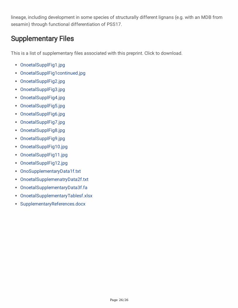
ConclusionRecent reports have described several genomic signatures of nuclear HGT from hosts to parasites but thefunctions of genes transferred to parasitic plants have remained largely elusive7,23,38,39,54,55. In this work,we identi�ed that PSS genes in C. campestris and C. australis are biochemically functional and weprovide a possible explanation based on genomic analysis as to how PSS genes were integrated into thegenomes of Cuscuta spp. Our �ndings support the idea that a common ancestor of the Cuscuta andGrammica subgenera in the Cuscuta genus gained a functional genomic PSS gene from an ancestor of S.indicum through parasitism-mediated HGT, rather than, as generally assumed, by CEV, and therebyacquired (+)-sesamin biosynthetic ability (Fig. 6, Supplementary Fig. 11). Furthermore, we suggested apossible mode of parasitism-mediated HGT from the host plant to the parasitic plant via locally inducedgene expression in the parasitic interface by uncovering genomic signatures of parasite Cuscuta CYP81Qgenes, as well as parasitism-mediated induction and transfer of host Si_CYP81Q1 transcripts into theparasite.
Our �ndings herein underscore that in addition to CEV, parasitism-mediated HGT provides anotherpossible driving force for the sporadic occurrence of specialized metabolites in the plant kingdom, as hasbeen observed for microorganisms. Furthermore, we note that Cuscuta plants are obligate parasites andcan parasitize a wide variety of host plants (Supplementary Fig. 12)6,23,56, and suggest that there mightbe more opportunities for HGT to occur in such parasitic plant species than in non-parasitic plants. Thebiological signi�cance of the presence of (+)-sesamin-derived lignans in Cuscuta and the molecularmechanism of HGT from host to parasite remain unclear. However, evidence of parasitism-mediated HGT
Page 12/26
of PSS in Cuscuta plants provides a new perspective on metabolic evolution in plants beyond typicalphylogenetic constraints and reproductive isolation, and establishes these parasites as new tools forinvestigating the biological activities of specialized metabolites. The unique ecological nature of Cuscutaplants as mediators of genetic and chemical information, given their promiscuous host range and abilityto disperse seeds at long distance, including across oceans1,2, suggest that they might play importantthough as yet to be elucidated roles in plant evolution and adaptation47.
MethodsPlant materials
Cuscuta campestris seeds were germinated and seedlings were parasitized onto host plants as describedpreviously57. Nicotiana tabacum plants were grown and parasitized by C. campestris as describedpreviously51. The C. campestris-N. tabacum parasitic complexes were grown at 25°C under a 16/8 hlight/dark cycle, and �owers and fruits of the C. campestris were harvested. The harvested tissues werefrozen in liquid nitrogen and stored at -80°C. Seeds of Sesamum indicum cv. ‘Masekin’ were germinatedand grown in soil (Sukoyakabaido, Yanmar, Osaka, Japan) mixed with the same volume of vermiculite(GS30L, Nittai Co., Ltd., Aichi, Japan) under natural sunlight illumination from May to August in 2019.Mature stems of 8- to 9-week-old S. indicum were parasitized by C. campestris.
Chemicals
(+)-Sesamin (Chromadex), (+)-sesaminol (Nagara Science), (+)-pinoresinol (Sigma-Aldrich), and (+)-piperitol, synthesized previously16, were prepared as the reference samples.
LC-MS analysis
Lignans in extracts of �owers and seeds of Cuscuta plants (C. campestris and C. japonica) wereanalyzed as follows. A 10 mg sample of lyophilized �owers or seeds was homogenized to a �ne powderusing a TissueLyser II (Qiagen). Next, 1 ml of 70% acetonitrile aqueous solution was added to thehomogenized samples and the samples were extracted using an ultrasonic cleaner at room temperaturefor 2 min. The �ltered extracts were analyzed using an ion-trap time-of-�ight mass spectrometer (LCMS-IT-TOF, Shimadzu) equipped with a photodiode array detector (Shimadzu). Each component wasseparated using a YMC Triart C18 column (TA12S03-1503WT, 150 mm × 3 mm I.D., 3 µm P.S.) withmobile phases A, 0.1% HCO2H-H2O; and B, 0.1% HCO2H-MeOH, in a linear gradient elution with 30-50-90-90-30-30% B (0-10-25-32-32.01-40 min) at a �ow rate of 0.3 ml/min.
PCR
Genomic DNA from C. campestris (Cc), C. japonica (Cj) and S. indicum (Si) was prepared from stemsusing the DNeasy Plant Mini kit (Qiagen) according to the manufacturer’s protocol. Total RNA wasprepared using the RNeasy Plant Mini kit (Qiagen) from the following tissue following lysis using a

Page 13/26
TissueLyser II (Qiagen): Cc seeds, Cc seedlings (at 7 days after germination), Si stem-Cc haustoriumjunction (parasitic interface tissues), Cc stems, Si stems with and without mechanical wounding with ablade, and Si leaves. The RNA samples were treated with DNase Set (Qiagen) to eliminate contaminatinggenomic DNA. After DNase I treatment, cDNA was synthesized using an oligo dT primer and thePrimeScript RT reagent kit (TAKARA BIO). Reactions were performed according to the manufactures’instructions. PCR products were ampli�ed using speci�c primer sets (listed in Supplemental Data 4) asdescribed previously17,58,59,60. Brie�y, genomic PCR, qPCR and RT-PCR was conducted using PrimeSTARMax DNA Polymerase (TAKARA BIO), GoTaq qPCR Master Mix (Promega) and PrimeSTAR GXL DNAPolymerase (TAKARA BIO), respectively.
RNA extraction for RT-PCR and RNA-seq
Total RNA for RT-PCR was prepared from S. indicum (Si) CYP81Q1 in C. campestris (Cc) stems and totalRNA for RNA-seq was prepared from Cc seedlings, Si-Cc haustorium junctions (parasitic interfacetissues), Si-Cc Si leaves, Cc stems growing on the Si stem, Si stems with and without mechanicalwounding with a blade, and Si leaves. Cc seedlings were harvested 7 days after germination, which wasgrown under 16h light/ 8h dark cycle for 5 days, blue-light illumination for 1h, under dark condition for23h, and a 16/8 h light/dark cycle 1 day. Si plants were used 4-6 weeks and 12-16 cm height as theparasitized plants and as the plant samples with and without mechanical wounding. After parasitizationor mechanical wounding, plant samples were under blue-light illumination for 1h, under dark conditionsfor 23h, and a 16/8 h light/dark cycle for 5 days. The samples were harvested 6 days later afterparasitization or mechanical wounding. Parasitic interface tissues were harvested from 1.5 cm lengthinclude the parasitic interface. Si stems with mechanical wounding were harvested from 1.5 cm lengthincluding the cutting point. Si stems without mechanical wounding were harvested from a 1.5 cm lengthof epicotyl. To collect Si leaves, the topmost portions of leaves that had grown to at least 3 cm werechosen. Cc stems were harvested at 10.5 -13.5 cm length from 1.5 cm near the parasitic interface to theCc stem tip to avoid cross-contamination with the host. Harvested tissues were washed twice with 70%EtOH for 2 min and rinsed with nuclease-free water for 2 min to clean the surface of the tissues. TotalRNA was extracted using the RNeasy Plant Mini kit (Qiagen) after lysis with a TissueLyser II (Qiagen) andthen treated using the TURBO DNA-free Kit (Thermo Fisher Scienti�c) according to the manufacturer’sprotocol. Each RNA sample was derived from a single organism.
Genome and transcriptome
We used open source Cuscuta transcriptome and genome data sets. The RNA-seq data for C. campestriswere obtained from the DNA Data Bank of Japan Sequenced Read Archive accession numberDRA009453 (https://trace.ddbj.nig.ac.jp/dra/index_e.html/DRA009453)30. The assembled genomesequence and annotations for C. campestris were obtained from the plaBi database(http://plabipd.de/portal/cuscuta-campestris) and for C. australis from the National Center forBiotechnology Information (NCBI) Sequence Read Archive(https://www.ncbi.nlm.nih.gov/bioproject/PRJNA394036). Cc_CPR1, Cc_CYP81Q110, Cc_CYP81Q111,
Page 14/26
Cc_CYP81AX6, and Ca_CYP81Q111 correspond to Cc043955, Cc046292, Cc015414, Cc047366, andRAL50776 (responsible for C65N0022E0.1), respectively. The DNA-seq data for C. americana wasobtained from SRA experiment ERR3569498 of BioProject PRJEB34450 and for C. californica, from SRAexperiment ERR3569499. Synteny analysis was performed using BLASTP to search for best hit proteinsequences in the databases indicated in Supplementary Table 3. All translation products of the geneslisted in Supplementary Table 3 were used as queries to search all of the databases.
Molecular phylogenetic analysis
The nucleotide sequences of Cuscuta CYP81-related genes used in this study are listed in SupplementaryTable 4 and Supplementary Data 1. The phylogenetic trees shown in Fig. 1, Supplementary Fig. 2, andSupplementary Fig. 9 were constructed using a maximum likelihood (ML) method in the Seaviewsoftware (phyML ln(L)=-23653.0, 1868 sites, GTR 4 rate classes)61. The timetree shown in SupplementaryFig. 5 was inferred by applying the RelTime method31, 62 to a phylogenetic tree whose branch lengthswere calculated using the ML method and the Tamura-Nei substitution model. Evolutionary analyseswere conducted in MEGA X software63.
Genome comparisons
The 10 kb genomic sequence containing the CYP81Q-related gene in the S. indicum genome wascompared to CYP81Q-related genes in Cuscuta genomes using nucmer in the Mummer -v3.1.0 package64.Structural similarities were visualized with dotplots using the DNAnexus Dot browser (available athttps://dnanexus.github.io/dot/) and Unipro UGENE software (http://ugene.unipro.ru/) (SupplementaryFig. 6).
Molecular cloning
Cc_CYP81Q110 (Cc15414), Cc_CYP81Q111 (Cc046292), and Cc_CYP81AX6 (Cc047366) were ampli�edby reverse transcription-polymerase chain reaction (RT-PCR) from cDNA prepared from a mixture of �owerbuds and ovary tissue from C. campestris with the speci�c primers described in Supplementary Table 4.Ca_CYP81Q111 was arti�cially synthetized without codon-optimization (Euro�ns Genomics), based onthe sequence of RAL50776. All of the P450 genes were cloned into yeast expression vectors andheterologously co-expressed in yeast with a C. campestris cytochrome P450 reductase, Cc_CPR1(Cc043955), as described previously29.
Biochemical analysis
Biochemical analysis of Cuscuta Cc_CYP81Q110, Cc_CYP81Q111, Cc_CYP81AX6, and Ca_CYP81Q111was performed basically as described previously29. Brie�y, yeast cells expressing Cuscuta CYP geneswere pre-cultured overnight at 30°C with rotary shaking at 120 rpm in 3 ml synthetic de�ned liquidmedium containing a set of amino acids appropriate for the designated expression vectors. Next, 50 µl ofstationary phase culture was transferred to 1 ml of fresh medium in 24-well plates supplemented with

Page 15/26
100 µM of lignans as substrates. The cultures were further incubated for 24 h at 30°C with rotary shakingat 120 rpm. For extraction, the cells were harvested with the medium and disrupted by sonication. Thehomogenate (50 µl) was mixed with 50 µl acetonitrile and centrifuged at 21,000×g for 10 min. Thesupernatant was collected, �ltered through a Millex-LH syringe �lter (Merck Millipore) and subjected to thefollowing HPLC analysis. Brie�y, the �ltered assay products were separated using a Cortecs UPLC C18+column (part# 186007401, 2.7 µm, 3 mm × 75 mm, Waters) with mobile phases (A; 0.1% tri�uoroaceticacid-H2O, and B; 0.1% tri�uoroacetic acid-acetonitrile) in a linear gradient with 30-80-80-30-30% B (0-1.4-1.8-2.0-2.5 min) at a �ow rate of 1.25 ml/min, and lignans were detected using a photodiode arraydetector at 280 nm.
Preparation and imaging of agarose-embedded sections
Parasitic interface tissues were �xed with 4% (w/v) paraformaldehyde in phosphate buffer solution(Fuji�lm Wako Pure Chemical Corporation, Osaka, Japan), embedded in 8% (w/v) agarose, and cut usinga MicroSlicerTM ZERO 1N (DOSAKA, Kyoto, Japan) into 200-μm sections. Histochemical staining ofsections was performed using a 0.5% (w/v) solution of Toluidine Blue O (1B-481, Waldeck GmbH & Co.,Munster, Germany) in distilled water. Stained slices were observed using a BX53 Biological Microscope(Olympus, Tokyo, Japan).
DeclarationsCon�ict of interest
The authors declare that the research was conducted in the absence of any commercial or �nancialrelationships that could be construed as a potential con�ict of interest.
Acknowledgements:
We thank D. Nelson (Univ. of Tennessee), T. Umezawa (Kyoto Univ.), Y. Tobimatsu (Kyoto Univ.), S.Yoshida (NAIST), K. Shirasu (RIKEN), H. Satake (SUNBOR), K. Shimamoto (SUNBOR), N. Okitsu (SIC), M.Takagi (SST), Y. Kawai (NCGM), M. Yamamoto (Toyama Univ.), Y. Ogata (Osaka Pref. Univ.), K. Nishitani(Kanagawa Univ.), and all the participants in the Society of Post Youth Agronomists (SPYA 2019, atOgoto, Shiga pref. Japan) and the Frontier Research Society of Plant Specialized Metabolism (FPSM2019, at Matsumoto, Nagano pref. Japan) for helpful comments on and support for this work.Computations were performed in part on the NIG supercomputer at ROIS National Institute of Genetics.
Funding
This work was partly supported by Grants-in-Aid for Scienti�c Research (18H03950 and 19H00944, JSPSto K.A.) and a Grant-in-Aid for JSPS Fellows (19J14848, JSPS to KS).
References

Page 16/26
1. García, M. A., Costea, M., Kuzmina, M. & Stefanović, S. Phylogeny, character evolution, andbiogeography of Cuscuta (dodders; Convolvulaceae) inferred from coding plastid and nuclearsequences. Am. J. Bot. 101, 670–690 (2014).
2. Costea, M., García, M., Baute, K. & Stefanović, S. Entangled evolutionary history of Cuscutapentagona clade: A story involving hybridization and Darwin in the Galapagos. Taxon 64, 1225–1242 (2015).
3. Sun, G. et al. Large-scale gene losses underlie the genome evolution of parasitic plant Cuscutaaustralis. Nat. Commun. 9, 2683 (2018).
4. Heide-Jørgensen, H.S. Establishment of the parasite. In Parasitic Flowering Plants. pp. 263–305.Koninklijke Brill NV, Leiden (2008).
5. Kim, G., LeBlanc, M. L., Wafula, E. K., dePamphilis, C. W. & Westwood, J. H. Genomic-scale exchangeof mRNA between a parasitic plant and its hosts. Science 345, 808–811 (2014).
�. Yang, Z. et al. Convergent horizontal gene transfer and cross-talk of mobile nucleic acids in parasiticplants. Nat. Plants 5, 991–1001 (2019).
7. Zhang, D. et al. Root parasitic plant Orobanche aegyptiaca and shoot parasitic plant Cuscutaaustralis obtained Brassicaceae-speci�c strictosidine synthase-like genes by horizontal genetransfer. BMC Plant Biol. 14, 19 (2014).
�. Sun, T., Xu, Y., Zhang, D., Zhuang, H., Wu, J., Sun, G. An acyltransferase gene that putatively functionsin anthocyanin modi�cation was horizontally transferred from Fabaceae into the genus Cuscuta.Plant Divers. 38:149–155 (2016).
9. Donnapee, S., Li, J., Yang, X., Ge, A., Donkor, P. O., Gao, X., Chang, Y. Cuscuta chinensis Lam.: Asystematic review on ethnopharmacology, phytochemistry and pharmacology of an importanttraditional herbal medicine. J. Ethnopharmacol. 157, 292–308 (2014).
10. Ahmad, A., Tandon, S., Xuan, T.D., Nooreen, Z. A Review on Phytoconstituents and Biologicalactivities of Cuscuta species. Biomed Pharmacother. 92, 772–795 (2017).
11. Du, K.Z., Li, Z., Guo, X., Li, Y., Chang, Y.X. Quantitative Analysis of Phenolic Acids and Flavonoids inCuscuta chinensis Lam. by Synchronous Ultrasonic-Assisted Extraction with Response SurfaceMethodology. J. Anal. Methods Chem. 2018, 6796720 (2018).
12. Huang, R., O'Donnell, A.J., Barboline, J.J., Barkman, T.J. Convergent evolution of caffeine in plants byco-option of exapted ancestral enzymes. Proc. Natl. Acad. Sci. U.S.A. 113, 10613–10618 (2016).
13. Mao, L. et al. Genomic evidence for convergent evolution of gene clusters for momilactonebiosynthesis in land plants. Proc. Natl. Acad. Sci. USA 117, 12472–12480 (2020).
14. Umezawa, T. Phylogenetic distribution of lignan producing plants. Wood Res. 90, 27–110 (2003).
15. Ono, E. et al. Formation of two methylenedioxy bridges by a Sesamum CYP81Q protein yielding afurofuran lignan, (+)-sesamin. Proc. Natl. Acad. Sci. USA 103, 10116–10121 (2006).
1�. Noguchi, A. et al. Mode-of-action and evolution of methylenedioxy bridge forming P450s in plantspecialized metabolism. Plant Biotechnol. 31, 493–503 (2014).
Page 17/26
17. Ono, E. et al. Formation of a methylenedioxy bridge in (+)-epipinoresinol by CYP81Q3 corroborateswith diastereomeric specialization in sesame lignans. Plant Cell Physiol. 59, 2278–2287 (2018).
1�. Srivastava, S., Gupta, M. M., Prajapati, V., Tripathi, A. K. & Kumar, S. Sesamin a potent antifeedantprinciple from Piper mullesua. Phytother. Res. 15, 70–72 (2001).
19. Jayasinghe, L. et al. Antifungal constituents of the stem bark of Bridelia retusa. Phytochemistry 62,637–641 (2003).
20. He, X. H. et al. Two new lignan glycosides from the seeds of Cuscuta chinensis. J. Asian Nat. Prod.Res. 12, 934–939 (2010).
21. Rho, T. & Yoon, K. D. Application of off-line two-dimensional high-performance countercurrentchromatography on the chloroform-soluble extract of Cuscuta auralis seeds. J. Sep. Sci. 41, 2169–2177 (2018).
22. Abu-La�, S. et al. Sesamin from Cuscuta palaestina natural plant extracts: Directions for newprospective applications. PLoS One 13, e0195707 (2018).
23. Vogel, A. et al. Footprints of parasitism in the genome of the parasitic �owering plant Cuscutacampestris. Nat. Commun. 9, 2515 (2018).
24. Akashi, T., Aoki, T. & Ayabe, S. CYP81E1, a cytochrome P450 cDNA of licorice (Glycyrrhiza echinataL.), encodes iso�avone 2'-hydroxylase. Biochem. Biophys. Res. Commun. 251, 67–70 (1998).
25. Liu, C.-J., Huhman, D., Sumner, L. W. & Dixon, R. A. Regiospeci�c hydroxylation of iso�avones bycytochrome P450 81E enzymes from Medicago truncatula. Plant J. 36, 471–484 (2003).
2�. Hoshino, A. et al. Genome sequence and analysis of the Japanese morning glory Ipomoea nil. Nat.Commun. 7, 13295 (2016).
27. Nelson, D.R., Schuler, M.A., Paquette, S.M., Werck-Reichhart, D. & Bak, S. Comparative genomics ofrice and Arabidopsis. Analysis of 727 cytochrome P450 genes and pseudogenes from a monocotand a dicot. Plant Physiol. 135, 756–772 (2004).
2�. Imai, M. et al. Uncoupling of the cytochrome P-450cam monooxygenase reaction by a singlemutation, threonine-252 to alanine or valine: possible role of the hydroxy amino acid in oxygenactivation. Proc. Natl. Acad. Sci. USA 86, 7823–7827 (1989).
29. Murata, J. et al. Oxidative rearrangement of (+)-sesamin by CYP92B14 co-generates twin dietarylignans in sesame. Nat. Commun. 8, 2155 (2017).
30. Kaga, Y. et al. Interspeci�c signaling between the parasitic plant and the host plants regulate xylemvessel cell differentiation in haustoria of Cuscuta campestris. Front. Plant Sci. 11, 193 (2020).
31. Tamura, K. et al. Estimating divergence times in large molecular phylogenies. Proc. Natl. Acad. Sci.USA 109, 19333–19338 (2012).
32. Kumar, S., Stecher, G., Suleski, M. & Hedges, S. B. TimeTree: a resource for timelines, timetrees, anddivergence times. Mol. Biol. Evol. 34, 1812–1819 (2017).
33. Wan, Y., Li, H., Fu, G., Chen, X., Chen, F. & Xie, M. The relationship of antioxidant components andantioxidant activity of sesame seed oil. J. Sci. Food Agric. 95, 2571–2578 (2015).
Page 18/26
34. Benson, G. Tandem repeats �nder: a program to analyze DNA sequences. Nucleic Acids Res. 27,573–580 (1999).
35. Habu, Y., Hisatomi, Y. & Iida, Molecular characterization of the mutable �aked allele for �owervariegation in the common morning glory. Plant J. 16, 371–376 (1998).
3�. Hoshino, A., Yoneya, Y. & Kuboyama, T. A Stowaway transposon disrupts the InWDR1 genecontrolling �ower and seed coloration in a medicinal cultivar of the Japanese morning glory. GenesGenet. Syst. 91, 37–40 (2016).
37. Afendi, F. M. et al. KNApSAcK family databases: integrated metabolite-plant species databases formultifaceted plant research. Plant Cell Physiol. 53, e1 (2012).
3�. Kado, T., Innan, H. Horizontal Gene Transfer in Five Parasite Plant Species in Orobanchaceae.Genome Biol. Evol. 10, 3196–3210 (2018).
39. Yoshida, S., Maruyama, S., Nozaki, H. & Shirasu, K. Horizontal gene transfer by the parasitic plantStriga hermonthica. Science 328, 1128 (2010).
40. Ge, Y., Porse, B. T. The functional consequences of intron retention: alternative splicing coupled toNMD as a regulator of gene expression. Bioessays 36, 236–243 (2014).
41. Yang, Y., Li, Y., Sancar, A., & Oztas, O. The circadian clock shapes the Arabidopsis transcriptome byregulating alternative splicing and alternative polyadenylation. J. Biol. Chem. 295, 7608–7619(2020).
42. Calderwood, A., Kopriva, S. & Morris, R. J. Transcript abundance explains mRNA mobility data inArabidopsis thaliana. Plant Cell 28, 610–615 (2016).
43. Wang, L. et al. Genome sequencing of the high oil crop sesame provides insight into oil biosynthesis.Genome Biol. 15, R39 (2014).
44. Wang, L., Yu, J., Li, D. & Zhang, X. Sinbase: an integrated database to study genomics, genetics andcomparative genomics in Sesamum indicum. Plant Cell Physiol. 56, e2 (2015).
45. Shi, Y. & Manley, J.L. The end of the message: multiple protein-RNA interactions de�ne the mRNApolyadenylation site. Genes Dev. 29, 889–897 (2015).
4�. Loke, J.C., Stahlberg, E.A., Strenski, D.G., Haas, B.J., Wood, P.C. & Li, Q.Q. Compilation of mRNApolyadenylation signals in Arabidopsis revealed a new signal element and potential secondarystructures. Plant Physiol. 138,1457–1468 (2005).
47. Xoconostle-Cázares, B., Xiang, Y., Ruiz-Medrano, R., Wang, H.L., Monzer, J., Yoo, B.C., McFarland, K.C.,Franceschi, V.R., & Lucas, W.J. Plant paralog to viral movement protein that potentiates transport ofmRNA into the phloem. Science 283, 94–98 (1999).
4�. Bera, S., Yamaguchi, K., Shigenobu, S., & Aoki, K. Trans-species small RNAs move long distances in aparasitic plant complex. Plant Biotechnol. (Tokyo) 38, 187–196, (2021).
49. Routh, A., Domitrovic, T. & Johnson, J.E. Host RNAs, including transposons, are encapsidated by aeukaryotic single-stranded RNA virus. Proc. Natl. Acad. Sci. U.S.A 109, 1907–1912 (2012).
Page 19/26
50. Olszewski, M., Dilliott, M., García-Ruiz, I., Bendarvandi, B. & Costea, M. Cuscuta seeds: Diversity andevolution, value for systematics/identi�cation and exploration of allometric relationships. PLoS One15, e0234627 (2020).
51. Lin, L.J., Tai, S.S., Peng, C.C., & Tzen, J.T. Steroleosin, a sterol-binding dehydrogenase in seed oilbodies. Plant Physiol. 128, 1200–1211 (2002).
52. Li, F., Asami, T., Wu, X., Tsang, E.W. & Cutler, A.J. A putative hydroxysteroid dehydrogenase involved inregulating plant growth and development. Plant Physiol., 145, 87–97 (2007).
53. Tera, M. et al. Identi�cation of a binding protein for sesamin and characterization of Its roles in plantgrowth. Sci. Rep. 14, 8631 (2019).
54. Yang, Z. et al. Horizontal gene transfer is more frequent with increased heterotrophy and contributesto parasite adaptation. Proc. Natl. Acad. Sci. USA 113, E7010–E7019 (2016).
55. Zhang, C. et al. Horizontal gene transfer has impacted cox1 gene evolution in Cassytha �liformis. J.Mol. Evol. 88, 361–371 (2020).
5�. Hettenhausen, C. et al. Stem parasitic plant Cuscuta australis (dodder) transfers herbivory-inducedsignals among plants. Proc. Natl. Acad. Sci. USA 114, E6703–E6709 (2017).
57. Hozumi, A. et al. Arabinogalactan proteins accumulate in the cell walls of searching hyphae of thestem parasitic plants, Cuscuta campestris and Cuscuta japonica. Plant Cell Physiol. 58, 1868–1877(2017).
5�. Narukawa, H., Yokoyama, R., Kuroha, T. & Nishitani, K. Host-produced ethylene is required for markedcell expansion and endoreduplication in dodder search hyphae. Plant Physiol. 185, 491–502 (2021).
59. Ono, E., et al. Glycoside-speci�c glycosyltransferases catalyze regioselective sequentialglucosylations for a sesame lignan, sesaminol triglucoside. Plant J. 101, 1221–1233 (2020).
�0. Shimizu, K., Hozumi, A. & Aoki, K. Organization of vascular cells in the haustorium of the parasitic�owering plant Cuscuta japonica. Plant Cell Physiol. 59, 720–728 (2018).
�1. Gouy, M., Guindon, S. & Gascuel, O. SeaView version 4: A multiplatform graphical user interface forsequence alignment and phylogenetic tree building. Mol. Biol. Evol. 27, 221–224 (2010).
�2. Tamura, K., Tao, Q. & Kumar, S. Theoretical foundation of the RelTime method for estimatingdivergence times from variable evolutionary rates. Mol. Biol. Evol. 35, 1770–1782 (2018).
�3. Kumar, S., Stecher, G., Li, M., Knyaz, C. & Tamura, K. MEGA X: molecular evolutionary geneticsanalysis across computing platforms. Mol. Biol. Evol. 35, 1547–1549 (2018).
�4. Kurtz, S. et al. Versatile and open software for comparing large genomes. Genome Biol. 5, R12(2004).
TablesTable 1: Structural comparison of CYP81Q-related genes

Page 20/26
Figures
Page 21/26
Figure 1
Phylogeny and genomic structures of CYP81Q-related genes a. Sesamin-producing plants are presentsporadically in the plant phylogeny. Numbers on the tree indicate estimated divergent times according tothe Timetree time scale of life31. n.d. indicates not detected in a BLAST search querying Si_CYP81Q1genes with default settings versus public genomes of Ginkgo biloba (id: 12840), Magnolia ashei (id:72476), and Piper nigrum (id: 529758). MYA, million years ago; CEV, convergent evolution; HGT, horizontalgene transfer. b. A phylogenetic tree was constructed using a maximum likelihood method in Seaviewsoftware (phyML ln(L)=-23653.0, 1868 sites, GTR 4 rate classes)61. Dotted rectangle, a structurallysimilar cytochrome P450 group that includes Sesamum PSS genes and Cuscuta homologs. Ca, Cuscutaaustralis; Cc, Cuscuta campestris; Si, Sesamum indicum; Sr, Sesamum radiatum; Sa, Sesamum alatum;Oe, Olea europaea; Pl, Phryma leptostachya; In, Ipomoea nil; At, Arabidopsis thaliana; Gm, Glycine max.Structurally distinct S. indicum P450 genes in the phenylpropanoid metabolic pathway (Si_CYP92B14,sesamolin/sesaminol synthase and Si_C3H; coumarate 3-hydroxylase) were set as an outgroupbranch29. c. Sesamum PSS, as represented by Si_CYP81Q1, has a single intron in the coding region15that corresponds to the second intron in Cuscuta CYP81Q-related genes. Boxes (Ex), lines (lnt), andnumbers indicate exon, intron, and sequence length in base pairs, respectively. Large and small trianglesin introns of Cuscuta CYP81Q genes indicate DNA transposons and their footprints, respectively.
Page 22/26
Figure 2
Enzymatic activity of Cuscuta PSS proteins (Top) Yeast enzyme activity assay products analyzed byHPLC. Chromatograms indicate the absorption at 280 nm of lignans. Blue, substrates; red, products.Chemicals on the left indicate substrate lignans tested in each bioconversion assay. a, (+)-pinoresinol; b,(-)-pinoresinol; c, (+)-epipinoresinol; d, (-)-epipinoresinol; e, (+)-piperitol; f, (+)-sesamin; g, (-)-sesamin; h, (+)-pluviatilol; i, putative (-)-pluviatilol. (Bottom) Diagram of the chemical reaction from (+)-pinoresinol to (+)-sesamin via (+)-piperitol that is catalyzed by PSS. Red, methylenedioxy bridges (MDB) on aromatic rings.

Page 23/26
Figure 3
Expression of Cuscuta PSS genes a. RNA-seq analyses of CYP81Q-related genes in Cuscuta campestris.Each bar indicates the mean and standard deviation for three replicates. TPM values were estimatedbased on mapping of RNA-seq reads in DRA00945330 onto cucam_0.32.annot.cds.fasta from the plaBidatabase (http://plabipd.de/portal/cuscuta-campestris)23. b. RNA-seq analyses of CYP81Q-related genes
Page 24/26
in Cuscuta australis. Each bar indicates the estimated TPM from a single-replicate RNA-seq experiment inPRJNA3940363. TPM, transcripts per million.
Figure 4
Comparative genomic synteny among Cuscuta and related species Overall gene synteny in the regionneighboring the CYP81Q-related gene is highly conserved among dodder, morning glory, and tomato(Solanales plants), whereas Sesamum indicum (Lamiales) has a different genome structure. Black boxes,CYP81Q-related genes in S. indicum and Cuscuta genomes. A functional annotation of each gene isshown in Supplemental Table S3.
Figure 5
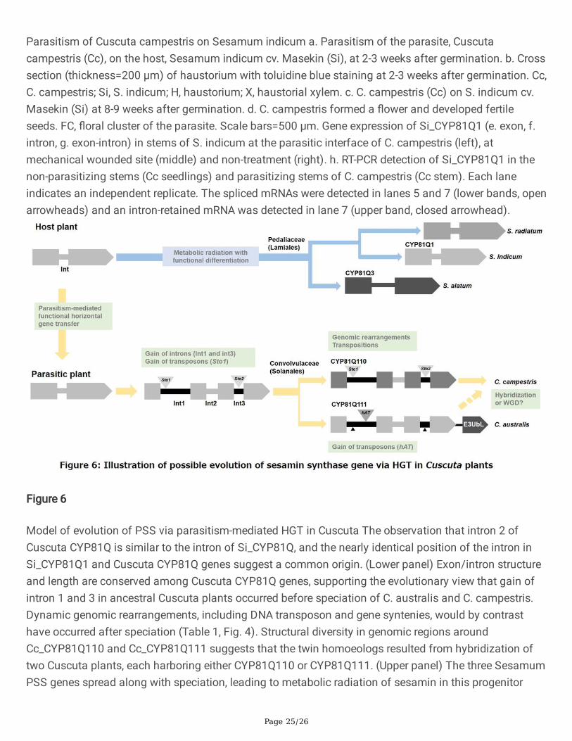
Page 25/26
Parasitism of Cuscuta campestris on Sesamum indicum a. Parasitism of the parasite, Cuscutacampestris (Cc), on the host, Sesamum indicum cv. Masekin (Si), at 2-3 weeks after germination. b. Crosssection (thickness=200 μm) of haustorium with toluidine blue staining at 2-3 weeks after germination. Cc,C. campestris; Si, S. indicum; H, haustorium; X, haustorial xylem. c. C. campestris (Cc) on S. indicum cv.Masekin (Si) at 8-9 weeks after germination. d. C. campestris formed a �ower and developed fertileseeds. FC, �oral cluster of the parasite. Scale bars=500 μm. Gene expression of Si_CYP81Q1 (e. exon, f.intron, g. exon-intron) in stems of S. indicum at the parasitic interface of C. campestris (left), atmechanical wounded site (middle) and non-treatment (right). h. RT-PCR detection of Si_CYP81Q1 in thenon-parasitizing stems (Cc seedlings) and parasitizing stems of C. campestris (Cc stem). Each laneindicates an independent replicate. The spliced mRNAs were detected in lanes 5 and 7 (lower bands, openarrowheads) and an intron-retained mRNA was detected in lane 7 (upper band, closed arrowhead).
Figure 6
Model of evolution of PSS via parasitism-mediated HGT in Cuscuta The observation that intron 2 ofCuscuta CYP81Q is similar to the intron of Si_CYP81Q, and the nearly identical position of the intron inSi_CYP81Q1 and Cuscuta CYP81Q genes suggest a common origin. (Lower panel) Exon/intron structureand length are conserved among Cuscuta CYP81Q genes, supporting the evolutionary view that gain ofintron 1 and 3 in ancestral Cuscuta plants occurred before speciation of C. australis and C. campestris.Dynamic genomic rearrangements, including DNA transposon and gene syntenies, would by contrasthave occurred after speciation (Table 1, Fig. 4). Structural diversity in genomic regions aroundCc_CYP81Q110 and Cc_CYP81Q111 suggests that the twin homoeologs resulted from hybridization oftwo Cuscuta plants, each harboring either CYP81Q110 or CYP81Q111. (Upper panel) The three SesamumPSS genes spread along with speciation, leading to metabolic radiation of sesamin in this progenitor
Page 26/26
lineage, including development in some species of structurally different lignans (e.g. with an MDB fromsesamin) through functional differentiation of PSS17.
Supplementary Files
This is a list of supplementary �les associated with this preprint. Click to download.
OnoetalSupplFig1.jpg
OnoetalSupplFig1continued.jpg
OnoetalSupplFig2.jpg
OnoetalSupplFig3.jpg
OnoetalSupplFig4.jpg
OnoetalSupplFig5.jpg
OnoetalSupplFig6.jpg
OnoetalSupplFig7.jpg
OnoetalSupplFig8.jpg
OnoetalSupplFig9.jpg
OnoetalSupplFig10.jpg
OnoetalSupplFig11.jpg
OnoetalSupplFig12.jpg
OnoSupplementaryData1f.txt
OnoetalSupplemenatryData2f.txt
OnoetalSupplementaryData3f.fa
OnoetalSupplementaryTablesf.xlsx
SupplementaryReferences.docx