Embed Size (px)
Citation preview

453
EVOLUTION OF CILIARY FEEDING IN THEPROSOBRANCHIA, WITH AN ACCOUNT OF
FEEDING IN GAPULUS UNGAR/GUS
By C. M. Yonge, D.Se.University of Bristol
(Text-figs. 1-6)
CONTENTSPAGE
Introduction
Rejection Currents in the Mantle Cavity of the ProsobranchiaEvolution of Ciliary Feeding
Vermetus novae-hollandiae .Crepidula fornicata and other CalyptraeidaeCapulus ungaricusModification of gill filaments
Discussion.
SummaryReferences.
453453455456457459461465
467468
INTRODUCTION
Ciliary feeding, of such widespread occurrence in the Lamellibranchia, isconfined in the Gastropoda to a few scattered groups. In freshwater Pul-monata, such as Limnaea, cilia on the foot assist in feeding when the animalis creeping suspended from the surface film (Brockmeier, 1898). Thecoso-matous Pteropoda feed exclusively by the aid of cilia on the unpaired middlelobe and the paired side lobes of the foot, and an evolutionary series-Cavolinia-Cymbulia-Gleba-can be traced in which there is a progressiveelaboration in the perfection of this mechanism and an accompanying reduc-tion in the buccal mass and associated structures handed down from carni-vorous ancestors (Yonge, 1926). Only in the few prosobranchs which haveacquired ciliary feeding mechanisms do these represent a modification of thectenidia as in the Lamellibranchia. They also, as it is the aim of this paper toshow, represent a modification of the rejection currents present in the mantlecavity of typical prosobranchs.
REJECTION CURRENTS IN THE MANTLE CAVITY OF THE PROSOBRANCHIA
In typical Prosobranchia a respiratory current, created by the beating of thelateral cilia on the gill filaments, is drawn into the mantle cavity by way of theinhalent opening (frequently prolonged into a siphon, e.g. in Buccinum) on
jOURN. MAR. BIOL. ASSOC.vol. XXII, 1938 29

454 C. M. YONGE
the left side of the head and, after passing between the gill filaments whererespiration takes place, passes out by the exhalent opening on the right side.Any sediment present in the water must inevitably be drawn in with thiscurrent and, if the gills and other important organs in the mantle cav~ty are.not to be smothered by it, it is essential that mechanisms should eXist forits rapid and efficient removal. In a recent paper on Aporrhais (Yonge, 1937)
~
H
---""
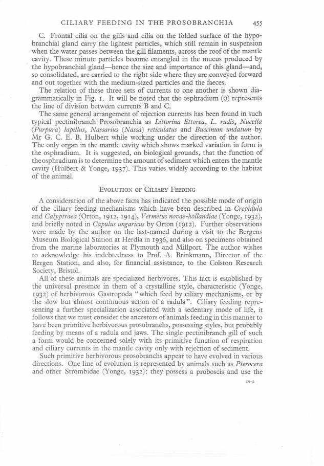
Fig. 1. Semi-diagrammatic representation of course of the ciliary currents within the mantlecavity of a typical pectinibranch prosobranch. A, B, C, three sets of rejection currentsrepresented by unbroken, broken and dotted arrows respecrively; E, exhalent current(downward arrows representing outward current at right angles to plane of figure);I, inhalent current (upward arrows representing inward current as before); G, gillfilament (feathered arrow representing respiratory current created by lateral cilia); H,leaflet of hypobranchial gland; 0, osphradium (with paired lateral plates as in Buccinum);R, rectum.
it was shown that disposal of suspended matter is brought about by thefollowing three well-defined sets of ciliary currents:
A. Cilia on the margin of the inhalent region carry the largest and heaviestparticles, which tend to settle almost at once, directly to the exterior, i.e. byway of the inhalent opening.
B. Cilia on the floor of the mantle cavity carry medium-sized particles,which settle farther within the cavity, across to the right Is ide where theyare caught in forwardly directed currents and conveyed to the exhalentopemng.
CILIARY FEEDING IN THE PROSOBRANCHIA 455
C. Frontal cilia on the gills and cilia on the folded surface of the hypo-branchial gland carry the lightest particles, which still remain in suspensionwhen the water passes between the gill filaments, across the roof of the mantlecavity. These minute particles become entangled in the mucus produced by
. the hypobranchial gland-hence the size and importance of this gland-and,so consolidated, are carried to the right side where they are conveyed forwardand out together with the medium-sized particles and the faeces.
The relation of these three sets of currents to one another is shown dia-
grammatically in Fig. 1. It will be noted that the osphradium (0) represents'the line of division between currents B and C.
The same general arrangement of rejection currents has been found in suchtypical pectinibranch Prosobranchia as Littorina littorea, L. rudis, Nucella(Purpura) lapillus, Nassarius (Nassa) reticulatus and Buccinum undatum byMr G. C. E. B. Hulbert while working under the direction of the author.The only organ in the mantle cavity which shows marked variation in form isthe osphradium. It is suggested, on biological grounds, that the function ofthe osphradium is to determine the amount of sediment which enters the mantlecavity (Hulbert & Yonge, 1937). This varies widely according to the habitatof the animal.
EVOLUTION OF CILIARY FEEDING
A consideration of the above facts has indicated the possible mode of originof the ciliary feeding mechanisms which have been described in Crepidulaand Calyptraea (Orton, 1912, 1914), Vermetus novae-hollandiae (Yonge, 1932),and briefly noted in Capulus ungaricus by Orton (1912). Further observationswere made by the author on the last-named during a visit to the BergensMuseum Biological Station at Herdla in 1936, and also on specimens obtainedfrom the marine laboratories at Plymouth and Millport. The author wishesto acknowledge his indebtedness to Prof. A. Brinkmann, Director of theBergen Station, and also, for financial assistance, to the Colston ResearchSociety, Bristol.
All of these animals are specialized herbivores. This fact is established bythe universal presence in them of a crystalline style, characteristic (Yonge,1932) of herbivorous Gastropoda "which feed by ciliary mechanisms, or bythe slow but almost continuous action of a radula". Ciliary feeding repre-senting a further specialization associated with a sedentary mode of life, itfollows that we must consider the ancestors of animals feeding in this manner tohave been primitive herbivorous prosobranchs, possessing styles, but probablyfeeding by means of a radula and jaws. The single pectinibranch gill of sucha form would be concerned solely with its primitive function of respirationand ciliary currents in the mantle cavity only with rejection of sediment.
Such primitive herbivorous prosobranchs appear to have evolved in variousdirections. One line of evolution is represented by animals such as Pteroceraand other Strombidae (Yonge, 1932): they possess a proboscis and use the
29-2

456 C. M. YONGE
jaws to crop fine algae which are broken down in the stomach by a cellulase.The radula shows little sign of wear" evidently acting as a conveyor belt"(Peile, 1937). Another line of evolution is represented by Aporrhais (Yonge,1937), which burrows in mud and collects detritus of plant origin with theaid of an extensile proboscis possessing grasping jaws and radula. Other linesare represented by groups-not necessarily closely related to one another-which have lost the power of movement, either as a result of cementation tothe substratum as in the Vermetidae, or effectively as in the Calyptraeidae andCapulidae. In these families, as in the Lamellibranchia, the gills and theciliary currents in the mantle cavity have become modified to enable theanimals to collect fine particles, chiefly phytoplankton, from the surroundingwater.
Vermetus novae-hollandiae.
The simplest case would appear to be that of Vermetus novae-hollandiae(Yonge, 1932). This large species lives, cemented to dead coral rock, on theexposed outer faces of Indo-Pacific coral reefs. Unlike the smaller V. gigaswhich lives in still waters in the Mediterranean and, as recently conclusivelydemonstrated by Boettger (1930), collects plankton by means of long
A
MP
FG
G
Fig. 2. Vermetus novae-hollandiae, mantle cavity opened along right side, seen from above.x 1~. A, anus; E, exhalent current; FG, food groove; G, gill; I, inhalent current; M,
mouth; MP, mucus from pedal gland; 0, osphnidium; R, position where large particlescollected prior to rejection by action of current A. Various currents represented by sametype of arrows as in Fig. 1. (Figure modified after Yonge, 1932.)

CILIARY FEEDING IN THE PROSOBRANCHIA 457
mucous threads extruded from the pedal gland, V. novae-hollandiae is aciliary feeder. As shown in Fig. 2, heavy material is accumulated on the leftside of the head and then rejected; this represents current A. Medium-sizedmaterial is carried across the floor of the mantle cavity in current B, where itpasses into a food groove which carries it to the mouth. Finely dividedmaterial-which will include phytoplankton on which the animal chieflyfeeds-is carried by frontal cilia on the gill filaments to the free edge of thegill where other cilia carry it anteriorly. In life this free edge is in closeapposition to the food grove (FG)on the right side of the mantle cavity. Thiscurrent clearly represents a modification of current C. It should further benoted that there is no hypobranchial gland. The increased size of the gill-the result of its added function as a collector of food-leaves no space for thisorgan, which is in any case unnecessary because material is no longer passedcompletely across the roof of the mantle cavity but carried at right angles,anteriorly, along the free edge of the .gill.
Food which enters the food groove from either current B or C is carriedround the right-hand side of the head to the mouth (M). As food streamsapproach, the small introvert is extruded over the opening of the pedal gland,the mouth opens and the exposed radula seizes the food which has been mixedwith mucus from the pedal gland (MP). Any excess of material passes fartherto the left and joins the material rejected by current A. The secretion of thepedal gland, no longer required to provide lubrication for movement, is thusconcerned with the consolidation of food particles but not with its directcapture as in V. gigas.
Crepidula fornicata and other Calyptraeidae.
In Crepidula fornicata (Orton, 1912, 1914) the elaboration of the ciliaryfeeding mechanisms has been carried several stages farther. The mantlecavity is of relatively enormous size, extending over the entire visceral massand also over the elongated" neck" region. This provides for the accommo-dation of the gill, which is many times larger than that of typical prosobranchsof the same body size (it is also relatively larger than that of V. novae-hollandiae,though not to the same extent). This increase in the gill is due to the greater
. current needed for feeding and the increased surface for food collection. Themodificaiions of the individual filaments will be discussed later. The osph-radium (Fig. 3, 0) is reduced to a small area at the anterior end of the gill, butan endostyle (EN) extends along the entire base of the gill.* This structure,described in detail by Orton (1912, 1914), is confined to the Calyptraeidae(Crepidula and Calyptraea) and represents a special adaptation for ciliaryfeeding in these animals, producing mucus which is carried on to the ventral,frontal surface of the gill filaments.
* In V. novae-hollandiae an elongated osphradium (Fig. 2, 0) runs along the base of thegill, not an endostyle as described in my paper (Yonge, 1932). I am glad to have an oppor-tunity of correcting this error. There are always abundant mucous glands in this region.

458 C. M. YONGE
Foodreachesthe mouthby oneof two routes. Large particles are carrieddirect into a food pouch (FP) in the mantle edge, which is situated, in life,just anterior to the mouth. They are there worked up into a pellet with mucusand passed to the mouth. If food is not required or excess of material ispresent, it is carried out by a current which runs parallel to and slightlyanterior to the food pouch. It is clear that this feeding current represents amodification of rejection current A. Medium and fine particles are carried
G
FP
FG
F
Fig. 3. Crepidulafornicata, mantle cavity opened along right side, seen from above. x I~.EN,endostyle; F, foot; FP, food pouch. Other lettering as in Fig. 2. (Figure modifiedafter Orton, 1914.)
over the floor of the mantle cavity in current B, but chiefly, entangled inmucus secreted by the endostyle, by way of the frontal cilia of the gill fila-ments (i.e. current C). By both routes particles pass into the food groove (FG)and are carried forward by the combined action of its cilia and those on the tipsof the gill filaments. The food particles are here worked up into pellets withmucus and are passed from time to time to the mouth (M). There they aregrasped and passed into the buccal cavity by the radula and retained there bythe jaws prior to swallowing. Peile (1937) has remarked on the absence ofwear in the teeth of the radula in both Crepidula fornicata and Calyptraeachinensis. There is no trace of a hypobranchial gland.
Orton found essentially the same conditions in Calyptraea and they almost

CILIARY FEEbING IN THE PROSOBRANCHIA 459
certainly prevail throughout the Calyptraeidae. Kleinsteuber (1913), in acomparison of the genera of this family, has pointed out that in Calyptraea,Crepidulaand Janacus the length of the gill filaments is about one-half, inTrochitasome three-quarters and in Crucibulumtwelve-thirteenths the widthof the body.
Capulus ungaricus.
The only existing account of feeding in this very interesting species beingconfined to a brief statement by Orton (1912), a more detailed account would
T RA
0
F
G
Fig. 4. Capulus ungaricus, animal seen from above after removal of shell; organs and currentsin mantle cavity shown. x 3. RA,radula; T, tentacle. Other lettering as before.
appear to be well justified, apart from its importance in connexion with thegeneral subject-matter of this paper. The animal has very limited powers ofmovement and is often found attached to the free edge of the valves of Lamelli-branchia. The appearance of the animal from the dorsal aspect after the limpet-like shell has been removed is shown in Fig. 4, which also indicates the

460 C. M. YONGE
position, within the mantle cavity, of the gills (G) and osphradium (0). Thehead with the stout tentacles (T) projects a little beyond the mantle edge. Theposition of the mouth and contained radula (RA)is shown but not that ofthe remarkable grooved proboscis. The proboscis appears in Fig. 5 (p), drawnafter it and the foot had been extended anteriorly. It is formed of theprolonged lips and extends downwards, the tip lying on the flat, upper surfaceof the anterior prolongation of the foot. The gill is very large and extendsobliquely across the roof of the mantle cavity, instead of along the left side
x
RA
A
Fig. 5. Capulus ungaricus, mantle cavity opened along right side, seen from above. x 3.GR,groove along proboscis; P, proboscis; x, region of propodiurn, below proboscis, wherefood material collects. Other lettering as before.
as in a typical prosobranch or in the other ciliary feeding species. This wouldappear to be a direct consequence of its large size; it could not be accom-modated in the restricted mantle cavity were it not so disposed. There is aconspicuous osphradium (Figs. 4 and 5, 0) but no hypobranchial gland.
The powerful inhalent current (I), created by the lateral cilia on the gillfilaments, enters the mantle cavity on the left side and impinges almost atright angles on the obliquely situated osphradium. The heavie~t suspended

CILIARY FEEDING IN THE PROSOBRANCHIA 461
material does not reach this organ, but is caught in current A and carriedround the upper surface of the anterior prolongation of the foot to theregion of the tip of the proboscis. The osphradium represents the divisionbetween currents Band C, the former carrying medium-sized particles overthe floor of the mantle cavity and the latter consisting of currents on the gills.These last currents are produced by frontal cilia which carry fine particles tothe tips of the filaments; here particles come under the influence of currentscaused by conspicuously large cilia and are conducted, embedded usually inmucous strings, along the tips of the filaments in an obliquely anterior direc-tion towards the exhalent opening. There are also abfrontal cilia, smaller thanthose on the frontal surface, which carry particles to the tips of the filaments.
Material in currents Band C unites in a common stream near the exhalentopening and passes anteriorly round and beneath the head to join materialfrom current A on the upper surface of the propodium (in the region markedX in Fig. 5). Material here collects in mucus-laden masses-sections revealabundant mucous glands in and below the epidermis in this region-and onthese the animal feeds by means of the proboscis. Food passes up the groovein the proboscis (GP) by means of ciliary, possibly aided by muscular, action;the radula appears to have an essentially conveying function. Peile (1937)states that in "Capulus ungaricus the teeth are slightly blunted in a few of thefront rows".
Orton (1912) states that the proboscis" appears to be held along the rightside of the animal to collect the food-particles from the tips of the gills whenthe animal is feeding". Nothing corresponding to this was observed in anyof the three specimens examined. Orton, however, admits that he had noopportunity of investigating Capulus fully.
Unlike the Calyptraeidae further evolution within the Capulidae has beentowards parasitism. Certain species of Capulus have been described asparasites, but this may be due to a misinterpretation of the true function ofthe proboscis; there is, however, no question as to the ecto-parasitic habits ofall species of the allied genus Thyca. Here the animal is attached throughoutadult life to the body of echinoderms. The foot is correspondingly reduced;the radula is lost, but the proboscis is elongated and penetrates deep into thebody of the host, apparently sucking in the coelomic fluid (see Schepman &Nierstrasz, 19°9; Koehler & Vaney, 1912). The species of the allied butcompletely sedentary family Hipponycidae probably feed in a similar mannerto Capulus.
Modification of gill filaments.
The elaboration of ciliary feeding mechanisms in these Prosobranchia hasinvolved, above all else, modifications of the gill filaments. The nature ofthese modifications can best be discussed by reference to Fig. 6, in whichappear lateral views of complete filaments and also transverse sections nearthe base, from the gills of the three species already discussed. Similar figures

462 C. M. YONGE
of Buccinum undatum, as an example of a pectinibranch prosobranch inwhich the gills are exclusively concerned with their primitive function ofrespiration, are also given. The filaments of Littorina littorea and of Aporrhaispespelecani, which were also examined, do not differ essentially from thoseof Buccinum. Except for Vermetus, the drawings of the complete filamentswere all made from living tissue.
The first point of interest is the progressive increase in the length of thefilaments in comparison to width at the base. In Buccinum (Fig. 6 A), andsimilar Prosobranchia, the filaments are rougWy triangular with the base andheight about equal; the other extreme is represented by Crepidula (Fig. 6 D),where the filament is narrow throughout and greatly elongated. The ratio ofheight to maximum width is in Buccinum (A) 1: 1, in Vermetus (B) 2.75: 1, inCapulus (c) 3: 1 and in Crepidula (D) 26: 1. In Vermetus the figures givenare based on measurements of preserved filaments which, as indicated bythe transverse section, contract considerably in width; they are thus not strictlycomparable with the others and the ratio should certainly be lower and sonearer to conditions in Buccinum.
In the general distribution of the main tracts of cilia, frontals (f), laterals (I)and abfrontals (a)-the last being always somewhat shorter and more sparsethan the frontals-and in the presence of the internal chitinous supportingrods (c), the various filaments do not differ fundamentally. In all the frontalsand abfrontals beat towards the tips of the filaments and the laterals trans-versely. In Buccinum the particles carried to the tip are then carried overfarther to the right (when the gill is in situ) and so on to the hypobranchialgland as shown in. Fig. 1. But in the other three genera particles are carriedforward along the tips from filament to filament at the region marked x(indicated approximately only in Vermetus).
Passing to a more detailed examination of the individual filaments, inBuccinum (A) there are well-developed frontal, lateral and abfrontal cilia.Between the regions occupied by the last two is a wide area which is sparselyciliated (as noted in living material but never revealed in sections). This regionis chiefly notable for the presence of many large mucous glands (m) and theirregular nature of the epithelium, in sharp contrast to its regular arrangementin the other ciliated regions. Dakin (1912), who studied sections cut parallelto the longitudinal axis of the filaments, speaks of the epithelium of the gillleaflet (the frontal region), the area of ciliated cells (the lateral region) andthe area of glandular cells. His sections did not pass through the abfrontalregion. Within the cavity of the filament are many blood corpuscles andmuch coagulated blood plasma, notably in the "glandular" region, which ispresumably concerned with respiration and also with the entangling, inmucus, of any particles which are carried between the filaments in thecurrent created by the lateral cilia. There are no mucous glands in thefron tal or abfrontal regions, although they are abundant at the sides of theabfrontal epithelium. Muscular activity was observed in living tissue and there

CILIARY FEEDING IN THE PROSOBRANCHIA
f
A
x
x/O
c
ff
463
x~\
~n
f ~
c
pa
fl
Fig. 6. Lateral view (above) and transverse section near base (below) of gill filaments of A,Buccinum undatum; E, Vermetus novae-hollandiae; c, Capulus ungaricus; D, CrepidulaJornicata. Sections all x 55; magnifications of whole filaments shown in figure. a, ab-frontal cilia; c, chitinous supporting rods; J, frontal cilia; I, lateral cilia; m, mucous glandcells; x, region of anteriorly directed current at tips of filaments.
aa.
¥I
'''.
B c
i1
IIIc-
x/6H
f
c-1I
t)aD

464 C. M. YONGE
are indications of muscle in the section, notably at the base of the abfrontalepithelium.
In Vermetus (B) the chitinous rods are exceptionally thick and broad andthe area occupied by the lateral cilia is reduced and confined to the abfrontalthird of the region supported by these rods. Mucous glands occur in allregions except in the epithelium which bears the lateral cilia. Their presencein both frontal and abfrontal epithelia indicates that their secretion assists inthe entanglement of food particles in these regions. As already noted thefilament has great powers of contraction, made possible by the presence, inthe" glandular" and abfrontal regions, of longitudinal and transverse fibresand also others running across the central cavity. The wrinkled condition ofthe section indicates the effect of the contraction of the second and third ofthese sets of muscles.
In Capulus (c) conditions, in transverse section, approximate more to thosein Buccinum, but mucous glands are here confined to the frontal region. Itmay be assumed, therefore, that collection of food particles by the frontalregion is so efficient that few pass between the filaments, as they presumablydo in the other two species. Muscle occurs, but not to the same extent as inthe other two, although muscular movements were observed when livingfilaments were examined. An important feature is the great length of theterminal cilia, which exceed IOOfL.
Finally in Crepidula (D), where the filament is no longer triangular, mucousglands are rare, a condition clearly correlated with the presence of theendostyle (Fig. 3, EN), a mucus-producing organ which has been described indetail by Orton (1912, 1914). The lateral cilia here lie nearer to the abfrontalregion and beyond the region of the chitinous rods. There is little indicationof muscle either in the behaviour of the living filaments or in sections. The tipsof the filaments are flattened somewhat laterally, as shown in Fig. 6, but muchmore so dorso-ventrally, as described and figured by Orton (1912, Fig. 3).The filaments of Crepidula have clearly been modified to a much greaterextent than those of Vermetus or Capulus.
The change in shape of the gill filaments from triangular to linear has hadthe important effect of increasing the extent of the region carrying lateralcilia-and so increasing the water current created in the mantle cavity-andalso the extent offrontal and abfrontal regions, thereby augmenting the collect-ing surface. The accompanying reduction in the middle, or " glandular", regionrepresents a reduction in the respiring surface. This is to some extent madegood by the increased length of the gill (though not so in Crepidula) but,owing to the sedentary habits of these ciliary feeding Prosobranchia, the needfor respiration will be distinctly less than that in actively moving genera, suchas Buccinum. There is a tendency also for a relative increase in thickness ofthe chitinous supporting rods, necessary owing to the increased length of thefilaments. The change in the distribution of the mucous glands may also becorrelated in all three genera with feeding on fine particles, culminating in

CILIARY FEEDING IN THE PROSOBRANCHIA 465
Crepidula with the acquisition of an endostyle and the loss of mucous glandson the filaments. The change in the beat of the terminal cilia has already beennoted.
It has thus been possible for the purely respiring gill filament of Bueeinumand similar Prosobranchia to be converted into an organ of food collectionby essentially minor modifications, representing the conversion of ciliatedrejection tracts into food-collecting tracts. The process, incidentally, indicatesthe manner in which the gill of the Filibranchia and Eulamellibranchia pro-bably evolved (starting from a paired aspidobranch gill and not from theunpaired pectinibranch gill here considered). The most important differencesin these groups are the folding back of the filaments, their increased cohesion bymeans of interlamellar and interfilamentary junctions of various kinds, and theappearance of latero-frontal cilia. The latter, which appear to be universallypresent in these two groups, although very small in more primitive Fili-branchia such as Glyeymeris and Area (Atkins, 1936), probably developedowing to the need for a straining mechanism, in the absence of which thenarrow spaces leading to the suprabranchial cavity in these complex gillswould become blocked. There is clearly no such need in the unfolded andunattached gill filaments of these Prosobranchia.
DISCUSSION
The mode of feeding in the three families here discussed, the Vermetidae,Calyptraeidae and Capulidae, is in essentials the same, and undoubtedlyrepresents a modification of ciliary mechanisms originally concerned with therejection of sediment from the mantle cavity. There appears to be noreason for postulating a common origin for these families; the Vermetidaeare admittedly widely removed from the other two, and they, though oftengrouped together (e.g. by Thiele (1925) in the Calyptraeacea), have manysignificant differences, their more superficial resemblances being very possiblythe result of convergence due to similar modes of life and not of commonorigin. Lebour (1937) has recently shown that the larvae of Capulus possessan echinospira shell indicating relationship to the Lamellarioidea.
Modification of the ciliary mechanisms and associated changes are alwayscorrelated with complete or effective loss of the power of movement andwith life in comparatively clear water. Loss of movement has involved thenecessity of drawing food to the animal by means of ciliary currents (and bythe unique method of mucous strings in Vermetus gigas), and this is possibleonly if the animal lives in water reasonably free from sediment which wouldclog the feeding tracts. Thus the Vermetidae live cemented to rocks and theCalyptraeidae and Capulus are frequently attached to the shells of Lamelli-branchia, such as Ostrea or Pecten, or to stones, and so raised above thebottom deposits. Those living on Lamellibranchs will profit by the currentsthey create.

466 C. M. YONGE
The necessity for providing powerful feeding currents and extensivecollecting surfaces has been met by the elongation of the original broadlytriangular gill filaments and by the greater extent of the entire gill. But therehas been no change in the direction of the ciliary currents except for theanteriorly directed current along the tips of the filaments, a current whichcorresponds in function to those present in the food grooves along the freeedges of the demibranchs in the Lamellibranchia. In the Calyptraeidae foodcollection has been further assisted by the appearance of an endostyle. In allgenera current B, essentially unmodified, assists current C in the collection offood-a ciliated tract on the floor of the mantle cavity carrying food finallytowards the mouth-while in all but the Vermetidae current A, by modificationof its direction, also assists in this process.
The invariable absence of a hypobranchial gland, so very well developed intypical Prosobranchia, is correlated with the increased size of the gill, thefilaments of which extend to the right side of the mantle cavity. Moreover,being an essential part of the original cleansing and rejection mechanism, aspostulated in the case of Aporrhais (Yonge, 1937), it is no longer necessary.
The radula and jaws have been retained but are modified in function. Peile(1937) has described the conspicuous absence of wear in the teeth of theradula of all of these animals and concludes, "we may sum up the functionof the radula as grasping, not rasping". Thus, although the gills havebecome modified in the direction of those of the Lamellibranchia and give someindication of the manner in whi(:;h they have evolved, the actual taking in offood at the mouth consists of the grasping, by radula and jaws, of mucus-laden masses which are passed back into the oesophagus by the conveyingaction of the radula. Nothing in the nature of a selective mechanism, repre-sented in the Lamellibranchia by the labial palps, the complex ciliated tractson which rigorously control the passage of material to the mouth, has beenevolved in these Gastropoda.
The evolution of ciliary feeding has taken significantly different directionsin the three families. In the Vermetidae it is associated with a retention, andremarkable enlargement, of the pedal gland, which provides mucus for theconsolidation of food particles in species such as Vermetus novae-hollandiae,while in V. gigas a still greater enlargement of this gland permits directfeeding by its secretion. In the Calyptraeidae the elaboration of ciliary feedingmechanisms has been taken to the highest degree of perfection recorded inthe Prosobranchia: by the enlargement of the mantle cavity and of the gill,modification of the filaments, possession of an endostyle and conversion of allthree rejection tracts into food streams. Finally in the Capulidae an increasein the size of the gill has been made possible by its oblique position in themantle cavity; but the most characteristic feature is the remarkable groovedproboscis, the further evolution of which has resulted in the parasitic habitfound in Thyca. The trends of evolutionary change in all three families maysuitably be indicated diagrammatically as follows:
l{

CILIARY FEEDING IN THE PROSOBRANCHIA 467
HERBIVOROUS STYLE-BEARING PROSOBRANCHIA/
1~./ ~ .
Enlarged gill; filaments Gill larger; filaments Enlarged gill transverselyelongate triangle; no linear; no hypo bran- extended; filamentshypobranchial gland; chial gland; current A elongate triangle; nocurrents Band C also modified for feed- hypobranchial gland;modified for feeding; ing; endostyle; no all currents modifiedfood groove; mucus pedal gland mucus from scatteredfrom pedal glandn co-
I
glands in propodium;solidates food lips prolonged into
/ ~ grooved proboscis./ ~ ~ / ~V. novae-hollandiae ~ Calyptraeidae / Capulus
"" / (? Hipponycidae)Pedal gland larger, ./
mucous strings for food Parasitic habit;. suckingcapture in still water proboscis; no radula
~ ./V.gigas Thyca
SUMMARY
1. Ciliary currents concerned with rejection of sediment from the mantlecavity of pectinibranch Prosobranchia consist of A, currents carrying heavierparticles to the inhalent opening; B, currents carrying medium particlesacross the floor of the mantle cavity; C, currents carrying fine particles overand between the gill filaments for later consolidation by the mucus from thehypobranchial gland. Material in currents Band C is rejected from theexhalent opening.
2. The feeding currents in ciliary feeding Prosobranchia represent modi-fications of these rejection currents.
3. In Vermetus novae-hollandiae currents B and C only are modified,material being passed to the mouth region, where it is mixed with mucusfrom the large pedal gland, by way of a food groove.
4. In Crepidula fornicata and other Calyptraeidae all currents are modifiedfor feeding and there is an endostyle for mucus secretion.
5. In Capulus ungaricus all currents are modified, food collecting on theupper surface of the propodium, where mucus is secreted and mucus-ladenmasses are passed to the mouth by way of the grooved proboscis.
6. In ciliary feeding species the gill is enlarged and the filaments tend tobecome longer and linear. This results in the production of an increased feedingcurrent and collecting surface and the loss of the hypobranchial gland. Ciliaat the tips of the filaments beat anteriorly. The distribution of mucous glandson the filaments can be correlated with the change in their function.
7. The possible mode of evolution of the lamellibranch gill is discussedin the light of these findings.
8. In the ciliary feeding Prosobranchia radula and jaws are retained forthe grasping of the mucus-laden food mas~es; nothing equivalent to thelabial palps of Lamellibranchia is present.

468 C. M. YONGE
9. Evolution of ciliary feeding has probably been independently achievedin the three families considered. In the Vermetidae further development ofthe pedal gland enables direct mucus feeding, as in Vermetus gigas; in theCapulidae evolution culminates in a parasitic habit as in Thyca; only in theCalyptraeidae, where ciliary feeding mechanisms are most highly organized,do all genera and species appear exclusively to be ciliary feeders.
REFERENCES
ATKINS, D., 1936. On the ciliary mechanisms and interrelationships of Lamelli-branchs. Part I. Quart. Journ. Micr. Sci., Vol. LXXIX,pp. 181-308.
BOETTGER,C. R., 193°. Studien zur Physiologie der Nahrungsaufnahme festgewach-sener Schnecken. Die Erniihrung der Wurmschnecke Vermetus. Bioi. Zbl.,Bd. L, pp. 581-97.
BROCKMEIER,H., 1898. Siisswasserschnecken als Planktonfischer. Planer Forschungs-ber., Bd. VI, p. 165.
DAKIN,W. J., 1912. Buccinum. L.M.B.C. Mem., No. xx.HULBERT,G. C. E. B. & YONGE,C. M., 1937. A possible function of the osphradium
in the Gastropoda. Nature, Vol. CXXXIX,p. 84°.KLEINSTEUBER,H., 1913. Die Anatomie von Trochita, Calyptraea und Janacus.
Zooi. Jahrb. Suppl. 13, Fauna chilensis, Bd. IV, pp. 385-476.KOEHLER,R. & VANEY,C., 1912. Nouvelles formes de Gasteropodes ectoparasites.
Bull. Sci. France Belg., T. XLVI,pp. 191-217.LEBOUR,M. V., 1937. The eggs and larvae of the British Prosobranchs with special
reference to those living in the plankton. JtJurn. Mar. BioI. Assoc., Vol. XXII,pp. 105-66.
ORTON,J. H., 1912. The mode offeeding in Crepidula, with an account of the current-producing mechanism in the mantle cavity. Journ. Mar. Bioi. Assoc., Vol. IX,pp. 444-78.
- 1914. On ciliary mechanisms in Brachiopods and some Polychaetes, with a com-parison of the ciliary mechanisms on the gills of molluscs. Journ. Mar. BioI.Assoc., Vol. x, pp. 283-311.
PElLE,A. J., 1937. Some Radula problems. Journ. Conch., Vol. XX,pp. 292-3°4.SCHEPMAN,M. M. & NIERSTRASZ,H. F., 1909. Parasitische Prosobranchier der
Siboga-Expedition. Siboga-Expeditie, Mon. XLIX2.THIELE,J., 1925. Prosobranchia. Handbuch der Zoologie, Kiikenthal und Krumbach,
Bd. v, pp. 40-94.YONGE,C. M., 1926. Ciliary feeding mechanisms in the Thecosomatous Pteropoda.
Journ. Linn. Soc. Lond., Zool., Vol. XXXVI,pp. 417-29.- 1932. Notes on feeding and digestion in Pterocera and Vermetus, with a discus-
sion on the occurrence of the crystalline style in the Gastropoda. Sci. Rpts.G. Barrier Reef Exped., Brit. Mus. (Nat. Hist.), Vol. I, pp. 259-81.
- 1937. The biology of Aporrhais pes-pelecani (L.) and A. serresiana (Mich.).Journ. Mar. BioI. Assoc., Vol. XXI,pp. 687-704.
Note. Since this paper went to press I have had the pleasure of seeing themanuscript of a forthcoming paper by Mr A. Graham on ciliary feeding inTurritellacommunis. Essentially the same mechanisms are involved as in thespecies here considered, but there are most interesting modifications due tothe fact that this animal is adapted for ciliary feeding in a muddy environment.