Embed Size (px)
Citation preview

ACTAUNIVERSITATIS
UPSALIENSISUPPSALA
2015
Digital Comprehensive Summaries of Uppsala Dissertationsfrom the Faculty of Medicine 1150
Evolution of Antibiotic Resistance
FRANZISKA PIETSCH
ISSN 1651-6206ISBN 978-91-554-9380-6urn:nbn:se:uu:diva-265018

Dissertation presented at Uppsala University to be publicly examined in A1:111a, BMC,Husargatan 3, Uppsala, Friday, 11 December 2015 at 09:15 for the degree of Doctor ofPhilosophy (Faculty of Medicine). The examination will be conducted in English. Facultyexaminer: Professor Josep Casadesús (Department of Genetics, University of Seville, Spain).
AbstractPietsch, F. 2015. Evolution of Antibiotic Resistance. Digital Comprehensive Summaries ofUppsala Dissertations from the Faculty of Medicine 1150. 67 pp. Uppsala: Acta UniversitatisUpsaliensis. ISBN 978-91-554-9380-6.
The emergence of antimicrobial resistance is a major global threat to modern medicine. Therapid dissemination of resistant pathogens and the associated loss of efficacy of many importantdrugs needs to be met with the development of new antibiotics and alternative treatment options.A better understanding of the evolution of resistance could help in developing strategies to slowdown the spread of antimicrobial drug resistance.
In this thesis we investigated the evolution of resistance to two important antibiotics,rifampicin and ciprofloxacin, paying special attention to the resistance patterns occurring withhigh frequency in clinical isolates.
Rifampicin is a first-line drug in tuberculosis treatment and resistance to this valuabledrug limits treatment options. Our work on rifampicin resistance helps to explain the extremebias seen in the frequency of specific resistance mutations in resistant clinical isolates of M.tuberculosis. We identified an important interplay between the level of resistance, relative fitnessand selection of fitness-compensatory mutations among the most common resistant isolates.
Fluoroquinlones are widely used to treat infections with Gram-negatives and the frequencyof resistance to these important drugs is increasing. Resistance to fluoroquinolones is the resultof a multi-step evolutionary process. Our studies on the development of resistance to thefluoroquinolone drug ciprofloxacin provide insights into the evolutionary trajectories and revealthe order in which susceptible wild-type E. coli acquire multiple mutations leading to high levelof resistance. We found that the evolution of ciprofloxacin resistance is strongly influenced bythe mutation supply rate and by the relative fitness of competing strains at each successive stepin the evolution. Our data show that different classes of resistance mutations arise in a particular,predictable order during drug selection. We also uncovered strong evidence for the existenceof a novel class of mutations affecting transcription and translation, which contribute to theevolution of resistance to ciprofloxacin.
Keywords: ciprofloxacin, rifampicin, bacterial fitness, compensation, mutation rate
Franziska Pietsch, Department of Medical Biochemistry and Microbiology, Box 582, UppsalaUniversity, SE-75123 Uppsala, Sweden.
© Franziska Pietsch 2015
ISSN 1651-6206ISBN 978-91-554-9380-6urn:nbn:se:uu:diva-265018 (http://urn.kb.se/resolve?urn=urn:nbn:se:uu:diva-265018)

Für meine Eltern
1
I was born in 1984 in Bernau, Germany and graduated from the University of Osnabrück with a Bachelor in Cell Biol-ogy in 2008. In summer 2008 I moved to Uppsala for my Master studies and received a Master’s degree in Microbi-ology 2011. The same year I joined Diarmaid Hughes’ research group and started my PhD studies on antibiotic resistance.
2


Main Supervisor Diarmaid Hughes, Professor
Department of Medical Biochemistry and Microbiology Uppsala University Uppsala, Sweden
Co-Supervisor Dan Andersson, Professor
Department of Medical Biochemistry and Microbiology Uppsala University Uppsala, Sweden
Faculty Opponent Josep Casadesús, Professor Department of Genetics University of Seville Seville, Spain Examining Committee Staffan Svärd, Professor Department of Cell Biology
Uppsala University Uppsala, Sweden
Glenn Björk, Professor Emeritus Department of Molecular Biology Umeå University Umeå, Sweden Göte Swedberg, Senior Lecturer
Department of Medical Biochemistry and Microbiology Uppsala University Uppsala, Sweden


List of Papers
This thesis is based on the following papers, which are referred to in the text by their Roman numerals.
I Brandis, G., Pietsch, F., Alemayehu, R., Hughes, D. (2015) Com-prehensive phenotypic characterization of rifampicin resistance mu-tations in Salmonella provides insight into the evolution of resistance in Mycobacterium tuberculosis. Journal of Antimicrobial Chemo-therapy, 70(3): 680-685
II Pietsch, F.*, Huseby, D.*, Brandis, G.*, Tegehall, A., Garoff, L.,
Hughes, D. Mutation supply and relative fitness shape the genotypes of ciprofloxacin-resistant Escherichia coli. Manuscript
III Pietsch, F., Bergman, J. M., Brandis, G., Marcusson, L. L., Zorzet,
A., Huseby, D., Hughes, D. RNA polymerase mutations contribute to the evolution of ciprofloxacin resistance in Escherichia coli. Manuscript
IV Pietsch, F.*, Garoff, L.*, Huseby, D.*, Lilja, T., Brandis, G., Hughes, D. Experimental evolution identifies a new class of genes selected during the development of ciprofloxacin resistance in Escherichia coli. Manuscript
*These authors contributed equally.
Reprint of paper I was made with permission from the publisher.


Contents
Introduction ................................................................................................... 13The discovery of antimicrobial drugs ....................................................... 13Classification of antibacterial drugs ......................................................... 15The origin of antibiotic resistance ............................................................ 17Development of antibiotic resistance ....................................................... 18Defining resistance ................................................................................... 20Resistance mechanisms ............................................................................ 22Resistance, bacterial fitness, and genetic compensation .......................... 25Mutation rates ........................................................................................... 26Fluoroquinolones ...................................................................................... 28Rifampicin ................................................................................................ 33
Present Investigations .................................................................................... 37Rifampicin resistance in M. tuberculosis (paper I) ................................... 37Evolution of rifampicin resistance in M. tuberculosis (paper I) ............... 38Evolution of ciprofloxacin resistance (paper II – IV) ............................... 39How clinical ciprofloxacin resistance E. coli strains build up their genotypes (paper II) .................................................................................. 41RNA polymerase mutations can contribute to ciprofloxacin resistance (paper III) .................................................................................................. 43Predicting the evolution of ciprofloxacin resistance in E. coli (paper IV) .................................................................................................................. 45
Perspectives on Resistance Development ..................................................... 48
Concluding Remarks ..................................................................................... 50
Svensk Sammanfattning ................................................................................ 52
Deutsche Zusammenfassung ......................................................................... 53
Acknowledgements ....................................................................................... 54
References ..................................................................................................... 57


Abbreviations
CIP CIPR CFU DNA ECDC E. coli EUCAST HGT MDR MIC MSC M. smegmatis M. tuberculosis NT OST QRDR RIF RIFR RNA RNAP RRDR S. enterica S. typhimurium TB WHO WT
Ciprofloxacin Ciprofloxacin resistance Colony Forming Unit Deoxyribonucleic acid European Centre for Disease Prevention and Control Escherichia coli European Committee on Antimicrobial Susceptibility Testing Horizontal Gene Transfer Multidrug Resistance Minimal Inhibitory Concentration Minimal Selective Concentration Mycobacterium smegmatis Mycobacterium tuberculosis Nucelotide Organic Solvent Tolerance Quinolone-Resistance Determining Region Rifampicin Rifampicin resistance Ribonucleic acid RNA polymerase Rifampicin-Resistance Determining Region Salmonella enterica Salmonella enterica serovar Typhimurium Tuberculosis World Health Organization Wild-type


13
Introduction
“Messieurs, c'est les microbes qui auront le dernier mot." (Gentlemen, it is the microbes who will have the last word.)” ― Louis Pasteur
The discovery of antimicrobial drugs Throughout most of human history bacterial infections could only be treated using folk medicines and herbal therapies. Those remedies were often insuf-ficient and for many infectious diseases there was no treatment available, so that bacterial infections frequently led to serious illnesses and caused high mortality rates.
The discoveries by Louis Pasteur and Robert Koch in the late 19th century of the existence of microbes, and the demonstration that they were responsi-ble for several recognized diseases including anthrax and cholera (Madigan et al. 2006) revolutionized the intellectual approach to treating infectious disease by providing an identified cause of disease and target for therapy. In parallel, the ongoing industrial and scientific revolution in Europe had creat-ed a chemical industry with the interest and capability to manufacture pure chemicals in large volumes. At one of these companies Paul Ehrlich, the founder of chemotherapy, initiated a search for a chemical ‘magic bullet’ to treat infectious diseases: a chemical that would selectively kill an infectious microbe but not harm the human patient (Strebhardt & Ullrich 2008). The success of that screen resulted in the discovery and introduction of Sal-varsan, an arsenic compound active against the syphilis spirochete (Strebhardt & Ullrich 2008) and stimulated the search for other small mole-cules with antimicrobial activities. The search led to the discovery of the antibacterial activity of sulphonamides, an important class of synthetic drugs introduced in the 1930s and still in use today (Madigan et al. 2006). A se-cond revolution in antimicrobial infection therapy began with the discovery of penicillin by Alexander Fleming in 1928, showing that microbes them-selves could produce antibacterial substances, so-called antibiotics (Fleming 1929). The development of penicillin for medical use, and its enormously successful application during the Second World War, led to a great interest in searching for other natural antibiotics. Use of the whole cell antibacterial-activity screening platform developed by Waksman (Kresge et al. 2004)

14
directed at a wide variety of fungi and bacteria, led to the “golden age” of antibiotic discovery in the 1950’s when about half of the antibiotics known today were found (Wright 2007). Subsequently, much of the progress in drug development involved generating synthetic or semisynthetic derivatives of natural antibiotics, with better pharmacokinetic and pharmacodynamics properties, and improved spectrum of activity (Fig. 1).
Figure 1. Timeline of antibiotic drug discovery
Today, the classical distinction between antimicrobials as synthetic mole-cules and antibiotics as natural compounds has lost its relevance, since al-most all antimicrobials in clinical use have been structurally modified in the course of development to enhance the antibacterial activity and reduce their toxic side effects.
The importance of antibiotics in modern medicine can hardly be overstat-ed. In addition to their critical role in helping to cure a variety of infectious diseases, effective prophylactic antibiotic therapy underpins most transplant surgery and cancer treatments.
1900 2000 1930 1940 1950 1960 1970
1940-1950 !-lactams (Penicillin)
Aminoglycosides (Streptomycin)
Salvarsan
Sulfonamides (Protonsil)
Rifamycins (Rifampicin)
1950-1960 Phenylpropanoils (Chloramphenicol)
Tetracyclines (Chlortetracycline) Lipopeptides (Polymyxin) Macrolides (Erythromycin)
Glycopeptides (Vancomycin)
Quinolones (Ciprofloxacin)
Oxazolidinones (Linezolid) Cyclic Lipopeptides (Daptomycin)

15
Classification of antibacterial drugs Antimicrobial agents, of natural or synthetic origin, generally work by inhib-iting or disrupting vital processes within the bacterial cell, targeting struc-tures or pathways sufficiently different or absent in mammalian cells.
Antibiotics can be grouped according to several different criteria: inhibi-tory effect, spectrum of activity, and molecular target. Some antimicrobial compounds are bactericidal at clinically used concentrations and thus capa-ble of killing the infecting bacteria, whereas others are bacteriostatic, inhibit-ing the growth or reproduction of the bacterial cells. Some drugs are regard-ed as having a broad spectrum of clinical activity, and are used against a wide range of Gram-negatives and Gram-positives, whereas others have a relatively narrow spectrum of clinical activity. Antibacterial drugs also differ in their bacterial targets and mechanisms of action (Fig. 2). Important targets for clinical antibacterial drugs include cell wall biosynthesis and membrane integrity, folic acid metabolism, protein synthesis, and DNA replication and transcription.
Figure 2. Major antibiotics and their targets
mRNA!
Protein!
DNA!
Replication!(DNA to DNA)!
Transcription!(DNA to RNA)!
Translation!(RNA to protein)!
DNA polymerase!+!
DNA gyrase!
RNA polymerase!
Ribosome!
Fluoroquinolones!
Rifamycins!
Tetracyclines!Aminoglycosides!Macrolides!Chloramphenicol!
30S!
50S! Cell wall!synthesis!
"-lactams!
Folic acid synthesis!
Sulfonamides!Trimethoprim!

16
The by far most important class of drugs in terms of clinical importance are the "-lactams. The majority of drugs used worldwide in infection control therapy belong to this biggest group of antimicrobials, including penicillins, cephalosporins, carbapenems and monobactams (Fig. 3). They target bacte-rial cell wall biosynthesis by inhibiting the final cross-linking of peptides required for building peptidoglycan chains. Other groups of antimicrobial drugs that are widely and frequently used are the macrolides, and tetracy-clines, each targeting different steps in protein synthesis on the ribosome, and the quinolones, targeting enzymes essential for DNA replication. The sulfonamides, originally launched in the 1930’s are still in use today and often administered in combination with trimethoprim. Both drugs target the folic acid metabolism pathway, needed for the synthesis of nucleic acids.
Figure 3. Distribution of worldwide antibiotic consumption by class in 2010 (Van Boeckel et al. 2014)
In addition there are many antimicrobial drugs that have limited use, often due to toxicity issues, difficulty in usage, spectrum of activity, or because they are purposely retained for particular uses. One of these drugs is rifam-picin, belonging to the rifamycin group, which interferes with the bacterial DNA-dependent RNA polymerase and inhibits RNA synthesis.
Penicillins!37%!
Cephalosporins!24%!
Macrolides!12%!
Fluoroquinolones!11%!
Trimethroprim!7%!
Tetracyclines!6%!
Others!3%!

17
The origin of antibiotic resistance Antibiotics by definition are of natural origin and a wide range of fungi and bacteria are natural producers of these bioactive molecules, which they re-lease into their surroundings. The origin of antibiotics is ancient and antibi-otic biosynthetic genes and resistance-conferring genes began to evolve mil-lions of years ago (Wright & Poinar 2012). Thus, antibiotics have been pre-sent in the environment long before humans started using them in clinical settings and many microorganisms have naturally been exposed to these bioactives over evolutionary timescales. It has been proposed that antibiotics have evolved to be global regulators within microbial communities, contrib-uting to quorum sensing and microbial communication in the natural milieu (Aminov 2009; Davies 2006). At the very low concentrations in which anti-biotics are thought to be naturally present, they can act as signaling mole-cules and as such trigger transcription responses important for environmental survival (Yim et al. 2006; Goh et al. 2002). A competitive role is only achieved once the antibiotic concentration is high enough to inhibit the growth of surrounding microorganisms (Sengupta et al. 2013). Transient high antibiotic concentrations in nature made it necessary for antibiotic pro-ducing organisms to harbor resistance genes needed for self-protection and stimulated the evolution of resistant genes in neighboring bacteria.
Given the billions of years of co-evolution of antibiotic producers and an-tibiotic resistant organisms it is not surprising that many bacteria are intrinsi-cally resistant to certain antibiotics, due to impermeability of their cell wall or lack of target. Gram-negative bacteria for examples are insensitive to many clinically effective Gram-positive antibiotics, because many molecules cannot penetrate their double membrane cell wall structure. Additionally, both Gram-negatives and Gram-positives express membrane-spanning efflux pumps, which can actively lower the intracellular antibiotic concentrations to sub-inhibitory levels (Cox & Wright 2013). Furthermore, recent findings have shown that a number of additional, less obvious genes can also contrib-ute to relative intrinsic resistance level and complement the ‘intrinsic resis-tome’ (Cox et al. 2014). Accordingly, many bacteria have evolved the ability to resist transient exposure to at least some antibiotics in their natural habi-tats. Hence, the concept of resistance development towards antibiotics is not new, but went largely unnoticed until human usage of highly concentrated doses of antibiotics in medicine began to select for bacteria with high-level resistance phenotypes. Indeed, once antibiotics were used to treat bacterial infections it did not take long for the first highly resistant strains to develop. Strains resistant to the first generation of antibiotics, including penicillin G and streptomycin, were isolated before or shortly after the drugs were intro-duced to the market (Madigan et al. 2006; Wright 2007).

18
Development of antibiotic resistance Antibiotics are produced by environmental bacteria and antibiotic resistance occurred long before humans started using the first antimicrobial substances to treat infectious diseases. During this “pre-antibiotic era”, antibiotic pro-ducing and antibiotic resistant bacteria coexisted in their natural habitats without facilitating the process of selecting for deadly resistant human path-ogens, so that those remained susceptible to most antibiotics found in nature (Sengupta et al. 2013). Only once humans started to use high concentrations of antibiotics in clinical medicine more than 70 years ago, did the selection for highly resistant strains begin and resistance emerged in human patho-gens. The selective pressure present in clinical settings selected for bacteria, which had either acquired resistance elements from the environmental mi-crobiota via horizontal gene transfer, or evolved their resistant phenotype by de novo mutations, transforming them into drug resistant pathogens (Mar-tinez 2009).
Because all major human pathogens were initial susceptible to antibiotic treatment, the effectiveness of antimicrobial therapy in the 1930s was re-markably successful and led to a significant decrease in human morbidity and mortality (Martinez 2009). Even though the emergence of antimicrobial resistance was recognized soon after the discovery of penicillin and has fol-lowed the introduction of every new drug, antibiotic resistance was for a long time not regarded as a serious problem, in large part because the availa-bility of many different classes of antibiotics meant that it was usually possi-ble to find an effective therapy. In addition, resistance per se did not rule out therapy as long as newer drug derivatives with higher potency were being developed. Thus, during the 1940’s – 1960’s, when the discovery and devel-opment of antibiotics kept pace with the emergence of resistant strains, the increasing occurrence of drug resistant pathogens did not raise major con-cerns. On the contrary, the initial efficacy of antibiotic chemotherapy misled many to believe that infectious diseases would soon become a problem of the past and be eradicated before too long (Aminov 2009). This belief caused the US Surgeon General William H. Stewart to make his famous declaration: “it is time to close the book on infectious diseases and declare the war against pestilence won” (Sengupta et al. 2013). The perception that the prob-lem with bacterial infections was solved also encouraged pharmaceutical companies to focus increasingly on the development of other types of drugs. In addition, because antibiotics typically require short dose therapy, and are relatively cheap, the pharmaceutical companies lost interest in developing new antibiotics in favour of drugs with a higher profit margin (Bush et al. 2011). This situation led to an “innovation gap” (Fig. 1), a period of almost 40 years between the mid 1960s and 2000 when no new class of antibiotics was released onto the market (Fischbach & Walsh 2009). The initial eupho-ria about the new wonder drugs ceased, as the cases of infections caused by

19
drug- and multidrug-resistant pathogens increased and the available antimi-crobials started losing efficacy. Adding to the lack of effective drugs was the fact that the ‘low-hanging fruits had all been picked’ during the early years of drug discovery and the development of new antibiotics became a lot more difficult, time consuming and costly than it was in the early years of antibi-otic development.
Today, resistance has been detected in every pathogen group and against all classes of antibiotics. Thus, the development of resistance to antibiotics appears to be almost inevitable and the frequency of multidrug-resistant pathogens is still increasing in many countries, including Europe and the USA. In other parts of the world, where controls on antibiotic overuse are poor and misuse is great, the situation with regard to resistance is even worse. Particularly the middle-income countries have a very high rate of antibiotic consumption per person and the rapid increase in the frequency of resistant pathogens during the past few decades has been driven by the ex-tensive use and misuse of - often inappropriate - antimicrobial drugs (Van Boeckel et al. 2014). In many parts of the world, antimicrobial agents are available over the counter and even in countries with firmer controls, antibi-otics are not restricted to the treatment of bacterial infections, but also used for farming purposes, so that the release of antibiotics that are not used to prevent or treat human infections is still very high (Martinez 2009; Cohen 2000).
Surveys in Europe, where high quality data is available, point to a strong correlation between the amount of antibiotics used in medicine and the fre-quency of resistance in particular countries (Fig. 4) (ECDC 2012; ECDC 2013). The European Centre for Disease Prevention and Control (ECDC) currently lists antimicrobial resistance as one of the most important health care challenges in Europe, since more and more cases of infections caused by resistant and multi-resistant strains are being recorded and the remaining options for effective antibiotic therapy for those infections are very limited or non-existent (Cars et al. 2011; Freire-Moran et al. 2011; EMEA 2009). ECDC estimates that in recent years at least 25,000 patients in Europe died annually because the treatment with the prescribed antibiotics failed. In addi-tion to the increase in morbidity and mortality, this therapeutic failure is also placing an economic burden estimated at 1.5 billion Euro/year on European health care systems (ECDC 2013).
Studies on the reversibility of resistance back to susceptibility in envi-ronments with reduced antibiotic concentrations give little hope for this re-version to happen in an adequate time span (Sundqvist et al. 2010; Anders-son & Hughes 2010).

20
Figure 4. Percentage of invasive E. coli isolates with resistance to fluoroquinolones, by country, EU/ EEA countries, 2012 (ECDC 2013)
In conclusion, antibiotic resistance has emerged as one of the most severe contemporary health care problems in community and hospital settings and poses a serious threat to our ability to treat bacterial infections. If this trend continues, we might soon approach a post-antibiotic era, in which bacterial infections that could be cured easily for more than sixty years are once again untreatable.
Defining resistance Antibiotics and antimicrobials were first identified and isolated on the basis of assays that showed visible growth inhibition of particular bacterial strains. For example, the Waksman screening platform was based on the detection of a zone of bacterial growth inhibition around a paper disk containing a poten-tially active cell extract or drug molecule (Strebhardt & Ullrich 2008; Kresge et al. 2004). Both for drug discovery and development, and for optimizing drug therapy, is important to have standard and agreed assays and definitions for the susceptibility or resistance to antimicrobial drugs. This requirement led to the introduction of the concept of the minimal inhibitory concentration (MIC) value (Strebhardt & Ullrich 2008; Kahlmeter 2003). MIC is defined

21
as the minimal concentration of drug that prevents visible bacterial growth under strictly defined in vitro conditions and it is measured using increasing concentration steps, with either broth dilution or epsilometer (Etest) assays (Madigan et al. 2006; Kahlmeter 2013). MIC gives a measure of growth inhibition under particular in vitro conditions, and its clinical usefulness requires that the values can somehow be translated into a prediction of clini-cal outcome. To address this clinical need, the distribution of MIC values in natural populations was determined by epidemiological studies of large numbers of bacterial isolates and modeling studies were performed to predict the drug exposure of pathogens during therapy (Fig. 5).
Figure 5. The breakpoint concept of ciprofloxacin in E. coli according to the EU-CAST MIC distribution (Kresge et al. 2004; Kahlmeter 2014). The figure is based on 16702 observations from different data sources. Epidemiological cut-off for wild-type E. coli ≤ 0.064 mg/L; clinical breakpoints S ≤ 0.5 mg/L, R > 1 mg/L.
To evaluate the therapeutic outcome, a variety of additional factors, includ-ing the pharmacokinetic properties of the antibiotic, drug toxicity, the clini-cal disease, and the patient’s general medical status also have to be taken

22
into account (Murray et al. 2005). In addition to these mathematical models, a lot of emphasis is also put on empirical data on failure and success of anti-biotic treatment gathered in the clinics during the past six decades in order to set appropriate length and dose parameters on drug therapy. The epidemio-logical studies have led to agreed definitions of an epidemiological break-point (defining the minimum drug concentration that inhibits growth of a susceptible wild-type of a particular species) and a clinical breakpoint (de-fining the minimum drug concentration that predicts a successful therapeutic outcome when used to treat an infection by a susceptible strain) (Wright 2007; Turnidge & Paterson 2007; Kahlmeter 2003). Thus, resistance devel-opment refers to the consequences of any genetic changes that increase the MIC of a strain to a level higher than the epidemiological breakpoint. Clini-cal resistance in contrast refers to the consequences of genetic changes that increase the MIC of a strain to a level higher than can be successfully treated with standard drug therapy. In clinical terminology bacterial strains are clas-sified according to the SIR system (Madigan et al. 2006; Turnidge & Pater-son 2007): S = susceptible (MIC lower than the clinical breakpoint, standard therapy likely to be successful); I = intermediate; R = resistant (MIC higher than the resistance breakpoint, standard therapy likely to be unsuccessful).
Resistance mechanisms In addition to intrinsic antibiotic resistance, there is also acquired resistance, which bacteria can obtain through two main mechanisms: spontaneous de novo mutations in their chromosome, or uptake of external genetic material via horizontal gene transfer (HGT). While chromosomal mutations can arise without any influences from the environment, the transfer of external genetic material, by the mechanisms of transformation, transduction or conjugation is dependent on direct contact between the human pathogen and DNA har-boring microorganisms which present the source of the resistance genes. As discussed above, it seems conceivable that the source of resistance genes transferred to human pathogens via HGT lies in the non-pathogenic microbi-osphere (Martinez 2009).
Resistance development refers to genetic alterations that increase the MIC of a previously susceptible bacterial strain. The mechanisms responsible for increased drug resistance can broadly be divided in three classes: (i) those that alter the drug target, leading to reduced target susceptibility, (ii) those that modify the drug, leading to a decrease drug-target affinity; (iii) and those that reduce the effective drug concentration that can reach the target, either via increased drug efflux or decreased drug influx (Fig. 6).

23
Figure 6. Basic mechanisms of resistance
All three mechanisms lead to reduced growth inhibition and could be due, either to the occurrence of de novo mutations, or to the acquisition of novel genetic material by HGT.
Drug targets can become less susceptible to drug inhibition by the occur-rence of mutations that reduce drug affinity. For example, mutations in the genes coding for DNA gyrase or DNA topoisomerase IV increase resistance to quinolones, single mutations in rpoB coding for the beta-subunit of RNA polymerase generate high-level resistance to rifampicin, and single muta-tions in ribosomal protein S12 generate high-level resistance to streptomycin (Lindgren et al. 2005; Brandis et al. 2012; Meier et al. 1994; Springer et al. 2001). Drug targets can also become less susceptible by homologous recom-bination of DNA sequences from related organisms. For example, Strep-toccus pneumoniae and Niesseria meningiditis become resistant to penicillin by acquisition of DNA from related species. The recombination creates mo-saic genes for penicillin binding proteins that retain functionality but are resistant to the antibiotic (Dowson et al. 1989; Hakenbeck & Coyette 1998; Laible et al. 1991). Resistance is also associated with mutations altering expression of the drug target, and target gene amplification can also cause phenotypic resistance leading on to the acquisition of other resistance muta-tions (Sandegren & Andersson 2009). Finally, there are examples of bypass
(iii) Increased efflux ! Decreased influx! !"
(i) Target modification!(ii) Drug modification!

24
mechanisms in which the drug target is bypassed or replaced by an alterna-tive drug-resistant target. An example is the mechanism of resistance to van-comycin in which a set of genes inherited on a transposon act coordinately to destroy the normal drug target, a peptide ending in D-Ala-D-Ala, and replace it with a vancomycin-resistant alternative, a peptide ending in D-Ala-D-Lac (Perichon & Courvalin 2009). There are many examples of horizontally acquired genes that code for en-zymes that modify or cleave various antibiotics resulting in a loss of antimi-crobial activity. Examples include many β-lactamases and carbapenemases that can cleave the β-lactam structure and are the major cause of resistance to β-lactam antibiotics; macrolide-modifying enzymes, aminoglycoside-modifying enzymes, and the ciprofloxacin-modifying variant of an amino-glycoside-modifying enzyme (Strahilevitz et al. 2009; Guan et al. 2013; Hawkey & Jones 2009).
There are also several examples where HGT has led to the acquisition of genes coding for proteins that protect a drug target. Examples include the Qnr proteins that apparently interact with DNA gyrase and reduce its suscep-tibility to fluoroquinolones, FusB that reduces the susceptibility of transla-tion elongation factor EF-G to fusidic acid, and the TetM family of proteins that interact with the ribosome and reduce susceptibility to tetracycline (Martínez-Martínez et al. 1998; Tran et al. 2005; O'Neill & Chopra 2006; Burdett 1980; Roberts et al. 1986). Finally, resistance can also be caused by genetic alterations that reduce the concentration of drug available to reach the target. Mutations that up-regulate the expression of drug efflux pumps (often by inactivating re-pressors) are a common contributor to quinolone resistance (Marcusson et al. 2009) and to multidrug resistance in Pseudomonas (Poole 2004). The con-verse of this, mutations affecting porins or otherwise affecting the make-up of the cell wall or membrane, can reduce drug entry into the cell (Fernandez & Hancock 2012). Each of the mechanisms described above, whether involving mutation and/or HGT, can make a significant contribution to the genetic development of resistance to antibiotics. Depending on the particular antibiotic and bacterial species, different mechanisms can be regarded as more or less important. In some cases, for example the development of resistance to fluoroquinolones in Gram-negatives, there are several different mechanisms that together con-tribute to the development of the clinically relevant resistance phenotype. In most cases, the genetic alterations leading to a reduction in drug susceptibil-ity also cause a reduction in the relative fitness of the mutant bacteria in a drug-free environment. This reduction in fitness, and its consequences are discussed below.

25
Resistance, bacterial fitness, and genetic compensation Antibiotics target important and essential functions in the bacterial cell and acquired resistance mutations frequently involve alterations in the drug tar-get proteins, causing a biological fitness cost. Resistance conferred by com-mon efflux-regulating mutations is likewise associated with a decrease in fitness of the resistant cell and also acquired novel genes might cause some disruption in bacterial physiology. This results in decreased fitness, which may be expressed as reduced growth rate or rate of reproduction, or for pathogenic organisms, as reduced virulence in form of transmission rate and rate of clearance from the host (Andersson & Levin 1999; Maisnier-Patin & Andersson 2004). Relative biological fitness can be measured as the maxi-mum growth rate during exponential phase and as such be compared be-tween isogenic susceptible and resistant strains. These data give valuable information about the growth potential of the mutant strains. Clonally or isogenically related susceptible and resistant strains can also be compared in competition experiments, which take additional parameters such as relative ability to use different carbon sources, length of lag phase, and survival dur-ing stationary phase into consideration. Using technology such as flow cy-tometry and fluorescently labeled bacteria, large cell populations can be analyzed, reducing experimental error and allowing the detection of very small fitness differences. However, it should be noted that fitness and fitness cost can vary drastically depended on the growth conditions and environ-mental factors (Paulander et al. 2009).
There are many examples of fitness cost associated with resistance muta-tions and relative fitness has been measured for a variety of species and for resistance by different mechanisms for different classes of antibiotics (Maisnier-Patin & Andersson 2004). For example, mutations in rpoB, con-ferring resistance to rifampincin, are frequently associated with a high fitness cost (Brandis & Hughes 2013). This is also true for mutations in elongation factor G, which give rise to fusidic acid resistant and rpsL mutations, which cause resistance to streptomycin (Nagaev et al. 2001; Paulander et al. 2009).
Low-fitness antibiotic-resistant variants should be at a competitive disad-vantage in the absence of antibiotic selection, raising the possibility that restricting the use of antibiotics might limit the frequency of resistant strains. However, epidemiological studies of clinical resistant strains, and laboratory evolution experiments have shown that this expectation can easily be con-founded (Maisnier-Patin & Andersson 2004; Andersson & Hughes 2010). Thus, some resistance mutations apparently confer no cost, or a very low cost and these have been found to be enriched among clinical isolates. For examples resistance mutations in DNA gyrase, the primary drug target of fluoroquinolones are often very low-cost or even cost free, and there are also examples of rifampicin resistant rpoB mutations, that do not impose a fitness cost on their carrier (Marcusson et al. 2009; Brandis et al. 2015).

26
Secondly, bacteria can acquire fitness-compensatory mutations that reduce the relative fitness costs of resistance without significantly reducing the level of resistance, resulting in the stabilization of resistant organisms in the popu-lation (Björkman et al. 2000; Nagaev et al. 2001; Marcusson et al. 2009). Compensation can occur via several different mechanisms. The majority of compensatory mutations ameliorate fitness by restoring the affected re-sistance protein by an intra –or intergenic mutation. Alternatively, the de-mand for the affected function of the resistance protein can be reduced, for example by a bypass mechanism, and finally, a defect enzyme can be com-pensated for by increasing the amount of enzyme (Maisnier-Patin & Anders-son 2004; Andersson & Hughes 2010). The list of resistance mutations fre-quently co-selected with second site mutations, which compensate the fitness cost of the initial resistance conferring mutation is long. Mutations in fusA, giving rise to fusidic acid resistant bacteria are often found in combination with additional compensatory mutations. The same holds true for streptomy-cin, where resistance mutations in rpsL are regularly associated with the emergence of compensatory mutations in other genes (Björkman et al. 1998; Björkman et al. 2000).
Low-cost and compensatory mutations function to stabilize the mainte-nance of resistant bacterial populations, even in the absence of drug selective pressure. Data from two studies on fusidic acid resistant fusA mutants and streptomycin resistant rpsL mutants mentioned above suggest that the target size for compensatory mutations is typically more than 20 times the size of that for reversion (Andersson & Hughes 2010; Björkman et al. 1998). Thus, it is unlikely that resistant bacteria will be outcompeted by their susceptible parental strains or revert back to their drug-susceptible ancestors and be lost from the population in a feasible time span, even in the absence of antibiot-ics, with reversion rates being very low and fitness compensatory mutations often readily available (Maisnier-Patin & Andersson 2004).
Mutation rates The selection of beneficial mutations facilitates adaptation to changing envi-ronments and is a major driving force of evolution. It is generally believed that spontaneous mutations arise stochastically at very low frequencies and they may be deleterious, neutral, or beneficial. Mutation rate is defined as the probability of a cell to obtain a mutation during its lifetime. These genet-ic alterations can include point mutations, deletions, insertions, or amplifica-tions and horizontal gene transfer events (Hershberg 2015). However, it is common to consider only chromosomal substitutions when mutational rates

27
are measured, which give an estimate of the base-substitution rate and is usually expressed as per base pair per generation. The rates of spontaneous mutations per base pair can differ greatly between species and also between regions in the chromosome within a single species. Intergenic or non-coding region are more variable and prone to accumulate mutations than DNA re-gions coding for highly transcribed genes. Random chromosomal mutations will most often be deleterious or neutral in terms of their impact on bacterial fitness, but some mutations might confer an advantage to their carrier, for example when growth conditions have changed, and become fixed in the bacterial population. Hence, the probability that a mutation becomes fixed in a genome depends on the balance between the fitness cost of a de novo mu-tation and the potential benefit conferred by that mutation in a particular environment. The mutation rate per genome is remarkably constant between different species, and this might reflect an evolutionary equilibrium in the average cost-benefit consequences of a random mutation (Drake 1991; Drake et al. 1998). Evolution involves the creation of genetic and phenotypic variation by mutation and the ability to quantify spontaneous mutation rates is crucial for a mechanistic understanding of evolution (Wielgoss et al. 2011). In the 1940s, Luria and Delbrück developed a ground-breaking meth-od to measure mutation rates in microorganisms (Luria & Delbrück 1943). In their so-called fluctuation experiments, they determined the distribution of the number of mutants from parallel bacterial cultures and from this data could calculate the mutation rate per replication cycle. More precisely, they measured the mutation rates of E. coli from a phage sensitive phenotype to a phage resistant phenotype. Bacterial cultures, which were exposed to the bacteriophage first became clear due to cell lysis, but would often turn turbid again after further incubation, because some phage-resistant cells would survive and be able to grow. Resistant mutations occurring early in the ex-periment, before exposure to the phage, would be present in much higher frequencies than resistant variants arising during viral exposure. Thus the frequencies of the resistant variants would differ greatly between independ-ent cultures by the end of the experiment, depending on how early a particu-lar mutant arose in the bacterial culture and how many generations it had to propagate. Due to this high variation in the number of mutants in each cul-ture, Luria and Delbrück argued that the distribution of mutant colonies did not follow a Poisson distribution and that resistant mutants were present in the culture before bacteriophage exposure. With these experiments Luria and Delbrück could show that beneficial mutations can arise independently, even in the absence of selection. Their method could be used to demonstrate that substitution rates can vary significantly between different phenotypic traits (Hershberg 2015; Luria & Delbrück 1943).
With the advantages of modern technology, such as next generation se-quencing, more precise evaluations can be made on mutational rates today.

28
In a long-term evolution experiment with E. coli, the point-mutation rate was calculated based on the accumulation of synonymous substitutions during 40 000 generations of growth and estimated to be 8.9 x 10⁻¹¹ per base-pair per generation (Wielgoss et al. 2011).
Fluoroquinolones Fluoroquinolones are amongst the most important and frequently used anti-microbials today (Fig. 3). The antimicrobial activity of quinolones was fortu-itously discovered during the development of the antimalarial drug chloro-quine, and synthetic quinolone drugs originated as a by-product of research into quinine synthesis (Lesher et al. 1962; Emmerson 2003). In 1967, na-lidixic acid was successfully introduced as the first quinolone drug (Emmer-son 2003). However, having almost no activity against Gram-positive organ-isms, nalidixic acid suffered from a limited spectrum of activity and from the fact that single mutations affecting the drug target could give rise to high-level resistance in Gram-negatives (Wolfson & Hooper 1985; Emmerson 2003). These two factors motivated a search for chemical analogues with an improved activity and resistance profile. A screening programme of chemi-cally modified analogues of nalidixic acid resulted in the development and launch of a second-generation of quinolones, the fluoroquinolones in the 1970’s (Wolfson & Hooper 1985; Emmerson 2003). These included nor-floxacin and ciprofloxacin, drugs with a broader spectrum of activity than nalidixic acid (active against aerobic, anaerobic, Gram-negative, and some Gram-positive bacteria) and greatly improved pharmacodynamic and phar-macokinetic properties. Their good distribution in body compartments and fluid, slow elimination, and high tolerance make them potent drugs, that can be applied for a wide range of bacterial infections. Indications include infec-tions of the gastrointestinal and respiratory tracts, and urinary tract infections caused by invasive E. coli strains. Currently there are four generations of fluoroquinolones on the market, with a fifth generation in development (Guan et al. 2013). Mechanism of action Fluoroquinolones target two essential enzymes, DNA gyrase and DNA topoisomerase IV, and inhibit thereby bacterial chromosome replication and transcription. Both target enzymes are type II topoisomerases that control the superhelicity of DNA by introducing negative supercoils into the chromo-some. This process involves a partial unwinding of the DNA strands and the

29
formation of double stranded DNA breaks. DNA gyrase is a tetramer build up by two subunits of GyrA (encoded by gyrA) and two subunits of GyrB (encoded by gyrB). The active part of the enzyme is formed by the two GyrA proteins, which bind to the DNA and catalyzes the supercoiling-reaction by cutting and re-ligating the DNA strands. The two GyrB proteins provide the energy for this reaction via ATP hydrolysis. DNA topoisomerase IV is a homologous enzyme to DNA gyrase with very similar function and activity. It is required for chromosome partitioning into daughter cells, acting to sepa-rate entangled daughter chromosomes at the end of the replication cycle. Like DNA gyrase, topoisomerase IV is build up as a tetramer, consisting of two subunits of ParC (encoded by parC) and two subunits of ParE (encoded by parE) (Wolfson & Hooper 1985; Emmerson 2003; Komp Lindgren et al. 2003). In Gram-negatives like E. coli, DNA gyrase is the primary drug target for fluoroquinoloes.
Fluoroquinolones form a complex with their target proteins and the DNA right in front of the replication fork. The enzyme-drug complex permits the creation of a double-strand DNA cleavage, but the drug remains bound to the cut DNA strand, preventing it from re-ligating (Hooper 1999). The presence of the drug blocks further DNA replication, or the movement of the RNA polymerase. At high drug concentrations, double-stranded DNA breaks ac-cumulate, leading to the bactericidal effect of fluoroquinolones (Wolfson & Hooper 1985; Hooper 1999). Resistance to fluoroquinolones E. coli infections, such as UTI’s are commonly and effectively treated with fluoroquinolones, and the clinical breakpoint for ciprofloxacin is 1 mg/L (Kahlmeter 2013). Even though there is no single mutation known in E. coli, or in other Gram-negatives, that can raise the level of fluoroquinolone re-sistance above this clinical breakpoint, resistance has developed and is the result of a multistep evolution process. Thus, only through the accumulation of several genetic alterations, each causing a small decrease in susceptibility, can clinically relevant levels of resistance be achieved.
Because of their board spectrum of activity and their few side effects, fluoroquinolones are not only administered to treat UTIs, but also frequently used as first-line treatment for many other kinds of infection-indication, of-ten leading to inappropriate prescriptions, as for example in the case of viral infections (Laxminarayan & Van Boeckel 2014). Due to their extensive use and misuse in human medicine and in the agriculture sector, the frequency of resistance to fluoroquinolones is high and still increasing in many countries. For example, resistance frequencies among invasive E. coli from human patients are currently 10% to 25% in Scandinavian countries and up to 25%

30
to 50% in several countries of eastern and southern Europe (Fig. 4) (ECDC 2013).
Several mutational and HGT-associated mechanisms have been described, that can each independently reduce susceptibility to fluoroquinolones in E. coli and other Gram-negatives (Fig. 7).
Figure 7. Resistance mechanisms to ciprofloxacin in E. coli. De novo: mutations in DNA gyrase (gyrA/B) and topoisomerase IV (parC/E), AcrAB TolC up-regulation, outer membrane proteins (porins; OMPs) downregulation; HGT: horizontal gene transfer: Qnr – target protection, aa (6’)-Ib – drug modification, Qep – additional efflux pump (encoded on different plasmids).
Mutations in one or several of the target genes, gyrA, gyrB, parC and parE are observed in all clinical isolates with a fluoroquinolone-resistance pheno-type (Komp Lindgren et al. 2003; Nazir et al. 2011; Marcusson et al. 2009; Deguchi et al. 1997). The most frequently identified mutations in clinical isolates are located in a small region of gyrA (in particular amino acid resi-dues S83 and D87) and parC (in particular amino acid residues S80 and E84), known as the quinolone resistance-determining region QRDR (Komp Lindgren et al. 2003; Nazir et al. 2011). In addition, many isolates also carry mutations in parE. Mutations in gyrB are also found, but are less frequent.
Mutations in genes coding for protein repressors that regulate drug efflux are also commonly present in clinical resistance strains. The major efflux
!"
!"
QepA!
Qnr!
#DNA gyrase!
OMPs!AcrAB!
transporter!
ciprofloxacin!
HGT!aac (6’)-Ib !
Resistance!plasmid!

31
pump associated with fluoroquinolone resistance in E. coli is the tripartite multidrug pump AcrAB-TolC (Marcusson et al. 2009; Lindgren et al. 2005; Goldman et al. 1996). The regulation of this pump is complex and involves several activator and repressor proteins (Fig. 8). Mutations that inactivate the global regulator protein MarR, or the local repressor AcrR, and thereby in-crease drug efflux are frequently found in clinical resistant isolates (Komp Lindgren et al. 2003; Marcusson et al. 2009; Singh et al. 2012). Isolates that overexpress the AcrAB-TolC pump have a multiple drug resistance pheno-type and reduced susceptibility to several other antibiotics including chlo-ramphenicol and tetracycline (Okusu et al. 1996; Fernandez & Hancock 2012). In addition, the overexpression of this efflux pump is also associated with an increased organic solvent tolerance (OST) phenotype (Wang et al. 2001).
Figure 8. Regulatory network controlling expression of the AcrAB-TolC efflux pump
Single drug target alterations typically increase the MIC to ciprofloxacin 12-25 fold, thus confer a stronger resistance phenotype than enhanced drug ef-flux, which is usually associated with a MIC-increase of 2-3 fold (Mar-cusson et al. 2009). Although the efflux-related mutations cause a smaller effect on the resistance phenotype than target mutations, do they present a
acrR! acrB!acrA!
SoxS! MarA! Rob!RamA!
SoxR! MarR!
tolC!marR box!
acrR!

32
much greater mutational target size than the primary and secondary drug target genes and are thus frequently selected during resistance evolution. A lower intracellular drug concentration can also be achieved by decreased drug influx. Mutations that reduce drug entry affect outer membrane porin proteins (Fernandez & Hancock 2012). The effect of altering porin proteins is small relative to other resistance mechanisms, probably because fluoro-quinolones can also directly cross the cell membrane and are not completely dependent on outer membrane porins for entry (Chapman & Georgopapa-dakou 1988; Cohen et al. 1989; Nikaido & Thanassi 1993; Cramariuc et al. 2012).
In addition to de novo mutations, resistance to fluoroquinolones in clinical isolates is also associated with the presence of several genes acquired by HGT (Guan et al. 2013). Those include the Qnr proteins, with QnrA to be the first one identified, which help to protect the quinolone drug-target pro-teins (Martínez-Martínez et al. 1998; Tran et al. 2005), the novel quinolone efflux pump QepA, which can be acquired by HGT and subsequently be expressed in the recipient cell (Yamane et al. 2007), and a variant of the aminoglycoside acetyltransferase, aac (6’)-Ib, an enzyme that modifies and thereby inactivates ciprofloxacin (Robicsek et al. 2006). Fitness cost and compensation As stated above, multiple chromosomal mutations, often in combination with horizontally acquired genes, are required for clinical resistance in E. coli and it is not surprising that, on average, fitness decreases as resistance mutations accumulate, which in turn stimulates selection of fitness compensatory muta-tions. Thus, in a stepwise laboratory evolution to increased fluoroquinolone resistance in E. coli, fitness was decreasing in most lineages as the number of resistance mutations was increasing, but in some lineages a partial restora-tion of fitness was observed with the accumulation of additional mutations in late selection steps (Lindgren et al. 2005). Likewise, Marcusson et al., (Mar-cusson et al. 2009) observed in their study on the impact of fluoroquinolone-resistance on bacterial fitness in vitro and in vivo that resistance mutations significantly decreased bacterial fitness, but for some triple mutants the ac-quisition of a fourth resistance mutation did not only dramatically decrease drug susceptibility, but also increased the fitness of the resistant strains. Hence, in this particular genetic context an additional mutation could in-crease both resistance and fitness, implying that selection for improved fit-ness might drive the selection for increased drug resistance or vice versa and that increased resistance could be selected for even in the absence of the quinolone drug (Marcusson et al. 2009; Andersson & Hughes 2010).

33
The focus of this thesis is on resistance to ciprofloxacin associated with the accumulation of de novo resistance mutations. Although there are increasing reports of HGT-associated resistance determinants being identified in re-sistant clinical isolates, they remain less frequent than chromosomal re-sistance mutations, and the occurrence of mutations appears to be the prima-ry mechanism leading to clinical resistance.
Rifampicin Like fluoroquinolones, rifampicin is a broad-spectrum antimicrobial com-pound with bactericidal effects on susceptible bacteria. Rifampicin is a sem-isynthetic derivative of rifamycin B produced by Streptomyces mediterranei and was introduced to the market in 1959 (Rana 2013). Rifampicin has a high efficacy against aerobic Gram-positive cocci, including staphylococci and streptococci. Importantly, its antibacterial spectrum also includes Myco-bacterium tuberculosis, which makes it a potent first-line drug for the treat-ment of tuberculosis (Brandis et al. 2012; WHO 2014b). Mechanism of action Rifampicin targets the bacterial DNA-dependent RNA polymerase, which transcribes all three species of RNA, namely rRNAs, mRNAs, and tRNAs. RNA polymerase is a large pentameric enzyme (~ 400 kDa), consisting of the five subunits α2ββ´ω (Fig. 9). The two α-subunits have an important function in forming a scaffold for assembly of the protein and also act in determining promoter-binding specificity by interacting with associated binding sites (Zhang et al. 1999). A structural role has also been suggested for the ω-subunit (Mukherjee & Chatterji 1997). The two biggest subunits β and β´ form the catalytic center of the enzyme. Single-stranded DNA enters the enzyme through the primary channel formed by the ββ´ heterodimer. Nucleotides enter the enzyme via the secondary channel and the polymerized RNA leaves through the exit channel (Trinh et al. 2006; Campbell et al. 2001; Zhang et al. 1999). Rifampicin binds to the active site within the DNA/RNA channel of the β-subunit of RNA polymerase, where it forms a stable complex with the enzyme. By blocking the path of elongating RNA when the transcript reaches two or three nucleotides in length, it inhibits RNA transcription shortly after its initiation (Campbell et al. 2001; Trinh et al. 2006).

34
Figure 9. Bacterial RNA polymerase and its subunits
Resistance to Rifampicin In contrast to the resistance towards fluoroquinlones, high-level resistance to rifampicin can be achieved in a single step, by the occurrence of any one of many different single nucleotide substitutions or small deletions and inser-tions in rpoB, the gene coding for the β-subunit of RNA polymerase (Bran-dis et al. 2012; Brandis & Hughes 2013; Campbell et al. 2001; Garibyan et al. 2003; Brandis et al. 2015; Reynolds 2000). There are four regions of nu-cleotide sequence in rpoB, referred to as the N-terminal cluster, and clusters I, II, and III, in which high-level resistance mutations have been identified. 96% of all high-level resistance mutations observed in clinical M. tuberculo-sis samples are located in cluster I, a stretch of 81 bp also known as the ri-fampicin-resistance-determining-region (RRDR) (Ramaswamy & Musser 1998; Garibyan et al. 2003; Brandis et al. 2015). Mutations occurring in this small region of the β-subunit surround the rifampicin-binding pocket close to the catalytic center of RNA polymerase and probably interfere with rifam-picin binding to RNA polymerase (Campbell et al. 2001; Trinh et al. 2006). There is a wide variety of mutations in the β-subunit of RNA polymerase, that can cause resistance to rifampicin, but only a small fraction of these
β"(rpoB)"
β’"(rpoC)"
α"
α"ω"

35
resistance mutations account for almost all clinical rifampicin resistant M. tuberculosis isolates.
In E. coli and Salmonella typhimurium a single mutation in the RRDR can increase the MIC from the wild-type level of 12 mg/L to over 3000 mg/L. Similarly, in M. tuberculosis, single resistance mutations in the RRDR raise the MIC well over its clinical breakpoint. Due to insufficient evidence no epidemiological cut-off has been determined for rifampicin and M. tubercu-losis (EUCAST 2010). To address the problem that resistance to rifampicin can occur by a single mutation, standard tuberculosis therapy involves the administration of a cocktail of four drugs, including rifampicin, isoniazid, ethambutol, and pyrazinamide (WHO 2010; WHO 2014b). Resistance to rifampicin is a major threat to effective tuberculosis therapy worldwide. In 2013, there were an estimated 9 million new cases of tuberculosis and more than 4100 deaths cause by the infection every day. During the past 20 years, the fraction of cases caused by multidrug-resistant (MRD) tuberculosis strains, resistant to isoniazid and rifampicin and possibly more anti-tuberculosis drugs, has steadily been increasing (WHO 2014b). Fitness cost and compensation The fact that prokaryotes use only one RNA polymerase to produce all types of RNA and that the bacterial growth rate is directly related to the rate of production of rRNAs, makes the bacterial RNA polymerase a fitness-determining enzyme and rifampicin resistance mutations in its β-subunit are usually associated with a fitness cost (Condon et al. 1995). Several studies have shown that in vitro derived mutants of M. tuberculosis with a rifampic-in resistance conferring mutation in the RRDR have reduced fitness com-pared to their drug-susceptible parental strains when grown in the absence of the antibiotic (Gagneux 2006; Mariam et al. 2004; Billington et al. 1999). In contrast, some M. tuberculosis clinical strains isolated from individuals with tuberculosis who developed rifampicin resistance during treatment showed no loss of fitness compared to their rifampicin-susceptible ancestors, despite harboring the same rpoB mutation as some of the laboratory-derived strains (Gagneux 2006). This phenomenon could be explained by the acquisition of compensatory mutations during the course of treatment. When the genotypes of clinical M. tuberculosis strains were compared at the beginning and at the end of drug treatment, genetic alterations were found in rpoA and rpoC (the genes coding for the α and β’ subunit, respectively) in the evolved rifampicin resistant strains, suggesting a compensatory role of those second-site RNA polymerase mutations (Comas et al. 2012). Furthermore, rifampicin resistant Salmonella strains evolved for improved fitness were shown to have ac-quired second-site mutation in either the α, β, or β’ subunit, and those addi-tional mutations were shown experimentally to ameliorate the fitness cost of

36
the original resistance mutation (Brandis et al. 2012; Brandis & Hughes 2013). Fitness-compensated rifampicin resistant E. coli could also easily be evolved in vitro (Reynolds 2000). These observations are in good agreement with whole genome sequencing data from clinical rifampicin resistant M. tuberculosis isolates, which frequently carry compensatory mutations in one of the RNA polymerase genes (Casali et al. 2012; Zhang et al. 2013; Comas et al. 2012; Farhat et al. 2013). We have used Salmonella and Mycobacteria smegmatis as our model organ-isms to study how fitness cost and resistance level influence the selection and evolution of rifampicin resistant M. tuberculosis isolates in clinical sam-ples.

37
Present Investigations
Rifampicin resistance in M. tuberculosis (paper I) Tuberculosis (Tb) is the second leading cause of death from infectious dis-eases worldwide, after the human immunodeficiency virus (HIV), with an estimated 1.5 million people dying annually from the disease. Most people infected with the airborne bacteria M. tuberculosis become asymptomatic carriers, but this latent stage can progress to the active, infective form of the disease. Tb is most prevalent in Asia and Africa, and India was reported as the country with the highest number of cases in 2013 (WHO 2014b). With-out treatment, Tb mortality rates are as high as 50% and effective drug treatments became only first available in 1944 (Diacon et al. 2012). In the 1960s, rifampicin was introduced to the market and remained the most effec-tive first-line drug in Tb medication until today. The current recommended treatment for new cases of drug-susceptible Tb is a six-month regimen of the four first-line drugs isoniazid, rifampicin, ethambutol, and pyrazinamide. During the 1990s, multidrug resistant (MRD) Tb, defined as resistance to isoniazid and rifampicin, emerged as a worldwide threat to Tb control (CDC 2006). Treatment for MRD Tb is longer, and requires more toxic and costlier second-line drugs and treatment success rates are much lower than for cases caused by drug susceptible M. tuberculosis. Globally, an estimated 3.5% of new Tb patients and 20.5% of previously treated patients are infected by MDR Tb strains. About 9% of those MDR cases were caused by extensively drug-resistant Tb strains, resistant to the two most important first-line drugs in tuberculosis treatment, rifampicin and isoniazid, to any of the second-line injectables and to any fluoroquinolone (WHO 2014b).
High-level resistance to rifampicin arises through single mutations in the rpoB gene, coding for the β-subunit of RNA polymerase. Tuberculosis sur-veillance is gradually improving and epidemiological data are accumulating. Furthermore, there have been big advanced in detection of M. tuberculosis infections and resistance within the past few years (Lawn & Nicol 2011; WHO 2014b). At the same time, the number of publicly accessible whole genome sequences of clinical M. tuberculosis isolates is increasing and gives an insight into the genetics of resistance development. Statistics on specific mutations leading to resistance also became available.

38
Evolution of rifampicin resistance in M. tuberculosis (paper I) Rifampicin is a critically important drug in human medicine because it is part of the standard first-line treatment for tuberculosis, one of the most widespread and difficult-to-treat bacterial infections worldwide (Diacon et al. 2012; WHO 2014b). Rifampicin is a bactericidal antibiotic that inhibits transcription by binding to the bacterial RNA polymerase (Vassylyev 2006). Structural analysis revealed that rifampicin targets the β-subunit of this pen-tameric enzyme, encoded by the rpoB gene (Campbell et al. 2001). Single mutations in rpoB that can lead to high-level resistance to rifampicin have been identified in four regions of rpoB: the N-terminal cluster (amino acids 146-148), cluster I (amino acids 507-534), cluster II (amino acids 563-574) and cluster III (amino acid 687) (using E. coli positions). In 96% of clinical rifampicin resistant M. tuberculosis isolates, point mutations, small duplica-tions or small deletions have been reported in cluster I, also known as the rifampicin resistance determining region (RRDR) (Campbell et al. 2001; Garibyan et al. 2003; Ramaswamy & Musser 1998). Although resistance to rifampicin can be acquired by many different mutations in rpoB, only a small fraction of these mutations is predominantly found among clinical M. tuberculosis isolates, with amino acid positions D516, H526 and S531 ac-counting for about 65% of the isolated mutations in the clinics (Campbell et al. 2001; Ramaswamy & Musser 1998; Heep et al. 2001). To explain this bias, it has been suggested that the clinically most common rifampicin re-sistant mutations are selected due to their low fitness cost. However, so far this hypothesis could not be confirmed and measurements on competitive fitness of drug-resistant M. tuberculosis mutants were limited and incon-sistent (Billington et al. 1999; Mariam et al. 2004; Gagneux 2006). The big variations observed in these fitness measurements might partly be caused by compensatory mutations present in some of the spontaneous selected rifam-picin resistant mutants. Due to the lack of a coherent dataset, we set up the study presented here with the aims to create a library of isogenic strains with rifampicin resistance mutations, to characterize the phenotypes of theses mutants, and to search for a correlation between the phenotypes of the ri-fampicin resistance mutations and their respective clinical frequency. We subsequently constructed a set of 122 distinct rifampicin resistance muta-tions using Salmonella enterica as a model system and characterized their phenotypes by measuring their minimal inhibitory concentration (MIC) and their fitness costs in the absence and in the presence of rifampicin.
The MICs associated with the different mutations ranged from 50 mg/L to 3000 mg/L (compared to the wild-type MIC of 12 mg/L), while the associat-ed fitness values ranged between 25% and 105% relative to the fitness of the

39
drug-susceptible wild-type. When comparing these results to available whole genome sequencing data of clinical M. tuberculosis isolates, we found that
(i) rifampicin resistant mutations frequently found in clinical M. tubercu-losis cause a fitness cost in Salmonella and there is no correlation between the fitness cost measured for each individual resistance mutation in Salmo-nella and its respective frequency in clinical M. tuberculosis strains.
(ii) each of the three clinically most relevant rifampicin resistant mutation individually confers a high MIC and is generally not found in combination with a second mutation in rpoB. In contrast, mutations that individually con-fer only a low-resistance phenotype are almost always found together with a second rifampicin resistance mutation in rpoB.
(iii) the higher the fitness cost of a rifampicin resistance mutation in Sal-monella, the more likely it is to be found in association with a putative com-pensatory mutation in clinical M. tuberculosis.
The same trends and correlations hold true when fitness is measured in competition assays instead of growth rate and also for rifampicin-resistance mutations isolated in M. smegmatis, suggesting that the determined fitness costs can be extrapolated across species boundaries. Taken together, our data indicate that mutations conferring only a low MIC are insufficient to satisfy clinical selection and therefore do not become fixed in M. tuberculosis popu-lations. Instead, clinical success seems primarily dependent on a high level of resistance, combined with a relatively low initial fitness cost and the abil-ity to acquire a second, fitness-compensatory mutation in rpoB, which re-duces the fitness cost of the initial mutation.
Evolution of ciprofloxacin resistance (paper II – IV) Ciprofloxacin is a critical antimicrobial drug and one of the most frequently used fluoroquinolones. The main target of fluoroquinolones in Gram-negatives is DNA gyrase, the enzyme responsible for regulating the superhe-licity of chromosomal DNA, but it also interferes with topoisomerase IV, an enzyme essential for chromosome partitioning. The binding of the drug to the gyrase-DNA complex leads to blockage of the replication forks and ac-cumulation of double stranded DNA breaks, triggering the SOS response and causing the bactericidal activity of these potent drugs (Chen et al. 1996). Due to its broad biological spectrum of activity and high tolerability, ciprof-loxacin is applied for a wide variety of bacterial infections in human medi-cine and also used as animal feed, especially in the poultry industry for growth stimulation (Madigan et al. 2006). Resulting from this extensive use

40
and overuse, resistance to ciprofloxacin is increasing, leading to loss of effi-cacy of this important drug (ECDC 2013).
Urinary tract infections caused by invasive E. coli are one example of an indication commonly treated with ciprofloxacin and laboratory strains of E. coli are widely used as model organisms to study infections and resistance development.
A lot is known about the two major classes of mutational targets involved in fluoroquinolone resistance: mutations in the genes coding for the drug target (gyrA and gyrB coding for DNA gyrase and parC and parE coding for topoisomerase IV), and mutations leading to up-regulated drug efflux, usual-ly affecting regulator genes of the major quinolone-efflux pump (marR, soxR, and acrR), leading to reduced intracellular drug concentrations (Fàbrega et al. 2010; Betitra et al. 2014; Hooper 1999; Kumagai et al. 1996; Wang et al. 2001). The clinical breakpoint for ciprofloxacin is 1 mg/L (Kahlmeter 2003) and a combination of several genetic alterations is needed to bring the resistance level of an infecting E. coli strain above this break-point (Komp Lindgren et al. 2003; Marcusson et al. 2009; Nazir et al. 2011). Hence, the evolution of ciprofloxacin resistance involves the accumulation of various resistance-contributing mutations in a multistep evolution process. The basis for a selection of stepwise increased resistance might be given during antibiotic therapy, when drug concentrations vary between different body compartments and sub-inhibitory drug concentrations, insufficient to eradicate the infection, might be present (for examples due to incompliance of the patient) and create an environment where first-step mutants will be selected and the accumulation of additional resistance mutations favored due to a gradually changing antibiotic pressure during treatment (Baquero & Negri 1997).
In the three projects described here, we were focused on ciprofloxacin re-sistance conferred by chromosomal de novo mutations in E. coli, which is still the dominant mechanism in resistance evolution to ciprofloxacin, even though resistance acquired via HGT is increasing (Aminov 2009; Guan et al. 2013).
There is a growing body of epidemiological data recording frequencies of fluoroquinolone resistant E. coli based on monitoring programmes carried out in Europe, and publicly available whole genome sequence data of clini-cal isolates is also accumulating (ECDC 2013).
Our overall aim with all three projects was to study the interplay between the acquisition of resistance mutations and their potential fitness costs and to identify novel genes contributing to resistance development, in order to get a deeper insight into the evolution of resistance to ciprofloxacin and a better understanding on the different trajectories taken to high-level resistance.
41
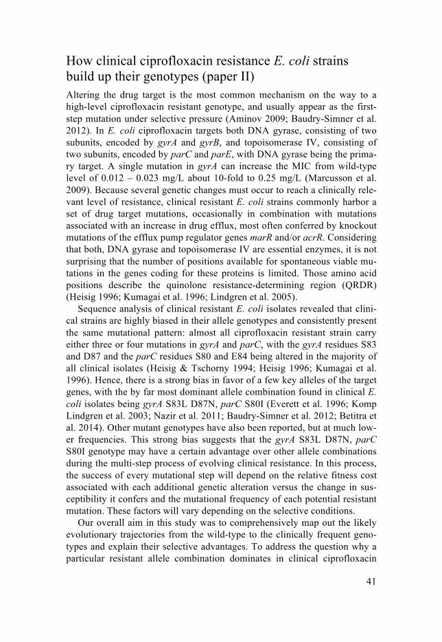
How clinical ciprofloxacin resistance E. coli strains build up their genotypes (paper II) Altering the drug target is the most common mechanism on the way to a high-level ciprofloxacin resistant genotype, and usually appear as the first-step mutation under selective pressure (Aminov 2009; Baudry-Simner et al. 2012). In E. coli ciprofloxacin targets both DNA gyrase, consisting of two subunits, encoded by gyrA and gyrB, and topoisomerase IV, consisting of two subunits, encoded by parC and parE, with DNA gyrase being the prima-ry target. A single mutation in gyrA can increase the MIC from wild-type level of 0.012 – 0.023 mg/L about 10-fold to 0.25 mg/L (Marcusson et al. 2009). Because several genetic changes must occur to reach a clinically rele-vant level of resistance, clinical resistant E. coli strains commonly harbor a set of drug target mutations, occasionally in combination with mutations associated with an increase in drug efflux, most often conferred by knockout mutations of the efflux pump regulator genes marR and/or acrR. Considering that both, DNA gyrase and topoisomerase IV are essential enzymes, it is not surprising that the number of positions available for spontaneous viable mu-tations in the genes coding for these proteins is limited. Those amino acid positions describe the quinolone resistance-determining region (QRDR) (Heisig 1996; Kumagai et al. 1996; Lindgren et al. 2005).
Sequence analysis of clinical resistant E. coli isolates revealed that clini-cal strains are highly biased in their allele genotypes and consistently present the same mutational pattern: almost all ciprofloxacin resistant strain carry either three or four mutations in gyrA and parC, with the gyrA residues S83 and D87 and the parC residues S80 and E84 being altered in the majority of all clinical isolates (Heisig & Tschorny 1994; Heisig 1996; Kumagai et al. 1996). Hence, there is a strong bias in favor of a few key alleles of the target genes, with the by far most dominant allele combination found in clinical E. coli isolates being gyrA S83L D87N, parC S80I (Everett et al. 1996; Komp Lindgren et al. 2003; Nazir et al. 2011; Baudry-Simner et al. 2012; Betitra et al. 2014). Other mutant genotypes have also been reported, but at much low-er frequencies. This strong bias suggests that the gyrA S83L D87N, parC S80I genotype may have a certain advantage over other allele combinations during the multi-step process of evolving clinical resistance. In this process, the success of every mutational step will depend on the relative fitness cost associated with each additional genetic alteration versus the change in sus-ceptibility it confers and the mutational frequency of each potential resistant mutation. These factors will vary depending on the selective conditions.
Our overall aim in this study was to comprehensively map out the likely evolutionary trajectories from the wild-type to the clinically frequent geno-types and explain their selective advantages. To address the question why a particular resistant allele combination dominates in clinical ciprofloxacin
42
resistant E. coli strains and which parameters influence the selection of the most successful genotype, we used a set of constructed isogenic strains rep-resenting different possible steps in the evolutionary process and newly se-lected mutants and measured mutation rates in fluctuation assays, drug sus-ceptibility as growth rate in increasing concentrations of ciprofloxacin and as standard MIC, and relative fitness in competition experiments. The results of those measurements were combined with experimental evolution and se-quence analysis. We found that the most common gyrA allele, S83L, has the highest fitness of the single gyrA mutations in the presence of ciprofloxacin and therefore presents the most successful first step mutant. In our evolution experiments from single to double mutant, we did not identify any double target mutants, like the clinically most frequent gyrA S83L, parC S80I. In-stead, all mutants selected for higher resistance had acquired efflux-associated mutations. Considering the mutational target size for efflux-associated mutations in comparison to specific target mutations and that the resistance level of a double target mutant and a mutant carrying a target mu-tation in combination with an efflux mutation are indistinguishable, this is not surprising. However, only about half of all clinical resistant isolates carry efflux-associated mutations, suggesting that they cause a significant negative impact on relative bacterial fitness and are counter-selected by the much fitter, but less frequently occurring target-only mutants in clinical settings. Thus, we argue that the rare acquisition of a second target mutation in gyrA or parC is a critical bottleneck in the evolutionary process to the clinically most successful genotype. Selection for acquisition of a third resistance mu-tation from the double gyrA S83L, parC S80I mutant resulted in the acquisi-tion of a second mutation in gyrA. These evolutionary findings are supported by the individual MICs of each mutant and by competition advantages of each successive mutant over its parental strain in the presence of ciprofloxa-cin. Thus, based on the MIC data and our evolution experiments we hypoth-esis that the most frequent trajectory taken to the high-resistant multi-target mutants found in the clinics goes via gyrA S83L to the double mutant gyrA S83L parC S80I, and finally to the triple mutant gyrA S83L parC S80I gyrA D87N. Our data show that a combination of mutation supply rate and rela-tive fitness of particular allele combinations are major factors determining the trajectories to the resistance genotypes frequently observed in clinical resistant isolates. Additionally, based on our measurements of mutation rates and relative fitness, we could compute a mathematical model, which enables us to predict that resistance genotypes frequently observed in clinical isolates are enriched under conditions where drug selection is weak and high fitness the major determinant for selection.

43
RNA polymerase mutations can contribute to ciprofloxacin resistance (paper III) E. coli is naturally susceptible to ciprofloxacin and it is well established that clinically relevant levels of resistance to this fluoroquinolone drug are only reached through the accumulation of several genetic alterations, of which each contributes to the decrease in susceptibility. Mutations that are linked to an increased resistance level have been described in the drug target genes (gyrA, gyrB, parC, and parE) and in efflux-related genes, commonly in regu-lators of the membrane spanning AcrAB-TolC pump (marR, acrR, and soxR), the major efflux pump in E. coli (Marcusson et al. 2009; Hooper 1999; Wang et al. 2001; Betitra et al. 2014; Kumagai et al. 1996).
The study described here was motivated by an earlier evolution experi-ment, during which an E. coli strain, evolved for high ciprofloxacin re-sistance, had acquired a mutation in rpoB, the gene coding for the β-subunit of RNA polymerase. This finding raised the questions whether the selection of rpoB mutations was a frequent event in the process of resistance devel-opment, in which genetic background they arise during the evolutionary process, if they contribute to resistance, and by which mechanism they con-fer a selective advantage.
We repeated the initial stepwise evolution, starting with ten independent lineages of wild-type E. coli and evolved them in increasing concentrations of ciprofloxacin, up to the clinical breakpoint. When we examined individual strains from the end-point cultures, we found that rpoB mutations had come up in three of the ten lineages and were present in 20 – 30 % of the endpoint cultures, suggesting that mutations affecting RNA polymerase arise fre-quently under our selective conditions. All of the rpoB mutations identified were located outside the rifampicin-resistance determining region and con-ferred only a low level of resistance to rifampicin. Whole genome sequence analysis of the endpoint strains revealed that each strain had acquired four to five mutations during the evolution experiment, including mutations in drug target genes (gyrA, gyrB) and in genes regulating drug efflux (marR, acrR, soxR), which have previously been associated with fluoroquinolone re-sistance (Heisig & Tschorny 1994; Hooper 1999; Marcusson et al. 2009). The temporal order in which these successive mutations occurred can be described according to the following pattern: first one or two mutations in the drug target genes; second mutations in efflux regulator genes; third, rela-tively late in the evolution of each strain, the RNA polymerase mutations; and finally an additional target or efflux regulator mutation. Based on this information we constructed a set of isogenic strains representing the differ-ent steps in the evolutionary process to the three endpoint strains carrying the different rpoB mutations and determined their MIC-values and fitness in terms of bacterial growth. Acquisition of each additional mutation, including
44
the mutations in RNA polymerase, lowered the susceptibility to ciprofloxa-cin. Furthermore, each additional mutation conferred a selective advantage to the resulting successive mutant over its parental strain in competition ex-periments in increasing drug concentrations. This competitive advantage was conferred as the drug concentration approached the MIC of the ancestral strain and it became apparent regardless of the initial cost of the additional mutation in the absence of ciprofloxacin. Each of the selected rpoB muta-tions also lowered the susceptibility level to ciprofloxacin in a wild-type background and in combination with the target and efflux mutation present in the respective other evolved strains. The same effect was seen for other fluoroquinolone drugs. This suggests that RNA polymerase mutations can contribute to an increased resistance to fluoroquinolones. We also observed in association with some rpoB alleles an increase in MIC for the unrelated antibiotics chloramphenicol and tetracycline. Chloramphenicol and tetracy-cline resistance can be caused by increased drug efflux (Okusu et al. 1996). To test whether the resistance observed in the rpoB mutant strains was due to an increase in drug efflux caused by an altered level of transcripts coding for efflux pump components, we measured expression levels for the three com-ponents of the major efflux pump AcrAB-TolC and for the two multidrug efflux transporters MdfA and MdtK by quantitative RT-PCR. The mRNA levels of mdtK were increased in all three strains carrying one of the ciprof-loxacin-selected rpoB mutations, and two of the three mutations also caused an increase in the AcrAB-TolC efflux pump components. In the third ciprof-loxacin-selected rpoB mutant transcription levels of the DNA gyrase genes gyrA and gyrB were up-regulated. In addition, all three rpoB mutations lead to an increased transcription of the gyrase inhibitor sbmC. This suggests that the mutant RNA polymerases cause changes in the transcription pattern. To strengthen these findings, we also determined the induction of SOS-response caused by double stranded DNA breaks created during exposure to ciprof-loxacin (Chen et al. 1996). The level of SOS-induction in the strains carrying a ciprofloxacin-selected polymerase mutation was generally lower than in strains carrying an rpoB mutation located in the rifampicin-resistance deter-mining region. We speculate that this is due to a lower intracellular drug concentration in the evolved rpoB mutant strains.
Concluding we could show that mutations in RNA polymerase frequently arise under selection with ciprofloxacin and that they contribute to the re-sistance phenotype. This observation can be explained by a changed tran-scription pattern of genes involved in susceptibility to ciprofloxacin by mu-tant RNA polymerase. Accordingly, this study identifies a new mutational class contributing to ciprofloxacin resistance.

45
Predictability in the evolution of ciprofloxacin resistance in E. coli (paper IV) Fluoroquinolones are important broad-spectrum antibiotics, frequently used to treat a wide variety of bacterial infections, including urinary tract infec-tions cause by invasive E. coli strains. Due to their extensive use, resistance development to fluoroquinolones has been increasingly observed (ECDC 2013). Ciprofloxacin is one of the fluoroquinlone drugs, with a clinical breakpoint for resistance in E. coli of 1 mg/L (Kahlmeter 2003). Multiple genetic changes are required to raise the MIC above this breakpoint (Heisig & Tschorny 1994; Komp Lindgren et al. 2003; Marcusson et al. 2009; Pid-dock 1999; Hooper 2001). There is a large literature on ciprofloxacin re-sistance-conferring mutations and chromosomal mutations linked to in-creased ciprofloxacin resistance have been noticed in the drug target genes gyrA, gryB, parC, and parE and in genes coding for the AcrAB-TolC efflux pump and its regulators, with knockout mutations in marR, acrR, and soxR being most common. A lot less is known about the trajectories taken during the complex development to a clinically relevant level of resistance, and about the interplay and internal competition between different resistant mu-tants arising in cultures under selection for ciprofloxacin resistance. Our recent finding that RNA polymerase mutations can significantly reduce sus-ceptibility to ciprofloxacin (paper III) raises the question if mutations in other genes that have not previously been reported can also confer a decrease in susceptibility to ciprofloxacin.
Thus, the aim of this project was to study the trajectories taken to drug re-sistance by wild-type E. coli cultures selected in the presence of ciprofloxa-cin and to investigate the possibility that mutations in genes other than the canonical target and efflux-regulating genes might be involved in this multi-step evolutionary process.
We evolved nine independent lineages of E. coli in increasing concentra-tions of ciprofloxacin in liquid and twenty independent lineages on agar plates until the clinical breakpoint was passed. The transmission bottleneck in the liquid evolution experiment was about 3x10⁸ CFU and accordingly mutant fixation in the population was expected to be strongly influenced by the relative fitness of the different arising genotypes. In contrast, the evolu-tion experiment on agar plates represented a single cell bottleneck at each step, reducing the relative influence of fitness. Consequently, the evolution-ary trajectory was instead expected to be more strongly influenced by genet-ic drift.
Deep sequencing was used on bacterial populations of all nine lineages evolved in liquid to identify the mutations that were prevalent at successive stages throughout the evolution, ranging from cycle two to the last step at cycle 23. Several individual strains from each evolved lineage were also

46
analyzed by whole genome sequencing to establish the genetic linkage of mutations on the chromosome. Additionally, we also sequenced the genomes of the endpoint strains of the lineages evolved on agar plates.
The deep sequencing analysis revealed that there were usually multiple al-lelic variants of particular genes present at the same time, of which one would typically be selected and be enriched or even go into fixation as the evolution continued. Strikingly, we could observe the same wave-like pat-tern in occurrence and selection of mutational targets in almost every line-age. The first wave of mutations affected primarily the drug target gene gyrA and one of the gyrA alleles would predictably reach 100% frequency early in the evolution. This was followed by a second wave with different mutations arising in one or more of the efflux-regulator genes marR, acrR, and soxR. Specific alleles of this mutational class typically reached a high frequency or fixation late in the evolution experiment. A third wave of mutations, which reached high frequencies in most of the lineages by cycle 14 or slightly later, involved mutations in genes coding for proteins involved in transcriptional and/or translational processes (rpoB, rpoN, thrV, and mnmA). A final muta-tional wave selected additional drug target mutations in gyrB, parC, and/or parE, respectively. To reveal which alleles were linked on the same chromo-some, we analyzed individual clones by whole genome sequencing, focusing on clones taken from the evolving populations at cycle 16. Each of the clones had acquired mutations in canonical resistance-associated genes (gy-rA, gyrB, parE, acrR, marR, soxR). The majority of the clones also carried one or more mutations in genes associated with transcription and translation in E. coli. The identified mutations included amino acid substitutions in rpoB and rpoC, encoding subunits of RNA polymerase, a complete deletion of a tRNA gene, and an out-of-frame deletion in a tRNA modification enzyme gene. Two other strains had mutations that potentially affected the expres-sion of genes involved in translation, one affecting the promoter of a tRNA gene, and the second one with possible effects on transcription of a release factor. Whole genome sequencing on the strains evolved on solid agar re-vealed a similar genomic pattern in terms of mutational classes. In addition to target and efflux regulator mutation, every clone had also acquired at least one mutation in a gene involved in transcription and/or translation. These included mutations in the RNA polymerase genes rpoB and rpoC, mutations in tRNA genes, tRNA modifying enzymes, and tRNA synthetases, and muta-tions in translation factors.
Concluding, we report that the evolution of resistance to ciprofloxacin follows a predictable pattern, in which mutations in different functional clas-ses of genes accumulate in successive waves and that the appearance of mu-tations affecting a wide variety of genes involved in transcription and trans-lation is a common occurrence following the acquisition of target and drug efflux mutations, both under conditions where selection for growth fitness is

47
high and in a situation where it is very relaxed. These evolution experiments strongly suggest that mutations affecting transcription and/or translation are a novel class of genes contributing to the evolution of resistance to ciprof-loxacin.

48
Perspectives on Resistance Development
The extensive use and misuse of antibiotics in medicine and agriculture has led to the emergence of drug resistant bacterial pathogens, which are now causing a severe global problem and gravely threaten our capacity to effi-ciently treat bacterial infections. This process of resistance development began with the introduction of the first antimicrobial drugs in the 1930s and it continues today. On a worldwide scale, the volume use of antibiotics in human medicine rose by 36% between 2000 and 2010, but one has to keep in mind that there are big variations in the annual per-person consumption of antibiotics between different countries (Van Boeckel et al. 2014). The in-creased consumption of antibiotics is tightly linked to the increased re-sistance observed and presents one of the major factors responsible for the enrichment of resistant pathogenic bacteria during the past 70 years. The use of any antimicrobial drug, however appropriate and conservative, will even-tually lead to selection of resistant bacteria, but the widespread unnecessary and excessive use of this valuable resource accelerates the accumulation of ineffective antibiotics (O'Neill 2014).
A recent review on antimicrobial resistance estimated the global costs of antimicrobial resistance until 2050 in the absence of any progress in tackling the challenge (O'Neill 2014). The predictions are grim even though they are thought to be an underestimation of the actual consequences of antimicrobial resistance. The scenario depicted includes a worldwide economic loss of up to $100 trillion and an estimated 300 million people dying by the year 2050. In order words: a continued rise in antimicrobial resistance would lead to enormous financial losses and 10 million premature deaths every year in the near future, presenting one of the leading causes of death, even more preva-lent than cancer (O'Neill 2014).
It is evident that actions have to be taken fast and on a global scale, in or-der to preserve the efficacy of the antibiotics we have. First and foremost, we have to find a responsible way of using the available antibiotics and restrict their usage to medical purposes. Because resistance development is associat-ed with the volume and distribution of antibiotics that are released into the environment, where they are present in sub-inhibitory concentrations and may select for resistance bacteria, the practice of using antibiotics as growth stimulator in animal feed and in aquaculture has to be banned in all parts of the world (Gullberg et al. 2011; Andersson & Hughes 2010). The records on

49
the dissemination and epidemiology of resistance bacteria need to be im-proved, as does the detection of multi-drug resistant bacteria. In the current situation, surveillance of antibacterial resistance is neither coordinated nor harmonized and a global consensus on methodology and data collection is lacking (WHO 2014a). Rapid detection methods are needed to help to identi-fy the infection causing organism and its resistance pattern in order to be able to prescribe an appropriate antimicrobial drug and avoid an unnecessary use of broad-spectrum antibiotics. In particular middle-income countries frequently use and over-use second– and third-line antibiotics, instead of first-line drugs for common infections and a more regulated handling of antibiotic prescriptions would allow conserving the antibiotic effectiveness for a longer time. Therefore, an appropriate use of antibiotics in developing countries should be encouraged and the use of last-resort antibiotic drugs avoided (Van Boeckel et al. 2014). The spread of resistant pathogens is con-nected to hygienic measures and transmission control in clinics and hospitals (Andersson & Hughes 2010). Consequently, taking better precautions in hospital settings and raising awareness in communities could help reduce the transmission of bacterial infections. Additionally, more funding should go into research on understanding the evolution of antibiotic resistance and into the discovery and development of new antimicrobial agents. In addition, research into discovery and development of antibiotic compounds has to be made attractive to pharmaceuticals companies. Complementary treatment options should also be investigated. Promising alternative therapies have been proposed, including the sensitization of the bacterial pathogen to a va-riety of antibiotic agents by disrupting intrinsic resistance elements (Cox et al. 2014), phage therapy, and the increased development and use of vaccines.
All of these measures to reduce the use of antimicrobial agents and the spread of antibiotic resistance can only be successful when taken as a coher-ent international collaboration (O'Neill 2014). Thus, to prevent a striking rise in drug resistance in low -and middle-income countries with large popula-tions and to preserve antibiotic efficacy worldwide, programmes that pro-mote rational use through coordinated efforts by the international community have to be devised and agreed upon (Van Boeckel et al. 2014).

50
Concluding Remarks
The latest report of the WHO on antimicrobial resistance and its surveillance is alarming: resistance to pathogenic bacteria has reached very high levels in many parts of the world and in some settings, few or none of the available treatments options remain effective for common infections (WHO 2014a). This severe global problem is the consequence of the widespread use and misuse of antibiotics in clinical and veterinary medicine, agriculture, aqua-culture, horticulture and other human activities over the past seven decades (Aminov 2009). With the large-scale production of antibiotics and a constant rise in antibiotic consumption, we exert a strong selective pressure towards bacteria to evolve resistance in various ecosystems, and today many of the pathogenic microorganisms found in the clinics are resistant to multiple an-tibiotics (Aminov 2009; Van Boeckel et al. 2014). The escalating number of resistant pathogens poses major problems on the treatment of patients with hospital or community-acquired multi-drug resistant infections (Putman et al. 2000). A deeper understanding of the molecular basis of the development of antibiotic resistance may allow a rational to circumvent the spread of drug resistant pathogens and guide us in the development of new antimicrobial therapy. If we would fail to prevent a further rapid increase in antibiotic re-sistance and to develop new antibiotics, then the level of medical care we take for granted today might be lost forever.
In this thesis, we focused on the evolution of resistance to two important antimicrobial drugs, rifampicin and ciprofloxacin.
Rifampicin is a first-line drug in tuberculosis treatment and resistance to rifampicin complicates the treatment of tuberculosis patients infected with the increasingly spreading multidrug-resistance M. tuberculosis. High-level resistance to rifampicin can be achieved through many different single muta-tions in the drug target, but there is a very strong bias towards a small set of resistance mutations seen in clinical resistant M. tuberculosis isolates. Paper I gives insight into the resistance evolution to rifampicin. We found that the success of the clinically most frequent rifampicin resistance mutations is depending on three parameters: a high resistance level conferred by the mu-tation, a relatively low initial fitness cost, and the ability to acquire a com-pensatory mutation to ameliorate bacterial fitness.

51
Paper II-IV focus on the evolution of resistance to ciprofloxacin, a widely used synthetic broad-spectrum antibiotic. Similar to the mutational bias ob-served in rifampicin resistant M. tuberculosis strains, a few alleles in the drug target of ciprofloxacin are overrepresented among clinical resistant E. coli strains. Clinical ciprofloxacin resistant isolates always carry multiple target mutations, sometimes in combination with efflux-related mutations. In paper II we discovered which trajectories are taken during the accumulation of ciprofloxacin resistance mutations and which parameters influence those pathways and the success of each mutational step on the way to a clinical relevant level of resistance and the most common allele combination in clin-ical isolates. Paper III and IV are based on laboratory evolution experiments during which wild-type E. coli was grown in increasing concentration of ciprofloxacin. Whole genome sequencing analysis of single strains and whole cultures from the evolutionary endpoint and different successive steps of the evolution experiments revealed that mutations in genes coding for proteins involved in transcriptional or translational cellular processes are frequently selected during resistance evolution. In paper III we show that mutations in the β-subunit of RNA polymerase were common events in our selections and that they contribute to ciprofloxacin resistance via transcrip-tional changes, including up-regulation of efflux-pump components, leading to lower intracellular drug concentrations. In paper IV we explored the muta-tional pattern that is the basis of the each multi-step evolutionary process to high-level resistance to ciprofloxacin. Confirming our observations descrip-ed in paper III, we found that the accumulation of resistance mutations oc-cured in a predictable order and that the mutational targets include genes involved in transcription and translation.
During the past years, awareness of the big threat we are facing by losing efficacy of every antibiotic class due to the rapid emergence of bacterial resistance strains has been increasing and several approaches and efforts need to be taken in order to improve the current situation. A better under-standing of resistance evolution in bacteria will allow us to make more pre-cise predictions about the rates and trajectories of the development of re-sistance towards new and old drugs and should also give us possibilities to take appropriate measures for a more intelligent use of antimicrobial drugs, so that the development of resistance will slow down and the useful lifespan of many drugs will be prolonged.

52
Svensk Sammanfattning
Upptäckten att infektionssjukdomar orsakade av bakterier kan behandlas snabbt och effektivt med antibiotika är en milstolpe i den medicinska histo-rien. Många ämnen med antibakteriell verkan upptäcktes mellan 1930- och 1960-talet. Denna enorma framgång, och insikten att många tidigare dödliga sjukdomar nu kunde botas enkelt, ledde till föreställningen att alla infek-tionssjukdomar snart skulle vara utrotade. Tyvärr blev det inte så, utan de första antibiotikaresistenta bakterierna upptäcktes kort efter att motsvarande antibiotika introducerats. Trots att bakterier blivit resistenta mot allt fler sorters antibiotika har nästan inga nya sorters antibiotika kommit ut på marknaden sedan den ”gyllene eran” under första hälften av 1900-talet. Idag finns resistens i alla grupper av sjukdomsalstrande bakterier och mot alla klasser av antibiotika. Om denna utveckling fortsätter i samma takt kommer vi väldigt snart att sakna effektiva läkemedel mot infektioner som fram tills alldeles nyligen varit okomplicerade att behandla.
Under arbetet som ligger till grund för denna avhandling har jag un-dersökt resistensutvecklingen mot två antibiotika: rifampicin, som används som behandling mot tuberkulos, och ciprofloxacin, ett bredspektrumantibi-otika, som används för att behandla t.ex. urinvägsinfektioner. Mot båda des-sa, kliniskt viktiga, antibiotika finns en ökad förekomst och spridning av resistenta bakterier. Syftet för mitt avhandlingsarbete var att öka vår förståelse för hur sjukdomsalstrande bakterier får speciella mutationer för att bli resistenta genom evolution i närvaro av antibiotika. Detta har gjorts med evolutionsexperiment i laboratoriemiljö och med genetiska och molekylära metoder.
Vi visade vilka faktorer som påverkar selektion och ansamling av de van-ligaste mutationerna som gör tuberkulosbakterien resistent mot rifampicin. Dessutom identifierade och studerade vi en ny typ av mutationer som kan bidra till ciprofloxacin-resistens och avslöjade deras mekanism. Olika muta-tioner som behövs för att en bakterie ska kunna bygga upp en klinisk resis-tens mot ciprofloxacin studerades. Effekten av de olika mutationerna och ordning i vilken de uppkommer beskrivs i avhandligen.
Sammanfattningsvis står vi inför enorma problem med antibiotikaresis-tenta bakterier. För att lösa detta behövs ökad kunskap om hur bakterier ut-vecklar resistens. Detta kan hjälpa oss att utveckla ny antibiotika och att använda de sorters antibiotika vi redan har på ett mer ansvarsfullt sätt.

53
Deutsche Zusammenfassung
Die Erkenntnis, dass durch Bakterien verursachte Infektionskrankenheiten schnell und effizient mit Antibiotika behandelt werden können, markierte einen Meilenstein der Medizingeschichte. Beginnend in den 30er Jahren des 20. Jahrhunderts bis über die Jahrhundertsmitte hinaus wurden zahlreiche Wirkstoffe mit antibakterieller Wirkung entdeckt und weiterentwickelt. Deren enormer Erfolg führte zu der irrtümlichen Annahme, dass Infektion-skrankheiten alsbald der Vergangenheit angehören würden. Schon kurze Zeit nach der Einführung der ersten Antibiotika wurden jedoch erste Resistenzen nachgewiesen. Während die Zahl der wegen resistenter Keime wirkungslos gewordenen Antibiotika stieg, gelangten nach der anfänglichen Entdecker-phase kaum neue antibakterielle Mittel auf den Markt. Heute sind Resisten-zen gegen alle Klassen von Antibiotika und in allen Bakteriengruppen bekannt. Sollte sich die Resistenzentwicklung so rapide fortsetzten, werden in naher Zukunft für die Behandlung von Krankheiten, die noch vor wenigen Jahren als unproblematisch galten, keine wirksamen Antibiotika mehr zur Verfügung stehen.
Im Rahmen meiner Dissertation wurde die Entstehung von Resistenzen zu zwei Antibiotika untersucht: Ciprofloxacin, ein wichtiges Breitbandantibi-tikum mit zunehmend kritischer Resistenzlage und Rifampicin, das als Mit-tel der ersten Wahl in der Tuberkulosebehandlung verwendet wird. Auch hier erschweren Resistenzen die Behandlung dieser weitverbreiteten Krank-heit. Ziel der vorliegenden Arbeit was es, anhand von Evolutionsexperimen-ten und mit genetischen und molekularen Verfahren ein besseres Verständis von der Entstehung der in klinischen Stämmen nachgewiesenen Re-sistenzmutationen zu bekommen.
Es wurde aufgedeckt, welche Kriterien die Selektion und Anreicherung der am häufigsten in klinisch resistenten Tuberkulosestämmen vorkom-menden Resistenzmutationen beeinflußen. Desweiteren konnten genauere Einblicke in die Entstehung von Resistenzen gegen Ciprofloxacin gewonnen werden. Es wurde eine neue Klasse von Mutationen, die zur Entwicklung von Resistenzen führen können identifiziert und der dahinterstehende Mech-anismus aufgedeckt. Zusätzlich wird beschrieben, welche verschiedenen Stufen an Mutationen durchlaufen werden müssen, um klinisch relevante Resistenzlevel gegen Ciprofloxacin zu erreichen und in welcher Reihenfolge dies in klinischen Isolaten geschieht.
54
Acknowledgements
It has been seven and a half years since I first came to Uppsala and many fantastic people have crossed my path since then. Without them, my time in Sweden would not have been as rich and colorful and I am grateful to every one of them.
I count myself very lucky to have been able to do my PhD studies in IM-BIM and the D7:3 corridor in particular. It was not only scientifically a great and inspiring place, but also socially and many of you became more than great and helpful colleagues to me. All the conferences, retreats, dinners, champagne-celebrations, pub nights, dissertation parties and various other outside-the-lab activities were a lot of fun thanks to all of you. First and foremost I would like to thank my supervisor Diarmaid. Thank you for taking me into your group as a completely inexperienced PhD student and introducing my to the world of bacterial genetics. I am grateful for all the support, patience and practical help I got from you on the way. It never ceased to amazed me to see you take that step back to look at what might seem puzzling results and put them into the context of the big scientific pic-ture.
I also want to thank my co-supervisor Dan for creating such a great, sci-entific atmosphere in D7:3 and for leaving the office door open when listen-ing to Jack Teagarden and Bobby Hackett. A big thank you also has to go to the past and present members of Diar-maid’s group: Disa, Marie and Linda, it was really nice to get to know you and I wish our overlapping time in the lab would have been longer. Jessica, thank you for being such a helpful colleague and good friend. Thank you also for hopping on to the rpoB paper and for help with the Swedish sum-mary. Eva, I’m glad you decided to say good-bye to sunny Spain to share you energy and enthusiasm with us. I envy the drive you have to work to-wards your goals. Rachel, thanks for many fun days and evenings in and outside the lab. The genial Gerrit, thank you for letting me participate in the rifampicin paper and most of all, thank you so much for your patience and always finding time to analyze various excel data etc. with me. In your hands, everything seems so simple. Doug, you were the perfect travel guide on our trip through Utah. Thank you for being such a fun and carrying col-

55
league and a supportive collaborator. Rahel, you are a sunshine and it was great to have you in the group. Lisa, I am happy you started the same day as me, and I could share all my first impressions with you. CaoSha, your cute comments often made my day. Kavita, welcome to the Hughes group. Lin-néa, thank you for enriching our group with your gentle character and being such an efficient collaborator. I also want to thank my students Veronika, Carl Fredrik, Yinyu, Dina and Angie for their valuable contributions to my projects.
A special thank you also has to go to everyone from Dan’s group. Anna and Jocke for enjoyable evenings with delicious food at your place. Jon, for first being the perfect supervisor and then being a wonderful colleague full of surprises and always up for some fun stuff. Marius, I am glad that we both ended up doing our PhD in D7:3. Michael, I hope our visits of the Christmas market in Würzburg are going to turn into a long lasting tradition. Erik G., thank you for adding so much character to the lab. Who else would organize the Thursday pancake-round or use the cold room to cool down? Hava, the best of luck with your defence. Also the best of luck for Lisa T. and Lisa A. for the last part of your PhDs. Hervé, thank you for spicing up our ImBact seminars with great sarcastic comments and questions. Karin and Ulrika, so nice that you also moved from ICM to IMBIM. And Omar, thank you for always smiling, you guys make D7:3 a happier place. Peter, Erik L. and Fredrika, thank you for contributing to the nice atmosphere in the lab. Jessi-ca, you are a great addition to Dan’s group. Sohaib and Nizar, thanks for your attempts to cheer me up when I seemed stressed. Lionel, it is very nice to have you around.
Linus, thank you for being such an optimistic person and always finding something positive to say.
I am also happy that I could meet and spend some good times with BMC people from outside our corridor. I thank Elin for being a great companion and support from the start to the end of our PhD studies. Britta for making my time at ICM wonderfully exciting. Alex H for company at the yoga clas-ses and fun nights out. Falk for always feeling up for a countdown tequila and Alvaro for enjoyable conversation about anything. Julia, Tabea, Andrew, Fempke, Sara, Behrooz and Rodrigo for a lot of after-work fun. Iris, your company in the writing office was highly appreciated.
Staffan, thank you for always saying yes: when I asked if I could do my masters project with you, when I asked if I could state you as my reference, and when I asked if you could be in my thesis committee. And to my dear friends, which I met during my master studies in Uppsala. Susanna, Mike, Alex, Kasia, Anja N., Anja M. and Marlen, I have so many

56
great memories of all of you, from the good old days when we were still master students until now and I value your friendship very highly. Susanna, Alex and Marlen, I am so glad I met you right at the beginning of my time in Uppsala. We had so much fun together and hopefully there is much more to come. Anja N, I am glad you are such a spontaneous friend, always ready to join whatever might turn up. Kasia, you were a great flat mate. Anja M., sometimes you are so much alike my sister that I would like to adopt you as my second twin. Marlen, how lucky I have been that you took me under your wings when I first got to Sweden and that you are always that step ahead, so I can turn to you with any silly question I might have. Abraham, thank you for all the good times we had and great memories we share and most of all, thank you for being open to all kinds of plans for the future. Ich danke meinen Eltern, die mir die beste Kindheit gegeben haben und die auch jetzt noch immer für mich da sind. Danke, dass ihr mich stets in Allem so liebevoll untersützt! Und danke an meine Laura, die mich versteht wie niemand sonst und ohne die ich nicht vollständig wäre.

57
References
Aminov, R.I., 2009. The role of antibiotics and antibiotic resistance in na-ture. Environmental microbiology, 11(12), pp.2970–2988.
Andersson, D.I. & Hughes, D., 2010. Antibiotic resistance and its cost: is it possible to reverse resistance? Nature Reviews Microbiolgy, 8(4), pp.260–271.
Andersson, D.I. & Levin, B.R., 1999. The biological cost of antibiotic re-sistance. Current opinion in microbiology, 2(5), pp.489–493.
Baquero, F. & Negri, M.C., 1997. Selective compartments for resistant mi-croorganisms in antibiotic gradients. BioEssays : news and reviews in molecular, cellular and developmental biology, 19(8), pp.731–736.
Baudry-Simner, P.J. et al., 2012. Mechanisms of reduced susceptibility to ciprofloxacin in Escherichia coli isolates from Canadian hospitals. The Canadian journal of infectious diseases & medical microbiology = Journal canadien des maladies infectieuses et de la microbiologie médi-cale / AMMI Canada, 23(3), pp.e60–4.
Betitra, Y. et al., 2014. Determinants of quinolone resistance in Escherichia coli causing community-acquired urinary tract infection in Bejaia, Alge-ria. Asian Pacific journal of tropical medicine, 7(6), pp.462–467.
Billington, O.J., McHugh, T.D. & Gillespie, S.H., 1999. Physiological cost of rifampin resistance induced in vitro in Mycobacterium tuberculosis. Antimicrobial Agents and Chemotherapy, 43(8), pp.1866–1869.
Björkman, J. et al., 2000. Effects of Environment on Compensatory Muta-tions to Ameliorate Costs of Antibiotic Resistance. Science, 287(5457), pp.1479–1482.
Björkman, J., Hughes, D. & Andersson, D.I., 1998. Virulence of antibiotic-resistant Salmonella typhimurium. Proceedings of the National Acade-my of Sciences, 95(7), pp.3949–3953.
58
Brandis, G. & Hughes, D., 2013. Genetic characterization of compensatory evolution in strains carrying rpoB Ser531Leu, the rifampicin resistance mutation most frequently found in clinical isolates. Journal of Antimi-crobial Chemotherapy, 68(11), pp.2493–2497.
Brandis, G. et al., 2015. Comprehensive phenotypic characterization of ri-fampicin resistance mutations in Salmonella provides insight into the evolution of resistance in Mycobacterium tuberculosis. Journal of Anti-microbial Chemotherapy, 70(3), pp.680–685.
Brandis, G. et al., 2012. Fitness-compensatory mutations in rifampicin-resistant RNA polymerase. Molecular Microbiology, 85(1), pp.142–151.
Burdett, V., 1980. Identification of tetracycline-resistant R-plasmids in Streptococcus agalactiae (group B). Antimicrobial Agents and Chemo-therapy, 18(5), pp.753–760.
Bush, K. et al., 2011. Tackling antibiotic resistance. Nature Publishing Group, 9(12), pp.894–896.
Campbell, E.A. et al., 2001. Structural mechanism for rifampicin inhibition of bacterial rna polymerase. Cell, 104(6), pp.901–912.
Cars, O., Hedin, A. & Heddini, A., 2011. The global need for effective anti-biotics - Moving towards concerted action. Drug Resistance Updates, 14(2), pp.68–69.
Casali, N. et al., 2012. Microevolution of extensively drug-resistant tubercu-losis in Russia. Genome Research, 22(4), pp.735–745.
CDC, 2006. Emergence of Mycobacterium tuberculosis with extensive re-sistance to second-line drugs--worldwide, 2000-2004. MMWR. Morbidi-ty and mortality weekly report, 55(11), pp.301–305.
Chapman, J.S. & Georgopapadakou, N.H., 1988. Routes of quinolone per-meation in Escherichia coli. Antimicrobial Agents and Chemotherapy, 32(4), pp.438–442.
Chen, C.R. et al., 1996. DNA gyrase and topoisomerase IV on the bacterial chromosome: quinolone-induced DNA cleavage. Journal of molecular biology, 258(4), pp.627–637.
Cohen, 2000. Changing patterns of infectious disease. Nature, 406(6797), pp.762–767.

59
Cohen et al., 1989. Cross-resistance to fluoroquinolones in multiple-antibiotic-resistant (Mar) Escherichia coli selected by tetracycline or chloramphenicol: decreased drug accumulation associated with mem-brane changes in addition to OmpF reduction. Antimicrobial Agents and Chemotherapy, 33(8), pp.1318–1325.
Comas, I. et al., 2012. Whole-genome sequencing of rifampicin-resistant Mycobacterium tuberculosis strains identifies compensatory mutations in RNA polymerase genes. Nature genetics, 44(1), pp.106–110.
Condon, C., Squires, C. & Squires, C.L., 1995. Control of rRNA transcrip-tion in Escherichia coli. Microbiological reviews, 59(4), pp.623–645.
Cox, G. & Wright, G.D., 2013. Intrinsic antibiotic resistance: mechanisms, origins, challenges and solutions. International journal of medical mi-crobiology : IJMM, 303(6-7), pp.287–292.
Cox, G., Koteva, K. & Wright, G.D., 2014. An unusual class of anthracy-clines potentiate Gram-positive antibiotics in intrinsically resistant Gram-negative bacteria. Journal of Antimicrobial Chemotherapy, 69(7), pp.1844–1855.
Cramariuc, O. et al., 2012. Mechanism for translocation of fluoroquinolones across lipid membranes. BBA - Biomembranes, 1818(11), pp.2563–2571.
Davies, J., 2006. Are antibiotics naturally antibiotics? Journal of industrial microbiology & biotechnology, 33(7), pp.496–499.
Deguchi, T. et al., 1997. Detection of mutations in the gyrA and parC genes in quinolone-resistant clinical isolates of Enterobacter cloacae. The Journal of antimicrobial chemotherapy, 40(4), pp.543–549.
Diacon, A., von, G.-B. & Donald, P.R., 2012. From Magic Mountain to Ta-ble Mountain. Swiss Medical Weekly.
Dowson, C. et al., 1989. Horizontal transfer of penicillin-binding protein genes in penicillin-resistant clinical isolates of Streptococcus pneumoni-ae. Proc. Natl. Acad. Sci., 86, pp.8842–8846.
Drake, J.W., 1991. A constant rate of spontaneous mutation in DNA-based microbes. Proceedings of the National Academy of Sciences, 88(16), pp.7160–7164.

60
Drake, J.W. et al., 1998. Rates of spontaneous mutation. Genetics, 148(4), pp.1667–1686.
ECDC, 2013. Antimicrobial resistance surveillance in Europe. pp.1–218.
ECDC, 2012. Summary of the latest data on antibiotic consumption in the European Union. pp.1–5.
EMEA, E., 2009. The bacterial challenge: time to react. pp.1–54.
Emmerson, A.M., 2003. The quinolones: decades of development and use. Journal of Antimicrobial Chemotherapy, 51(90001), pp.13–20.
EUCAST, 2010. Rifampicin Rationale for the EUCAST clinical breakpoints. pp.1–10.
Everett, M.J. et al., 1996. Contributions of individual mechanisms to fluoro-quinolone resistance in 36 Escherichia coli strains isolated from humans and animals. Antimicrobial Agents and Chemotherapy, 40(10), pp.2380–2386.
Farhat, M.R. et al., 2013. Genomic analysis identifies targets of convergent positive selection in drug-resistant Mycobacterium tuberculosis. Nature genetics, 45(10), pp.1183–1189.
Fàbrega, A. et al., 2010. Constitutive SoxS expression in a fluoroquinolone-resistant strain with a truncated SoxR protein and identification of a new member of the marA-soxS-rob regulon, mdtG. Antimicrobial Agents and Chemotherapy, 54(3), pp.1218–1225.
Fernandez, L. & Hancock, R.E.W., 2012. Adaptive and Mutational Re-sistance: Role of Porins and Efflux Pumps in Drug Resistance. Clinical microbiology reviews, 25(4), pp.661–681.
Fischbach, M.A. & Walsh, C.T., 2009. Antibiotics for emerging pathogens. Science, 325(5944), pp.1089–1093.
Fleming, A., 1929. On the antibacterial action of culutres of a penicillium, with special reference to their use in the isolation of B. influenzae. The British Journal of Experimental Pathology, 10, pp.226–236.
Freire-Moran, L. et al., 2011. Critical shortage of new antibiotics in devel-opment against multidrug-resistant bacteria—Time to react is now. Drug Resistance Updates, 14(2), pp.118–124.

61
Gagneux, S., 2006. The Competitive Cost of Antibiotic Resistance in Myco-bacterium tuberculosis. Science, 312(5782), pp.1944–1946.
Garibyan, L. et al., 2003. Use of the rpoB gene to determine the specificity of base substitution mutations on the Escherichia coli chromosome. DNA repair, 2(5), pp.593–608.
Goh, E.-B. et al., 2002. Transcriptional modulation of bacterial gene expres-sion by subinhibitory concentrations of antibiotics. Proceedings of the National Academy of Sciences, 296(2-3), pp.17025–17030.
Goldman, J.D., White, D.G. & Levy, S.B., 1996. Multiple antibiotic re-sistance (mar) locus protects Escherichia coli from rapid cell killing by fluoroquinolones. Antimicrobial Agents and Chemotherapy, 40(5), pp.1266–1269.
Guan, X. et al., 2013. Plasmid-mediated quinolone resistance - current knowledge and future perspectives. Journal of International Medical Research, 41(1), pp.20–30.
Gullberg, E. et al., 2011. Selection of Resistant Bacteria at Very Low Anti-biotic Concentrations M. Lipsitch, ed. PLoS Pathogens, 7(7), p.e1002158.
Hakenbeck, R. & Coyette, J., 1998. Resistant penicillin-binding proteins. Cellular and molecular life sciences : CMLS, 54(4), pp.332–340.
Hawkey, P.M. & Jones, A.M., 2009. The changing epidemiology of re-sistance. Journal of Antimicrobial Chemotherapy, 64(Supplement 1), pp.i3–i10.
Heep, M. et al., 2001. Frequency of rpoB mutations inside and outside the cluster I region in rifampin-resistant clinical Mycobacterium tuberculo-sis isolates. Journal of clinical microbiology, 39(1), pp.107–110.
Heisig, P., 1996. Genetic evidence for a role of parC mutations in develop-ment of high-level fluoroquinolone resistance in Escherichia coli. Anti-microbial Agents and Chemotherapy, 40(4), pp.879–885.
Heisig, P. & Tschorny, R., 1994. Characterization of fluoroquinolone-resistant mutants of escherichia coli selected in vitro. Antimicrobial Agents and Chemotherapy, 38(6), pp.1284–1291.

62
Hershberg, R., 2015. Mutation-The Engine of Evolution: Studying Mutation and Its Role in the Evolution of Bacteria. Cold Spring Harbor perspec-tives in biology, 7(9).
Hooper, D.C., 2001. Emerging mechanisms of fluoroquinolone resistance. Emerging infectious diseases, 7(2), pp.337–341.
Hooper, D.C., 1999. Mechanisms of fluoroquinolone resistance. Drug Re-sistance Updates, pp.1–18.
Kahlmeter, G., 2014. Defining antibiotic resistance-towards international harmonization. Upsala journal of medical sciences, 119(2), pp.78–86.
Kahlmeter, G., 2003. European harmonization of MIC breakpoints for anti-microbial susceptibility testing of bacteria. Journal of Antimicrobial Chemotherapy, 52(2), pp.145–148.
Kahlmeter, G., 2013. MIC distribution 2013.
Komp Lindgren, P., Karlsson, A. & Hughes, D., 2003. Mutation Rate and Evolution of Fluoroquinolone Resistance in Escherichia coli Isolates from Patients with Urinary Tract Infections. Antimicrobial Agents and Chemotherapy, 47(10), pp.3222–3232.
Kresge, N., Simoni, R.D. & Hill, R.L., 2004. Selman Waksman: the Father of Antibiotics. The Journal of Biological Chemistry, 279(48), pp.1–3.
Kumagai, Y. et al., 1996. Quinolone-resistant mutants of escherichia coli DNA topoisomerase IV parC gene. Antimicrobial Agents and Chemo-therapy, 40(3), pp.710–714.
Laible, G., Spratt, B.G. & Hakenbeck, R., 1991. Interspecies recombination-al events during the evolution of altered PBP 2x genes in penicillin-resistant clinical isolates of Streptococcus pneumoniae, Mol Microbiol.
Lawn, S.D. & Nicol, M.P., 2011. Xpert® MTB/RIF assay: development, evaluation and implementation of a new rapid molecular diagnostic for tuberculosis and rifampicin resistance. Future microbiology, 6(9), pp.1067–1082.
Laxminarayan, R. & Van Boeckel, T.P., 2014. The value of tracking antibi-otic consumption. The Lancet. Infectious diseases, 14(5), pp.360–361.

63
Lesher, G.Y. et al., 1962. 1,8-Naphthyridine Derivatives. A Class of Chemo-therapeutic Agents. Journal of medicinal and pharmaceutical chemistry, 91, pp.1063–1065.
Lindgren, P.K. et al., 2005. Biological Cost of Single and Multiple Norflox-acin Resistance Mutations in Escherichia coli Implicated in Urinary Tract Infections. Antimicrobial Agents and Chemotherapy, 49(6), pp.2343–2351.
Luria, S.E. & Delbrück, M., 1943. Mutations of Bacteria from Virus Sensi-tivity to Virus Resistance. Genetics, 28(6), pp.491–511.
Madigan, M.T., Martinko, J.M. & Brock, T.D., 2006. Brock Biology of Mi-croorganisms 11 ed., Pearson Prentice Hall.
Maisnier-Patin, S. & Andersson, D.I., 2004. Adaptation to the deleterious effects of antimicrobial drug resistance mutations by compensatory evo-lution. Research in microbiology, 155(5), pp.360–369.
Marcusson, L.L., Frimodt-Møller, N. & Hughes, D., 2009. Interplay in the selection of fluoroquinolone resistance and bacterial fitness. PLoS Path-ogens, 5(8), p.e1000541.
Mariam, D.H. et al., 2004. Effect of rpoB Mutations Conferring Rifampin Resistance on Fitness of Mycobacterium tuberculosis. Antimicrobial Agents and Chemotherapy, 48(4), pp.1289–1294.
Martinez, J.L., 2009. The role of natural environments in the evolution of resistance traits in pathogenic bacteria. Proceedings. Biological sciences / The Royal Society, 276(1667), pp.2521–2530.
Martínez-Martínez, L., Pascual, A. & Jacoby, G.A., 1998. Quinolone re-sistance from a transferable plasmid. Lancet (London, England), 351(9105), pp.797–799.
Meier, A. et al., 1994. Genetic alterations in streptomycin-resistant Myco-bacterium tuberculosis: mapping of mutations conferring resistance. An-timicrobial Agents and Chemotherapy, 38(2), pp.228–233.
Mukherjee, K. & Chatterji, D., 1997. Studies on the omega subunit of Esche-richia coli RNA polymerase. European journal of biochemistry / FEBS, 247(3), pp.884–889.
Murray, P.R., Rosenthal, K.S. & Pfaller, M.A., 2005. Medical Microbiology 5 ed., Philadelphia : Elsevier Mosby.

64
Nagaev, I. et al., 2001. Biological cost and compensatory evolution in fusid-ic acid-resistant Staphylococcus aureus. Molecular Microbiology, 40(2), pp.433–439.
Nazir, H. et al., 2011. Can phylogenetic type predict resistance develop-ment? Journal of Antimicrobial Chemotherapy, 66(4), pp.778–787.
Nikaido, H. & Thanassi, D.G., 1993. Penetration of lipophilic agents with multiple protonation sites into bacterial cells: tetracyclines and fluoro-quinolones as examples. Antimicrobial Agents and Chemotherapy, 37(7), pp.1393–1399.
O'Neill, 2014. Antimicrobial Resistance: Tackling a crisis for the health and wealth of nations. pp.1–20.
O'Neill, A.J. & Chopra, I., 2006. Molecular basis of fusB-mediated re-sistance to fusidic acid in Staphylococcus aureus. Molecular Microbiol-ogy, 59(2), pp.664–676.
Okusu, H., Ma, D. & Nikaido, H., 1996. AcrAB efflux pump plays a major role in the antibiotic resistance phenotype of Escherichia coli multiple-antibiotic-resistance (Mar) mutants. Journal of bacteriology, 178(1), pp.306–308.
Paulander, W., Maisnier-Patin, S. & I, A.D., 2009. The fitness cost of strep-tomycin resistance depends on rpsL mutation, carbon source and RpoS (sigmaS). Genetics, 183(2), pp.539–46– 1SI–2SI.
Perichon, B. & Courvalin, P., 2009. VanA-Type Vancomycin-Resistant Staphylococcus aureus. Antimicrobial Agents and Chemotherapy, 53(11), pp.4580–4587.
Piddock, L.J., 1999. Mechanisms of fluoroquinolone resistance: an update 1994-1998. Drugs, 58 Suppl 2, pp.11–18.
Poole, K., 2004. Efflux‐mediated multiresistance in Gram‐negative bacteria. Clinical microbiology and infection, 10(1), pp.12–26.
Putman, M. et al., 2000. Antibiotic resistance: era of the multidrug pump. Molecular Microbiology, 36(3), pp.772–773.
Ramaswamy, S. & Musser, J.M., 1998. Molecular genetic basis of antimi-crobial agent resistance in Mycobacterium tuberculosis: 1998 update. Tubercle and lung disease : the official journal of the International Un-ion against Tuberculosis and Lung Disease, 79(1), pp.3–29.
65
Rana, F., 2013. Rifampicin - an overview. International Journal of Research in Pharmacy and Chemistry, 1(3), pp.83–87.
Reynolds, M.G., 2000. Compensatory evolution in rifampin-resistant Esche-richia coli. Genetics, 156(4), pp.1471–1481.
Roberts, M.C. et al., 1986. Tetracycline resistance and tetM in pathogenic urogenital bacteria. Antimicrobial Agents and Chemotherapy, 30(5), pp.810–812.
Robicsek, A. et al., 2006. Fluoroquinolone-modifying enzyme: a new adap-tation of a common aminoglycoside acetyltransferase. Nature medicine, 12(1), pp.83–88.
Sandegren, L. & Andersson, D.I., 2009. Bacterial gene amplification: impli-cations for the evolution of antibiotic resistance. Nature Publishing Group, 7(8), pp.578–588.
Sengupta, S., Chattopadhyay, M.K. & Grossart, H.-P., 2013. The multifacet-ed roles of antibiotics and antibiotic resistance in nature. Frontiers in microbiology, 4, p.47.
Singh, R. et al., 2012. Temporal Interplay between Efflux Pumps and Target Mutations in Development of Antibiotic Resistance in Escherichia coli. Antimicrobial Agents and Chemotherapy, 56(4), pp.1680–1685.
Springer, B. et al., 2001. Mechanisms of streptomycin resistance: selection of mutations in the 16S rRNA gene conferring resistance. Antimicrobial Agents and Chemotherapy, 45(10), pp.2877–2884.
Strahilevitz, J. et al., 2009. Plasmid-Mediated Quinolone Resistance: a Mul-tifaceted Threat. Clinical microbiology reviews, 22(4), pp.664–689.
Strebhardt, K. & Ullrich, A., 2008. Paul Ehrlich's magic bullet concept: 100 years of progress. Nature Reviews: Cancer, 8(6), pp.473–480.
Sundqvist, M. et al., 2010. Little evidence for reversibility of trimethoprim resistance after a drastic reduction in trimethoprim use. Journal of Anti-microbial Chemotherapy, 65(2), pp.350–360.
Tran, J.H., Jacoby, G.A. & Hooper, D.C., 2005. Interaction of the plasmid-encoded quinolone resistance protein Qnr with Escherichia coli DNA gyrase. Antimicrobial Agents and Chemotherapy, 49(1), pp.118–125.

66
Trinh, V. et al., 2006. Structural Perspective on Mutations Affecting the Function of Multisubunit RNA Polymerases. Microbiology and Molecu-lar Biology Reviews, 70(1), pp.12–36.
Turnidge, J. & Paterson, D.L., 2007. Setting and revising antibacterial sus-ceptibility breakpoints. Clinical microbiology reviews, 20(3), pp.391–408– table of contents.
Van Boeckel, T.P. et al., 2014. Global antibiotic consumption 2000 to 2010: an analysis of national pharmaceutical sales data. The Lancet. Infectious diseases, 14(8), pp.742–750.
Vassylyev, I.A.D.G., 2006. Is it Easy to Stop RNA Polymerase? Cell Cycle, 5(4), pp.399–404.
Wang, H. et al., 2001. Genetic Characterization of Highly Fluoroquinolone-Resistant Clinical Escherichia coli Strains from China: Role of acrR Mutations. Antimicrobial Agents and Chemotherapy, 45(5), pp.1515–1521.
WHO, 2014a. Antimicrobial Resistance. WHO Press, pp.1–256.
WHO, 2014b. Global Tuberculosis Report 2014. WHO Press, pp.1–171.
WHO, 2010. Treatment of Tuberculosis: Guidelines 4 ed., Geneva: World Health Organization.
Wielgoss, S. et al., 2011. Mutation Rate Inferred From Synonymous Substi-tutions in a Long-Term Evolution Experiment With Escherichia coli. G3 (Bethesda, Md.), 1(3), pp.183–186.
Wolfson, J.S. & Hooper, D.C., 1985. The fluoroquinolones: structures, mechanisms of action and resistance, and spectra of activity in vitro. An-timicrobial Agents and Chemotherapy, 28(4), pp.581–586.
Wright, G.D., 2007. The antibiotic resistome: the nexus of chemical and genetic diversity. Nature Publishing Group, 5(3), pp.175–186.
Wright, G.D. & Poinar, H., 2012. Antibiotic resistance is ancient: implica-tions for drug discovery. Trends in microbiology, 20(4), pp.157–159.
Yamane, K. et al., 2007. New Plasmid-Mediated Fluoroquinolone Efflux Pump, QepA, Found in an Escherichia coli Clinical Isolate. Antimicro-bial Agents and Chemotherapy, 51(9), pp.3354–3360.

67
Yim, G., Huimi Wang, H. & Davies, J., 2006. The truth about antibiotics. The truth of antibiotics, 296(2-3), pp.163–170.
Zhang et al., 1999. Crystal structure of Thermus aquaticus core RNA poly-merase at 3.3 A resolution. Cell, 98(6), pp.811–824.
Zhang et al., 2013. Genome sequencing of 161 Mycobacterium tuberculosis isolates from China identifies genes and intergenic regions associated with drug resistance. Nature genetics, 45(10), pp.1255–1260.

Acta Universitatis UpsaliensisDigital Comprehensive Summaries of Uppsala Dissertationsfrom the Faculty of Medicine 1150
Editor: The Dean of the Faculty of Medicine
A doctoral dissertation from the Faculty of Medicine, UppsalaUniversity, is usually a summary of a number of papers. A fewcopies of the complete dissertation are kept at major Swedishresearch libraries, while the summary alone is distributedinternationally through the series Digital ComprehensiveSummaries of Uppsala Dissertations from the Faculty ofMedicine. (Prior to January, 2005, the series was publishedunder the title “Comprehensive Summaries of UppsalaDissertations from the Faculty of Medicine”.)
Distribution: publications.uu.seurn:nbn:se:uu:diva-265018
ACTAUNIVERSITATIS
UPSALIENSISUPPSALA
2015