Embed Size (px)
Citation preview

HAL Id: hal-03093969https://hal.archives-ouvertes.fr/hal-03093969
Submitted on 4 Jan 2021
HAL is a multi-disciplinary open accessarchive for the deposit and dissemination of sci-entific research documents, whether they are pub-lished or not. The documents may come fromteaching and research institutions in France orabroad, or from public or private research centers.
L’archive ouverte pluridisciplinaire HAL, estdestinée au dépôt et à la diffusion de documentsscientifiques de niveau recherche, publiés ou non,émanant des établissements d’enseignement et derecherche français ou étrangers, des laboratoirespublics ou privés.
Evidence of a functional reorganization in the auditorydorsal stream following unilateral hearing loss
Nicolas Vannson, Kuzma Strelnikov, Chris James, Olivier Deguine, PascalBarone, Mathieu Marx
To cite this version:Nicolas Vannson, Kuzma Strelnikov, Chris James, Olivier Deguine, Pascal Barone, et al.. Evidence ofa functional reorganization in the auditory dorsal stream following unilateral hearing loss. Neuropsy-chologia, Elsevier, 2020, 149, pp.107683. �10.1016/j.neuropsychologia.2020.107683�. �hal-03093969�

Evidence of a functional reorganization in the auditory dorsal stream
following unilateral hearing loss
Nicolas Vannson a, b, e,*, Kuzma Strelnikov d, Chris J. James e, Olivier Deguine a, b, c,
Pascal Barone a, b, 1, Mathieu Marx a, b, c, 1
a Brain and Cognition Research Centre, University of Toulouse Paul Sabatier, Toulouse, France
b Brain and Cognition Research Centre, CNRS-UMR, 5549, Toulouse, France
c Service d’Otologie, Otoneurologie et ORL pediatrique, Hopital Pierre-Paul Riquet, CHU Toulouse Purpan, France
d Hopital Pierre-Paul Riquet, CHU Toulouse Purpan, France
e Cochlear France SAS, Toulouse, France
A R T I C L E I N F O
Keyword:
Spatial hearing
Localization
Unilateral hearing loss
Dorsal/ventral auditory streams
A B S T R A C T
Unilateral hearing loss (UHL) generates a disruption of binaural hearing mechanisms, which impairs sound
localization and speech understanding in noisy environments. We conducted an original study using fMRI and
psychoacoustic assessments to investigate the relationships between the extent of cortical reorganization across
the auditory areas for UHL patients, the severity of unilateral hearing loss, and the deficit in binaural abilities.
Twenty-eight volunteers (14 UHL patients) were recruited (twenty-two females and six males). The brain im-
aging analysis demonstrated that UHL induces a shift in aural dominance favoring the better ear, with a cortical
reorganization located in the non-primary auditory areas, ipsilateral (same side) to the better ear. This reorga -
nization is correlated not only to the hearing loss severity but also to spatial localization abilities. A regression
analysis between brain activity and patient’s performance clearly showed that the spatial hearing deficit was
linked to a functional alteration of the posterior auditory areas known to process spatial hearing. Altogether, our
study reveals that UHL alters the dorsal auditory stream, which is deleterious to spatial hearing.
1. Introduction
The ability to locate sound sources in the environment facilitates
inter-individual communication and is a key skill for self-protection.
Sound localization deficits result from binaural hearing alterations and
are usually accompanied by other hearing impairments (e.g., poor
speech recognition in noise), which may lead to poorer quality of life
(Heinrich et al., 2019; Vannson et al., 2015). Based on connectivity
(Kaas and Hackett, 1998) and electrophysiological (Rauschecker and
Tian, 2000) studies in nonhuman primates, several authors have sug-
gested that spatial hearing is supported by a dorsal where stream, in
opposition to a ventrally directed auditory stream involved in sound
identification (Arnott et al., 2004; Rauschecker and Scott, 2009; van der
Heijden et al., 2019). In humans, lesion studies support the duality of
segregated networks for spatial and nonspatial auditory information
processing, extending the segregation to the parieto-frontal and tem-
poral lobes (Adriani et al., 2003; Clarke et al., 2002; Zündorf et al.,
2016). Similarly, focal inactivation of activity targeting either the
anterior or posterior auditory cortex impairs respectively, identification
or localization performance (Ahveninen et al., 2013). Lastly, brain im-
aging studies support the notion of a dual pathway, and it is now evident
that in the early stages, the planum temporale is more specifically
modulated by sound position than the primary (core) auditory cortex
(PAC) (van der Heijden et al., 2018; Van der Zwaag et al., 2011).
In the case of unilateral hearing loss (UHL), binaural processing is
disrupted, leading to poorer localization (Abel and Lam, 2008) and
speech recognition performance in noisy environments (Bronkhorst and
Plomp, 1989). At the brain level, a small number of studies have
explored the functional reorganization that takes place in adults after
UHL. As neuronal sensitivity to binaural disparities in terms of time and
level is present all along the central auditory pathway, from the lower
brainstem to the auditory cortex (Grothe et al., 2010; Sadoun et al.,
2020), UHL would be expected to impact binaural integration at all
stages. Interestingly, functional reorganization has been observed at the
* Corresponding author. CNRS CERCO UMR 5549 CHU Toulouse Purpan - Pavillon Baudot BP 25202 31052, Toulouse Cedex, France.
E-mail address: [email protected] (N. Vannson).1 Pascal Barone and Mathieu Marx equally contributed.
https://doi.org/10.1016/j.neuropsychologia.2020.107683
Received 21 July 2020; Received in revised form 16 October 2020; Accepted 8 November 2020
Available online 17 November 2020

N. Vannson et al.
2
=
=
=
level of the auditory cortex after acquired UHL but seems to be absent at
subcortical levels (Langers et al., 2005), a result that could be explained
by the weak spatial resolution of deep brain structures (subcortical
structures such as the inferior colliculus for instance that are hardly
defined with fMRI (Schonwiesner et al., 2007). Human brain imaging
studies in UHL thus report a decrease in normal interhemispheric
asymmetry in cortical auditory activation after stimulation of the pre-
served ear. Whereas monaural stimulation induces a more pronounced
level of activity in the contralateral (opposite) auditory cortex in normal
hearing persons, more bilateral activation has been demonstrated in
cases of UHL, using either fMRI (Burton et al., 2012; Scheffler et al.,
1998; Suzuki et al., 2002), or EEG/MEG (Hanss et al., 2009; Vasama and
Makela, 1995). However, contradictory results make it unclear whether
the functional subdivisions of the auditory cortex are differently affected
by acquired UHL (Hanss et al., 2009; Scheffler et al., 1998). The
disruption of binaural processes should predominantly impact the dorsal
auditory stream, but this has not been explored. Furthermore, several
reports have indicated that patients with congenital unilateral deafness
(Slattery III & Middlebrooks, 1994) exhibit close to normal localization
performances. This suggests that patients can develop adaptive
compensatory strategies, which should have a specific signature in brain
activity. Altogether, many questions remain to be resolved, concerning
the brain plasticity mechanisms that may occur in patients with acquired
UHL and their putative impact on behavioral performances.
The psychoacoustic effects of acquired unilateral hearing loss can be
evaluated within a normal hearing population using a plug in one ear.
Under these conditions, the deficit for spatial hearing has been
demonstrated in several studies (for instance Van Wanrooij and Van
Opstal, 2007). Still, performances for spatial hearing remain poorer than
that observed following long-term hearing loss (Alzaher et al., in prep;
Kumpik et al., 2010; Vannson et al., 2017); where a behavioral adap-
tation has occurred. In addition, no evidence of immediate plastic
reorganization has been described in such acute deprivation (Van
Wanrooij and Van Opstal, 2007). The behavioral and cortical conse-
quences of postlingual acquired unilateral hearing loss should be
distinguished from those occurring after congenital unilateral hearing
loss. In the latter case, the development is characterized by the presence
of different sensitive periods, expressed as a susceptibility to behavior-
ally adapt to sensory privation as well as a potential of brain plasticity
that decreases with age (A. Kral, 2013; Syka, 2002).
We conducted a study that aimed at investigating the relationships
between UHL severity, the resulting deficit in spatial hearing, and the
extent of cortical reorganization across auditory areas. Our results
clearly demonstrate that a unilateral deafness results in a sound locali-
zation deficit that is related to a functional shift towards ipsilateral aural
dominance. The core of the auditory cortex appears relatively preserved
but UHL impacts specifically the posterior auditory areas of the dorsal
pathway involved in spatial hearing while the ventral temporal stream
areas maintain their normal implication in processing non-spatial
acoustical information.
2. Materials and methods
This study was approved by the local institutional review board
(Comite de Protection des Personnes du Sud Ouest et Outre-Mer IV; no.
CPP14-021/2014-A00498-39) in accordance with the Declaration of
Helsinki.
2.1. Participants
Fourteen right-handed (according to the Edinburgh Handedness In-
ventory; Oldfield, 1971) adults (11 females; median age 39 years, SD
13,06) and native French speakers with an acquired UHL were
recruited. Ten of them had a better right ear (left UHL), and four had a
better-left ear (right UHL). The only inclusion criterion was UHL
established by pure-tone audiometry, ranging from mild to total in order
to ensure a representative sample. All participants with UHL had normal
contralateral hearing thresholds (i.e. below 20 dB HL between 125 Hz
and 8 kHz). Mean pure-tone average (PTA) was 87 dB HL, covering a
range of 37 to over 120 dB. The main characteristics of this population
can be found in Table 1.
Furthermore, 14 age- and sex-matched normal-hearing subjects
(NHS) were recruited with a comparable median age (36.5 years, SD
14; bootstrap confidence intervals, p > 0.05) and with hearing thresh-
olds within the normal range for both ears. All participants underwent
psychoacoustic tests and fMRI scans. The fMRI data of one participant
with UHL (subject 11) were excluded because of technical problems
during the fMRI acquisition.
2.2. Psychoacoustic procedures
Clinical pure-tone and speech audiometry was carried out with a GN
Otometrics Madsen Itera 2 audiometer with TDH-39 headphones.
Binaural hearing was assessed in sound field in a calibrated soundproof
booth. Binaural hearing encompassed the French Matrix test (FrMatrix;
Jansen et al., 2012) for speech-in-noise recognition and a clinical set-up
sound localization test routinely used in the ENT department (Vannson
et al., 2015, 2017).
2.2.1. Speech-in-noise recognition test: FrMatrix
The FrMatrix is an adaptive and standardized test for speech-in-noise
recognition that features closed-set sentences. Each sentence has the
same syntactic structure: name - verb - number - object - color. For
instance: “Felix draws six blue bikes”. The background noise is a sta-
tionary long-term average speech spectrum noise that was generated by
superimposing all 280 sentences Speech and noise were generated by an
IBM PC running the OMA software and presented by three loudspeakers
and an amplifier (Studio Lab, SLB sat 200). Speech was set at 65 dB SPL,
and competing noise was adjusted to obtain a signal-to-noise ratio of
50% (SNR50) for correct recognition. SNR50 was measured in three
sound field listening conditions: i) the dichotic condition with the signal
presented to the poorer ear (PE) and the noise to the contralateral, better
ear (BE); ii) the diotic condition with both signal and noise collocated
and presented from the loudspeaker located in front of the subject; and
iii) the reverse-dichotic condition with the signal presented to the better
ear loudspeaker and the noise to the poorer ear loudspeaker. Subjects
were asked to repeat any words they heard.
2.2.2. Sound localization test
Localization ability was assessed in the ENT department with a
horizontal array of 7 loudspeakers and an amplifier located at intervals
of 30◦ from – 90◦ to 90◦ in a frontal semicircle diameter of 1.2 m at the
subject’s head level. Localization stimulus was made up of two 150 msec
white Gaussian noise from 20 Hz to 20 kHz with a 0.05 msec ramp. A
silence of 150 msec was encompassed between the two noises for total
stimulus duration of 450 msec. This stimulus was similar to the one used
by Slattery and Middlebrooks (Slattery III & Middlebrooks, 1994) and
was presented 63 times (9 presentations per loudspeaker) with a period
of 2 s silence between each presentation to allow the subjects to give
orally the loudspeaker location. Localization ability was measured with
the standard root-mean-square (RMS) errors. The examiner was in the
room with the patient to control that he/she keeps his/her head in front
of the speaker 1 (0◦) throughout the experiment. The patient had a sheet
of paper with the position and the number of all speakers to help
him/her. If the patient was not able to point out to a loudspeaker,
his/her answer was marked as “No answer”. For each patient, we had at
least 60 out of 63 valid responses (less than 5% missing data).
N. Vannson et al.
3
±
×
×
=
=
= × × ×
= =
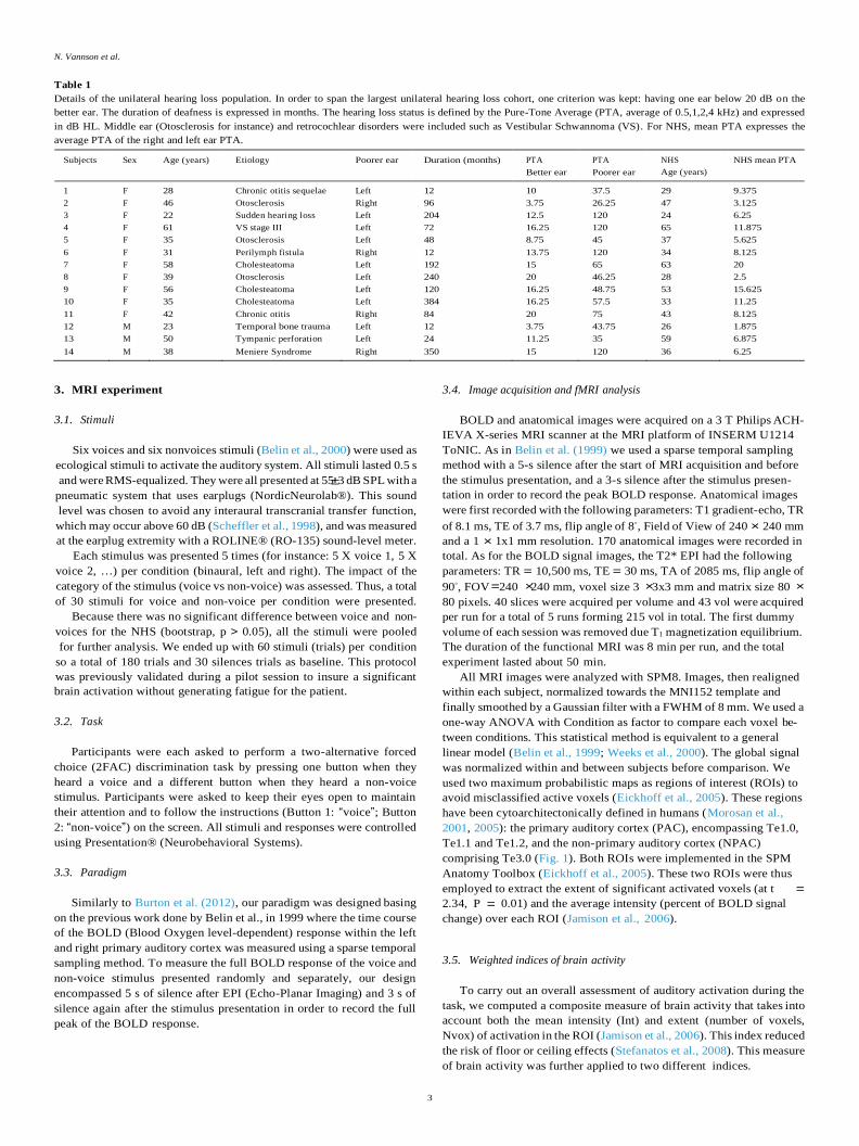
Table 1
Details of the unilateral hearing loss population. In order to span the largest unilateral hearing loss cohort, one criterion was kept: having one ear below 20 dB on the
better ear. The duration of deafness is expressed in months. The hearing loss status is defined by the Pure-Tone Average (PTA, average of 0.5,1,2,4 kHz) and expressed
in dB HL. Middle ear (Otosclerosis for instance) and retrocochlear disorders were included such as Vestibular Schwannoma (VS). For NHS, mean PTA expresses the
average PTA of the right and left ear PTA.
Age (years)
3. MRI experiment
3.1. Stimuli
Six voices and six nonvoices stimuli (Belin et al., 2000) were used as
ecological stimuli to activate the auditory system. All stimuli lasted 0.5 s
and were RMS-equalized. They were all presented at 55 3 dB SPL with a
pneumatic system that uses earplugs (NordicNeurolab®). This sound
level was chosen to avoid any interaural transcranial transfer function,
which may occur above 60 dB (Scheffler et al., 1998), and was measured
at the earplug extremity with a ROLINE® (RO-135) sound-level meter.
Each stimulus was presented 5 times (for instance: 5 X voice 1, 5 X
voice 2, …) per condition (binaural, left and right). The impact of the
category of the stimulus (voice vs non-voice) was assessed. Thus, a total
of 30 stimuli for voice and non-voice per condition were presented.
Because there was no significant difference between voice and non-
voices for the NHS (bootstrap, p > 0.05), all the stimuli were pooled
for further analysis. We ended up with 60 stimuli (trials) per condition
so a total of 180 trials and 30 silences trials as baseline. This protocol
was previously validated during a pilot session to insure a significant
brain activation without generating fatigue for the patient.
3.2. Task
Participants were each asked to perform a two-alternative forced
choice (2FAC) discrimination task by pressing one button when they
heard a voice and a different button when they heard a non-voice
stimulus. Participants were asked to keep their eyes open to maintain
their attention and to follow the instructions (Button 1: “voice”; Button
2: “non-voice”) on the screen. All stimuli and responses were controlled
using Presentation® (Neurobehavioral Systems).
3.3. Paradigm
Similarly to Burton et al. (2012), our paradigm was designed basing
on the previous work done by Belin et al., in 1999 where the time course
of the BOLD (Blood Oxygen level-dependent) response within the left
and right primary auditory cortex was measured using a sparse temporal
sampling method. To measure the full BOLD response of the voice and
non-voice stimulus presented randomly and separately, our design
encompassed 5 s of silence after EPI (Echo-Planar Imaging) and 3 s of
silence again after the stimulus presentation in order to record the full
peak of the BOLD response.
3.4. Image acquisition and fMRI analysis
BOLD and anatomical images were acquired on a 3 T Philips ACH-
IEVA X-series MRI scanner at the MRI platform of INSERM U1214
ToNIC. As in Belin et al. (1999) we used a sparse temporal sampling
method with a 5-s silence after the start of MRI acquisition and before
the stimulus presentation, and a 3-s silence after the stimulus presen-
tation in order to record the peak BOLD response. Anatomical images
were first recorded with the following parameters: T1 gradient-echo, TR
of 8.1 ms, TE of 3.7 ms, flip angle of 8◦, Field of View of 240 240 mm
and a 1 1x1 mm resolution. 170 anatomical images were recorded in
total. As for the BOLD signal images, the T2* EPI had the following
parameters: TR 10,500 ms, TE 30 ms, TA of 2085 ms, flip angle of
90◦, FOV 240 240 mm, voxel size 3 3x3 mm and matrix size 80
80 pixels. 40 slices were acquired per volume and 43 vol were acquired
per run for a total of 5 runs forming 215 vol in total. The first dummy
volume of each session was removed due T1 magnetization equilibrium.
The duration of the functional MRI was 8 min per run, and the total
experiment lasted about 50 min.
All MRI images were analyzed with SPM8. Images, then realigned
within each subject, normalized towards the MNI152 template and
finally smoothed by a Gaussian filter with a FWHM of 8 mm. We used a
one-way ANOVA with Condition as factor to compare each voxel be-
tween conditions. This statistical method is equivalent to a general
linear model (Belin et al., 1999; Weeks et al., 2000). The global signal
was normalized within and between subjects before comparison. We
used two maximum probabilistic maps as regions of interest (ROIs) to
avoid misclassified active voxels (Eickhoff et al., 2005). These regions
have been cytoarchitectonically defined in humans (Morosan et al.,
2001, 2005): the primary auditory cortex (PAC), encompassing Te1.0,
Te1.1 and Te1.2, and the non-primary auditory cortex (NPAC)
comprising Te3.0 (Fig. 1). Both ROIs were implemented in the SPM
Anatomy Toolbox (Eickhoff et al., 2005). These two ROIs were thus
employed to extract the extent of significant activated voxels (at t
2.34, P 0.01) and the average intensity (percent of BOLD signal
change) over each ROI (Jamison et al., 2006).
3.5. Weighted indices of brain activity
To carry out an overall assessment of auditory activation during the
task, we computed a composite measure of brain activity that takes into
account both the mean intensity (Int) and extent (number of voxels,
Nvox) of activation in the ROI (Jamison et al., 2006). This index reduced
the risk of floor or ceiling effects (Stefanatos et al., 2008). This measure
of brain activity was further applied to two different indices.
Subjects Sex Age (years) Etiology Poorer ear Duration (months) PTA
Better ear
PTA
Poorer ear
NHS NHS mean PTA
1 F 28 Chronic otitis sequelae Left 12 10 37.5 29 9.375
2 F 46 Otosclerosis Right 96 3.75 26.25 47 3.125
3 F 22 Sudden hearing loss Left 204 12.5 120 24 6.25
4 F 61 VS stage III Left 72 16.25 120 65 11.875
5 F 35 Otosclerosis Left 48 8.75 45 37 5.625
6 F 31 Perilymph fistula Right 12 13.75 120 34 8.125
7 F 58 Cholesteatoma Left 192 15 65 63 20
8 F 39 Otosclerosis Left 240 20 46.25 28 2.5
9 F 56 Cholesteatoma Left 120 16.25 48.75 53 15.625
10 F 35 Cholesteatoma Left 384 16.25 57.5 33 11.25
11 F 42 Chronic otitis Right 84 20 75 43 8.125
12 M 23 Temporal bone trauma Left 12 3.75 43.75 26 1.875
13 M 50 Tympanic perforation Left 24 11.25 35 59 6.875
14 M 38 Meniere Syndrome Right 350 15 120 36 6.25

N. Vannson et al.
4
=
= +
+ -
=
=
=
- =
- =
=
- =
= - - =
= -
Fig. 1. Auditory region of interests. The primary auditory cortex (PAC, in red) encompasses areas Te 1.0, Te 1.1, Te 1.2 and the non-primary auditory (NPAC, in
blue) the Te 3.0 region. (For interpretation of the references to color in this figure legend, the reader is referred to the Web version of this article.)
First, hemispheric lateralization was computed using the weighted
laterality index (WLI; Stefanatos et al., 2008) and based on the com-
posite activity computation:
WLI (left (Nvox x Int) – right (Nvox x Int))/(left (Nvox x Int)
right (Nvox x Int))
A WLI close to 0 means symmetrical brain activation. When WLI
tends towards either 1 or 1, it reflects asymmetrical brain activation
towards the left or right hemisphere.
Second, we computed a binaural integration index (BII) using the
composite activity, in order to assess how the auditory cortex responded
to a sound that was simultaneously presented to both ears (BIN),
compared with the summed activity obtained after separate stimulation
of each ear (R + L):
BII = BIN / (R + L)
A BII below 1 corresponds to a suppression mechanism induced by
binaural integration. By contrast, a BII above 1 reflects facilitation
processes.
3.6. Experimental design and statistical analysis
All the analyses were implemented in MATLAB r2014b and R (3.3.3).
For all the behavioral measures, such as localization, speech-in-noise
recognition and hit rate for voice vs non-voice distinction, mean dif-
ferences between participants with UHL and NHS were compared using
a nonparametric, bootstrap technique to generate 95% confidence in-
tervals (10,000 samples; bias corrected and accelerated confidence in-
tervals; alpha 0.05; Carpenter and Bithell, 2000). In addition, this
bootstrap technique was also applied to compare groups on brain ac-
tivity indices (WLI and BII). Finally, we performed a regression analysis
between behavioral measures and brain activation, and all significant
clusters (p < 0.05, with familywise error correction, FWE) were identi-
fied with probabilistic cytoarchitectonic maps (Eickhoff et al., 2005) and
the Harvard/Oxford Atlas in MRIcron (2016).
4. Results
4.1. Binaural hearing assessment
We assessed binaural hearing abilities of all participants using a
localization test and the FrMatrix (Fig. 2A and B). Average performance
of participants with UHL on these two tasks clearly revealed a sub-
stantial deterioration in their binaural hearing processing, as their mean
RMS error for localization was 27.70◦ (SD 28.33), which was
considerably poorer (bootstrap, alpha 0.05) than for NHS (1.19◦, SD
1.24) (Fig. 2A, right). The deficit was observed for all azimuths but
was especially important for sounds on the side of the poor ear, for
which most RMS values exceeded 40◦ (Fig. 2A, left).
For speech-in-noise recognition (FrMatrix; Fig. 2B), SNR50s did not
differ between dichotic and reverse-dichotic conditions for NHS (boot-
strap, alpha 0.05), and were therefore pooled. Mean SNR50 was
14.50 dB (SD 1.39) in the dichotic and reverse-dichotic conditions,
and 5.37 dB (SD 1.20) in the diotic condition. Compared with the
NHS group, participants with UHL scored lower in each condition
(bootstrap, alpha 0.05; Fig. 2B). Their mean SNR50s were 2.00 dB
(SD 5.31) in the dichotic condition, 3.14 dB (SD 1.19) in the
diotic condition, and 10.62 dB (SD 2.62) in the reverse-dichotic
condition.
In addition, the degree of UHL, as measured by the poorer ear PTA,
was significantly correlated with speech-in-noise recognition (dichotic
SNR50s, rho = 0.80, p < 0.001) and horizontal sound localization
abilities (mean RMS values, rho = 0.75, p = 0.002). In the UHL group,

N. Vannson et al.
5
=
=
=
=
=
=
= =
Fig. 2. Binaural hearing evaluation. (A) Sound
localization performances of NHS (blue) and UHL
participants (red). RMS errors (+/- SEM) are dis-
played according to the sound position (left panel) or
averaged across speakers (right panel). Participants
with UHL exhibited a strong deficit, reflected by a
higher RMS compared with NHS, especially on the
side of the deaf ear. (B) FrMatrix test. The perfor-
mances of the NHS (blue) and participants with UHL
(red) on speech-in-noise recognition are expressed as
the speech recognition score corresponding to the
50% correct threshold (see text) in the three different
presentation conditions. PE = poorer ear; BE = better
ear; S = speech; N = noise. In all the panels, asterisks
indicate a significant difference based on bootstrap
95% confidence intervals (α = 0.05). (For interpre-
tation of the references to color in this figure legend,
the reader is referred to the Web version of this
article.)
poor localization abilities were also significantly associated with a
deficit in speech-in-noise recognition in the dichotic condition (rho
0.69, p 0.006).
In conclusion, participants with UHL exhibited a significant deficit in
integrating binaural cues, the magnitude of this deficit being related to
UHL severity, in line with previous studies (Vannson et al., 2015, 2017).
5. Sound categorization task
Both groups performed a voice/non-voice discrimination task
(Massida et al., 2011) in the scanner, in three listening conditions (right,
left, and binaural). The hit rate for participants with UHL in the binaural
listening condition (voices and non-voices summed) was similar to that
observed for NHS (93.66%, SD 6.13 vs. 95.80%, SD 3.77, bootstrap,
alpha 0.05), reflecting a near-to-normal BE performance level. As
expected, poorer ear performances of participants with UHL were low
(16.66%, SD 28.72), with a tendency to systematically classify the
stimulus as a non-voice one (Sup Fig.1).
5.1. Neurobiological investigation by fMRI
5.1.1. BOLD activation at whole-brain level
Brain activation was induced by environmental sounds delivered to
either one ear or both ears. As responses to voice and non-voice stimuli
did not differ significantly within the NHS group (bootstrap, alpha
0.05), both were summed. Besides, the impact of the side of unilateral
hearing loss on cortical reorganization is still controversial. Some EEG
studies (Hanss et al., 2009; Khosla et al., 2003; Po-Hung Li et al., 2003)
showed a clear impact of the deafness side, with more plasticity in case
of left hearing loss. In contrast, this right ear/left ear effect was not
found in several fMRI studies (Bilecen et al., 2000; Scheffler et al., 1998),
which showed similar patterns of reorganization whatever the affected
side. Therefore, to avoid stirring misleading conclusions we decided to
split our population into two subgroups: good right better ear (n 10)
and 3-left better ear. In the Results section, we focus on the left UHL
subgroup, as it contained more participants. The data for the right UHL
subgroup are provided in the Supplementary Information (Sup Fig. 3 and
4).
Overall, in the NHS group, monaural stimulation led to greater
contralateral brain activation, whatever the presentation side, whereas
binaural stimulation induced symmetrical brain activation (Fig. 3, left).
These results were observed at the whole-brain level, but also at the level
of the two ROIs within the auditory cortex.
In the UHL group with right BE (Fig. 3, right), left PE stimulation
induced low but significant activation of the auditory cortices, owing to
the broad range of hearing loss severity. Right BE stimulation induced
strong contralateral activation (left hemisphere), as in NHS, but sub-
stantial activity was also observed in the right, ipsilateral auditory areas.
This trend towards a pattern of bilateral activation after BE stimulation
was also observed when the BE and PE were simultaneously stimulated.
To gain better insight into the mechanisms of brain reorganization
after UHL, we conducted separate analyses on the PAC and NPAC.
5.1.2. Composite auditory brain activity in unilateral hearing loss
The levels of activity induced by the stimuli were first analyzed in the
PAC and NPAC of participants with UHL. As at the whole-brain level
(Fig. 3), the level of activity induced after left-ear stimulation was
extremely weak in both left and right PAC and NPAC (Sup Fig. 2 ). Right-
ear and binaural stimulation both led to greater activity in the left (i.e.,
contralateral to the BE) PAC, but the levels of activity appeared to be
different in the NPAC, with a trend towards greater mean activity in the
right hemisphere (i.e., side ipsilateral to the BE).
In order to provide a better account of individual variability than
mean brain activity, we analyzed how the binaural integration and
lateralization indices were modified in patients with UHL compared
with NHS.
5.1.3. Binaural interaction revealed by the binaural integration index (BII)
Binaural integration mechanisms occur when the BII is significantly
different from 1.0 (Fig. 4). In the NHS group, whatever the hemisphere,

N. Vannson et al.
6
Fig. 3. BOLD activation at the whole-brain level.
Brain activity for NHS (left column) and participants
with a left poorer ear (left UHL, right column). In
both groups, monaural stimulation induced bilateral
activation in auditory areas with a tendency in NHS
towards more contralateral activity. In UHL, deaf ear
stimulation did not elicit a significant response, while
stimulation of the intact ear appeared to similarly
activate both left and right cortices. For the purposes
of display, sagittal, coronal and horizontal views are
illustrated at p uncorrected = 0.001.
Fig. 4. Binaural integration index. This index (BII)
was obtained with a composite computation of signal
intensity and number of activated voxels. BII values
are presented for NHS (blue) and participants with
UHL (red), separately for the left and right hemi-
spheres, and in both cortical areas (PAC and NPAC). A
BII below +1.0 indicates that the activity observed
following binaural stimulation was lower that the
summed activity resulting from each monaural stim-
ulation. This suppression mechanism was observed in
NHS in both the PAC and NPAC. In participants with
UHL, the suppression mechanisms were reduced in
the PAC and absent in the NPAC. The black asterisks
show a significant difference between NHS and UHL.
The Ns vs. 1 (red) indicates that the BII values for
participants with UHL did not differ from 1, based on
bootstrap 95% confidence intervals (α = 0.05). (For
interpretation of the references to color in this figure
legend, the reader is referred to the Web version of
this article.)
N. Vannson et al.
7
+
=
+
=
= =
= +
=
= = = =
= = = =
=
= = = =
= = =
=
=
=
=
=
=
=
= +
+ + =
mean BII values were significantly below 1.0 (bootstrap, alpha 0.05)
within the PAC (left hem 0.79 (SD 0.48); right hem 0.58 (SD
0.31)) and NPAC (left hem 0.60 (SD 0.31); right hem 0.50 (SD
0.10)) reflecting a binaural suppression mechanism, as the composite
activation level induced by the simultaneous stimulation of both ears
was significantly lower than the sum of individual activations. In the
PAC and NPAC, mean BII values were statistically similar, but there was
a trend toward greater binaural suppression in the NPAC than in the
PAC.
In the UHL group, none of the mean BII values differed significantly
from 1 (bootstrap, alpha 0.05) in the PAC (left hem 0.86 (SD
0.29); right hem 0.87 (SD 0.60)) and NPAC (left hem 0.97 (SD
0.27); right hem 0.97 (SD 0.39)), whatever the hemisphere. These
results can be interpreted as a loss of binaural integration, which was
particularly apparent within the NPAC, with mean BII values signifi-
cantly higher and closer to values of 1, than those observed in NHS. In
the PAC, mean BII values did not differ significantly from those observed
in the NHS group.
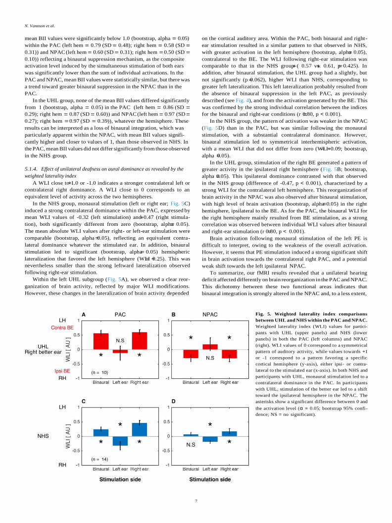
5.1.4. Effect of unilateral deafness on aural dominance as revealed by the
weighted laterality index
A WLI close to 1.0 or -1.0 indicates a stronger contralateral left or
contralateral right dominance. A WLI close to 0 corresponds to an
equivalent level of activity across the two hemispheres.
In the NHS group, monaural stimulation (left or right ear; Fig. 5C)
induced a strong contralateral dominance within the PAC, expressed by
mean WLI values of -0.32 (left stimulation) and 0.47 (right stimula-
tion), both significantly different from zero (bootstrap, alpha 0.05).
The mean absolute WLI values after right- or left-ear stimulation were
comparable (bootstrap, alpha 0.05), reflecting an equivalent contra-
lateral dominance whatever the stimulated ear. In addition, binaural
stimulation led to significant (bootstrap, alpha 0.05) hemispheric
lateralization that favored the left hemisphere (WLI 0.25). This was
nevertheless smaller than the strong leftward lateralization observed
following right-ear stimulation.
Within the left UHL subgroup (Fig. 5A), we observed a clear reor-
ganization of brain activity, reflected by major WLI modifications.
However, these changes in the lateralization of brain activity depended
on the cortical auditory area. Within the PAC, both binaural and right-
ear stimulation resulted in a similar pattern to that observed in NHS,
with greater activation in the left hemisphere (bootstrap, alpha 0.05),
contralateral to the BE. The WLI following right-ear stimulation was
comparable to that in the NHS group ( 0.57 vs. 0.61, p 0.425). In
addition, after binaural stimulation, the UHL group had a slightly, but
not significantly (p 0.062), higher WLI than NHS, corresponding to
greater left lateralization. This left lateralization probably resulted from
the absence of binaural suppression in the left PAC, as previously
described (see Fig. 4), and from the activation generated by the BE. This
was confirmed by the strong individual correlation between the indices
for the binaural and right-ear conditions (r 0.80, p < 0.001).
In the NHS group, the pattern of activation was weaker in the NPAC
(Fig. 5D) than in the PAC, but was similar following the monaural
stimulation, with a substantial contralateral dominance. However,
binaural stimulation led to symmetrical interhemispheric activation,
with a mean WLI that did not differ from zero (WLI 0.09; bootstrap,
alpha 0.05).
In the UHL group, stimulation of the right BE generated a pattern of
greater activity in the ipsilateral right hemisphere (Fig. 5B; bootstrap,
alpha 0.05). This ipsilateral dominance contrasted with that observed
in the NHS group (difference of -0.47, p < 0.001), characterized by a
strong WLI for the contralateral left hemisphere. This reorganization of
brain activity in the NPAC was also observed after binaural stimulation,
with high level of brain activation (bootstrap, alpha 0.05) in the right
hemisphere, ipsilateral to the BE. As for the PAC, the binaural WLI for
the right hemisphere mainly resulted from BE stimulation, as a strong
correlation was observed between individual WLI values after binaural
and right-ear stimulation (r 0.80, p < 0.001).
Brain activation following monaural stimulation of the left PE is
difficult to interpret, owing to the weakness of the overall activation.
However, it seems that PE stimulation induced a strong significant shift
in brain activation towards the contralateral right PAC, and a potential
weak shift towards the left ipsilateral NPAC.
To summarize, our fMRI results revealed that a unilateral hearing
deficit affected differently on brain reorganization in the PAC and NPAC.
This dichotomy between these two functional areas indicates that
binaural integration is strongly altered in the NPAC and, to a less extent,
Fig. 5. Weighted laterality index comparisons
between UHL and NHS within the PAC and NPAC.
Weighted laterality index (WLI) values for partici-
pants with UHL (upper panels) and NHS (lower
panels) in both the PAC (left columns) and NPAC
(right). WLI values of 0 correspond to a symmetrical
pattern of auditory activity, while values towards +1
or -1 correspond to a pattern favoring a specific
cortical hemisphere (y-axis), either ipsi- or contra-
lateral to the stimulated ear (x-axis). In both NHS and
participants with UHL, monaural stimulation led to a
contralateral dominance in the PAC. In participants
with UHL, stimulation of the better ear led to a shift
toward the ipsilateral hemisphere in the NPAC. The
asterisks show a significant difference between 0 and
the activation level (α = 0.05; bootstrap 95% confi-
dence; NS = no significant).
N. Vannson et al.
8
= =
= =
= =
= =
= =
= = = =
in the PAC. When the better ear is stimulated, cortical reorganization is
marked by normal contralateral dominance within the PAC and a sig-
nificant shift of activation towards the ipsilateral hemisphere in the
NPAC.
5.1.5. Relationship between binaural abilities and brain reorganization
UHL leads to poorer binaural hearing performances and to brain
reorganization mechanisms that mainly and significantly affect the
NPAC, as expressed by changes in aural dominance (WLI) and binaural
interaction (BII). Two approaches were used to further investigate the
relationship between cortical reorganization and spatial hearing
deficits.
First, we assessed the relationship between hearing performances
(PTA, FrMatrix dichotic score and RMS error) and NPAC WLI and BII
values. No correlation was found between BII and hearing performances
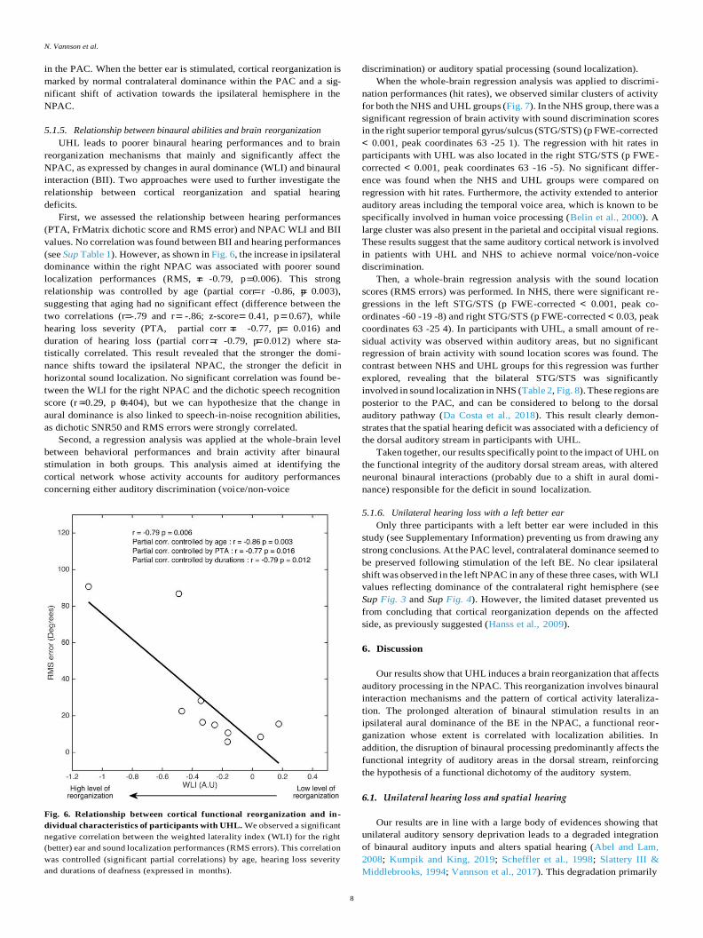
(see Sup Table 1). However, as shown in Fig. 6, the increase in ipsilateral
dominance within the right NPAC was associated with poorer sound
localization performances (RMS, r -0.79, p 0.006). This strong
relationship was controlled by age (partial corr r -0.86, p 0.003),
suggesting that aging had no significant effect (difference between the
two correlations (r -.79 and r -.86; z-score 0.41, p 0.67), while
hearing loss severity (PTA, partial corr r -0.77, p 0.016) and
duration of hearing loss (partial corr r -0.79, p 0.012) where sta-
tistically correlated. This result revealed that the stronger the domi-
nance shifts toward the ipsilateral NPAC, the stronger the deficit in
horizontal sound localization. No significant correlation was found be-
tween the WLI for the right NPAC and the dichotic speech recognition
score (r -0.29, p 0.404), but we can hypothesize that the change in
aural dominance is also linked to speech-in-noise recognition abilities,
as dichotic SNR50 and RMS errors were strongly correlated.
Second, a regression analysis was applied at the whole-brain level
between behavioral performances and brain activity after binaural
stimulation in both groups. This analysis aimed at identifying the
cortical network whose activity accounts for auditory performances
concerning either auditory discrimination (voice/non-voice
Fig. 6. Relationship between cortical functional reorganization and in-
dividual characteristics of participants with UHL. We observed a significant
negative correlation between the weighted laterality index (WLI) for the right
(better) ear and sound localization performances (RMS errors). This correlation
was controlled (significant partial correlations) by age, hearing loss severity
and durations of deafness (expressed in months).
discrimination) or auditory spatial processing (sound localization).
When the whole-brain regression analysis was applied to discrimi-
nation performances (hit rates), we observed similar clusters of activity
for both the NHS and UHL groups (Fig. 7). In the NHS group, there was a
significant regression of brain activity with sound discrimination scores
in the right superior temporal gyrus/sulcus (STG/STS) (p FWE-corrected
< 0.001, peak coordinates 63 -25 1). The regression with hit rates in
participants with UHL was also located in the right STG/STS (p FWE-
corrected < 0.001, peak coordinates 63 -16 -5). No significant differ-
ence was found when the NHS and UHL groups were compared on
regression with hit rates. Furthermore, the activity extended to anterior
auditory areas including the temporal voice area, which is known to be
specifically involved in human voice processing (Belin et al., 2000). A
large cluster was also present in the parietal and occipital visual regions.
These results suggest that the same auditory cortical network is involved
in patients with UHL and NHS to achieve normal voice/non-voice
discrimination.
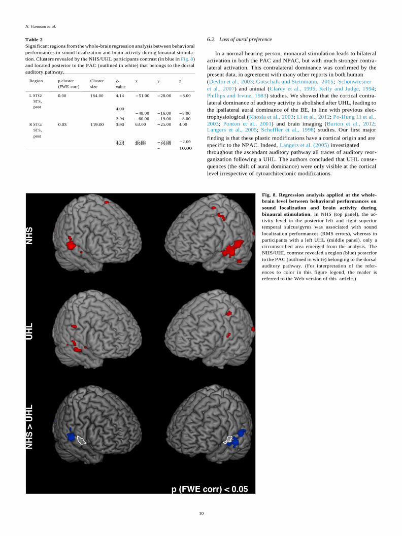
Then, a whole-brain regression analysis with the sound location
scores (RMS errors) was performed. In NHS, there were significant re-
gressions in the left STG/STS (p FWE-corrected < 0.001, peak co-
ordinates -60 -19 -8) and right STG/STS (p FWE-corrected < 0.03, peak
coordinates 63 -25 4). In participants with UHL, a small amount of re-
sidual activity was observed within auditory areas, but no significant
regression of brain activity with sound location scores was found. The
contrast between NHS and UHL groups for this regression was further
explored, revealing that the bilateral STG/STS was significantly
involved in sound localization in NHS (Table 2, Fig. 8). These regions are
posterior to the PAC, and can be considered to belong to the dorsal
auditory pathway (Da Costa et al., 2018). This result clearly demon-
strates that the spatial hearing deficit was associated with a deficiency of
the dorsal auditory stream in participants with UHL.
Taken together, our results specifically point to the impact of UHL on
the functional integrity of the auditory dorsal stream areas, with altered
neuronal binaural interactions (probably due to a shift in aural domi-
nance) responsible for the deficit in sound localization.
5.1.6. Unilateral hearing loss with a left better ear
Only three participants with a left better ear were included in this
study (see Supplementary Information) preventing us from drawing any
strong conclusions. At the PAC level, contralateral dominance seemed to
be preserved following stimulation of the left BE. No clear ipsilateral
shift was observed in the left NPAC in any of these three cases, with WLI
values reflecting dominance of the contralateral right hemisphere (see
Sup Fig. 3 and Sup Fig. 4). However, the limited dataset prevented us
from concluding that cortical reorganization depends on the affected
side, as previously suggested (Hanss et al., 2009).
6. Discussion
Our results show that UHL induces a brain reorganization that affects
auditory processing in the NPAC. This reorganization involves binaural
interaction mechanisms and the pattern of cortical activity lateraliza-
tion. The prolonged alteration of binaural stimulation results in an
ipsilateral aural dominance of the BE in the NPAC, a functional reor-
ganization whose extent is correlated with localization abilities. In
addition, the disruption of binaural processing predominantly affects the
functional integrity of auditory areas in the dorsal stream, reinforcing
the hypothesis of a functional dichotomy of the auditory system.
6.1. Unilateral hearing loss and spatial hearing
Our results are in line with a large body of evidences showing that
unilateral auditory sensory deprivation leads to a degraded integration
of binaural auditory inputs and alters spatial hearing (Abel and Lam,
2008; Kumpik and King, 2019; Scheffler et al., 1998; Slattery III &
Middlebrooks, 1994; Vannson et al., 2017). This degradation primarily

N. Vannson et al.
9
Fig. 7. Regression analysis applied at the whole-
brain level between behavioral performances on
sound discrimination and brain activity during
binaural stimulation. In both NHS (upper panels)
and participants with UHL, we observed a similar
cortical network whose functional activity was pre-
dictive of spatial performances (hit rates; see text). It
included the superior and middle temporal gyri and
ventral temporal areas. Interestingly, in both cases,
the occipital visual areas were also involved. Sagittal,
coronal and horizontal views are illustrated at p un-
corrected = 0.001, for illustration purposes.
affects the binaural auditory cues known as interaural level and time
differences (ILD and ITD), which is the first cause for the deficit in sound
localization and speech in noise comprehension, as observed in all of our
patients. This is clearly reinforced by the observed correlation between
hearing thresholds in the deaf ear (PTA) and the spatial hearing (see also
Vannson et al., 2017). Furthermore, we showed that the asymmetric
binaural inputs lead to abnormal binaural interactions and altered
pattern of cortical lateralization. In addition to the alteration of ITD and
ILD perception, we suggest that the spatial hearing deficit also relies on
the cortical reorganization induced by UHL. Such an assumption is based
on the fact that the cortical reorganization is present only in a specific
cortical region, the NPAC, not in AI and presumably not at lower stages
while the convergence of binaural inputs occurs as early as the brain-
stem level. In a separate population of patients with UHL, as in other
valuable studies (Slattery III & Middlebrooks, 1994), we observed that a
restricted set of patients present near to normal performance for spatial
localization (Alzaher et al. in prep). Such adaptation is probably based
on the optimized use of monaural spectral cues (Kumpik et al., 2010)
and is associated to a pattern of cortical activity different from that
observed in hearing controls (Alzaher et al., in prep). These results add
further evidence that the specific cortical reorganization induced after
long term UHL directly impacts behavioral performances in the spatial
domain.
N. Vannson et al.
10
Table 2
Significant regions from the whole-brain regression analysis between behavioral
performances in sound localization and brain activity during binaural stimula-
tion. Clusters revealed by the NHS/UHL participants contrast (in blue in Fig. 8)
and located posterior to the PAC (outlined in white) that belongs to the dorsal
auditory pathway.
6.2. Loss of aural preference
In a normal hearing person, monaural stimulation leads to bilateral
activation in both the PAC and NPAC, but with much stronger contra-
lateral activation. This contralateral dominance was confirmed by the
present data, in agreement with many other reports in both human Region p cluster
(FWE-corr)
Cluster
size
Z-
value
x y z (Devlin et al., 2003; Gutschalk and Steinmann, 2015; Schonwiesner
et al., 2007) and animal (Clarey et al., 1995; Kelly and Judge, 1994;
L STG/
STS,
post
0.00 184.00 4.14 -51.00 -28.00 -8.00
4.00
Phillips and Irvine, 1983) studies. We showed that the cortical contra-
lateral dominance of auditory activity is abolished after UHL, leading to
the ipsilateral aural dominance of the BE, in line with previous elec-
R STG/
STS,
3.94
0.03 119.00 3.90
-48.00 -16.00 -8.00
-60.00 -19.00 -8.00
63.00 -25.00 4.00
trophysiological (Khosla et al., 2003; Li et al., 2012; Po-Hung Li et al.,
2003; Ponton et al., 2001) and brain imaging (Burton et al., 2012; Langers et al., 2005; Scheffler et al., 1998) studies. Our first major
post
3.70 45.00 3.43 45.00
-22.0025.00
-2.00finding is that these plastic modifications have a cortical origin and are
specific to the NPAC. Indeed, Langers et al. (2005) investigated - 10.00
throughout the ascendant auditory pathway all traces of auditory reor-
ganization following a UHL. The authors concluded that UHL conse-
quences (the shift of aural dominance) were only visible at the cortical
level irrespective of cytoarchitectonic modifications.
Fig. 8. Regression analysis applied at the whole-
brain level between behavioral performances on
sound localization and brain activity during
binaural stimulation. In NHS (top panel), the ac-
tivity level in the posterior left and right superior
temporal sulcus/gyrus was associated with sound
localization performances (RMS errors), whereas in
participants with a left UHL (middle panel), only a
circumscribed area emerged from the analysis. The
NHS/UHL contrast revealed a region (blue) posterior
to the PAC (outlined in white) belonging to the dorsal
auditory pathway. (For interpretation of the refer-
ences to color in this figure legend, the reader is
referred to the Web version of this article.)
N. Vannson et al.
11
Contralateral dominance has been reported at the thalamic level for
individuals with UHL (Langers et al., 2005) and can be observed in the
PAC (present data). This suggests that the ipsilateral dominance
observed in the NPAC results from either local mechanisms or cortico-
cortical interactions. In the NHS group, contralateral dominance was
less pronounced in the NPAC than in the PAC. This smaller imbal-
ance between the inputs from each ear in the NPAC could explain a
greater susceptibility to shift towards the regions ipsilateral to the BE
once contralateral input has decreased owing to UHL. The changes in
lateralization reported here correspond to those observed in animal
models of congenital UHL (Kral et., 2013a, 2013b; Tillein et al., 2016) or
induced (McAlpine et al., 1997; Popelar et al., 1994) with recordings of
neuronal activity demonstrating a clear ipsilateral dominance with
respect to the BE, reflected in a larger population of ipsilateral
responding neurons with an increased excitability and reduced latency.
Of interest, animal models of UHL tend to suggest that this reorganiza-
tion occurs at subcortical levels in the inferior colliculus (McAlpine
et al., 1997) and is present in cortical field A1 (Popelar et al., 1994;
Tillein et al., 2016), whereas in humans, it is only clearly visible at a
higher cortical stage. The origins of these differences are unclear, but
they may be due to a stronger functional specialization of human
auditory cortical areas. Besides interspecies differences for auditory
functions, this discrepancy is probably explained by the fact that most
animal studies on hearing loss explore congenital deafness. Indeed, in
induced animal neonatal deafness, structural and functional modifica-
tion are observed from the cochlear nucleus (Syka, 2002) up to the
primary auditory cortex (Tillein et al., 2016) with the restriction that the
reorganization of the primary auditory cortex is present only if the onset
of unilateral hearing loss appears early in development before 2 years of
age (Kral, 2013). Furthermore, in animal models, changes in ipsilateral
dominance are quite rapid, if not immediate (McAlpine et al., 1997),
whereas in humans, after cochlear nerve resection, they can take several
months (Bilecen et al., 2000; Burton et al., 2012). Altogether, it is
possible that in humans, changes in lateralization rely on gradual
cortical interactions and not on the unmasking of existing inputs.
Several reports suggest that left or right monaural deprivation
differently impacts cortical reorganization (Burton et al., 2012; Moore
et al., 2003), with the assumption that the left hemisphere (right-ear
deafness) is more resistant to cortical plasticity (Hanss et al., 2009).
Here, most of our results originated from participants with left (poorer
ear) UHL, and only a small number of participants with right UHL were
included, preventing us from reaching any firm conclusion as to whether
side of deafness differently impacts brain reorganization and behavioral
performance.
6.3. Alteration of binaural processing for spatial hearing
Our second major finding is that there is a strong relationship be-
tween the extent of cortical reorganization in the NPAC and the
behavioral performances of individuals with UHL. In our study, partic-
ipants with poorer hearing thresholds performed more poorly on sound
localization and had greater ipsilateral lateralization. This indicates that
when the plastic reorganization shifts the lateralization of activity to-
wards an ipsilateral dominance, it reduces the ability to locate sounds in
a free field. We attribute this relation to the alteration of cortical
binaural integration after deafness. At the cortical level, the majority of
neurons are spatially selective to contralateral sounds (Barone et al.,
1996) and have restricted receptive fields constructed through the
excitatory/inhibitory interactions of inputs from each ear (Clarey et al.,
1995; Rajan and Irvine, 1996; Samson et al., 1993). The lack of inhibi-
tory inputs from the ipsilateral ear makes the neurons’ receptive fields
omnidirectional and contributes to a sound localization deficit in
monaural conditions (Clarey et al., 1995). We present evidence that this
alteration of binaural integration is also present in the human auditory
cortex.
In normal hearing persons, binaural stimulation does not correspond
to a linear, additive summation of inputs from each ear, but instead to
suppression (or occlusion; 37) mechanisms (BII below 1; see Fig. 3).
Similar observations of a lower response elicited by binaural stimulation
than the sum of monaural stimulations have been made in fMRI studies
(Jancke et al., 2002; Krumbholz et al., 2005), and have been attributed
to the suppression of ipsilateral inputs when tested in dichotic situations
(Amaral and Langers, 2013; Brancucci et al., 2004; Della Penna et al.,
2006; Fujiki et al., 2002). Consequently, the binaural index reveals the
presence of excitatory/inhibitory interactions in the PAC and NPAC of
hearing humans. UHL profoundly alters binaural integration, reflecting
the reduction (in PAC) or absence (in NPAC) of inhibitory inputs from
the PE. In animal models of congenital UHL, electrophysiological re-
cordings have provided evidence of altered binaural integration pro-
cessing (Tillein et al., 2016), resulting in decreased inhibition in the
ipsilateral hemisphere and increased inhibition in the hemisphere
opposite the BE. Thus, both human and animal studies point to the same
conclusion of an alteration of inhibitory interactions following UHL.
6.4. Consequences of altered spatial hearing on cortical reorganization for
auditory rehabilitation
The main conclusion of our analysis is that UHL induces reorgani-
zation of cortical inputs that modifies the functional asymmetry of the
auditory cortex, a plastic phenomenon that is deleterious to spatial
hearing abilities. In order to assess the potential UHL auditory rehabil-
itation strategy, the level of cortical asymmetry could be used as an
objective marker of brain plasticity. Indeed, within developing children,
this plastic reorganization has been shown to circumvent the rehabili-
tation of the deaf ear with a cochlear implant (Polonenko et al., 2017)
and has impacts beyond auditory functions, including on language
acquisition (Schaefer et al., 2019). In many cases, however, any alter-
ation of a sensory function is accompanied by intra- or crossmodal
compensatory mechanisms, brought about plastic adaptation (Singh
et al., 2018). Thus, the impact of UHL on binaural integration may favor
processing by the hearing ear (Gordon and Kral, 2019) and produce
some perceptual advantages in patients with UHL. These advantages
need to be established, although improvements in intensity discrimi-
nation (Maslin et al., 2015) or spatial hearing through monaural spectral
cues extraction have been reported (Slattery III & Middlebrooks, 1994).
6.5. Spatial hearing and dorsal stream in UHL
The regression analysis based on either spatial (sound location) or
non-spatial (sound discrimination) performances showed that they are
supported by different auditory cortical networks in UHL. Rather than
revealing areas that were differently activated by the two tasks, the
regression analysis revealed areas whose functional integrity (capacity
to respond) could be predictive of behavioral performances.
The areas involved in voice/non-voice discrimination are manifold,
as the different acoustic attributes of these two types of stimulus need to
be processed in order to form semantic representations. In the partici-
pants with UHL, the auditory areas related to their behavioral perfor-
mances perfectly matched those observed in NHS, and included the
lateral parts of the STG, middle temporal gyrus, and ventral temporal
areas. This regression highlighted areas of the STS, including the tem-
poral voice area, an auditory area specifically sensitive to the human
voice (Belin et al., 2000). In both groups, the involvement of the visual
occipital and parietal areas probably reflects the creation of an internal
representation when making a decision about semantic sound
categorization.
When the analysis was applied to spatial hearing performance, it
showed that the cortical network which supports sound localization
skills was very circumscribed in participants with UHL compared to that
of NHS. In the latter, sound location performance was associated with a
small set of auditory areas that mainly encompassed the posterior parts
of the STG/STS region, which is known to belong to the dorsal auditory

N. Vannson et al.
12
stream (Da Costa et al., 2018). Furthermore, the contrast with NHS
revealed that this posterior region was functionally absent in partici-
pants with UHL, concerning sound localization performances, in line
with a study that reported a deficit in sound localization when TMS
pulses targeted this posterior region (Ahveninen et al., 2013). Thus, our
data clearly demonstrate that the functional integrity of the dorsal
stream areas is affected following UHL, which impairs sound localiza-
tion processing.
These results are of theoretical importance, as they provide addi-
tional support for the hypothesis of a functional dichotomy of the
auditory cortex, which is now widely accepted for both carnivores and
nonhuman primates on anatomical and electrophysiological grounds
(Kaas and Hackett, 1998; Lomber and Malhotra, 2008; Rauschecker and
Tian, 2000). In humans, a similar dichotomy has been proposed, based
on observation of behavioral deficits following cortical lesions (Clarke
et al., 2002; Duffour-Nikolov et al., 2012; Zündorf et al., 2016) and brain
activation analysis (Arnott et al., 2004; Van der Zwaag et al., 2011). Our
study focusing on brain plasticity in UHL provides an original argument
in favor of this functional cortical dissociation.
Lastly our results are of interest for the rehabilitation of patients with
single-sided deafness treated by a cochlear implant (single-sided
deafness-SSD; Arndt et al., 2011; Cabral Junior et al., 2016; Vander-
auwera et al., 2020). Indeed, the importance of brain plasticity mech-
anisms has been shown for a successful hearing rehabilitation (Lazard
et al., 2014) and the restoration of a normal aural dominance could be
beneficial to an optimal functional recuperation. A handful of studies in
developing children (Polonenko et al., 2017; Sharma et al., 2016) and
adult (Legris et al., 2018) SSD patients are encouraging and suggest that
a normal pattern of cortical activity could be obtained after restoring the
binaural auditory inputs using cochlear implantation in the deaf ear.
7. Conclusion
The present study revealed that the auditory cortex undergoes major
functional reorganization after acquired UHL. Monaural sensory depri-
vation leads to an increase in the aural dominance of the hearing ear,
which impacts mainly on the NPAC and disrupts binaural integration.
This plastic reorganization is deleterious to spatial hearing abilities,
because it disrupts the functional integrity of the dorsal stream network.
Authors contibutions
NV, KS, BP, MM designed the experiment. NV, OD, MM recruited the
patients and run the experiments. NV and KS analyzed the data. NV, KS,
CJ, OD, BP, MM wrote and corrected the manuscript.
Fundings
This study was supported by an ANRT-Cochlear CIFRE PhD funding
(N◦2012/1463) to NV, a grant Agir pour l’audition (APA-RD2015-6B) to
MM, OD and BP and the recurrent funding of the CNRS.
Declaration of competing interest
NV and CJ are Cochlear employees.
Acknowledgements
The authors are very thankful to all patients and control subjects
included in this study. The authors are also grateful to Yohan Gallois,
Charles Edouard Molinier, and Gaetan Iversenc for their help in
recruiting and testing the patients. The authors would also like to
acknowledge Nathalie Vayssiere, Helene Gros-Dagnac and all the tech-
nical team of the MRI platform of INSERM U1214 ToNIC for the scan-
ning sessions.
Appendix A. Supplementary data
Supplementary data to this article can be found online at https://doi.
org/10.1016/j.neuropsychologia.2020.107683.
Data availability
All data are available from the authors upon reasonable request.
References
Abel, S.M., Lam, K., 2008. Impact of unilateral hearing loss on sound localization. Appl.
Acoust. 69 (9), 804–811.
Adriani, M., Bellmann, A., Meuli, R., Fornari, E., Frischknecht, R., Bindschaedler, C.,
Rivier, F., Thiran, J.-P., Maeder, P., Clarke, S., 2003. Unilateral hemispheric lesions
disrupt parallel processing within the contralateral intact hemisphere: an auditory
fMRI study. Neuroimage 20, S66–S74.
Ahveninen, J., Huang, S., Nummenmaa, A., Belliveau, J.W., Hung, A.-Y., Jaaskelainen, I.
P., Rauschecker, J.P., Rossi, S., Tiitinen, H., Raij, T., 2013. Evidence for distinct
human auditory cortex regions for sound location versus identity processing. Nat.
Commun. 4, 2585. https://doi.org/10.1038/ncomms3585.
Amaral, A.A., Langers, D.R., 2013. The relevance of task-irrelevant sounds: hemispheric
lateralization and interactions with task-relevant streams. Front. Neurosci. 7, 264.
Arndt, S., Aschendorff, A., Laszig, R., Beck, R., Schild, C., Kroeger, S., Ihorst, G.,
Wesarg, T., 2011. Comparison of pseudobinaural hearing to real binaural hearing
rehabilitation after cochlear implantation in patients with unilateral deafness and
tinnitus. Otol. Neurotol. 32 (1), 39–47.
Arnott, S.R., Binns, M.A., Grady, C.L., Alain, C., 2004. Assessing the auditory dual-
pathway model in humans. Neuroimage 22 (1), 401–408.
Barone, P., Clarey, J.C., Irons, W.A., Imig, T.J., 1996. Cortical synthesis of azimuth-
sensitive single-unit responses with nonmonotonic level tuning: a thalamocortical
comparison in the cat. J. Neurophysiol. 75 (3), 1206–1220.
Belin, P., Zatorre, R.J., Hoge, R., Evans, A.C., Pike, B., 1999. Event-related fMRI of the
auditory cortex. Neuroimage 10 (4), 417–429.
Belin, P., Zatorre, R.J., Lafaille, P., Ahad, P., Pike, B., 2000. Voice-selective areas in
human auditory cortex. Nature 403 (6767), 309.
Bilecen, D., Seifritz, E., Radü, E.W., Schmid, N., Wetzel, S., Probst, R., Scheffler, K., 2000.
Cortical reorganization after acute unilateral hearing loss traced by fMRI. Neurology
54 (3), 765, 765.
Brancucci, A., Babiloni, C., Babiloni, F., Galderisi, S., Mucci, A., Tecchio, F.,
Zappasodi, F., Pizzella, V., Romani, G.L., Rossini, P.M., 2004. Inhibition of auditory
cortical responses to ipsilateral stimuli during dichotic listening: evidence from
magnetoencephalography. Eur. J. Neurosci. 19 (8), 2329–2336.
Bronkhorst, A.W., Plomp, R., 1989. Binaural speech intelligibility in noise for hearing-
impaired listeners. J. Acoust. Soc. Am. 86 (4), 1374–1383.
Burton, H., Firszt, J.B., Holden, T., Agato, A., Uchanski, R.M., 2012. Activation
lateralization in human core, belt, and parabelt auditory fields with unilateral
deafness compared to normal hearing. Brain Res. 1454, 33–47.
Cabral Junior, F., Pinna, M.H., Alves, R.D., Malerbi, A.F., dos, S., Bento, R.F., 2016.
Cochlear implantation and single-sided deafness: a systematic review of the
literature. Int. Arch. Otorhinolaryngol. 20 (1), 69–75.
Carpenter, J., Bithell, J., 2000. Bootstrap confidence intervals: when, which, what? A
practical guide for medical statisticians. Stat. Med. 19 (9), 1141–1164.
Clarey, J.C., Barone, P., Irons, W.A., Samson, F.K., Imig, T.J., 1995. Comparison of noise
and tone azimuth tuning of neurons in cat primary auditory cortex and medical
geniculate body. J. Neurophysiol. 74 (3), 961–980.
Clarke, S., Thiran, A.B., Maeder, P., Adriani, M., Vernet, O., Regli, L., Cuisenaire, O.,
Thiran, J.-P., 2002. What and where in human audition: selective deficits following
focal hemispheric lesions. Exp. Brain Res. 147 (1), 8–15.
Da Costa, S., Clarke, S., Crottaz-Herbette, S., 2018. Keeping track of sound objects in
space: the contribution of early-stage auditory areas. Hear. Res. 366, 17–31.
Della Penna, S., Brancucci, A., Babiloni, C., Franciotti, R., Pizzella, V., Rossi, D.,
Torquati, K., Rossini, P.M., Romani, G.L., 2006. Lateralization of dichotic speech
stimuli is based on specific auditory pathway interactions: neuromagnetic evidence.
Cerebr. Cortex 17 (10), 2303–2311.
Devlin, J.T., Raley, J., Tunbridge, E., Lanary, K., Floyer-Lea, A., Narain, C., Cohen, I.,
Behrens, T., Jezzard, P., Matthews, P.M., 2003. Functional asymmetry for auditory
processing in human primary auditory cortex. J. Neurosci. 23 (37), 11516–11522.
Duffour-Nikolov, C., Tardif, E., Maeder, P., Thiran, A.B., Bloch, J., Frischknecht, R.,
Clarke, S., 2012. Auditory spatial deficits following hemispheric lesions: dissociation
of explicit and implicit processing. Neuropsychol. Rehabil. 22 (5), 674–696.
Eickhoff, S.B., Stephan, K.E., Mohlberg, H., Grefkes, C., Fink, G.R., Amunts, K., Zilles, K.,
2005. A new SPM toolbox for combining probabilistic cytoarchitectonic maps and
functional imaging data. Neuroimage 25 (4), 1325–1335.
Fujiki, N., Jousmaki, V., Hari, R., 2002. Neuromagnetic responses to frequency-tagged
sounds: a new method to follow inputs from each ear to the human auditory cortex
during binaural hearing. J. Neurosci. 22 (3), RC205. RC205.
Gordon, K., Kral, A., 2019. Animal and human studies on developmental monaural
hearing loss. Hear. Res. 380, 60–74.
Grothe, B., Pecka, M., McAlpine, D., 2010. Mechanisms of sound localization in
mammals. Physiol. Rev. 90 (3), 983–1012.
Gutschalk, A., Steinmann, I., 2015. Stimulus dependence of contralateral dominance in
human auditory cortex. Hum. Brain Mapp. 36 (3), 883–896.

N. Vannson et al.
13
Hanss, J., Veuillet, E., Adjout, K., Besle, J., Collet, L., Thai-Van, H., 2009. The effect of
long-term unilateral deafness on the activation pattern in the auditory cortices of
French-native speakers: influence of deafness side. BMC Neurosci. 10 (1), 23.
Heinrich, A., Mikkola, T.M., Polku, H., Tormakangas, T., Viljanen, A., 2019. Hearing in
real-life environments (HERE): structure and reliability of a questionnaire on
perceived hearing for older adults. Ear Hear. 40 (2), 368–380.
Jamison, H.L., Watkins, K.E., Bishop, D.V., Matthews, P.M., 2006. Hemispheric
specialization for processing auditory nonspeech stimuli. Cerebr. Cortex 16 (9),
1266–1275.
Jancke, L., Wüstenberg, T., Scheich, H., Heinze, H.-J., 2002. Phonetic perception and the
temporal cortex. Neuroimage 15 (4), 733–746. https://doi.org/10.1006/
nimg.2001.1027.
Jansen, S., Luts, H., Wagener, K.C., Kollmeier, B., Del Rio, M., Dauman, R., James, C.,
Fraysse, B., Vormes, E., Frachet, B., 2012. Comparison of three types of French
speech-in-noise tests: a multi-center study. Int. J. Audiol. 51 (3), 164–173.
Kaas, J.H., Hackett, T.A., 1998. Subdivisions of AuditoryCortex and levels of processing
in primates. Audiology and Neurotology 3 (2–3), 73–85. https://doi.org/10.1159/
000013783.
Kelly, J.B., Judge, P.W., 1994. Binaural organization of primary auditory cortex in the
ferret (Mustela putorius). J. Neurophysiol. 71 (3), 904–913.
Khosla, D., Ponton, C.W., Eggermont, J.J., Kwong, B., Dort, M., Vasama, J.-P., 2003.
Differential ear effects of profound unilateral deafness on the adult human central
auditory system. Journal of the Association for Research in Otolaryngology 4 (2),
235–249.
Kral, A., 2013. Auditory critical periods: a review from system ’s perspective.
Neuroscience 247, 117–133.
Kral, A., Land, R., Baumhoff, P., Tillein, J., Hubka, P., Lomber, S.G., 2013. C ross-modal
plasticity in the congenitally deaf cat. Multisensory Res. 26, 37, 37.
Kral, Andrej, Hubka, P., Heid, S., Tillein, J., 2013. Single-sided deafness leads to
unilateral aural preference within an early sensitive period. Brain 136 (1), 180–193.
Krumbholz, K., Schonwiesner, M., Rübsamen, R., Zilles, K., Fink, G.R., Von Cramon, D.Y.,
2005. Hierarchical processing of sound location and motion in the human brainstem
and planum temporale. Eur. J. Neurosci. 21 (1), 230–238.
Kumpik, D.P., Kacelnik, O., King, A.J., 2010. Adaptive reweighting of auditory
localization cues in response to chronic unilateral earplugging in humans.
J. Neurosci. 30 (14), 4883–4894.
Kumpik, D.P., King, A.J., 2019. A review of the effects of unilateral hearing loss on
spatial hearing. Hear. Res. 372, 17–28.
Langers, D.R., van Dijk, P., Backes, W.H., 2005. Lateralization, connectivity and
plasticity in the human central auditory system. Neuroimage 28 (2), 490–499.
Lazard, D.S., Innes-Brown, H., Barone, P., 2014. Adaptation of the communicative brain
to post-lingual deafness. Evidence from functional imaging. Hear. Res. 307, 136–
143.
Legris, E., Galvin, J., Roux, S., Gomot, M., Aoustin, J.-M., Marx, M., He, S., Bakhos, D.,
2018. Cortical reorganization after cochlear implantation for adults with single-
sided deafness. PloS One 13 (9), e0204402.
Li, L.P.-H., Shiao, A.-S., Chen, K.-C., Lee, P.-L., Niddam, D.M., Chang, S.-Y., Hsieh, J.-C.,
2012. Neuromagnetic index of hemispheric asymmetry prognosticating the outcome
of sudden hearing loss. PloS One 7 (4), e35055.
Lomber, S.G., Malhotra, S., 2008. Double dissociation of’what’and’where’processing in
auditory cortex. Nat. Neurosci. 11 (5), 609.
Maslin, M.R., Taylor, M., Plack, C.J., Munro, K.J., 2015. Enhanced intensity
discrimination in the intact ear of adults with unilateral deafness. J. Acoust. Soc. Am.
137 (6), EL408–EL414.
Massida, Z., Belin, P., James, C., Rouger, J., Fraysse, B., Barone, P., Deguine, O., 2011.
Voice discrimination in cochlear-implanted deaf subjects. Hear. Res. 275 (1–2),
120–129.
McAlpine, D., Martin, R.L., Mossop, J.E., Moore, D.R., 1997. Response properties of
neurons in the inferior colliculus of the monaurally deafened ferret to acoustic
stimulation of the intact ear. J. Neurophysiol. 78 (2), 767–779.
Moore, D.R., Hartley, D.E., Hogan, S.C., 2003. Effects of otitis media with effusion (OME)
on central auditory function. Int. J. Pediatr. Otorhinolaryngol. 67, S63–S67.
Morosan, P., Rademacher, J., Palomero-Gallagher, N., Zilles, K., 2005. Anatomical
organization of the human auditory cortex: cytoarchitecture and transmitter
receptors. The Auditory Cortex. Psychology Press, pp. 45–68.
Morosan, P., Rademacher, J., Schleicher, A., Amunts, K., Schormann, T., Zilles, K., 2001.
Human primary auditory cortex: cytoarchitectonic subdivisions and mapping into a
spatial reference system. Neuroimage 13 (4), 684–701.
Oldfield, R.C., 1971. The assessment and analysis of handedness: the Edinburgh
inventory. Neuropsychologia 9 (1), 97–113.
Phillips, D.P., Irvine, D.R., 1983. Some features of binaural input to single neurons in
physiologically defined area AI of cat cerebral cortex. J. Neurophysiol. 49 (2), 383–
395.
Po-Hung Li, L., Shiao, A.-S., Lin, Y.-Y., Chen, L.-F., Niddam, D.M., Chang, S.-Y., Lien, C.-
F., Chou, N.-S., Ho, L.-T., Hsieh, J.-C., 2003. Healthy-side dominance of cortical
neuromagnetic responses in sudden hearing loss. Ann. Neurol. 53 (6), 810–815.
Polonenko, M.J., Gordon, K.A., Cushing, S.L., Papsin, B.C., 2017. Cortical organization
restored by cochlear implantation in young children with single sided deafness. Sci.
Rep. 7 (1), 16900.
Ponton, C.W., Vasama, J.-P., Tremblay, K., Khosla, D., Kwong, B., Don, M., 2001.
Plasticity in the adult human central auditory system: evidence from late-onset
profound unilateral deafness. Hear. Res. 154 (1–2), 32–44.
Popelar, J., Erre, J.-P., Aran, J.-M., Cazals, Y., 1994. Plastic changes in ipsi-contralateral
differences of auditory cortex and inferior colliculus evoked potentials after injury to
one ear in the adult Guinea pig. Hear. Res. 72 (1–2), 125–134.
Rajan, R., Irvine, D.R., 1996. Features of, and boundary conditions for, lesion-induced
reorganization of adult auditory cortical maps. Auditory System Plasticity and
Regeneration 224–237.
Rauschecker, J.P., Scott, S.K., 2009. Maps and streams in the auditory cortex: nonhuma n
primates illuminate human speech processing. Nat. Neurosci. 12 (6), 718.
Rauschecker, J.P., Tian, B., 2000. Mechanisms and streams for processing of “what” and
“where” in auditory cortex. Proc. Natl. Acad. Sci. Unit. States Am. 97 (22), 11800–
11806.
Sadoun, A., Chauhan, T., Mameri, S., Zhang, Y.F., Barone, P., Deguine, O., Strelnikov, K.,
2020. Stimulus-specific information is represented as local activity patterns across
the brain. Neuroimage 223, 117326. https://doi.org/10.1016/j.
neuroimage.2020.117326.
Samson, F.K., Clarey, J.C., Barone, P., Imig, T.J., 1993. Effects of ear plugging on single-
unit azimuth sensitivity in cat primary auditory cortex. I. Evidence for monaural
directional cues. J. Neurophysiol. 70 (2), 492–511.
Schaefer, S.M., de Kruijf, M., Henderson, L., Metryka, A., O ’Driscoll, M., Bruce, I.A.,
2019. Improved speech and language development after unilateral cochlear
implantation in children with a potentially useable contralateral ear. Cochlear
Implants Int. 20 (1), 39–46.
Scheffler, K., Bilecen, D., Schmid, N., Tschopp, K., Seelig, J., 1998. Auditory cortical
responses in hearing subjects and unilateral deaf patients as detected by functional
magnetic resonance imaging. New York, NY: 1991 Cerebr. Cortex 8 (2), 156–163.
Schonwiesner, M., Novitski, N., Pakarinen, S., Carlson, S., Tervaniemi, M., Naatanen, R.,
2007. Heschl ’s gyrus, posterior superior temporal gyrus, and mid-ventrolateral
prefrontal cortex have different roles in the detection of acoustic changes.
J. Neurophysiol. 97 (3), 2075–2082.
Sharma, A., Glick, H., Campbell, J., Torres, J., Dorman, M., Zeitler, D.M., 2016. Cortical
plasticity and Re-organization in pediatric single-sided deafness pre-and post-
cochlear implantation: a case study. Otology & neurotology: Official Publication of
the American Otological Society, American Neurotology Society [and] European
Academy of Otology and Neurotology 37 (2), e26.
Singh, A.K., Phillips, F., Merabet, L.B., Sinha, P., 2018. Why does the cortex reor ganize
after sensory loss? Trends Cognit. Sci. 22 (7), 569–582.
Slattery III, W.H., Middlebrooks, J.C., 1994. Monaural sound localization: acute versus
chronic unilateral impairment. Hear. Res. 75 (1–2), 38–46.
Stefanatos, G.A., Joe, W.Q., Aguirre, G.K., Detre, J.A., Wetmore, G., 2008. Activation of
human auditory cortex during speech perception: effects of monaural, binaural, and
dichotic presentation. Neuropsychologia 46 (1), 301–315.
Suzuki, M., Kouzaki, H., Nishida, Y., Shiino, A., Ito, R., Kitano, H., 2002. Cortical
representation of hearing restoration in patients with sudden deafness. Neuroreport
13 (14), 1829–1832.
Syka, J., 2002. Plastic changes in the central auditory system after hearing loss,
restoration of function, and during learning. Physiol. Rev. 82 (3), 601–636.
Tillein, J., Hubka, P., Kral, A., 2016. Monaural congenital deafness affects aural
dominance and degrades binaural processing. Cerebr. Cortex 26 (4), 1762–1777.
van der Heijden, K., Rauschecker, J.P., de Gelder, B., Formisano, E., 2019. Cortical
mechanisms of spatial hearing. Nat. Rev. Neurosci. 20 (10), 609–623.
van der Heijden, K., Rauschecker, J.P., Formisano, E., Valente, G., de Gelder, B., 2018.
Active sound localization sharpens spatial tuning in human primary auditory cortex.
J. Neurosci. 38 (40), 8574–8587.
Van der Zwaag, W., Gentile, G., Gruetter, R., Spierer, L., Clarke, S., 2011. Where sound
position influences sound object representations: a 7-T fMRI study. Neuroimage 54
(3), 1803–1811.
Van Wanrooij, M.M., Van Opstal, A.J., 2007. Sound localization under perturbed
binaural hearing. J. Neurophysiol. 97 (1), 715–726.
Vanderauwera, J., Hellemans, E., Verhaert, N., 2020. Research insights on neural effects
of auditory deprivation and restoration in unilateral hearing loss: a systematic
review. J. Clin. Med. 9 (3), 812.
Vannson, N., James, C., Fraysse, B., Strelnikov, K., Barone, P., Deguine, O., Marx, M.,
2015. Quality of life and auditory performance in adults with asymmetric hearing
loss. Audiology and Neurotology 20 (Suppl. 1), 38–43.
Vannson, N., James, C.J., Fraysse, B., Lescure, B., Strelnikov, K., Deguine, O., Barone, P.,
Marx, M., 2017. Speech-in-noise perception in unilateral hearing loss: relation to pure-
tone thresholds and brainstem plasticity. Neuropsychologia 102, 135–143.
Vasama, J.-P., Makela, J.P., 1995. Auditory pathway plasticity in adult humans after
unilateral idiopathic sudden sensorineural hearing loss. Hear. Res. 87 (1–2), 132–
140.
Weeks, R., Horwitz, B., Aziz-Sultan, A., Tian, B., Wessinger, C.M., Cohen, L.G.,
Hallett, M., Rauschecker, J.P., 2000. A positron emission tomographic study of
auditory localization in the congenitally blind. J. Neurosci. 20 (7), 2664–2672.
Zündorf, I.C., Lewald, J., Karnath, H.-O., 2016. Testing the dual-pathway model for
auditory processing in human cortex. Neuroimage 124, 672–681.