Embed Size (px)
Citation preview
1.1 Editors
© German Guideline Program in Oncology | Evidence-based Guideline Breast Cancer EN - V4.4 | May 2021
0
Evidence-based Guideline
for the Early Detection,
Diagnosis, Treatment and
Follow-up of Breast
Cancer
Version 4.4 - May 2021
AWMF-Registernummer: 032/045OL
1.1 Editors
© German Guideline Program in Oncology | Evidence-based Guideline Breast Cancer - V4.4 | May 2021
0
Important Updates
Concerning editorial changes in versions 4.1, 4.2, and 4.3, see chapter 1.6.
Version 4.4 (May 2021) was updated as an amendment.
The following chapters were updated:
Chapter 6.4.1
Chapter 6.4.2.
The update in the scope of the amendment was triggered by the authroisation of
numerous CDK4/6 inhibitors.
In versions 4.1 to 4.3, no content-related changes were made (see see chapter 1.6).
Version 4.0 (2017)
The recommendations contained in the chapter on patient infeormation and
Eductaion have been expanded and adpated to the information needs of the patients
(see chapter 3.2)
Early Detection (previously a separate S3 Practice Guideline) has been incorporated
herein: Specific recommendations therein describe the approach to be used in
patients with a high breast density and how to handle specific examination
procedures (e.g. tomosynthesis) (see Chapter 3.2).
In the past years, a growing amount of data has become available on women with an
elevated familial or genetic predisposition and on the cancer occurrence in such
special settings. In other words, the recommendations give explicit instructions and
options on how to proceed (e.g. platinum-containing systemic therapy, counseling
services and dealing with the increased lifetime risk of further malignancies) (see
Chapter 3.3).
The Diagnostics chapter contains new recommendations on staging alongside the
conventional breast-related gynecological examinations. For example, a CT scan of
the chest and abdomen is now explicitly recommended in patients with a high risk of
recurrence or metastases (see Chapter 4.1).
In the current version, Chapter 4.4 Surgical treatment of invasive carcinoma takes into
account more modern surgical procedures and the method applied to axillary
staging, while particularly focusing on the growing use of neoadjuvant therapies.
Overall, the new recommendations are intended to reduce radical surgeries
(narrowing the safety margin in the resection of invasive carcinoma, omitting axillary
dissection under pre-defined conditions).
The recommendations on pathomorphological examinations also include more recent
markers. This Guideline also counts Ki67 among the conventional prognostic factors
and allows the use of multigene assays in defined settings (see Chapter 4.5).
As in the surgical setting, the recommendations on radiotherapy increasingly follow
de-escalation strategies: Procedures like hypofractionation and partial breast
radiation alone can be discussed with the patients, especially those with advanced
age (see Chapter 4.6).
1.1 Editors
© German Guideline Program in Oncology | Evidence-based Guideline Breast Cancer - V4.4 | May 2021
1
The updated S3 Practice Guideline outlines in detail the options for adjuvant
endocrine therapy, including that of prolonged administration (5-10 years) and the
administration of chemotherapies (see Chapter 4.7).
New recommendations regarding influenceable lifestyle factors are designed to
increase patients’ physical activity and weight loss in order to lower their risk of
recurrence and heighten their subjective well-being (see Chapter 4.7.7).
The updated Guideline also gives clear recommendations relating to settings with
metastasis and local recurrences: In patients with local recurrences, for example,
options for repeated radiotherapy and reinduction of cytostatics should be evaluated
besides complete resection (see Chapters 5.3 and 5.4).
In addition to the previous and now updated chapters from the 2012 Guideline, new
chapters have been developed by the authors to do justice to the high therapeutic
relevance of their subject matter and more extensive evidence. The following
chapters have been added:
Chapter 4.7.6 Bone-targeted therapy
Chapter 4.7.7 Influenceable lifestyle factors
Chapter 7 Breast cancer during pregnancy and lactation, pregnancy after breast
cancer, fertility preservation
Chapter 8 Breast cancer in elderly patients
Chapter 9. Breast cancer in men
1.1 Editors
© German Guideline Program in Oncology | Evidence-based Guideline Breast Cancer - V4.4 | May 2021
2
1. Information about this Guideline ..................................................... 9
1.1. Editors .................................................................................................................................. 9
1.2. Leading Scientific Societies .................................................................................................. 10
1.3. Funding of the Guideline ..................................................................................................... 10
1.4. Contact ............................................................................................................................... 10
1.5. How to cite.......................................................................................................................... 10
1.6. Previous Changes ................................................................................................................ 10
1.7. Special Comment ................................................................................................................ 11
1.8. Objectives of the Guideline Program for Oncology ............................................................... 11
1.9. Additional Documents relating to this Guideline .................................................................. 12
1.10. Composition of the Guideline Group .................................................................................... 12
1.10.1.Guideline Coordination ....................................................................................................... 12
1.10.2.Participating professional associations and organizations .................................................... 12
1.10.3.Additional Parties without voting Power .............................................................................. 21
1.10.4.Patient Involvement ............................................................................................................ 23
1.10.5.Methodological Support ...................................................................................................... 23
1.11. Abbreviations Used ............................................................................................................. 24
2. Introduction .................................................................................. 28
2.1. Scope and Purpose .............................................................................................................. 28
2.1.1.Objective and Key Questions ................................................................................................. 28
2.1.2.Validity and Update Process .................................................................................................. 29
2.2. Methodology ....................................................................................................................... 29
2.2.1.Levels of Evidence (LoE) ........................................................................................................ 29
2.2.2.Grades of Recommendation (GoR) ......................................................................................... 32
2.2.3.Statements ........................................................................................................................... 33
2.2.4.Expert Consensus (EK) .......................................................................................................... 33
2.2.5.Independence and Disclosure of Possible Conflicts of Interest ............................................... 33
3. General ......................................................................................... 35
3.1. Patient information and education ....................................................................................... 35
3.1.1.Informing the patient about diagnosis ................................................................................... 37
3.1.2.Educating the patient about treatment .................................................................................. 38

1.1 Editors
© German Guideline Program in Oncology | Evidence-based Guideline Breast Cancer - V4.4 | May 2021
3
3.2. Early detection, mammographic screening ........................................................................... 41
3.2.1.Shared decision-making ....................................................................................................... 44
3.2.2.Mammographic screening ..................................................................................................... 45
3.2.3.Measures for the early detection of breast cancer .................................................................. 49
3.2.3.1. Sonography ................................................................................................................. 50
3.2.3.2. Complementary diagnostic imaging in high mammographic density for early detection 51
3.2.4.Needs for research for the early detection of breast cancer .................................................... 52
3.3. Women at increased risk of developing breast cancer .......................................................... 53
3.3.1.Familial breast cancer ........................................................................................................... 53
4. Locoregional primary disease ......................................................... 64
4.1. General diagnostic and therapeutic concepts ....................................................................... 64
4.2. Diagnostics on abnormal findings and pretherapeutic diagnosis of spread in confirmed breast
cancer 65
4.2.1.Basic diagnostic workup........................................................................................................ 65
4.2.2.Imaging methods .................................................................................................................. 67
4.2.3.Diagnostic confirmation ........................................................................................................ 71
4.3. DCIS and high-risk lesions .................................................................................................. 76
4.3.1.Preliminary remarks .............................................................................................................. 76
4.3.2.DCIS ..................................................................................................................................... 76
4.3.2.1. Clinical presentation, risk and course in DCIS ............................................................... 76
4.3.2.2. Surgical therapy of DCIS ............................................................................................... 78
4.3.2.3. Radiotherapy of the DCIS ............................................................................................. 80
4.3.2.4. Antihormone therapy in DCIS ....................................................................................... 81
4.3.3.Risk lesions .......................................................................................................................... 81
4.3.3.1. Preliminary remarks ..................................................................................................... 81
4.3.3.2. Atypical ductal hyperplasia (ADH) in punch or vacuum biopsy ....................................... 82
4.3.3.3. Lobular neoplasia (LN) in punch or vacuum biopsy ....................................................... 83
4.3.3.4. Flat epithelial atype (FEA) in punch or vacuum biopsy ................................................... 84
4.3.3.5. ADH, LN, FEA in open biopsy ........................................................................................ 84
4.3.3.6. Papilloma in the punch or vacuum biopsy ..................................................................... 85
4.3.3.7. Papilloma in open PE .................................................................................................... 86
4.4. Surgical therapy of invasive carcinoma................................................................................. 86
4.4.1.General recommendation ...................................................................................................... 86
4.4.2.Breast conserving therapy ..................................................................................................... 87
4.4.3.Mastectomy .......................................................................................................................... 89
4.4.4.Plastic reconstructive surgeries ............................................................................................. 91
4.4.5.Surgical therapy of the axilla ................................................................................................. 91
1.1 Editors
© German Guideline Program in Oncology | Evidence-based Guideline Breast Cancer - V4.4 | May 2021
4
4.5. Pathomorphological examination......................................................................................... 96
4.5.1.Preliminary remarks .............................................................................................................. 96
4.5.2.General principles ................................................................................................................. 96
4.5.2.1. General patient data, preliminary findings, anamnestic information .............................. 96
4.5.2.2. Documentation of the macroscopic processing ............................................................. 97
4.5.2.3. Documentation of the microscopic processing and assessment .................................... 97
4.5.2.4. Clarification of mammographically detected microcalcification ..................................... 98
4.5.2.5. frozen section examination .......................................................................................... 99
4.5.2.6. Histological classification and grading ......................................................................... 99
4.5.2.6.1. Histological classification ................................................................................... 100
4.5.2.6.2. Expansion of intraductal tumor component......................................................... 100
4.5.2.6.3. Histological grading ........................................................................................... 100
4.5.2.6.4. DCIS-Grading ..................................................................................................... 101
4.5.2.7. Multifocality/multicentricity ....................................................................................... 101
4.5.2.8. Peritumoral lymph vessel invasion .............................................................................. 101
4.5.3.Determination of hormone receptor and HER2 status and the Ki-67 proliferation index of
invasive carcinomas ..................................................................................................................... 102
4.5.3.1. Interpretation Hormonrezeptorstatus ......................................................................... 104
4.5.3.2. Evaluation Ki-67 proliferation index ........................................................................... 108
4.5.4.Prognostic and predictive factors ........................................................................................ 109
4.5.4.1. uPA/PAI-1 ................................................................................................................. 112
4.5.4.2. Ki-67......................................................................................................................... 112
4.5.4.3. Intrinsic subtypes ...................................................................................................... 113
4.5.4.4. Multi-gene tests ........................................................................................................ 114
4.5.5.Percutaneous biopsies in the context of interventional diagnostics ...................................... 120
4.5.5.1. Percutaneous biopsy (high-speed punch biopsy, vacuum biopsy) ............................... 121
4.5.5.1.1. Macroscopic processing...................................................................................... 121
4.5.5.1.2. Microscopic processing and assessment ............................................................. 121
4.5.5.2. Fine needle puncture/aspiration cytology (FNAC) ........................................................ 124
4.5.6.Excision biopsies ................................................................................................................ 124
4.5.6.1. Macroscopic processing ............................................................................................. 124
4.5.6.2. Microscopic processing and assessment ..................................................................... 126
4.5.7.Mastectomy specimens ....................................................................................................... 128
4.5.7.1. Macroscopic examination ........................................................................................... 128
4.5.7.2. Microscopic examination and assessment .................................................................. 129
4.5.8.Lymph nodes ...................................................................................................................... 129
4.5.8.1. Macroscopic examination ........................................................................................... 130
4.5.8.2. Microscopic examination and assessment .................................................................. 130
4.6. Adjuvant radiothepary of breastc ancer.............................................................................. 132

1.1 Editors
© German Guideline Program in Oncology | Evidence-based Guideline Breast Cancer - V4.4 | May 2021
5
4.7. Systemic adjuvant therapy (endocrine, chemo-, antibody therapy) ..................................... 155
4.7.1.Selection of adjuvant therapy and risk assessment .............................................................. 155
4.7.2.Endocrine therapy ............................................................................................................... 156
4.7.3.Adjuvant chemotherapy ...................................................................................................... 161
4.7.4.Neoadjuvant therapy ........................................................................................................... 164
4.7.5.Antibody Therapy ............................................................................................................... 167
4.7.6.Bone-directed therapy ........................................................................................................ 169
4.7.6.1. Therapy and prevention of cancer treatment induced bone loss .................................. 169
4.7.6.1.1. Therapy of cancer therapy induced osteoporosis ................................................. 172
4.7.6.2. Adjuvant therapy to improve bone metastasis-free and overall survival ...................... 172
4.7.6.3. Bone-directed therapy for patients with bone metastases ........................................... 173
4.7.6.4. Tolerance of bisphosphonates ................................................................................... 174
4.7.7.Lifestyle factors that can be influenced ............................................................................... 174
5. Recurrent or metastatic breast cancer ........................................... 182
5.1. Definition and prognosis ................................................................................................... 182
5.1.1.Definition ........................................................................................................................... 182
5.1.2.Incidence and prognosis ..................................................................................................... 182
5.2. Diagnostic for locale or locoregional recurrences ............................................................... 183
5.3. Treatment of local/locoregional recurrence ....................................................................... 186
5.3.1.Local (intramammary) recurrence ........................................................................................ 186
5.3.2.Local recurrence after mastectomy ...................................................................................... 187
5.3.3.Axillary lymph node recurrence ........................................................................................... 188
5.3.4.Pharmacological therapy ..................................................................................................... 189
5.3.5.Radiation therapy ............................................................................................................... 190
5.4. Distant metastases ............................................................................................................ 192
5.4.1.Systemic therapy in pre- and perimenopausal patients and positive hormone receptor status
and negative HER2 status............................................................................................................. 192
5.4.2.Systemic therapy in postmenopausal patients and positive hormone receptor status and
negative HER2 status. .................................................................................................................. 196
5.4.2.1. First-line therapy ....................................................................................................... 197
5.4.2.2. Second and follow-up line therapy ............................................................................. 198
5.4.3.Chemotherapy of metastatic breast cancer .......................................................................... 201
5.4.3.1. Bevacizumab in metastatic breast cancer (first line) .................................................... 203
5.4.3.2. Regimens .................................................................................................................. 204
5.4.4.Metastatic HER2-positive breast cancer ............................................................................... 217
5.4.5.Specific metastatic localization............................................................................................ 217
5.4.5.1. Basic management of distance metastases ................................................................. 217
1.1 Editors
© German Guideline Program in Oncology | Evidence-based Guideline Breast Cancer - V4.4 | May 2021
6
5.4.5.2. Specific management of bone metastases .................................................................. 218
5.4.5.2.1. Indications for radiation therapy ......................................................................... 219
5.4.5.2.2. Indications for surgical tretment ......................................................................... 220
5.4.5.2.3. Bone protective therapy ...................................................................................... 221
5.4.5.3. Treatment of brain metastases ................................................................................... 222
5.4.5.4. Treatment of liver metastases .................................................................................... 227
5.4.5.5. Treatment of lungmetastases ..................................................................................... 228
5.4.5.5.1. Malign pleural effusion ....................................................................................... 228
5.4.5.6. Cutaneous and soft tissue metastases ........................................................................ 229
5.5. Palliative medicine ............................................................................................................. 229
5.5.1.Patients' needs ................................................................................................................... 231
5.5.2.Family carers' needs ........................................................................................................... 232
6. Treatment, support and continuing care........................................ 232
6.1. General concept ................................................................................................................ 232
6.2. Psycho-oncological aspects ............................................................................................... 233
6.2.1.Basic principles of psycho-oncological care ......................................................................... 233
6.2.2.Psycho-oncological care strategies and interventions .......................................................... 234
6.3. Supportive therapy ............................................................................................................ 237
6.3.1.Definition ........................................................................................................................... 237
6.3.2.Significance and qualification of side effects ....................................................................... 237
6.3.3.Principle of supportive therapy ............................................................................................ 238
6.3.4.Medication-induced nausea and vomiting ........................................................................... 238
6.3.4.1. Diagnostics ................................................................................................................ 239
6.3.4.2. Prophylactic pharmacotherapy ................................................................................... 239
6.3.4.3. Highly emetic cancer chemotherapy ........................................................................... 242
6.3.4.4. Anthracycline/Cyclophosphamide (AC)-based chemotherapy specifically for patients with
breast cancer ............................................................................................................. 243
6.3.4.5. Moderately emetic cancer chemotherapy .................................................................... 243
6.3.4.6. Low emetic cancer chemotherapy ............................................................................... 244
6.3.4.7. Minimally emetic cancer chemotherapy ...................................................................... 244
6.3.4.8. Anticipatory nausea ad vomiting ................................................................................ 245
6.3.4.9. Nausea and vomiting despite optimal prophylaxis ...................................................... 245
6.3.4.10. Nonpharmacological treatment options ...................................................................... 245
6.3.5.Radiotherapy-induced nausea and vomiting ........................................................................ 246
6.3.6.Neutropenia, febrile neutropenia (FN), infections ................................................................. 247
6.3.6.1. Infections in neutropenia ........................................................................................... 251
6.3.7.Cancer therapy-induced anemia ......................................................................................... 251
1.1 Editors
© German Guideline Program in Oncology | Evidence-based Guideline Breast Cancer - V4.4 | May 2021
7
6.3.7.1. Definition of anemia .................................................................................................. 252
6.3.7.2. Anemia in cancer, anemia of chronic disease (ACD) .................................................... 252
6.3.7.3. Incidence of cancer therapy-induced anemia .............................................................. 252
6.3.7.4. Diagnostics ................................................................................................................ 252
6.3.7.4.1. Laboratory parameters ....................................................................................... 252
6.3.7.4.2. Therapy options in cancer therapy-induced anemia ............................................ 253
6.3.7.4.3. Erythropoiesis-stimulating agents (ESA) in chemotherapy-induced anemia ......... 253
6.3.7.4.4. Iron substitution ................................................................................................. 254
6.3.7.4.5. Differential diagnosis and diagnostic workup ...................................................... 254
6.3.7.4.6. Transfusion of packed erythrocytes .................................................................... 256
6.3.8.Neurotoxicity ...................................................................................................................... 259
6.3.8.1. Taxane-associated neuropathy .................................................................................. 259
6.3.8.2. Diagnostics ................................................................................................................ 260
6.3.8.3. Patient education ....................................................................................................... 261
6.3.8.4. Prophylaxis of CIPN .................................................................................................... 261
6.3.8.5. Therapy of CIPN ......................................................................................................... 261
6.3.8.6. Other toxicities .......................................................................................................... 263
6.4. Follow-up and long-term care ........................................................................................... 264
6.4.1.Objectives .......................................................................................................................... 264
6.4.2.Examinations to detect locoregional and in-breast recurrences or contralateral breast cancer
265
6.4.3.Men with breast cancer ....................................................................................................... 267
6.4.4.Examination for metastases ................................................................................................ 267
6.4.5.Diagnostic workup and treatment of side effects and sequelae of primary and long-term
treatments ................................................................................................................................... 268
6.4.5.1. Lymphedema ............................................................................................................. 268
6.4.5.2. Cardiotoxicity ............................................................................................................ 269
6.4.5.3. Leukemia ................................................................................................................... 269
6.4.5.4. Menopausal syndrome ............................................................................................... 269
6.4.5.5. Antibody therpay ....................................................................................................... 270
6.4.5.6. Thromboembolic events ............................................................................................. 270
6.4.5.7. Osteoporosis ............................................................................................................. 270
6.4.5.8. Fatigue ...................................................................................................................... 271
6.4.5.9. Reprodction ............................................................................................................... 271
6.4.6.Frequency of follow-up examinations ................................................................................. 271
6.5. Rehabilitation .................................................................................................................... 274
6.6. Complementary medicine .................................................................................................. 280
6.6.1.Diagnostics......................................................................................................................... 284
6.6.2.Complementary medical interventions for anxiety/anxiety disorders/depression ................. 284
1.1 Editors
© German Guideline Program in Oncology | Evidence-based Guideline Breast Cancer - V4.4 | May 2021
8
6.6.3.Complementary medical interventions for fatigue ................................................................ 284
6.6.4.Complementary medical interventions for the prophylaxis of chemotherapy-induced nausea
and vomiting ............................................................................................................................... 285
6.6.5.Complementary medical interventions for the prophylaxis and treatment of oral mucositis .. 285
6.6.6.Complementary medical interventions for the treatment of acute radiation-induced skin
reactions285
6.6.7.Food supplements .............................................................................................................. 285
6.6.8.Mistletoe therapy ................................................................................................................ 286
6.6.9.Traditional Chinese medicine (TCM) .................................................................................... 287
6.6.9.1. Treatment with herbal products ................................................................................. 287
6.6.10.Meditation and mindfulness-based stress reduction .......................................................... 288
6.6.11.Complementary medical interventions for the treatment of sleep disorders in breast cancer
patients288
6.6.12.Complememntary medical interventions for the treatment of pain in breast cancer patients 289
6.6.13.Complementary medical approaches for the treatment of taxane-induced neuropathy ....... 290
6.6.14.6.6.14. Complementary medical approaches for the treatment of hot flushes/vasomotor
symptoms291
6.6.15.Alternative medical methods ............................................................................................. 291
6.7. Documentation, care coordination and quality management .............................................. 292
6.7.1.Documentation ................................................................................................................... 292
6.7.2.Care coordination and quality management ........................................................................ 294
6.7.2.1. Structural elements of good care coordination............................................................ 294
7. Breast cancer during pregnancy and lactation, pregnancy after breast
cancer, fertility preservation ........................................................ 296
7.1. Pregnancy after breast cancer ............................................................................................ 296
7.2. Breast cancer during pregnancy ......................................................................................... 298
7.3. Fertility preservation ......................................................................................................... 300
8. Breast cancer in elderly patients ................................................... 302
8.1. General ............................................................................................................................. 302
8.2. Geriatrics .......................................................................................................................... 302
8.3. Local therapy .................................................................................................................... 303
8.4. Adjuvant endocrine therapy ............................................................................................... 305
8.5. Adjuvant chemotherapy ..................................................................................................... 305
1.1 Editors
© German Guideline Program in Oncology | Evidence-based Guideline Breast Cancer - V4.4 | May 2021
9
8.6. Anti-HER2-Therapy ........................................................................................................... 307
9. Breast cancer in men .................................................................... 308
10. Quality Indicators ........................................................................ 313
11. Appendices ................................................................................. 319
11.1. Clinical algorithm of the diagnostic chain for the early detection of breast cancer .............. 319
11.1.1.Options and indications for plastic reconstruction ............................................................. 321
11.1.2.Classification of procedures .............................................................................................. 322
11.2. Pathomorphological examination....................................................................................... 322
11.3. TNM and pTNM Classification and UICC Staging ................................................................. 345
11.4. Follow-up and long-term care ........................................................................................... 351
11.5. 2012 Guideline Working Groups ........................................................................................ 354
12. Evidence Tables ........................................................................... 356
13. List of Figures .............................................................................. 356
14. List of Tables ............................................................................... 356
15. Bibliography ................................................................................ 361
1. Information about this Guideline
1.1. Editors
German Guideline Program in Oncology of the Association of Scientific Medical
Societies in Germany (Arbeitsgemeinschaft der Wissenschaftlichen Medizinischen
Fachgesellschaften e. V., AWMF), the German Cancer Society (Deutsche
Krebsgesellschaft e. V., DKG), and German Cancer Aid (Deutsche Krebshilfe, DKH).

1.2 Leading Scientific Societies
© German Guideline Program in Oncology | Evidence-based Guideline Breast Cancer - V4.4 | May 2021
10
1.2. Leading Scientific Societies
Deutsche Gesellschaft für Gynäkologie und
Geburtshilfe e.V. (DGGG)
Deutsche Krebsgesellschaft vertreten durch Ihre
Arbeitsgemeinschaften (DKG)
1.3. Funding of the Guideline
This Guideline was funded by the German Cancer Aid (Deutsche Krebshilfe, DKH) as
part of the German Guideline Program in Oncology.
1.4. Contact
Guideline Program in Oncology Office
c/o German Cancer Society (DKG)
Kuno-Fischer-Strasse
814057 Berlin, Germany
www.leitlinienprogramm-onkologie.de
1.5. How to cite
German Guideline Program in Oncology (German Cancer Society, German Cancer Aid,
AWMF):
Interdisciplinary Evidenced-based Practice Guideline for the Early Detection, Diagnosis,
Treatment and Follow-up of Breast Cancer
Long version 4.4, Mai 2021, AWMF Registration Number: 032/045OL,
http://www.leitlinienprogramm-onkologie.de/leitlinien/mammakarzinom/Accessed
1.6. Previous Changes
April 2018 Version 4.1: General editorial revision. The background text in Chapter
3.2.2. was edited, as were Figure 4 and Figure 5. In the recommendation boxes 3.8,
3.13, 4.40, 4.53, 4.69, 4.72, “Recommendation” was changed to “Statement”. The
parentheses around “(SLNE ≥ 3 Lnn.)“ were omitted in Table 9. Correction made to QI
6 (mi added).

1.7 Special Comment
© German Guideline Program in Oncology | Evidence-based Guideline Breast Cancer - V4.4 | May 2021
11
1.7. Special Comment
Medicine is constantly evolving. This makes all information—particularly
about diagnostic and therapeutic procedures—only as good as the state of
knowledge at the time the Guideline went to print. The greatest possible care
has been taken with the recommendations given herein for treatment as well
as for the choice and dosage of medications. Nevertheless, guideline users
are advised to refer to the manufacturer's instruction leaflet and the
prescribing information and, in case of doubt, consult a specialist. In
everybody's general interest, please report any inconsistencies or
discrepancies you may find to the editorial board of the German Guideline
Program in Oncology.
The user remains personally liable for all diagnostic and therapeutic
applications, medications and dosages.
Registered trademarks and brand names are not specifically identified in
these care guidelines. It therefore cannot be inferred that a trademark is free
merely by the lack of any such reference.
This work is protected in whole and in part. Any use that could infringe on
copyright laws is prohibited and constitutes a criminal offence unless written
permission for said use has been obtained from the editorial board of the
German Guideline Program in Oncology. No part of this book may be
reproduced in any form whatsoever without the written permission of the
editorial board of the German Guideline Program in Oncology. This holds in
particular for copies, translations, microfiche, as well as the saving, use, and
exploitation in electronic systems, intranets, and the internet.
1.8. Objectives of the Guideline Program for Oncology
With the German Guideline Program in Oncology, the Association of the Scientific
Medical Societies in Germany (AWMF), the German Cancer Society (DKG), and the
German Cancer Aid (DKH) have set themselves the task of jointly promoting and
supporting the development, updating and use of scientifically founded and practicable
guidelines in oncology. The program is based on medical-scientific knowledge of the
professional societies and the DKG, the consensus of medical experts, users and
patients, as well as regulations of the guideline preparation of the AWMF and the expert
support and funding by the German Cancer Aid. To map current medical knowledge
and account for progress in the field of medicine, guidelines have to be reviewed and
updated regularly. The AWMF Guidance Manual forms the basis for the development of
high-quality oncologic guidelines. As guidelines constitute an important quality
assurance and quality management tool in oncology, they should be incorporated
specifically and consistently into routine care. Active implementation measures and
evaluation programs are therefore an important component in promoting the Guideline
Program in Oncology. The objective of the program is to create professional and
medium-term financially secure preconditions for the development and production of
high-quality guidelines in Germany. These high-quality guidelines serve not only the
structured transfer of knowledge, but may also find their place in health care system
structures. Worth mentioning here are evidence-based guidelines as the basis for
preparing and updating disease management programs or for implementing guideline-
derived quality indicators (QIs) for the certification of organ cancer centers.

1.9 Additional Documents relating to this Guideline
© German Guideline Program in Oncology | Evidence-based Guideline Breast Cancer - V4.4 | May 2021
12
1.9. Additional Documents relating to this Guideline
This document is the comprehensive version of the Interdisciplinary S3 Practice
Guideline for the Early Detection, Diagnosis, Treatment and Follow-up of Breast Cancer
and can be accessed via the links listed below: In addition to this comprehensive
version, the following supplementary documents are available:
• Short version of the guideline
• Lay version (patient guideline)
• Guideline Report on its development process
• Evidence tables This Guideline and all additional documents are available via the
following websites.
• Guideline Program in Oncology (http://www.leitlinienprogramm-
onkologie.de/leitlinien/mammakarzinom/)
• AWMF (www.leitlinien.net)
• Guidelines International Network (www.g-i-n.net)
In addition, a print version of the Patient Guidelines can be ordered from the DKH
(https://www.krebshilfe.de/informieren/ueber-krebs/infothek/)
1.10. Composition of the Guideline Group
1.10.1. Guideline Coordination
Guideline coordination
Prof. Achim Wöckel, M.D., Clinic for Women (Department of Obstetrics and
Gynecology), University Hospital of Würzburg, Josef-Schneider-Str. 4, 97080 Würzburg,
Germany
Co-coordinators:
Prof. Rolf Kreienberg, M.D., Landshut Prof. Wolfgang Janni, M.D., Department of
Obstetrics and GynecologyUlm University Hospital
Guideline Secretariat
Katharina Brust, B.Sc., Clinic for Women (Department of Obstetrics and Gynecology)
University Hospital of Würzburg Josef-Schneider-Str. 4,97080 Würzburg, Germany
1.10.2. Participating professional associations and organizations
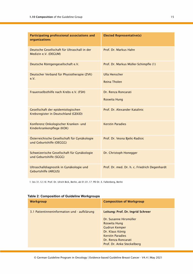
Table 1: Participating professional associations and organizations
Participating professional associations and
organizations
Elected Representative(s)
Arbeitsgemeinschaft Deutscher Tumorzentren
e.V. (ADT)
Prof. Dr. Jutta Engel
Prof. Dr. Dieter Hölzel
Prof. Dr. Jutta Engel
Prof. Dr. Dieter Hölzel
Prof. Dr. Jutta Engel

1.10 Composition of the Guideline Group
© German Guideline Program in Oncology | Evidence-based Guideline Breast Cancer - V4.4 | May 2021
13
Participating professional associations and
organizations
Elected Representative(s)
Prof. Dr. Dieter Hölzel
Arbeitsgemeinschaft Gynäkologische
Onkologie der DGGG und DKG (AGO)
Prof. Dr. Tanja Fehm
Prof. Dr. Anton Scharl
Arbeitsgemeinschaft Internistische Onkologie
in der DKG (AIO)
Dr. Anja Welt
Dr. Matthias Zaiss
Arbeitsgemeinschaft Prävention und
integrative Medizin in der Onkologie der
Deutschen Krebsgesellschaft (PRiO)
Prof. Dr. med. Volker Hanf
Prof. Dr. Karsten Münstedt
Arbeitsgemeinschaft Psychoonkologie der
Deutschen Krebsgesellschaft (PSO)
Prof.Dr. Joachim Weis
Arbeitsgemeinschaft Radiologische Onkologie
(ARO)
Prof. Dr. Wilfried Budach
Prof.Dr. Frederik Wenz
Arbeitsgemeinschaft Supportive Maßnahmen in
der Onkologie, Rehabilitation und
Sozialmedizin (ASORS)
Prof. Dr. Hartmut Link
Prof. Dr. Oliver Rick
Arbeitskreis Frauengesundheit (AKF) Prof. Dr. Anke Steckelberg
Gudrun Kemper
Berufsverband Deutscher Strahlentherapeuten
e.V. (BVDST)
Prof. Dr. Petra Feyer
Prof. Dr. Volker Budach
Berufsverband für Frauenärzte e. V. Dr. Klaus König
BRCA-Netzwerk e. V. Andrea Hahne
Traudl Baumgartner
Bundesverband Deutscher Pathologen e.V.
(BDP)
Prof. Dr. Annette Lebeau
Prof. Dr. Hans-Peter Sinn

1.10 Composition of the Guideline Group
© German Guideline Program in Oncology | Evidence-based Guideline Breast Cancer - V4.4 | May 2021
14
Participating professional associations and
organizations
Elected Representative(s)
Chirurgische Arbeitsgemeinschaft für
Onkologie (CAO-V)
Prof. Dr. Wolfram Trudo Knoefel
Deutsche Gesellschaft der Plastischen,
Rekonstruktiven und Ästhetischen Chirurgen
(DGPRÄC)
Prof. Dr. Christoph Heitmann
Deutsche Gesellschaft für Geratrie e. V. (DGG) Prof. Dr. Michael Denkinger
Deutsche Gesellschaft für Gynäkologie und
Geburtshilfe e.V. (DGGG)
Prof. Dr. Sara Brucker
Prof. Dr. Bernd Gerber
Deutsche Gesellschaft für Hämatologie und
Medizinische Onkologie e.V. (DGHO)
Prof.Dr. Diana Lüftner
Prof. Dr. Hans Tesch
Deutsche Gesellschaft für Humangenetik (GfH) Prof. Dr. Christian Kubisch
Deutsche Gesellschaft für Nuklearmedizin e.V.
(DGN)
Prof. Dr. Andreas Buck
Deutsche Gesellschaft für Palliativmedizin e.V.
(DGP)
Dr. Christina Gerlach M.Sc.
Dr. Susanne Hirsmüller
Deutsche Gesellschaft für Pathologie e.V.
(DGP)
Prof. Dr. Hans H. Kreipe
Prof. Dr. Carsten Denkert
Deutsche Gesellschaft für Psychosomatische
Frauenheilkunde und Geburtshilfe (DGPFG)
Dr. Friederike Siedentopf
Deutsche Gesellschaft für Radioonkologie e.V.
(DEGRO)
Prof. Dr. Cordula Petersen
Prof. Dr. Jürgen Dunst
Deutsche Gesellschaft für
Rehabilitationswissenschaften (DGRW)
Prof. Dr. Hans Helge Bartsch
Deutsche Gesellschaft für Senologie e.V. (DGS) Prof. Dr. Rüdiger Schulz-Wendtland
1.10 Composition of the Guideline Group
© German Guideline Program in Oncology | Evidence-based Guideline Breast Cancer - V4.4 | May 2021
15
Participating professional associations and
organizations
Elected Representative(s)
Deutsche Gesellschaft für Ultraschall in der
Medizin e.V. (DEGUM)
Prof. Dr. Markus Hahn
Deutsche Röntgengesellschaft e.V. Prof. Dr. Markus Müller-Schimpfle (1)
Deutscher Verband für Physiotherapie (ZVK)
e.V.
Ulla Henscher
Reina Tholen
Frauenselbsthilfe nach Krebs e.V. (FSH) Dr. Renza Roncarati
Roswita Hung
Gesellschaft der epidemiologischen
Krebsregister in Deutschland (GEKID)
Prof. Dr. Alexander Katalinic
Konferenz Onkologischer Kranken- und
Kinderkrankenpflege (KOK)
Kerstin Paradies
Österreichische Gesellschaft für Gynäkologie
und Geburtshilfe (OEGGG)
Prof. Dr. Vesna Bjelic-Radisic
Schweizerische Gesellschaft für Gynäkologie
und Geburtshilfe (SGGG)
Dr. Christoph Honegger
Ultraschalldiagnostik in Gynäkologie und
Geburtshilfe (ARGUS)
Prof. Dr. med. Dr. h. c. Friedrich Degenhardt
1: bis 31.12.16: Prof. Dr. Ulrich Bick, Berlin, ab 01.01.17: PD Dr. E. Fallenberg, Berlin
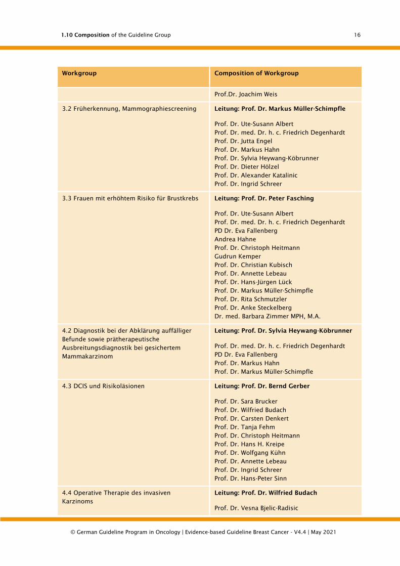
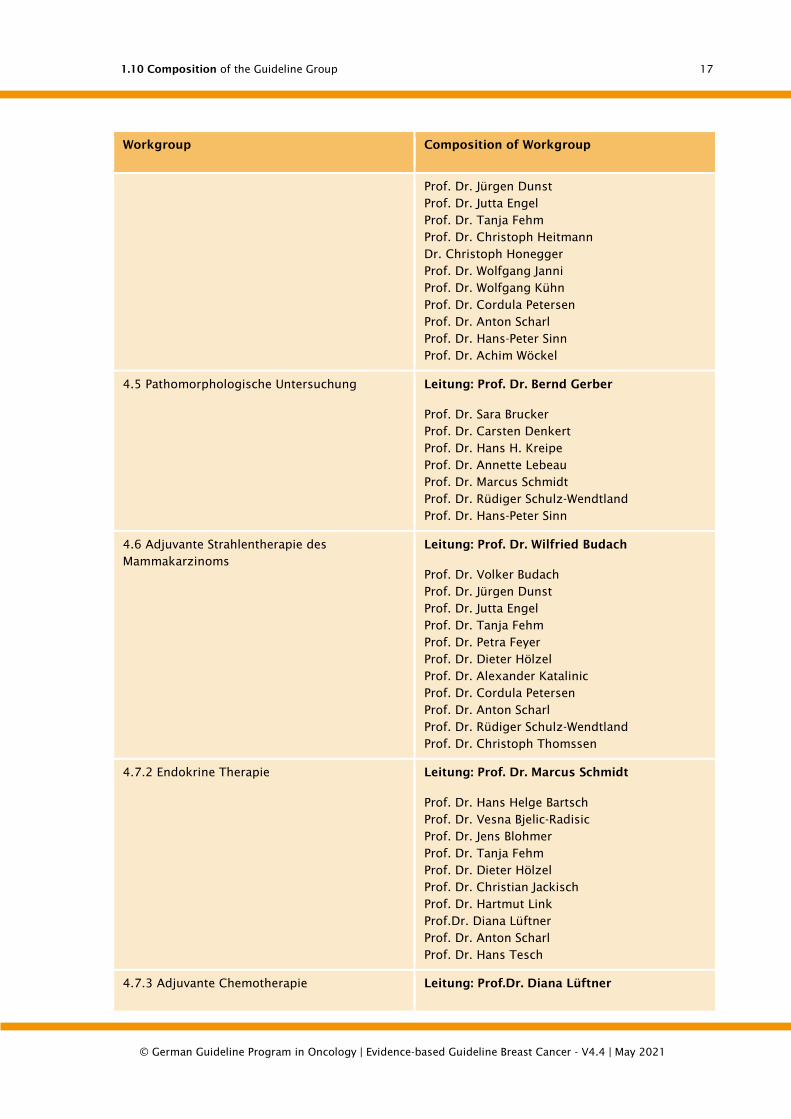
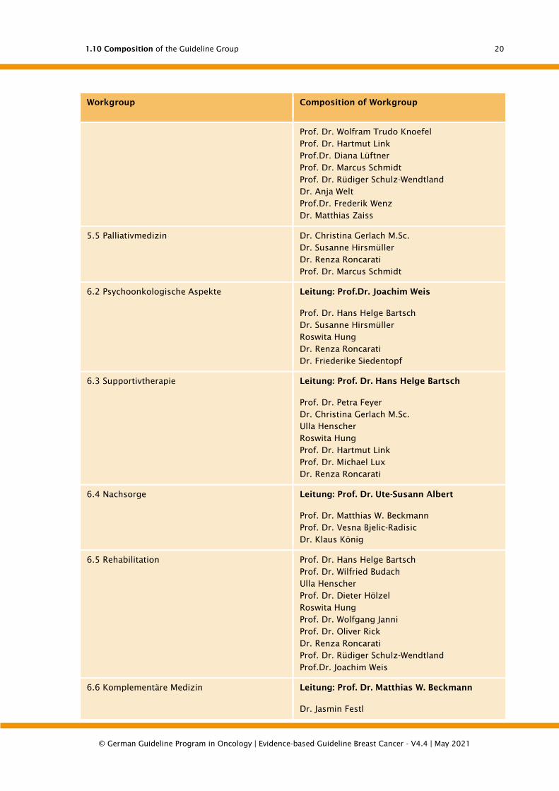
Table 2: Composition of Guideline Workgroups
Workgroup Composition of Workgroup
3.1 Patientinneninformation und - aufklärung Leitung: Prof. Dr. Ingrid Schreer
Dr. Susanne Hirsmüller
Roswita Hung
Gudrun Kemper
Dr. Klaus König
Kerstin Paradies
Dr. Renza Roncarati
Prof. Dr. Anke Steckelberg
1.10 Composition of the Guideline Group
© German Guideline Program in Oncology | Evidence-based Guideline Breast Cancer - V4.4 | May 2021
16
Workgroup Composition of Workgroup
Prof.Dr. Joachim Weis
3.2 Früherkennung, Mammographiescreening Leitung: Prof. Dr. Markus Müller-Schimpfle
Prof. Dr. Ute-Susann Albert
Prof. Dr. med. Dr. h. c. Friedrich Degenhardt
Prof. Dr. Jutta Engel
Prof. Dr. Markus Hahn
Prof. Dr. Sylvia Heywang-Köbrunner
Prof. Dr. Dieter Hölzel
Prof. Dr. Alexander Katalinic
Prof. Dr. Ingrid Schreer
3.3 Frauen mit erhöhtem Risiko für Brustkrebs Leitung: Prof. Dr. Peter Fasching
Prof. Dr. Ute-Susann Albert
Prof. Dr. med. Dr. h. c. Friedrich Degenhardt
PD Dr. Eva Fallenberg
Andrea Hahne
Prof. Dr. Christoph Heitmann
Gudrun Kemper
Prof. Dr. Christian Kubisch
Prof. Dr. Annette Lebeau
Prof. Dr. Hans-Jürgen Lück
Prof. Dr. Markus Müller-Schimpfle
Prof. Dr. Rita Schmutzler
Prof. Dr. Anke Steckelberg
Dr. med. Barbara Zimmer MPH, M.A.
4.2 Diagnostik bei der Abklärung auffälliger
Befunde sowie prätherapeutische
Ausbreitungsdiagnostik bei gesichertem
Mammakarzinom
Leitung: Prof. Dr. Sylvia Heywang-Köbrunner
Prof. Dr. med. Dr. h. c. Friedrich Degenhardt
PD Dr. Eva Fallenberg
Prof. Dr. Markus Hahn
Prof. Dr. Markus Müller-Schimpfle
4.3 DCIS und Risikoläsionen Leitung: Prof. Dr. Bernd Gerber
Prof. Dr. Sara Brucker
Prof. Dr. Wilfried Budach
Prof. Dr. Carsten Denkert
Prof. Dr. Tanja Fehm
Prof. Dr. Christoph Heitmann
Prof. Dr. Hans H. Kreipe
Prof. Dr. Wolfgang Kühn
Prof. Dr. Annette Lebeau
Prof. Dr. Ingrid Schreer
Prof. Dr. Hans-Peter Sinn
4.4 Operative Therapie des invasiven
Karzinoms
Leitung: Prof. Dr. Wilfried Budach
Prof. Dr. Vesna Bjelic-Radisic
1.10 Composition of the Guideline Group
© German Guideline Program in Oncology | Evidence-based Guideline Breast Cancer - V4.4 | May 2021
17
Workgroup Composition of Workgroup
Prof. Dr. Jürgen Dunst
Prof. Dr. Jutta Engel
Prof. Dr. Tanja Fehm
Prof. Dr. Christoph Heitmann
Dr. Christoph Honegger
Prof. Dr. Wolfgang Janni
Prof. Dr. Wolfgang Kühn
Prof. Dr. Cordula Petersen
Prof. Dr. Anton Scharl
Prof. Dr. Hans-Peter Sinn
Prof. Dr. Achim Wöckel
4.5 Pathomorphologische Untersuchung Leitung: Prof. Dr. Bernd Gerber
Prof. Dr. Sara Brucker
Prof. Dr. Carsten Denkert
Prof. Dr. Hans H. Kreipe
Prof. Dr. Annette Lebeau
Prof. Dr. Marcus Schmidt
Prof. Dr. Rüdiger Schulz-Wendtland
Prof. Dr. Hans-Peter Sinn
4.6 Adjuvante Strahlentherapie des
Mammakarzinoms
Leitung: Prof. Dr. Wilfried Budach
Prof. Dr. Volker Budach
Prof. Dr. Jürgen Dunst
Prof. Dr. Jutta Engel
Prof. Dr. Tanja Fehm
Prof. Dr. Petra Feyer
Prof. Dr. Dieter Hölzel
Prof. Dr. Alexander Katalinic
Prof. Dr. Cordula Petersen
Prof. Dr. Anton Scharl
Prof. Dr. Rüdiger Schulz-Wendtland
Prof. Dr. Christoph Thomssen
4.7.2 Endokrine Therapie Leitung: Prof. Dr. Marcus Schmidt
Prof. Dr. Hans Helge Bartsch
Prof. Dr. Vesna Bjelic-Radisic
Prof. Dr. Jens Blohmer
Prof. Dr. Tanja Fehm
Prof. Dr. Dieter Hölzel
Prof. Dr. Christian Jackisch
Prof. Dr. Hartmut Link
Prof.Dr. Diana Lüftner
Prof. Dr. Anton Scharl
Prof. Dr. Hans Tesch
4.7.3 Adjuvante Chemotherapie Leitung: Prof.Dr. Diana Lüftner

1.10 Composition of the Guideline Group
© German Guideline Program in Oncology | Evidence-based Guideline Breast Cancer - V4.4 | May 2021
18
Workgroup Composition of Workgroup
Prof. Dr. Sara Brucker
Prof. Dr. Bernd Gerber
Prof. Dr. Nadia Harbeck
Prof. Dr. Volker Möbus
Prof. Dr. Volkmar Müller
Prof. Dr. Andreas Schneeweiss
Prof. Dr. Rüdiger Schulz-Wendtland
Prof. Dr. Elmar Stickeler
Prof. Dr. Hans Tesch
4.7.4 Neoadjuvante Therapie Leitung: Prof. Dr. Andreas Schneeweiss
Prof. Dr. Sara Brucker
Prof. Dr. Bernd Gerber
Prof. Dr. med. Jens Huober
Prof. Dr. Sibylle Loibl
Prof. Dr. Michael Untch
Prof. Dr. Gunter von Minckwitz
4.7.5 Antikörpertherapie Leitung: Prof.Dr. Diana Lüftner, Prof. Dr.
Marcus Schmidt, Prof. Dr. Andreas
Schneeweiss
Prof. Dr. Jens Blohmer
Prof. Dr. Elmar Stickeler
Prof. Dr. Michael Untch
4.7.6 Knochengerichtete Therapie Leitung: Prof. Dr. Peyman Hadji
Prof. Dr. Rüdiger Schulz-Wendtland
Prof. Dr. Florian Schütz
Prof. Dr. Elmar Stickeler
4.7.7 Beeinflussbare Lebensstilfaktoren Leitung: PD Dr. Freerk Baumann
Prof. Dr. med. Volker Hanf
Prof. Dr. Hans Hauner
Prof. Dr. Wolfgang Janni
Prof. Dr. Ute Nöthlings
5.2 Diagnostik des lokalen/lokoregionalen
Rezidivs
Leitung: PD Dr. Eva Fallenberg
Prof. Dr. Jens Blohmer
Prof. Dr. med. Dr. h. c. Friedrich Degenhardt
Prof. Dr. Markus Hahn
Dr. Klaus König
Prof. Dr. Markus Müller-Schimpfle
Prof. Dr. Anton Scharl
Prof. Dr. Elmar Stickeler

1.10 Composition of the Guideline Group
© German Guideline Program in Oncology | Evidence-based Guideline Breast Cancer - V4.4 | May 2021
19
Workgroup Composition of Workgroup
5.3 Therapie des lokalen/lokoregionalen
Rezidivs
Leitung: Prof. Dr. Wilfried Budach
Prof. Dr. Sara Brucker
Prof. Dr. Bernd Gerber
Prof. Dr. Christoph Heitmann
Dr. Susanne Hirsmüller
Prof. Dr. Christian Jackisch
Prof. Dr. Michael Lux
5.4 Fernmetastasen - Chemo Leitung: Prof. Dr. Hans Tesch
Prof. Dr. Hans Helge Bartsch
Prof. Dr. Sara Brucker
Prof. Dr. Wilfried Budach
Prof. Dr. Bernd Gerber
Dr. Christina Gerlach M.Sc.
Dr. Susanne Hirsmüller
Prof. Dr. med. Jens Huober
Prof. Dr. Wolfram Trudo Knoefel
Prof. Dr. Hartmut Link
Prof.Dr. Diana Lüftner
Prof. Dr. Rüdiger Schulz-Wendtland
Dr. Anja Welt
Prof.Dr. Frederik Wenz
Dr. Matthias Zaiss
5.4 Fernmetastasen - Endokrin Leitung: Prof. Dr. Hans-Jürgen Lück
Prof. Dr. Hans Helge Bartsch
Prof. Dr. Sara Brucker
Prof. Dr. Wilfried Budach
Prof. Dr. Bernd Gerber
Dr. Christina Gerlach M.Sc.
Dr. Susanne Hirsmüller
Prof. Dr. Wolfram Trudo Knoefel
Prof. Dr. Hartmut Link
Prof.Dr. Diana Lüftner
Prof. Dr. Volkmar Müller
Prof. Dr. Rüdiger Schulz-Wendtland
Dr. Anja Welt
Prof.Dr. Frederik Wenz
Dr. Matthias Zaiss
5.4 Fernmetastasen – Spez.
Metastasenlokalisation
Leitung: Prof. Dr. Cordula Petersen
Prof. Dr. Hans Helge Bartsch
Prof. Dr. Sara Brucker
Prof. Dr. Wilfried Budach
Prof. Dr. Bernd Gerber
Dr. Christina Gerlach M.Sc.
Dr. Susanne Hirsmüller
1.10 Composition of the Guideline Group
© German Guideline Program in Oncology | Evidence-based Guideline Breast Cancer - V4.4 | May 2021
20
Workgroup Composition of Workgroup
Prof. Dr. Wolfram Trudo Knoefel
Prof. Dr. Hartmut Link
Prof.Dr. Diana Lüftner
Prof. Dr. Marcus Schmidt
Prof. Dr. Rüdiger Schulz-Wendtland
Dr. Anja Welt
Prof.Dr. Frederik Wenz
Dr. Matthias Zaiss
5.5 Palliativmedizin Dr. Christina Gerlach M.Sc.
Dr. Susanne Hirsmüller
Dr. Renza Roncarati
Prof. Dr. Marcus Schmidt
6.2 Psychoonkologische Aspekte Leitung: Prof.Dr. Joachim Weis
Prof. Dr. Hans Helge Bartsch
Dr. Susanne Hirsmüller
Roswita Hung
Dr. Renza Roncarati
Dr. Friederike Siedentopf
6.3 Supportivtherapie Leitung: Prof. Dr. Hans Helge Bartsch
Prof. Dr. Petra Feyer
Dr. Christina Gerlach M.Sc.
Ulla Henscher
Roswita Hung
Prof. Dr. Hartmut Link
Prof. Dr. Michael Lux
Dr. Renza Roncarati
6.4 Nachsorge Leitung: Prof. Dr. Ute-Susann Albert
Prof. Dr. Matthias W. Beckmann
Prof. Dr. Vesna Bjelic-Radisic
Dr. Klaus König
6.5 Rehabilitation Prof. Dr. Hans Helge Bartsch
Prof. Dr. Wilfried Budach
Ulla Henscher
Prof. Dr. Dieter Hölzel
Roswita Hung
Prof. Dr. Wolfgang Janni
Prof. Dr. Oliver Rick
Dr. Renza Roncarati
Prof. Dr. Rüdiger Schulz-Wendtland
Prof.Dr. Joachim Weis
6.6 Komplementäre Medizin Leitung: Prof. Dr. Matthias W. Beckmann
Dr. Jasmin Festl
1.10 Composition of the Guideline Group
© German Guideline Program in Oncology | Evidence-based Guideline Breast Cancer - V4.4 | May 2021
21
Workgroup Composition of Workgroup
Prof. Dr. med. Volker Hanf
Roswita Hung
Prof. Dr. Karsten Münstedt
Dr. Renza Roncarati
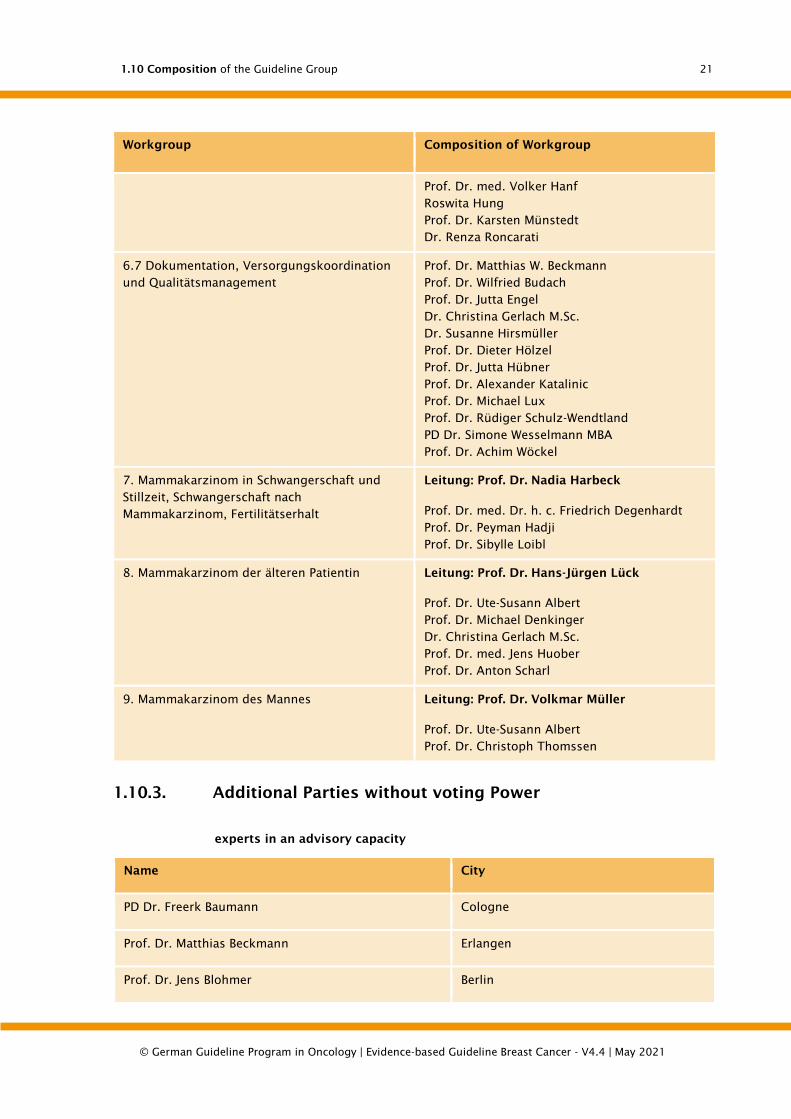
6.7 Dokumentation, Versorgungskoordination
und Qualitätsmanagement
Prof. Dr. Matthias W. Beckmann
Prof. Dr. Wilfried Budach
Prof. Dr. Jutta Engel
Dr. Christina Gerlach M.Sc.
Dr. Susanne Hirsmüller
Prof. Dr. Dieter Hölzel
Prof. Dr. Jutta Hübner
Prof. Dr. Alexander Katalinic
Prof. Dr. Michael Lux
Prof. Dr. Rüdiger Schulz-Wendtland
PD Dr. Simone Wesselmann MBA
Prof. Dr. Achim Wöckel
7. Mammakarzinom in Schwangerschaft und
Stillzeit, Schwangerschaft nach
Mammakarzinom, Fertilitätserhalt
Leitung: Prof. Dr. Nadia Harbeck
Prof. Dr. med. Dr. h. c. Friedrich Degenhardt
Prof. Dr. Peyman Hadji
Prof. Dr. Sibylle Loibl
8. Mammakarzinom der älteren Patientin Leitung: Prof. Dr. Hans-Jürgen Lück
Prof. Dr. Ute-Susann Albert
Prof. Dr. Michael Denkinger
Dr. Christina Gerlach M.Sc.
Prof. Dr. med. Jens Huober
Prof. Dr. Anton Scharl
9. Mammakarzinom des Mannes Leitung: Prof. Dr. Volkmar Müller
Prof. Dr. Ute-Susann Albert
Prof. Dr. Christoph Thomssen
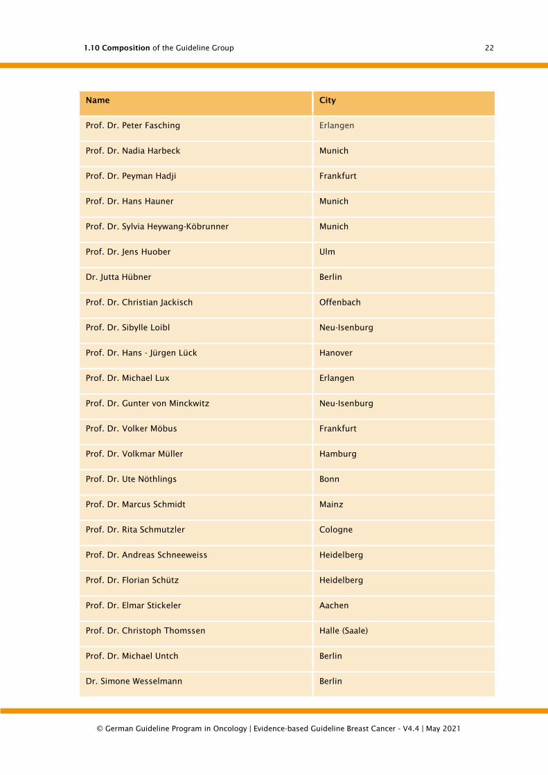
1.10.3. Additional Parties without voting Power
experts in an advisory capacity
Name City
PD Dr. Freerk Baumann Cologne
Prof. Dr. Matthias Beckmann Erlangen
Prof. Dr. Jens Blohmer Berlin
1.10 Composition of the Guideline Group
© German Guideline Program in Oncology | Evidence-based Guideline Breast Cancer - V4.4 | May 2021
22
Name City
Prof. Dr. Peter Fasching Erlangen
Prof. Dr. Nadia Harbeck Munich
Prof. Dr. Peyman Hadji Frankfurt
Prof. Dr. Hans Hauner Munich
Prof. Dr. Sylvia Heywang-Köbrunner Munich
Prof. Dr. Jens Huober Ulm
Dr. Jutta Hübner Berlin
Prof. Dr. Christian Jackisch Offenbach
Prof. Dr. Sibylle Loibl Neu-Isenburg
Prof. Dr. Hans - Jürgen Lück Hanover
Prof. Dr. Michael Lux Erlangen
Prof. Dr. Gunter von Minckwitz Neu-Isenburg
Prof. Dr. Volker Möbus Frankfurt
Prof. Dr. Volkmar Müller Hamburg
Prof. Dr. Ute Nöthlings Bonn
Prof. Dr. Marcus Schmidt Mainz
Prof. Dr. Rita Schmutzler Cologne
Prof. Dr. Andreas Schneeweiss Heidelberg
Prof. Dr. Florian Schütz Heidelberg
Prof. Dr. Elmar Stickeler Aachen
Prof. Dr. Christoph Thomssen Halle (Saale)
Prof. Dr. Michael Untch Berlin
Dr. Simone Wesselmann Berlin

1.10 Composition of the Guideline Group
© German Guideline Program in Oncology | Evidence-based Guideline Breast Cancer - V4.4 | May 2021
23
Name City
Dr. Barbara Zimmer, MPH, MA (Competence Center
Oncology, MDK North Rhine, no author at the explicit
request of the MDK)
Düsseldorf
Other research assistants:
Katharina Brust, B.Sc. (Guidelines Secretariat) Würzburg
Dr. Jasmin Festl (guideline evaluation, literature
selection)
Würzburg
Steffi Hillmann, MPH (guideline research and
evaluation)
Würzburg
PD Dr. Mathias Krockenberger (literature selection) Würzburg
Stephanie Stangl, MPH Würzburg
Dr. Tanja Stüber (literature selection) Würzburg
1.10.4. Patient Involvement
This Guideline was prepared with the direct involvement of 4 patient advocates.
Ms. Roncarati and Ms. Hung of the German Self-Help Group for Women after Cancer
(Frauenselbsthilfe nach Krebs) were involved from the very beginning in the preparation
of the guideline and participated in the consensus conferences with their own voting
rights. Prof. Steckelberg and Ms. Kemper “Working Group on Women’s Health” (AKF)
were also involved and took part in the consensus conferences with their own right to
vote.
1.10.5. Methodological Support
Methodological support was provided by the Guideline Program in Oncology:
• Monika Nothacker, M.D. MPH (AWMF)
• Professor Ina Kopp, M.D. (AWMF)
• Dr. Markus Follmann MPH, MSc. (DKG)
• Thomas Langer, Social Scientist (DKG)
Through external sub-contractors:
• Simone Wesselmann, M.D., MBA (update of quality indicators)

1.11 Abbreviations Used
© German Guideline Program in Oncology | Evidence-based Guideline Breast Cancer - V4.4 | May 2021
24
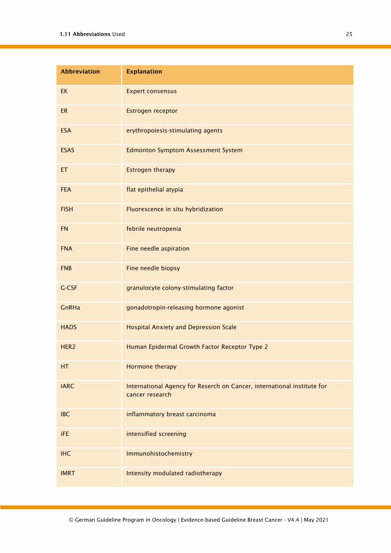
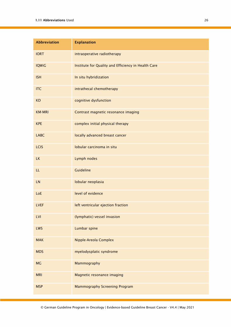
1.11. Abbreviations Used
Table 3: Abbreviations Used
Abbreviation Explanation
ADH (intra-)ductal atypical hyperplasia
AI aromatase inhibitor
AML acute myeloid leukaemia
APBI accelerated partial breast radiation
ASCO American Society of Clinical Oncology
ATL activities of daily living
AUC area under the curve
BÄK German Medical Association
bds on both sides
BET breast-conserving therapy
CISH Chromogenic in situ hybridization
CNB Core Needle Biopsy
CT Computed tomography
DBT digital breast tomosynthesis
DCIS Ductal carcinoma in situ
DFS disease-free survival (DFS)
DGS German Society for Senology
DKG German Cancer Society
ECE extracapsular tumor growth at the lymph nodes
EIC extensive intraductal component
1.11 Abbreviations Used
© German Guideline Program in Oncology | Evidence-based Guideline Breast Cancer - V4.4 | May 2021
25
Abbreviation Explanation
EK Expert consensus
ER Estrogen receptor
ESA erythropoiesis-stimulating agents
ESAS Edmonton Symptom Assessment System
ET Estrogen therapy
FEA flat epithelial atypia
FISH Fluorescence in situ hybridization
FN febrile neutropenia
FNA Fine needle aspiration
FNB Fine needle biopsy
G-CSF granulocyte colony-stimulating factor
GnRHa gonadotropin-releasing hormone agonist
HADS Hospital Anxiety and Depression Scale
HER2 Human Epidermal Growth Factor Receptor Type 2
HT Hormone therapy
IARC International Agency for Reserch on Cancer, international institute for
cancer research
IBC inflammatory breast carcinoma
iFE intensified screening
IHC Immunohistochemistry
IMRT Intensity modulated radiotherapy
1.11 Abbreviations Used
© German Guideline Program in Oncology | Evidence-based Guideline Breast Cancer - V4.4 | May 2021
26
Abbreviation Explanation
IORT intraoperative radiotherapy
IQWiG Institute for Quality and Efficiency in Health Care
ISH In situ hybridization
ITC intrathecal chemotherapy
KD cognitive dysfunction
KM-MRI Contrast magnetic resonance imaging
KPE complex initial physical therapy
LABC locally advanced breast cancer
LCIS lobular carcinoma in situ
LK Lymph nodes
LL Guideline
LN lobular neoplasia
LoE level of evidence
LVEF left ventricular ejection fraction
LVI (lymphatic) vessel invasion
LWS Lumbar spine
MAK Nipple-Areola Complex
MDS myelodysplatic syndrome
MG Mammography
MRI Magnetic resonance imaging
MSP Mammography Screening Program

1.11 Abbreviations Used
© German Guideline Program in Oncology | Evidence-based Guideline Breast Cancer - V4.4 | May 2021
27
Abbreviation Explanation
NACT neoadjuvant chemotherapy
NCCN National Comprehensive Cancer Network
NICE National Institute for Health and Care Excellence
NNT Number Needed to Treat
NZGG New Zealand Guidelines Group
OP Operation
OS Overall Survival
PBI partial breast radiation
pCR pathological complete remission (Engl.: pathological complete remission)
PET Positron Emission Tomography
PFS progression-free survival (PFS)
PI Proliferation Index
PMRT postoperative radiotherapy
PNP Polyneuropathy
POS Palliative Care Outcome Scale
PST primary systemic therapy
QoL Quality of Life
RCT randomized controlled trial
RFA Radio Frequency Ablation
ROR risk of recurrence
RR Relative risk

2.1 Scope and Purpose
© German Guideline Program in Oncology | Evidence-based Guideline Breast Cancer - V4.4 | May 2021
28
Abbreviation Explanation
RS recurrence score
SABCS San Antonio Breast Cancer Symposium
SBRT stereotactic radiation
SGB Social Security Code
SIB simultaneous integrated boost
SIGN Scottish Intercollegiate Guidelines Network
SISH Silver enhanced in situ hybridization
2. Introduction
2.1. Scope and Purpose
2.1.1. Objective and Key Questions
The main rationale for updating the guideline is the consistently high epidemiological
importance of breast cancer and the associated burden of disease. In this context, the
effects of new care concepts in their implementation must be examined. The need to
update the guideline also arises from the existence of new scientific findings and the
further development of the guideline methodology. In addition, an editorial and content
review and revision of the core statements and recommendations of the guideline is
required at regular intervals. The objectives of the S3 guideline for the early detection,
diagnosis, therapy and aftercare of breast cancer were retained from the original
version and the first two updates and supplemented or specified for the third new
edition:
• Consideration of current findings of evidence-based medicine and recognised
treatment concepts
• consideration of the findings from disseminated guidelines and the comprehensive
coverage of guideline-based quality indicators in the updating and implementation
of the guideline
• Supporting the involvement of patients in therapy decisions and positioning their
individual needs
• Comprehensive implementation of multidisciplinary, quality-assured and cross-
sectoral care for breast cancer
• concrete efforts to improve the provision of needs-based and quality-assured
psychosocial care and rehabilitation
• Support of the documentation of epidemiology and progression of breast cancer
diseases by clinical cancer registers

2.2 Methodology
© German Guideline Program in Oncology | Evidence-based Guideline Breast Cancer - V4.4 | May 2021
29
• systematic consideration of the recommendations of initial, continuing and further
training and in quality management systems
• systematic consideration of the recommendations and quality indicators derived
from them in disease management programmes (DMPs), certification procedures
of breast centres, cancer registries and external comparative quality assurance and
standardisation of documentation requirements.
Improving the knowledge of the disease among non-affected persons and patients is
an important goal for which there is a clear potential for improvement. It is a
prerequisite for empowering women to participate in therapy decisions. At present,
information is increasingly being made available on the Internet, but in many cases
with very varying, sometimes unacceptable quality. Particularly in the area of breast
cancer, a flood of information and educational material is available, the quality of which
is predominantly assessed as poor. Within the framework of the OL-program, different
versions of the patient guideline have been created, which are regularly adapted after
the corresponding updates. The respective valid versions of the Women's and Patient
Guidelines are available free of charge (see Chapter 1.9).
Addressees:
The recommendations of the interdisciplinary guideline (LL) are addressed to all
physicians and members of professional groups involved in the care of citizens in the
context of early detection and patients with breast cancer (gynaecologists, general
practitioners, radiologists, pathologists, radio-oncologists, haemato-oncologists,
psycho-oncologists, physiotherapists, nursing staff, etc.) and all women with breast
cancer and their relatives.
Other indirect addressees are:
• medical and scientific societies and professional associations
• Representation of the interests of women (women's health organisations, patient
and self-help organisations)
• Quality assurance institutions and projects at federal and state level
• health policy institutions and decision-makers at federal and state level
• those responsible for DMP programmes and integrated care contracts
• Cost unit
• as well as the public for information on good medical practice.
2.1.2. Validity and Update Process
The S3 guideline is valid until the next update, the validity period is estimated at 5
years. Shorter-term updates are planned in case of urgent need for changes. Comments
and notes on updating the guideline are expressly requested and can be sent to the
following address: [email protected]
2.2. Methodology
The methodological procedure for the preparation of the guideline is described in the
guideline report. It is freely available on the Internet, e.g. on the pages of the Oncology
Guidelines Program (http://www.leitlinienprogramm-
onkologie.de/leitlinien/mammakarzinom/) and the AWMF pages
(http://www.awmf.org/).
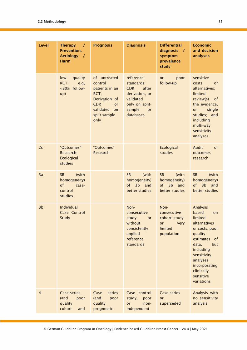
2.2.1. Levels of Evidence (LoE)

2.2 Methodology
© German Guideline Program in Oncology | Evidence-based Guideline Breast Cancer - V4.4 | May 2021
30
To classify the risk of bias in the identified studies, this guideline uses the system of
the Oxford Centre for Evidence-based Medicine in the 2009 version, as shown in Table
5. This system provides for the classification of studies for different clinical questions
(benefit of therapy, prognostic significance, diagnostic value).
Scheme of evidence grading according to Oxford (version March 2009)
Level Therapy /
Prevention,
Aetiology /
Harm
Prognosis Diagnosis Differential
diagnosis /
symptom
prevalence
study
Economic
and decision
analyses
1a SR (with
homogeneity)
of RCTs
SR (with
homogeneity)
inception
cohort
studies; CDR
validated in
different
populations
SR (with
homogeneity)
of Level 1
diagnostic
studies; CDR
with 1b
studies from
different
clinical
centers
SR (with
homogeneity)
of
prospective
cohort
studies
SR (with
homogeneity)
of Level
1economic
studies
1b Individual
RCT (with
narrow
Confidence
Interval)
Individual
inception
cohort study
with > 80%
follow-up;
CDR
validated in a
single
population
Validating
cohort study
with good
reference
standards; or
CDR tested
within one
clinical centre
Prospective
cohort study
with good
follow-up
Analysis
based on
clinically
sensitive
costs or
alternatives;
systematic
review(s) of
the evidence;
and including
multi-way
sensitivity
analyses
2a SR (with
homogeneity)
of cohort
studies
SR (with
homogeneity)
of either
retrospective
cohort
studies or
untreated
control
groups in
RCTs
SR (with
homogeneity)
of Level >2
diagnostic
studies
SR (with
homogeneity)
of Level 2b
and better
studies
SR (with
homogeneity)
of Level >2
economic
studies
2b Individual
cohort study
(including
Retrospective
cohort study
or follow-up
Exploratory
cohort study
with good
Retrospective
cohort study,
Analysis
based on
clinically
2.2 Methodology
© German Guideline Program in Oncology | Evidence-based Guideline Breast Cancer - V4.4 | May 2021
31
Level Therapy /
Prevention,
Aetiology /
Harm
Prognosis Diagnosis Differential
diagnosis /
symptom
prevalence
study
Economic
and decision
analyses
low quality
RCT; e.g,
<80% follow-
up)
of untreated
control
patients in an
RCT;
Derivation of
CDR or
validated on
split-sample
only
reference
standards;
CDR after
derivation, or
validated
only on split-
sample or
databases
or poor
follow-up
sensitive
costs or
alternatives;
limited
review(s) of
the evidence,
or single
studies; and
including
multi-way
sensitivity
analyses
2c "Outcomes"
Research;
Ecological
studies
"Outcomes"
Research
Ecological
studies
Audit or
outcomes
research
3a SR (with
homogeneity)
of case-
control
studies
SR (with
homogeneity)
of 3b and
better studies
SR (with
homogeneity)
of 3b and
better studies
SR (with
homogeneity)
of 3b and
better studies
3b Individual
Case Control
Study
Non-
consecutive
study; or
without
consistently
applied
reference
standards
Non-
consecutive
cohort study;
or very
limited
population
Analysis
based on
limited
alternatives
or costs, poor
quality
estimates of
data, but
including
sensitivity
analyses
incorporating
clinically
sensitive
variations
4 Case-series
(and poor
quality
cohort and
Case series
(and poor
quality
prognostic
Case control
study, poor
or non-
independent
Case-series
or
superseded
Analysis with
no sensitivity
analysis
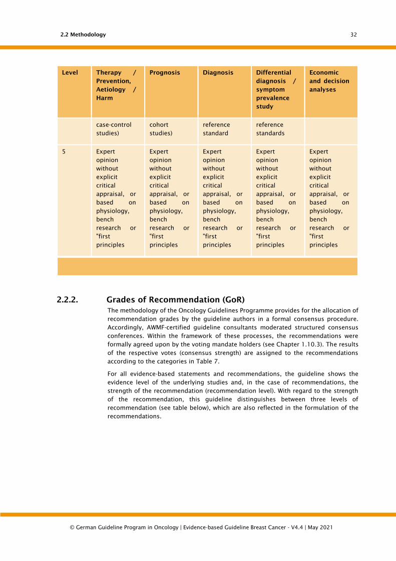
2.2 Methodology
© German Guideline Program in Oncology | Evidence-based Guideline Breast Cancer - V4.4 | May 2021
32
Level Therapy /
Prevention,
Aetiology /
Harm
Prognosis Diagnosis Differential
diagnosis /
symptom
prevalence
study
Economic
and decision
analyses
case-control
studies)
cohort
studies)
reference
standard
reference
standards
5 Expert
opinion
without
explicit
critical
appraisal, or
based on
physiology,
bench
research or
"first
principles
Expert
opinion
without
explicit
critical
appraisal, or
based on
physiology,
bench
research or
"first
principles
Expert
opinion
without
explicit
critical
appraisal, or
based on
physiology,
bench
research or
"first
principles
Expert
opinion
without
explicit
critical
appraisal, or
based on
physiology,
bench
research or
"first
principles
Expert
opinion
without
explicit
critical
appraisal, or
based on
physiology,
bench
research or
"first
principles
2.2.2. Grades of Recommendation (GoR)
The methodology of the Oncology Guidelines Programme provides for the allocation of
recommendation grades by the guideline authors in a formal consensus procedure.
Accordingly, AWMF-certified guideline consultants moderated structured consensus
conferences. Within the framework of these processes, the recommendations were
formally agreed upon by the voting mandate holders (see Chapter 1.10.3). The results
of the respective votes (consensus strength) are assigned to the recommendations
according to the categories in Table 7.
For all evidence-based statements and recommendations, the guideline shows the
evidence level of the underlying studies and, in the case of recommendations, the
strength of the recommendation (recommendation level). With regard to the strength
of the recommendation, this guideline distinguishes between three levels of
recommendation (see table below), which are also reflected in the formulation of the
recommendations.

2.2 Methodology
© German Guideline Program in Oncology | Evidence-based Guideline Breast Cancer - V4.4 | May 2021
33
Scheme of recommendation grading
Level of recommendation Description Language
A Strong recommendation shall/shall not
B Recommendation should/should not
C Recommendation open can/can be waived
Consensus strength
Consensus strength Percentage approval
Strong consensus > 95% of those entitled to vote
Consensus > 75 - 95% of those entitled to vote
Majority approval > 50 - 75% of those entitled to vote
Dissent < 50% der Stimmberechtigten
The decision criteria for determining the degrees of recommendation are explained in
the guideline report on this guideline.
2.2.3. Statements
Statements are descriptions or explanations of specific facts or questions without
immediate call for action. In line with the procedure for recommendations, they are
adopted in a formal consensus procedure and can be based either on study results or
on expert opinions.
2.2.4. Expert Consensus (EK)
Statements/recommendations for which it has been decided to work on the basis of
expert consensus of the Guidelines Group are identified as "Expert Consensus". No
symbols or letters were used for the graduation of the recommendations based on
expert consensus; the strength here results from the wording used
(should/should/can) according to the graduation in Table 6.
2.2.5. Independence and Disclosure of Possible Conflicts of
Interest
The German Cancer Aid provided the financial means through the Oncology Guidelines
Programme (OL). These funds were used for personnel costs, office material, literature
procurement and the consensus conferences (room rent, technology, catering,
moderator fees and travel expenses of the participants). The guideline was developed
with editorial independence from the financing organization. During the guideline
2.2 Methodology
© German Guideline Program in Oncology | Evidence-based Guideline Breast Cancer - V4.4 | May 2021
34
process, all members submitted a written declaration of any existing conflicts of
interest.
Obtaining declarations of conflict of interest
From March 2016 onwards, declarations of conflict of interest were obtained from all
persons involved using the updated AWMF form (see Guidelines Report), with step-by-
step indication of the level of remuneration and explicit request for intellectual
(academic) conflicts of interest (schools, publication activity, etc.). The completeness
of the declarations was achieved in October 2016. Including methodologists, individual
experts and deputies of mandate holders of the professional societies, the guideline
group comprises about 90 persons.
Assessment of conflicts of interest
In the form used, those who had to fill in the form were asked to indicate whether there
was a thematic link with the present guideline topic. However, a final self-assessment
of whether a conflict of interest exists was no longer carried out. The conflict of interest
declarations were evaluated by a working group nominated by the steering group. The
working group consisted of Prof. Dr. R. Kreienberg (Senior Coordinator, Gynaecologist,
former Director of the University Women's Hospital Ulm, DGGG), Prof. Dr. U.S. Albert
(Senologist, DGS), Prof. Dr. W. Budach (Director of the Clinic for Radiotherapy of the
University Hospital in Düsseldorf, DEGRO) and Dr. M. Nothacker, MPH (Methodologist,
AMWF). All results were presented to an external consultant (Prof. Dr. Ludwig of the
AKDÄ) and confirmed.
Evaluation criteria
The initial assessment of the declarations of conflict of interest was carried out by all
evaluators for all persons, completely independently of the other evaluators, according
to the classification: 0 = none, 1 = low, 2 = moderate, 3 = serious in terms of the
assessed level of conflict of interest. Reasons for this were recorded. The assessments
of the working group members were combined and the range of the assessments was
determined. In a joint telephone conference, the assessments were discussed which
had at least a 2 (moderate conflict of interest n=31) and/or a 3 (serious conflict of
interest, n=15). After discussion, the final ranking was determined. The following
criteria were examined with regard to the existing thematic reference and the absolute
level of remuneration and the strength of the relationship:
1. Lectures financed by the industry
2. Review/Advisory Board: Paid review/advisory work for industrial companies
(Scientific Advisory Board: work for industry)
3. Third-party funds directly financed by industry
With only a few presentations, the evaluation was "1 - low". In the case of the existence
of relevant expert opinions/consultancy or activities in a scientific field, the assessment
was "1 - low". In the case of relevant activities as an assessor/consultant or in a scientific
advisory board and/or receipt of third-party funding from industry, the assessment was
either "2 - moderate" or "3 - serious". The ratings were assigned by consensus in the
overall view of the information and are subject to a subjective assessment, as the
information provided in the conflict of interest forms did not consistently allow for a
reliable quantitative assessment and there was no rationale for a fixed cut-off.
Finally, the evaluations were assigned as follows:
44 times = assessment 2 - 14 elected officials and 30 experts were affected 0 times =
assessment 3

3.1 Patient information and education
© German Guideline Program in Oncology | Evidence-based Guideline Breast Cancer - V4.4 | May 2021
35
Dealing with conflicts of interest
The following approach was implemented:
• 0 -1 (no or minor conflict of interest): no special measures.
• 2 (moderate conflict of interest): double vote on the topics concerned (additional
calculation of the result of the vote if persons with a moderate conflict of interest
are excluded).
• 3 (serious conflict of interest): no vote on the topics concerned, review of the
chapter by third parties is obligatory, optional exclusion from the discussion
(review should be carried out by methodologists or members of the guideline group
who are not biased with regard to content).
The issues potentially affected by conflicts of interest were identified on the basis of
the substances listed.
Guidance coordinators and members of the steering group were excluded from the
vote as a matter of principle. In order to create an empirical basis for the actual risk of
bias in relation to the overall group assessment, the persons with moderate conflicts
of interest were identified by means of a preceding question before the electronic
voting process. Subsequently, this blinded grouping made it possible to conduct
sensitivity analyses with regard to the group of persons with a moderate conflict of
interest (result if all persons with a conflict of interest were involved vs.)
This procedure was discussed at the end of November with an independent expert and
expert on conflicts of interest, Prof. Dr. Ludwig of the AKDÄ, who confirmed that the
procedure was appropriate.
At this point we would like to thank all employees for their exclusively voluntary work
on the project.
3. General
3.1. Patient information and education
Thanks to the utilization of new information technologies such as the Internet and the
increasing need on the part of patients for information and involvement in decision-
making about the treatment of their disease, the provision of appropriate information
to patients plays a more important role than ever before. Numerous studies have
confirmed the importance of this issue for the doctor-patient relationship, the course
of the disease and for achieving the therapeutic aim [9], [10], [11].Non-prescriptive
patient information combined with shared decision-making constitutes the basis for
action by doctors. Two ethical principles are at work in this interaction: the patient’s
self-determination (autonomy) and the physician’s duty of care [12]. The patient’s
autonomy takes priority in this context. A decision made by a patient is always
voluntary and binding for action taken by doctors. Patients can make decisions for or
against diagnostic and therapeutic measures or can decide in favor of “not wanting to
know”. Any existing information deficits are to be remedied by the physician so that
the patient can make informed decisions (informed consent). The personal discussion
between the patient and the doctor takes on special importance as the basis for an
understanding based on mutual trust and respect. In this context, increased emphasis
is placed on shared decision-making, i.e. enabling the patient to participate in
decisions. Shared decision-making is characterized by a consultation process that
follows certain rules and an intensive exchange of information between doctor and
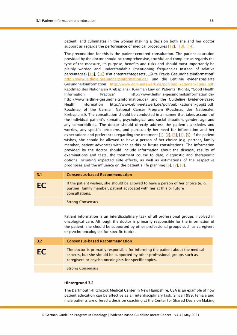
3.1 Patient information and education
© German Guideline Program in Oncology | Evidence-based Guideline Breast Cancer - V4.4 | May 2021
36
patient, and culminates in the woman making a decision both she and her doctor
support as regards the performance of medical procedures [12], [13], [14].
The precondition for this is the patient-centered consultation. The patient education
provided by the doctor should be comprehensive, truthful and complete as regards the
type of the measure, its purpose, benefits and risks and should most importantly be
plainly worded and understandable (mentioning frequencies instead of relative
percentages) [15], [16] (Patientenrechtegesetz, „Gute Praxis Gesundheitsinformation“
http://www.leitlinie-gesundheitsinformation.de/ und die Leitlinie evidenzbasierte
Gesundheitsinformation http://www.ebm-netzwerk.de/pdf/publikationen/gpgi2.pdf;
Raodmap des Nationalen Krebsplans). (German Law on Patients’ Rights, “Good Health
Information Practice” http://www.leitlinie-gesundheitsinformation.de/
http://www.leitlinie-gesundheitsinformation.de/ and the Guideline Evidence-Based
Health Information http://www.ebm-netzwerk.de/pdf/publikationen/gpgi2.pdf;
Roadmap of the German National Cancer Program (Roadmap des Nationalen
Krebsplans]). The consultation should be conducted in a manner that takes account of
the individual patient’s somatic, psychological and social situation, gender, age and
any comorbidities. The doctor should directly address the patient’s anxieties and
worries, any specific problems, and particularly her need for information and her
expectations and preferences regarding the treatment [1], [2], [3], [4], [5]. If the patient
wishes, she should be allowed to have a person of her choice (e.g. partner, family
member, patient advocate) with her at this or future consultations. The information
provided by the doctor should include information about the disease, results of
examinations and tests, the treatment course to date, diagnostic and therapeutic
options including expected side effects, as well as estimations of the respective
prognoses and the influence on the patient’s life planning [6], [7], [8].
3.1 Consensus-based Recommendation
EC If the patient wishes, she should be allowed to have a person of her choice (e. g.
partner, family member, patient advocate) with her at this or future
consultations.
Strong Consensus
Patient information is an interdisciplinary task of all professional groups involved in
oncological care. Although the doctor is primarily responsible for the information of
the patient, she should be supported by other professional groups such as caregivers
or psycho-oncologists for specific topics.
3.2 Consensus-based Recommendation
EC The doctor is primarily responsible for informing the patient about the medical
aspects, but she should be supported by other professional groups such as
caregivers or psycho-oncologists for specific topics.
Strong Consensus
Hintergrund 3.2
The Dartmouth-Hitchcock Medical Center in New Hampshire, USA is an example of how
patient education can be effective as an interdisciplinary task. Since 1999, female and
male patients are offered a decision coaching at the Center for Shared Decision Making
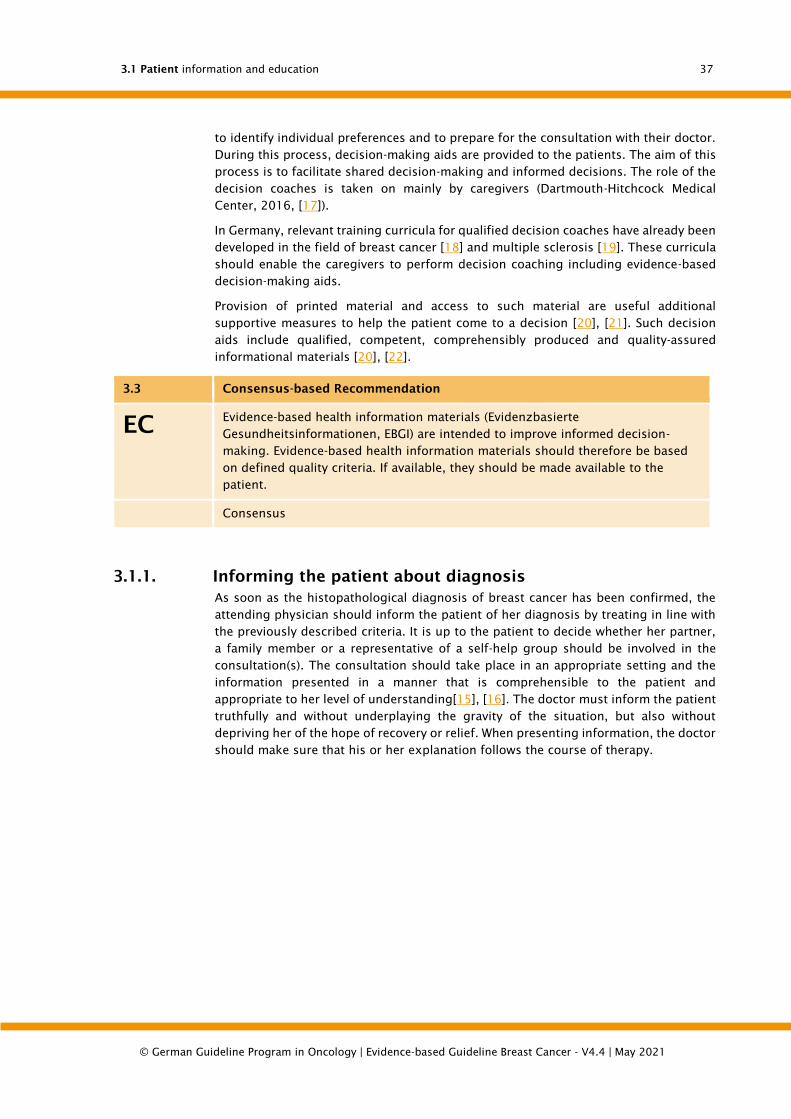
3.1 Patient information and education
© German Guideline Program in Oncology | Evidence-based Guideline Breast Cancer - V4.4 | May 2021
37
to identify individual preferences and to prepare for the consultation with their doctor.
During this process, decision-making aids are provided to the patients. The aim of this
process is to facilitate shared decision-making and informed decisions. The role of the
decision coaches is taken on mainly by caregivers (Dartmouth-Hitchcock Medical
Center, 2016, [17]).
In Germany, relevant training curricula for qualified decision coaches have already been
developed in the field of breast cancer [18] and multiple sclerosis [19]. These curricula
should enable the caregivers to perform decision coaching including evidence-based
decision-making aids.
Provision of printed material and access to such material are useful additional
supportive measures to help the patient come to a decision [20], [21]. Such decision
aids include qualified, competent, comprehensibly produced and quality-assured
informational materials [20], [22].
3.3 Consensus-based Recommendation
EC Evidence-based health information materials (Evidenzbasierte
Gesundheitsinformationen, EBGI) are intended to improve informed decision-
making. Evidence-based health information materials should therefore be based
on defined quality criteria. If available, they should be made available to the
patient.
Consensus
3.1.1. Informing the patient about diagnosis
As soon as the histopathological diagnosis of breast cancer has been confirmed, the
attending physician should inform the patient of her diagnosis by treating in line with
the previously described criteria. It is up to the patient to decide whether her partner,
a family member or a representative of a self-help group should be involved in the
consultation(s). The consultation should take place in an appropriate setting and the
information presented in a manner that is comprehensible to the patient and
appropriate to her level of understanding[15], [16]. The doctor must inform the patient
truthfully and without underplaying the gravity of the situation, but also without
depriving her of the hope of recovery or relief. When presenting information, the doctor
should make sure that his or her explanation follows the course of therapy.
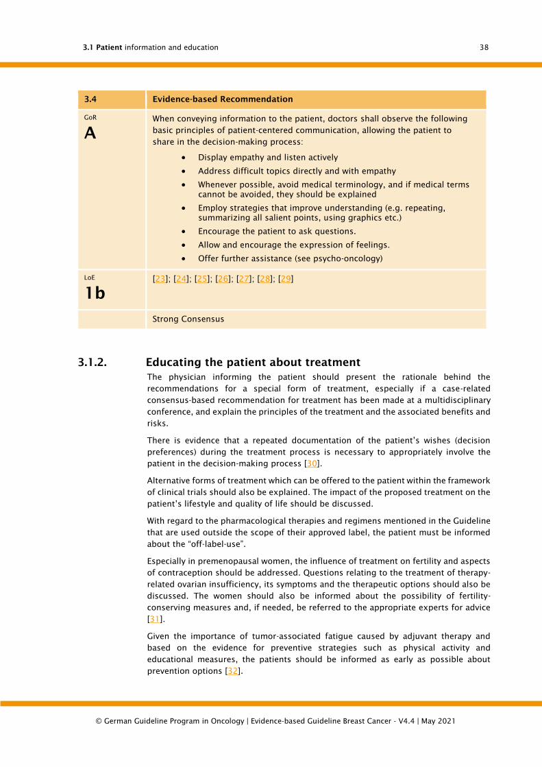
3.1 Patient information and education
© German Guideline Program in Oncology | Evidence-based Guideline Breast Cancer - V4.4 | May 2021
38
3.4 Evidence-based Recommendation
GoR
A
When conveying information to the patient, doctors shall observe the following
basic principles of patient-centered communication, allowing the patient to
share in the decision-making process:
• Display empathy and listen actively
• Address difficult topics directly and with empathy
• Whenever possible, avoid medical terminology, and if medical terms
cannot be avoided, they should be explained
• Employ strategies that improve understanding (e.g. repeating,
summarizing all salient points, using graphics etc.)
• Encourage the patient to ask questions.
• Allow and encourage the expression of feelings.
• Offer further assistance (see psycho-oncology)
LoE
1b
[23]; [24]; [25]; [26]; [27]; [28]; [29]
Strong Consensus
3.1.2. Educating the patient about treatment
The physician informing the patient should present the rationale behind the
recommendations for a special form of treatment, especially if a case-related
consensus-based recommendation for treatment has been made at a multidisciplinary
conference, and explain the principles of the treatment and the associated benefits and
risks.
There is evidence that a repeated documentation of the patient’s wishes (decision
preferences) during the treatment process is necessary to appropriately involve the
patient in the decision-making process [30].
Alternative forms of treatment which can be offered to the patient within the framework
of clinical trials should also be explained. The impact of the proposed treatment on the
patient’s lifestyle and quality of life should be discussed.
With regard to the pharmacological therapies and regimens mentioned in the Guideline
that are used outside the scope of their approved label, the patient must be informed
about the “off-label-use”.
Especially in premenopausal women, the influence of treatment on fertility and aspects
of contraception should be addressed. Questions relating to the treatment of therapy-
related ovarian insufficiency, its symptoms and the therapeutic options should also be
discussed. The women should also be informed about the possibility of fertility-
conserving measures and, if needed, be referred to the appropriate experts for advice
[31].
Given the importance of tumor-associated fatigue caused by adjuvant therapy and
based on the evidence for preventive strategies such as physical activity and
educational measures, the patients should be informed as early as possible about
prevention options [32].

3.1 Patient information and education
© German Guideline Program in Oncology | Evidence-based Guideline Breast Cancer - V4.4 | May 2021
39
The patient should also be informed about measures for preventing lymphedema,
about the necessity of oncological follow-up-care, about rehabilitation (see below), and
about social, financial and psycho-oncological support [29] informiert werden. Für If
necessary, the patient should be advised to obtain further professional advice on the
topics mentioned above (rehabilitation, social counseling and psycho-oncology), and
the necessary arrangements made.
Any treatment requires the patient’s cooperation. Aspects which are the patient’s own
responsibility should be discussed.
3.5 Consensus-based Recommendation
EC During the consultation to inform the patient about treatment, the following
issues should be addressed and information on the benefits and harms
communicated:
• Surgical therapy: Breast-conserving therapy options with mandatory
radiotherapy as equivalent to mastectomy with different variants of
primary and secondary reconstruction or the provision of an external
prosthesis
• Systemic therapy: Principles and desired treatment targets of (neo-
)adjuvant or palliative therapy, therapy duration and mode of
administration, its side effects and possible late sequelae, and the
treatment options for side effects
• Radiotherapy: Principles and desired treatment targets, duration and
follow-up surveillance, possible acute and late sequelae, treatment
options for side effects
• Participation in clinical trials, principles behind the treatment and
treatment targets, duration and mode of administration of the
therapy, effects and side-effects known to date, special features (e.g.
monitoring, additional measures, cooperation, data storage and
processing)
• Other: Options for prevention and treatment of therapy-related side
effects and sequelae (e.g. fatigue, nausea, osteoporosis,
lymphedema, etc.), necessity for follow-up, possibilities for
rehabilitation and psycho-oncological support as well as services
offered by self-help groups, aspects that are the responsibility of the
patient and cooperation (e.g. reporting symptoms and problems,
treatment compliance).
Strong Consensus
Hintergrund 3.5
The physician must take the patient's somatic, psychological and social situation, age
and any comorbidities she may have into account during the consultation. The patient’s
anxieties, worries, resilience, need for information, expectations regarding treatment
and preferences should be documented by the doctor at diagnosis, at the beginning of
and during therapy, after completion of therapy, in the event of recurrence as well as
in the event of disease progression [1], [2], [3], [5], [28]. This also includes informing
patients about “normal and unremarkable” test results and the course of treatment in
order to provide reassurance, while giving prognostic information to facilitate them in
planning their future lives [6], [7], [8].
Breast cancer is not an emergency. The patient must always be allowed sufficient time
for decision-making. She can reject a particular procedure or treatment or withdraw
previously granted consent to participate in a therapeutic trial or clinical study. She has
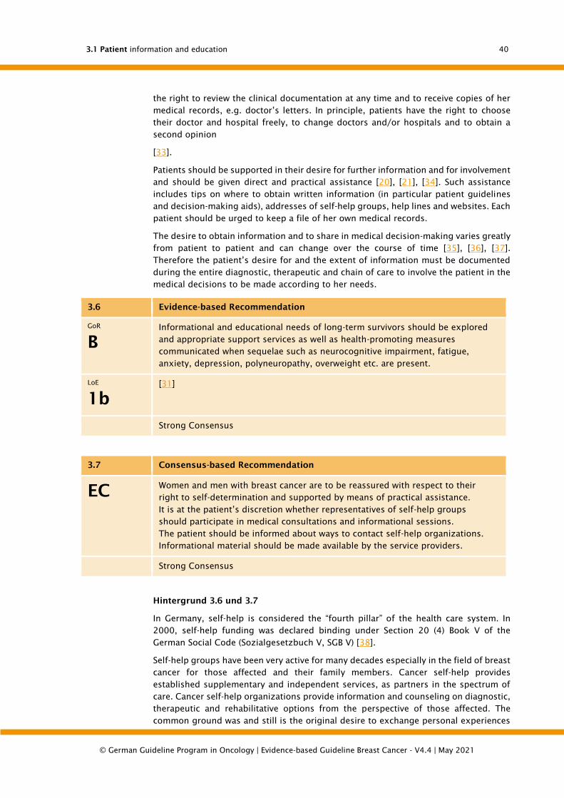
3.1 Patient information and education
© German Guideline Program in Oncology | Evidence-based Guideline Breast Cancer - V4.4 | May 2021
40
the right to review the clinical documentation at any time and to receive copies of her
medical records, e.g. doctor’s letters. In principle, patients have the right to choose
their doctor and hospital freely, to change doctors and/or hospitals and to obtain a
second opinion
[33].
Patients should be supported in their desire for further information and for involvement
and should be given direct and practical assistance [20], [21], [34]. Such assistance
includes tips on where to obtain written information (in particular patient guidelines
and decision-making aids), addresses of self-help groups, help lines and websites. Each
patient should be urged to keep a file of her own medical records.
The desire to obtain information and to share in medical decision-making varies greatly
from patient to patient and can change over the course of time [35], [36], [37].
Therefore the patient’s desire for and the extent of information must be documented
during the entire diagnostic, therapeutic and chain of care to involve the patient in the
medical decisions to be made according to her needs.
3.6 Evidence-based Recommendation
GoR
B
Informational and educational needs of long-term survivors should be explored
and appropriate support services as well as health-promoting measures
communicated when sequelae such as neurocognitive impairment, fatigue,
anxiety, depression, polyneuropathy, overweight etc. are present.
LoE
1b
[31]
Strong Consensus
3.7 Consensus-based Recommendation
EC Women and men with breast cancer are to be reassured with respect to their
right to self-determination and supported by means of practical assistance.
It is at the patient’s discretion whether representatives of self-help groups
should participate in medical consultations and informational sessions.
The patient should be informed about ways to contact self-help organizations.
Informational material should be made available by the service providers.
Strong Consensus
Hintergrund 3.6 und 3.7
In Germany, self-help is considered the “fourth pillar” of the health care system. In
2000, self-help funding was declared binding under Section 20 (4) Book V of the
German Social Code (Sozialgesetzbuch V, SGB V) [38].
Self-help groups have been very active for many decades especially in the field of breast
cancer for those affected and their family members. Cancer self-help provides
established supplementary and independent services, as partners in the spectrum of
care. Cancer self-help organizations provide information and counseling on diagnostic,
therapeutic and rehabilitative options from the perspective of those affected. The
common ground was and still is the original desire to exchange personal experiences

3.2 Early detection, mammographic screening
© German Guideline Program in Oncology | Evidence-based Guideline Breast Cancer - V4.4 | May 2021
41
and knowledge and to encourage and mutually reassure each other. By offering
empathic and pragmatic exchanges of experience, cancer self-help services can be very
meaningful, especially, but not exclusively in those sectors where the disease has a
significant impact on everyday life. This is why the involvement of self-help services in
the healthcare system in general and their role as providers of information and
assistance is of such inestimable value and so irreplaceable, especially to anyone
seeking advice or affected by the disease.
Krebsselbsthilfeorganisationen bieten u. a. Informationen und Beratung über
diagnostische, therapeutische und rehabilitative Möglichkeiten aus der Perspektive von
Betroffenen an. Gemeinsam war und ist der ursprüngliche Wunsch, die persönlichen
Erfahrungen und das erlebte Wissen miteinander auszutauschen, sich gegenseitig zu
ermutigen und zu bestärken. Nicht nur, aber gerade in Bereichen, in denen die
Krankheit erheblichen Einfluss auf das Alltagsleben nimmt, ist die Krebsselbsthilfe
durch den empathischen und auch pragmatischen Erfahrungsaustausch von großer
Bedeutung. Dies macht ihr Mitwirken im Gesundheitssystem generell und als
Informations- und Hilfsangebot speziell für Ratsuchende und Betroffene so wertvoll
und unersetzbar.
The services offered by cancer self-help groups are cross-sectoral and can be used free-
of-charge whenever breast cancer is suspected, at diagnosis, during therapy and follow-
up care as well. Members (and their relatives) are also offered psychosocial support
and help "to help themselves" (empowerment strategies) as well as a forum sharing
experiences and information aimed at sustainably improving quality of life [39].
Affected sufferers’ work for cancer self-help services is of a voluntary nature.
There is a wide range of assistance and support services offered by cancer self-help
organizations. Besides local discussion groups, information is available via the various
media. These include written and illustrated brochures, websites and forums that
enable modern interaction by means of chats, blogs and interaction online and in real-
time. State and federal cancer self-help organizations are increasingly assuming patient
advocacy roles on social and healthcare policy committees.
It is recommended in the S3 Practice Guideline “Psycho-oncology" [29] to inform
patients about cancer self-help services and to offer informational materials. An
agreement on the cooperation between self-help and service providers should ideally
be made in writing in the form of a contract of cooperation. There is a great cross-
sectoral need for research of the structure and efficacy of self-help services.
Up-to-date contact details of self-help groups and other self-help providers are
accessible on the website of the National Contact Point for Self-Help (Nationale
Kontaktstelle für Selbsthilfe) (www.nakos.de) If and how they use the services of the
offered by cancer self-help organizations is left to the discretion of each affected
individual and their family members.
3.2. Early detection, mammographic screening
3.8 Consensus-based Statement
ST The most important population-related risk factor for the development of breast
cancer in women and men is advanced age.
Consensus
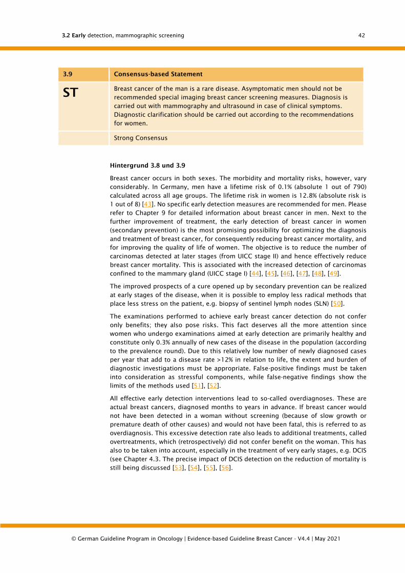
3.2 Early detection, mammographic screening
© German Guideline Program in Oncology | Evidence-based Guideline Breast Cancer - V4.4 | May 2021
42
3.9 Consensus-based Statement
ST Breast cancer of the man is a rare disease. Asymptomatic men should not be
recommended special imaging breast cancer screening measures. Diagnosis is
carried out with mammography and ultrasound in case of clinical symptoms.
Diagnostic clarification should be carried out according to the recommendations
for women.
Strong Consensus
Hintergrund 3.8 und 3.9
Breast cancer occurs in both sexes. The morbidity and mortality risks, however, vary
considerably. In Germany, men have a lifetime risk of 0.1% (absolute 1 out of 790)
calculated across all age groups. The lifetime risk in women is 12.8% (absolute risk is
1 out of 8) [43]. No specific early detection measures are recommended for men. Please
refer to Chapter 9 for detailed information about breast cancer in men. Next to the
further improvement of treatment, the early detection of breast cancer in women
(secondary prevention) is the most promising possibility for optimizing the diagnosis
and treatment of breast cancer, for consequently reducing breast cancer mortality, and
for improving the quality of life of women. The objective is to reduce the number of
carcinomas detected at later stages (from UICC stage II) and hence effectively reduce
breast cancer mortality. This is associated with the increased detection of carcinomas
confined to the mammary gland (UICC stage I) [44], [45], [46], [47], [48], [49].
The improved prospects of a cure opened up by secondary prevention can be realized
at early stages of the disease, when it is possible to employ less radical methods that
place less stress on the patient, e.g. biopsy of sentinel lymph nodes (SLN) [50].
The examinations performed to achieve early breast cancer detection do not confer
only benefits; they also pose risks. This fact deserves all the more attention since
women who undergo examinations aimed at early detection are primarily healthy and
constitute only 0.3% annually of new cases of the disease in the population (according
to the prevalence round). Due to this relatively low number of newly diagnosed cases
per year that add to a disease rate >12% in relation to life, the extent and burden of
diagnostic investigations must be appropriate. False-positive findings must be taken
into consideration as stressful components, while false-negative findings show the
limits of the methods used [51], [52].
All effective early detection interventions lead to so-called overdiagnoses. These are
actual breast cancers, diagnosed months to years in advance. If breast cancer would
not have been detected in a woman without screening (because of slow growth or
premature death of other causes) and would not have been fatal, this is referred to as
overdiagnosis. This excessive detection rate also leads to additional treatments, called
overtreatments, which (retrospectively) did not confer benefit on the woman. This has
also to be taken into account, especially in the treatment of very early stages, e.g. DCIS
(see Chapter 4.3. The precise impact of DCIS detection on the reduction of mortality is
still being discussed [53], [54], [55], [56].
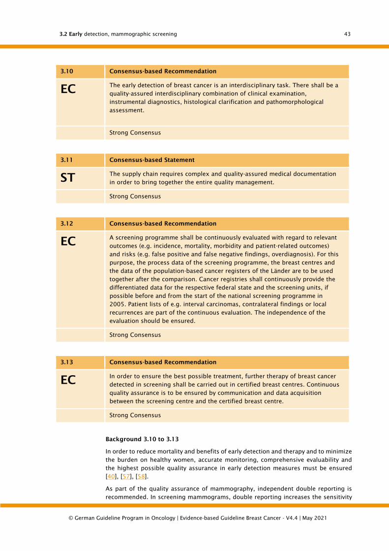
3.2 Early detection, mammographic screening
© German Guideline Program in Oncology | Evidence-based Guideline Breast Cancer - V4.4 | May 2021
43
3.10 Consensus-based Recommendation
EC The early detection of breast cancer is an interdisciplinary task. There shall be a
quality-assured interdisciplinary combination of clinical examination,
instrumental diagnostics, histological clarification and pathomorphological
assessment.
Strong Consensus
3.11 Consensus-based Statement
ST The supply chain requires complex and quality-assured medical documentation
in order to bring together the entire quality management.
Strong Consensus
3.12 Consensus-based Recommendation
EC A screening programme shall be continuously evaluated with regard to relevant
outcomes (e.g. incidence, mortality, morbidity and patient-related outcomes)
and risks (e.g. false positive and false negative findings, overdiagnosis). For this
purpose, the process data of the screening programme, the breast centres and
the data of the population-based cancer registers of the Länder are to be used
together after the comparison. Cancer registries shall continuously provide the
differentiated data for the respective federal state and the screening units, if
possible before and from the start of the national screening programme in
2005. Patient lists of e.g. interval carcinomas, contralateral findings or local
recurrences are part of the continuous evaluation. The independence of the
evaluation should be ensured.
Strong Consensus
3.13 Consensus-based Recommendation
EC In order to ensure the best possible treatment, further therapy of breast cancer
detected in screening shall be carried out in certified breast centres. Continuous
quality assurance is to be ensured by communication and data acquisition
between the screening centre and the certified breast centre.
Strong Consensus
Background 3.10 to 3.13
In order to reduce mortality and benefits of early detection and therapy and to minimize
the burden on healthy women, accurate monitoring, comprehensive evaluability and
the highest possible quality assurance in early detection measures must be ensured
[40], [57], [58].
As part of the quality assurance of mammography, independent double reporting is
recommended. In screening mammograms, double reporting increases the sensitivity

3.2 Early detection, mammographic screening
© German Guideline Program in Oncology | Evidence-based Guideline Breast Cancer - V4.4 | May 2021
44
of carcinoma detection by 2.9 -13.7 % (median 7.8 %). The specificity can be reduced
(up to 2.1 %) or increased (up to 2.8 %) [40], depending on the decision procedure after
double findings. The use of computer-assisted detection systems cannot replace
double findings [40], [59], [60], [61] due to the current study situation.
An individually supplementary recommendation of further early detection
examinations can be useful, taking into account the individual risk-benefit ratio. In
women with increased risk (usually increased familial risk, presence of histologically
confirmed risk lesions, condition following malignancy of the breast or ovaries),
supplementary methods or an additional examination at intervals may be considered.
Individually adapted screening tests can also be considered for women before the age
of 50 and after the age of 70, depending on the individual risk. At an older age, in
addition to the individual risk of breast cancer, the overall survival prognosis must be
taken into account, since the risk of overdiagnosis increases significantly in the
presence of competing risks, especially in older women.
Overall, it can be assumed that side effects such as overdiagnosis occur in other early
detection examinations to at least a comparable extent as in quality assured screening,
false positive findings to a far greater extent.
Therefore, these groups should be involved in a quality assurance programme for the
implementation and evaluation of structural, process and outcome quality as well as in
the diagnostic and care chain (anamnesis, risk counselling, information on health
behaviour, clinical examination, instrumental diagnostics, interventional tissue
removal techniques, surgical clarification and histopathological findings). In terms of
age, risk and findings, management in the context of early detection of breast cancer
should be based on the algorithm of the diagnostic chain (see Chapter 12.1) [40] [41],
[62], [63], [64], [65].
3.2.1. Shared decision-making
3.14 Consensus-based Recommendation
EC Early detection examinations can lead to physical and psychological stress. This
circumstance shall be taken into account through careful education and an
effective communication strategy.
Strong Consensus
3.15 Consensus-based Recommendation
EC In the context of breast cancer screening, information and education shall not
be limited to pre-formulated texts, but require a medical information interview
that takes into account the preferences, needs, concerns and fears of the
woman and allows for participatory decision-making. In mammography
screening, information and clarification shall be provided to the woman
primarily in writing, with a supplementary reference to the possibility of a
medical consultation in the invitation letter.
Consensus

3.2 Early detection, mammographic screening
© German Guideline Program in Oncology | Evidence-based Guideline Breast Cancer - V4.4 | May 2021
45
Background 3.14 and 3.15
Informed self-determination and participation in medical decision-making processes is
a particularly high priority for women interested in participating in screening. During
the medical consultation, anamnesis, the individual risk constellation, fears and worries
must be addressed and women must be supported in their decision-making processes
in a participatory manner. In risk communication, absolute and relative figures and
their reference periods should be included. Benefits and harms should be assessed and
communicated in relation to the relevant periods. In particular, life-saving and
overdiagnosis should be determined, estimated and communicated in relation to the
life of the woman according to their definition [40], [66] see also Chapter 4.1: Patient
information and education. In addition to the hoped-for effect, the side effects must be
adequately explained according to their occurrence in any screening tests, even outside
the screening programme.
For information purposes and as a decision-making aid for women, the patient
information on the Oncology Guidelines Programme is available (http://www.leitlinien-
programm.de) as well as the website http://www.mammo-programm.de, in accordance
with the quality requirements "Good Practice Health Information" [67]. The decision
support of the Joint Federal Committee of Physicians and Health Insurance Funds (GBA
fact sheet updated 2017) is based on IQWIG's Rapid Review and can be accessed at
http://www.mammo-programm.de The figures given there are very conservative
estimates. Nevertheless, it is confirmed that mammography screening is the only
imaging method with a proven reduction in breast cancer mortality and, if there is a
desire for early detection, participation in the quality-assured screening programme is
recommended.
3.2.2. Mammographic screening
3.16 Evidence-based Statement
ST Mammography is the only method with guaranteed reduction of breast cancer
mortality.
LoE
1a
[40]; [41]; [42]; [44]; [53]; [54]; [58]; [65]; [68]
Strong Consensus
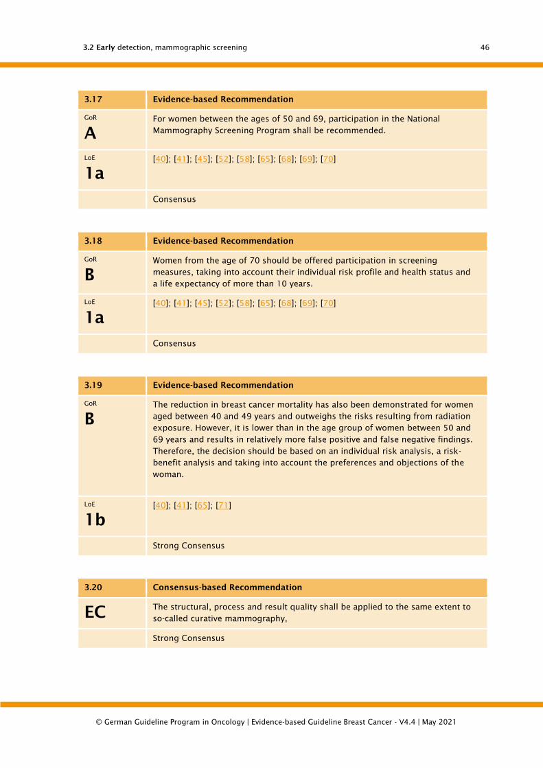
3.2 Early detection, mammographic screening
© German Guideline Program in Oncology | Evidence-based Guideline Breast Cancer - V4.4 | May 2021
46
3.17 Evidence-based Recommendation
GoR
A
For women between the ages of 50 and 69, participation in the National
Mammography Screening Program shall be recommended.
LoE
1a
[40]; [41]; [45]; [52]; [58]; [65]; [68]; [69]; [70]
Consensus
3.18 Evidence-based Recommendation
GoR
B
Women from the age of 70 should be offered participation in screening
measures, taking into account their individual risk profile and health status and
a life expectancy of more than 10 years.
LoE
1a
[40]; [41]; [45]; [52]; [58]; [65]; [68]; [69]; [70]
Consensus
3.19 Evidence-based Recommendation
GoR
B
The reduction in breast cancer mortality has also been demonstrated for women
aged between 40 and 49 years and outweighs the risks resulting from radiation
exposure. However, it is lower than in the age group of women between 50 and
69 years and results in relatively more false positive and false negative findings.
Therefore, the decision should be based on an individual risk analysis, a risk-
benefit analysis and taking into account the preferences and objections of the
woman.
LoE
1b
[40]; [41]; [65]; [71]
Strong Consensus
3.20 Consensus-based Recommendation
EC The structural, process and result quality shall be applied to the same extent to
so-called curative mammography,
Strong Consensus
3.2 Early detection, mammographic screening
© German Guideline Program in Oncology | Evidence-based Guideline Breast Cancer - V4.4 | May 2021
47
3.21 Consensus-based Recommendation
EC After a mammographic finding of categories 0, III, IV and V, further clarification
should take place within one week in order to keep the psychological stress on
the woman as low as possible.
Strong Consensus
Background 3.16 to 3.21
In Germany, mammography screening for women from the age of 50 until the end of
the 70th year of life is part of the guideline of the Joint Federal Committee on the early
detection of cancer (source: Guideline of the Joint Federal Committee on the early
detection of cancer (Cancer Screening Guideline / KFE-RL) in the version of 18. June
2009 published in the Federal Gazette 2009, No. 148a entered into force on 3 October
2009 last amended on 21 April 2016, published in the Federal Gazette AT 08.07.2016
B2, entered into force on 1 January 2017 https://www.g-
ba.de/informationen/richtlinien/17/).
For mammography screening, the reduction in mortality with regard to breast cancer
can be regarded as assured for the group of invited versus uninvited women. The
evaluations of randomized studies show a mortality reduction of 20% for all age groups
from [45], [52], [68], [69], [70]. In preparing the present update of the S3 guideline
recommendations, the recommendations of the American Cancer Society ACS (ACS)
[41], the US Preventive Services Task Force [51], [65], [70] and the evaluation of cancer
prevention and control measures by the International Agency for Research on Cancer
(IARC) of WHO [42], [45], [68] have been taken into account.
The IARC assessment of the WHO will be considered in more detail below, as it is of
great importance in the international context of drawing up national health measures.
29 experts from 16 countries were involved in the preparation of the comprehensive
data analysis of the effects and side effects of IARC mammography screening. IARC
confirms the results of the meta-analyses on the old RCTs. However, considering the
considerable progress in diagnostics (mammographic technique, quality assurance of
the screening chain) and therapy, the relevance of these studies, which are more than
20 years old, is questioned. Instead, IARC assesses well-controlled observational
studies of modern screening programmes as better suited to assess the effectiveness
of current mammography screening. Special emphasis has been placed on studies with
sufficient follow-up to reduce the so-called length time bias, taking into account
temporal and regional trends. Based on the available literature, approximately 20
incidence-based cohort studies and another approximately 20 case-control studies
fulfilled these requirements.
The following statements were made:
For regular participants, a reduction in mortality compared to non-participants can be
expected through mammography screening. It is currently assumed that in Europe up
to 8 lives can be saved by screening women between 50 and 69 years of age (about 10
rounds) for every 1000 participants who are healthy today. Further data are needed to
map the specific effect strength of the screening measure in the screening population.
A further reduction in mortality through continued screening up to and including the
age of 74 is considered proven.

3.2 Early detection, mammographic screening
© German Guideline Program in Oncology | Evidence-based Guideline Breast Cancer - V4.4 | May 2021
48
The data available for the age group between 40 and 49 years was still considered to
be limited.
For none of the other imaging tests (tomosynthesis, sonography, MRI or other
procedures) is there sufficient evidence for a reduction in breast cancer mortality. This
applies to both complementary and substitutive use for mammography screening.
With regard to screening of BRCA1- or -2-positive women, IARC recognizes the
significant increase in sensitivity in the context of intensified monitoring, but with
significantly reduced specificity. For the proof of a mortality reduction the data
situation was still insufficient.
Among the potential side effects, the IARC counts overdiagnosis and false positive
findings in addition to the very low risk of radiation exposure.
The overdiagnosis rate was calculated only on the basis of studies that had sufficient
follow-up and that sufficiently considered length time bias and other disturbing
influences. (To understand: If follow-up is insufficient, early diagnoses are erroneously
counted as overdiagnoses, which leads to an overestimation of the overdiagnosis rate
and an underestimation of the mortality reduction). The rate of overdiagnosis after 25
years of follow-up is given as 6.5% (1%-10%) of breast cancer diagnoses. This means
that out of 1000 women regularly screened over 20 years, 71 instead of 67 women
learn of their actually existing breast cancer or DCIS. These 4 additionally diagnosed
diseases would not have become known to women during their lifetime without
screening.
In screening, false positive diagnoses are defined as findings on the basis of which a
woman is again invited for additional examinations because of an ultimately benign
finding. The additional examinations usually consist of imaging procedures, if
necessary also histological clarifications (mostly punch or vacuum biopsies). Re-
appointments for additional imaging in case of ultimately benign findings affect about
2% of the screened women per subsequent round in Europe. Histological clarifications
(usually image-guided minimally invasive biopsies) with a benign result occur in up to
0.6% of screened women. Cumulated over 20 years of screening, this corresponds to a
one-time order of about 20% of the participants in 20 years or biopsies in about 6% of
the participants because of a finally benign finding.
Technological developments: 2D mammography and 3D mammography (digital
breast tomosynthesis (DBT))
For 3D mammography (digital breast tomosynthesis (DBT)), there are very large
numerical evaluations from the USA, although only retrospectively collected, as well as
at least 3 prospective controlled studies from European screening programmes. The
latter are based on systematic double examinations using digital full-field 2D
mammography and 3D mammography. There are now 3 systematic reviews of the
studies, [68], [72], [73].
The comparisons of all studies almost exclusively concern the comparison between 2D
mammography and 2D + 3D mammography. However, an additional study on one type
of device indicates that an "2D synthetic mammography" additionally calculated from
3D mammography should be comparable to primary 2D mammography and thus no
double image (with double the radiation dose) should be necessary in the future [74].
For another type of device, wide-angle tomosynthesis, only 3D was compared with 2D
(at least with regard to the first and second findings). For this device type, additional
2D mammography also did not appear necessary [75].
3.2 Early detection, mammographic screening
© German Guideline Program in Oncology | Evidence-based Guideline Breast Cancer - V4.4 | May 2021
49
Data to date indicate a significant increase in detection rate for the combination of 2D
+ 3D mammography, which was present at all density levels, but appeared to be most
pronounced at density levels ACR 2-3. This increase in the detection rate almost
exclusively related to an increased detection of invasive carcinomas while maintaining
the same detection at DCIS. With regard to the individual findings, 2D + 3D
mammography showed an increase in specificity in most studies, but according to
consensus a slight deterioration. In addition, a slight increase in the biopsy rate was
observed with 3D-DBT, although the PPV remained the same. Summarizing these
results, 2D + 3D mammography shows a significantly improved detection proven in
prospective screening with comparable specificity and slightly increased radiation
exposure.
First results show a slight reduction of the interval carcinoma rate. At present, no
statements on overdiagnosis are possible. These would have to be taken into account
in screening and especially in any application for early detection outside the screening
program. There are still open questions regarding the comparability of the results of
the device types, the quality assurance of findings and technology and logistical issues
(compatibility; effects of a learning effect; fatigue in systematic reading) and require
further studies. Nevertheless, due to the significant gain in sensitivity and the very
good specificity, this method currently appears to be the most promising method for
screening applications.
3.2.3. Measures for the early detection of breast cancer
3.22 Consensus-based Recommendation
EC As part of the statutory early cancer diagnosis, women shall be offered an
anamnesis and an explanation of possible risk factors.
Consensus
3.23 Evidence-based Statement
ST Breast self-examination, even with regular use and training, is not the only
method capable of reducing breast cancer mortality.
LoE
1a
[40]; [41]
Strong Consensus
3.24 Consensus-based Recommendation
EC Through qualified information, women should be encouraged to become
familiar with the normal changes in their own bodies. This includes the
appearance and feel of the breasts in order to detect any deviations themselves.
Strong Consensus

3.2 Early detection, mammographic screening
© German Guideline Program in Oncology | Evidence-based Guideline Breast Cancer - V4.4 | May 2021
50
3.25 Consensus-based Recommendation
EC Clinical breast examination, i.e. inspection, palpation of the breast and
assessment of lymphatic drainage, should be offered to women aged 30 years
and older as part of the statutory early detection examinations.
Clinical examination of the breast and axilla should not be recommended as the
sole method for early detection of breast cancer.
Strong Consensus
3.2.3.1. Sonography
3.26 Consensus-based Recommendation
EC The systematic use of sonography cannot be recommended as the sole method
for early detection of breast cancer.
Strong Consensus
Background 3.26
No studies are available on the sole use of sonography instead of mammography for
the early detection of breast cancer. Sonography has not yet been recommended by the
international committee for systematic screening [76], [77], [78].
In the context of complementary complementary diagnostics, the use of sonography
can lead to an increase in sensitivity, especially in women with an increased risk of
breast cancer,
Technological enhancements through the use of automated 3D-sonography
(ABUS/AVUS) in comparison to medical 2D-sonography (HHUS) have not yet been able
to identify any advantages in terms of sensitivity and specificity. Initial study data
indicate that the use of automated ultrasound may be able to reduce examiner
dependency. A lack of systematic quality assurance for diagnostics and screening,
different device applications and availability as well as a heterogeneous study situation
currently allow only limited recommendation of the method within the framework of
studies [79], [80],[81], [82], [83], [84], [85], [86], [87], [88].
3.2 Early detection, mammographic screening
© German Guideline Program in Oncology | Evidence-based Guideline Breast Cancer - V4.4 | May 2021
51
3.2.3.2. Complementary diagnostic imaging in high mammographic density for
early detection
3.27 Evidence-based Statement
ST Increased mammographic density is an independent, moderate risk factor for
the occurrence of breast cancer. Mammographic density and sensitivity correlate
negatively.
LoE
3a
[40]; [89]; [90]; [91]
Strong Consensus
3.28 Evidence-based Recommendation
GoR
B
The evidence on the use of complementary imaging methods is limited. Outside
of the high-risk situation, sonography currently appears to be the most suitable
method to complement mammography. Sonography can increase the density
dependent sensitivity, a reduction in mortality is not proven. In early detection it
is associated with a higher rate of biopsies than the National Mammography
Screening Programme.
LoE
3a
[40]; [65]; [68]; [72]; [73]; [92]; [93]
Strong Consensus
3.29 Evidence-based Recommendation
GoR
B
Tomosynthesis can increase sensitivity. Its testing in a quality assured program
should be considered.
LoE
1b
[74]; [75]; [94]
Strong Consensus
Background 3.27 to 3.29
Increasing mammographic density is associated with a decrease in sensitivity and
specificity and an increase in the risk of interval carcinoma. High mammographic
density also represents an independent risk factor [89], [90], [95], which, however, is
only low in relation to the risk of the normal population with a factor of approx. 1.3
[91], [96].
The data basis on which the density-related risk calculations are based has so far been
based exclusively on visual density estimates using the previous density definition of
classes ACR1-4 (corresponding to ACR-Lexicon, 4th ed. [97]) or (semi-) quantitative
3.2 Early detection, mammographic screening
© German Guideline Program in Oncology | Evidence-based Guideline Breast Cancer - V4.4 | May 2021
52
density measurements. According to this previous definition, 4 density classes were
defined according to the percentage of dense tissue in the mammogram (ACR1=
density up to 25%, ACR2 = density of 25-50%; ACR3 = density of 50-75%; ACR4= density
> 75%).
Overall, the reproducibility of the density categories is unsatisfactory (re-classification
of 12.6-18.7% of mammograms) [73]. According to the updated version of the 2013
classification [98], the density is no longer given according to the percentage of density
areas as ACR 1-4, but according to BIRADS density ad according to descriptive
characteristics. However, objective criteria for a standardized density measurement are
still missing. This limits the reliability of recommendations for the use of additional
imaging for mammography screening as well as for mammography in early detection
a [45], [73].
In principle, the use of additional imaging (sonography, KM-MRI, tomosynthesis) at high
parenchyma density leads to the detection of additional (mostly invasive) carcinomas,
but is associated with an increased false positive rate as well as increased control
examination and biopsy rates. There is also a lack of long-term data on the effect on
survival and overdiagnosis. When using complementary methods, data on the
correlation between age and other influencing breast cancer risk factors [99] are
missing.
On the basis of previous studies, medical sonography (HHUS, handheld US) performed
and evaluated as a complement to mammography appears to show the best balance
between benefit and risk, which is indicated by first evaluations from an Italian
screening program [93].
Women should therefore be included in the decision on complementary imaging in
cases of high mammographic density and negative 2D mammography and informed
about the benefits and risks of such complementary imaging, taking into account the
overall risk of disease.
3.2.4. Needs for research for the early detection of breast cancer
After discussion of the current data situation in the working groups as well as in the
plenum of the S3 Guidelines Commission, the testing of the following topics is
considered necessary:
• Optimisation of the screening programme with regard to age limits
• and the benefit/risk ratio regarding screening between 45-49 years and > 70 years
• Optimization of the screening interval, especially for younger women (
• Optimization of the investigation methodology for identified problem areas
- stratified addition of sonography (2D/3D)
- selective substitution/addition by 3D mammography (digital breast
tomosynthesis (DBT))
• Optimised adaptation of therapy for prognostically favourable early breast
carcinomas and pre-stages with the aim of reducing possible overtherapies without
significant loss of efficancy

3.3 Women at increased risk of developing breast cancer
© German Guideline Program in Oncology | Evidence-based Guideline Breast Cancer - V4.4 | May 2021
53
3.3. Women at increased risk of developing breast
cancer
3.3.1. Familial breast cancer
About 30% of all women with a breast cancer in Germany have a family history of breast
cancer and meet the inclusion criteria for genetic testing established and validated by
the German Consortium for Familial Breast and Ovarian Cancer (see statement 3.14)
[101]. These are based on a mutation detection rate of at least 10% [100].
3.30 Evidence-based Recommendation
GoR
B
Genetic testing should be offered if there is a familial or individual exposure
that is associated with at least a 10 % probability of mutation detection.
This is the case when, in one line of the family
• at least 3 women have breast cancer
• at least 2 women suffer from breast cancer, 1 of which before the
age of 51
• at least 1 woman has breast cancer and 1 woman has ovarian cancer
• at least 2 women have ovarian cancer
• at least 1 woman has breast and ovarian cancer
• at least 1 woman with 35 years or younger is suffering from breast
cancer
• at least 1 woman aged 50 years or younger has bilateral breast
cancer
• at least 1 man has breast cancer and 1 woman has breast or ovarian
cancer
A reasonable period of reflection should be allowed before making the
diagnosis.
LoE
2a
For mutation
likelihood
5
[102]
Consensus
Background 3.30
In about 25% of these women a germline mutation can be detected in one of the known
predisposing high-risk genes BRCA1 or BRCA2 [101]. Women with a BRCA1 or BRCA2
mutation fall ill about 20 years earlier than women without family risk and have a
lifelong risk of developing breast cancer of on average 60%, of an average of 40% of a
contralateral breast cancer and 16 - 55% of an ovarian cancer [103].
In unselected patients with a triple-negative breast carcinoma (TNBC) a mutation
prevalence of BRCA1 mutations in 8.5% and of BRCA2 mutations in 2.7% could be
detected [104]. However, the exact prevalence rates for gene mutations in the presence
of TNBC without further familial predisposition have not yet been conclusively clarified.
3.3 Women at increased risk of developing breast cancer
© German Guideline Program in Oncology | Evidence-based Guideline Breast Cancer - V4.4 | May 2021
54
In the meantime, other risk genes have also been identified, e.g. CHEK2, PALB2 and
RAD51C. While CHEK2 is associated with a moderate breast cancer risk [105], PALB2
seems to be associated with a similarly high risk as BRCA1/2 [106], RAD51C is primarily
associated with an increased ovarian cancer risk [107].
Even if gene panel analyses are already offered, genotype-phenotype studies should be
awaited in order to recommend concrete preventive measures based on the clinical
appearance [104].
3.31 Evidence-based Recommendation
GoR
A
The consultation shall enable participatory decision-making. This requires
comprehensive information for women and the clarification and inclusion of
women's preferences in the decision-making process. Evidence-based decision-
making aids can improve women's decisions.
LoE
1a
[108]; [109]; [110]; [111]; [112]; [113]
Consensus
3.3 Women at increased risk of developing breast cancer
© German Guideline Program in Oncology | Evidence-based Guideline Breast Cancer - V4.4 | May 2021
55
3.32 Evidence-based Recommendation
GoR
B
In risk counselling prior to genetic testing, the following contents should be
considered in particular:
• Probability for the presence of a mutation
• Disease risks in the case of positive findings
• the benefits and harms of preventive and therapeutic options,
including the option of doing nothing
• Probability of false negative findings
• The importance of genetic testing for family members
After receipt of the genetic findings, the following contents in particular should
be deepened in the risk consultation before offering preventive measures:
• Disease risk depending on genetic findings, age and concomitant
diseases (natural course)
• Probability of false positive and false negative test results of
intensified screening
• Use of preventive options (intensified early detection, prophylactic
surgeries, drug therapies) with regard to mortality reduction,
morbidity reduction and quality of life
• Risks of the preventive options including long-term consequences
• Competing risks, prognosis and treatability in case of disease
occurrence without preventive measures taking into account the
specific appearance of the genetically defined tumor subtype
• Possible risks for associated tumours,
• Psycho-oncological counselling services
LoE
5
1a
[114]; [115]; [116]; [117]; [118]; [119]
Consensus
Background 3.31 and 3.32
The desire of women and men for detailed information and a joint decision on
prevention and treatment options has been documented several times. These findings
apply, at least in Germany, largely independent of educational level, age or state of
health [112]. Such so-called evidence-based health information is a prerequisite for
participation and informed decisions. It has also been shown that evidence-based
health information can improve decision-making [120].
Although various working groups worldwide have been working for about 20 years on
the question of how information on health and disease topics can be presented in such
a way that it can serve as a basis for informed decisions [116], implementation in
practice and in concrete health information is currently hardly successful [117].
3.3 Women at increased risk of developing breast cancer
© German Guideline Program in Oncology | Evidence-based Guideline Breast Cancer - V4.4 | May 2021
56
3.33 Evidence-based Statement
ST BRCA1-associated breast carcinomas often exhibit a characteristic
histopathological and immunohistochemical phenotype:
• invasive carcinoma with medullary properties
• G3 morphology
• Estrogen receptor, progesterone receptor and HER2 negativity (triple
negative)
LoE
2a
for
histopatholo
gic
characteristi
c
[118]; [121]
Strong Consensus
3.34 Consensus-based Recommendation
EC If these characteristics are present, the pathologist should point out the
possibility of a hereditary background.
Strong Consensus
Background 3.33 and 3.34
Breast carcinomas, which develop on the basis of a genetic disposition, can have a
distinct driver gene profile, which can manifest itself in phenotypic peculiarities. This
has been demonstrated for BRCA1-associated breast carcinomas. While BRCA2-
associated breast carcinomas are sporadic carcinomas, BRCA1-associated carcinomas
frequently exhibit a particular phenotype which shows characteristics of a medullary
carcinoma without, however, forming the full picture of the classic medullary type of
breast carcinoma [103], [122], [123], [124], [125]. These special features include
macroscopically relatively smooth outer boundaries with displacing rather than
infiltrating growth and a prominent, less coarse aspect. Also characteristic are a G3
morphology with high grade nuclear pleomorphism, high mitotic activity and lack of
tubule formation with often syncytial growth as well as a lack of expression of steroid
hormone receptors and HER2 (triple-negative). The Ki-67 proliferation index is usually
above 30% and the tumor cells often show expression of basal cytokeratins (CK5/6,
CK14). The tumour stroma shows a pronounced lymphoplasmacellular infiltration and
in the neighbouring tumour-free breast tissue a so-called lymphocytic lobulitis is more
frequently observed, which is a weaker indication criterion, however. The presence of
these characteristics should be a reason to consider a genetic disposition and to
stimulate a family anamnestic survey.
3.3 Women at increased risk of developing breast cancer
© German Guideline Program in Oncology | Evidence-based Guideline Breast Cancer - V4.4 | May 2021
57
3.35 Evidence-based Recommendation
GoR
B/0
In patients with a pathogenic BRCA1/2 mutation (IARC class 4/5)
(recommendation level B) and in patients with a remaining lifetime risk of >/=
30% (recommendation level 0), an intensified early detection with the addition of
MRI should only be carried out within the framework of a transparent quality
assurance and corresponding evaluation.
The additional mammography from the age of 40 should be performed within
the framework of a transparent quality assurance and corresponding evaluation
(recommendation grade B).
LoE
2a
[100]; [126]; [127]; [128]; [129]; [130]; [131]
Strong Consensus
Background 3.35
On average, women with a genetic risk for breast cancer develop the disease earlier
than women from the general population. Therefore, the usual early detection
measures do not appear to be sufficient. The German Mammography Screening
Program (MSP) according to § 25 para. 2 and 3 SGB V for the early detection of breast
cancer in the general female population is aimed at asymptomatic women between the
ages of 50 and 69. Special features of women from familial/hereditary high-risk
collectives (including higher lifetime risks for breast cancer, mostly younger at the time
of first diagnosis) are not separately considered in the MSP. Internationally, different
recommendations are given on measures for intensified early detection (iFE) (including
age limits, inclusion criteria and/or the scope of measures), whereby the breast MRI is
included (e.g. [100], [129], [130], [132]). However, the significance of iFE with regard
to patient-relevant outcomes has not been conclusively clarified.
Evaluations of the diagnostic quality of the procedures as iFE measures (e.g. for
mammography or MRI, including age and/or mutation status) have been published
[128].
In the systematic search for clinical studies on the significance of iFE measures in
relation to outcome parameters (mortality, incidence rates, tumour stages, quality of
life), which were carried out within the framework of the S3 guideline update, one
prospective and two retrospective cohort studies (LoE 2a-3c) with limited significance
of their results on high-risk collectives and BRCA mutation carriers were identified
[123], [133], [134], which allow impressions to be made, e.g. on detection or incidence
rates. For the evaluation of the benefit of the intensified early detection measures in
high-risk collectives or in BRCA mutation carriers, there is no direct evidence of a
reduction in mortality through intensified early detection. However, iFE can detect
breast carcinomas in early stages [134]. However, intensified monitoring is also
associated with an increase in the number of investigations due to false positive
findings.
In addition, only a part of the family-related risk has so far been clarified. Based on new
and inexpensive high-throughput methods of gene analysis, new risk genes have
recently been identified, and more are to be expected. For these new genes, the clinical
appearance, e.g. the age-related disease risks and the possible occurrence of special
3.3 Women at increased risk of developing breast cancer
© German Guideline Program in Oncology | Evidence-based Guideline Breast Cancer - V4.4 | May 2021
58
genetically defined histological tumour types with possible effects on the natural
course of the disease and the effectiveness of imaging procedures, is still largely
unknown. Against this background, special demands must be placed on the assurance
of structural, process and result quality.
For this reason, structured measures of intensified early detection (iFE) (including MRI)
were implemented within the framework of contracts pursuant to § 140a SGB V at the
nationwide centers of the German Consortium for Familial Breast and Ovarian Cancer.
These address women with proven pathogenic germline mutation in the genes BRCA1
and BRCA2, as well as women from negatively tested families with a lifetime disease
risk of > 30% or heterozygote risk of > 20%. An essential component of this care is the
recording of the quality of the iFE results on the basis of accompanying pseudonymised
documentation. A mortality-based evaluation is only possible and absolutely desirable
by linking to cancer registers.
Extensive measures have been established to ensure structural and process quality in
breast diagnostics, e.g. within the framework of the mammography screening
programme or diagnostic breast examinations. Recommendations and qualification
measures of professional associations support the process of strengthening quality in
the application of breast diagnostics. The German Radiological Society takes this
process into account by establishing a structured continuing education program in
senological radiology. A closer networking and cooperation of already established care
structures with externally audited breast and screening centres should be promoted
and consolidated.
3.36 Evidence-based Statement
ST The surgical therapy of BRCA-associated breast cancer is based on the
guidelines for sporadic breast cancer.
Mastectomy has no survival advantage over breast-conserving therapy.
Drug therapy for BRCA-associated breast cancer is based on the guidelines for
sporadic breast cancer.
LoE
2b
[135]; [136]; [137]; [138]; [139]; [140]
Strong Consensus
3.37 Evidence-based Statement
ST There are indications that chemotherapy containing platinum can lead to a
better response than standard chemotherapy.
LoE
2b
[135]; [136]; [137]; [138]; [139]; [140]
Consensus
3.3 Women at increased risk of developing breast cancer
© German Guideline Program in Oncology | Evidence-based Guideline Breast Cancer - V4.4 | May 2021
59
Background 3.36 and 3.37
If a woman with a mutation in the BRCA1 or BRCA2 genes develops breast cancer,
treatment is currently based on the recommendations for sporadic breast cancer.
However, several preclinical and retrospective studies indicate a reduced sensitivity of
BRCA-incompetent cells to spindle toxins such as vinca alkaloids and taxanes [141],
[142] and an increased sensitivity to DNA-intercalating substances such as platinum
derivatives [137]. These observations are currently being reviewed in prospective
randomized studies. However, a retrospective mutation analysis of study patients of
the TNBC arm of the Geparsixto study did not show a benefit for the BRCA1/2 mutation
carriers through the addition of platinum [143]. The direct comparison between
platinum and taxane will be examined in the ongoing TNT study in the metastatic
situation. Preliminary results here indicate a benefit of platinum [144].
Research on BRCA-deficient cell lines has led to the substance class of PARP inhibitors
being used in clinical trials [145], [146]. While efficacy has already been proven in
metastatic BRCA1/2-associated ovarian cancer and has already led to the approval of
PARP inhibitors, final proof of efficacy for breast cancer is still pending and is currently
being tested in prospective clinical trials.
3.38 Evidence-based Statement
ST Risk reducing surgery in healthy BRCA1/2 mutation carriers (IARC class
4/5): prophylactic mastectomy:
Healthy women with a BRCA1 or BRCA2 mutation have an increased lifetime risk
of developing breast cancer.
In healthy women with a pathogenic BRCA1 or BRCA2 gene mutation, bilateral
prophylactic mastectomy leads to a reduction in the incidence of breast cancer.
A reduction of breast cancer-specific mortality or total mortality by bilateral
prophylactic mastectomy is not sufficiently ensured.
Therefore, a decision for or against a bilateral prophylactic mastectomy always
requires case-related comprehensive information and detailed multidisciplinary
consultation on the potential advantages and disadvantages of such an
intervention, taking into account the possible alternatives.
LoE
2a
[100]; [147]; [148]; [149]; [150]; [151]; [152]; [153]; [154]; [155]
Strong Consensus
Background 3.38
As risk-reducing surgical procedures in healthy women affected by corresponding gene
mutations, bilateral prophylactic mastectomy (BPM) and bilateral prophylactic salpingo-
oophorectomy (BPSO) are available. BPM reduces the risk of breast cancer by over 95%.
An effect of BPM on the reduction of breast cancer-specific mortality is not conclusively
proven. Whether BPM has an influence on overall survival has not yet been sufficiently
proven by 90% [147], [148], [149], [152], [153], [154], [155].
Prophylactic bilateral salpingo-oophorectomy reduces the risk of ovarian cancer by
97%. Whether this prophylactic intervention also reduces the risk of breast cancer is
3.3 Women at increased risk of developing breast cancer
© German Guideline Program in Oncology | Evidence-based Guideline Breast Cancer - V4.4 | May 2021
60
not clearly established at present. First retrospective examinations described a risk
reduction for the first carcinoma by 50%, 30-50% for the contralateral second carcinoma
[156], [157], [158]. In addition, a 75% reduction of total mortality could be shown for
the prophylactic bilateral salpingo-oophorectomy [149], [158]. More recent prospective
studies indicate a significantly lower effect or could not prove it at all [159], [160].
However, both studies are also subject to possible bias, so that the question is not
finally clarified at present. The prophylactic bilateral salpingo-oophorectomy is
recommended in women affected by BRCA mutations by laparoscopiam around the age
of 40 years and after completed family planning. Hormone replacement therapy is
indicated until the age of about 50 years.
The rate of metachronous ipsilateral secondary carcinomas (newly developed
carcinoma of the same side) does not seem to be significantly increased in patients
with proven BRCA1/2 mutation according to the current state of knowledge, so that a
breast-conserving therapy is adequate [161]. However, these patients have an increased
risk for a contralateral breast carcinoma of about 25-45% in 15 years [161], [162], [163],
[164]. The risk depends mainly on the affected gene and the age at the time of the first
disease. Bilateral or contralateral mastectomy reduces the incidence of secondary
breast cancer. Studies also indicate an improvement in overall survival by contralateral
mastectomy, although its significance has not yet been conclusively assessed. The
prognosis of the first carcinoma must also be considered [149], [153], [165], [166].
For healthy women or women already suffering from a breast carcinoma from BRCA1/2
negatively tested risk families the benefit of prophylactic surgery is not proven [153]
The indications should therefore be very strict. This also applies to women with
evidence of a mutation in a non-BRCA1/2 risk gene.
Before every prophylactic surgery a comprehensive clarification with risk calculation is
necessary which takes into account the affected gene and, if applicable, the age at first
disease and the prognosis after first disease [162], [164]. During the preoperative
consultation the possibilities for immediate reconstruction (expanders, implants,
pedicled and free flap plasty) should also be discussed in detail.
A possible risk reduction through the prophylactic administration of tamoxifen has not
been clearly proven. While in one study a significant reduction of the contralateral
second carcinoma by 70% was described [167], another study in multivariate analysis
showed no significance [157].
3.3 Women at increased risk of developing breast cancer
© German Guideline Program in Oncology | Evidence-based Guideline Breast Cancer - V4.4 | May 2021
61
3.39 Evidence-based Statement
ST Risk reducing surgery in healthy BRCA1/2 mutation carriers (IARC class
4/5): prophylactic adnexectomy
Women with a pathogenic BRCA1 or BRCA2 mutation have an increased lifetime
risk of ovarian cancer, tuberculosis and/or primary peritoneal carcinoma.
In healthy women with a pathogenic BRCA1 or BRCA2 gene mutation,
prophylactic adnexectomy leads to a reduction in ovarian cancer incidence and
total mortality.
Therefore, prophylactic bilateral salpingo-oophorectomy should be discussed
and recommended on a case-by-case basis in the context of a comprehensive,
multidisciplinary consultation on the potential advantages and disadvantages of
such an intervention, taking into account the lack of effective early detection
options.
LoE
2a
[123]; [147]; [149]; [151]; [156]; [159]; [168]
Strong Consensus
3.40 Evidence-based Statement
ST Risk reducing surgery for BRCA1/2 mutation carriers (IARC class 4/5)
already unilaterally infected with breast cancer: contralateral mastectomy
and prophylactic adnexectomy
Women with a pathogenic BRCA1 or BRCA2 gene mutation who already have
breast cancer have an increased risk of developing contralateral breast cancer.
This risk depends, among other things, on the affected gene and the age of the
first disease and must be taken into account during the consultation.
In women with a pathogenic BRCA1 or BRCA2 gene mutation, contralateral
secondary prophylactic mastectomy leads to a reduction of the contralateral
carcinoma risk. The prognosis of the initial carcinoma should be taken into
account when determining the indication for contralateral secondary
prophylactic mastectomy.
In patients with a pathogenic BRCA1 or BRCA2 gene mutation, prophylactic
adnexectomy leads to a reduction in breast cancer-specific mortality and an
increase in overall survival.
LoE
2a
[102]; [157]; [162]; [164]; [166]; [169]; [170]; [171]; [172]
3.3 Women at increased risk of developing breast cancer
© German Guideline Program in Oncology | Evidence-based Guideline Breast Cancer - V4.4 | May 2021
62
3.41 Evidence-based Statement
ST Risk reducing surgery in risk individuals without proven pathogenic (IARC
class 4/5) BRCA1/2 mutation
In women without a proven BRCA1 or BRCA2 gene mutation, the benefit of
prophylactic or secondary prophylactic contralateral mastectomy has not been
proven.
LoE
2a
[164]; [173]; [174]
Strong Consensus
Background 3.39 to 3.41
Bilateral prophylactic mastectomy (BPM) and bilateral prophylactic salpingo-
oophorectomy (BPSO) are available as risk-reducing surgical procedures for healthy
mutation carriers. The prophylactic bilateral mastectomy reduces the risk of breast
cancer by more than 95%. An effect of BPM on the reduction of breast cancer-specific
mortality is not conclusively proven. Whether BPM has an influence on overall survival
has not yet been sufficiently proven [147], [148], [149], [152], [153], [154], [155].
3.42 Consensus-based Recommendation
EC Contact with cancer self-help should be offered to healthy and high-risk women
and men, in order to meet their need for further information and to support
their right to self-determination.
They should be supported:
• in the event of suspected family problems
• in the context of genetic testing
• before prophylactic measures
Appropriate written information material should be kept available.
Strong Consensus
Background 3.42
A confirmed or suspected genetic disposition leads to further burdens (genetic testing,
early detection of cancer, prophylactic surgeries, desire to have children, socio-legal
consequences) in addition to the actual cancer. They do not only concern concern about
one's own health and/or the individual course of the disease, but also about children
and grandchildren. Feelings of guilt for having inherited a predisposition are not rare.
Children of mutation carriers worry about losing their mother or have already
accompanied the death of close relatives and have to deal with their own potential
predisposition in parallel. Partners of mutation carriers are afraid of losing their partner
and having to experience a recurrence of cancer in the near future in their children.
Within a relationship, women also have the fear of losing their physical integrity and
attractiveness. The genetic disposition is often experienced as a flaw, as a "disability",
which is perceived as a constant threat.
In addition to an interdisciplinary care structure, which also includes psycho-
oncological services, the experience of those affected also offers support. Women and

3.3 Women at increased risk of developing breast cancer
© German Guideline Program in Oncology | Evidence-based Guideline Breast Cancer - V4.4 | May 2021
63
men who have a family disposition have an experienced, experience-based knowledge
that can supplement the decision-making process with specialist medical information.
The time spent at the doctor's office is often not sufficient to make decisions regarding
this complex topic. Fears and worries additionally prevent the cognitive reception of all
the information provided. Therefore, the processing of the information during the
conversation and the exchange of experiences with people who are also affected can
be helpful. In particular, descendants of confirmed mutation carriers require
comprehensive information in order to be able to decide for or against the performance
of a predictive genetic test and preventive measures. Persons from families with an
increased risk of breast and ovarian cancer should therefore be offered contact with
cancer self-help organisations. Whether and in what form this is used is at the discretion
of each and every individual. Further information on cancer self-help for families with
a family history of cancer can be found at https://www.brca-netzwerk.de/.

4.1 General diagnostic and therapeutic concepts
© German Guideline Program in Oncology | Evidence-based Guideline Breast Cancer - V4.4 | May 2021
64
4. Locoregional primary disease
4.1. General diagnostic and therapeutic concepts
The incidence of breast cancer increased in Germany until the end of the 1980s and
has only decreased significantly in recent years. Since 1990, the mortality rate has also
been declining. In the USA and England a decrease in mortality of more than 20% is
also observed, which is associated with consistent early detection and adjuvant
systemic therapy. It is to be hoped that the mammography screening now implemented
throughout Germany on the basis of the Cancer Early Detection Guidelines (KFÜ) and
the federal mammography contracts created for this purpose, in which asymptomatic
women between the ages of 50 and 69 are personally invited to mammography
screening, will lead to the earlier detection of breast carcinomas and a further reduction
in mortality in the medium term.
Decisive progress has been made in imaging diagnostics both for palpable as well as
clinically unclear or suspicious findings and the establishment of interventional
methods in preoperative diagnostic clarification.
For patients with breast carcinoma, unclear or suspicious findings and precancerous
lesions, in addition to careful clinical examination
• Mammography including additional mammographic images (e.g. enlargement
mammography),
• Mammary sonography with radiofrequency probes (7.5-12 MHz analogous to the
DEGUM recommendation),
• the interventional methods such as punch biopsy and vacuum biopsy,
• magnetic resonance imaging (MRT) with the administration of contrast medium,
• the galactography,
• the rarely used pneumocystography (largely replaced by high-frequency
sonography technology) and
• fine needle puncture only in special individual cases (e.g. lymph node puncture
axilla).
at your disposal. The arsenal of these non-invasive and invasive diagnostic methods, in
combination with the histological processing of the preoperatively removed punches
including the immunohistochemical findings obtained there (estrogen and
progesterone receptor, HER2 status), allows targeted surgical planning within the
framework of a pretherapeutic consultation. Here, the extent of the surgery, taking into
account the oncological safety margins, any oncoplastic surgeries that may be
necessary to reconstruct the surgical defect and the patient's wishes can be brought
together to form an overall surgical concept.
In addition to this early, comprehensive surgical planning, the introduction of sentinel
node biopsy in particular has led to progress in the surgical treatment of primary breast
cancer. The restriction of conventional axillary lymphonodectomy to cases with
clinically or sonographically affected axilla allows for a limitation of surgical radicality
in the axilla with a significant reduction of short and long-term morbidity for almost
70-80% of our patients. Here the surgical standard has changed substantially.
The same applies to the oncoplastic surgical techniques. The increased use of
intramammary reconstructions using the glandular rotational flap technique to avoid
larger tissue defects and defect coverage using local flap techniques, in particular
thoracoepigastric displacement flaps, today make it possible to preserve breasts with

4.2 Diagnostics on abnormal findings and pretherapeutic diagnosis of spread in confirmed breast
cancer
© German Guideline Program in Oncology | Evidence-based Guideline Breast Cancer - V4.4 | May 2021
65
acceptable cosmetic results and restored body integrity with maximum oncological
safety even in the case of larger tissue resections.
The diagnostic and surgical advances in the therapy of primary breast cancer are
complemented by the success of primary systemic therapy. Here, chemotherapy - for
receptor-negative tumors - has led to remarkable histopathological complete remission
rates. With the help of this primary systemic therapy, breast carcinomas that were
previously considered inoperable can be operated on and the rate of breast-
conserving surgeries can be increased.
Postoperative radiotherapy leads to an improvement in local tumor control. Meta-
analyses have shown that mortality is also significantly reduced. The effects are largely
independent of the patient's age. This applies to percutaneous radiotherapy after
breast-conserving surgery as well as after mastectomy. The effects of radiotherapy on
regional lymph drainage have not been conclusively clarified.
The adjuvant systemic therapy has gained a new significance, especially due to the
consensus meetings in St. Gallen as a result of the renaissance of adjuvant endocrine
therapy in post-menopausal women with hormone receptor positive tumors. Especially
in post-menopausal patients with endocrine sensitive tumours, the use of aromatase
inhibitors as upfront therapy, as sequence therapy ("switch"), i.e. the use of aromatase
inhibitors following a shortened tamoxifen therapy of 2-3 years and a total therapy
duration of 5 years, as well as in the form of an extended adjuvant therapy with
aromatase inhibitors after regular 5-year tamoxifen therapy have proven to be
promising.
The data available to date from large, multi-centre prospective randomised studies
must be supported by the long-term results, especially in order to better sound out
previously unrecognised late effects of long-term treatment with aromatase inhibitors.
In adjuvant systemic chemotherapy, too, further therapeutic successes can be expected
in the short and medium term if the taxanes or the dose-dense and dose-intensified
chemotherapy are used optimally. The results of adjuvant therapy with trastuzumab
(Herceptin®) have received particular attention. US studies show a significant
prolongation of the absence of relapse and a reduction in the rate of metastasis as well
as an improvement in overall survival through the use of this antibody.
Overall, the treating physicians have a large arsenal of diagnostic and therapeutic
options at their disposal for their patients. It is certainly crucial for the improvement of
the overall results that our patients are treated according to the recommendations of
these guidelines. Under- or over-therapy, i.e. therapy that does not comply with the
guidelines, reduces the quality of results (disease-free survival, overall survival).
4.2. Diagnostics on abnormal findings and
pretherapeutic diagnosis of spread in confirmed
breast cancer
4.2.1. Basic diagnostic workup
Basic diagnostics as described in Recommendation 4.1 are recommended for the
clarification of abnormal findings and as part of the pretherapeutic diagnosis of
confirmed breast cancer. An algorithm for the diagnostic procedure for women with
abnormal findings of the breast can be found in Chapter 12.1 (Algorithm: Diagnostics
of women with abnormal or suspicious findings of the breast from early detection) and
4.2 Diagnostics on abnormal findings and pretherapeutic diagnosis of spread in confirmed breast
cancer
© German Guideline Program in Oncology | Evidence-based Guideline Breast Cancer - V4.4 | May 2021
66
also applies to women whose suspicious findings were collected outside of screening
programmes.
4.1 Consensus-based Recommendation
EC The following are considered basic tests:
• Medical history and clinical breast examination: inspection, palpation
of breast and lymph drainage areas
• Mammography
• Ultrasound
If the clinical breast examination reveals abnormal findings, the diagnosis shall
be completed by appropriate imaging procedures and, if necessary, a
histological examination.
Strong Consensus
4.2 Evidence-based Recommendation
GoR
B
The effects of endogenous and exogenous hormones should be taken into
account when performing and reporting diagnostic measures.
LoE
2b
[175]; [176]; [177]; [178]
Strong Consensus
Background 4.1 and 4.2
If the assessability of the diagnostic procedures is only possible to a limited extent due
to the effect of hormone treatment, an individual decision must be made on how to
proceed. The following measures must be taken into account [175], [176], [177], [178],
[179], [180]:
Modification, discontinuation or interruption of hormone intake (taking into account
the histological result)
Adapted choice of imaging methods
Education about hormone-related limitations of diagnostic safety (increased false
positive and false negative rates) of all methods. In particular, the use of MRI is to be
examined under consideration of increased false-positive rates [181], [182].

4.2 Diagnostics on abnormal findings and pretherapeutic diagnosis of spread in confirmed breast
cancer
© German Guideline Program in Oncology | Evidence-based Guideline Breast Cancer - V4.4 | May 2021
67
4.2.2. Imaging methods
Mammography
4.3 Consensus-based Recommendation
EC Women over 40 years of age shall be given a mammography if the findings are
abnormal.
Strong Consensus
4.4 Consensus-based Recommendation
EC In women under 40 years of age, mammography shall be used where a
suspected malignancy cannot be ruled out with sufficient certainty on the basis
of clinical examination, sonography and - if indicated - percutaneous biopsy.
Strong Consensus
4.5 Consensus-based Recommendation
EC Suitable additional images shall be considered for mammographic clarification.
Strong Consensus
4.6 Consensus-based Recommendation
EC In the case of currently detected malignant tumours, a mammography shall be
performed pre-therapeutically.
Strong Consensus
4.7 Evidence-based Recommendation
GoR
A
In cases of high mammographic density or limited mammographic assessability,
a sonography shall be performed as a supplement.
LoE
3a
[73]; [92]; [183]
Strong Consensus
Background 4.3 to 4.7
Mammography in symptomatic and pre-therapeutic patients serves to assess the
original findings as correctly as possible (with regard to dignity and extent) and thus
to ensure optimal therapy planning and to exclude further changes requiring
clarification.
4.2 Diagnostics on abnormal findings and pretherapeutic diagnosis of spread in confirmed breast
cancer
© German Guideline Program in Oncology | Evidence-based Guideline Breast Cancer - V4.4 | May 2021
68
Good sensitivity and high accuracy for mammography are comprehensively
documented for women over 40 years of age [45], [64], [92]. The pre-test probability
increases by a factor of 2 or more even in asymptomatic women if there is a familial
risk or a breast carcinoma already detected on the opposite side. In the case of clinical
suspicion this increase is usually even significantly higher depending on the type of
clinical findings.
Thus, in symptomatic women > 40 years of age, the risk of overlooking or misjudging
a carcinoma far exceeds the risk of carcinoma initiation through exposure to X-rays in
quality-assured mammography [51], [184]. Therefore, mammography should be used
in symptomatic patients from the age of 40 onwards. Pretherapeutically, the mutual
breast should also be completely examined mammographically if a mammary
carcinoma has already been found on one side [28], [185].
In order to exploit the full potential of mammography, suitable additional images
(individually or anatomically adapted projections, so-called rolled images, spot and
magnification images or, if available, tomosynthesis) should be used in addition to
standard images if necessary to clarify unclear findings.
For the targeted use of tomosynthesis in the diagnostic situation, there is now sufficient
evidence for at least equivalent results for the differentiation of soft tissue changes
compared to additional mammographic images before [186], [187], [188], [189], [190],
[191].
There is insufficient data for the primary use of tomosynthesis in the symptomatic
situation. However, results from studies on the use of tomosynthesis in the screening
situation suggest an increase in sensitivity even in combination with synthetic
mammography. The extent to which these can be transferred from the screening
situation to the curative situation has not been conclusively clarified [74], [192].
If a reliable exclusion of malignant tumours is not possible with the above mentioned
procedures including sonography, interventional clarification (punch biopsy) is
primarily indicated.
In cases in which no reliable diagnosis can be made with the above techniques or in
which a biopsy is problematic (multiple findings, pronounced scarring, extreme
localization), the use of contrast medium MRI can be considered.
If MRI is not feasible (e.g. pacemaker, cochlear implant, claustrophobia), studies are
available for contrast agent mammography that demonstrate an improvement in
detection, especially in dense glandular tissue. These studies show a comparable
diagnostic accuracy for contrast medium mammography as for contrast medium MRI
with regard to detection and expansion assessment [193], [194], [195], [196], [197],
[198], [199].
The preparation and reporting of mammography images should be restricted to
equipment and persons who are subject to appropriate quality assurance (as part of
the screening programme or existing quality assurance agreements).
Like any imaging method, mammography does not achieve 100% sensitivity. In women
≥ 40, it is usually between 85-90% [92]. Therefore, depending on the findings and
glandular tissue in symptomatic women ≥ 40, mammography must be supplemented
with suitable additional imaging or interventional methods (usually sonography and
percutaneous biopsy) until a definite clarification is achieved with the highest possible
certainty.
In women below the age of 40, benign changes and tumours are far more common
than malignancies. In total, less than 5% of all breast cancer cases occur before the age
4.2 Diagnostics on abnormal findings and pretherapeutic diagnosis of spread in confirmed breast
cancer
© German Guideline Program in Oncology | Evidence-based Guideline Breast Cancer - V4.4 | May 2021
69
of 40 (Tumor Register Munich 2016). In addition, in the glandular tissue which is often
denser in adolescents the accuracy of mammography decreases [200] and the risk of
X-ray radiation increases with younger age. Nevertheless, about 50% of breast
carcinomas can be reliably detected with mammography.
These changed conditions have to be taken into account in the indexing of
mammography examinations. Screening mammograms are not recommended before
the age of 40 for women without risk. In symptomatic patients, clinical and sonographic
examination should be performed first. If sonography (and possibly percutaneous
biopsy) cannot exclude breast cancer with sufficient certainty, the use of
mammography should be considered depending on the clinical suspicion or remaining
uncertainty, individual risk and age of the patient. (For the use of early detection
mammography before the age of 40 at high risk, see Chapter 4.3).
Sonography
4.8 Evidence-based Recommendation
GoR
A
Sonography shall be used for the clarification of clinically unclear and
mammographic as well as MR-tomographic findings of evaluation categories 0,
III, IV and V.
LoE
1b
[185]; [201]; [202]; [203]; [204]; [28]; [73]; [92]; [183]
Strong Consensus
4.9 Consensus-based Recommendation
EC The aim of standardized mammary sonography is the systematic and
reproducible examination of the mammary gland and axilla. The findings shall
be reproducibly documented.
Strong Consensus
4.10 Consensus-based Recommendation
EC Structural, process and result quality should also be demonstrated as a basic
requirement for the application of mammary sonography.
Strong Consensus
Background 4.8 to 4.10
Sonography should be used for clarification of clinically unclear and mammographic as
well as MR-tomographic findings of evaluation categories 0, III, IV and V. The
examination should be performed systematically and reproducibly. Additional criteria
to the B-image such as Doppler sonography, 3D sonography and elastography can be
helpful in differentiating between malignant and benign findings [205], [206], [207].
Automated 3D chest ultrasound can be performed using a system positioned by
assistant personnel. The images are automatically generated and forwarded to a
workstation. Image interpretation and reporting by the physician takes place at the

4.2 Diagnostics on abnormal findings and pretherapeutic diagnosis of spread in confirmed breast
cancer
© German Guideline Program in Oncology | Evidence-based Guideline Breast Cancer - V4.4 | May 2021
70
workstation. The diagnostic indices are essentially identical to the hand-held
ultrasound [213], [214], [208].
The intraoperative use of ultrasound in the context of carcinoma surgery (direct
visualization of the incision boundaries) can reduce the postoperative resection rate
[209], [210], [211]. It may be helpful to use ultrasound for postoperative quality control
of residual glandular tissue after prophylactic mastectomy in high-risk patients.
Regular participation in certified advanced training courses on breast diagnostics (e.g.
via DEGUM) is generally recommended [212].
Contrast medium MRI
4.11 Evidence-based Recommendation
GoR
B
In the diagnostic situation, KM-MRI should be limited to those cases that cannot
be resolved with sufficient certainty using conventional diagnostics (MG, US) and
percutaneous biopsy.
LoE
2a
[215]
Strong Consensus
4.12 Evidence-based Recommendation
GoR
B
The performance of a pre-therapeutic KM-MRI for a diagnosed breast carcinoma
only makes sense in justified cases. The indication for this should be made in a
multidisciplinary conference.
LoE
1a
[216]; [217]; [218]
Strong Consensus
4.13 Consensus-based Recommendation
EC A KM-MRI of the breast shall only be performed where the possibility of an MRI-
supported intervention exists or is bindingly regulated and the histological
results of the MRI intervention are presented in a multidisciplinary conference in
the sense of documenting the quality of results.
Strong Consensus
Background 4.11 to 4.13
In the majority of cases in the diagnostic situation, the confirmation or exclusion of a
breast carcinoma can be achieved with sufficient certainty by a combination of clinical
examination, mammography, sonography and percutaneous biopsy. In the following
situations, however, the complementary performance of a KM-MRI can help to solve the
problem:
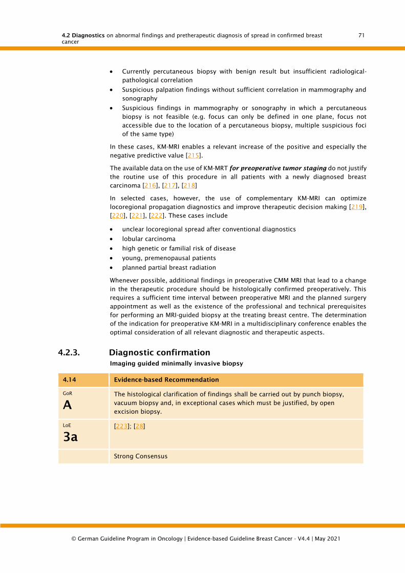
4.2 Diagnostics on abnormal findings and pretherapeutic diagnosis of spread in confirmed breast
cancer
© German Guideline Program in Oncology | Evidence-based Guideline Breast Cancer - V4.4 | May 2021
71
• Currently percutaneous biopsy with benign result but insufficient radiological-
pathological correlation
• Suspicious palpation findings without sufficient correlation in mammography and
sonography
• Suspicious findings in mammography or sonography in which a percutaneous
biopsy is not feasible (e.g. focus can only be defined in one plane, focus not
accessible due to the location of a percutaneous biopsy, multiple suspicious foci
of the same type)
In these cases, KM-MRI enables a relevant increase of the positive and especially the
negative predictive value [215].
The available data on the use of KM-MRT for preoperative tumor staging do not justify
the routine use of this procedure in all patients with a newly diagnosed breast
carcinoma [216], [217], [218]
In selected cases, however, the use of complementary KM-MRI can optimize
locoregional propagation diagnostics and improve therapeutic decision making [219],
[220], [221], [222]. These cases include
• unclear locoregional spread after conventional diagnostics
• lobular carcinoma
• high genetic or familial risk of disease
• young, premenopausal patients
• planned partial breast radiation
Whenever possible, additional findings in preoperative CMM MRI that lead to a change
in the therapeutic procedure should be histologically confirmed preoperatively. This
requires a sufficient time interval between preoperative MRI and the planned surgery
appointment as well as the existence of the professional and technical prerequisites
for performing an MRI-guided biopsy at the treating breast centre. The determination
of the indication for preoperative KM-MRI in a multidisciplinary conference enables the
optimal consideration of all relevant diagnostic and therapeutic aspects.
4.2.3. Diagnostic confirmation
Imaging guided minimally invasive biopsy
4.14 Evidence-based Recommendation
GoR
A
The histological clarification of findings shall be carried out by punch biopsy,
vacuum biopsy and, in exceptional cases which must be justified, by open
excision biopsy.
LoE
3a
[223]; [28]
Strong Consensus

4.2 Diagnostics on abnormal findings and pretherapeutic diagnosis of spread in confirmed breast
cancer
© German Guideline Program in Oncology | Evidence-based Guideline Breast Cancer - V4.4 | May 2021
72
4.15 Consensus-based Recommendation
EC The biopsy shall be controlled by means of imaging that clearly shows the
findings.
When selecting the collection method, diagnostic certainty and the risk of side
effects shall be taken into account.
The examiner shall take suitable measures to ensure that the localisation of the
finding can be found again (e.g. by clip insertion).
Strong Consensus
4.16 Consensus-based Recommendation
EC Even if the findings are primarily detected by mammography or MRI, the
sonographically controlled punch biopsy shall be performed if the sonographic
correlate is reliable.
Strong Consensus
4.17 Evidence-based Recommendation
GoR
A
If microcalcification is present without accompanying focal findings,
stereotactically controlled vacuum biopsy shall be used.
LoE
2b
[223]
Strong Consensus
4.18 Consensus-based Recommendation
EC Vacuum biopsy should be used for mammographic or MRI-guided tissue
collection.
Strong Consensus
4.19 Consensus-based Recommendation
EC For all biopsies, the correlation between the histological result and the
suspected clinical diagnosis shall be checked and documented.
Strong Consensus
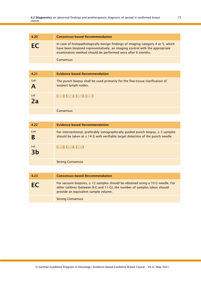
4.2 Diagnostics on abnormal findings and pretherapeutic diagnosis of spread in confirmed breast
cancer
© German Guideline Program in Oncology | Evidence-based Guideline Breast Cancer - V4.4 | May 2021
73
4.20 Consensus-based Recommendation
EC In case of histopathologically benign findings of imaging category 4 or 5, which
have been biopsied representatively, an imaging control with the appropriate
examination method should be performed once after 6 months.
Consensus
4.21 Evidence-based Recommendation
GoR
A
The punch biopsy shall be used primarily for the fine-tissue clarification of
suspect lymph nodes.
LoE
2a
[224]; [225]; [226]; [227]
Consensus
4.22 Evidence-based Recommendation
GoR
B
For interventional, preferably sonographically guided punch biopsy, ≥ 3 samples
should be taken at ≤ 14 G with verifiable target detection of the punch needle.
LoE
3b
[228]; [229]; [230]
Strong Consensus
4.23 Consensus-based Recommendation
EC For vacuum biopsies, ≥ 12 samples should be obtained using a 10 G needle. For
other calibres (between 8-G and 11-G), the number of samples taken should
provide an equivalent sample volume.
Strong Consensus
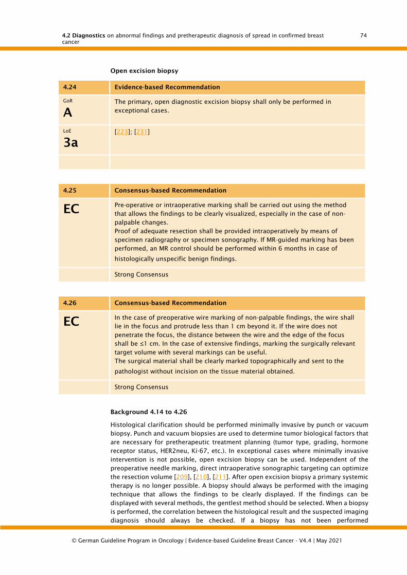
4.2 Diagnostics on abnormal findings and pretherapeutic diagnosis of spread in confirmed breast
cancer
© German Guideline Program in Oncology | Evidence-based Guideline Breast Cancer - V4.4 | May 2021
74
Open excision biopsy
4.24 Evidence-based Recommendation
GoR
A
The primary, open diagnostic excision biopsy shall only be performed in
exceptional cases.
LoE
3a
[223]; [231]
4.25 Consensus-based Recommendation
EC Pre-operative or intraoperative marking shall be carried out using the method
that allows the findings to be clearly visualized, especially in the case of non-
palpable changes.
Proof of adequate resection shall be provided intraoperatively by means of
specimen radiography or specimen sonography. If MR-guided marking has been
performed, an MR control should be performed within 6 months in case of
histologically unspecific benign findings.
Strong Consensus
4.26 Consensus-based Recommendation
EC In the case of preoperative wire marking of non-palpable findings, the wire shall
lie in the focus and protrude less than 1 cm beyond it. If the wire does not
penetrate the focus, the distance between the wire and the edge of the focus
shall be ≤1 cm. In the case of extensive findings, marking the surgically relevant
target volume with several markings can be useful.
The surgical material shall be clearly marked topographically and sent to the
pathologist without incision on the tissue material obtained.
Strong Consensus
Background 4.14 to 4.26
Histological clarification should be performed minimally invasive by punch or vacuum
biopsy. Punch and vacuum biopsies are used to determine tumor biological factors that
are necessary for pretherapeutic treatment planning (tumor type, grading, hormone
receptor status, HER2neu, Ki-67, etc.). In exceptional cases where minimally invasive
intervention is not possible, open excision biopsy can be used. Independent of the
preoperative needle marking, direct intraoperative sonographic targeting can optimize
the resection volume [209], [210], [211]. After open excision biopsy a primary systemic
therapy is no longer possible. A biopsy should always be performed with the imaging
technique that allows the findings to be clearly displayed. If the findings can be
displayed with several methods, the gentlest method should be selected. When a biopsy
is performed, the correlation between the histological result and the suspected imaging
diagnosis should always be checked. If a biopsy has not been performed
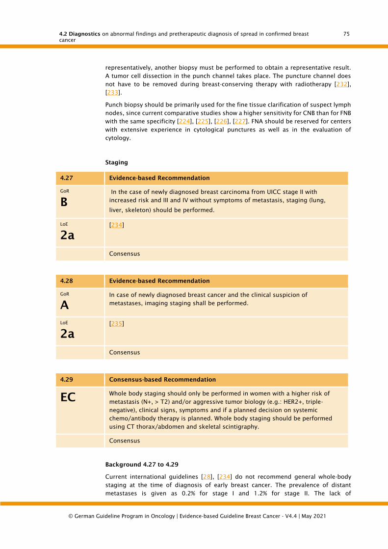
4.2 Diagnostics on abnormal findings and pretherapeutic diagnosis of spread in confirmed breast
cancer
© German Guideline Program in Oncology | Evidence-based Guideline Breast Cancer - V4.4 | May 2021
75
representatively, another biopsy must be performed to obtain a representative result.
A tumor cell dissection in the punch channel takes place. The puncture channel does
not have to be removed during breast-conserving therapy with radiotherapy [232],
[233].
Punch biopsy should be primarily used for the fine tissue clarification of suspect lymph
nodes, since current comparative studies show a higher sensitivity for CNB than for FNB
with the same specificity [224], [225], [226], [227]. FNA should be reserved for centers
with extensive experience in cytological punctures as well as in the evaluation of
cytology.
Staging
4.27 Evidence-based Recommendation
GoR
B
In the case of newly diagnosed breast carcinoma from UICC stage II with
increased risk and III and IV without symptoms of metastasis, staging (lung,
liver, skeleton) should be performed.
LoE
2a
[234]
Consensus
4.28 Evidence-based Recommendation
GoR
A
In case of newly diagnosed breast cancer and the clinical suspicion of
metastases, imaging staging shall be performed.
LoE
2a
[235]
Consensus
4.29 Consensus-based Recommendation
EC Whole body staging should only be performed in women with a higher risk of
metastasis (N+, > T2) and/or aggressive tumor biology (e.g.: HER2+, triple-
negative), clinical signs, symptoms and if a planned decision on systemic
chemo/antibody therapy is planned. Whole body staging should be performed
using CT thorax/abdomen and skeletal scintigraphy.
Consensus
Background 4.27 to 4.29
Current international guidelines [28], [234] do not recommend general whole-body
staging at the time of diagnosis of early breast cancer. The prevalence of distant
metastases is given as 0.2% for stage I and 1.2% for stage II. The lack of
4.3 DCIS and high-risk lesions
© German Guideline Program in Oncology | Evidence-based Guideline Breast Cancer - V4.4 | May 2021
76
recommendation for staging in the early stage serves to avoid unnecessary further
examinations and stress due to false positive incidence findings in imaging staging.
The prevalence of distant metastases is increased in N+ [236]. In addition, this
guideline contains a consensus recommendation for staging in aggressive tumor
biology if this has a decisive influence on the therapeutic procedure.
The available evidence on the selection of staging methods is limited. Studies have
shown sensitivities and specificities of the individual methods. However, there are no
studies on the influence of the selected methods on the actual outcome (survival as a
function of any resulting changes in therapy or quality of life).
Due to better sensitivity and specificity, CT thorax/abdomen and skeletal scinitgram
have replaced the earlier staging with thoracic x-ray and abdominal ultrasound as basic
staging examinations (ESMO 2015/17).
A general recommendation for PET or PET-CT is not given based on the NCCN 2014,
ESMO and NCGBCI Ireland 2015 guidelines, since even here false negative findings (for
slower growing metastases and metastases
The question of when further imaging examinations are useful, there is a layman's
understanding "Make smart decisions together" -Recommendation based on this
guideline.
4.3. DCIS and high-risk lesions
4.3.1. Preliminary remarks
DCIS and risk lesions are usually clinically occult changes that are detected during early
clinical diagnosis or mammography screening. This chapter deals with ductal
carcinoma in situ (DCIS), atypical ductal epithelial hyperplasia (ADH), lobular neoplasia
(LN), flat epithelial epidermis (FEA) and intraductal papilloma. These changes have the
character of a neoplasia in common, but with quite different risk of progression. The
aim of diagnostic and therapeutic measures, especially excision, is on the one hand to
avoid progression into invasive carcinoma and the associated morbidity and mortality.
Since it is not only a question of precursor but also indicator changes, the aim is also
to reduce the risk of ipsi- or contralateral carcinoma not directly associated with the
lesion.
4.3.2. DCIS
4.3.2.1. Clinical presentation, risk and course in DCIS
4.30 Consensus-based Recommendation
EC DCIS - General
In the treatment of a patient with ductal carcinoma in situ (DCIS) without
invasive components, the advantages and disadvantages of available forms of
therapy or their combination shall be explained. The relative and absolute
effects of adjuvant therapy measures in relation to the local recurrence
probability and overall survival shall be presented.
Strong Consensus
4.3 DCIS and high-risk lesions
© German Guideline Program in Oncology | Evidence-based Guideline Breast Cancer - V4.4 | May 2021
77
Background 4.30
The incidence of DCIS has increased significantly in the last decades and amounts to
approximately 5,500 per year [43] in Germany. Clinically, about 80% of DCIS behave
asymptomatically and only 20% of DCIS manifest themselves as symptom-related
disease [237].
Recently, studies have shown a low breast-cancer-specific long-term mortality of pure
DCIS in the US population [238] of 3.3% after 20 years (and 1.7% if cases with
contralateral second carcinoma are excluded [eTable 5 in the supplement]), thus
triggering a discussion about the importance of early detection of DCIS and its
treatment [239]. The mortality rate after diagnosis of DCIS is partly due to cases with
undiscovered occult invasion or microinvasion [240], and partly to invasive tumor
recurrence or ipsilateral second cancers which may occur many years after diagnosis
of DCIS [241]. The overall low mortality rate can be regarded as an expression of an
adequate therapy for patients with DCIS according to current standards. The
epidemiological data do not allow the conclusion that DCIS generally takes an indolent
natural course of the disease. Rather, case series with overlooked DCIS indicate a long-
term mortality of breast cancer which can be attributed to tumor progression [242],
whereas the mean time interval between overlooked DCIS and the occurrence of
invasive breast cancer in the Nurses Health Study was about 9.0 years [243]. As these
were low risk cases, the time interval for the untreated DCIS until the occurrence of an
invasive carcinoma should be altogether shorter, however, concrete data are not
available in the literature. For the DCIS discovered in the screening a connection with
the avoidance of an invasive interval carcinoma could be proven [55].
With regard to the risk factors for an unfavorable course, i.e. for the occurrence of an
intramammary tumor recurrence, clinical factors play a role, in particular the age of the
patient and the findings of a clinically manifest disease compared to a disease detected
in screening, as well as radiological factors such as the density of glandular tissue, and
last but not least histological and tumor biological factors. The age of the patient was
a significant risk factor for tumor recurrence in several prospective-randomized and
retrospective clinical studies [244], [245], and in patients
Much of the uncertainty regarding the clinical management of DCIS is due to the fact
that DCIS histologically and biologically represents a heterogeneous disease with
varying malignancy potential, and that an invasive disease can only be excluded when
the lesion has been pathomorphologically fully examined [247]. Pathomorphological
factors which reflect the malignancy potential of the DCIS are the grading and subtype
of the DCIS [248], the size of the lesion and (to a certain extent) the immunohistological
phenotype. Recently, gene expression analysis has also been discussed to better
estimate the aggressiveness of the DCIS [249]. Among these parameters, grading is the
most important risk factor [244], followed by lesion size and detection of comedotype
necrosis [246]. It could be shown that the group of patients with high-grade DCIS and
extensive comedotype necroses in > 50 % of ductuli has a significantly increased risk
of both ipsilateral invasive and non-invasive tumor recurrence [250]. The role of gene
expression analysis in this context has not yet been conclusively clarified. In a low-risk
collective defined by means of a DCIS-specific multiassay the 10-year local recurrence
rate after breast-conserving surgery without radiotherapy was still 10.8% [251], which
is similar or worse compared to collectives with breast-conserving surgery without
radiotherapy defined by conventional risk factors [252], [253].
The determination of the size of the DCIS is not necessary for the assessment of the
T/pT category according to TNM, but nevertheless important for the pathological-
radiological correlation and for the management of the patient [254]. The larger the

4.3 DCIS and high-risk lesions
© German Guideline Program in Oncology | Evidence-based Guideline Breast Cancer - V4.4 | May 2021
78
DCIS, the more probable the presence of multifocality and marginal infestation as well
as tumor residuals, and thus the probability of a local recurrence with the possibility of
a metachronous tumor invasion [255], [256], [257], [258], [259], [260]. In addition, the
risk of an occult invasion increases with an increase in tumor size [261]. A threshold
value for a critical size of the DCIS, up to which breast-conserving therapy is possible,
cannot be given. The DCIS size is often underestimated due to mammographic
evaluation or preparation radiography, but can also be overestimated in cases with well
differentiated DCIS at the base of a mastopathy with benign calcifications. A relatively
accurate estimation of the total extent of the DCIS is possible by serial, lamellar
processing with an accuracy of 3 - 5 mm. Small DCIS of 1 cm or less can be measured
directly histologically in the sectional preparation [262]. It should be noted that,
especially with low-grade DCIS, the propagation of the DCIS can be discontinuous. It is
therefore recommended to indicate the total size including these gaps and not the size
of the individual foci in the case of a supposed multifocality [262]. When resecting a
DCIS in several parts, the pathologist should try to reconstruct the size of the DCIS
taking into account the topography of the individual sub-preparations [263].
4.3.2.2. Surgical therapy of DCIS
4.31 Evidence-based Recommendation
GoR
A
Complete excision is the therapeutic basis for the treatment of DCIS. The
resection limits shall be at least 2 mm for pure DCIS when adjuvant radiotherapy
is connected.
LoE
2b
[264]; [265]; [266]; [267]; [268]
Strong Consensus
Background 4.31
Just as in invasive breast cancer, breast-conserving therapy can be considered standard
today in DCIS, especially when there is a favourable relation between the extent of the
lesion and the breast size. However, in contrast to invasive carcinoma, there is no
randomized controlled trial for DCIS that has compared the outcome after breast
conservation therapy with that after mastectomy. Histopathological studies on the
spread pattern of DCIS show that DCIS is a usually (90%) unicentric, but potentially
multifocal lesion, which can theoretically be rehabilitated by surgery alone, provided
that a targeted (segmentally oriented) surgery with sufficient resection limits is
performed [269], [270].
The question of which resection limits have to be adhered to in the breast-conserving
therapy of DCIS is directly related to the implementation or the renunciation of
postoperative radiotherapy. With a resection margin of 10 mm or more, the benefit of
radiotherapy is only slight [271], [272]. A 2 mm wide safety margin is associated with
a lower risk of recurrence than a 1 mm wide margin, but does not differ statistically
from a 5 mm wide resection margin if breast-conserving surgery is performed with
subsequent whole breast radiation [273]. In case of mastectomy and affected resection
margin or narrow resection of
Taking into account careful pathological work-up and radiological-pathological
correlation with respect to the topography and the size of the DCIS, a minimum
4.3 DCIS and high-risk lesions
© German Guideline Program in Oncology | Evidence-based Guideline Breast Cancer - V4.4 | May 2021
79
resection margin of at least 2 mm therefore appears to be sufficient if adjuvant
radiotherapy is performed [264], [265], [266], [268], [274]. If radiotherapy is not
performed, wider safety margins should be aimed for, although no optimum margin
can be given here. Here, further risk factors should be taken into account, especially
whether the DCIS extends over a wide area or only with an extension close to the edge
[271] as well as size and grading of the DCIS [259]. If a complete excision cannot be
achieved by resection(s), a secondary mastectomy should be considered. In the case of
a marginal margin of less than 2 mm, the necessity of a follow-up resection should be
discussed as an individual case decision in the interdisciplinary team, taking into
account the clinical situation (topography of the marginal situation, size and grading
of the DCIS, age of the patient, etc.).
Radiotherapy of DCIS
4.32 Evidence-based Recommendation
GoR
A
An axillary dissection shall not be performed in DCIS. A sentinel node biopsy
shall only be performed if a secondary sentinel node biopsy is not possible for
technical reasons, e.g. in the case of a mammary ablatio.
LoE
1b
[185]; [275]
Strong Consensus
Background 4.32
Since DCIS is by definition a non-metastatic lesion, no staging examination is required,
and usually no sentinel lymph node (SN) biopsy is needed. However, an SN biopsy can
be performed in the case of primary mastectomy or very peripheral tumour site, as the
morbidity of the SN intervention is low and the SN biopsy cannot be performed in this
constellation for technical reasons if an invasive carcinoma has been subsequently
identified [185], [275], [276], [277]. When a DCIS is diagnosed by means of punch or
vacuum biopsy, an invasive carcinoma is found in about 20% of the surgical specimens
subsequently obtained [278], [279], [280]. There is an increased risk in palpable lesions
and DCIS with a radiological size of > 4 cm [279], [281], [282]. However, even in this
situation, the probability of a clinically relevant, positive sentinel lymph node being
present in the case of occult tumor invasion is low and can be clinically neglected [279],
[283], [284]. An SN biopsy should therefore be performed secondarily in BET because
of DCIS, if necessary.
4.3 DCIS and high-risk lesions
© German Guideline Program in Oncology | Evidence-based Guideline Breast Cancer - V4.4 | May 2021
80
4.3.2.3. Radiotherapy of the DCIS
4.33 Evidence-based Recommendation
GoR
B
Adjuvant radiotherapy reduces the risk of local recurrence after breast-
conserving therapy by up to 50%, but at low risk the benefit for the patient is
small. The possibility of radiotherapy should be offered to the patient
depending on the individual risk profile.
LoE
1a
[238]; [285]; [286]; [287]; [288]
Strong Consensus
Background 4.33
The radiotherapy of DCIS aims to reduce the intramammary recurrence risk and the
associated morbidity [285], [289]. However, it is not an obligatory component of the
breast-conserving therapy of DCIS, but an individual therapy decision which depends
on clinical, radiological and pathological criteria. In the EBCTCG meta-analysis the 10-
year in-breast recurrence rate with negative resection margin was 26.0% without
radiotherapy compared to 12.0% with postoperative whole breast radiation (p 3 mm
6.7% and 0.9% (p = 0.0003) for the sole excision compared to excision plus whole
breast radiation (p = 0.0003) in the RTOG-9804 study [252]. The risk of an
intramammary recurrence after 10 years is currently given as 11% for patients with
postoperative follow-up radiation and 19% without follow-up radiation. Thus, the risk
of recurrence has been significantly reduced compared to older data [290]. This can be
attributed to improvements in diagnosis and therapy of DCIS. These include the
radiological diagnosis and early detection of the DCIS, the pathomorphological
processing and evaluation of the tissue samples [291] and last but not least the surgical
treatment of the DCIS. In a low risk collective (G1/G2 to 2.5 cm) the intramammary
recurrence rate after 12 years without radiotherapy was 12.5% and thus approx. 1% per
year [252], and in a further prospective-retrospective analysis of 209 low risk patients
without radiotherapy (size 2 cm or less, age 50 years or more, grade 1 or 2) the 12-
year intramammary recurrence rate was only 7.8% and thus 0.65% per year [253]. These
recurrence rates are lower than the 15% after 10 years considered acceptable by
EUSOMA for invasive carcinomas [292].
Although half of the tumor recurrences after DCIS are invasive tumor recurrences, no
survival benefit from postoperative radiotherapy of DCIS has been shown in several
prospective randomized studies and a follow-up of up to 20 years [285], [286]. In a
retrospective analysis, however, the hazard ratio for the mortality probability after
breast cancer for the invasive intramammary recurrence was 18.1 (95% AI: 14.0 - 23.6)
[238]. There are indications for the association of breast cancer mortality in DCIS with
pathological risk factors (grading, size, comedotype necroses of DCIS) [287]. In low-
grade DCIS no significant mortality could be detected in this cohort, independent of
the performance of surgical intervention [293]. The possible reduction of the
intramammary recurrence risk by radiotherapy must therefore be seen together with
the unclear effect on mortality and requires an individual therapy decision taking into
account the risk factors for an intramammary recurrence.
According to the available data, the number needed to treat (NNT) to prevent a local
recurrence by follow-up radiation is 7 [288] for all DCIS and 17 for a low-risk collective

4.3 DCIS and high-risk lesions
© German Guideline Program in Oncology | Evidence-based Guideline Breast Cancer - V4.4 | May 2021
81
( 50 years). The patient should be informed about the personal (absolute) benefit as
well as the possible risks of radiation on the basis of an individual risk assessment.
There is evidence that hypofractionation, similar to invasive carcinoma, can reduce
radiotherapy-associated morbidity in DCIS and is similarly effective in preventing
intramammary recurrence [294], [295].
4.3.2.4. Antihormone therapy in DCIS
Similar to radiotherapy the risk of intramammary recurrence after BET due to DCIS can
be reduced by adjuvant antihormonal therapy [289]. This also applies to the
contralateral risk of an invasive second carcinoma [296]. However, the effect on the
intramammary risk of recurrence is less than for adjuvant radiotherapy, and likewise
without a detectable influence on survival [296], [297], [298], [299]. The number of
patients to be treated (NNT), for the detection of a locally protective effect by the
administration of tamoxifen over 5 years is 15 patients [297]. The NNT rate for
subgroups is: ipsilateral DCIS 47; contralateral DCIS 93; ipsilateral invasive carcinoma
63 and contralateral invasive carcinoma 54 patients [297]. Aromatase inhibitors have a
similar protective effect in DCIS as tamoxifen [300]. Due to the relatively low benefit
for the individual patient with DCIS, [301] is recommended as a cautious indication for
adjuvant antihormonal therapy in DCIS. This shows a higher benefit in postmenopause
than in premenopause [302]. If an indication for antihormonal therapy is given, the
determination of the estrogen receptor in DCIS is necessary [296].
4.3.3. Risk lesions
4.3.3.1. Preliminary remarks
4.34 Consensus-based Recommendation
EC The therapeutic concept for high-risk lesions shall be developed on an
interdisciplinary basis (radiodiagnostics, surgeon, pathology) after the
histological findings from a punch/vacuum biopsy are available.
Strong Consensus
Background 4.34
Clinical screening and mammography screening programs are increasingly detecting
clinically occult risk lesions of the mamma, which may be associated with
microcalcifications or architectural defects. The risk of progression of these lesions
[57], grouped together as "benign lesions with uncertain malignant potential (B3)", is
quite heterogeneous and generally lower than in DCIS. The recommendations for
excision of risk lesions refer to the natural course, subtype and extent of the lesions.
In recent years the literature base for recommendations for the management of occult
risk lesions has broadened significantly and this has led to a more conservative
approach [303]. However, the literature on risk lesions is mainly based on non-
randomized, retrospective case series of individual institutions, where information on
the radiological-pathological correlation is often missing as well as information on the
indication for surgery, if the lesions caused excision in only a part of the patients. This
could at least partly explain the range of variation in the published upgrade risk with
open biopsy. In principle, the procedure for risk lesions should be decided individually
in an interdisciplinary conference, taking into account pathological-radiological
correlation diagnostics.
4.3 DCIS and high-risk lesions
© German Guideline Program in Oncology | Evidence-based Guideline Breast Cancer - V4.4 | May 2021
82
4.3.3.2. Atypical ductal hyperplasia (ADH) in punch or vacuum biopsy
4.35 Consensus-based Recommendation
EC If ADH is diagnosed, an open PE shall be performed to exclude a higher grade
lesion.
Strong Consensus
Background 4.35
Atypical ductal epithelial hyperplasia (ADH) is a proliferation of isomorphic ductal
epithelia of terminal ductuli or duktulo-lobular units (TDLUs) with cytological and
structural features of a well differentiated DCIS. ADH is morphologically similar to a
small, well differentiated DCIS and is only differentiated by quantitative criteria. A well-
differentiated DCIS can only be diagnosed if the change in a TDLU exceeds a size of 2
mm or at least two separate duct structures are homogeneously affected [248], [304].
Even in the WHO expert group there is no agreement on which of the two quantitative
criteria is to be preferred [248], [304]. In the punch biopsy, the diagnosis of a well
differentiated DCIS should be made with caution due to the problematic differential
diagnosis. In most cases it is sufficient to describe an alteration as atypical ductal
epithelial proliferation to induce an excision [303]. However, it can be assumed that an
ADH is usually small and extends over less than 2 - 3 mm [305]
(http://www.euref.org/european-guidelines). If no further changes are found in the
resectate, the therapy should be based on the diagnosis of ADH [248], [304]. The risk
of breast carcinoma in the long-term course of ADH is 3 - 5 times higher [306], [307],
[308] and the cumulative risk of invasive or insitu carcinoma is indicated with 21% (95%
CI 14-28%) [309] or with 20% for a median follow-up of 17 years [310].
In clinical studies in which ADH was diagnosed by punch biopsy, the upgrade rate to
DCIS or invasive carcinoma in open biopsy is 28% to 56% [311], [312], [313], [314],
[315], but is lower if there is concordance between radiological and pathological
findings [316]. In a meta-analysis, the upgrade rate for Z. n. vacuum biopsy and
diagnosis of ADH was significantly lower and was 20.9% [317]. There are different
approaches to achieve a better estimation of the risk in ADH. These include, for
example, a postinterventional mammogram and close monitoring [313], the number of
affected TDLUs [318], [319], [320] or cytological atypes or necroses [320], [321].
Suggestions to avoid an open biopsy try to define subgroups by combining these
criteria or by complete removal of microcalcifications [322], [323]. This may help in
individual cases to plan the therapy. However, the predictive criteria cannot be regarded
as sufficiently validated due to the small number of cases in the available studies.
Therefore, open PE is currently recommended for diagnosis of ADH in punch or vacuum
biopsy [303], [324].
4.3 DCIS and high-risk lesions
© German Guideline Program in Oncology | Evidence-based Guideline Breast Cancer - V4.4 | May 2021
83
4.3.3.3. Lobular neoplasia (LN) in punch or vacuum biopsy
4.36 Consensus-based Recommendation
EC In case of isolated or incidential findings of an LN (classic variant) in punch or
vacuum biopsy and concordance with imaging, further bioptical clarification is
not necessary. In the case of LN with increased risk (pleomorphic LN, florid or
tumorous LN, LN with comedotype necroses) an excision of the change should
be performed, as well as in the case of discordance with the radiological
findings.
Strong Consensus
Background 4.36
Lobular neoplasia (LN) is characterized by an atypical proliferation of loosely cohesive
or discohesive and generally small, uniform epithelia within a terminal ducto-lobular
unit with or without pagetoid spread into the ducts. It includes both atypical lobular
hyperplasia (ALH) with distension and distortion of less than 50% of the azini in the
affected terminal dukto-lobular unit (TDLU) [325], and lobular carcinoma in situ (LCIS).
The LN is considered a non-obligate precursor lesion for breast carcinoma [326] and at
the same time a risk lesion with increased tumor risk ipsi- and contralateral. The risk
for invasive carcinoma is given as about 1 - 2% per year and the lifetime risk is
calculated at 30 - 40% [327], [328]. The risk for pleomorphic LCIS is possibly higher
[329].
The management of diagnosing an LN in punch biopsy depends on whether the LN is
the only pathological change, on the subtype of the LN and on any associated
pathological changes. In case of an incidence of a classic LN in punch biopsy
(ultrasound or vacuum biopsy) there is no definite indication for an open biopsy for
further clarification and the further procedure should be determined in the
interdisciplinary findings conference. A wait-and-see approach with follow-up is
acceptable in this situation, since the risk of carcinoma at the biopsy site within 3 - 5
years (i.e. the probability of an overlooked carcinoma) is indicated with ≤ 2% [303]. In
contrast, in the case of radiological-pathological discordance of the findings, i.e. if the
pathological findings do not explain the radiological index lesion (e.g. focal findings
or malignoma-typical microcalcifications), there is a higher risk. In this situation further
clarification should be carried out by means of a representative open biopsy [248],
[330], [331].
Among the variants of the LN and at the same time lobular intraepithelial neoplasias
with increased risk of later invasive carcinoma are the LCIS with comedotype necroses,
the pleomorphic LCIS and the florid LCIS. These special forms of LCIS can be clinically
associated with a focal finding or radiologically extensive microcalcifications, as in
DCIS, and are frequently associated with a (micro-)invasion [332], [333]. In pleomorphic
LCIS, higher grade cellular atypia are found, sometimes accompanied by massively
distended lobules and infestation of the ducts as well as formation of comedotype
necroses and microcalcifications. The pleomorphic LCIS differs from a ductal carcinoma
in situ by the cellular morphology and the loss of the E-cadherin expression [334]. If
these variants of an LN with increased risk are present (pleomorphic LN, florid or tumor-
like LN, LN with comedotype necroses) an excision of the alteration should be
performed [303], [330], [335], [336]. The B classification of the classic LN is B3, unless
the criteria for a pleomorphic LN or an LN with comedotype necroses are present which
justify classification as in the B5a category [337].

4.3 DCIS and high-risk lesions
© German Guideline Program in Oncology | Evidence-based Guideline Breast Cancer - V4.4 | May 2021
84
4.3.3.4. Flat epithelial atype (FEA) in punch or vacuum biopsy
4.37 Consensus-based Recommendation
EC In the case of diagnosis of FEA, an open PE can be forgone with if the suspicious
calcifications have already been completely or almost completely removed by
means of vacuum biopsy imaging. In the case of radiologically extensive
calcifications or in case of discordance with the radiological findings, a
representative open PE shall be performed.
Strong Consensus
Background 4.37
The term flat epithelia (FEA) is the term preferred by the WHO for columnar cell
hyperplasia with nuclear atypes [248], [338]. In a typical case, intraluminal
microcalcifications, which represent secretion calcium, are associated with FEA and
lead to the clarification and diagnosis of FEA. Morphologically and on the molecular
level there are multiple relationships and similarities of FEA with atypical ductal
epithelial hyperplasia (ADH) and lobular neoplasia [339]. Not infrequently, these
changes occur together on [340] and are considered early, non-obligatory precursor
lesions in the low-grade pathway for multistep carcinogenesis of breast cancer [341],
[342], [343].
The risk of a higher grade lesion (DCIS or invasive carcinoma) in a subsequent open
biopsy is less than 10% [344], [345], [346], [347] in more recent studies and taking into
account the pathological-histological correlation. In case of complete or largely
complete removal of microcalcifications, further clarification by means of open biopsy
is therefore not necessary [303], [348], [349]. This concerns the removal of
microcalcifications by means of 9G or 11G vacuum biopsy [350].
4.3.3.5. ADH, LN, FEA in open biopsy
4.38 Consensus-based Recommendation
EC Bei Vorliegen einer klassischen LN oder einer FEA (alleine oder in Kombination
mit einem invasiven Karzinom) am Resektionsrand ist keine Nachresektion
erforderlich. Bei Vorliegen einer isolierten ADH am Resektionsrand sollte eine
Nachresektion erfolgen. Ebenso sollte bei Vorliegen einer LN mit erhöhtem
Risiko am Resektionsrand (alleine oder in Kombination mit einem invasiven
Karzinom) eine Nachresektion erfolgen.
Strong Consensus
Background 4.38
For the procedure in the diagnosis of a risk lesion (ADH, LN, FEA) in open biopsy, a
distinction must be made as to whether it is an accompanying finding associated with
an invasive carcinoma or ductal carcinoma in situ or an isolated change. In the low-risk
lesions (classic LN or FEA) at the resection margin, no resection is necessary [330],
[351]. On the other hand, in the case of an LN with increased risk at the resection
margin (alone or in combination with an invasive carcinoma), resection is
recommended because this change spreads intraductally and is biologically similar to
a low-grade DCIS [330], [335], [351], [352]. In the case of isolated atypical ductal
4.3 DCIS and high-risk lesions
© German Guideline Program in Oncology | Evidence-based Guideline Breast Cancer - V4.4 | May 2021
85
hyperplasia (ADH) at the resection margin in the open biopsy, similar to ADH in the
punch biopsy, it must be taken into account that the findings may not be
representative. Since ADH is only differentiated from a well differentiated DCIS on the
basis of quantitative criteria, a resection should be performed here if necessary to
exclude higher grade changes.
4.3.3.6. Papilloma in the punch or vacuum biopsy
4.39 Consensus-based Recommendation
EC In the case of histological diagnosis of a lactiferous papilloma detected by
imaging or a papilloma with ADH, an excision shall be performed; this can also
be performed as a vacuum biopsy, provided that no atypia are present.
Strong Consensus
Background 4.39
The diagnosis of a lactiferous papilloma in the punch biopsy (ultrasound-guided or
vacuum biopsy) means, similar to the other B3 lesions, a risk of a higher grade lesion
in a subsequent open biopsy. This applies less to the histological differential diagnosis
compared with a papillary DCIS or a papillary or solid-papillary carcinoma, which can
be made without further ado at the punch biopsy. Rather, there is the problem of the
heterogeneity of the lactiferous papilloma and the possibility of association of the
lesion with a DCIS or an invasive carcinoma. The background is the question of the
representativeness of the punch biopsy. Therefore, it must first be considered whether
the finding is an incident lesion, i.e. a small papilloma or micropapilloma that is only
histologically detectable (with negligible risk), or an imaging finding that is only
partially recorded in the punch biopsy. Furthermore, the upgrade risk depends on
whether atypical papilloma is present, i.e. whether it is a papilloma with ADH or an
atypical papilloma.
In a recent meta-analysis of 34 studies, the upgrade rate for papilloma was 7.0%
compared to 36.9% for atypical papilloma. Furthermore, a lower tendency to upgrade
was found in the newer studies after 2005 [353]. Despite the relatively low risk of
upgrade in benign papilloma, the literature predominantly recommends post-excision
in punch biopsy diagnosis of papilloma [354], [355], [356], [357]. An exclusively
radiological evaluation of the course of the disease is justifiable in the case of small or
incident papillomas, also if the papilloma has already been completely excised by the
punch biopsies or vacuum biopsies and the mamma can be easily monitored by
imaging [303], [358]. As an alternative to open biopsy, completion can also be carried
out by vacuum biopsy [359].
4.4 Surgical therapy of invasive carcinoma
© German Guideline Program in Oncology | Evidence-based Guideline Breast Cancer - V4.4 | May 2021
86
4.3.3.7. Papilloma in open PE
4.40 Consensus-based Recommendation
EC If a papilloma or papilloma with ADH is detected in an open biopsy, no further
intervention is necessary. If atypia are present at the edge of the resection, a
follow-up resection should be performed.
Strong Consensus
Background 4.40
The further procedure in case of a benign lactiferous papilloma or a papilloma with
ADH in an open biopsy depends on the question of possibly accompanying atypical
changes, for example a DCIS in the surrounding parenchyma. In the absence of such
changes, no further intervention is required. The recommendation to complete the
excision of an atypical papilloma in the resection margin is based on the possible
association with a well differentiated DCIS, which otherwise would not be completely
excised [360], [361].
4.4. Surgical therapy of invasive carcinoma
4.4.1. General recommendation
4.41 Evidence-based Recommendation
GoR
A
The basis of therapy for all non-advanced breast carcinomas is tumour resection
in sano (R0 status).
LoE
1a
[362]; [363]
Strong Consensus
4.42 Evidence-based Statement
ST The resection margin status has a prognostic effect in invasive breast cancer.
There is a significant correlation between the resection margin status (positive
vs. negative) and the local recurrence rate.
LoE
1a
[362]; [364]
Strong Consensus
Background 4.41 and 4.42
The complete removal of the tumor with free resection limits is a prerequisite for a low
local recurrence risk. The local recurrence risk is decisively determined by the tumor
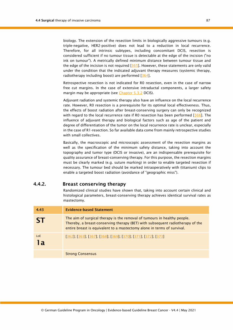
4.4 Surgical therapy of invasive carcinoma
© German Guideline Program in Oncology | Evidence-based Guideline Breast Cancer - V4.4 | May 2021
87
biology. The extension of the resection limits in biologically aggressive tumours (e.g.
triple-negative, HER2-positive) does not lead to a reduction in local recurrence.
Therefore, for all intrinsic subtypes, including concomitant DCIS, resection is
considered sufficient if no tumour tissue is detectable at the edge of the incision ("no
ink on tumour"). A metrically defined minimum distance between tumour tissue and
the edge of the incision is not required [365]. However, these statements are only valid
under the condition that the indicated adjuvant therapy measures (systemic therapy,
radiotherapy including boost) are performed [364].
Retrospective resection is not indicated for R0 resection, even in the case of narrow
free cut margins. In the case of extensive intraductal components, a larger safety
margin may be appropriate (see Chapter 5.3.2 DCIS).
Adjuvant radiation and systemic therapy also have an influence on the local recurrence
rate. However, R0 resection is a prerequisite for its optimal local effectiveness. Thus,
the effects of boost radiation after breast-conserving surgery can only be recognized
with regard to the local recurrence rate if R0 resection has been performed [366]. The
influence of adjuvant therapy and biological factors such as age of the patient and
degree of differentiation of the tumor on the local recurrence rate is unclear, especially
in the case of R1-resection. So far available data come from mainly retrospective studies
with small collectives.
Basically, the macroscopic and microscopic assessment of the resection margins as
well as the specification of the minimum safety distance, taking into account the
topography and tumor type (DCIS or invasive), are an indispensable prerequisite for
quality assurance of breast-conserving therapy. For this purpose, the resection margins
must be clearly marked (e.g. suture marking) in order to enable targeted resection if
necessary. The tumour bed should be marked intraoperatively with (titanium) clips to
enable a targeted boost radiation (avoidance of "geographic miss").
4.4.2. Breast conserving therapy
Randomized clinical studies have shown that, taking into account certain clinical and
histological parameters, breast-conserving therapy achieves identical survival rates as
mastectomy.
4.43 Evidence-based Statement
ST The aim of surgical therapy is the removal of tumours in healthy people.
Thereby, a breast-conserving therapy (BET) with subsequent radiotherapy of the
entire breast is equivalent to a mastectomy alone in terms of survival.
LoE
1a
[362]; [363]; [367]; [368]; [369]; [370]; [371]; [372]; [373]
Strong Consensus
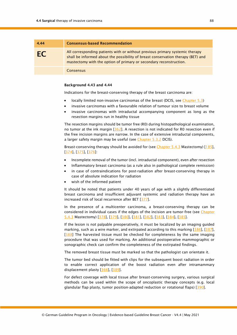
4.4 Surgical therapy of invasive carcinoma
© German Guideline Program in Oncology | Evidence-based Guideline Breast Cancer - V4.4 | May 2021
88
4.44 Consensus-based Recommendation
EC All corresponding patients with or without previous primary systemic therapy
shall be informed about the possibility of breast conservation therapy (BET) and
mastectomy with the option of primary or secondary reconstruction.
Consensus
Background 4.43 and 4.44
Indications for the breast-conserving therapy of the breast carcinoma are:
• locally limited non-invasive carcinomas of the breast (DCIS, see Chapter 5.3)
• invasive carcinomas with a favourable relation of tumour size to breast volume
• invasive carcinomas with intraductal accompanying component as long as the
resection margins run in healthy tissue
The resection margins should be tumor free (R0) during histopathological examination,
no tumor at the ink margin [362]. A resection is not indicated for R0 resection even if
the free incision margins are narrow. In the case of extensive intraductal components,
a larger safety margin may be useful (see Chapter 5.3.2 DCIS).
Breast-conserving therapy should be avoided for (see Chapter 5.4.3 Mastectomy) [185],
[374], [375], [376]:
• Incomplete removal of the tumor (incl. intraductal component), even after resection
• Inflammatory breast carcinoma (as a rule also in pathological complete remission)
• in case of contraindications for post-radiation after breast-conserving therapy in
case of absolute indication for radiation
• wish of the informed patient
It should be noted that patients under 40 years of age with a slightly differentiated
breast carcinoma and insufficient adjuvant systemic and radiation therapy have an
increased risk of local recurrence after BET [377].
In the presence of a multicenter carcinoma, a breast-conserving therapy can be
considered in individual cases if the edges of the incision are tumor-free (see Chapter
5.4.3 Mastectomy) [378], [379], [380], [381], [382], [383], [384], [385].
If the lesion is not palpable preoperatively, it must be localized by an imaging guided
marking, such as a wire marker, and extirpated according to this marking [386], [387],
[388] The harvested tissue must be checked for completeness by the same imaging
procedure that was used for marking. An additional postoperative mammographic or
sonographic check can confirm the completeness of the extirpated findings.
The removed breast tissue must be marked so that the pathologist can orientate it.
The tumor bed should be fitted with clips for the subsequent boost radiation in order
to enable correct application of the boost radiation even after intramammary
displacement plasty [366], [389].
For defect coverage with local tissue after breast-conserving surgery, various surgical
methods can be used within the scope of oncoplastic therapy concepts (e.g. local
glandular flap plasty, tumor position-adapted reduction or rotational flaps) [390].
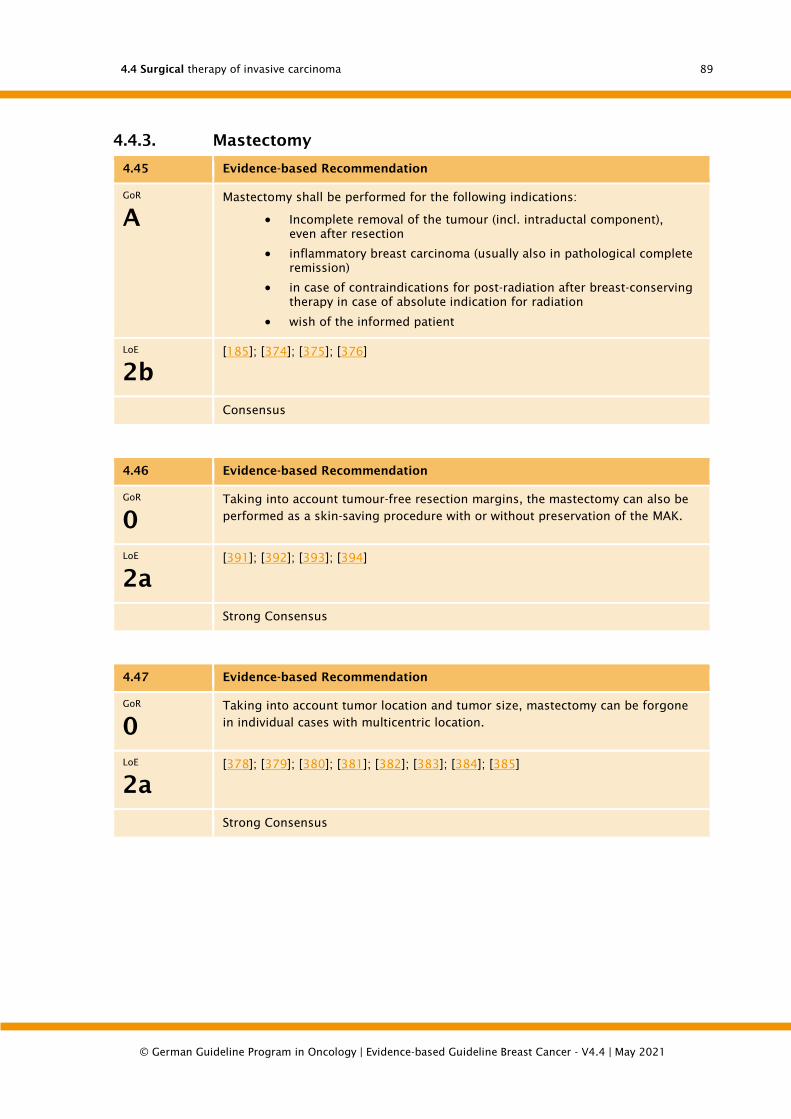
4.4 Surgical therapy of invasive carcinoma
© German Guideline Program in Oncology | Evidence-based Guideline Breast Cancer - V4.4 | May 2021
89
4.4.3. Mastectomy
4.45 Evidence-based Recommendation
GoR
A
Mastectomy shall be performed for the following indications:
• Incomplete removal of the tumour (incl. intraductal component),
even after resection
• inflammatory breast carcinoma (usually also in pathological complete
remission)
• in case of contraindications for post-radiation after breast-conserving
therapy in case of absolute indication for radiation
• wish of the informed patient
LoE
2b
[185]; [374]; [375]; [376]
Consensus
4.46 Evidence-based Recommendation
GoR
0
Taking into account tumour-free resection margins, the mastectomy can also be
performed as a skin-saving procedure with or without preservation of the MAK.
LoE
2a
[391]; [392]; [393]; [394]
Strong Consensus
4.47 Evidence-based Recommendation
GoR
0
Taking into account tumor location and tumor size, mastectomy can be forgone
in individual cases with multicentric location.
LoE
2a
[378]; [379]; [380]; [381]; [382]; [383]; [384]; [385]
Strong Consensus
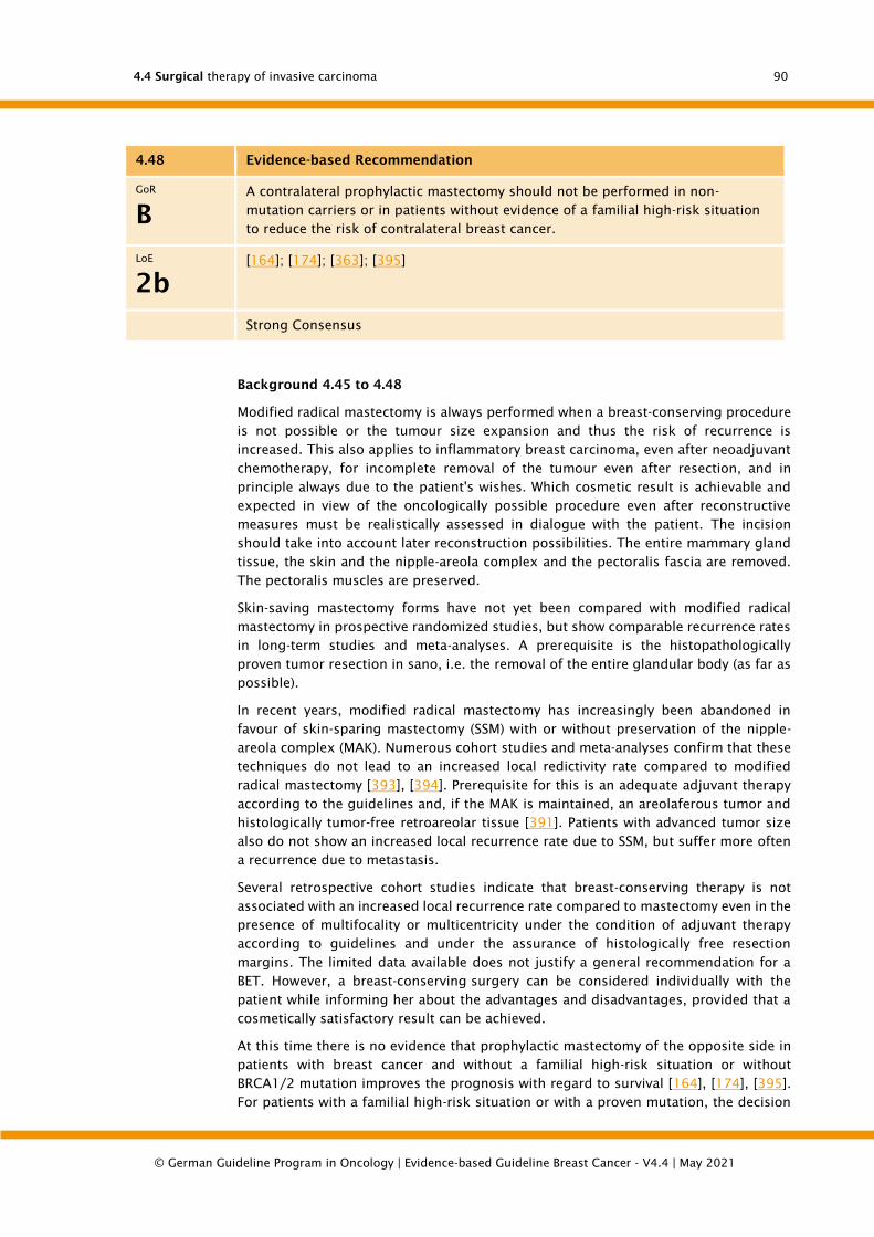
4.4 Surgical therapy of invasive carcinoma
© German Guideline Program in Oncology | Evidence-based Guideline Breast Cancer - V4.4 | May 2021
90
4.48 Evidence-based Recommendation
GoR
B
A contralateral prophylactic mastectomy should not be performed in non-
mutation carriers or in patients without evidence of a familial high-risk situation
to reduce the risk of contralateral breast cancer.
LoE
2b
[164]; [174]; [363]; [395]
Strong Consensus
Background 4.45 to 4.48
Modified radical mastectomy is always performed when a breast-conserving procedure
is not possible or the tumour size expansion and thus the risk of recurrence is
increased. This also applies to inflammatory breast carcinoma, even after neoadjuvant
chemotherapy, for incomplete removal of the tumour even after resection, and in
principle always due to the patient's wishes. Which cosmetic result is achievable and
expected in view of the oncologically possible procedure even after reconstructive
measures must be realistically assessed in dialogue with the patient. The incision
should take into account later reconstruction possibilities. The entire mammary gland
tissue, the skin and the nipple-areola complex and the pectoralis fascia are removed.
The pectoralis muscles are preserved.
Skin-saving mastectomy forms have not yet been compared with modified radical
mastectomy in prospective randomized studies, but show comparable recurrence rates
in long-term studies and meta-analyses. A prerequisite is the histopathologically
proven tumor resection in sano, i.e. the removal of the entire glandular body (as far as
possible).
In recent years, modified radical mastectomy has increasingly been abandoned in
favour of skin-sparing mastectomy (SSM) with or without preservation of the nipple-
areola complex (MAK). Numerous cohort studies and meta-analyses confirm that these
techniques do not lead to an increased local redictivity rate compared to modified
radical mastectomy [393], [394]. Prerequisite for this is an adequate adjuvant therapy
according to the guidelines and, if the MAK is maintained, an areolaferous tumor and
histologically tumor-free retroareolar tissue [391]. Patients with advanced tumor size
also do not show an increased local recurrence rate due to SSM, but suffer more often
a recurrence due to metastasis.
Several retrospective cohort studies indicate that breast-conserving therapy is not
associated with an increased local recurrence rate compared to mastectomy even in the
presence of multifocality or multicentricity under the condition of adjuvant therapy
according to guidelines and under the assurance of histologically free resection
margins. The limited data available does not justify a general recommendation for a
BET. However, a breast-conserving surgery can be considered individually with the
patient while informing her about the advantages and disadvantages, provided that a
cosmetically satisfactory result can be achieved.
At this time there is no evidence that prophylactic mastectomy of the opposite side in
patients with breast cancer and without a familial high-risk situation or without
BRCA1/2 mutation improves the prognosis with regard to survival [164], [174], [395].
For patients with a familial high-risk situation or with a proven mutation, the decision
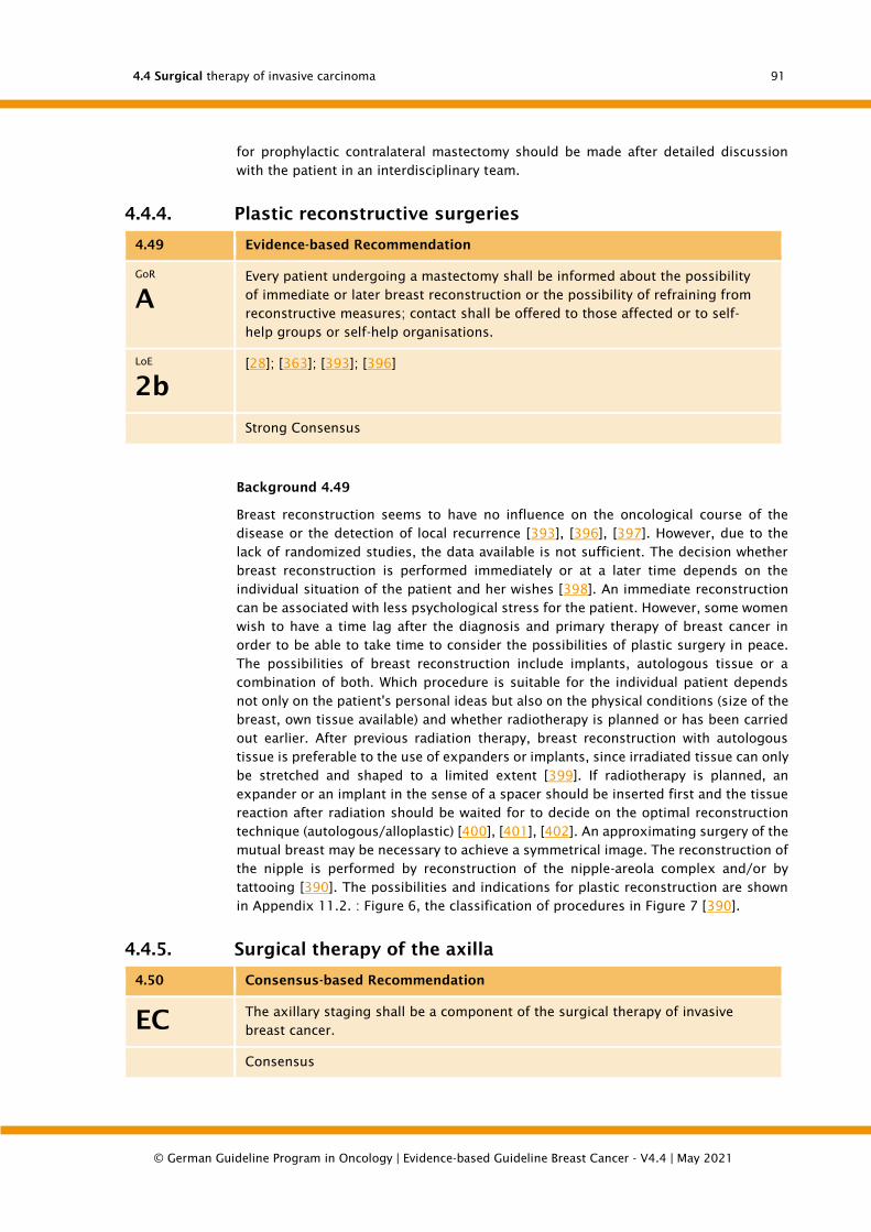
4.4 Surgical therapy of invasive carcinoma
© German Guideline Program in Oncology | Evidence-based Guideline Breast Cancer - V4.4 | May 2021
91
for prophylactic contralateral mastectomy should be made after detailed discussion
with the patient in an interdisciplinary team.
4.4.4. Plastic reconstructive surgeries
4.49 Evidence-based Recommendation
GoR
A
Every patient undergoing a mastectomy shall be informed about the possibility
of immediate or later breast reconstruction or the possibility of refraining from
reconstructive measures; contact shall be offered to those affected or to self-
help groups or self-help organisations.
LoE
2b
[28]; [363]; [393]; [396]
Strong Consensus
Background 4.49
Breast reconstruction seems to have no influence on the oncological course of the
disease or the detection of local recurrence [393], [396], [397]. However, due to the
lack of randomized studies, the data available is not sufficient. The decision whether
breast reconstruction is performed immediately or at a later time depends on the
individual situation of the patient and her wishes [398]. An immediate reconstruction
can be associated with less psychological stress for the patient. However, some women
wish to have a time lag after the diagnosis and primary therapy of breast cancer in
order to be able to take time to consider the possibilities of plastic surgery in peace.
The possibilities of breast reconstruction include implants, autologous tissue or a
combination of both. Which procedure is suitable for the individual patient depends
not only on the patient's personal ideas but also on the physical conditions (size of the
breast, own tissue available) and whether radiotherapy is planned or has been carried
out earlier. After previous radiation therapy, breast reconstruction with autologous
tissue is preferable to the use of expanders or implants, since irradiated tissue can only
be stretched and shaped to a limited extent [399]. If radiotherapy is planned, an
expander or an implant in the sense of a spacer should be inserted first and the tissue
reaction after radiation should be waited for to decide on the optimal reconstruction
technique (autologous/alloplastic) [400], [401], [402]. An approximating surgery of the
mutual breast may be necessary to achieve a symmetrical image. The reconstruction of
the nipple is performed by reconstruction of the nipple-areola complex and/or by
tattooing [390]. The possibilities and indications for plastic reconstruction are shown
in Appendix 11.2. : Figure 6, the classification of procedures in Figure 7 [390].
4.4.5. Surgical therapy of the axilla
4.50 Consensus-based Recommendation
EC The axillary staging shall be a component of the surgical therapy of invasive
breast cancer.
Consensus
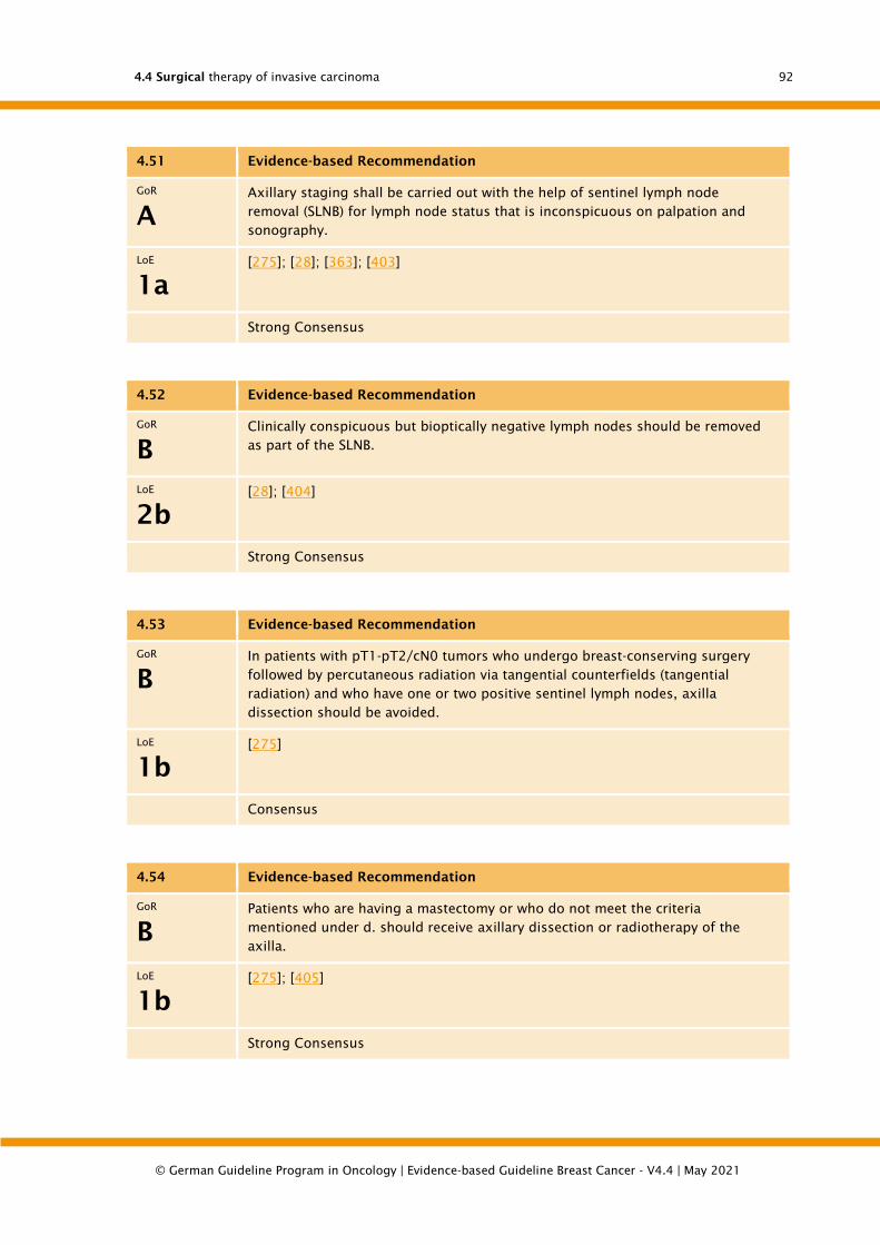
4.4 Surgical therapy of invasive carcinoma
© German Guideline Program in Oncology | Evidence-based Guideline Breast Cancer - V4.4 | May 2021
92
4.51 Evidence-based Recommendation
GoR
A
Axillary staging shall be carried out with the help of sentinel lymph node
removal (SLNB) for lymph node status that is inconspicuous on palpation and
sonography.
LoE
1a
[275]; [28]; [363]; [403]
Strong Consensus
4.52 Evidence-based Recommendation
GoR
B
Clinically conspicuous but bioptically negative lymph nodes should be removed
as part of the SLNB.
LoE
2b
[28]; [404]
Strong Consensus
4.53 Evidence-based Recommendation
GoR
B
In patients with pT1-pT2/cN0 tumors who undergo breast-conserving surgery
followed by percutaneous radiation via tangential counterfields (tangential
radiation) and who have one or two positive sentinel lymph nodes, axilla
dissection should be avoided.
LoE
1b
[275]
Consensus
4.54 Evidence-based Recommendation
GoR
B
Patients who are having a mastectomy or who do not meet the criteria
mentioned under d. should receive axillary dissection or radiotherapy of the
axilla.
LoE
1b
[275]; [405]
Strong Consensus

4.4 Surgical therapy of invasive carcinoma
© German Guideline Program in Oncology | Evidence-based Guideline Breast Cancer - V4.4 | May 2021
93
4.55 Evidence-based Recommendation
GoR
A
In the case of exclusive micrometastasis, a targeted therapy of the lymph
drainage areas (surgery, radiotherapy) shall be avoided.
LoE
1b
[406]; [407]
Strong Consensus
4.56 Evidence-based Recommendation
GoR
B
In patients receiving primary systemic therapy (PST) and who have a
pretherapeutically and sonographically negative lymph node status, the SLN
should be performed after PST.
LoE
2b
[363]; [408]; [409]
Consensus
4.57 Evidence-based Recommendation
GoR
B
In patients who receive primary systemic therapy (PST) and have a punch biopsy
positive (cN1) and a clinically negative nodal status after PST (ycN0), an axilla
dissection should be performed.
LoE
2b
[410]; [411]
Consensus
4.58 Consensus-based Recommendation
EC In patients who receive primary systemic therapy (PST) and have a positive nodal
status before and after PST, an axilla dissection shall be performed.
Strong Consensus
4.59 Consensus-based Recommendation
EC In the case of evidence of distant metastasis, axial staging shall be avoided.
Strong Consensus
4.4 Surgical therapy of invasive carcinoma
© German Guideline Program in Oncology | Evidence-based Guideline Breast Cancer - V4.4 | May 2021
94
Background 4.50 to 4.59
The sentinel node biopsy (SLNB) is a targeted surgical procedure for determining the
nodal status of breast cancer. The procedure is used to identify patients with increased
systemic and local risk and to plan adjuvant therapy.
Provided that the procedure is standardized and quality-assured, the SLNB has a high
staging accuracy and a significantly reduced shoulder-arm morbidity. The SLNB goes
with a safe local control (axillary recurrence
The SLNB is indicated in all patients with a clinically negative lymph node status and
for whom axillary staging is necessary [275].
The SLNB is not indicated in cases of clinical suspicion of advanced lymph node
involvement and tumour-transmitted lymph nodes [275]. To clarify preoperatively
whether lymph node metastasis is actually present in clinically and/or sonographically
suspicious lymph nodes, an ultrasound-guided FNA or a biopsy of the suspicious lymph
nodes may be helpful. Histological evidence of lymph node metastasis precludes the
use of sentinel node biopsy.
In patients with histologically proven tumor involvement of the axilla, surgical removal
of the axillary lymph nodes may be indicated. In patients with positive nodal status,
the number of affected lymph nodes or the ratio of affected to examined lymph nodes
may provide information for the selection of subsequent antineoplastic systemic
therapy and adjuvant radiotherapy.
A randomized study of the American College of Surgeons Oncology Group (ASOG
Z0011-study) examined the significance of axilla dissection in patients with T1 and T2
tumors and 1-2 positive SLN, in whom a breast-conserving therapy with subsequent
percutaneous radiotherapy of the entire affected breast via tangential radiation fields
(WBI) had been performed, with regard to the locoregional recurrence rate and overall
survival. No advantage for axilla dissection could be identified [406], [412]. Due to
methodological limitations of the study, an evidence search was performed in which
the probability of error of the study was classified as "unclear" [413].
In further randomized studies, the clinical value of a dissection of axillary or other
regional lymph nodes was examined in different patient groups with a low risk profile
[370], [414], [415], [416], [417]. In these studies, axillary dissection was not associated
with a survival benefit either.
Apart from the NSABP-B-04 study, the above mentioned studies generally included
tangential field radiation of the breast and thus partial radiation of the axilla (at least
in Level I). The NSABP-B-04 study is thus the only available study in which no further
therapy (i.e. no surgery, no systemic therapy, and no radiotherapy) was performed on
positive axillas. In this study 38% of the patients in the axillary operated collective had
histological lymph node metastases. In the non-treated group, 19% had follow-up
surgery for axillary recurrence. This allows the conclusion that with untreated axilla
tumour-affected lymph nodes become clinically apparent in only about 50% of cases. It
is therefore not clear what influence radiotherapy has on the risk of axillary recurrence
in affected sentinel lymph nodes and the absence of axillary dissection, and which
therapeutic procedure is most suitable for reducing the regional risk of recurrence with
the least side effects or late effects.
Based on the available data, there is a well-founded option to refrain from axilla
dissection in the above-mentioned group of patients in the Z0011 study with a positive
SLN. This is particularly relevant for patients in whom no further affected lymph nodes
are expected and/or in whom the identification of additional lymph node metastases
4.4 Surgical therapy of invasive carcinoma
© German Guideline Program in Oncology | Evidence-based Guideline Breast Cancer - V4.4 | May 2021
95
would not result in changes in adjuvant therapy. Patients with a T1 or T2 tumour and
1-2 positive SLN can thus be offered the option of foregoing axillary dissection with
breast-conserving therapy, provided that they are made aware of the current data
situation. A replacement of the axilla dissection by radiotherapy of the axilla can be
considered as an alternative.
Patients with micrometastases in the SLN should not be recommended for axillary
dissection because there is no evidence for an increased regional risk of local
recurrence [407].
Patients who undergo a mastectomy or who do not undergo postoperative radiotherapy
of the affected breast are usually not suitable patients for waiving axilla dissection.
In exceptional situations (e.g. old age, multimorbidity, etc.), a waiver of any axillary
intervention may be considered [370], [414], [415], [417]. In Germany, the Insema
Study (LKP: Prof. Dr. Th. Reimer, sponsored by the DKH) is currently being conducted
to prospectively identify patients who can do without axillary staging. Axillary staging
is not indicated in patients in the stage of distant metastasis (M1).
In patients treated with primary systemic therapy (PST), the optimal procedure in the
area of lymphatic outflow pathways has not been conclusively clarified. A distinction
must be made between women who primarily have clinically (palpatory and
sonographic) inconspicuous lymph nodes and those whose nodal status is primarily
classified as suspicious.
Women with a clinically unremarkable lymph node status benefit from axillary staging
after PST. They avoid surgery (before PST) and possible follow-up therapies for clinically
occult, positive lymph nodes that are successfully treated by systemic therapy [418]. In
addition, histopathological complete remission (response of systemic therapy to the
tumor in the breast and lymph nodes), an important endpoint in clinical trials, can only
be determined if the lymph node status is recorded after systemic therapy. The
feasibility (detection rate) as well as the accuracy (false negative rate) seem to be
comparable in this collective with primarily operated patients [408].
In patients without clinical and/or imaging suspicion of axillary metastasis and in whom
PST is planned, SLNB can therefore be performed either before or after PST. The
decision depends on the weighing of the clinical relevance and the consequences for
possible therapy decisions [418].
In patients with primarily suspect lymph node status, a minimally invasive clarification
of the suspicious lymph node should be performed by punch biopsy or fine needle
aspiration. In the case of a confirmed primary lymph node involvement, the success
rates of SLNB (detection rate, false negative rate) after PST are clearly limited [410],
[411]. Therefore, an axilla dissection after PST is recommended for these patients.
Newer procedures that could possibly lead to an improved detection and accuracy of
SLNB (in primarily nodal-positive patients) after PST, such as the clip or seed marking
[419], are currently still insufficiently evaluated and cannot be recommended for
clinical routine.
Sufficient data regarding the locoregional risk of recurrence of an SLNB after PST are
not yet available. In particular, it is not clear whether the unfavourable false negative
rate of SLNB after PST in women with primary positive nodal status translates into an
increased rate of locoregional recurrence or unfavourable overall survival.
In view of the insufficient information available, the conduct of clinical studies on this
issue and the encouragement of suitable patients to participate in these studies are
expressly supported.

4.5 Pathomorphological examination
© German Guideline Program in Oncology | Evidence-based Guideline Breast Cancer - V4.4 | May 2021
96
4.5. Pathomorphological examination
4.5.1. Preliminary remarks
This chapter deals with the pathomorphological examination of the tissue samples.
This is used to establish the diagnosis and to determine prognostic and predictive
factors that are helpful in assessing the course of the disease and the response to
therapy.
Recommendations from internationally recognized guidelines and published protocols
[420], [421], [422], [423], [424], [425], [426], [427], [428] were used as a basis.
Preceded by "General Principles", which give general information on the qualitatively
good performance of the pathomorphological examination and its prerequisites.
Special aspects of examinations of "Percutaneous biopsies in the context of
interventional diagnostics", "Excision biopsies", "Mastectomy preparations" and "Lymph
nodes" are presented separately and are divided into the following topics:
• Macroscopic processing ("cutting") with removal of tissue for histological
examination
• Microscopic processing (sectional planes, staining, special methods) and review
(including criteria of classification)
These instructions are supplemented in the appendix (see Chapter 12.3) by the
recommended classifications and grading systems as well as proposed forms for the
"Begleitschein zur Einsendung" and the "Dokumentation der Gutachterlichen
diagnostischen Beurteilung".
4.5.2. General principles
4.5.2.1. General patient data, preliminary findings, anamnestic information
The patient data, preliminary findings and further information to the pathologist are
best communicated using a form (see Chapter 12.3: Figure 9), which records the
following information:
• Patient data (name, date of birth, gender, identification number, if available)
• Physician in charge
• day of collection
• Clinical diagnosis or indication for tissue sampling
• further clinical information:
- location of tissue sample collection (e.g. right breast, upper outer
quadrant)
- Type of collection (e.g. high-speed punch biopsy, modified radical
mastectomy)
- Clinical findings and imaging (e.g. findings palpable/non-palpable;
microcalcification present/not present; if necessary with transmission of
the preparation radiography)
- Possibly previous neoadjuvant therapy
- Preliminary findings and essential details of the medical history
4.5 Pathomorphological examination
© German Guideline Program in Oncology | Evidence-based Guideline Breast Cancer - V4.4 | May 2021
97
General principles for surgical material
4.60 Consensus-based Recommendation
EC The surgical material shall be marked clearly topographically with details of the
problem and the clinical-radiological findings and sent in full to the pathologist.
Strong Consensus
Background 4.60
A well-organized cooperation between the disciplines involved is a prerequisite for
pathomorphological diagnostics.
For the most accurate pathomorphological diagnosis possible, the biopsy or surgical
material must meet certain conditions, which are listed below:
• All biopsy and surgical material should be sent to the pathologist.
• The excisate/mastectomy preparations must be clearly topographically marked by
the surgeon (e.g. with different coloured sutures); the location of the markings
must be noted on the clinical accompanying form (see Chapter 12.3: : : Figure 9).
• If material is to be taken from the tumour (or other tissue) (e.g. for scientific
investigations, tumour bank), this must be done under the control of the
pathologist. For this purpose, the surgical specimens are to be sent to the
pathologist immediately after removal, unfixed.
• In such a material removal it must be taken into account that necessary
classifications of a tumor (especially R-classification, pTNM-classification, tumor
heterogeneity) must not be affected.
• Tissue fixation is carried out in 10 % neutral buffered formalin in sufficient
quantity. A fixation period between 6 h and 72 h [426] is recommended.
4.5.2.2. Documentation of the macroscopic processing
For documentation of the macroscopic processing, see the sections on the respective
type of tissue sample.
4.5.2.3. Documentation of the microscopic processing and assessment
The following information is documented, possibly using a form (see Figure 10 and
Figure 11):
• Type of tissue sample
• Page reference
• Major pathological changes (e.g. invasive carcinoma, intraductal carcinoma,
atypical ductal hyperplasia, fibrocystic mastopathy, mastitis)
• Carcinoma:
- Histological type
- Grading (for invasive carcinomas and DCIS)
- Presence of an associated intraductal carcinoma/DCIS (for invasive
carcinomas)
- tumour size*\footnote {not collected in percutaneous biopsies} (DCIS and
invasive carcinomas; for invasive carcinomas with extensive intraductal
component [Def. see extent of intra-ductal tumour component]: indication
of the size of the invasive component and additionally indication of the
size of the associated DCIS)
4.5 Pathomorphological examination
© German Guideline Program in Oncology | Evidence-based Guideline Breast Cancer - V4.4 | May 2021
98
- If necessary, indication of additional, additional tumour foci, if present
(multifocality/multicentricity)*
- resection margin*\footnote {not collected in percutaneous biopsies} (for
invasive carcinomas and DCIS)
▪ Tumour directly at the edge of the resection ("border forming")
▪ tumour not directly at the edge of the resection; then minimum
safety distance of the tumour from the edge of the resection in
mm with indication of localisation (if necessary, separate for
intraductal components)
- Peritumoral vascular invasion (if present under light microscopy)
- pTNM Classification*\footnote {not collected in percutaneous biopsies}
[429] (if necessary including further tissue samples)
- Additional immunohistological tests:
▪ Estrogen receptor (ER) or progesterone receptor (PgR) status (for
invasive carcinomas; for DCIS if therapeutically relevant)
▪ Human Epidermal Growth Factor Receptor (HER)2 and Ki-67 status
(for invasive carcinomas)
• Microcalcifications, if available: Location or indication of association with benign
or malignant lesion
• Comments:
- Reference to the intraoperatively communicated frozen section findings
- Reference to clinical/radiological findings (especially microcalcifications:
e.g. "finding is compatible with" or "correlation not certain")
• Reference to findings on other tissue samples/preliminary examinations (in the
case of findings on surgical specimens after percutaneous mammary biopsy:
statement required whether or not biopsy cavity is included in the surgical
specimen).
4.5.2.4. Clarification of mammographically detected microcalcification
In the clarification of mammographically suspect microcalcifications, the correlation of
the histopathological findings with the findings of the imaging procedures is necessary
(preparation radiography or disc radiography required).
It should be noted that mammography not only detects calcium phosphate (type II
microcalcifications), which can be stained with hematoxylin-eosin, but also calcium
oxalate (type I microcalcifications) in rare cases. The latter can be detected in polarized
light or dark field.
If there is no evidence of radiologically relevant microcalcification (> 100 µm) in the
initial sections, further sections should be made, possibly supplemented by special
staining (Kossa), and any remaining material should be embedded.
Occasionally, radiography of the paraffin blocks or of the remaining tissue not yet
embedded is also helpful for finding the radiologically relevant microcalcification in
larger tissue samples.
When detecting radiologically relevant microcalcifications, their localization in relation
to the histopathological change should be indicated.

4.5 Pathomorphological examination
© German Guideline Program in Oncology | Evidence-based Guideline Breast Cancer - V4.4 | May 2021
99
4.5.2.5. frozen section examination
4.61 Consensus-based Recommendation
EC The intraoperative frozen section examination shall only be performed if there is
an immediate surgical consequence.
Fields of application are:
• Dignity assessment of palpable focal findings >10 mm, if a
preoperative diagnosis by means of minimally invasive biopsy was
not possible
• Determination of the resection margin status
• Assessment of the sentinel lymph nodes
• A frozen section examination shall not be performed for non-
palpable lesions and minimally invasive biopsies (punch biopsies,
vacuum-assisted biopsies).
Strong Consensus
Background 4.61
The aim of the intraoperative frozen section examination on surgical preparations of
the breast is to evaluate those criteria which directly influence the further surgical
procedure:
• Dignity of the lesion: benign or malignant (DCIS or invasive carcinomas), if a
preoperative diagnosis by means of minimally invasive biopsy was not possible,
• Size and extent of a tumour (if necessary, detection of multiple tumour foci),
• Tumor detection at the edge of the incision [430], [431].
The intraoperative examination of the sentinel lymph nodes (SLN) allows in positive
cases a one-stage surgery of the axilla. However, if the result is negative, it must be
taken into account that subsequent processing of the formalin-fixed and paraffin-
embedded residual material will still find metastases in the sentinel lymph nodes in up
to 21% of cases [432], [433]. An intraoperative work-up of the lymph nodes in sections
is not justified in view of the limited assessability of frozen sections and the effort
involved.
As an alternative to frozen section examination, a preoperative punch biopsy or fine
needle aspiration can be performed to examine the lymph node status under certain
conditions. With appropriate expertise, aspiration cytology can also be performed.
The material examined in the frozen section is to be processed using the paraffin
technique.
4.5.2.6. Histological classification and grading
4.62 Consensus-based Recommendation
EC All invasive carcinomas shall be histologically classified (according to the current
WHO classification [118] ).
Strong Consensus
4.5 Pathomorphological examination
© German Guideline Program in Oncology | Evidence-based Guideline Breast Cancer - V4.4 | May 2021
100
4.5.2.6.1. Histological classification
The WHO classification of breast tumours is the international basis for the classification
and nomenclature of breast carcinomas [118]; see Chapter 12.3: Table 17.
The histological classification is carried out both on the punch and vacuum biopsies
and on the surgical specimens. In particular with the special types of breast carcinoma
(e.g. tubular, mucinous) a final classification is only possible on the surgical specimen.
Due to intratumoral heterogeneity, there may be differences between the
punch/vacuum biopsy and the surgical specimen. The published agreement rates are
between 65% and 100% [434].
4.5.2.6.2. Expansion of intraductal tumor component
If the invasive carcinoma is accompanied by an intraductal component that extends
beyond the border of the invasive carcinoma, not only the distance of the invasive
carcinoma but also the distance of the intraductal component to the nearest resection
edges should be given in mm.
The evidence for the fact that with a negative resection margin ("no ink on tumor"/no
color on the tumor), an extensive intraductal component (EIC) is associated with an
increased risk of local recurrence is contradictory and depends on the definition of the
EIC [435], [436], [437]. The presence of an EIC is associated with a significantly
increased risk of local recurrence if the size of the intraductal tumor component in one
dimension is at least twice the size of the invasive carcinoma [437]. It is therefore
appropriate to apply this definition and to indicate the presence of such an EIC.
In addition, significant DCIS residuals are found more frequently in patients with EIC
with positive or dense resection margins in post-resectates than in patients without EIC
[438]. Therefore, the guidelines of the Society of Surgical Oncology (USA) and the
American Society for Radiation Oncology also recommend that postoperative
mammography should be considered in micro calcified EIC-positive carcinomas, as
residual calcifications would justify a post-excision [435]. It is also pointed out that the
characteristics of young age and several narrow resection margins in patients with EIC-
positive carcinoma are associated with an increased local recurrence risk. Patients with
these characteristics could benefit from a post-excision [365], [436].
Grading of invasive carcinomas
4.63 Consensus-based Recommendation
EC For all invasive breast carcinomas a grading according to Elston and Ellis [439]
(modification of Bloom and Richardson grading) shall be performed.
Strong Consensus
4.5.2.6.3. Histological grading
Histological grading is also carried out on the punch and vacuum biopsies as well as
on the surgical specimens of the breast (see Chapter 12.3: Table 18). It should generally
also be indicated numerically (G1, G2 or G3).
The agreement between the histological grading of punch biopsies and surgical
specimens is moderate (kappa value: 0.35-0.65) [440], [441], [442], [443], [444], [445],
[446], [447], [448], [449], [450]. The punch biopsies tend to show a lower grading,
4.5 Pathomorphological examination
© German Guideline Program in Oncology | Evidence-based Guideline Breast Cancer - V4.4 | May 2021
101
especially due to an underestimated mitosis number [440], [441], [442], [444], [445],
[447], [448], [451]. Possibly the Ki-67 (Mib-1) proliferation index in the punch biopsies
correlates better than the mitosis number with the low and high mitosis rates in the
excidates [451].
For the decision for or against neoadjuvant therapy, the differentiation between grade
2 and grade 3 is of particular relevance preoperatively. In particular for carcinomas with
minimal tubular differentiation (3 score points) and high nuclear pleomorphism (3
score points), there are various suggestions for modified mitosis scoring on punch
biopsies for better assignment of the grade on the non-invasive biopsies. On the one
hand, the halving of the limit values for the mitosis scores is recommended [452],
[453], on the other hand, the use of the Ki-67 index instead of the mitosis score is
advocated [454], so that breast carcinomas with a Ki-67 index > 25% would receive a
score of 3 for proliferative activity.
Neither of these two proposals has yet been sufficiently validated and generally
accepted internationally. However, they can be helpful in borderline cases to achieve a
better match of the grading of the punch biopsy and the surgical specimen.
4.5.2.6.4. DCIS-Grading
The grading is based on the grading scheme according to WH0 [118] (see Chapter 12.3,
Table 16).
For all DCIS, the following parameters should be listed in addition to the grading:
• Core degree according to "Consensus Conference on the Classification of DCIS
in Philadelphia, 1997" [455] (see Chapter 12.3 Table 15)
• Comedo-like necroses existing/not present
4.5.2.7. Multifocality/multicentricity
Currently there is no internationally uniform definition of the terms "multifocality" and
"multicentricity" before [456], [457]. The following classification is recommended:
• Multifocality: Occurrence of macroscopically recognizable, separate carcinoma
foci in one quadrant or according to Faverly [458] with a distance between the
foci of less than 4 cm.
• Multicentricity: Occurrence of separate carcinoma foci in more than one
quadrant or according to Faverly [458] at a distance of at least 4 cm between
foci.
4.5.2.8. Peritumoral lymph vessel invasion
The presence of peritumoral (lymphatic) vascular invasion (LVI) should be reported, as
LVI is an important independent prognostic factor [459], [460]. This is especially true
for nodal-negative T1 tumors, where the detection of LVI means that the risk of
recurrence and the development of distant metastases is significantly increased [461],
[462].
In the diagnosis of peritumoral LVI, strict criteria must be applied to delimit tumor cell
complexes located in artificially created tissue gaps (e.g. as a result of shrinkage
artifacts) [118], [463], [464]:
• Detection in peritumoral tissue.

4.5 Pathomorphological examination
© German Guideline Program in Oncology | Evidence-based Guideline Breast Cancer - V4.4 | May 2021
102
• The tumor cells are located in capillary vascular spaces that are lined by an
endothelial border.
• The arrangement of the tumor cells often doesn't match the shape of the vascular
space.
• Occurs at the site of normal lymphatic vessels:
- Associated with other vascular structures
- Periductal
- In the interlobular stroma
Occasionally, immunohistochemistry (e.g. D2-40 detection) can be helpful.
4.5.3. Determination of hormone receptor and HER2 status and the
Ki-67 proliferation index of invasive carcinomas
4.64 Evidence-based Recommendation
GoR
A
In invasive breast carcinoma, the primary diagnosis of invasive breast cancer
shall determine the estrogen and progesterone receptor status as well as the
HER2 status, preferably already at the punch biopsy.
LoE
2a
[421]; [426]; [465]; [466]
Strong Consensus
4.65 Consensus-based Recommendation
EC Additionally, the proliferation rate can be determined by immunohistochemical
detection of Ki-67.
Strong Consensus
4.66 Consensus-based Recommendation
EC The determination of estrogen and progesterone receptor status shall be
performed immunohistochemically. The percentage of positive tumour cell
nuclei and the average intensity of staining shall be reported. In addition, scores
can be obtained by stating the method (Allred (Quick) Score, Immunore
according to Remmele and Stegner). The evaluation as ER- or PgR-positive
requires at least 1 % positive tumour cell nuclei.
Strong Consensus
4.5 Pathomorphological examination
© German Guideline Program in Oncology | Evidence-based Guideline Breast Cancer - V4.4 | May 2021
103
4.67 Evidence-based Recommendation
GoR
A
As a prerequisite for trastuzumab therapy, HER2 positivity is defined as
immunohistochemically proven protein overexpression with a score of 3+ or
gene amplification, preferably detected by in situ hybridization (ISH).
LoE
1b
[426]; [467]; [468]
Strong Consensus
4.68 Consensus-based Recommendation
EC When determining the hormone receptor and HER2 status and the Ki-67
proliferation index, the reliability of the detection methods used shall be
ensured. This includes internal test validation, the use of standardized
protocols, on slide and internal controls as well as regular successful
participation in external quality assurance measures.
Strong Consensus
Background 4.64 to 4.68
The quality of the detection methods used, including pre-analytics (including fixation)
and evaluation, should be ensured by the use of SOPs and regular internal and external
quality controls. For external quality control, the regular, annual successful
participation in interlaboratory comparisons is recommended, which is offered, for
example, by the "Pathology Quality Assurance Initiative" (QuIP), a joint venture of the
German Society of Pathology (DGP) and the Federal Association of German Pathologists
(BDP).
In the guidelines of the American Society of Clinical Oncology (ASCO) and the College
of American Pathologists (CAP) it is recommended that the ER/PgR determination is
preferably performed on punch biopsies [421] because of the faster fixation of the
tissue. The HER2 determination can be carried out primarily on punch biopsies as well
as on surgical specimens [426]. The decisive factor is the time until fixation (cold
ischemia time, /= 0.81 or concordance >/= 95%). It must also be ensured that artificially
altered tissue (edge, retraction or squeezing artifacts) is excluded from the assessment.
The validity and reproducibility of the HER2 determination can be more easily ensured
with standardized test kits, which is why the use of such test kits is recommended. For
the detection of HER2 gene amplification, the various methods of in situ hybridization
(fluorescence, chromogenic, silver-amplified) can be used, taking into account quality
requirements. If test kits are used, the application follows exactly the manufacturer's
instructions.
The expected HER2-positivity rate for invasive breast cancer is currently averaging 15%.
Deviations from this average value can result from the composition of the examination
collective. Potential influencing variables are the histological degree of differentiation,
the hormone receptor status, the histological tumor type as well as the nodal status of
the examined carcinomas and the age of the patients (all P<0.0001) [469]. Es wird

4.5 Pathomorphological examination
© German Guideline Program in Oncology | Evidence-based Guideline Breast Cancer - V4.4 | May 2021
104
empfohlen, die HER2-Positivitätsrate zur Qualitätssicherung kontinuierlich zu
überwachen.
4.5.3.1. Interpretation Hormonrezeptorstatus
Interpretation of immunohistochemical reaction results should follow the
recommendations of the ASCO/CAP guidelines [421]:
Evaluation as ER- or PgR-positive requires at least 1% positive tumor cell nuclei [421],
[470], [471]. Tumors are evaluated as ER- or PgR-negative when less than 1% of tumor
cell nuclei are immunoreactive with positive internal control. The recommendation of
the 1% threshold is based on the results of a systematic review [472]. However, this
review is based exclusively on retrospective studies in which a cutoff value for
endocrine therapy was established. Different threshold levels were not tested against
each other in the studies.
There is now evidence that tumors with low ER positivity (1-9% positive cells) should be
considered separately [470]. Several studies have shown that they are tumor
biologically and prognostically closer to ER-/PgR-negative or triple-negative breast
carcinomas (ER-, PgR-, and HER2-negative) than ER-positive (>10% positive tumor cells)
[473], [474], [475], [476], [477].
Therefore, the following subdivision is recommended [472], [478]:
• ER-/PgR-positive: >10% positive tumor cells.
• ER-/PgR-gering positive: 1%-9% positive tumor cells
• ER-/PgR-negative: <1% positive tumor cells
In addition to the percentage of positive tumor cell nuclei, the average staining intensity
must also be reported according to the ASCO/CAP guidelines [421]. As a supplement,
the internationally accepted Allred score [479 or the immunoreactive score (IRS)
according to Remmele and Stegner [480] can be reported (see Chapter 12.3 Table 21).
Immunohistochemistry is not usable and should possibly be repeated on another
specimen if
• external or on-slide controls do not give the expected result,
• artifacts occupy the majority of the material,
• normal epithelial cells within the specimen do not show nuclear staining,
• the tissue has been decalcified in strong acids,
• an ER-negative/PgR-positive phenotype is present (to exclude false negative ER
or false positive PgR testing),
fixation of the tissue was not optimal (cold ischemia time > 1h, fixati-on < 6h or > 72h)
and the test result is negative in the absence of internal control.
Even in the presence of a histological type that is usually ER-/PgR-positive (tubular,
mucinous), testing should be repeated as a precaution (possibly also on another tissue
sample) if the result is ER-/PgR-negative.
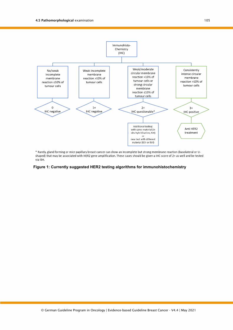
4.5 Pathomorphological examination
© German Guideline Program in Oncology | Evidence-based Guideline Breast Cancer - V4.4 | May 2021
105
Figure 1: Currently suggested HER2 testing algorithms for immunohistochemistry

4.5 Pathomorphological examination
© German Guideline Program in Oncology | Evidence-based Guideline Breast Cancer - V4.4 | May 2021
106
Figure 2: Currently suggested HER2 testing algorithms for in-situ hybridization, adapted from [426][468]
The increase of the immunohistochemical cut-off for the score 3+ from 10% to 30%
[481] recommended in the 2007 ASCO/CAP guidelines has been withdrawn. Thus, the
prerequisites for an immunohistochemically HER2-positive test result again meet the
criteria applied in the pivotal studies for anti-HER2 therapy (Figure 1). The update
committee also cited as reasons for the reduction of the cut-off to the original level the
fact that the increase affected only a few patients [0.15%; [482]] and that the analytical
quality of HER2 testing had generally improved since 2007, [426].
A weak point of the ASCO/CAP guideline published in 2013 is the contradictory
definition of the 2+score with unclear differentiation from the 1+-score [426]. This
ambiguity was corrected in 2015 by revising the definition to reapply the original
criteria from 2007 [468].
The threshold value for an ISH-positive test result was also readjusted to the criteria
used in the approval studies for anti-HER2 therapy. Accordingly, a HER2/CEN17 ratio >
2.0 is considered HER2-positiveFigure 2).
However, the addition of the pericentromere region of chromosome 17 can lead to false
negative results if only the ratio is taken into account as a criterion for distinguishing
HER2-positive and negative cases. Therefore, according to the current update, a ratio
of 4.0 (Figure 2). A test result in the borderline category should trigger re-testing as
before (other validated method on the same material or re-testing on other material,
for example on excidate if borderline result on needle biopsy). The ultimate goal is to
4.5 Pathomorphological examination
© German Guideline Program in Oncology | Evidence-based Guideline Breast Cancer - V4.4 | May 2021
107
achieve a clear HER2 test result (negative or positive) as a basis for clinical decision
making. The "borderline" category is a subgroup that has not yet been adequately
investigated and where it is uncertain how many of the affected patients will benefit
from anti-HER2 therapy.
In the 2013 update, criteria for the evaluation of HER2-heterogeneous tumours were
defined for the first time, i.e. for tumours containing cell populations with and without
HER2 gene amplification. The experts assume that a HER2-amplified cell population is
only clinically relevant if the HER2-amplified cells can be separated as aggregated
complexes from HER2-negative cell assemblies. To identify areas with HER2-amplified
cells, the preparation should be completely screened before at least 20 cell nuclei are
evaluated. Alternatively, areas with potential HER2 amplification can be narrowed down
using IHC. If a second contiguous cell population with elevated HER2 signaling levels
exists, representing more than 10% of the cells on the section, it will be evaluated
separately. In both populations the signals should be counted in at least 20 adjacent
cells in at least 2 areas each and evaluated separately. Tumours that contain amplified
and non-amplified areas under these conditions are considered HER2-positive. In the
report of findings, the percentage of the tumour with HER2 gene amplification should
be stated.
In summary, the 2013 update recommends that primary testing be performed on the
punch biopsy. If the test result is clearly negative or positive according to the criteria
in Fig. 1, no re-testing is usually required.
Exceptions are, among other things, test results that are discrepant to the
histopathological findings or if the tumor in the excidate shows a different tumor type
or histological grading than in the punch biopsy.
A new HER2 test on the excidate of the tumor should be requested if
• HER2 status at punch biopsy unclear (IHC and ISH in borderline area)
• heterogeneous HER2 status at the punch biopsy
• HER2 test positive for invasive carcinomas, G1, of the following histological
types:
o ductal or lobular, ER- and PgR-positive
o tubular, mucinous, cribriform
o adenoid-cystic carcinoma (usually triple-negative)
• HER2 test negative and
o less invasive tumor contained in punch biopsy
o resectate G3 carcinoma, which is morphologically different from the
carcinoma in the punch biopsy
o there are doubts about the sample handling of the punch biopsy (too
long ischemia time, too short fixation, etc.)
A general retesting of G3 carcinomas on the resected tissue, whose testing on the
needle biopsy showed a HER2 negative result, is not necessary [468], [483]
HER immunohistochemistry is not usable and should be repeated or replaced by ISH if
• controls do not deliver the expected result,
• artifacts take up most of the material,
• normal gangetic epithelia show a strong membrane coloration (internal
control).
The HER-ISH is not usable and should be repeated if
• controls do not deliver the expected result,
• at least two tumour areas cannot be evaluated,
4.5 Pathomorphological examination
© German Guideline Program in Oncology | Evidence-based Guideline Breast Cancer - V4.4 | May 2021
108
• > 25 % of the signals are too weak to be evaluated,
• > 10% of the signals appear in the cytoplasm,
• Core resolution is bad,
• autofluorescence is strong (FISH).
4.5.3.2. Evaluation Ki-67 proliferation index
For the determination of the Ki-67 proliferation index (PI), no generally accepted,
comprehensive recommendation for standardisation is available to date. The
background to this is that the methodological approach in the studies that have
demonstrated a prognostic or predictive relevance of Ki-67 is very heterogeneous
[484]. As a consequence of the lack of standardization, increased interobserver
variability in the determination of Ki-67-PI was repeatedly observed in reproducibility
studies, especially in the middle range or in G2 mammary carcinomas [484], [485],
[486], [487]. Due to the heterogeneous approach in the studies, the consensus
recommendation of an international working group on Ki-67 in breast cancer also
focuses only on certain key points regarding the methodological procedure including
evaluation and interpretation of the results [488]. The experts emphasized that it is not
possible to give generally valid Ki-67 limits for prognosis, prediction and monitoring.
Limit values defined in studies could only be applied locally if the local results were
validated against the study results.
In the meantime, however, the above mentioned international working group and other
study groups have shown that the reproducibility of the Ki-67 determination can be
significantly improved by systematic training and application of uniform criteria [489],
[490], [491]. However, an acceptable reliability of the determination can be achieved
with different procedures (e.g. counting and semi-quantitative estimation). This means
that it is currently not possible to favor a particular procedure, especially since different
evaluation strategies were used in the recently published studies that prove the
prognostic or predictive relevance of Ki-67-PI [492], [493], [494], [495], [496].
Nevertheless, it is possible to define certain framework conditions on which there is
general consensus in order to improve reproducibility:
Application of a standardized staining protocol with internal and external quality
control
• Evaluation preferably at the invasion front of at least 3 visual fields at high
magnification, 400x (exception: needle biopsy, if the invasion front cannot be
delimited or the extent of the infiltration
• Exclusive evaluation of the nuclear staining, which stands out from the nuclear
counterstain (nucleoli alone are not counted, staining intensity is irrelevant)
• Indication of the percentage of Ki-67-positive tumour cells in relation to the
total number of tumour cells
• Determination of percentage by single cell counting, semi-quantitative
estimation in 5% steps or by image analysis possible [489], [492], [497], [498],
[499]
The use of image analysis procedures requires that the result of each individual
measurement is critically reviewed by the pathologist and the size of the measurement
field is adjusted if necessary [500].
There is no uniform view on the question of which cell number should be analysed. The
recommendations range from 100 to 2000 cells [488], [489], [497]. In the studies on
the reproducibility of the Ki-67 determination, it was unanimously shown that -
regardless of the number of cells analysed - the agreement<10%) und hoch
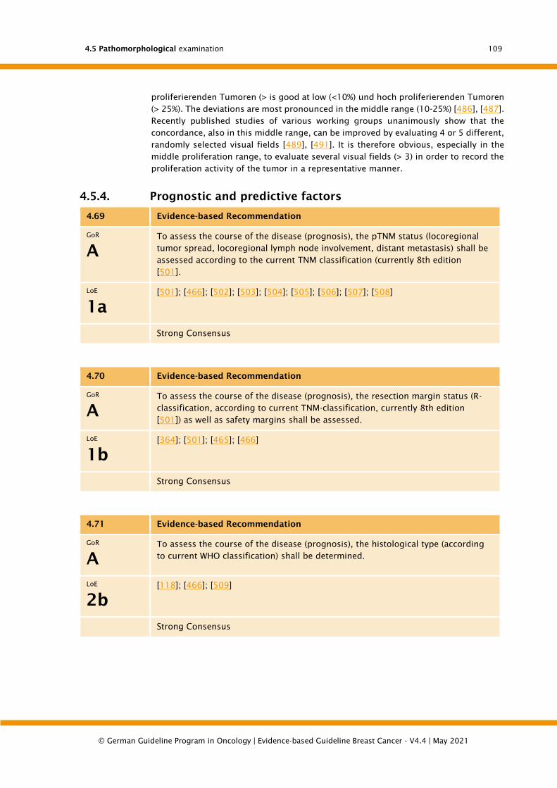
4.5 Pathomorphological examination
© German Guideline Program in Oncology | Evidence-based Guideline Breast Cancer - V4.4 | May 2021
109
proliferierenden Tumoren (> is good at low (<10%) und hoch proliferierenden Tumoren
(> 25%). The deviations are most pronounced in the middle range (10-25%) [486], [487].
Recently published studies of various working groups unanimously show that the
concordance, also in this middle range, can be improved by evaluating 4 or 5 different,
randomly selected visual fields [489], [491]. It is therefore obvious, especially in the
middle proliferation range, to evaluate several visual fields (> 3) in order to record the
proliferation activity of the tumor in a representative manner.
4.5.4. Prognostic and predictive factors
4.69 Evidence-based Recommendation
GoR
A
To assess the course of the disease (prognosis), the pTNM status (locoregional
tumor spread, locoregional lymph node involvement, distant metastasis) shall be
assessed according to the current TNM classification (currently 8th edition
[501].
LoE
1a
[501]; [466]; [502]; [503]; [504]; [505]; [506]; [507]; [508]
Strong Consensus
4.70 Evidence-based Recommendation
GoR
A
To assess the course of the disease (prognosis), the resection margin status (R-
classification, according to current TNM-classification, currently 8th edition
[501]) as well as safety margins shall be assessed.
LoE
1b
[364]; [501]; [465]; [466]
Strong Consensus
4.71 Evidence-based Recommendation
GoR
A
To assess the course of the disease (prognosis), the histological type (according
to current WHO classification) shall be determined.
LoE
2b
[118]; [466]; [509]
Strong Consensus

4.5 Pathomorphological examination
© German Guideline Program in Oncology | Evidence-based Guideline Breast Cancer - V4.4 | May 2021
110
4.72 Evidence-based Recommendation
GoR
A
To assess the course of the disease (prognosis) the histological grading
according to Elston and Ellis [510] shall be determined.
LoE
2a
[510]; [466]; [492]; [506]; [511]; [512]
Strong Consensus
4.73 Evidence-based Recommendation
GoR
A
To assess the course of the disease (prognosis) the peritumoral lymph vessel
invasion (according to the current TNM classification, currently 8th edition
[501]) shall be assessed.
LoE
2b
[459]; [460]; [466]; [506]; [513]
Strong Consensus
4.74 Consensus-based Recommendation
EC In order to assess the course of the disease (prognosis), age shall be
determined.
Strong Consensus
Ki-67
4.75 Evidence-based Statement
ST The addition of Ki-67 to the conventional prognostic factors (age, pT, pN, grade,
ER, PR, HER2) improves the prognostic assessment in women with histologically
confirmed ER/PR-positive and HER2-negative invasive breast cancer for the
decision whether adjuvant chemotherapy should be given.
LoE
1b
[484]; [492]; [493]; [496]
Consensus
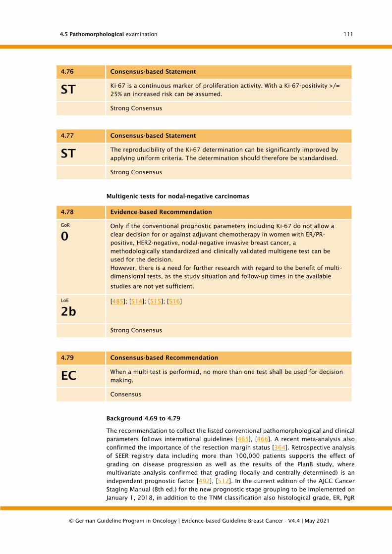
4.5 Pathomorphological examination
© German Guideline Program in Oncology | Evidence-based Guideline Breast Cancer - V4.4 | May 2021
111
4.76 Consensus-based Statement
ST Ki-67 is a continuous marker of proliferation activity. With a Ki-67-positivity >/=
25% an increased risk can be assumed.
Strong Consensus
4.77 Consensus-based Statement
ST The reproducibility of the Ki-67 determination can be significantly improved by
applying uniform criteria. The determination should therefore be standardised.
Strong Consensus
Multigenic tests for nodal-negative carcinomas
4.78 Evidence-based Recommendation
GoR
0
Only if the conventional prognostic parameters including Ki-67 do not allow a
clear decision for or against adjuvant chemotherapy in women with ER/PR-
positive, HER2-negative, nodal-negative invasive breast cancer, a
methodologically standardized and clinically validated multigene test can be
used for the decision.
However, there is a need for further research with regard to the benefit of multi-
dimensional tests, as the study situation and follow-up times in the available
studies are not yet sufficient.
LoE
2b
[485]; [514]; [515]; [516]
Strong Consensus
4.79 Consensus-based Recommendation
EC When a multi-test is performed, no more than one test shall be used for decision
making.
Consensus
Background 4.69 to 4.79
The recommendation to collect the listed conventional pathomorphological and clinical
parameters follows international guidelines [465], [466]. A recent meta-analysis also
confirmed the importance of the resection margin status [364]. Retrospective analysis
of SEER registry data including more than 100,000 patients supports the effect of
grading on disease progression as well as the results of the PlanB study, where
multivariate analysis confirmed that grading (locally and centrally determined) is an
independent prognostic factor [492], [512]. In the current edition of the AJCC Cancer
Staging Manual (8th ed.) for the new prognostic stage grouping to be implemented on
January 1, 2018, in addition to the TNM classification also histological grade, ER, PgR

4.5 Pathomorphological examination
© German Guideline Program in Oncology | Evidence-based Guideline Breast Cancer - V4.4 | May 2021
112
and HER2 status and the Oncotype DX® Recurrence Score are considered [517]. The
anatomical stage grouping remains unaffected.
4.5.4.1. uPA/PAI-1
Although the invasion factors uPA/PAI-1 according to the current ASCO guidelines can
be used to decide for or against adjuvant systemic therapy in ER/PgR-positive and
HER2-negative nodal-negative breast cancer [485], the majority of the guideline group
has spoken out against a renewed recommendation in the current revision of the S3
guideline. The main reason for this is that the results of the prospective randomized
chemo-N0 study in nodal-negative breast cancer are not transferable to current
treatment standards. This is also the conclusion reached by the experts of the ASCO
guideline in the explanatory text to the statement [485] - despite their open
recommendation. The patients in the group with low uPA/PAI-1 did not receive any
systemic treatment in the chemo-N0 study, i.e. also no endocrine therapy as is standard
today for ER/PgR-positive tumors [518]. It is therefore not possible to say what the
additional benefit of chemotherapy for high uPA/PAI-1 in ER/PgR-positive tumours
would be under current conditions if the patients in the comparative arm received
endocrine therapy. In addition, the HER2 status of the tumours analysed in the chemo-
N0 trial is unknown. There are indications that there is a correlation between uPA/PAI-
1 and intrinsic subtypes. HER2-positive or triple-negative carcinomas are significantly
less likely to be uPA/PAI-1-negative than luminal A-type carcinomas [519]. The question
therefore arises whether uPA/PAI-1 is actually an independent prognostic parameter.
Furthermore, the prognostic value seems to be different for the individual subtypes
and is not detectable in hormone receptor-positive breast carcinomas if the HER2-
positive tumors are excluded [520]. The assessment of the S3 guideline group is
therefore in line with that of the Institute for Quality and Efficiency in Health Care
(IQWiG) in its final report on the evaluation of uPA/PAI-1 in primary breast cancer with
intermediate risk of recurrence after R0 resection. It concludes that the patient-relevant
benefit or harm of an uPA/PAI-1-supported therapy decision is unclear due to a lack of
suitable studies [521].
4.5.4.2. Ki-67
The correlation between the immunohistochemically determined Ki-67 proliferation
index and the prognosis of breast cancer has been shown in numerous clinical studies.
Nevertheless, Ki-67 is not recommended in the current ASCO biomarker guidelines as
a decision aid for or against adjuvant chemotherapy [485]. In the S3 guideline update,
the question was raised whether more recent evidence is available to show that the
addition of Ki-67 to conventional factors improves the prognosis estimate for invasive
breast cancer. For this purpose, a systematic literature review was conducted for the
period 2015-10/2016. A meta-analysis was identified that included more than 64,196
patients from 41 studies. The meta-analysis concludes that Ki-67 is an independent
prognostic parameter for the overall survival of patients with breast cancer. The
prognosis of tumours with high Ki-67-positivity is significantly worse than that of Ki-
67-low-expressing tumours (hazard ratio, HR=1.57; 95% CI 1.33-1.87; p<0,00001).
Dies gilt ebenso für die Subgruppe der ER-positiven Tumoren (HR=1,51; 95% CI 1,25-
1,81; p<0,0001) [484]. In kürzlich publizierten prospektiven Studien wurde außerdem
der stärkere Nutzen einer Chemotherapie bei Frauen mit hoch proliferierenden ER-
/PgR-positiven, HER2-negativen invasiven Mammakarzinomen gezeigt [492], [493],
[496]. Aus Sicht der Leitliniengruppe liegt daher ausreichende Evidenz vor, den Ki-67-
Proliferationsindex bei Frauen mit histologisch gesichertem ER-/PgR-positivem, HER2-
negativem invasiven Mammakarzinom bei der Entscheidung für oder gegen eine
adjuvante Chemotherapie einzubeziehen.
4.5 Pathomorphological examination
© German Guideline Program in Oncology | Evidence-based Guideline Breast Cancer - V4.4 | May 2021
113
Nevertheless, there are objections to the use of Ki-67 as a prognostic factor (Harris
2016). One of the points of criticism is that the studies use very different threshold
values (1-30%) [484] and that there is no uniform threshold value to differentiate the
risk groups. For daily practice it would be desirable to be able to clearly differentiate
between different prognosis groups on the basis of defined limit values. However, it
should be remembered that Ki-67 is to be understood as a continuous marker of the
proliferation rate of a tumor. Ki-67 must probably also be understood in the context of
subgroups of breast carcinoma (e.g. hormone receptor-positive versus negative
tumours), which have different proliferation activity.
<=10% als niedrig. Gemäß der Metaanalyse von Petrelli et al. [484] ist der Grenzwert
mit der höchsten prognostischen Signifikanz bislang noch nicht bekannt. Allerdings
konnte anhand der Auswertung von 25 Studien, die hinsichtlich des Zusammenhangs
zwischen Grenzwert und Überleben informativ waren, gezeigt werden, dass das
Gesamtüberleben bei einer Ki-67-Positivität>The general consensus is that carcinomas
can be divided into those with low, intermediate and high proliferation activity. In the
case of hormone receptor-positive, HER2-negative breast carcinoma, Ki-67-positivity is
generally valid /= 25% was significantly lower than Ki-67-positivity < 25% (HR=2,05;
95% CI 1,66-2,53, p<0,00001). Diese Schlussfolgerung der Meta-Analyse bildet die
Grundlage für die offene Empfehlung der S3-Leitliniengruppe, dass bei einer Ki-67-
Positivität >/= 25% an increased risk can be assumed. This is also supported by the
multicenter data from one of the German clinical cancer registries [494]. In the
intermediate range of more than 10% to 25% Ki-67 is not safe to use for therapy
decisions in daily practice.
Another criticism of Ki-67 as a prognosis marker is the lack of reproducibility between
laboratories and examiners [485].
The international working group on Ki-67 in breast cancer as well as other working
groups have therefore carried out a number of studies and interlaboratory comparisons
to improve the analytical validity of Ki-67, from which conclusions can be drawn for a
standardised evaluation (see Section 4.5.3 Evaluation Ki-67). It can also be assumed
that the variability of the determination has improved over the years due to increasing
automation of immunohistochemistry, training and participation in interlaboratory
comparisons. The data for the already mentioned study of a clinical cancer registry
were collected multicenter in different pathologies [494] and prove the prognostic
relevance of Ki-67.
Ki-67 can also provide useful information on the proliferation rate of hormone receptor
negative tumors.
4.5.4.3. Intrinsic subtypes
By analyzing gene expression profiles, molecular subtypes of breast cancer were
identified that differ significantly in their clinical course and treatment response:
Luminal A and Luminal B, HER2-positive, basal-like [522], [523]. Since the array analyses
required for this are not feasible in daily diagnostics, the commercially available PAM50
gene expression test was developed, which can reliably detect these intrinsic subtypes
in clinical diagnostics and has prognostic significance [524]. Alternatively, a simplified
classification was proposed in which the tumors are differentiated by an
immunohistochemical algorithm [525], [526], [527]:
• Luminal A: ER and/or PgR positive and HER2 negative and Ki-67 low
• Luminal B:
o HER2 negative: ER and/or PgR positive and HER2 negative and Ki-67
high

4.5 Pathomorphological examination
© German Guideline Program in Oncology | Evidence-based Guideline Breast Cancer - V4.4 | May 2021
114
o HER2-positive: ER- and/or PgR-positive and HER2-positive and Ki-67
high or low
• HER2 positive: ER and PgR negative and HER2 positive
• triple negative: ER, PgR and HER2 negative.
This stratification of breast carcinomas offers itself as a biological model for
therapeutic strategies. Accordingly, the last St. Gallen Consensus recommendations
were based on this biological model [470], [471], [528], [529]: For a tumor of the type
Luminal A, a sole endocrine therapy is considered necessary, while for the other
subtypes, chemotherapy is recommended in the majority of cases, in HER2-positive
disease supplemented by anti-HER2 therapy. However, when transferring the
molecularly defined subtypes into immunohistochemical categories, it should be borne
in mind that these are not congruent. Thus, with 79% most but not all triple-negative
tumors are basal-like and on the other hand 31% of basal-like tumors are not triple-
negative [530]. The indication for chemotherapy in ER/PgR-positive tumors respectively
the differentiation of Luminal A and Luminal B HER2-negative depends in the proposed
immunohistological classification exclusively on the Ki-67 proliferation index. The
question of the optimal threshold value for Ki-67 to differentiate Luminal A and B is not
answered (see also section Ki-67). When stratifying luminal tumors for treatment
planning, it must also be taken into account that their leading feature is ER expression.
The PgR status can provide helpful additional information, especially if high positivity
is present. If ER is highly expressed and Ki-67-PI is low, strong PgR expression confirms
the presence of a luminal A type tumor. A low PgR expression (1-9% positive tumor cell
nuclei) is not sufficient to classify ER-negative tumors into the group of luminal tumors.
Like tumors with low ER-positivity, they are biologically more likely to be classified as
ER/PgR-negative or triple-negative breast carcinomas (see also Chapter 5.5.3.1:
Interpretation of hormone receptor status). In the meantime, other factors are also
being considered to stratify the luminal tumors for treatment planning. In addition to
the extent of ER/PgR positivity, these include grading and risk stratification based on
gene expression profiles (multigene tests) [470], [471], [528]. However, a prospective
validation of selectivity is also lacking for most of these parameters. The risk groups
of the gene expression tests described in the following section can be helpful for
clinical decisions in the borderline area.
4.5.4.4. Multi-gene tests
It is undisputed that patients with breast cancer benefit overall from adjuvant drug
therapy [531]. In addition to the acute toxicities (e.g. nausea, vomiting, alopecia),
chemotherapy can also have a not inconsiderable rate of undesirable side effects, which
can have a potentially long-term negative impact on quality of life (e.g. amenorrhoea,
fatigue, sensory polyneuropathy, "chemobrain") or even life-threatening (e.g. heart
failure, secondary leukemias) are [532], [533], [534], [535], [536]. Therefore, an
accurate risk assessment plays a decisive role. In addition to the classic clinical-
pathological prognostic factors, multi-test methods are playing an increasingly
important role. Risk assessment is of great importance, since patients with a low
absolute risk have only a low absolute benefit from adjuvant chemotherapy [531]. For
this reason it is crucial that predictive biomarkers in general and multi-test in breast
cancer in particular are assessed according to clear and stringent criteria of evidence
[537].
Different commercially available multigene tests for breast cancer
This compilation includes some of the most frequently used gene expression tests in
Germany (EndoPredict®, MammaPrint®, Oncotype DX®, Pro-signa®), it does not claim
to be complete.

4.5 Pathomorphological examination
© German Guideline Program in Oncology | Evidence-based Guideline Breast Cancer - V4.4 | May 2021
115
Three of these multigene tests were extensively investigated prospectively and
retrospectively in early hormone receptor-positive breast cancer. Both EndoPredict® (EP)
[538], [539], [540], [541] as well as Oncotype DX® [542], [543], [544], [545], [546] and
Prosigna® were evaluated. (PAM50) [547], [548], [549], [550] could consistently show
in these studies that they were able to identify patients in a low-risk group with a 10-
year risk of distant metastases below 10% with pure endocrine therapy and without
adjuvant chemotherapy. The multivariate analysis showed the prognostic significance
independent of classical clinical-pathological factors such as age, tumor size, nodal
status [105].
For both Oncotype DX® and MammaPrint® prospective evidence is available in the
meantime. In the prospective-randomized PlanB study, Gluz and collaborators were
able to show that patients with a low recurrence score (RS) ≤11 with pure endocrine
therapy had an excellent 3-year disease-free survival of 98% even without adjuvant
chemotherapy [492]. The very good survival of patients with hormone receptor-
positive, HER2-negative and nodal-negative tumors with a low RS (<11) unter rein
endokriner Therapie wurde ebenfalls im Rahmen der prospektiv randomisierten
TAILORx-Studie gezeigt [516]. Patientinnen, die als low-risk mit einem RS 32% hatten
diskordante Ergebnisse zwischen genomischer (G) und klinisch-pathologischer (C)
Risikoeinteilung. C high-risk / G low-risk-Patientinnen hatten ein erkrankungsfreies
Überleben von 90,3%, wenn sie zur genomischen Risikoeinteilung randomisiert wurden
und dementsprechend keine Chemotherapie erhielten. In der Chemotherapiegruppe
wurde für die Patientinnen eine Risikominderung von absolut etwa 3 Prozentpunkten
beobachtet, die in der Per-Protokoll-Auswertung statistisch signifikant war (p=0,03;
Hazard Ratio 0,64). Daraus ergibt sich, dass ein relevanter Effekt der Chemotherapie
nicht ausgeschlossen werden kann.
Concordance of different gene expression tests
In the OPTIMA (Optimal Personalised Treatment of early breast cancer usIng
Multiparameter Analysis) Prelim Feasibility Study, 313 patients with early ER-positive,
HER2-negative breast cancer were randomised prospectively between chemotherapy
followed by endocrine therapy and therapy after risk assessment using Oncotype DX®
[551], [552]. In this study, the results of the risk assessments between Oncotype DX®,
Prosigna®, MammaPrint®, MammaTyper®, NexCourse Breast® (IHC4-AQUA) and the
classification into subtypes using Blueprint®, MammaTyper® and Prosigna® were then
compared. The agreement between different tests was only moderate in terms of risk
classification (kappa 0.33-0.60) and subtype determination (kappa 0.39-0.55). These
results show that for the individual patient, different tests can result in different risk
assessments and thus divergent recommendations for or against chemotherapy.
Comparison of prognostic significance between different multi-tests
A comparison of the prognostic significance between the two gene expression assays
Oncotype DX® and PAM50 in the TransATAC study showed that the risk of recurrence
(ROR) determined by PAM50 provided more prognostic information in ER-positive,
endocrine treated patients than the recurrence score (RS) [553] calculated by Oncotype
DX®. A further comparison of different gene expression tests (Endopredict® and
PAM50) was performed retrospectively in 536 nodal-positive, ER-positive, HER2-
negative patients treated in the randomized GEICAM/9906 Phase III study [554].
Between PAM50-ROR and EP a 20-21% recurrence score was found. Both gene
expression tests identified a low risk group without significant differences between the
tests (10-year MFS: ROR-S 87%, ROR-P 89%, EP 93%). The addition of pathological
parameters achieved a superior prognostic significance (10-year MFS ROR-T 88%, ROR-
PT 92%, EPclin 100%). A further direct comparison of two gene expression signatures
4.5 Pathomorphological examination
© German Guideline Program in Oncology | Evidence-based Guideline Breast Cancer - V4.4 | May 2021
116
was performed in the TransATAC study in 928 ER-positive/HER2-negative patients
treated with either tamoxifen or anastrozole [514]. EP and EPclin showed a greater
prognostic significance than RS (EP: LR-χ2=49.3; EPclin: LR-χ2=139.3; RS: LR-χ2=29.1).
This effect was particularly strong in nodal-positive tumours and late metastases.
However, the comparison of the RS with the EPclin score in this study cannot be readily
interpreted clinically, as the established limits of the RS were not considered.
Systematic review of biomarkers by the American Society of Clinical Oncology (ASCO)
In the meantime, extensive studies have been carried out for numerous biomarkers in
breast cancer. The American Society of Clinical Oncology Clinical (ASCO) published in
2016 a guideline for the use of biomarkers for adjuvant systemic therapy decisions in
patients with early breast cancer and known ER/PgR and HER2 status [485]. Systematic
reviews, meta-analyses and randomized studies were used in the literature search. For
the preparation of this guideline 50 studies from the years 2006-2014 were used. One
randomized prospective and 18 prospective-retrospective studies had evaluated the
clinical benefit of additional biomarkers for the decision on adjuvant systemic therapy.
Following the publication of the MINDACT study, a focused update of the ASCO
biomarker guideline for the use of the MammaPrint® was also published in July 2017
[555]. In summary, no study could be identified for the selection of a specific therapy.
In addition to ER, PgR, and HER2, sufficient evidence for a clinical benefit of the above
mentioned multi-target tests Oncotype DX®, EndoPredict®, and Prosigna® and
MammaPrint were confirmed in nodal-negative patients with ER/PR positive, HER2
negative carcinomas. The quality of evidence for Oncotype DX®, Prosigna® and
MammaPrint® was rated "high" and for EndoPredict® "medium". The focused update
also identified a potential benefit of MammaPrint® in nodal-positive patients with
ER/PR-positive, HER2-negative breast carcinomas and 1 to 3 affected lymph nodes as
well as high clinical risk (according to MINDACT categorization) (quality of evidence:
high).
Final report "Biomarker-based tests for the decision for or against adjuvant systemic
chemotherapy in primary breast cancer" (D14-01) by the Institute for Quality and
Efficiency in Health Care (IQWiG)
The final report of IQWiG [556] is contradictory. For this report, 3 randomized [515],
[541], [543] and 5 prognosis studies [514], [540], [546], [549], [557] were initially
identified. However, none of the five forecast studies and only two of the three
randomized studies [515], [541] were considered for IQWiG's evidence assessment. In
the final report, the work result was classified as follows: "In the present report, only
results of 2 of the 8 included studies could be used for the present report. The results
of the remaining 6 studies were not used for the evaluation due to the high proportion
of data not included. The data available on the basis of the 8 included studies were not
sufficient to answer the question".
Justification of the level of recommendation of the S3 Guidelines presented here
The statement in the present guideline is based on the current ASCO Biomarker
Guideline and a systematic literature review, which was conducted in the context of the
S3 guideline update and covers the period 2015 to 10/2016. The statement was
carefully formulated after extensive discussion at the meeting of the S3 Guidelines
Commission in December 2016 and achieved consensus in the current wording.
IQWIG's report on gene expression tests for breast cancer, which was published at the
same time, was also discussed and taken into account in the S3 Guidelines
Commission's decision.
4.5 Pathomorphological examination
© German Guideline Program in Oncology | Evidence-based Guideline Breast Cancer - V4.4 | May 2021
117
It is noticeable that the ASCO biomarker guideline takes into account the evidence from
a total of 19 publications, whereas only 2 studies were used for IQWiG's decision. In
IQWiG's final report, a major counter-argument against the prognosis studies referred
to above and largely used in the current systematic review for ASCO biomarkers was
that less than randomly adjusted 70% of the tumour samples recruited in the studies
could be examined with the above-mentioned multi-test. It should be critically noted
here that the 70% required by IQWiG cannot be derived from the literature. Since there
is no plausible evidence for an exact percentage of the archive material to be examined,
in prognosis studies it is important that the patients examined are representative of
the study population and have sufficient statistical power to be able to demonstrate
differences in survival. Both are given in the above-mentioned studies, which were not
considered by IQWiG. A further point of criticism by IQWiG of the prospective-
retrospective prognosis studies was that the 95 % confidence interval includes the limit
of 5% of the occurrence of metastases after 10 years, as defined by IQWiG. The problem
here is that 10% is not accepted as the limit between low and high risk, as is common
international practice.
According to IQWIG's defined methodology, test combinations that combine both
molecular and clinical factors to a common score were explicitly not considered (final
report p. 23) [556]. These include EPclin, but also the ROR-T and ROR-PT scores. This
appears problematic, since recent studies with a direct comparison of the different test
systems show that the test combinations are superior to the purely molecular tests,
especially in nodal-positive patients and late metastases [514], [558]. However, this can
also be interpreted to the effect that the test combinations have sufficient prognostic
significance only by the addition of clinical parameters, whereby the clinical factors in
EPclin are of great importance [540]. In the clinical context the consideration of both
molecular and clinical factors is of central importance for a valid risk assessment.
Another critical point raised by IQWiG is the rate of recurrences or deaths that seem
"acceptable" for patients to avoid chemotherapy. If metastases / recurrences / deaths
due to breast cancer are to be avoided with the highest probability, each patient would
have to be treated with adjuvant chemotherapy in addition to endocrine therapy, as
this would be the only way to do the maximum possible. However, since chemotherapy,
as mentioned above, can have a considerable rate of undesirable side effects, every
patient has the right to the best possible medical advice in order to make the best
possible decision for herself (for or against chemotherapy). Even if the arguments
against multi-dimensional tests, which IQWiG has carefully compiled on 212 pages in
the present final report [556], are to be respected, from a medical perspective it must
be asked whether we currently have better instruments for our counselling than multi-
dimensional tests. As sufficiently published in the prognosis studies mentioned above,
the different gene expression signatures consistently have an independent and
superior prognostic significance in comparison to the classical prognostic factors such
as tumor size, nodal status, histological differentiation grade, lymphangioinvasion or
Ki-67. These classical prognostic factors are what we have available to advise patients
when multi-tests are not used. After a detailed and critical discussion of the limitations
of multi-tests and the above mentioned pros and cons, the guideline group therefore
assigned recommendation level 0 after weighing the benefits and harms. Therefore, if
in women with ER-/PgR-positive, HER2-negative, nodal-negative invasive breast cancer
the conventional prognostic parameters including Ki-67 do not allow for a clear
decision for or against adjuvant chemotherapy, a methodologically standardised and
clinically validated multigene test can be used for the decision.
For the use of gene expression tests in the nodal-positive situation, the following
statement was discussed at the guideline meeting: "In women with ER/PgR-positive,
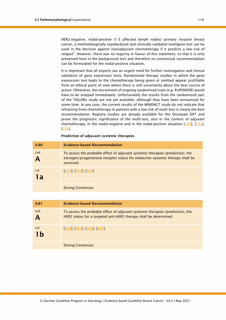
4.5 Pathomorphological examination
© German Guideline Program in Oncology | Evidence-based Guideline Breast Cancer - V4.4 | May 2021
118
HER2-negative, nodal-positive (1-3 affected lymph nodes) primary invasive breast
cancer, a methodologically standardized and clinically validated multigene test can be
used in the decision against (neo)adjuvant chemotherapy if it predicts a low risk of
relapse". However, there was no majority in favour of this statement, so that it is only
presented here in the background text and therefore no consensual recommendation
can be formulated for the nodal-positive situation.
It is important that all experts see an urgent need for further investigation and clinical
validation of gene expression tests. Randomised therapy studies in which the gene
expression test leads to the chemotherapy being given or omitted appear justifiable
from an ethical point of view where there is still uncertainty about the best course of
action. Otherwise, the recruitment of ongoing randomised trials (e.g. RxPONDER) would
have to be stopped immediately. Unfortunately the results from the randomized part
of the TAILORx study are not yet available, although they have been announced for
some time. In any case, the current results of the MINDACT study do not indicate that
refraining from chemotherapy in patients with a low risk of multi-test is clearly the best
recommendation. Registry studies are already available for the Oncotype DX® and
prove the prognostic significance of the multi-test, also in the context of adjuvant
chemotherapy, in the nodal-negative and in the nodal-positive situation [554], [555],
[556].
Prediction of adjuvant systemic therapies
4.80 Evidence-based Recommendation
GoR
A
To assess the probable effect of adjuvant systemic therapies (prediction), the
estrogen/progesterone receptor status for endocrine systemic therapy shall be
assessed.
LoE
1a
[421]; [559]; [560]
Strong Consensus
4.81 Evidence-based Recommendation
GoR
A
To assess the probable effect of adjuvant systemic therapies (prediction), the
HER2 status for a targeted anti-HER2 therapy shall be determined.
LoE
1b
[426]; [465]; [466]; [467]
Strong Consensus
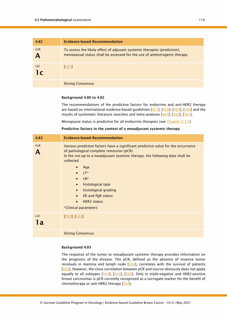
4.5 Pathomorphological examination
© German Guideline Program in Oncology | Evidence-based Guideline Breast Cancer - V4.4 | May 2021
119
4.82 Evidence-based Recommendation
GoR
A
To assess the likely effect of adjuvant systemic therapies (prediction),
menopausal status shall be assessed for the use of antiestrogenic therapy.
LoE
1c
[561]
Strong Consensus
Background 4.80 to 4.82
The recommendations of the predictive factors for endocrine and anti-HER2 therapy
are based on international evidence-based guidelines [421], [426], [465], [466] and the
results of systematic literature searches and meta-analyses [467], [560], [561].
Menopause status is predictive for all endocrine therapies (see Chapter 5.7.2)
Predictive factors in the context of a neoadjuvant systemic therapy
4.83 Evidence-based Recommendation
GoR
A
Various predictive factors have a significant predictive value for the occurrence
of pathological complete remission (pCR).
In the run-up to a neoadjuvant systemic therapy, the following data shall be
collected
• Age
• cT*
• cN*
• histological type
• histological grading
• ER and PgR status
• HER2 status
*Clinical parameters
LoE
1a
[562]; [563]
Strong Consensus
Background 4.83
The response of the tumor to neoadjuvant systemic therapy provides information on
the prognosis of the disease. The pCR, defined as the absence of invasive tumor
residuals in mamma and lymph node [564], correlates with the survival of patients
[565]. However, the close correlation between pCR and course obviously does not apply
equally to all subtypes [564], [565], [566]. Only in triple-negative and HER2-positive
breast carcinomas is pCR currently recognized as a surrogate marker for the benefit of
chemotherapy or anti-HER2 therapy [564].

4.5 Pathomorphological examination
© German Guideline Program in Oncology | Evidence-based Guideline Breast Cancer - V4.4 | May 2021
120
According to the pooled data from 7 German neoadjuvant studies including 3332
patients [563] and the results of a meta-analysis including 11,695 patients from 30
studies [562], pCR rate and subtype are significantly associated. The highest pCR rates
are achieved in triple-negative and HER2+/HR- subtypes. In patients with a positive
HER2 status, an additional neoadjuvant anti-HER2 treatment further increases the pCR
rate.
In addition, there is a significant correlation between age, pre-therapeutic tumor size
and nodal status as well as histological tumor type and degree of differentiation with
the pCR [563].
Other factors are currently under discussion to predict a higher response to
neoadjuvant systemic therapy. These include in particular:
• Ki-67 proliferation index
• Tumor infiltrating lymphocytes (TILs)
Based on the determination of the Ki-67 proliferation index in 1166 punch biopsies of
the GeparTrio study it was shown that Ki-67 is a significant predictive marker for the
response to neoadjuvant chemotherapy [567]. However, the discussion about the
results of the study also made clear that the definition of a uniform threshold value for
the prediction of the therapy effect is difficult [568], [569] Ki-67 showed a significant
predictive and prognostic value over a wide range of cut-offs, so that an optimization
of the threshold value based on study data might be problematic. Rather, it can be
assumed that the level of the optimal cut-off value is context-dependent, i.e. influenced
by the composition of the study collective, the molecular tumor types and the
therapeutic agents. The decision for a neoadjuvant therapy is mainly controlled by the
hormone receptor and HER2 status. Triple-negative and HER2-positive carcinomas are
usually treated neoadjuvantly. In this situation the Ki-67 level is no longer necessarily
relevant. Therefore, the majority of the S3 guideline group decided against a
recommendation of Ki-67 as a predictive factor for the response to neoadjuvant
systemic therapy.
Tumor infiltrating lymphocytes (TILS) can be understood as an immunological
biomarker. In recent years, an increasing number of studies have been published
showing that the quantification of TILs has a prognostic and predictive value, especially
in HER2-positive and triple-negative breast carcinomas [570], [571], [572]. A meta-
analysis including 13,100 patients from 23 studies concluded that an increased
number of TILs predicts the achievement of pCR and is associated with an improved
survival rate [572]. However, different methods for the detection of TILs are used in the
studies (HE staining; immunohistochemistry: CD45, CD3, CD8) [572], [573]. Also the
procedure of evaluation and the amount of cut-off are different [572], [573], so that
from the point of view of the S3-guideline group no recommendation for this parameter
can be derived from the study data available so far.
4.5.5. Percutaneous biopsies in the context of interventional
diagnostics
Currently available methods for interventional diagnostics:
• High-speed punch biopsy (e.g. 14 gauge)
• vacuum biopsy (e.g. 11 gauge or 8 gauge)

4.5 Pathomorphological examination
© German Guideline Program in Oncology | Evidence-based Guideline Breast Cancer - V4.4 | May 2021
121
4.5.5.1. Percutaneous biopsy (high-speed punch biopsy, vacuum biopsy)
Indications: Diagnostic confirmation for therapy planning, clarification of unclear and
suspected malignant findings.
4.5.5.1.1. Macroscopic processing
Description:
• High-speed punch biopsy: number and (total) length of punch cylinders/parts;
• vacuum biopsy: number of punching cylinders/parts; if necessary, further
description (colour, consistency)
Tissue embedding:
• complete embedding of the tissue samples sent
4.5.5.1.2. Microscopic processing and assessment
Processing:
• Sectional stages (see Statement 4.34.; H&E; additional examinations if
required)
• special additional examinations in case of detection of invasive carcinoma
(hormone receptors; HER2, Ki-67)
Cutting steps for percutaneous biopsies
4.84 Consensus-based Recommendation
EC At least 3 HE incisions should be made and examined from tissue cylinders
removed for the clarification of calcifications.
For tissue cylinders that were removed for the clarification of focal findings, one
HE incision may be sufficient.
Further incision steps may be necessary if there is no correlation to the clinical-
radiological findings or to clarify the diagnosis.
Strong Consensus
Background 4.84
The diagnostic reliability at the punch and vacuum biospies is improved, especially in
the presence of microcalcifications, by the preparation of cutting steps [574]. This is
also related to the diameter of the cylinders, which is usually larger in biopsies from
microcalcifications associated lesions, since vacuum biopsies are increasingly used
here. The addition of 2 incision steps on a collective of 40 punch and 63 vacuum
biopsies changed the diagnostic category in 13% of biopsies from lesions with
microcalcifications. On the other hand, the addition of 266 punch biopsies and 6
vacuum biopsies resulted in a change in the diagnostic category in only 1.5% of the
biopsies from other lesions.
Assessment:
• The information is documented as in Chapter 5.5.2.3, possibly using a form
(see Chapter 12.3: Figure 10).
In punch biopsies, the displacement of benign or malignant epithelial cell clusters into
the stroma and/or vessels is possible; in individual cases, the differentiation of true
stroma and/or vessel invasion may be difficult.
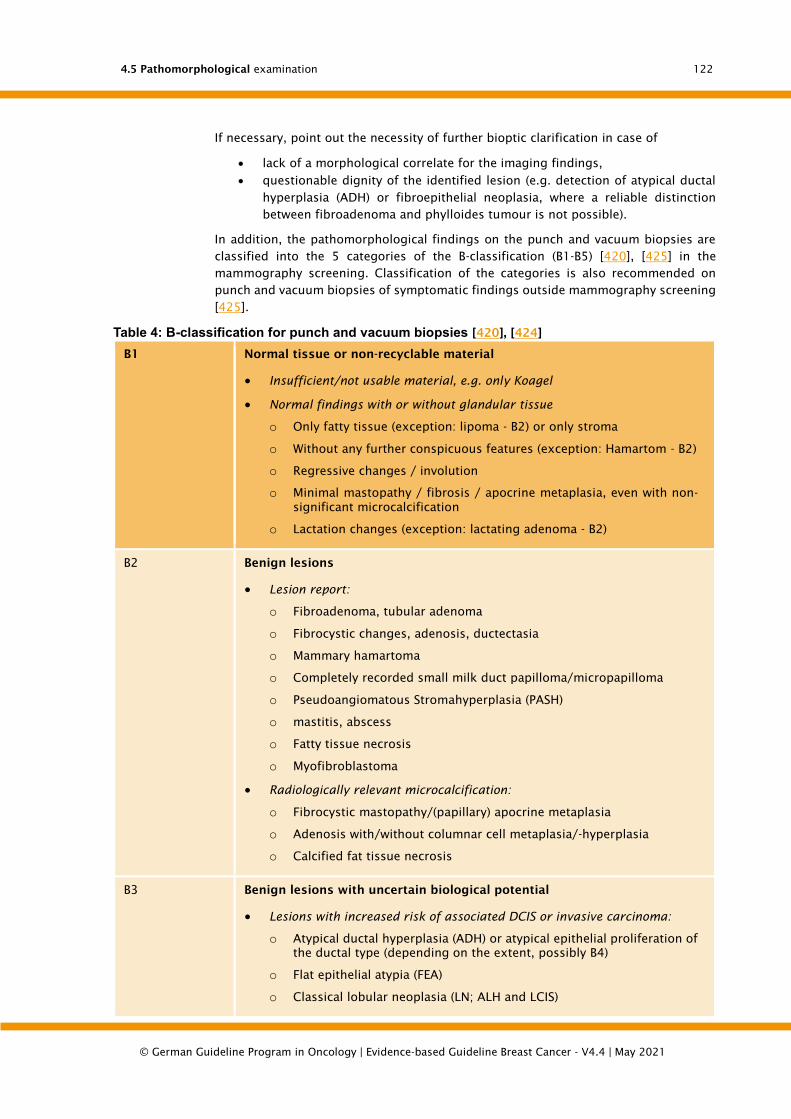
4.5 Pathomorphological examination
© German Guideline Program in Oncology | Evidence-based Guideline Breast Cancer - V4.4 | May 2021
122
If necessary, point out the necessity of further bioptic clarification in case of
• lack of a morphological correlate for the imaging findings,
• questionable dignity of the identified lesion (e.g. detection of atypical ductal
hyperplasia (ADH) or fibroepithelial neoplasia, where a reliable distinction
between fibroadenoma and phylloides tumour is not possible).
In addition, the pathomorphological findings on the punch and vacuum biopsies are
classified into the 5 categories of the B-classification (B1-B5) [420], [425] in the
mammography screening. Classification of the categories is also recommended on
punch and vacuum biopsies of symptomatic findings outside mammography screening
[425].
Table 4: B-classification for punch and vacuum biopsies [420], [424]
B1 Normal tissue or non-recyclable material
• Insufficient/not usable material, e.g. only Koagel
• Normal findings with or without glandular tissue
o Only fatty tissue (exception: lipoma - B2) or only stroma
o Without any further conspicuous features (exception: Hamartom - B2)
o Regressive changes / involution
o Minimal mastopathy / fibrosis / apocrine metaplasia, even with non-
significant microcalcification
o Lactation changes (exception: lactating adenoma - B2)
B2
Benign lesions
• Lesion report:
o Fibroadenoma, tubular adenoma
o Fibrocystic changes, adenosis, ductectasia
o Mammary hamartoma
o Completely recorded small milk duct papilloma/micropapilloma
o Pseudoangiomatous Stromahyperplasia (PASH)
o mastitis, abscess
o Fatty tissue necrosis
o Myofibroblastoma
• Radiologically relevant microcalcification:
o Fibrocystic mastopathy/(papillary) apocrine metaplasia
o Adenosis with/without columnar cell metaplasia/-hyperplasia
o Calcified fat tissue necrosis
B3 Benign lesions with uncertain biological potential
• Lesions with increased risk of associated DCIS or invasive carcinoma:
o Atypical ductal hyperplasia (ADH) or atypical epithelial proliferation of
the ductal type (depending on the extent, possibly B4)
o Flat epithelial atypia (FEA)
o Classical lobular neoplasia (LN; ALH and LCIS)
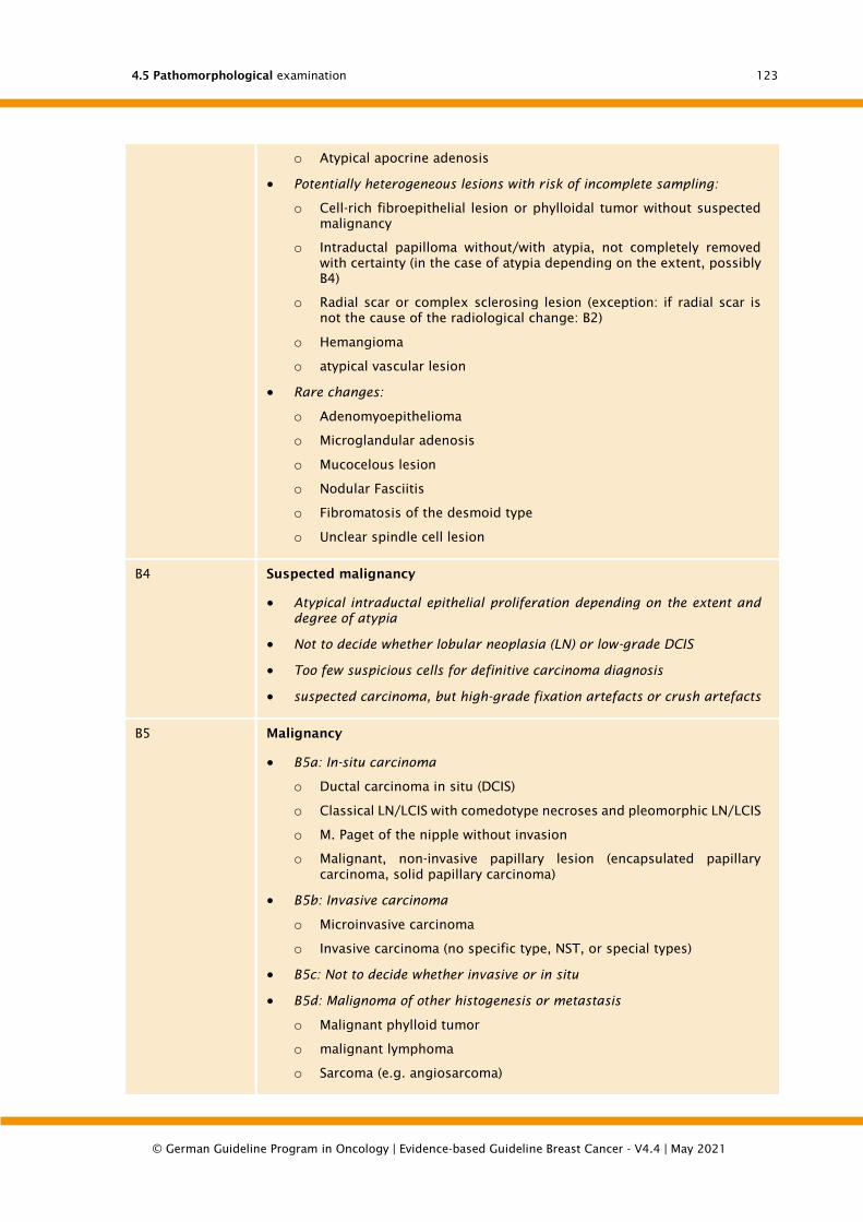
4.5 Pathomorphological examination
© German Guideline Program in Oncology | Evidence-based Guideline Breast Cancer - V4.4 | May 2021
123
o Atypical apocrine adenosis
• Potentially heterogeneous lesions with risk of incomplete sampling:
o Cell-rich fibroepithelial lesion or phylloidal tumor without suspected
malignancy
o Intraductal papilloma without/with atypia, not completely removed
with certainty (in the case of atypia depending on the extent, possibly
B4)
o Radial scar or complex sclerosing lesion (exception: if radial scar is
not the cause of the radiological change: B2)
o Hemangioma
o atypical vascular lesion
• Rare changes:
o Adenomyoepithelioma
o Microglandular adenosis
o Mucocelous lesion
o Nodular Fasciitis
o Fibromatosis of the desmoid type
o Unclear spindle cell lesion
B4 Suspected malignancy
• Atypical intraductal epithelial proliferation depending on the extent and
degree of atypia
• Not to decide whether lobular neoplasia (LN) or low-grade DCIS
• Too few suspicious cells for definitive carcinoma diagnosis
• suspected carcinoma, but high-grade fixation artefacts or crush artefacts
B5 Malignancy
• B5a: In-situ carcinoma
o Ductal carcinoma in situ (DCIS)
o Classical LN/LCIS with comedotype necroses and pleomorphic LN/LCIS
o M. Paget of the nipple without invasion
o Malignant, non-invasive papillary lesion (encapsulated papillary
carcinoma, solid papillary carcinoma)
• B5b: Invasive carcinoma
o Microinvasive carcinoma
o Invasive carcinoma (no specific type, NST, or special types)
• B5c: Not to decide whether invasive or in situ
• B5d: Malignoma of other histogenesis or metastasis
o Malignant phylloid tumor
o malignant lymphoma
o Sarcoma (e.g. angiosarcoma)
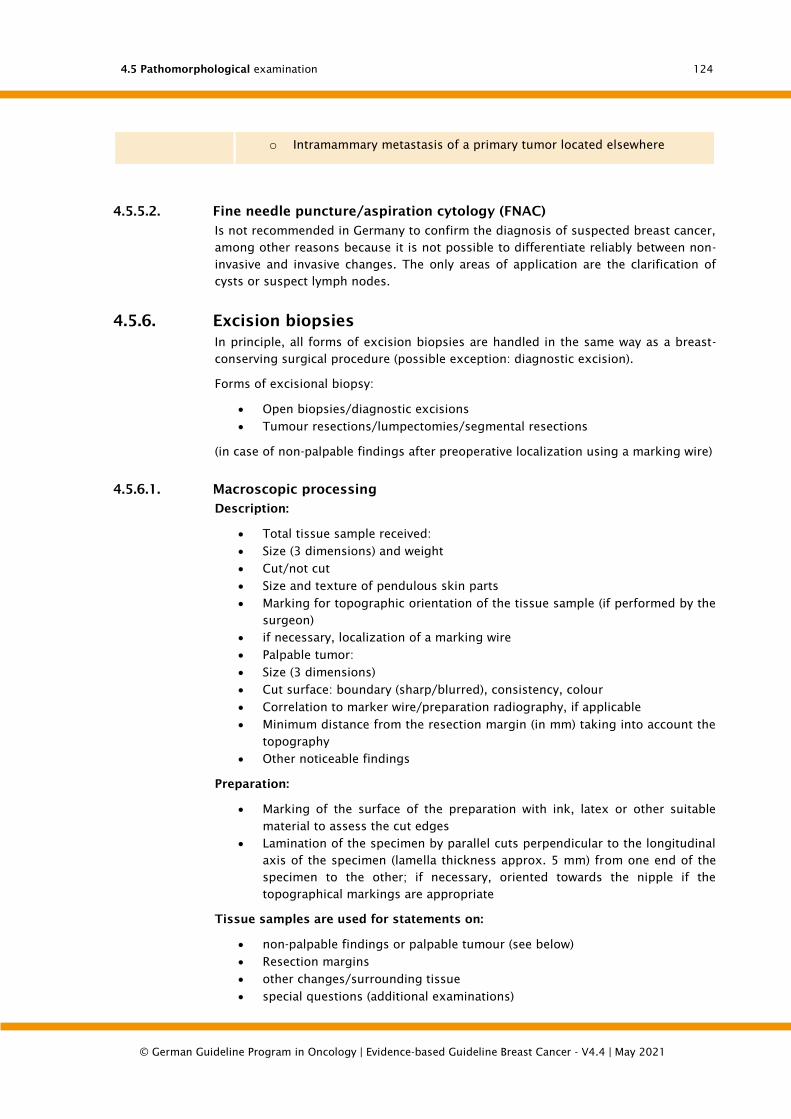
4.5 Pathomorphological examination
© German Guideline Program in Oncology | Evidence-based Guideline Breast Cancer - V4.4 | May 2021
124
o Intramammary metastasis of a primary tumor located elsewhere
4.5.5.2. Fine needle puncture/aspiration cytology (FNAC)
Is not recommended in Germany to confirm the diagnosis of suspected breast cancer,
among other reasons because it is not possible to differentiate reliably between non-
invasive and invasive changes. The only areas of application are the clarification of
cysts or suspect lymph nodes.
4.5.6. Excision biopsies
In principle, all forms of excision biopsies are handled in the same way as a breast-
conserving surgical procedure (possible exception: diagnostic excision).
Forms of excisional biopsy:
• Open biopsies/diagnostic excisions
• Tumour resections/lumpectomies/segmental resections
(in case of non-palpable findings after preoperative localization using a marking wire)
4.5.6.1. Macroscopic processing
Description:
• Total tissue sample received:
• Size (3 dimensions) and weight
• Cut/not cut
• Size and texture of pendulous skin parts
• Marking for topographic orientation of the tissue sample (if performed by the
surgeon)
• if necessary, localization of a marking wire
• Palpable tumor:
• Size (3 dimensions)
• Cut surface: boundary (sharp/blurred), consistency, colour
• Correlation to marker wire/preparation radiography, if applicable
• Minimum distance from the resection margin (in mm) taking into account the
topography
• Other noticeable findings
Preparation:
• Marking of the surface of the preparation with ink, latex or other suitable
material to assess the cut edges
• Lamination of the specimen by parallel cuts perpendicular to the longitudinal
axis of the specimen (lamella thickness approx. 5 mm) from one end of the
specimen to the other; if necessary, oriented towards the nipple if the
topographical markings are appropriate
Tissue samples are used for statements on:
• non-palpable findings or palpable tumour (see below)
• Resection margins
• other changes/surrounding tissue
• special questions (additional examinations)
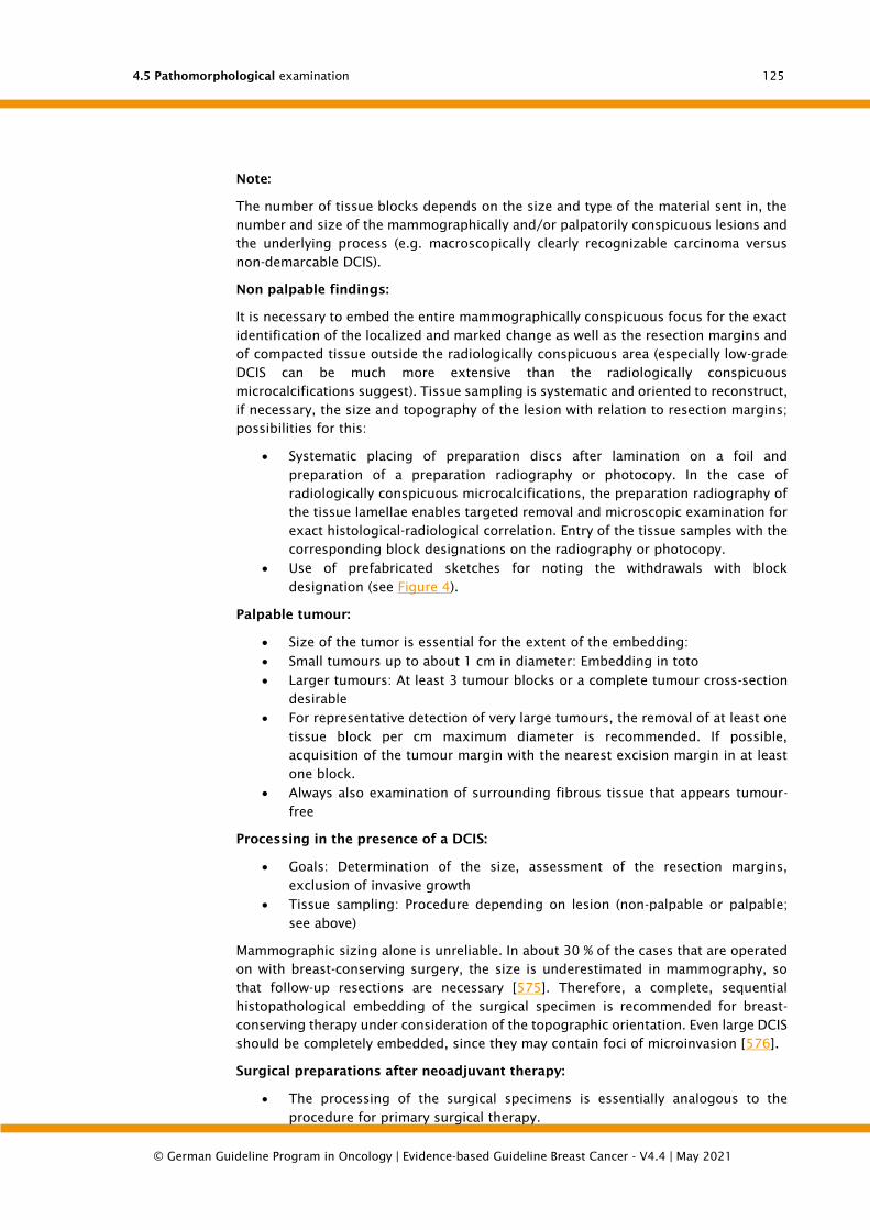
4.5 Pathomorphological examination
© German Guideline Program in Oncology | Evidence-based Guideline Breast Cancer - V4.4 | May 2021
125
Note:
The number of tissue blocks depends on the size and type of the material sent in, the
number and size of the mammographically and/or palpatorily conspicuous lesions and
the underlying process (e.g. macroscopically clearly recognizable carcinoma versus
non-demarcable DCIS).
Non palpable findings:
It is necessary to embed the entire mammographically conspicuous focus for the exact
identification of the localized and marked change as well as the resection margins and
of compacted tissue outside the radiologically conspicuous area (especially low-grade
DCIS can be much more extensive than the radiologically conspicuous
microcalcifications suggest). Tissue sampling is systematic and oriented to reconstruct,
if necessary, the size and topography of the lesion with relation to resection margins;
possibilities for this:
• Systematic placing of preparation discs after lamination on a foil and
preparation of a preparation radiography or photocopy. In the case of
radiologically conspicuous microcalcifications, the preparation radiography of
the tissue lamellae enables targeted removal and microscopic examination for
exact histological-radiological correlation. Entry of the tissue samples with the
corresponding block designations on the radiography or photocopy.
• Use of prefabricated sketches for noting the withdrawals with block
designation (see Figure 4).
Palpable tumour:
• Size of the tumor is essential for the extent of the embedding:
• Small tumours up to about 1 cm in diameter: Embedding in toto
• Larger tumours: At least 3 tumour blocks or a complete tumour cross-section
desirable
• For representative detection of very large tumours, the removal of at least one
tissue block per cm maximum diameter is recommended. If possible,
acquisition of the tumour margin with the nearest excision margin in at least
one block.
• Always also examination of surrounding fibrous tissue that appears tumour-
free
Processing in the presence of a DCIS:
• Goals: Determination of the size, assessment of the resection margins,
exclusion of invasive growth
• Tissue sampling: Procedure depending on lesion (non-palpable or palpable;
see above)
Mammographic sizing alone is unreliable. In about 30 % of the cases that are operated
on with breast-conserving surgery, the size is underestimated in mammography, so
that follow-up resections are necessary [575]. Therefore, a complete, sequential
histopathological embedding of the surgical specimen is recommended for breast-
conserving therapy under consideration of the topographic orientation. Even large DCIS
should be completely embedded, since they may contain foci of microinvasion [576].
Surgical preparations after neoadjuvant therapy:
• The processing of the surgical specimens is essentially analogous to the
procedure for primary surgical therapy.
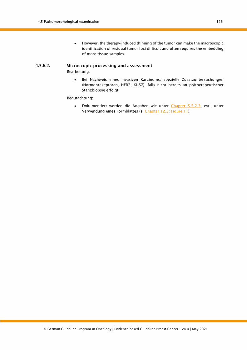
4.5 Pathomorphological examination
© German Guideline Program in Oncology | Evidence-based Guideline Breast Cancer - V4.4 | May 2021
126
• However, the therapy-induced thinning of the tumor can make the macroscopic
identification of residual tumor foci difficult and often requires the embedding
of more tissue samples.
4.5.6.2. Microscopic processing and assessment
Bearbeitung:
• Bei Nachweis eines invasiven Karzinoms: spezielle Zusatzuntersuchungen
(Hormonrezeptoren, HER2, Ki-67), falls nicht bereits an prätherapeutischer
Stanzbiopsie erfolgt
Begutachtung:
• Dokumentiert werden die Angaben wie unter Chapter 5.5.2.3, evtl. unter
Verwendung eines Formblattes (s. Chapter 12.3: Figure 11).

4.5 Pathomorphological examination
© German Guideline Program in Oncology | Evidence-based Guideline Breast Cancer - V4.4 | May 2021
127
Figure 3: Sketch of tissue samples
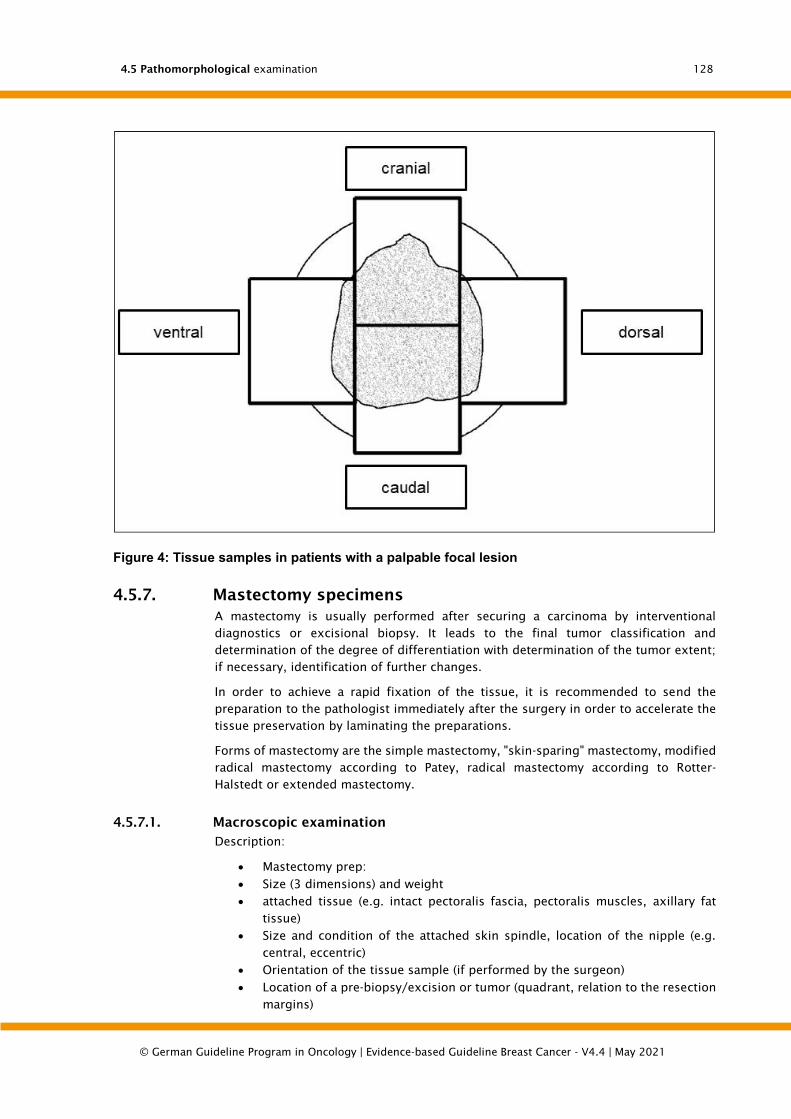
4.5 Pathomorphological examination
© German Guideline Program in Oncology | Evidence-based Guideline Breast Cancer - V4.4 | May 2021
128
Figure 4: Tissue samples in patients with a palpable focal lesion
4.5.7. Mastectomy specimens
A mastectomy is usually performed after securing a carcinoma by interventional
diagnostics or excisional biopsy. It leads to the final tumor classification and
determination of the degree of differentiation with determination of the tumor extent;
if necessary, identification of further changes.
In order to achieve a rapid fixation of the tissue, it is recommended to send the
preparation to the pathologist immediately after the surgery in order to accelerate the
tissue preservation by laminating the preparations.
Forms of mastectomy are the simple mastectomy, "skin-sparing" mastectomy, modified
radical mastectomy according to Patey, radical mastectomy according to Rotter-
Halstedt or extended mastectomy.
4.5.7.1. Macroscopic examination
Description:
• Mastectomy prep:
• Size (3 dimensions) and weight
• attached tissue (e.g. intact pectoralis fascia, pectoralis muscles, axillary fat
tissue)
• Size and condition of the attached skin spindle, location of the nipple (e.g.
central, eccentric)
• Orientation of the tissue sample (if performed by the surgeon)
• Location of a pre-biopsy/excision or tumor (quadrant, relation to the resection
margins)
4.5 Pathomorphological examination
© German Guideline Program in Oncology | Evidence-based Guideline Breast Cancer - V4.4 | May 2021
129
• Tumor:
• Size (3 dimensions)
• cut surface: boundary (sharp/unsharp), consistency, colour
• Reference to the resection margin
• other conspicuous findings (e.g. prosthesis capsule, fibrocystic changes)
Preparation:
• if necessary, staining of the preparation margin with ink or pigments to identify
the resection margin
• Lamination of the preparation from lateral to medial in parallel slices of 5-10
mm thickness, with lamellae remaining in contact with the skin
Tissue samples are used for statements on:
• Nipple/submammary tissue
• Tumor (number of paraffin blocks depending on size)
• Pre-biopsy/excision cave margin (3-4 tissue samples in total)
• Resection margins
• other changes
• additional breast gland tissue from the 4 quadrants (at least 1 block each)
• special questions/additional examinations
If the mastectomy was performed because of a DCIS or if there were radiologically
extensive microcalcifications, a preparation radiography of the tissue lamellae can be
helpful in order to localize the changes precisely and to be able to carry out a targeted
removal to determine the extent and reference to the resection edges.
4.5.7.2. Microscopic examination and assessment
The information is documented as in Chapter 5.5.2.3, possibly using a form (see
Chapter 12.3: Figure 11).
4.5.8. Lymph nodes
4.85 Consensus-based Recommendation
EC Lymph node status
The lymph node status is determined by histological examination of all removed
lymph nodes.
The following assessments shall be obligatory: Number of removed and affected
lymph nodes, extent of the largest tumour settlement, capsule rupture, pN
category (according to TNM classification, 8th edition UICC 2017).
The aim of the work-up is to detect all macrometastases (> 2.0 mm).
Strong Consensus
Background 4.85
Sentinel lymph node biopsy (SLNB):
• Removal of the so-called sentinel lymph node (dye and/or radionuclide
labeling).
• Nowadays common primary procedure for the determination of the nodal
status. Prerequisite is compliance with the recommended quality criteria [577].
4.5 Pathomorphological examination
© German Guideline Program in Oncology | Evidence-based Guideline Breast Cancer - V4.4 | May 2021
130
• The minimal aim of the histological examination is the detection of all
macrometastases (> 2 mm) [427], [532]. Desirable, but not obligatory, is also
the identification of micrometastases (> 0.2 mm and/or more than 200 tumor
cells, but not larger than 2 mm). In the presence of micrometastases, it is to
be expected that other lymph nodes will be affected in approx. 20 % of cases
[578], and with a size of > 1 mm even in approx. 30 % of cases [579]. The
histological examination of the SLN does not aim to detect isolated tumor cells
(ITC). If ITCs are detected, their correct classification (see below) must be
ensured.
Axillary lymphadenectomy:
• Nowadays only in exceptional cases primary surgical procedure to determine
the lymph node status. Mostly for completion in case of affected SLN.
• The aim of the histological examination is to detect all macrometastases (> 2
mm).
4.5.8.1. Macroscopic examination
Description:
• Size (3 dimensions) and weight of the entire tissue sample (for axillary lymph
node dissection)
• Orientation (if marked)
• Number of lymph nodes
• Dimension of the largest lymph node
Preparation:
• Careful examination of the fatty tissue for lymph nodes
• Histological examination of all contained lymph nodes
• In macroscopically affected and interconnected lymph nodes: Examination of
a representative cross-section
• In case of macroscopically not clearly affected lymph nodes: complete
embedding for histological examination
• If the size of the lymph nodes permits, they should be halved along the
longitudinal axis or laminated in slices 2-3 mm thick
4.5.8.2. Microscopic examination and assessment
Processing:
• Sentinel lymph node [427], [577]:
• For macroscopically affected lymph nodes: one H&E incision per block
• In macroscopically not clearly affected lymph nodes: Cutting steps (distance /=
500 µm) H&E stained; number for reliable detection of all macrometastases
depending on tissue thickness
• Immunohistochemical reactions with antibodies against cytokeratins are not
recommended as standard procedures, but may be diagnostically helpful in
individual cases (e.g. invasive lobular carcinoma)
• Axillary lymph node dissections:
• For macroscopically affected lymph nodes: one H&E cut per block
• In macroscopically not clearly affected lymph nodes: According to international
guidelines one H&E incision is sufficient [427], [428]. However, depending on
the thickness of the total blocked lymph node tissue, it is recommended to
4.5 Pathomorphological examination
© German Guideline Program in Oncology | Evidence-based Guideline Breast Cancer - V4.4 | May 2021
131
make at least 2-3 incisions (distance 100-500 µm) to ensure the detection of
all macrometastases (> 2 mm).
• Assessment:
• The following information is documented, possibly using a form (see Chapter
12.3: Figure 11):
• Type of tissue sample
• Page reference
• Number of lymph nodes examined (with localization, if marked)
• Number of lymph nodes affected
• Extent of the greatest metastatic infiltration
• Extranodal infiltration, if any
• pTNM stage (including additional tissue samples if necessary) (see Chapter
5.5.6 and Chapter 5.5.7)
Remarks:
• If the pathological classification is based on a sentinel lymph node
examination, this is indicated by the suffix (sn), for example pN0(sn) [429].
The detection of isolated tumor cells (ITC) in regional lymph nodes is classified as
pN0(i+). ITC are defined as single tumor cells or small clusters of cells not larger than
0.2 mm in the largest dimension. As an additional criterion it has been proposed to
include a cluster of less than 200 cells (in a histological section) in this category [429].
4.6 Adjuvant radiothepary of breastc ancer
© German Guideline Program in Oncology | Evidence-based Guideline Breast Cancer - V4.4 | May 2021
132
4.6. Adjuvant radiothepary of breastc ancer
4.86 Evidence-based Recommendation
GoR
A
After breast-conserving surgery due to invasive carcinoma, radiation of the
affected breast shall be performed.
For patients with clearly limited life expectancy (<10 years) and a small (pT1),
node-negative (pN0), hormone receptor-positive HER2-negative tumor receiving
endocrine adjuvant therapy, conditional upon free excision margins and taking
an increased risk of local recurrence into account, radiation can be dispensed
with after individual counselling.
Note for all recommendations: All individual items are "or" combinations. "And" links are
represented by an "and".
LoE
1a
[580]; [581]; [582]; [583]; [584]; [585]; [586]; [587]
Strong Consensus
Background 4.86
Postoperative radiation is the most important and effective measure to reduce the risk
of intramammary recurrence; the effectiveness is proven with the highest evidence by
numerous randomized studies and meta-analyses [580], [581] To date, no subgroup
has been identified in randomized studies where there is no significant effect in terms
of improving local tumor control. According to the current state of knowledge, the
effectiveness of radiotherapy is also independent of tumor-specific or patient-related
prognostic factors. In recent studies, in which the effectiveness of radiotherapy was
examined in tumor subgroups defined by molecular biology or gene expression
analyses, significant effects of radiotherapy were found in all subgroups [557].
Therefore, although it is possible to identify patient groups with a low risk of relapse,
these patients also benefit from radiotherapy with regard to an optimization of local
tumor control. Therefore, in contrast to the individualized indication of systemic
therapies, predictive markers cannot be used to make a decision on radiotherapy after
breast-conserving surgery.
In meta-analyses [580], [581] the radiation reduced the locoregional relapse rate as
well as the rate of all relapses (locoregional and distal relapses). Although primary or
adjuvant drug systemic therapies also increase locoregional tumor control, their effect
is not sufficient to dispense with radiotherapy after breast-conserving surgery. If the
relative risk reduction (i.e. the hazard ratio, similar to the evaluation of drug therapy
procedures) is used as a measure of the effectiveness of radiotherapy, the effectiveness
of radiotherapy on local tumour control has actually improved considerably in recent
years. The hazard ratio in the meta-analyses, which mainly contain studies from the
1970s and 1980s, is about 0.35; of 10 possible recurrences without radiation, 6 to 7
were prevented by radiotherapy [581]. In more recent studies, hazard ratios of about
0.2 or even less are consistently reported, i.e. of 10 possible recurrences without
radiation, 8 are prevented by radiotherapy [583], [584], [586]. This improvement in
relative effectiveness can be explained radiobiologically well and is based essentially
on standardized surgical techniques and improved pathohistological diagnostics (fewer

4.6 Adjuvant radiothepary of breastc ancer
© German Guideline Program in Oncology | Evidence-based Guideline Breast Cancer - V4.4 | May 2021
133
undetected R1 resections, thus less "tumor burden" with a higher success rate with
standardized radiation dose).
The improved local tumor control leads to a reduction of breast cancer-specific
mortality; this effect was confirmed in consecutive meta-analyses. In the last meta-
analysis of the EBCTCG data [581] percutaneous radiotherapy leads to a reduction of
disease-specific mortality in pN0 patients by 3.3% in absolute terms and by 8.5% in
absolute terms in pN+ patients after 15 years. These effects have been proven for all
age groups, but the benefit decreases in older patients [588].
The effect of radiotherapy of the breast on survival - since it is generated secondarily
by optimizing local tumor control - is detectable only after a longer follow-up period
and increases over time [581]. The greatest benefit of radiotherapy is achieved by
patients in whom the risk of relapse is reduced by > 10% by adjuvant radiotherapy.
Thus, statistically speaking, 4 local recurrences prevented within the first 10 years can
prevent cancer-related deaths within 15 years [580], [581]. The positive effect on
survival is lower in more recent studies with overall more favorable tumor stages or is
not given in very favorable collectives in the first 10 years after therapy. Nevertheless,
radiotherapy remains the most important measure to optimize local control. In more
recent studies the risk for intramammary recurrence is lower than the risk for
contralateral new carcinomas, so that a protective effect on new carcinomas is
discussed [589].
Patients with low-risk tumors also benefit from adjuvant radiotherapy of the breast.
The positive effect with regard to local tumor control is clearly documented in all
randomized studies [583], [584], [586], [590], [591]. The most important risk factor for
a local recurrence in the breast is also in these patients with very favorable tumors the
renunciation of radiotherapy [584]. The risk for side effects was not different with and
without radiotherapy; most of the side effects result from the adjuvant endocrine
therapy [586]. Data from population-based analyses also show that the renunciation of
radiotherapy in patients at an advanced age is associated with an increased mortality
rate from breast cancer [592]. Radiotherapy of the breast should therefore also be the
rule for these patients.
If adjuvant radiotherapy is used, the free resection margin plays a minor role; a R0
resection ("no ink on tumor") is sufficient from [362], [593]. Radiotherapy can therefore
help to avoid unnecessary follow-up resections and limit the resection volume (a factor
relevant for the final cosmetic outcome).
The risks of radiation therapy are low nowadays; radiation of the breast has the lowest
risks of all therapy methods used in the adjuvant therapy of breast cancer. The risk of
symptomatic pneumonitis is less than 1%. In older studies, increased cardiac mortality
was found in patients with left-sided breast cancer and adjuvant radiotherapy; cardiac
excess mortality correlated with the cardiac radiation dose [594]. In studies conducted
after about 1990 (i.e. after the introduction of 3D conformational radiation), significant
cardiac excess mortality was no longer detectable. In more recent studies, neither co-
radiation of the regional lymph nodes nor combination with trastuzumab was observed
to increase the risk of cardiac events [595], [596], [597], [598]. In addition, with
Intensity Modulated Radio Therapy (IMRT) and Volu-Metric-Arc-Therapy (VMAT)
modern therapy methods with sufficient Level Ib/IIa evidence are available today, which
report lower acute skin reactions, late fibrosis and telangiectasia of the breast and thus
better cosmetic results than after 3D-radiation. At the same time, effective protection
of the lungs and heart is guaranteed. However, IMRT/VMAT should not be used
generally, but should be limited to patients with larger breasts and/or abnormal chest
curvature (e.g. funnel chest) [599], [600], [601], [602]. A further method by which the
4.6 Adjuvant radiothepary of breastc ancer
© German Guideline Program in Oncology | Evidence-based Guideline Breast Cancer - V4.4 | May 2021
134
radiation dose to the heart can be minimized to non-critical values even with left-sided
radiation is radiation in controlled deep inspiration [603]. The risk for radiation-induced
secondary malignancies is basically given with radiation application and amounted in
the early studies (therapy before 1990) to about 1% after 20 years. However, more
recent data from cancer registries (e.g. from the SEER database) show that only a small
proportion (<10%) der nach Strahlentherapie auftretenden Zweitmalignome durch die
Strahlentherapie bedingt ist [604], [605].
Fractionation
4.87 Evidence-based Recommendation
GoR
B/0
Radiotherapy of the breast should be performed in hypofractionation (total dose
approx. 40 Gy in approx. 15-16 fractions in approx. 3 to 5 weeks) or can be
performed in conventional fractionation (total dose approx. 50 Gy in approx. 25-
28 fractions in approx. 5-6 weeks).
LoE
1a
[606]; [607]; [608]; [609]; [610]; [611]; [612]
Consensus
Background 4.87
Until a few years ago, treatment with small single doses (daily 1.80 Gy to 2.00 Gy, so-
called conventional fractionation) was considered the only sensible fractionation for
curative therapy intentions. This applied to all tumours and was well justified from a
radiobiological point of view. In the meantime, data are available from four large
randomized studies with a total of more than 7000 patients, which have shown equally
good results both in terms of tumor control and late toxicity with moderate
hypofractionation (about half as many fractions as with conventional fractionation) in
breast cancer [607], [608], [611], [612]. This surprising result is explained by an
unexpectedly low α/β value of breast carcinomas, which according to more recent
calculations is in the order of 3 to 3.5 Gy.
Hypofractionation means that the number of fractions is reduced; the individual dose
increases, and the total dose is reduced according to radiobiological calculations.
However, a distinction must be made between pure hypofractionation (the total
treatment time remains the same, e.g. only every second day of radiation, e.g. START-
A study) and accelerated hypofraction (the treatment time is shortened by daily
radiation). From the data available so far, it can be concluded that moderate
hypofractionation (15 to 16 fractions for breast radiation instead of 25 to 28 fractions
for conventional fractionation) is well possible and can be combined with moderate
acceleration (reduction of the total treatment time from 5-6 weeks to three weeks).
These variants are well justified not only by studies but also by radiobiological model
calculations.
It is unclear whether a further reduction in the number of fractions (extreme
hypofractionation) or the treatment time (extreme acceleration) is possible. Two British
studies (FAST and FAST-FORWARD) test these concepts [613]. So far, only data on acute
toxicity are available, which (as expected) is not amplified [614]. The crucial questions
of tumor control and late toxicity are expected to be answered from about 2020.
4.6 Adjuvant radiothepary of breastc ancer
© German Guideline Program in Oncology | Evidence-based Guideline Breast Cancer - V4.4 | May 2021
135
In summary, based on the data available, a moderate hypofractionation with moderate
acceleration (i.e. about 15 to 16 fractions with radiation on workdays, total treatment
time about 3 weeks) can be recommended for the follow-up radiation of the whole
breast in breast cancer; a possibly indicated boost should be administered sequentially
(after the hypofractionated radiation of the breast). The advantage of hypofractionation
in comparison to conventional fractionation with the same local tumor control is a clear
reduction in acute skin reactions and also a tendency to slightly lower late sequelae
[607], [615]. A further advantage for the patients is the shortened treatment period of
only 3 to 5 weeks in connection with hypofractionation. So far, there is no evidence
that clinical or molecular biological factors can be used to identify subgroups which
benefit from the use of a specific fractionation regime [616]. The recommendation for
hypofractionation is therefore currently valid across the board for all adjuvant
radiotherapy of the mammary gland without lymph drainage channels.
Exceptions concern patients with radiation of the lymphatic outflow pathways. These
were underrepresented in the hypofractionation studies. Although the data available to
date (in accordance with radiobiological models) do not show any increased late toxicity
for this collective with significantly larger radiation volumes, the data situation is
considered weak. Furthermore, an increased risk of late sequelae at the nerve plexus
and with regard to the occurrence of lymphedema is possible [617] The late toxicity of
hypofractionated regimens when irradiating also the regional lymph nodes is currently
being investigated in large studies. In practice (outside of studies), conventional
fractionation is therefore recommended as the fractionation of first choice when the
lymph nodes are also irradiated.
4.88 Evidence-based Recommendation
GoR
A/B
Boost radiation
Local dose saturation (boost radiation) of the tumor bed lowers the local
recurrence rate in the breast without providing a significant survival benefit.
The boost radiation
• shall therefore be used for all /= 50 year old patients and
• should only be performed in > 51-year-old patients with an increased
local risk of relapse (G3, HER2-positive, triple-negative, > T1).
LoE
1a
[618]; [619]; [620]; [621]
Strong Consensus
Background 4.88
A local dose saturation of the tumor bed (so-called boost) can further reduce the relapse
rate after breast-conserving surgery. Data are available from two large randomized
studies. Both studies compared radiation of the whole breast (dose 50 Gy) versus
radiation of the breast with subsequent boost. In the French study the boost dose was
10 Gy [621]; in the larger EORTC study the boost dose was 16 Gy [619]. In both studies
the risk of relapse was significantly reduced. Follow-up data from the larger EORTC
study are available for 20 years, which were updated and published every 5 years;
furthermore, several analyses of prognostic factors have been published from this
study [366], [618], [619], [620], [622], [623], [624]. The benefit of the boost on local
tumor control has remained stable over 20 years or even increased. Local control was
also significantly improved for prognostically favorable subgroups; there is no
4.6 Adjuvant radiothepary of breastc ancer
© German Guideline Program in Oncology | Evidence-based Guideline Breast Cancer - V4.4 | May 2021
136
subgroup that does not benefit from a boost with respect to local tumor control. The
relative risk reduction is similar in all subgroups; the absolute advantage is greater in
younger patients with higher risk than in older patients with low-risk tumours. The rate
of late complications (fibrosis grade 3 in boost volume) was low in the EORTC study
and higher in older patients than in younger ones; therefore, the individual benefit-risk
ratio (local control versus fibrosis) is very favourable in younger patients and decreases
with age.
As a consequence of these data, a boost is strongly recommended and clearly indicated
in all premenopausal patients as well as in postmenopausal patients with an increased
risk of relapse or histological risk factors (G3, HER2-positive, tripelnegative, > T1) -
Elderly patients with evidence of concomitant DCIS, affected lymph nodes, missing
hormone receptors, lymph vessel invasion and after close R0 or R1 resection may also
benefit from a boost. A boost is generally unnecessary in older patients without risk
factors.
In the past, the boost was predominantly (e.g. in the EORTC study) applied with
electrons with five to eight fractions following the radiation of the breast (so-called
sequential boost). After the introduction of 3D radiation planning, techniques with
photons have become standard. Alternative procedures are interstitial brachytherapy
or a single intraoperative radiation (with KV-radiation or electron radiation). All
procedures have advantages for certain clinical situations and limitations. However,
there is no evidence to date that any of these procedures is clearly superior to others.
For boost radiation, therefore, the procedures that are available locally and with which
experience exists should be selected.
A new procedure is the simultaneous integrated boost (SIB) during external radiation.
Here, the sequential boost dose of 10-16 Gy/5-8 fractions, which was previously
administered after breast radiation, is divided into the number of fractions (25-28)
required for breast radiation and integrated at the tumor bed as a simultaneous boost.
The SIB has physical-technical and biological advantages (overdoses outside the boost
volume are reduced) and leads to a reduction of the total treatment time by about 1 to
1.5 weeks depending on the amount of the boost dose [625], [626], [627]. A major
disadvantage of the boost (namely the extension of the treatment time of the external
radiation) is eliminated by the SIB. When using a SIB, the indication for boost radiation
can therefore be set rather generously. The combination of conventional fractionation
when irradiating the breast with SIB has been evaluated in large prospective non-
randomized studies and is considered safe and effective [589], [628]. The combination
of hypofractionation when irradiating the breast and SIB is still considered
experimental. Several randomized studies worldwide are currently investigating this
question (in Germany: HYPOSIB study).
Partial breast radiation and IORT
4.89 Evidence-based Recommendation
GoR
0
Partial breast radiation alone (as an alternative to post-radiation of the entire
breast) can be performed in patients with a low risk of recurrence.
LoE
1a
[629]; [630]; [631]; [632]; [633]; [634]
Strong Consensus

4.6 Adjuvant radiothepary of breastc ancer
© German Guideline Program in Oncology | Evidence-based Guideline Breast Cancer - V4.4 | May 2021
137
Background 4.89
An individual risk-based indication for adjuvant partial breast radiation (PBI (partial
breast radiation) or APBI (accelerated partial breast radiation)) after complete tumor
excision with breast preservation and limitation of the radiation volume to the tumor
region can be performed in patients with a low (local) risk of recurrence.
The different concepts with different radiation qualities and technical procedures for
partial breast radiation mostly correspond to those used in PBI as boost RT before or
after radiation of the entire breast (WBI) [635], [636], [637], [638], [639], [640], [641],
[642], [643], [644], [645], [646].
In multi-catheter brachytherapy, the applicator tubes can be placed during or even after
the surgery. The same applies to balloon brachytherapy, in which the balloon is inserted
into the wound cavity with a central brachytherapy catheter already during the surgery
("open cavity"), but also afterwards ("closed cavity"). Radiation is typically performed
over a few days, sometimes several times a day (e.g. twice a day for 5 days).
Two prospective randomized studies with long follow-up times are available for
multicatheter brachytherapy. The Budapest study [630] showed similar results with
APBI and WBI up to 10 years after therapy, but this monoinstitutional study was closed
early due to poor recruitment, so that it did not have enough power for the hypothesis
of non-inferiority of APBI. Results from the randomized phase III study of the GEC-
ESTRO group were recently published [633]. 1184 patients (> 40 years, tumor up to
3cm) were randomized between 2004 and 2009. The primary endpoint was ipsilateral
local recurrence with a non-inferiority margin of 3% after 5 years. The 5-year recurrence
rate was 0.9% for EBRT and 1.4% for APBI as the sole RT modality (p = 0.42). Overall
survival was 95.6% for EBRT and 97.3% for APBI (p = 0.11).
The IORT as sole radiotherapy modality represents the extreme variant of the
combination of hypofractionation and APBI and is performed immediately after surgical
tumor extirpation as a single radiation treatment limited to the tumor resection cavity
with application of a total dose considered curative to the expanded tumor bed. For an
IORT, electrons of a linear accelerator (= IOERT), an orthovolt therapy with 50 kV X-rays
of a miniature X-ray device or a balloon brachytherapy technique are used [636], [637],
[640], [642], [647], [648]. Altogether, almost 5000 patients were randomized in two
prospective randomized studies (TARGIT, ELIOT). Although the results of the studies
available to date showed a slightly increased recurrence rate in the breast for the overall
group of patients, local tumor control rates for certain subgroups of older patients with
unifocal small breast cancer were comparable to those achievable with radiotherapy of
the entire breast [631], [632], [649].
A monocentric study [650] randomized 520 patients over 40 years of age with tumors
up to 2.5 cm for percutaneous APBI with 5 x 6 Gy as IMRT compared to conventional
whole breast radiation. After a median follow-up of 5 years the local recurrence rate
was 1.5% in both arms with a more favorable toxicity profile and better quality of life
according to APBI [651].
A recently published meta-analysis [652] summarized all published prospective studies
on APBI that had published survival data and could clearly show that there is by no
means an inferiority of APBI in terms of overall survival after 5 years to whole breast
radiation, whereby the studies essentially included older patients with small, clinically
nodal-negative breast cancer. In terms of disease-free survival, APBI also does not
appear inferior to whole breast radiation, although possibly increased local recurrence
rates and reduced non-breast-cancer mortality seem to compensate.
4.6 Adjuvant radiothepary of breastc ancer
© German Guideline Program in Oncology | Evidence-based Guideline Breast Cancer - V4.4 | May 2021
138
Radiation treatment of parts of the breast (PBI) limited to the primary tumor area as the
sole ("definitive") intra- or postoperative radiation treatment may also be an option for
selected patients with a higher risk of local recurrence in whom homogeneous radiation
of the entire breast is not feasible (e.g., patients with a high risk of localized tumor
recurrence, patients with a high risk of localized tumor recurrence, patients with a high
risk of localized tumor recurrence, patients with a high risk of localized tumor
recurrence, patients with a high risk of localized tumor recurrence, patients with a high
risk of localized tumor recurrence, patients with a high risk of localized tumor
recurrence, patients with a high risk of localized tumor recurrence, patients with a high
risk of localized tumor recurrence). e.g. pre-radiation, age, comorbidity) [28], [466],
[629], [643], [653], [654].
Postmastectomy radiotherapy (PMRT)
4.90 Evidence-based Statement
ST Postoperative radiotherapy of the chest wall after mastectomy reduces the risk
of locoregional recurrence and improves overall survival in locally advanced and
nodal-positive breast cancer.
LoE
1a
[655]
Strong Consensus
4.6 Adjuvant radiothepary of breastc ancer
© German Guideline Program in Oncology | Evidence-based Guideline Breast Cancer - V4.4 | May 2021
139
4.91 Evidence-based Recommendation
GoR
A
Radiation therapy of the chest wall after mastectomy shall be indicated in the
following situations
• PT4,
• pT3 pN0 R0 in the presence of risk factors (lymph vessel invasion
(L1), grading G3, premenopausal, age < 50 years),
•
• R1-/R2-resection and lack of the possibility of a rehabilitative
resection.
a) In more than 3 affected axillary lymph nodes, postmastectomy radiation
shall be performed regularly.
b) In 1-3 axillary lymph nodes affected by tumours, post-mastectomy
radiation shall be performed if there is an increased risk of recurrence
(e.g. if HER2-positive, triple-negative, G3, L1, Ki-67 > 30%, > 25% of the
removed lymph nodes are tumour-infected; age ≤ 45 years with
additional risk factors such as medial tumor localization or tumor size >
2cm, or ER negative).
c) PMRT shall be avoided in 1-3 tumour-infected axillary lymph nodes and
tumours with a low risk of local recurrence (pT1, G1, ER-positive, HER2-
negative, at least 3 characteristics must apply).
d) For all other patients with 1-3 tumour-infected axillary lymph nodes, the
individual indication shall be determined on an interdisciplinary basis.
LoE
1a
[274]; [655]; [656]; [657]; [658]; [659]; [660]; [661]; [662]; [663]; [664]; [665];
[666]; [667]; [668]
Consensus
Background 4.90 and 4.91
This update of the S3 guideline presents the current evidence on the indication for
postmastectomy radiotherapy. Compared to the S3-Guideline version of 2012, the
indication is more clearly differentiated according to risk constellations and, under
certain circumstances, completely withdrawn. The authors emphasize that the
indication for postoperative radiotherapy always includes information about possible
treatment alternatives with all their consequences and is based on a clear "informed
consent" of the patients. With a normal risk constellation, postoperative radiation of
the chest wall and lymph drainage channels can be avoided after a mastectomy. There
are, however, situations with a high risk of locoregional recurrence, in which adjuvant
radiotherapy of the chest wall with possibly lymph drainage pathways is indispensable
in view of the otherwise threatening high locoregional recurrence rates. Analogous to
the situation after breast-conserving surgical therapy, postoperative radiotherapy also
has a positive influence on local and regional tumour control after mastectomy and
ultimately on tumour-specific and overall survival. The locoregional risk of relapse and
the indication for postoperative radiotherapy correlate positively. The higher the risk
of locoregional relapse, the higher the absolute improvement of locoregional tumor
control and survival by postoperative radiotherapy. The high risk of relapse is defined
as > 20% and the intermediate risk as 10-20% probability of a locoregional relapse.
4.6 Adjuvant radiothepary of breastc ancer
© German Guideline Program in Oncology | Evidence-based Guideline Breast Cancer - V4.4 | May 2021
140
A high risk of relapse exists for patients with T4 tumours, pT3 pN0 R0 with risk factors
(lymph vessel invasion (L1), grading G3, premenopausal status, age
There is an intermediate risk of relapse for patients with T1 or T2 tumors with only 1-
3 tumor-affected axillary lymph nodes with the simultaneous presence of other risk
factors, such as grading G3, lymph vessel invasion (L1), intravascular tumor cell
detection (V1), Ki-67 > 30%, > 25% of removed lymph nodes tumor-affected, age ≤ 45
years and medial tumor site.
There is a low risk of relapse for patients with 1-3 tumor-affected axillary lymph nodes
if 3 of the 4 following factors are present simultaneously: pT1, G1, ER-positive, HER2-
negative. For other patients with 1-3 tumour-infected lymph nodes, the individual
indication must be determined on an interdisciplinary basis.
In general, the benefit of chest wall radiotherapy in patients with a high risk of
locoregional relapse is assured and is recommended internationally in this situation.
For patients with pN1 (1-3 LC) and intermediate risk, radiotherapy may have a survival
benefit. For patients with pN0 and intermediate risk, the indication for radiotherapy
after mastectomy is not secured due to pending data.
After primary (neoadjuvant) systemic therapy, the indication for radiotherapy of the
chest wall, possibly including locoregional lymph drainage areas, continues to be based
on the pre-therapeutic T-N category due to the lack of available RCTs, regardless of the
extent of response to primary systemic therapy. In analogy to adjuvant radiotherapy
after mastectomy, the indication for radiotherapy after neoadjuvant chemotherapy is
also given in the case of histopathologically confirmed full remission (ypT0), although
prospectively randomized phase III studies that could support this recommendation are
currently lacking.
Postmastectomy radiotherapy after primary systemic therapy
4.92 Evidence-based Recommendation
GoR
A
After primary (neoadjuvant) systemic therapy, the indication for postmastectomy
radiotherapy shall be based on the pretherapeutic clinical stage; for pCR (ypT0
and ypN0), the indication shall be determined in the interdisciplinary tumour
board depending on the risk profile.
LoE
1a
[669]; [670]; [671]; [672]
Strong Consensus
Background 4.92
The neoadjuvant systemic therapy aims to bring the tumour into remission. The degree
of remission is on the one hand associated with the prognosis of the disease, on the
other hand it could also have an influence on the local therapy [565]. The significance
of adjuvant radiotherapy of the chest wall and the lymphatic outflow pathways within
the neoadjuvant therapy concept is not clarified by evidence from randomized studies
[673]. Data from retrospective observational studies mainly refer to the indication for
postmastectomy radiation.
Under the hypothesis that the combination of all therapy modalities contributes to the
survival benefit, the order in which the therapies are administered should be irrelevant.
This would speak for the indication for radiotherapy after the initial stage [674], [675],
4.6 Adjuvant radiothepary of breastc ancer
© German Guideline Program in Oncology | Evidence-based Guideline Breast Cancer - V4.4 | May 2021
141
[676]. In view of the lack of randomized studies on this question, this will continue to
be the standard, especially for high-risk patients. This is especially true for patients
who have not achieved pathologically complete remission in the breast or axilla [677].
On the other hand the histopathologically proven complete remission after neoadjuvant
therapy is a strong prognostic factor independent of the primary tumor biology. In
cases of a pathologically complete remission in the tumor area as in the axilla, it seems
obvious to reduce the extent of local adjuvant radiotherapy [678], [679]. However, data
of randomized clinical studies are missing.
The indication for radiotherapy after neoadjuvant therapy is complicated by the fact
that following national and international therapy recommendations the excision of the
sentinel lymph node is preferred after neoadjuvant therapy [680], [681]. The pre-
therapeutic information on axillary lymph node infiltratrion is lost in some cases.
However, this concerns only a very small group of patients, because in the case of
suspicious pretherapeutic findings the diagnosis should be histologically confirmed
[680], [681]. A potential undertherapy would only exist in patients with mastectomy
who had three or more clinically occult lymph node metastases, but which were no
longer detectable in the posttherapeutic sentinel excision.
Breast-conserving therapy: Regardless of the response to neoadjuvant chemotherapy,
radiotherapy of the breast within the framework of breast-conserving therapy is
generally indicated [682].
Complete remission: In pathologically proven complete remission (ypT0/is ypN0), most
patients undergo breast-conserving surgery. The indication for radiotherapy of the
lymph drainage system is under discussion. Arguments for radiation of the lymphatic
drainage system are preoperative nodal status (≥ 3 lymph nodes), estrogen receptor
status (ER-negative), grading (G3), tumor size (T3), age (
A rule of thumb could be [663], [682], [683]:
1. in pathologically complete remission of primarily clinically positive lymph
nodes (generally histologically confirmed by needle biopsy), radiotherapy of
the lymphatic outflow pathways can be limited to the high-risk cases according
to current guidelines (see footnote 4).
2. in patients with pathologically complete remission (ypT0/is ypN0) with
primarily clinically negative lymph nodes, radiotherapy should be performed
within the framework of BET as in the situation without neoadjuvant therapy
(radiotherapy of the breast with standard tangent). This would correspond to
the procedure of the ACOSOG Z0011 study [412].
3. no recommendation can be made for the rare situations in which a mastectomy
has been performed and primarily no high-risk situation was present, but only
the suspicion (cN1) or possibility (cN0) of axillary lymph node metastases.
No remission (non-pCR): It is generally agreed that there is an increased risk of relapse
in the absence of remission of the primary tumor or axillary lymph nodes. The
indication for postoperative radiotherapy (radiotherapy of the lymphatic outflow
pathways and - in case of mastectomy - also of the chest wall) should be determined
after the stage before the start of therapy and adapted to the current tumor
development if necessary [663].
In inflammatory breast carcinoma (IBC) and primarily inoperable breast carcinoma the
therapy concept basically consists of primary systemic therapy, mastectomy and
radiation; a reduction of this concept is associated with a shorter mean survival time
[684], [685]. Postoperative radiotherapy (PMRT) must therefore be considered

4.6 Adjuvant radiothepary of breastc ancer
© German Guideline Program in Oncology | Evidence-based Guideline Breast Cancer - V4.4 | May 2021
142
obligatory. Radiotherapy of the lymphatic outflow pathways in inflammatory breast
carcinoma should be based on the original tumor stage.
In any case the indication and the radiation field should be discussed
interdisciplinary and should be determined bindingly for all treatment partners.
Table 5: Indication algorithm for radiation therapy after neoadjuvant therapy
Pretherapeutic Posttherapeutic RT-BET¹ PMRT² RT-LAW³
Locally advanced pCR / no pCR yes yes yes
cT1/2 cN1+ ypT1+ or ypN1+ (no
pCR)
yes yes yes
cT1/2 cN1+ ypT0/is ypN0 yes At-risk cases⁴
cT1/2 cN0 (Sonogr.
obligato)
ypT0/is ypN0 yes no no
1 with classic tangent,
2 if a mastectomy was performed,
3 together with PMRT or RT because of BET
4 criteria for high risk of relapse:
- pN0 premenopausal, high risk: central or medial seat, and (G2-3 and ER/PgR negative)
- pN1a high risk: central or medial seat and (G2-3 or ER/PgR negative) or premenopausal, lateral
seat and (G2-3 or ER/PgR negative)
Radiotherapy of the regional lymph nodes
4.93 Evidence-based Statement
ST Adjuvant radiation of regional lymph drainage areas improves disease-free
survival and overall survival in subgroups of patients.
LoE
1a
[595]; [596]; [597]; [686]; [687]
Consensus
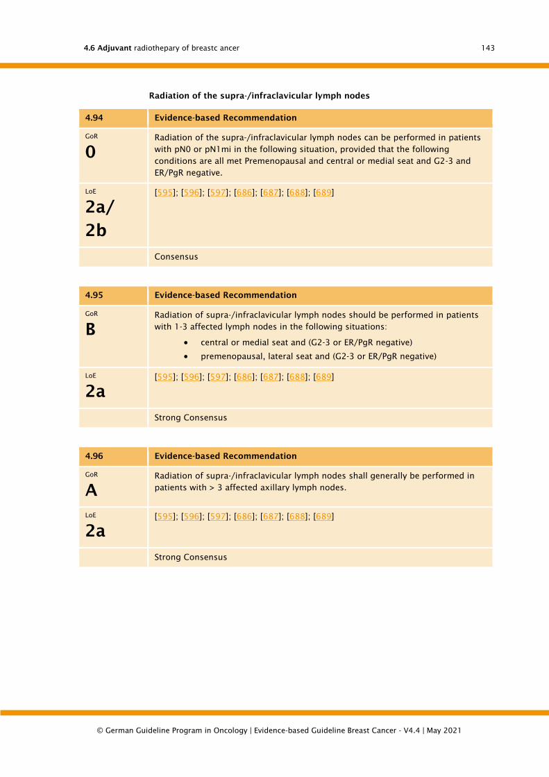
4.6 Adjuvant radiothepary of breastc ancer
© German Guideline Program in Oncology | Evidence-based Guideline Breast Cancer - V4.4 | May 2021
143
Radiation of the supra-/infraclavicular lymph nodes
4.94 Evidence-based Recommendation
GoR
0
Radiation of the supra-/infraclavicular lymph nodes can be performed in patients
with pN0 or pN1mi in the following situation, provided that the following
conditions are all met Premenopausal and central or medial seat and G2-3 and
ER/PgR negative.
LoE
2a/
2b
[595]; [596]; [597]; [686]; [687]; [688]; [689]
Consensus
4.95 Evidence-based Recommendation
GoR
B
Radiation of supra-/infraclavicular lymph nodes should be performed in patients
with 1-3 affected lymph nodes in the following situations:
• central or medial seat and (G2-3 or ER/PgR negative)
• premenopausal, lateral seat and (G2-3 or ER/PgR negative)
LoE
2a
[595]; [596]; [597]; [686]; [687]; [688]; [689]
Strong Consensus
4.96 Evidence-based Recommendation
GoR
A
Radiation of supra-/infraclavicular lymph nodes shall generally be performed in
patients with > 3 affected axillary lymph nodes.
LoE
2a
[595]; [596]; [597]; [686]; [687]; [688]; [689]
Strong Consensus
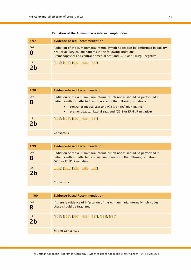
4.6 Adjuvant radiothepary of breastc ancer
© German Guideline Program in Oncology | Evidence-based Guideline Breast Cancer - V4.4 | May 2021
144
Radiation of the A. mammaria interna lymph nodes
4.97 Evidence-based Recommendation
GoR
0
Radiation of the A. mammaria internal lymph nodes can be performed in axillary
pN0 or axillary pN1mi patients in the following situation:
Premenopausal and central or medial seat and G2-3 and ER/PgR negative
LoE
2b
[595]; [596]; [597]; [686]; [687]
4.98 Evidence-based Recommendation
GoR
B
Radiation of the A. mammaria interna lymph nodes should be performed in
patients with 1-3 affected lymph nodes in the following situations:
• central or medial seat and (G2-3 or ER/PgR negative)
• premenopausal, lateral seat and (G2-3 or ER/PgR negative)
LoE
2b
[595]; [596]; [597]; [686]; [687]
Consensus
4.99 Evidence-based Recommendation
GoR
B
Radiation of the A. mammaria interna lymph nodes should be performed in
patients with > 3 affected axillary lymph nodes in the following situation:
G2-3 or ER/PgR negative
LoE
2b
[595]; [596]; [597]; [686]; [687]
Consensus
4.100 Evidence-based Recommendation
GoR
B
If there is evidence of infestation of the A. mammaria interna lymph nodes,
these should be irradiated.
LoE
2b
[595]; [596]; [597]; [686]; [687]; [688]; [689]
Strong Consensus

4.6 Adjuvant radiothepary of breastc ancer
© German Guideline Program in Oncology | Evidence-based Guideline Breast Cancer - V4.4 | May 2021
145
4.101 Evidence-based Recommendation
GoR
A
Radiation of the A. mammaria interna lymph nodes should be decided
individually and interdisciplinarily in case of increased cardiac risk or therapy
with trastuzumab.
LoE
4
[690]; [691]
Strong Consensus
Background 4.93 to 4.101
Adjuvant radiotherapy of supra- and infraclavicular and parasternal lymph nodes was
investigated in 3 large randomized studies [595], [597], [686] and a population-based
cohort study [596], [687] in more than 10,000 patients. In a meta-analysis of the 3
randomized studies, a consistent, statistically significant survival advantage for
radiation [687] was demonstrated, which is also supported by the Danish cohort study
[596]. The improvement in survival results from a reduced remote metastasis rate (HR
= 0.84). The absolute survival advantage is 2-3% on average of all included patients
after 10 years. In patients who received both chemotherapy and hormone therapy, the
survival advantage was significantly higher in the EORTC study [595]. In the Canadian
study [597], patients with hormone receptor negative tumors benefited the most from
lymph drainage radiation. In both studies the supra- and infraclavicular as well as
parasternal lymph nodes were irradiated either together or both regions were not
irradiated. In contrast, radiation of the breast/breast wall was performed in all patients.
In the French study [686] an advantage could only be shown for the subgroup of
patients who also received adjuvant chemotherapy. In the Danish cohort study, the
greatest benefit was found in the group of premenopausal patients and those with > 3
affected axillary lymph nodes. In the latter two studies, supra- and infraclavicular lymph
drainage was irradiated in all patients and only the additional effect of irradiating
parasternal lymph drainage was investigated. Although the patients included and the
subgroups analysed were different in the studies, the results of all studies indicate that
patients with an increased risk of distant metastasis have the greatest benefit from
lymph drainage radiation. As HER2 status is not available in any of the trials, no
conclusions can be drawn in this respect.
Fears that lymph drainage radiations more frequently lead to more late effects for the
patients due to the significantly more extensive radiation volumes have not been
confirmed. Although a significantly increased rate of low-grade radiogenic pneumonitis
(grade I-II) was reported [595], [597], but not for higher-grade pneumonitis. In the
Canadian study [597], lymphatic drainage radiation led to a 4% higher probability of
grade II and III lymphedema of the arm (p 10 years). Studies [595], [686] have not
observed an increased rate of cardiac late sequelae due to lymphatic drainage radiation.
Whether the radiation of the parasternal lymph nodes after more than 10 years still
induces a higher cardiac toxicity or whether more secondary tumors are induced by the
higher load on the lungs cannot be conclusively assessed at present.
In the EORTC [595] and the French study [686], patients without axillary lymph node
involvement could be treated with central or medial tumor site, and in the Canadian
study [597] with an increased risk of relapse regardless of the location (>/= 5 cm tumor,
>/= 2 cm tumor and
4.6 Adjuvant radiothepary of breastc ancer
© German Guideline Program in Oncology | Evidence-based Guideline Breast Cancer - V4.4 | May 2021
146
In the case of nodal-positive breast carcinomas, the advantage of additional lymph
drainage radiation is not generally greater than in nodal-negative patients, but due to
the higher number of cases, it is statistically more reliable. Especially in the Danish
study [596], patients with > 3 affected axillary lymph nodes benefited significantly from
lymph drainage radiation, whereas in 1-3 affected lymph nodes a clinically relevant
advantage of lymph drainage radiation results only in premenopausal patients and
higher grading (G2-3) [687]. The indications for radiation of the mammary internal
lymph nodes differ from those of supra/infraclavicular lymph drainage only in so far
that in case of proven lymph node infestation the mammary internal lymph nodes
should be irradiated independently of other risk factors, predominantly based on the
results of the Danish study [596]. The importance of radiation of only the mammaria
internal lymph nodes without additional radiation of the supra/infraclavicular lymph
nodes was not investigated in any study and therefore no recommendation could be
made in this respect.
In summary, the benefit of lymph drainage radiation in patients with an increased risk
of relapse clearly outweighs the risks. Since the currently available study results do not
allow a good differentiation between the benefits of radiation of the supra- and
infraclavicular lymph nodes and the benefits of radiation of the parasternal lymph
nodes, the indication for lymph drainage radiation was largely seen as a package in the
statements of the guideline. Isolated radiation of the supra- and infraclavicular lymph
nodes is only recommended for a smaller proportion of patients, especially those with
previous cardiac diseases and with additional adjuvant therapy with trastuzumab. If the
radiation of the parasternal lymph nodes exceeds the typical dose loads on the heart
and lungs [692], [693], which can be a problem especially with left-sided tumors, the
benefits and risks of radiation therapy must be weighed individually and a waiver of
parasternal radiation is a sensible option in these cases. In these cases a radiation with
held breath in deep inspiration can often reduce the dose to heart and lungs in such a
way that a low-risk radiation of the parasternal lymph nodes is possible after all [694].
Whether radiation of the supra- and infraclavicular lymph nodes alone without radiation
of the parasternal lymph nodes in addition to radiation of the breast/breast wall leads
to a survival advantage cannot be answered from randomized studies. However, in
more than 3 affected axillary lymph nodes, the relapse rate in the supraclavicular lymph
nodes is so high (approx. 17% [688]) that radiation of the supra- and infraclavicular
lymph nodes is indicated for this reason alone. In patients with 1-3 affected axillary
lymph nodes with additional risk factors, the supraclavicular relapse rate without
radiotherapy is 9.6% (G2 with 2 positive lymph nodes or G3 with 1 positive lymph node)
and 21% (G3 with 2-3 positive lymph nodes or G2 with 3 positive lymph nodes),
respectively, and is also so high that radiation of the supra- and infraclavicular lymph
nodes alone is also indicated [689].

4.6 Adjuvant radiothepary of breastc ancer
© German Guideline Program in Oncology | Evidence-based Guideline Breast Cancer - V4.4 | May 2021
147
Radiation of the axillary lymph nodes
4.102 Evidence-based Recommendation
GoR
A/0
An extended axillary radiation can be performed in patients with 1-2 affected
axillary sentinel lymph nodes, provided that no axillary dissection has been
performed or no further local axillary therapy has been agreed upon (analogous
to ACOSOG Z0011). The decision on the appropriate procedure shall be made
on an interdisciplinary basis.
LoE
2b
[406]; [695]; [696]; [697]
Strong Consensus
Background 4,102
All radiation techniques used for homogeneous radiation of the entire breast lead to
co-radiation of parts of the ipsilateral axillary lymph drainage area. The amount of this
portion and the doses achieved there differ depending on the volume of the breast to
be irradiated in Levels I and II and also depend on the individual positioning of the
patient during the radiation treatment. The total doses resulting in these axilla portions
are between 20 - 40 Gy for a standard 50 Gy CT of the entire breast and thus influence
the rate of axillary recurrence [698].
If radiation of supra- and infraclavicular lymph nodes is performed, according to the
current recommendations of ESTRO and RTOG the axillary lymph node level III is
included in the clinical target volume [699], [700] and the radiation technique is chosen
in such a way that there is no gap between the target volume for lymph drainage and
the target volume for the breast/breast wall. Because of the positioning uncertainties
in the shoulder region, a safety margin is added around the clinical target volumes to
generate the planning target volumes. This ensures that the medial portion of Level II
in particular is irradiated with the full dose. Consequently, in radiation therapy of the
supra- and infraclavicular lymph nodes and the entire breast/breast wall, only the
cranial portions of Level I and the lateral portions of Level II are not irradiated with the
full dose. Radiation of the axilla is usually understood by the radiooncologist as an
expansion of the target volume to include the lateral portions of level II and the cranial
portion of level I according to the expansion in the ESTRO consensus [700].
Radiation of the entire axilla, also called "extended axillary radiation" in the statements,
is only sensible if there is a high risk of infestation of these regions and no surgical
removal has been performed. In the AMAROS study [695], patients were randomized to
receive either axillary radiotherapy (50 Gy in 25 fractions in 5 weeks) or dissection of
the axillary lymph nodes prior to axillary sentinel lymph node biopsy in the case of
axillary lymph node involvement (n =1425). The majority of the included patients had
1-2 positive sentinel lymph nodes. The axillary relapse rate, DFS and overall survival
after 5 and 10 years did not differ statistically and clinically significantly in both arms.
The rate of arm edema of all degrees was significantly higher after axillary dissection
(23% after 5 years) than after axillary radiotherapy (11% after 5 years).
In the Z0011 study [406], after breast-conserving surgery with 1-2 positive sentinel
lymph nodes, either an axillary dissection was randomized to receive an axillary
dissection or no targeted axillary therapy was performed. According to the protocol,

4.6 Adjuvant radiothepary of breastc ancer
© German Guideline Program in Oncology | Evidence-based Guideline Breast Cancer - V4.4 | May 2021
148
radiation of the breast was planned for all patients. Overall survival and DFS in the 856
evaluable patients were not significantly different after 5 years (p=0.24/p=0.14).
According to this, a further axillary therapy can be dispensed with in 1-2 affected
sentinel lymph nodes. However, the study shows a number of weaknesses: The study
was closed prematurely before the planned number of patients was reached; 38% of
the patients had only micrometastases; and the radiation techniques were only
examined in a subgroup of 228 patients. Of these 228 patients, about 50% had a so-
called high tangent to the radiation of the breast and another about 17% had radiation
of the entire axilla, i.e. a considerable proportion of the patients had received partial
or complete radiation of the axilla [697]. Consequently, there remains an uncertainty
as to which patients with positive sentinel lymph nodes can be spared further axillary
therapy by dissection or radiation therapy without risk. If more than 2 sentinel lymph
nodes are affected and the axilla is not dissected, extended axillary radiation is clearly
recommended. In 1-2 affected lymph nodes, the indication must be discussed in the
interdisciplinary tumor board. If only micrometastases are present, further axillary
therapy is not necessary. Since caked lymph nodes or lymph nodes with massive
extracapsular tumor growth (ECE) were an exclusion criterion for treatment in the
Z0011 study, the further procedure should also be coordinated interdisciplinarily in
these cases. Although ECE is an independent negative prognostic factor for DFS and
overall survival [701], the axillary relapse rate after axillary dissection without
radiotherapy is low [696] and does not represent an indication for extended axillary
radiotherapy. In the AMAROS study, ECE was not an exclusion criterion, although the
results for patients with ECE were not reported separately. If there is no axillary
dissection [702], extended axillary radiotherapy may be considered in these cases.
Radiation therapy is to be considered if a residual tumor is detected in the axilla.
Dose and fractionation for radiation of the regional lymph drainage
4.103 Consensus-based Recommendation
EC Radiotherapy of lymphatic drainage should be performed in conventional
fractionation (1.8 Gy to 2.0 Gy 5 times a week, total dose approx. 50 Gy in
approx. 5-6 weeks) or can be performed in hypofractionation (total dose approx.
40 Gy in approx. 15-16 fractions in approx. 3 to 5 weeks).
Strong Consensus
Background 4,103
All studies on radiation of the lymphatic drainage system were carried out in
conventional fractionation with 5x2 Gy per week up to a total dose of 50 Gy. In the
studies on hypofractionated radiotherapy of the breast or breast wall, less than
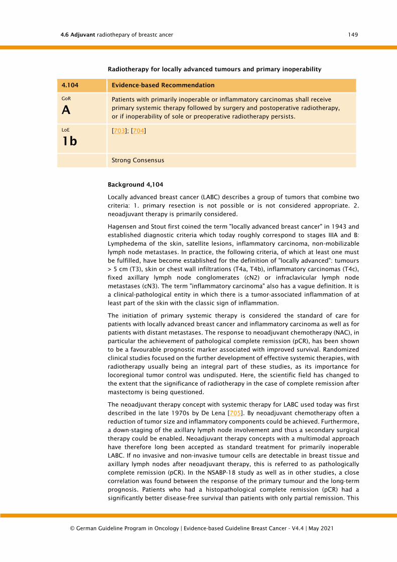
4.6 Adjuvant radiothepary of breastc ancer
© German Guideline Program in Oncology | Evidence-based Guideline Breast Cancer - V4.4 | May 2021
149
Radiotherapy for locally advanced tumours and primary inoperability
4.104 Evidence-based Recommendation
GoR
A
Patients with primarily inoperable or inflammatory carcinomas shall receive
primary systemic therapy followed by surgery and postoperative radiotherapy,
or if inoperability of sole or preoperative radiotherapy persists.
LoE
1b
[703]; [704]
Strong Consensus
Background 4,104
Locally advanced breast cancer (LABC) describes a group of tumors that combine two
criteria: 1. primary resection is not possible or is not considered appropriate. 2.
neoadjuvant therapy is primarily considered.
Hagensen and Stout first coined the term "locally advanced breast cancer" in 1943 and
established diagnostic criteria which today roughly correspond to stages IIIA and B:
Lymphedema of the skin, satellite lesions, inflammatory carcinoma, non-mobilizable
lymph node metastases. In practice, the following criteria, of which at least one must
be fulfilled, have become established for the definition of "locally advanced": tumours
> 5 cm (T3), skin or chest wall infiltrations (T4a, T4b), inflammatory carcinomas (T4c),
fixed axillary lymph node conglomerates (cN2) or infraclavicular lymph node
metastases (cN3). The term "inflammatory carcinoma" also has a vague definition. It is
a clinical-pathological entity in which there is a tumor-associated inflammation of at
least part of the skin with the classic sign of inflammation.
The initiation of primary systemic therapy is considered the standard of care for
patients with locally advanced breast cancer and inflammatory carcinoma as well as for
patients with distant metastases. The response to neoadjuvant chemotherapy (NAC), in
particular the achievement of pathological complete remission (pCR), has been shown
to be a favourable prognostic marker associated with improved survival. Randomized
clinical studies focused on the further development of effective systemic therapies, with
radiotherapy usually being an integral part of these studies, as its importance for
locoregional tumor control was undisputed. Here, the scientific field has changed to
the extent that the significance of radiotherapy in the case of complete remission after
mastectomy is being questioned.
The neoadjuvant therapy concept with systemic therapy for LABC used today was first
described in the late 1970s by De Lena [705]. By neoadjuvant chemotherapy often a
reduction of tumor size and inflammatory components could be achieved. Furthermore,
a down-staging of the axillary lymph node involvement and thus a secondary surgical
therapy could be enabled. Neoadjuvant therapy concepts with a multimodal approach
have therefore long been accepted as standard treatment for primarily inoperable
LABC. If no invasive and non-invasive tumour cells are detectable in breast tissue and
axillary lymph nodes after neoadjuvant therapy, this is referred to as pathologically
complete remission (pCR). In the NSABP-18 study as well as in other studies, a close
correlation was found between the response of the primary tumour and the long-term
prognosis. Patients who had a histopathological complete remission (pCR) had a
significantly better disease-free survival than patients with only partial remission. This
4.6 Adjuvant radiothepary of breastc ancer
© German Guideline Program in Oncology | Evidence-based Guideline Breast Cancer - V4.4 | May 2021
150
led to the fact that pCR serves as an early surrogate marker for survival and therefore
different neoadjuvant therapy regimes can be compared against each other.
Careful examination of the local tumor extent with lymph node determination as well
as good staging examinations are essential for the determination of the therapeutic
concept, since the pre-therapeutic disease extent influences the type of surgery and
the radiotherapy.
In 1971, EORTC's phase III study for the treatment of locally advanced and inflammatory
breast cancer was published. A total of 410 patients were randomized to receive either
radiotherapy alone, radiotherapy plus chemotherapy (CMF), radiotherapy combined
with tamoxifen, or radiotherapy combined with tamoxifen and CMF. The surgery was
not a planned part of the treatment sequence, but was reserved as a "salvage measure"
in case of local recurrence. With radiotherapy alone, a 10-year overall survival of 13%
and a distant metastasis-free survival of 15% was achieved, which was considered an
indication of curative potential. As expected, the addition of chemo- or hormone
therapy led to a significant increase in the time to local recurrence, to distant
metastasis or to an improvement in overall survival. With the combination treatment
the greatest therapeutic effect could be achieved. The 10-year overall survival could be
improved by 8-15% by the addition of the systemic therapy.
In the following years, the systemic therapy could be improved by the addition of
anthracyclines and taxanes within the framework of corresponding studies, with a
gradual improvement in 5-year overall survival for LABC of approx. 70% and 40% for
inflammatory breast carcinomas, respectively. Radiation after mastectomy (in case of
resectability) after neoadjuvant chemotherapy was performed in the majority of
patients. Here, too, it was shown that patients with pCR during mastectomy had
substantially better overall survival than patients with partial response or residual
tumour. This supported the use of the pCR rate as a surrogate marker of overall survival
for future treatment concepts. However, the subgroup analyses also showed that the
pCR rate was determined to a greater extent by the biology of the tumours than by the
chosen chemotherapy regimen.
Regardless of the response to the neoadjuvant therapy, the question arose to what
extent additional local treatments would improve the results. Randomized studies on
the necessity of surgery and radiotherapy in this situation are not available. Radiation
therapy is usually recommended for persistent resectability after NAC. Resectability
should then be re-examined approximately 6-12 weeks after completion of radiation.
In stage III patients with resectable tumors after neoadjuvant chemotherapy (NAC), the
effect of surgery and radiotherapy was investigated in three small randomized trials.
Patients undergoing neoadjuvant chemotherapy (in two studies with anthracyclines, in
one study with CMF) were randomized to undergo either mastectomy or radiotherapy.
Overall survival, disease-free survival and locoregional tumour control were comparable
between both local therapies. However, the local relapse rate after 2-5 years was in the
order of 40-60%, regardless of whether surgery or radiation was performed. Finally, an
indirect indication that patients should be operated and irradiated in order to reduce
the local recurrence rates. In none of these studies was the response rate after
neoadjuvant therapy documented. In this respect, the question remains open as to the
extent to which surgery and radiation therapy are required after complete remission
after neoadjuvant chemotherapy.
The challenge for radiotherapy lies in the decision which patients after NAC followed
by mastectomy will benefit from post-mastectomy radiotherapy (PMRT). Ultimately, this
decision is subject to numerous (additional) influences. At present, the current data
situation tends to favour an extension of the indication for PMRT to nodal positive

4.6 Adjuvant radiothepary of breastc ancer
© German Guideline Program in Oncology | Evidence-based Guideline Breast Cancer - V4.4 | May 2021
151
carcinomas with low lymph node involvement (1-3 positive lymph nodes). The addition
of different systemic therapies has increased the rate of patients with complete
remission after NAC. It could be shown that a ypN0 status after neoadjuvant
chemotherapy was associated with a low risk of locoregional recurrence, even without
the use of PMRT.
The role of PMRT was finally substantiated in the two large studies of the Danish Breast
Cooperative Group (DBCG) and the so-called British Columbia Trial. In the DBCG study
82 b and c [667], [668] as well as in the so-called British Columbia Clinical Trail [706],
more than 3500 women were randomized in the period 1979-1990, either in favor of
PMRT or only follow-up after surgery and systemic therapy. Systemic therapy in these
studies included either CMF or Tamoxifen. More than 90% of these patients in these
studies had positive lymph node metastases (pN+). These studies showed a substantial
decrease in the long-term locoregional recurrence rate, which was also reflected in
improved breast cancer-specific and overall survival. Based on these results, PMRT is
ultimately recommended without restriction for patients with ≥ 4 pathologically
affected lymph nodes or patients in stage III.
Two smaller studies investigated the significance of surgery after radiotherapy (after
neoadjuvant chemotherapy):
Merajver et al. [707] initially treated 90 patients in stage III with 9 cycles of an
anthracycline-containing neoadjuvant chemotherapy. After the last cycle the initial
tumor region was biopsied again. In case of complete remission patients received
radiotherapy of the thoracic wall and regional lymph node stations without surgery.
Patients with residual tumor as part of the biopsy received a mastectomy followed by
radiotherapy at the same dosage. Renouncing surgery in patients with pCR was not
associated with a higher local recurrence rate, which was about 20% after five years of
follow-up.
Ring et al [708] (n=136) and Daveau et al [709] (n=165) extracted from their prospective
data collections the treatment results of stage III patients with complete remission after
neoadjuvant chemotherapy who had received radiotherapy but no surgery. These were
compared to patients with pCR after chemotherapy, who had received a mastectomy
including postoperative radiation. The treatment results regarding overall survival,
disease-free survival and distant metastasis-free survival were almost identical. A trend
towards improved locoregional tumor control was observed in patients who received
both surgery and radiation. Even in the absence of evidence, surgery is ultimately
recommended after neoadjuvant systemic therapy regardless of response, if complete
resection seems possible.
The determination of the type of surgical procedure following neoadjuvant
chemotherapy (NAC) is complex and not standardised. There are no defined criteria
according to which breast-conserving therapy (BET) may/can be sought for patients
after NAC and for which a mastectomy offers more safety. Due to the high local
recurrence rate, mastectomy with axilla clearance is generally recommended for
primarily inoperable LABC and inflammatory tumours, even if good remission is
achieved after preoperative therapy. In the case of "large operable" tumours, the
primary goal is to operate in a breast-conserving manner. The pattern of tumour
reduction also influences the further surgical procedure. If there is concentric shrinkage
to a single tumour node, it may also be possible to remove it completely by BET.
There are two retrospective studies on the extent to which radiotherapy is required
after mastectomy, especially in cases of complete pathological remission:
4.6 Adjuvant radiothepary of breastc ancer
© German Guideline Program in Oncology | Evidence-based Guideline Breast Cancer - V4.4 | May 2021
152
Huang et al. [710] examined the clinical outcome of 670 breast cancer patients who
had received six different neoadjuvant chemotherapy regimens in clinical trials after
mastectomy and axilla dissection. Of these patients, 134 did not receive PMRT, while
542 patients received radiotherapy after mastectomy. The patients had been treated
between 1974 and 2000, with a median follow-up of 69 months. As expected, a
selection bias was found in the non-randomised comparison, with significantly higher
T and N stages among the patients who were irradiated: In the PMRT cohort 83% of the
women represented stage III (-IV) compared to only 50% in the cohort that did not
receive radiation. The pCR rate after neoadjuvant chemotherapy was 14% in the PMRT
arm versus 6% without radiation. Overall, the 10-year local recurrence rate was 11% for
women receiving PMRT versus 22% without radiation (p=0.0001) and overall survival
was also better with radiation therapy. In a subgroup of 46 patients in clinical stage III
(-IV) (35 received PMRT, 11 were not irradiated), who each achieved complete remission
after neoadjuvant chemotherapy, the 10-year local recurrence rate was 3% with
radiotherapy (PMRT) compared to 33% without PMRT (p=0.0006).
In the multivariate analysis regarding the factors associated with local recurrence, the
most significant factor was found to be the decision not to undergo postmastectomy
radiotherapy (PMRT) (HR 4.7; 95% CI: 2.7-8.1). Other factors were ≥ 20% pathological
lymph nodes involved according to NICE, clinical stage ≥ I IIB, no tamoxifen, and
hormone receptor negative status. Overall cancer-specific survival after 10 years was
comparable in both treatment groups: 58% versus 55%. However, the univariate analysis
showed an improvement in disease-specific survival for the subgroup of patients with
PMRT, including those with clinical stage ≥ IIIB, cN2-N3 status and stage ≥ IV
pathological lymph nodes.
In another publication McGuire et al. [674] identified 106 patients in stage II-III who
had been treated with neoadjuvant chemotherapy (92% anthracycline-based
chemotherapy, 38% with taxanes) and mastectomy between 1982 and 2002 and who
had pCR at the time of surgery. Of these, 34 patients received PMRT, compared to 72
patients who did not receive radiation. Overall, the 10-year local recurrence rate did
not differ between the PMRT and non-PMRT group (5% versus 10%, p=0.4), probably
most influenced by the 0% 10-year recurrence rate of the 32 patients in clinical stage I-
II. In contrast, PMRT led to a significant decrease in the 10-year local recurrence rate
for the 74 patients in stage III (7% versus 33%, p=0.04). Overall survival was also
significantly improved for stage III patients who received PMRT (77.3% vs. 33.3%,
p=0.002). Consequently, the authors concluded that PMRT should be performed in
stage III patients who had achieved complete pathologic remission after neoadjuvant
chemotherapy.
From this database, further specific subgroups were retrospectively analysed. Garg et
al. [711] analysed the effect of radiotherapy on 107 younger patients (younger than 35
years) in stage IIA-IIIC treated with neoadjuvant chemotherapy and mastectomy with
PMRT (n = 80) or without PMRT (n = 20) in the period 1975 to 2005. 84% of stage III
patients received PMRT compared to 42% who did not receive any. The pCR rate was
19% in the PMRT group versus 1% in the non-irradiated group. As documented in the
preliminary analysis (Huang et al.), PMRT significantly improved locoregional control
(88% versus 63%) and overall survival (67% versus 48%) in this subgroup of young
patients.
Nagar et al. [675] reported on 162 women in clinical stage T3 N0 treated from 1985-
2004 after neoadjuvant chemotherapy followed by mastectomy and PMRT (n =119) or
no PMRT (n = 43). The 5-year local recurrence rate was 9%, but was significantly higher
in patients who had not received PMRT (24% versus 4%, p
4.6 Adjuvant radiothepary of breastc ancer
© German Guideline Program in Oncology | Evidence-based Guideline Breast Cancer - V4.4 | May 2021
153
In summary, these study results show that women in clinically advanced stage III
(especially those with cN2-N3 involvement and with ypN+ lymph nodes that were still
affected at the time of surgery) are at the highest local recurrence risk after neoadjuvant
chemotherapy and mastectomy. The risk of local recurrence seems to be significantly
lower for patients in clinical T3N0 stage, especially with a ypN0 status at the time of
surgery.
Locoregional radiation in patients with synchronous metastasis
Locoregional radiation is currently being investigated in the oligometastasis concept
together with other tumor entities in various ongoing studies, without any reliable
results so far. Radiation therapy can certainly contribute to symptom relief, but the
influence on survival is inconsistent in the retrospective data. Some retrospective
analyses document improved survival for both surgery and radiotherapy, but in some
cases there is no influence from local therapy.
By Badwe et al. [712] the data of the randomized study from India were finally published
in 2015. Untreated patients with "de novo" metastasis were randomized to surgical
local therapy followed by radiation (n =173) vs. no local therapy (n =177) or chemo-
/hormone therapy alone [712]. Median overall survival was 19.2 months with local
therapy vs. 20.5 months without local therapy (p = 0.79). Therefore, local therapy is
not recommended as standard for initial metastasis. However, this negative result does
not exclude a possible benefit for subgroups. A second randomized study on the same
issue was initiated in Turkey. Results have so far only been published in abstract form
at congresses. For the endpoint overall survival no benefit for local therapy was seen
in this data collective: after 54 months 35% with local therapy vs. 31% without local
therapy. However, subgroup analyses (HR+, patients
Therapy sequence of adjuvant systemic therapy and radiotherapy
4.105 Evidence-based Recommendation
GoR
A
Postoperative chemotherapy and radiotherapy shall be sequential.
Note: the superiority of a specific sequence (first chemotherapy or first
radiotherapy) is not proven. In clinical practice, the sequence of chemotherapy
followed by radiotherapy has become established.
LoE
1b
[713]; [714]; [715]; [716]
Strong Consensus
4.106 Evidence-based Recommendation
GoR
B
In the case of single RT, this should be initiated postoperatively within an 8-
week period.
LoE
2b
[717]; [718]
Strong Consensus

4.6 Adjuvant radiothepary of breastc ancer
© German Guideline Program in Oncology | Evidence-based Guideline Breast Cancer - V4.4 | May 2021
154
4.107 Evidence-based Recommendation
GoR
0
Adjuvant endocrine therapy can be initiated independently of radiotherapy. (1a)
A therapy with trastuzumab can be continued during radiotherapy. In the case of
simultaneous A. mammaria lymph node radiation, the procedure should be
determined on an interdisciplinary basis. (4)
LoE
1a
4
[598]; [690]; [691]; [719]
Strong Consensus
Background 4.105 to 4.107
So far, there is no evidence that a specific sequence of radiotherapy and drug therapy
is clearly superior in the adjuvant situation. In principle, both drug therapy and
radiotherapy should be started as early as possible and not unnecessarily delayed
[677], [720], [721].
Antihormonal therapy can be carried out independently of radiotherapy (even
simultaneously); this has now been proven by randomized studies [719], [722], [723],
[724], [725], [726]. If only endocrine therapy (without chemotherapy) and radiotherapy
are indicated, radiotherapy should therefore normally be started within 4 to 6 weeks
after surgery; anti-hormonal therapy can be started before, during or even shortly after
radiotherapy. A delayed start of radiotherapy (time interval between surgery and the
start of radiotherapy 8 - 12 weeks) may not be disadvantageous; in the case of justified
delays (e.g. intercurrent diseases) a longer time interval can be accepted. However,
unnecessary delays should be avoided.
A sequential approach is recommended for the combination of chemotherapy and
radiotherapy (first chemotherapy, then radiotherapy or vice versa); randomized studies
and meta-analyses showed no advantage for a specific temporal sequence [713], [714],
[715], [716]. The sequence of the therapeutic procedures should take into account the
dominant individual risk of recurrence (systemic or locoregional) and be determined on
an interdisciplinary basis. In most cases chemotherapy is performed first and then
(usually within 2 to 4 weeks after the last course of chemotherapy) radiation therapy is
followed by [727]. This is particularly useful for patients with a high systemic risk of
relapse. In the case of very long adjuvant therapy regimens, this can result in a time
interval of about 6 months up to a maximum between surgery and the start of
radiotherapy. Fears that this long delay in radiotherapy (which is considered to be
clearly disadvantageous in the case of radiotherapy alone without chemotherapy) could
be associated with a reduced effect of radiotherapy and an increased locoregional
relapse rate have not been confirmed. However, if the planned chemotherapy cannot
be started postoperatively within the usual time frame for individual reasons,
radiotherapy should be brought forward if necessary. This should be discussed in
particular when using hypofractioned radiotherapy regimens.
A therapy with targeted drugs for HER2-positive tumours can be continued during
radiotherapy. All studies show no increased toxicity or increased rate of cardiac events,
not even with left-sided radiotherapy [728]. However, there is very little data concerning
the combination of trastuzumab and simultaneous radiotherapy of the left parasternal
4.7 Systemic adjuvant therapy (endocrine, chemo-, antibody therapy)
© German Guideline Program in Oncology | Evidence-based Guideline Breast Cancer - V4.4 | May 2021
155
lymph nodes; in these cases indication and therapy sequence should be determined
interdisciplinary.
4.7. Systemic adjuvant therapy (endocrine, chemo-,
antibody therapy)
4.7.1. Selection of adjuvant therapy and risk assessment
The recommendations for adjuvant therapy of breast cancer consider tumor size,
lymph node status, grading, hormone receptor status, HER2 status, menopause status
and age as the most important factors in deciding on the necessity and type of adjuvant
therapy [367], [529].
The St. Gallen recommendations of 2009 point to endocrine sensitivity and the
recommendations of 2011 to molecular subtypes as decisive criteria for the indication
of adjuvant chemotherapy [529]. The immunohistochemically determined markers ER,
PgR, HER2 and Ki-67 [529] are considered as surrogate parameters for the molecular
subtypes. ER- and/or PgR-positive, HER2-negative tumours with a low proliferation rate
are considered to be Luminal A, while Luminal B is these tumours with a high
proliferation rate. It must be taken into account that there is no validated threshold
value for Ki-67 (e.g. for the classification of Luminal A vs. B or for the decision
for/against adjuvant chemotherapy).
Indications for adjuvant chemotherapy are
• In HER2-positive tumors, simultaneous anti-HER2 therapy with trastuzumab for
1 year in combination with (neo)adjuvant chemotherapy is standard
• for endocrine non-sensitive tumours (ER- and PgR-negative)
• for endocrine sensitive tumours
• in the case of nodal-positive tumours (studies are currently evaluating whether
adjuvant chemotherapy can be dispensed with in patients with low nodal
infestation (1-3 affected LK) and favourable tumour biology (Luminal A))
• G 3
• young age of disease (
Chemotherapy is always indicated when the individual expected benefit is higher than
possible side effects and late damage. This requires a differentiated education of the
patients, especially if the expected benefit is only small.
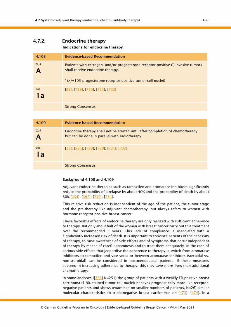
4.7 Systemic adjuvant therapy (endocrine, chemo-, antibody therapy)
© German Guideline Program in Oncology | Evidence-based Guideline Breast Cancer - V4.4 | May 2021
156
4.7.2. Endocrine therapy
Indications for endocrine therapy
4.108 Evidence-based Recommendation
GoR
A
Patients with estrogen- and/or progesterone receptor-positive (°) invasive tumors
shall receive endocrine therapy.
° (>/=10% progesterone receptor-positive tumor cell nuclei)
LoE
1a
[28]; [729]; [730]; [731]; [732]
Strong Consensus
4.109 Evidence-based Recommendation
GoR
A
Endocrine therapy shall not be started until after completion of chemotherapy,
but can be done in parallel with radiotherapy.
LoE
1a
[28]; [585]; [729]; [730]; [731]; [732]
Strong Consensus
Background 4.108 and 4.109
Adjuvant endocrine therapies such as tamoxifen and aromatase inhibitors significantly
reduce the probability of a relapse by about 40% and the probability of death by about
30% [234], [367], [730], [734].
This relative risk reduction is independent of the age of the patient, the tumor stage
and the pre-therapy like adjuvant chemotherapy, but always refers to women with
hormone receptor-positive breast cancer.
These favorable effects of endocrine therapy are only realized with sufficient adherence
to therapy. But only about half of the women with breast cancer carry out this treatment
over the recommended 5 years. This lack of compliance is associated with a
significantly increased risk of death. It is important to convince patients of the necessity
of therapy, to raise awareness of side effects and of symptoms that occur independent
of therapy by means of careful anamnesis and to treat them adequately. In the case of
serious side effects that jeopardise the adherence to therapy, a switch from aromatase
inhibitors to tamoxifen and vice versa or between aromatase inhibitors (steroidal vs.
non-steroidal) can be considered in postmenopausal patients. If these measures
succeed in increasing adherence to therapy, this may save more lives than additional
chemotherapy.
In some analyses ([733] N=251) the group of patients with a weakly ER-positive breast
carcinoma (1-9% stained tumor cell nuclei) behaves prognostically more like receptor-
negative patients and shows (examined on smaller numbers of patients, N=26) similar
molecular characteristics to triple-negative breast carcinomas on [473], [474]. In a

4.7 Systemic adjuvant therapy (endocrine, chemo-, antibody therapy)
© German Guideline Program in Oncology | Evidence-based Guideline Breast Cancer - V4.4 | May 2021
157
study of 314 patients with a low ER staining (1-9%), they showed a similar frequency of
BRCA-1 mutations as ER-negative patients [476].
Since this group of patients (1-9% positive ER) obviously behaves prognostically
differently than those with an ER-positivity of > 10% [735], an additional adjuvant or
neoadjuvant chemotherapy should be considered.
Carcinomas with a weak PR staining (<10%) und negativen ER-Färbung könnten
molekular dem triple-negativen Mammakarzinom entsprechen [736], [737]. Diese
Daten werden unterstützt durch die EBCTCG-Analyse von 2008 “Adjuvant
chemotherapy in oestrogen-receptor-poor breast cancer”, in der nachgewiesen wurde,
dass Tamoxifen nur einen geringen Effekt auf die Rezidivrate und das Überleben bei
Patientinnen mit einer schwachen Östrogenrezeptor-Expression hatte und einen nur
geringen zusätzlichen Effekt zur adjuvanten Chemotherapie.
Data from some studies (such as Tam-02, [738], [739]) indicate that a later start (up to
5 years after completion of local therapy and/or chemotherapy) with adjuvant
endocrine therapy is better than dispensing with this endocrine therapy altogether.
This later start of endocrine therapy also prolonged DFS and OS or DDFS. The MA.17
study [740], which also allowed a longer therapy-free interval between tamoxifen and
letrozole, showed similar results. These are indications that in case of side effects a
therapy break is preferable to a general discontinuation.
Endocrine therapy
4.110 Evidence-based Recommendation
GoR
A/B
After 5 years of tamoxifen, the indication for extended endocrine therapy shall
be evaluated for each patient with ER+ breast cancer.
Indications should be based on the weighing of the risk of relapse and the
therapy-associated side effects (toxicity, reduced adherence).
The current menopausal status of the patient shall be taken into account when
choosing endocrine therapy.
LoE
1a
[741]
Strong Consensus
Background 4,110
Endocrine adjuvant treatment of early breast cancer is one of the most effective
therapeutic options. Recent publications prove this for a time interval of up to 15 years.
A distinction was made between initial adjuvant therapy (IAT, years 0-5) and extended
adjuvant therapy (EAT: years 6-10).
The therapy-associated side effect rate of these continuous therapies reduces therapy
adherence and leads to a loss of effectiveness. Therefore, studies should be conducted
to determine whether intermittent adjuvant endocrine therapy could be as effective as
EAT.
Currently, there is a lack of reliable diagnostic tools to reliably predict the risk of late
metastasis (after year 5) for such an intervention. To this end, multi-gene assays, for
example, could be prospectively evaluated in studies.

4.7 Systemic adjuvant therapy (endocrine, chemo-, antibody therapy)
© German Guideline Program in Oncology | Evidence-based Guideline Breast Cancer - V4.4 | May 2021
158
After 5 years of tamoxifen, a further 5 years of adjuvant tamoxifen reduces the
recurrence rate (-2.8% in absolute terms in the ATLAS study) and prolongs overall
survival (-2.48% in absolute terms in the ATLAS study, [742], [743], [744]) in patients
with hormone receptor-positive breast carcinomas, regardless of menopausal status
(however, only 9% of patients in the ATLAS study were premenopausal). The incidence
of pulmonary embolism and endometrial carcinoma was significantly increased after
10 years of tamoxifen compared to 5 years of tamoxifen without effect on mortality.
Ischemic heart disease and myocardial infarction were significantly less common after
10 years than after 5 years of tamoxifen.
If the patients became postmenopausal after 5 years of adjuvant tamoxifen, the
subsequent administration of letrozole for 5 years adjuvant improved DFS and OS,
especially in women who were premenopausal before letrozole or had lymph node
metastases [740]. Approved for this EAT in Germany after 5 years of tamoxifen are
tamoxifen and letrozole.
Therapy for premenopausal patients
4.111 Evidence-based Recommendation
GoR
A
Premenopausal patients shall be treated with tamoxifen for at least 5 years.
Antiestrogenic therapy with tamoxifen 20 mg per day shall be carried out over a
period of 5 - 10 years or until relapse, depending on the risk of relapse.
The indication for extended therapy depends on the risk of recurrence and the
patient's wishes.
LoE
1a
[729]; [730]; [742]; [743]; [745]
Strong Consensus
Background 4,111
Extending the administration of tamoxifen from 5 to 10 years will reduce ipsi and
contralateral recurrence rates and prolong overall survival in the ATLAS study. However,
the rates of pulmonary embolism and endometrial carcinoma are increased without any
influence on mortality [742], [743], [744]. The risk-adapted duration of tamoxifen
administration (longer administration with increased risk of recurrence) is
recommended by ASCO [741].
Endocrine therapy
4.112 Consensus-based Recommendation
EC For patients with an ER+ breast cancer and increased risk, who are still
premenopausal after completion of chemotherapy, an aromatase inhibitor can
be used to eliminate ovarian function.
Consensus
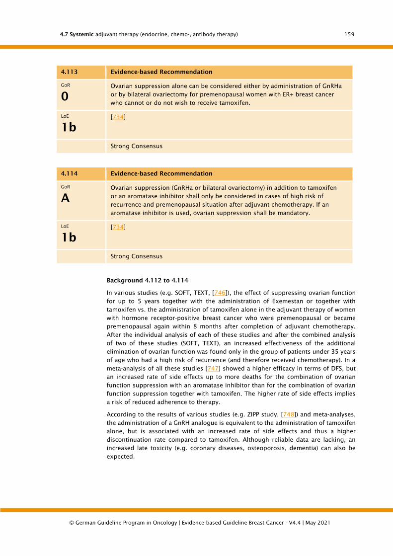
4.7 Systemic adjuvant therapy (endocrine, chemo-, antibody therapy)
© German Guideline Program in Oncology | Evidence-based Guideline Breast Cancer - V4.4 | May 2021
159
4.113 Evidence-based Recommendation
GoR
0
Ovarian suppression alone can be considered either by administration of GnRHa
or by bilateral ovariectomy for premenopausal women with ER+ breast cancer
who cannot or do not wish to receive tamoxifen.
LoE
1b
[734]
Strong Consensus
4.114 Evidence-based Recommendation
GoR
A
Ovarian suppression (GnRHa or bilateral ovariectomy) in addition to tamoxifen
or an aromatase inhibitor shall only be considered in cases of high risk of
recurrence and premenopausal situation after adjuvant chemotherapy. If an
aromatase inhibitor is used, ovarian suppression shall be mandatory.
LoE
1b
[734]
Strong Consensus
Background 4.112 to 4.114
In various studies (e.g. SOFT, TEXT, [746]), the effect of suppressing ovarian function
for up to 5 years together with the administration of Exemestan or together with
tamoxifen vs. the administration of tamoxifen alone in the adjuvant therapy of women
with hormone receptor-positive breast cancer who were premenopausal or became
premenopausal again within 8 months after completion of adjuvant chemotherapy.
After the individual analysis of each of these studies and after the combined analysis
of two of these studies (SOFT, TEXT), an increased effectiveness of the additional
elimination of ovarian function was found only in the group of patients under 35 years
of age who had a high risk of recurrence (and therefore received chemotherapy). In a
meta-analysis of all these studies [747] showed a higher efficacy in terms of DFS, but
an increased rate of side effects up to more deaths for the combination of ovarian
function suppression with an aromatase inhibitor than for the combination of ovarian
function suppression together with tamoxifen. The higher rate of side effects implies
a risk of reduced adherence to therapy.
According to the results of various studies (e.g. ZIPP study, [748]) and meta-analyses,
the administration of a GnRH analogue is equivalent to the administration of tamoxifen
alone, but is associated with an increased rate of side effects and thus a higher
discontinuation rate compared to tamoxifen. Although reliable data are lacking, an
increased late toxicity (e.g. coronary diseases, osteoporosis, dementia) can also be
expected.
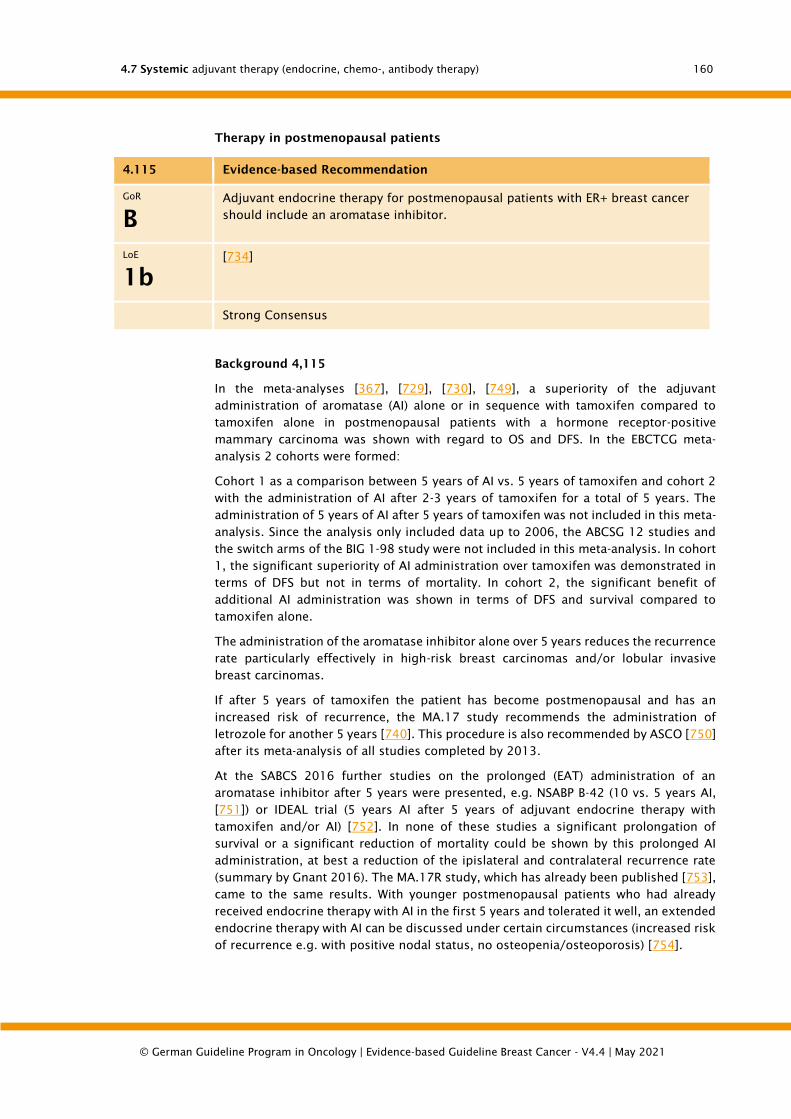
4.7 Systemic adjuvant therapy (endocrine, chemo-, antibody therapy)
© German Guideline Program in Oncology | Evidence-based Guideline Breast Cancer - V4.4 | May 2021
160
Therapy in postmenopausal patients
4.115 Evidence-based Recommendation
GoR
B
Adjuvant endocrine therapy for postmenopausal patients with ER+ breast cancer
should include an aromatase inhibitor.
LoE
1b
[734]
Strong Consensus
Background 4,115
In the meta-analyses [367], [729], [730], [749], a superiority of the adjuvant
administration of aromatase (AI) alone or in sequence with tamoxifen compared to
tamoxifen alone in postmenopausal patients with a hormone receptor-positive
mammary carcinoma was shown with regard to OS and DFS. In the EBCTCG meta-
analysis 2 cohorts were formed:
Cohort 1 as a comparison between 5 years of AI vs. 5 years of tamoxifen and cohort 2
with the administration of AI after 2-3 years of tamoxifen for a total of 5 years. The
administration of 5 years of AI after 5 years of tamoxifen was not included in this meta-
analysis. Since the analysis only included data up to 2006, the ABCSG 12 studies and
the switch arms of the BIG 1-98 study were not included in this meta-analysis. In cohort
1, the significant superiority of AI administration over tamoxifen was demonstrated in
terms of DFS but not in terms of mortality. In cohort 2, the significant benefit of
additional AI administration was shown in terms of DFS and survival compared to
tamoxifen alone.
The administration of the aromatase inhibitor alone over 5 years reduces the recurrence
rate particularly effectively in high-risk breast carcinomas and/or lobular invasive
breast carcinomas.
If after 5 years of tamoxifen the patient has become postmenopausal and has an
increased risk of recurrence, the MA.17 study recommends the administration of
letrozole for another 5 years [740]. This procedure is also recommended by ASCO [750]
after its meta-analysis of all studies completed by 2013.
At the SABCS 2016 further studies on the prolonged (EAT) administration of an
aromatase inhibitor after 5 years were presented, e.g. NSABP B-42 (10 vs. 5 years AI,
[751]) or IDEAL trial (5 years AI after 5 years of adjuvant endocrine therapy with
tamoxifen and/or AI) [752]. In none of these studies a significant prolongation of
survival or a significant reduction of mortality could be shown by this prolonged AI
administration, at best a reduction of the ipislateral and contralateral recurrence rate
(summary by Gnant 2016). The MA.17R study, which has already been published [753],
came to the same results. With younger postmenopausal patients who had already
received endocrine therapy with AI in the first 5 years and tolerated it well, an extended
endocrine therapy with AI can be discussed under certain circumstances (increased risk
of recurrence e.g. with positive nodal status, no osteopenia/osteoporosis) [754].

4.7 Systemic adjuvant therapy (endocrine, chemo-, antibody therapy)
© German Guideline Program in Oncology | Evidence-based Guideline Breast Cancer - V4.4 | May 2021
161
4.7.3. Adjuvant chemotherapy
4.116 Evidence-based Recommendation
GoR
B
An indication for adjuvant chemotherapy should be provided at:
• HER2-positive tumours (from pT1b, N0; pT1a, N0 if further risk: G3,
ER/PR neg., Ki-67 high)
• Triple-negative tumors (ER- and PgR-negative, HER2-negative)
• Luminal B tumors with high risk of recurrence (Ki-67 high, G 3, high
risk multigene assay, young age, lymph node involvement)
LoE
1a
[185]; [367]; [755]; [756]; [757]; [758]
Strong Consensus
4.117 Evidence-based Recommendation
GoR
A
Chemotherapy shall be administered in the recommended dosages.
If the cycles are underdosed or reduced, there is a risk of loss of effectiveness.
LoE
1a
[757]; [759]; [760]; [761]; [762]; [763]
Strong Consensus
Administration of the cytostatic drugs
4.118 Evidence-based Recommendation
GoR
B
Cytostatic drugs can be administered simultaneously or sequentially (according
to evidence-based protocols).
In cases of high tumour-related mortality risk and suitable patients, dose-
controlled therapies should be used.
LoE
1b
[764]; [765]; [766]; [767]; [768]; [769]
Strong Consensus
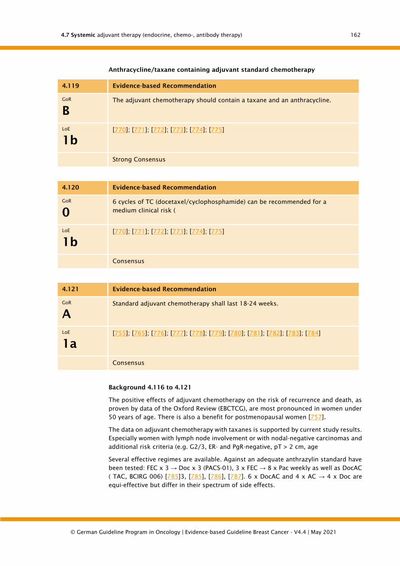
4.7 Systemic adjuvant therapy (endocrine, chemo-, antibody therapy)
© German Guideline Program in Oncology | Evidence-based Guideline Breast Cancer - V4.4 | May 2021
162
Anthracycline/taxane containing adjuvant standard chemotherapy
4.119 Evidence-based Recommendation
GoR
B
The adjuvant chemotherapy should contain a taxane and an anthracycline.
LoE
1b
[770]; [771]; [772]; [773]; [774]; [775]
Strong Consensus
4.120 Evidence-based Recommendation
GoR
0
6 cycles of TC (docetaxel/cyclophosphamide) can be recommended for a
medium clinical risk (
LoE
1b
[770]; [771]; [772]; [773]; [774]; [775]
Consensus
4.121 Evidence-based Recommendation
GoR
A
Standard adjuvant chemotherapy shall last 18-24 weeks.
LoE
1a
[755]; [765]; [776]; [777]; [778]; [779]; [780]; [781]; [782]; [783]; [784]
Consensus
Background 4.116 to 4.121
The positive effects of adjuvant chemotherapy on the risk of recurrence and death, as
proven by data of the Oxford Review (EBCTCG), are most pronounced in women under
50 years of age. There is also a benefit for postmenopausal women [757].
The data on adjuvant chemotherapy with taxanes is supported by current study results.
Especially women with lymph node involvement or with nodal-negative carcinomas and
additional risk criteria (e.g. G2/3, ER- and PgR-negative, pT > 2 cm, age
Several effective regimes are available. Against an adequate anthrazylin standard have
been tested: FEC x 3 → Doc x 3 (PACS-01), 3 x FEC → 8 x Pac weekly as well as DocAC
( TAC, BCIRG 006) [785]3, [785], [786], [787]. 6 x DocAC and 4 x AC → 4 x Doc are
equi-effective but differ in their spectrum of side effects.
4.7 Systemic adjuvant therapy (endocrine, chemo-, antibody therapy)
© German Guideline Program in Oncology | Evidence-based Guideline Breast Cancer - V4.4 | May 2021
163
In the sequence after 4 x AC, 4 x docetaxel every 3 weeks (100 mg/m2 ) and 12 x
paclitaxel weekly (80 mg/m2 ) are to be considered equivalent to [756], [788]. 4 cycles
of AC - 4 x paclitaxel q21 ( Henderson-like ) are inferior to 6 x CEF (MA-21 [789]).
The dual combination 4 x DocC (TC) is superior to the old standard 4 x AC with regard
to DFS and OS and avoids anthracycline-associated toxicities [756]0, [790].
In general, the administration of a longer adjuvant chemotherapy seems to be
advantageous, as the comparison of 4 cycles vs. 8 cycles has shown [763], [779].
Several studies show that 6 cycles of TC are as effective as an anthracycline-taxane
sequence therapy for certain patient groups. Whether these current efficacy data for 6
cycles of TC also apply to 4 cycles of TC cannot be decided at present on the basis of
the available data. A corresponding reduction in the number of cycles should therefore
only be made in the case of intolerable toxicities.
The Danish DBCG-07-READ study in TOPO2A-normal early breast cancer showed no
difference in DFS and OS for 6x TC vs. 3x EC - 3x DOC [783]. The WSG-PlanB study also
showed no difference between 6x TC and 4x EC - 4x docetaxel for HER2-negative early
breast cancer [784]. The pooled analysis of 3 US studies (ABC Trials) could not formally
confirm the non-inferiority between 6x TC and an anthracycline-taxane-containing
sequence therapy (iDFS HR 1.202; 95% KI 0.97-1.49 with a predefined threshold value
of 1.18). However, the absolute difference between the two arms was small (difference
4-year iDFS 2.5%) and subgroup analyses showed that the greatest benefit in favour of
anthracycline-taxane-containing sequence therapy was found in [782] when the clinical
risk was high (e.g. > 3 affected lymph nodes).
The significance of new substances in adjuvant therapy (e.g. gemcitabine, capecitabine)
as the fourth substance in addition to anthracyclines, taxanes and cyclophosphamide
cannot yet be conclusively assessed. While the addition of Gemcitabine is not
associated with an advantage [791], some data for Capecitabine indicate a trend
towards a further improvement of DFS or OS [792], [793]. However, this improvement
was associated with increased toxicity [767]0, [793]. In the FinXX study, the
improvement of DFS and OS was no longer significant after 5 years in the overall
collective, only in risk patients (triple-negative, > 3 LK), Cave: Standard arm [794].
In recent studies, a higher efficacy of the dose-dense (q2w) [765], [769] or the dose-
intensified dose-dense chemotherapy [768], [795] could be shown compared to
conventional chemotherapy (q3w). Especially for patients at high risk (≥ 4 affected LK),
dose-intensified dose-dense chemotherapy (ETC) is a standard regime. Patients with a
low or moderate risk of recurrence, however, do not benefit from dose-intensified
therapy compared to standard chemotherapy [796], [797].
Myeloablative high-dose chemotherapies currently have no place in unselected high-
risk collectives: Compared to conventional chemotherapy they show a better event-free
survival, but overall survival remains unaffected [798], [776]0. Therapy-associated
mortality and side effects are significantly increased [799], [800].
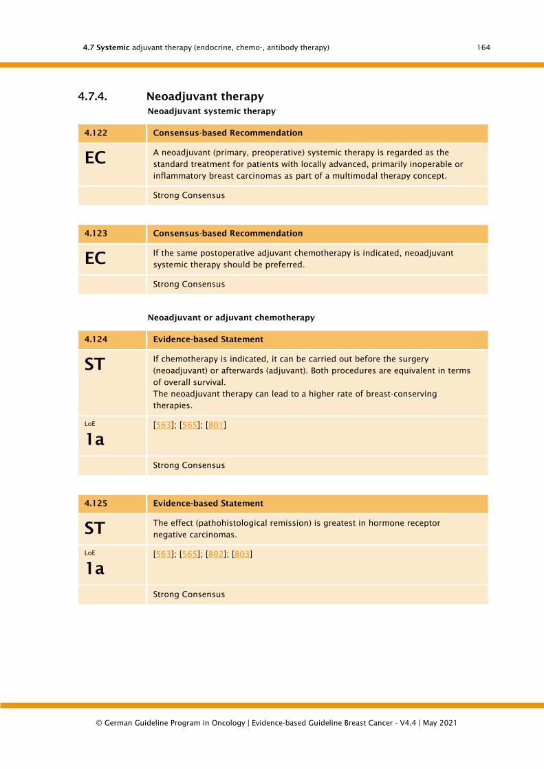
4.7 Systemic adjuvant therapy (endocrine, chemo-, antibody therapy)
© German Guideline Program in Oncology | Evidence-based Guideline Breast Cancer - V4.4 | May 2021
164
4.7.4. Neoadjuvant therapy
Neoadjuvant systemic therapy
4.122 Consensus-based Recommendation
EC A neoadjuvant (primary, preoperative) systemic therapy is regarded as the
standard treatment for patients with locally advanced, primarily inoperable or
inflammatory breast carcinomas as part of a multimodal therapy concept.
Strong Consensus
4.123 Consensus-based Recommendation
EC If the same postoperative adjuvant chemotherapy is indicated, neoadjuvant
systemic therapy should be preferred.
Strong Consensus
Neoadjuvant or adjuvant chemotherapy
4.124 Evidence-based Statement
ST If chemotherapy is indicated, it can be carried out before the surgery
(neoadjuvant) or afterwards (adjuvant). Both procedures are equivalent in terms
of overall survival.
The neoadjuvant therapy can lead to a higher rate of breast-conserving
therapies.
LoE
1a
[563]; [565]; [801]
Strong Consensus
4.125 Evidence-based Statement
ST The effect (pathohistological remission) is greatest in hormone receptor
negative carcinomas.
LoE
1a
[563]; [565]; [802]; [803]
Strong Consensus
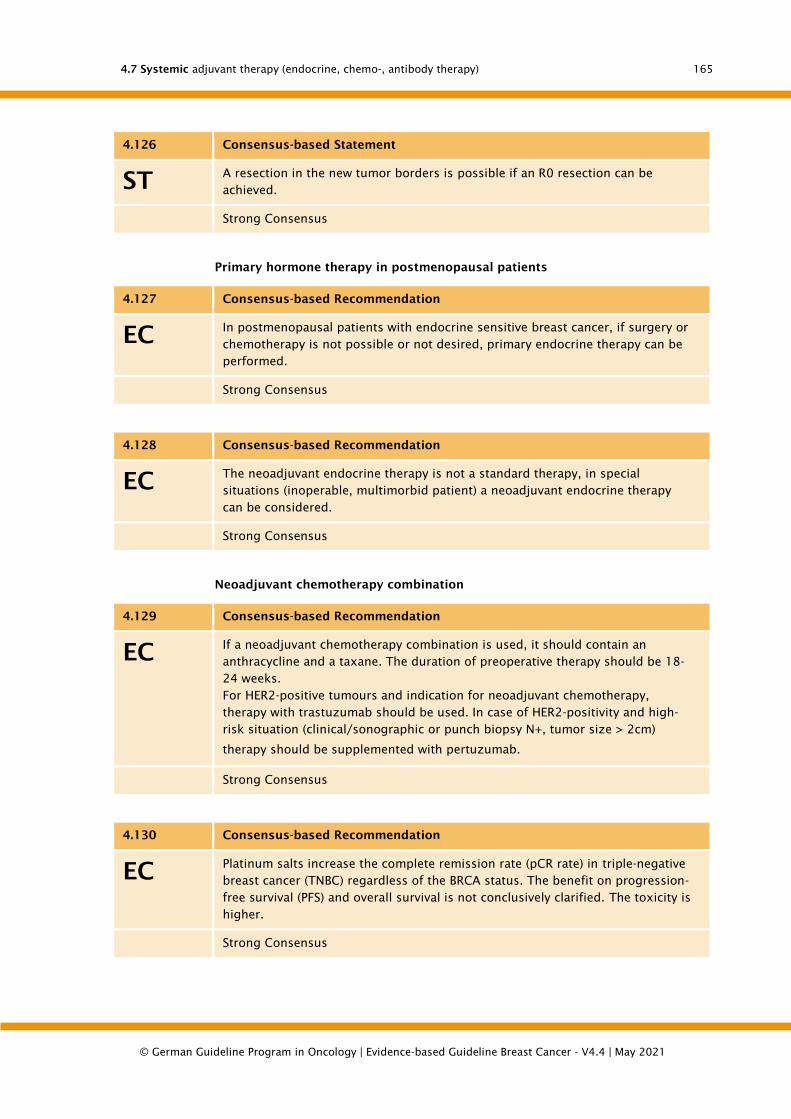
4.7 Systemic adjuvant therapy (endocrine, chemo-, antibody therapy)
© German Guideline Program in Oncology | Evidence-based Guideline Breast Cancer - V4.4 | May 2021
165
4.126 Consensus-based Statement
ST A resection in the new tumor borders is possible if an R0 resection can be
achieved.
Strong Consensus
Primary hormone therapy in postmenopausal patients
4.127 Consensus-based Recommendation
EC In postmenopausal patients with endocrine sensitive breast cancer, if surgery or
chemotherapy is not possible or not desired, primary endocrine therapy can be
performed.
Strong Consensus
4.128 Consensus-based Recommendation
EC The neoadjuvant endocrine therapy is not a standard therapy, in special
situations (inoperable, multimorbid patient) a neoadjuvant endocrine therapy
can be considered.
Strong Consensus
Neoadjuvant chemotherapy combination
4.129 Consensus-based Recommendation
EC If a neoadjuvant chemotherapy combination is used, it should contain an
anthracycline and a taxane. The duration of preoperative therapy should be 18-
24 weeks.
For HER2-positive tumours and indication for neoadjuvant chemotherapy,
therapy with trastuzumab should be used. In case of HER2-positivity and high-
risk situation (clinical/sonographic or punch biopsy N+, tumor size > 2cm)
therapy should be supplemented with pertuzumab.
Strong Consensus
4.130 Consensus-based Recommendation
EC Platinum salts increase the complete remission rate (pCR rate) in triple-negative
breast cancer (TNBC) regardless of the BRCA status. The benefit on progression-
free survival (PFS) and overall survival is not conclusively clarified. The toxicity is
higher.
Strong Consensus
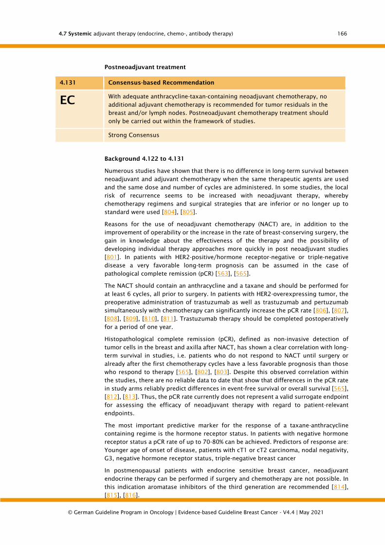
4.7 Systemic adjuvant therapy (endocrine, chemo-, antibody therapy)
© German Guideline Program in Oncology | Evidence-based Guideline Breast Cancer - V4.4 | May 2021
166
Postneoadjuvant treatment
4.131 Consensus-based Recommendation
EC With adequate anthracycline-taxan-containing neoadjuvant chemotherapy, no
additional adjuvant chemotherapy is recommended for tumor residuals in the
breast and/or lymph nodes. Postneoadjuvant chemotherapy treatment should
only be carried out within the framework of studies.
Strong Consensus
Background 4.122 to 4.131
Numerous studies have shown that there is no difference in long-term survival between
neoadjuvant and adjuvant chemotherapy when the same therapeutic agents are used
and the same dose and number of cycles are administered. In some studies, the local
risk of recurrence seems to be increased with neoadjuvant therapy, whereby
chemotherapy regimens and surgical strategies that are inferior or no longer up to
standard were used [804], [805].
Reasons for the use of neoadjuvant chemotherapy (NACT) are, in addition to the
improvement of operability or the increase in the rate of breast-conserving surgery, the
gain in knowledge about the effectiveness of the therapy and the possibility of
developing individual therapy approaches more quickly in post neoadjuvant studies
[801]. In patients with HER2-positive/hormone receptor-negative or triple-negative
disease a very favorable long-term prognosis can be assumed in the case of
pathological complete remission (pCR) [563], [565].
The NACT should contain an anthracycline and a taxane and should be performed for
at least 6 cycles, all prior to surgery. In patients with HER2-overexpressing tumor, the
preoperative administration of trastuzumab as well as trastuzumab and pertuzumab
simultaneously with chemotherapy can significantly increase the pCR rate [806], [807],
[808], [809], [810], [811]. Trastuzumab therapy should be completed postoperatively
for a period of one year.
Histopathological complete remission (pCR), defined as non-invasive detection of
tumor cells in the breast and axilla after NACT, has shown a clear correlation with long-
term survival in studies, i.e. patients who do not respond to NACT until surgery or
already after the first chemotherapy cycles have a less favorable prognosis than those
who respond to therapy [565], [802], [803]. Despite this observed correlation within
the studies, there are no reliable data to date that show that differences in the pCR rate
in study arms reliably predict differences in event-free survival or overall survival [565],
[812], [813]. Thus, the pCR rate currently does not represent a valid surrogate endpoint
for assessing the efficacy of neoadjuvant therapy with regard to patient-relevant
endpoints.
The most important predictive marker for the response of a taxane-anthracycline
containing regime is the hormone receptor status. In patients with negative hormone
receptor status a pCR rate of up to 70-80% can be achieved. Predictors of response are:
Younger age of onset of disease, patients with cT1 or cT2 carcinoma, nodal negativity,
G3, negative hormone receptor status, triple-negative breast cancer
In postmenopausal patients with endocrine sensitive breast cancer, neoadjuvant
endocrine therapy can be performed if surgery and chemotherapy are not possible. In
this indication aromatase inhibitors of the third generation are recommended [814],
[815], [816].
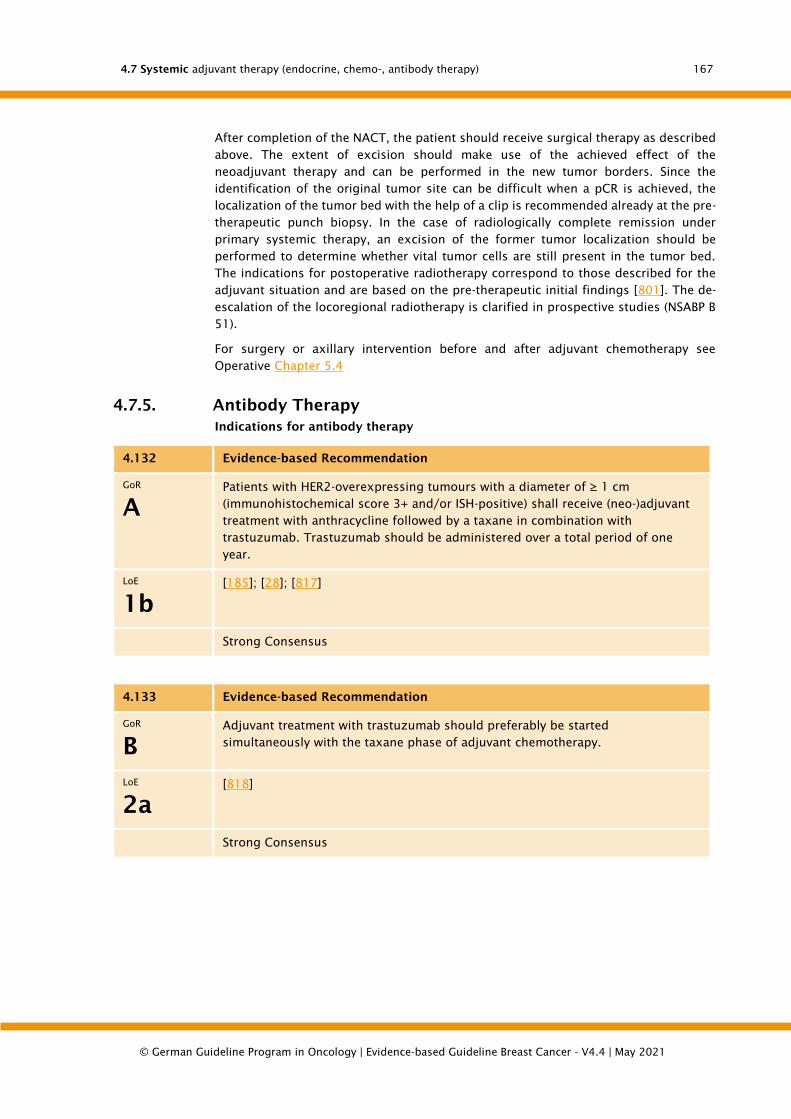
4.7 Systemic adjuvant therapy (endocrine, chemo-, antibody therapy)
© German Guideline Program in Oncology | Evidence-based Guideline Breast Cancer - V4.4 | May 2021
167
After completion of the NACT, the patient should receive surgical therapy as described
above. The extent of excision should make use of the achieved effect of the
neoadjuvant therapy and can be performed in the new tumor borders. Since the
identification of the original tumor site can be difficult when a pCR is achieved, the
localization of the tumor bed with the help of a clip is recommended already at the pre-
therapeutic punch biopsy. In the case of radiologically complete remission under
primary systemic therapy, an excision of the former tumor localization should be
performed to determine whether vital tumor cells are still present in the tumor bed.
The indications for postoperative radiotherapy correspond to those described for the
adjuvant situation and are based on the pre-therapeutic initial findings [801]. The de-
escalation of the locoregional radiotherapy is clarified in prospective studies (NSABP B
51).
For surgery or axillary intervention before and after adjuvant chemotherapy see
Operative Chapter 5.4
4.7.5. Antibody Therapy
Indications for antibody therapy
4.132 Evidence-based Recommendation
GoR
A
Patients with HER2-overexpressing tumours with a diameter of ≥ 1 cm
(immunohistochemical score 3+ and/or ISH-positive) shall receive (neo-)adjuvant
treatment with anthracycline followed by a taxane in combination with
trastuzumab. Trastuzumab should be administered over a total period of one
year.
LoE
1b
[185]; [28]; [817]
Strong Consensus
4.133 Evidence-based Recommendation
GoR
B
Adjuvant treatment with trastuzumab should preferably be started
simultaneously with the taxane phase of adjuvant chemotherapy.
LoE
2a
[818]
Strong Consensus
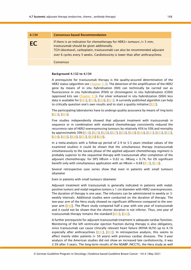
4.7 Systemic adjuvant therapy (endocrine, chemo-, antibody therapy)
© German Guideline Program in Oncology | Evidence-based Guideline Breast Cancer - V4.4 | May 2021
168
4.134 Consensus-based Recommendation
EC If there is an indication for chemotherapy for HER2+ tumours /= 5 mm,
trastuzumab should be given additionally.
TCH (docetaxel, carboplatin, trastuzumab) can also be recommended adjuvant
over 6 cycles every 3 weeks. Cardiotoxicity is lower than after anthracyclines.
Consensus
Background 4.132 to 4.134
A prerequisite for trastuzumab therapy is the quality-assured determination of the
HER2 status (algorithm see Chapter 5.5). The detection of the amplification of the HER2
gene by means of in situ hybridisation (ISH) can technically be carried out as
fluorescence in situ hybridisation (FISH) or chromogenic in situ hybridisation (CISH)
(approved kits see Chapter 5.5). For silver enhanced in situ hybridization (SISH) less
data is available for [481], [819], [820], [821]. A currently published algorithm can help
to critically question one's own results and to start a quality initiative [822].
The participating laboratories have to undergo quality assurance by means of ring tests
[823], [824].
Five studies independently showed that adjuvant treatment with trastuzumab in
sequence or in combination with standard chemotherapy consistently reduced the
recurrence rate of HER2-overexpressing tumours by relatively 45% to 50% and mortality
by approximately 30% [818], [825], [826], [827], [828], [829], [830], [831], [832], [833],
[834], [835], [836], [837], [838] [839].
In a meta-analysis with a follow-up period of 2.9 to 5.5 years (median values of the
examined studies) it could be shown that the simultaneous therapy (trastuzumab
simultaneously to the taxane phase of the applied adjuvant chemotherapy regimen) is
probably superior to the sequential therapy with trastuzumab after completion of the
adjuvant chemotherapy: for DFS HRsim = 0.62 vs. HRseq = 0.74, for OS significant
benefit only with simultaneous application with an HRsim = 0.68 [817], [818].
Several retrospective case series show that even in patients with small tumours
(diameter
Even in patients with small tumours (diameter
Adjuvant treatment with trastuzumab is generally indicated in patients with nodal-
positive tumors and nodal-negative tumors ≥ 1 cm diameter with HER2 overexpression.
The duration of therapy is one year. The infusions can be administered in weekly or 3-
weekly intervals. Additional studies were conducted on the duration of therapy. The
two-year arm of the Hera study showed no significant difference compared to the one-
year arm [840]. The Phare study compared half a year with one year of trastuzumab
and it could not be shown that the shorter duration is not inferior. Thus, one year of
trastuzumab therapy remains the standard [841], [842].
A further prerequisite for adjuvant trastuzumab treatment is adequate cardiac function.
Monitoring of the left ventricular ejection fraction during therapy is also obligatory,
since trastuzumab can cause clinically relevant heart failure (NYHA III/IV) up to 4.1%
especially after anthracyclines [831], [843]. In retrospective analysis, this seems to
affect mainly older patients (> 50 years) with previous cardiac diseases. The 3-year
analysis of the American studies did not show an increased late cardiotoxicity, it was
2.5% after 3 years. The long-term results of the NSABP /NCCTG, the Hera study as well
4.7 Systemic adjuvant therapy (endocrine, chemo-, antibody therapy)
© German Guideline Program in Oncology | Evidence-based Guideline Breast Cancer - V4.4 | May 2021
169
as the BCIRG study showed that after 18 months no additional trastuzumab-related
cardiotoxicity occurred [837], [838], [839], [842].
In the Cochrane analysis by Moja et al (2012), the absolute benefits and risks are
summarised as follows: Mortality is relatively reduced by 30% with trastuzumab and
the cardiac risk is five times higher compared to chemotherapy alone. If 1,000 patients
are treated with chemotherapy without trastuzumab, 900 survive and 5 have
cardiotoxicity.
If 1,000 patients were treated with chemotherapy and trastuzumab, 933 would survive
(i.e. 33 more than without trastuzumab), 740 would survive without disease relapse
(95 more than in the arm without trastuzumab) and 26 would have cardiotoxic side
effects (i.e. 21 more than without trastuzumab). This Cochrane publication is based on
the 2010 analysis, but long-term data are now available from these trials. No additional
cardiotoxicities occurred.
4.7.6. Bone-directed therapy
In breast cancer, interactions between the bone and its metabolism on the one hand
and the tumor cells on the other hand can be observed. These effects can be achieved
directly by the breast carcinoma cells or indirectly via systemic therapeutics.
Bone targeted treatment plays an important role in breast cancer in several respects:
• Therapy and prevention of cancer treatment induced bone density and
structure loss (cancer treatment induced bone loss)
• Adjuvant therapy of primary breast carcinoma to improve bone metastasis-free
and overall survival
• Preventive therapy of skeletal related events (SRE) in osseous metastasized
breast cancer
Bone modifying agents used in breast cancer are bisphosphonates and the antibody
Denosumab, which is directed against the ligand of the Receptor Activator of Nuclear
Factor-Kappa B (RANK ligand).
4.7.6.1. Therapy and prevention of cancer treatment induced bone loss
In malignant diseases the risk of loss of bone density, destruction of bone structure
and thus therapy-associated osteoporosis with a consecutively increased risk of
fracture is significantly increased [844]. In addition to the frequently observed
immobilization and lifestyle changes (e.g. discontinuation of estrogen therapy), drug
therapies are the most important factors for the osseous changes. Supportive therapies
(e.g. cortisone preparations) can damage the bone as well as cytotoxic or endocrine
medications. In particular, the high cure rates of numerous solid tumours and
especially breast carcinoma are causing the problem to become increasingly
prominent.
In premenopausal women with a hormone receptor-positive breast cancer a
suppression of ovarian function (ovarian function suppression, e.g. by GnRH
analogues) alone as well as in combination with tamoxifen or an aromatase inhibitor
and therapy with tamoxifen alone lead to a loss of bone density and to an increased
incidence of osteoporosis compared to a healthy control collective [845], [846], [847].
The combination of ovarian suppression combined with an aromatase inhibitor leads
to the greatest decrease in bone density [845].

4.7 Systemic adjuvant therapy (endocrine, chemo-, antibody therapy)
© German Guideline Program in Oncology | Evidence-based Guideline Breast Cancer - V4.4 | May 2021
170
In postmenopausal women, therapy with aromatase inhibitors also leads to a loss of
bone density and an increased incidence of fractures compared to women treated with
tamoxifen [848], [849], [850], [851].
Chemotherapy can also lead to a relevant loss of bone density [852], [853].
An indication for preventive treatment should be based on gender, age and bone
density, taking into account family history and lifestyle. Primary prevention of cancer-
therapy induced bone loss should be considered in particular if there is a special risk
constellation [854], [855]. This includes, among other things: advanced age, low body
mass index, nicotine abuse, aromatase inhibitor therapy, family disposition, long-term
cortisone therapy, immobility, endocrine diseases, medication intake (Dachverband der
deutschsprachigen wissenschaftlichen Osteologischen Gesellschaft e.V.,
http://www.dv-osteologie.org; [856].
Bone-directed therapy
4.135 Consensus-based Recommendation
EC Patients with an increased risk of bone loss due to medical history or cancer
therapy should have a bone density measurement performed at the beginning of
therapy.
Depending on the result and other risk factors, the bone density measurement
should be repeated at regular intervals.
Strong Consensus
4.136 Consensus-based Recommendation
EC To avoid cancer-therapy induced osteoporosis, preventive treatment should be
considered depending on the individual risk constellation for the development
of osteoporosis (http://www.dv-osteologie.org; ESMO bone health guidance).
Consensus
4.137 Evidence-based Recommendation
GoR
B
Bone protective therapy should be considered in premenopausal patients with
GnRH and/or tamoxifen and in postmenopausal patients on aromatase inhibitor
therapy.
LoE
1b
[845]; [848]; [850]; [856]
Strong Consensus
Background 4,135 to 4,137
Preventive treatment of cancer-associated bone loss differs little from the treatment of
non-cancer-associated osteoporosis. The following general recommendations can be
given to affected patients:
• Avoidance of underweight

4.7 Systemic adjuvant therapy (endocrine, chemo-, antibody therapy)
© German Guideline Program in Oncology | Evidence-based Guideline Breast Cancer - V4.4 | May 2021
171
• Avoidance of noxious substances (e.g. nicotine (ab)usus)
• Avoidance of cortisone preparations and drug therapies that negatively
influence bone metabolism as far as possible
• Avoidance of vitamin D deficiency and reduced daily calcium intake
• Avoidance of immobilization or increase of physical activity as far as possible
4.138 Evidence-based Recommendation
GoR
B
Hormone therapy with estrogens should not be used in breast cancer patients to
prevent cancer-associated osteoporosis, as an increased recurrence rate cannot
be ruled out, especially in hormone receptor-positive patients.
LoE
1a
[857]
Strong Consensus
4.139 Consensus-based Recommendation
EC In addition to the general recommendations, bisphosphonates or denosumab
can be used for the primary prevention of cancer therapy-induced bone loss.
Strong Consensus
Background 4.138 and 4.139
In a primary prevention study in postmenopausal breast cancer patients treated with
aromatase inhibitors, the RANK ligand antibody Denosumab led to a prevention of bone
density loss and to a significant reduction of the fracture rate by 50% [860],
independent of age and bone density.
A primary preventive use of bisphosphonates could significantly prevent a loss of bone
density under endocrine therapy in both pre and postmenopause (postmenopausal:
[861], [862], [863]; premenopausal: [858], [859]). However, in no study could a
significant reduction of osteoporotic fractures be demonstrated by the
bisphosphonates. The greatest evidence for the prevention of bone density loss exists
for the bisphosphonate zoledronate [858], [862], [863], but other bisphosphonates
have also been investigated in smaller RCTs for the prevention of therapy-induced bone
density loss in breast cancer: clodronate, ibandronate, pamidronate, alendronate,
risedronate.
4.140 Evidence-based Statement
ST A risk reduction for fractures as part of endocrine therapy has only been clearly
demonstrated for Denosumab, but not for bisphosphonates at present.
LoE
1
[860]
Strong Consensus
4.7 Systemic adjuvant therapy (endocrine, chemo-, antibody therapy)
© German Guideline Program in Oncology | Evidence-based Guideline Breast Cancer - V4.4 | May 2021
172
4.141 Consensus-based Recommendation
EC The bone-directed therapy to prevent therapy-associated osteoporosis should be
carried out as long as the endocrine therapy is performed.
Strong Consensus
4.7.6.1.1. Therapy of cancer therapy induced osteoporosis
4.142 Consensus-based Recommendation
EC If a fracture occurs without adequate trauma, a bone metastasis should be ruled
out.
Strong Consensus
Background 4.142
After exclusion of the bone metastasis an adequate therapy of the osteoporosis should
be carried out. This should consider all possible aspects and, if necessary, be
interdisciplinary (e.g. pain therapy, surgical stabilization and reconstruction, minimally
invasive procedures (e.g. vertebroplasty, kyphoplasty, radiotherapy, drug therapy)).
4.7.6.2. Adjuvant therapy to improve bone metastasis-free and overall survival
According to the "seed and soil" theory, luminal breast cancer cells in particular
metastasize preferentially in the bone and can be detected there as disseminated tumor
cells [864], [865], [866]. Bisphosphonates and probably also denosumab seem to have
a therapeutic effect on the persistence of these cells and thus on the incidence of
secondary bone metastases [867].
Two meta-analyses investigated studies on the adjuvant use of different
bisphosphonates. Ben-Aharon and colleagues found a positive effect on survival in
postmenopausal breast cancer patients (HR 0.81 (0.69-0.95) [868]. An Oxford meta-
analysis by Coleman and colleagues showed a significant, positive effect on bone
metastasis-free survival of 34% and overall survival of 17% for postmenopausal patients
(including premenopausal patients under ovarian suppression with GnRH analogues;
ABCSG-12) [869].
For premenopausal patients (without ovary suppression using GnRH analogues), the
meta-analyses did not show a significant advantage with regard to disease-free, bone
metastasis-free and overall survival. An evaluation of the secondary endpoint in a
subpopulation of premenopausal patients (largely without ovary suppression) showed
no effect on the prognosis despite a higher therapy density at the beginning of
treatment (AZURE study [856]).
To date, however, no bisphosphonate has been approved for the indication of adjuvant
therapy in the European Union, so that treatment can only be carried out outside the
approval status (off-label use).

4.7 Systemic adjuvant therapy (endocrine, chemo-, antibody therapy)
© German Guideline Program in Oncology | Evidence-based Guideline Breast Cancer - V4.4 | May 2021
173
4.143 Evidence-based Statement
ST Adjuvant bisphosphonate therapy prolongs bone metastasis-free survival and
overall survival in postmenopausal breast cancer patients as well as in
premenopausal patients under ovarian suppression (outside the approval
status).
LoE
1
[868]; [869]
Strong Consensus
Background 4.143
A preventive effect of Denosumab against the occurrence of bone metastases and the
prolongation of overall survival could not be clearly demonstrated so far. Prospective
studies with this question are currently being conducted.
4.144 Evidence-based Recommendation
GoR
0
For premenopausal patients without ovary suppression, no recommendation can
currently be made for the adjuvant use of bisphosphonates or Denosumab.
LoE
1b
[856]; [868]; [869]
Strong Consensus
Background 4.144
Bisphosphonates with evidence of an adjuvant therapeutic benefit (according to meta-
analysis by Coleman et al., 2015 [870]):
• Alendronate p.o. 70 mg/w
• Clodronate p.o. 1600 mg/d
• Clodronate p.o. 1040 mg/d
• Ibandronate p.o. 50 mg/d
• Pamidronate p.o. (in oral form not available in D)
• Risedronate p.o. 35 mg/w
• Zoledronate i.v. 4 mg/6 m
4.7.6.3. Bone-directed therapy for patients with bone metastases
In the case of breast carcinoma, metastases are most frequently found in the bones
carrying bone marrow. Especially the luminal tumors show an affinity to the skeletal
system. The most frequent complications in bone metastases are pain, pathological
fractures, vertebral compression syndromes and hypercalcemia [870]. If the above-
mentioned symptoms (except for pain) occur, morbidity is significantly increased.
Various measures can be taken to prevent these serious complications.
In the interdisciplinary AWMF-S3 guideline 032-054OL "Supportive Therapy in
Oncological Patients" the diagnosis and therapy of bone metastases are discussed in
detail [871]).
4.7 Systemic adjuvant therapy (endocrine, chemo-, antibody therapy)
© German Guideline Program in Oncology | Evidence-based Guideline Breast Cancer - V4.4 | May 2021
174
4.7.6.4. Tolerance of bisphosphonates
Possible side effects of bisphosphonates are:
When administered intravenously
• flu-like symptoms ("flu like symptoms") especially with the first i. v.
administrations
• Deterioration of renal function, especially in cases of reduced renal function
prior to the start of therapy until renal failure develops
For peroral administration
• gastrointestinal complaints (e.g. B. Nausea, vomiting, diarrhoea).
The rate of jaw bone necrosis caused by the bisphosphonates and Denosumab was not
increased in the studies on adjuvant use [848], [872]. Only in the AZURE study, which
showed a higher dose density in the first 30 months of treatment (see above), were
increased rates of jawbone necrosis described [856].
4.145 Consensus-based Recommendation
EC Before the start of adjuvant osteoprotective therapy, a visit to a dentist shall
take place. Otherwise, the recommendations of the S3 guideline on
"Antiresorptive-associated jaw necrosis" apply.
Strong Consensus
4.7.7. Lifestyle factors that can be influenced
Exercise therapy and physical activity
4.146 Evidence-based Recommendation
GoR
A
Patients shall be motivated to be physically active and to normalize their body
weight (in case of an increased BMI). Assistance shall be provided. It is especially
recommended:
a) to avoid physical inactivity and return to normal everyday activity as
soon as possible after diagnosis (LoE 2a)
b) to achieve the goal of 150 minutes of moderate or 75 minutes of
strenuous physical activity per week (LoE 1a)
LoE
2a/
1a
[873]; [874]; [875]; [876]; [877]; [878]
Strong Consensus

4.7 Systemic adjuvant therapy (endocrine, chemo-, antibody therapy)
© German Guideline Program in Oncology | Evidence-based Guideline Breast Cancer - V4.4 | May 2021
175
Background 4.146
Follow-up care should not only focus on the detection of a relapse of the disease, but
also on general health maintenance with training and counselling. This includes
information about lifestyle, e.g. exercise and nutrition, especially in the case of obesity
with a BMI ≥ 30 kg/m². The increasing body weight is related to the mortality due to a
breast cancer disease [879]. Retrospective studies have shown that patients with a BMI
≥ 30 kg/m² compared to a BMI
Insufficient physical activity increases the risk of osteoporosis [881]. Initial strategies
to reduce morbidity associated with osteoporosis include education about risk factors
and a healthy lifestyle. In addition, overweight and obese survivors should be advised
to reduce the consumption of high-calorie foods and beverages, promote physical
activity and thereby achieve weight loss.
It is recommended that primary health care providers provide the following
interventions based on clinical indication against the background of musculoskeletal
symptoms, including pain acupuncture, physical activity, recommendation for physical
therapy and rehabilitation. Exercise therapy could also reduce the risk of cardiotoxicity
and cardiovascular disease [873].
Table 6: Definitions of weight categories according to body-mass-index
Category BMI (kg/m²)
massive underweight < 16,00
moderate underweight 16,0 - Underweight
slight underweight 17,0 -
Normal weight 18,5 - Normal weight
Preadiposity 25,0 - Overweight
Obesity grade I 30,0 -
Obesity grade II 35,0 - Obesity
Obesity grade III ≥ 40,0
Source: WHO, 2003
Meanwhile, numerous systematic reviews and meta-analyses document the many
health effects of physical activity in breast cancer patients, including the reduction of
treatment-specific symptoms (e.g. fatigue), improvement of quality of life and physical
functions. Data from a meta-analysis of 16 studies suggest an average relative risk of
0.72 for physically active breast cancer patients (95% CI, 0.60-0.85) and 0.52 for all-
cause mortality (95% CI, 0.42 0.64) [882].
Breast cancer patients should return to normal daily activities as soon as possible after
diagnosis and should also be advised to continue regular physical activity. Breast
cancer patients should exercise at least 150 minutes of moderate or 75 minutes of

4.7 Systemic adjuvant therapy (endocrine, chemo-, antibody therapy)
© German Guideline Program in Oncology | Evidence-based Guideline Breast Cancer - V4.4 | May 2021
176
intensive physical activity per week. Exercise therapy should include strength training
at least 2 days per week [878], [883], [880]0.
On lifestyle factors in breast cancer there are it's a layman's terms Deciding Wisely
Together " -Recommendation based on this guideline.
4.147 Evidence-based Recommendation
GoR
B
Patients should be offered strength training programmes, especially under
chemo- and hormone therapy.
LoE
1b
[873]; [884]; [885]; [886]; [887]
Consensus
Background 4.147
Movement therapy, including stretching and other methods of movement therapy,
show effective effects in the treatment of postoperative musculoskeletal symptoms
[888], [889]. Recent data from the Hormones and Physical Exercise Trial, a prospective
RCT study, show that participation in an intensive exercise program reduced aromatase
inhibitor-associated pain by 20% [886]. To date, apart from acupuncture [890], only
exercise therapy shows statistically significant improvements in aromatase inhibitor-
associated symptoms according to [886]. This RCT study was able to prove that
primarily intensive strength training units led to a reduction of pain and aromatase
inhibitor-associated symptoms. Breast cancer patients under radiation also benefit
from strength training, which is safe, feasible and an effective method to reduce the
fatigue syndrome is [887].
Physical activity reduces pain in breast cancer patients, as shown in a meta-analysis of
RCT studies [891].
4.148 Evidence-based Recommendation
GoR
B
Patients should be advised and guided to regular sports therapy and physical
activity for the treatment of breast cancer-associated fatigue.
LoE
1a
[892]; [893]; [894]; [895]
Strong Consensus
Background 4.148
It is recommended that primary health care providers should advise and guide patients
to regular physical activities to treat fatigue. A regular, physical exercise program can
reduce fatigue, help survivors to feel better physically and emotionally, and to cope
with the disease, as several RCTs show [878], [893], [894].

4.7 Systemic adjuvant therapy (endocrine, chemo-, antibody therapy)
© German Guideline Program in Oncology | Evidence-based Guideline Breast Cancer - V4.4 | May 2021
177
4.149 Evidence-based Recommendation
GoR
B
In manifest chemotherapy-induced polyneuropathy, exercise therapy should be
used to improve functionality.
This may include:
• Balance exercises,
• sensomotoric training,
• Coordination training,
• Vibration training,
• Fine Motor Training
LoE
1a/2a
[885]; [886]; [891]; [896]
Strong Consensus
Background 4.149
The following section on polyneuropathy and exercise therapy is based almost entirely
on the S3 guideline supportive therapy (http://leitlinienprogramm-
onkologie.de/Supportive-Therapie.95.0.html).
It is recommended that primary care should offer interventions such as physical activity
to treat neuropathy and relieve pain. The effectiveness of non-drug interventions is
evaluated in a review by Streckmann et al. [896]. In this review 18 studies on "exercise
interventions for neuropathic patients" were analysed. The majority of the studies (11
of 18) refer to diabetes mellitus-induced neuropathy and the others to neuropathy of
different etiologies (such as polyneuropathy after liver transplantation, Charcot-Marie-
Tooth syndrome and others). Only one study refers primarily to neuropathy in the
context of an oncological disease [896].
In this study, a total of 61 patients with lymphoma were randomized to different
therapy times. The patients received a combined training of aerobic endurance,
strength and sensorimotor training. The study was terminated prematurely due to clear
effects in favour of the intervention group regarding quality of life, depth sensitivity,
activity level (measured in metabolic equivalent (MET)/week), balance control, lactate
values and side effects of oncological therapy (recorded by SGA; HADS and
questionnaire of experienced deficits in attention). In further RCTs with breast cancer
patients, physical activity showed that arthralgias, neuropathies and neuropathy
symptoms improved, but in which "reduction of neuropathy" was not the primary
endpoint [885], [886].
In the review, a positive effect for exercise therapy for the treatment of PNP of different
genesis is documented [897]. Training methods such as endurance training, balance
training, vibration training, Tai Chi, walking and standing training, also using weights,
are used. Basically, this review results in an advantage for balance training regardless
of the underlying genesis. For CIPN, a combination of endurance, strength and
sensomotoric training is found to be effective. Whether a prophylactic effect can also
be generated is currently still the subject of several ongoing studies.
For CIPN in comparison to PNP of other genesis a deviating/different pathogenetic
mechanism is suspected depending on the aetiology.

4.7 Systemic adjuvant therapy (endocrine, chemo-, antibody therapy)
© German Guideline Program in Oncology | Evidence-based Guideline Breast Cancer - V4.4 | May 2021
178
This limits the comparability of studies on PNP and should give reason for caution when
transferring results from studies on PNP of other aetiology to CIPN.
Based on data from a randomized study on healthy volunteers to improve balance
(tested on a one-legged stand), it could be shown that exclusive strength training or
also strength and endurance training had no effects, but that sensorimotor training
was required [898].
With regard to other therapeutic measures such as occupational therapy or physical
therapy/electrotherapy, no randomized studies on the treatment of CIPN are available.
In a review from 2011, therapies used in oncological rehabilitation are described as
beneficial [899]. In the intervention group, occupational therapy sensitivity training
(e.g. exercises in a bean bath, electrotherapy) was used and the course during
rehabilitation was evaluated. Success criteria were the reduction of CIPN symptoms and
assessment of future performance in working life. The intervention group showed a
clear benefit in this respect.
A systematic review of rehabilitative interventions also lists publications that show an
improvement in functional limitations. However, these reviews include patients with
Guillain-Barré syndrome [900].
In line with this, positive effects of Tai Chi in the training of balance could be achieved
with older people [901].
In summary, there are clear indications for the improvement of functional limitations
by non-drug methods such as sports therapy, occupational therapy, physiotherapy and
physical therapy including electrotherapy. In addition, there is no evidence of a
damaging effect of the intervention. Furthermore, there is evidence for the
effectiveness of exercise therapy to reduce further side effects in female tumor
patients, such as Fatigue [894]. Exercise training, primarily as sensomotoric and
balance training, as well as occupational therapy, physiotherapy and physical therapy
including electrotherapy under consideration of the contraindications are therefore
recommended.
4.150 Evidence-based Recommendation
GoR
B
Patients after surgical treatment of breast cancer and the occurrence of
lymphedema should be introduced to supervised, slowly progressive strength
training for lymphedema treatment.
LoE
1b
[876]; [902]; [903]; [904]; [905]; [906]; [907]
Strong Consensus
Background 4.150
Patients after surgical treatment of breast cancer and the occurrence of lymphedema
should be introduced to supervised, slowly progressive strength training for
lymphedema treatment. This is safe and effective in reducing arm swelling in high risk
breast cancer survivors (five or more lymph nodes). In the past, patients with axillary
lymphonodectomy/radiation have been advised to avoid physical activity and heavy
lifting with the arm of the affected side. However, one study found that supervised,
slowly progressive strength training after breast cancer is safe and effective. In
addition, this type of physical activity can reduce the likelihood of arm swelling in high-

4.7 Systemic adjuvant therapy (endocrine, chemo-, antibody therapy)
© German Guideline Program in Oncology | Evidence-based Guideline Breast Cancer - V4.4 | May 2021
179
risk breast cancer survivors (five or more lymph nodes) and improve symptoms of
existing lymphedema [876].
Body Weight
4.151 Evidence-based Recommendation
GoR
A
Patients shall be advised (a) to achieve and maintain a healthy body weight and
(b) in the case of overweight or obesity, to limit the intake of high-calorie foods
and beverages and to increase physical activity in order to promote moderate
weight loss and maintain it in the long term.
LoE
5
[873]
Strong Consensus
Background 4.151
Increased body weight is accompanied by a reduction in life expectancy. As causes of
death, cardiovascular diseases are in the foreground, but cancers as a whole also
increase the mortality [908]. A more recent analysis by IARC recently confirmed that an
increase in fat mass increases the risk of postmenopausal breast cancer and other
cancers [909]. In another analysis using the GLOBOCAN database, 10.2% of all
postmenopausal breast cancers were due to obesity (population attribute fraction)
[910].
If there is a breast carcinoma, the presence of obesity (BMI 30 ≥ kg/m²) increases the
overall mortality risk by 75% for women with premenopausal breast carcinoma and by
34% for women with postmenopausal breast carcinoma. Per BMI increase of 5 kg/m² a
risk increase for total mortality of 8-17% and for breast cancer-related mortality of 17-
29% was observed [911].
In recent years, several weight loss studies have been conducted in obese women with
breast cancer. These studies showed that a moderate weight reduction with
improvement of various accompanying phenomena (biomarkers, psychosocial
parameters, quality of life) is possible with moderately energy-reduced diets and
increased exercise, and no particular side effects are to be expected [912]. In a more
recent evaluation of the ENERGY trial it was reported that a program with dietary
changes and increased exercise improves the quality of life in obese women with breast
cancer, but this effect weakened over time starting with [913].
In older persons with obesity (BMI 30 kg/m²) there is only weak evidence that these
persons benefit from weight reduction with reduction diet and increased exercise
[914].

4.7 Systemic adjuvant therapy (endocrine, chemo-, antibody therapy)
© German Guideline Program in Oncology | Evidence-based Guideline Breast Cancer - V4.4 | May 2021
180
Nutrition
4.152 Evidence-based Recommendation
GoR
A
Patients shall be advised to achieve and maintain a diet rich in vegetables, fruits,
whole grains and legumes, low in saturated fats and limited in alcohol intake.
LoE
5
[873]
Strong Consensus
Background 4.152
So far there are only few nutritional intervention studies in women with breast cancer.
These studies were mostly of short duration and almost exclusively recorded surrogate
parameters. Two larger intervention studies started more than 20 years ago led to
contradictory results, which may be explained by different compliance [915], [916]. In
both studies the intervention aimed at a significant reduction of fat intake. The WHEL
study also recommended a high consumption of vegetables, fruit and wholemeal
products [916].
From prospective cohort studies, similar findings are found for breast cancer as for
chronic diseases in general. According to these findings, a western diet rich in fat and
sugar also increases the risk of breast cancer, while a health-promoting diet reduces
the risk. The WCRF therefore also recommends a balanced mixed diet for cancer
patients. Recently, a secondary analysis of the PREDIMED study reported that a
Mediterranean diet, supplemented with olive oil or nuts, was associated with a 62% and
34% lower incidence of breast cancer [917].
Concerning the specific role of individual food groups, a number of analyses and meta-
analyses have now been published from cohort studies, which essentially show an
inverse relationship between dietary fiber consumption and breast cancer risk [918],
while a high consumption of meat and processed meat products was associated with
an increased risk of breast cancer [919]. In another meta-analysis, the consumption of
milk and dairy products was associated with a reduced risk [920]. In sum, it follows
that a diet according to the Nutrition Circle of the German Society for Nutrition (DGE)
[921] is also recommended for women with breast cancer. Alternatively, a
Mediterranean diet or a diet according to the principles of the Mediterranean diet can
be recommended.
The recommendations made so far are almost exclusively from prospective cohort
studies; controlled intervention studies are largely lacking and are urgently needed in
order to be able to make nutritional recommendations with greater evidence.
The data on alcohol consumption in women with breast cancer is contradictory [922].
According to expert panels, alcohol consumption should be limited to a maximum of
10 grams per day [923].
4.7 Systemic adjuvant therapy (endocrine, chemo-, antibody therapy)
© German Guideline Program in Oncology | Evidence-based Guideline Breast Cancer - V4.4 | May 2021
181
Avoidable toxins
4.153 Evidence-based Recommendation
GoR
A
Patients shall be advised not to smoke, and smoking cessation programmes
should be recommended to women smokers.
LoE
2a
[873]
Strong Consensus
Background 4,153
The meta-analysis of various observational studies by Berube et al. [924] shows a 33%
increased risk of breast cancer-specific mortality in patients who smoked at the time of
initial diagnosis compared to previous smokers. Already during the primary therapy
and later also during the aftercare the patients should be influenced by suitable means
to keep abstinence from tobacco. In this context, reference is made to the S3 guideline
"Screening, diagnosis and treatment of harmful and dependent tobacco consumption"
AWMF-Register No. 076-006.
4.154 Evidence-based Recommendation
GoR
B
To avoid later relapses (> 5 years after initial diagnosis), patients with receptor-
positive disease should avoid a daily alcohol consumption of > 12 g pure
alcohol.
LoE
2a
[925]
Strong Consensus
Background 4,154
While the data on the increase in breast cancer risk for healthy women through regular
alcohol consumption can be regarded as robust, there has been inconsistent and
contradictory evidence in recent years on the significance of alcohol consumption for
the probability of recurrence. In the USA, about 7% of patients with primary breast
cancer say that they consume more alcohol [873]. The OAS and DFS of breast cancer
patients seems to be unaffected by a daily intake of less than 12g of pure alcohol per
day ("one drink" in the US guidelines). However, the pooled analysis of prospective
cohort studies in receptor-positive disease with a daily intake of > 12g showed a
significant deterioration of DFS after the first 5 years ("late recurrences") [925]. In this
context, the authors explain that regular consumption from the time of diagnosis was
decisive for the classification into the respective group. Thus, the consumption
behaviour 5 years after diagnosis was not decisive.
5.1 Definition and prognosis
© German Guideline Program in Oncology | Evidence-based Guideline Breast Cancer - V4.4 | May 2021
182
5. Recurrent or metastatic breast cancer
5.1. Definition and prognosis
5.1.1. Definition
Local or locoregional recurrences are: the recurrence of breast cancer in the ipsilateral
breast, on the ipsilateral thoracic wall including the overlying skin, the regional lymph
nodes of the axilla, the supra- and infraclavicular region and along the internal
mammary vessels.
The local or locoregional recurrence can be isolated or in combination with distant
metastases in other organ systems [926], [927].
Early detection of isolated local or locoregional recurrence has a positive influence on
survival. Therefore, regular monitoring of local and axillary tumor freedom is an
important task of aftercare. Accordingly, local/local-regional recurrences are mainly
treated with curative (50-70%) and only about 30% with palliative targeting [928].
5.1.2. Incidence and prognosis
Local relapses after breast-conserving surgery and radiation occur with a frequency of
5-10% (after 10 years). The median 5-year survival rate is 65 (45-79)% [929].
Recurrences on the thoracic wall after mastectomy are observed in 4 (2-20)% and
recurrences in the axilla in 1 (0.1-8)%. These patients show a 5-year survival of 50 (24-
78)% and 55 (31-77)% respectively on [929]. Locoregional recurrences occurring
simultaneously at different sites are observed with a frequency of 16 (8-19)% and are
associated with a 5-year survival of 21 (18-23)% [930]. The course and biological
behavior of the inbreast recurrence after BET and the local recurrence after MRM do not
differ significantly [931], [932], [933], [934]. For both constellations the same
prognostic factors for the clinical course are found without differences. In the event of
a local recurrence, the primary prognostic factors continue to apply, differences exist
only between "early" ( 2 years) local recurrences. The "early" recurrences can be cured
to a lesser extent and are also correlated with a higher rate of secondary recurrence
and distant metastasis [935], [936], [937], [938], [939].
Prognostic factors for the occurrence of local/local recurrence after MRM or BET:
• Number of lymph nodes affected
• Tumor size (maximum diameter)
• Grading
• Hormone receptor status
• Resection status (R0/R1/R2)
• Focality (unifocal > multifocal > inflammatory LR)
A local relapse or a locoregional relapse is treated locally. In operable cases, a complete
excision of the recurrent tumor should be aimed for. Postoperative radiotherapy after
extirpation may improve local tumor control [940]. If local tumor control is achieved
with this therapy, long-term survival is possible [931]. In case of inoperability
radiotherapy is the therapeutic procedure of choice [940]. Due to the high risk for a
subsequent systemic progression a systemic therapy can be considered in addition to
the local therapy of the recurrence (surgery and/or radiotherapy) [936]. The effect of
systemic chemotherapy has not yet been proven by prospective randomized studies
[936].

5.2 Diagnostic for locale or locoregional recurrences
© German Guideline Program in Oncology | Evidence-based Guideline Breast Cancer - V4.4 | May 2021
183
Prognostic factors for disease progression after local/local recurrence after MRM or
BET:
• Resection status of the local relapse (R0, R1, R2)
• Tumor size of the local recurrence
• Localization (scars vs. far away from scars)
• Focus
• Grading
• Hormone receptor status
• Length of the disease-free interval
• primary lymph node status
5.2. Diagnostic for locale or locoregional recurrences
5.1 Evidence-based Recommendation
GoR
B
Patients should be informed about the clinical signs of a relapse.
LoE
5
[31]; [873]
Strong Consensus
5.2 Evidence-based Recommendation
GoR
B
Further diagnostic methods in addition to those recommended in the follow-up
should not be used in asymptomatic patients.
LoE
5
[31]; [873]
Strong Consensus
5.3 Evidence-based Recommendation
GoR
A
Mammography and breast ultrasound shall be used for the imaging clarification
in case of suspected local/local recurrence - as in the diagnosis of primary
breast carcinoma.
LoE
2a-2b
[941]; [942]
Strong Consensus

5.2 Diagnostic for locale or locoregional recurrences
© German Guideline Program in Oncology | Evidence-based Guideline Breast Cancer - V4.4 | May 2021
184
5.4 Evidence-based Recommendation
GoR
B
Breast MRI should be used if no sufficiently reliable diagnostic statement can be
made with other methods with regard to the risk situation of the woman.
LoE
2b
[30]; [943]; [944]
Strong Consensus
5.5 Evidence-based Recommendation
GoR
B
For the primary histological clarification of a locoregional recurrence, mammary
sonography and minimally invasive biopsy methods are suitable.
LoE
2b
[28]
Strong Consensus
5.6 Evidence-based Recommendation
GoR
A
If distant metastases are suspected, they can be excluded by appropriate
diagnostic measures.
In case of newly diagnosed breast cancer and the clinical suspicion of
metastases, imaging staging shall be performed.
As staging examinations, a contrast-enhanced CT (thorax, abdomen, pelvis) and
a bone scintigram shall be performed.
LoE
2b
[234]; [28]; [941]
Strong Consensus
5.7 Evidence-based Recommendation
GoR
B
A PET-CT should only be used if there is a strong suspicion of remote metastasis
in symptomatic patients using other methods and this metastasis cannot be
reliably detected or ruled out.
LoE
2b
[234]; [28]
Consensus
5.2 Diagnostic for locale or locoregional recurrences
© German Guideline Program in Oncology | Evidence-based Guideline Breast Cancer - V4.4 | May 2021
185
Background 5.1 to 5.7
The goals of follow-up care are mentioned in Chapter 7. These include the diagnosis
of curable local recurrence and locoregional recurrence.
Since there were different definitions of recurrence, an international consensus group
was formed to define the different localizations of recurrence [945]:
Local recurrence: Any epithelial invasive breast cancer and any DCIS in the ipsilateral
breast or in the skin or subcutaneous tissue in the ipsilateral thorax
Regional recurrence: Occurrence of the recurrence in the ipsilateral lymph nodes of the
axilla and around the clavicle
The locoregional recurrence summarizes the local recurrence and the regional
recurrence.
In Germany the following constellations are called local or locoregional recurrences
according to the previous S3 guidelines for early detection, diagnosis, therapy and
aftercare of breast cancer:
The recurrence of breast cancer in the ipsilateral breast, on the ipsilateral thoracic wall
including the overlying skin, the regional lymph nodes of the axilla, the supra- and
infraclavicular region and/or along the internal mammary vessels.
In case of local and locoregional recurrence without distant metastasis there is usually
a curative therapy chance. Factors for a good prognosis of patients with local and
locoregional recurrence are age of the patient below 70 years, a finding as small as
possible at diagnosis of the recurrence, a longer disease-free interval and the complete
removal of the recurrence [946], [947], [948].
Early detection of these recurrences is therefore important. Patients in follow-up care
after completion of locoregional primary therapy are therefore offered regular
examinations to detect intramammary or locoregional recurrence as early as possible.
At the same time the patient must be informed about the typical clinical signs of a local
and locoregional recurrence [28], [31], [873].
After a mastectomy, a clinical and sonographic examination should be performed at
least annually to monitor the ipsilateral thoracic wall and the regional lymph node
stations. After breast-conserving therapy, regular mammography with supplementary
mammary sonography is also recommended [28], [941], [949], [950].
Apparative diagnostics (mammography, sonography) to assess the ipsilateral and
contralateral thoracic wall and axilla should be performed at least once a year.
Although in principle a stratification of the aftercare with regard to the imaging
procedures used and the examination frequency according to individual risk
constellation seems to be reasonable, there is no sufficient data available in the
literature on this subject [951] Recommendations for the indication and
implementation of the various diagnostic and interventional procedures for the primary
diagnosis of breast cancer (see Chapter 5.2) can largely be transferred to the aftercare
situation. There are differences, however, in that changes in the breast after surgery
and radiotherapy can lead to limited assessability in mammography and sonography.
If the differentiation between scarred and carcinomatous lesions cannot be made in
mammography and sonography when a recurrence after BET is suspected, a magnetic
resonance imaging (MRI) examination of the breast should be performed. As in the
primary diagnosis of breast cancer, histological confirmation should be sought before
determining the therapy in the case of recurrence - if technically feasible using
percutaneous minimally invasive biopsy procedures.
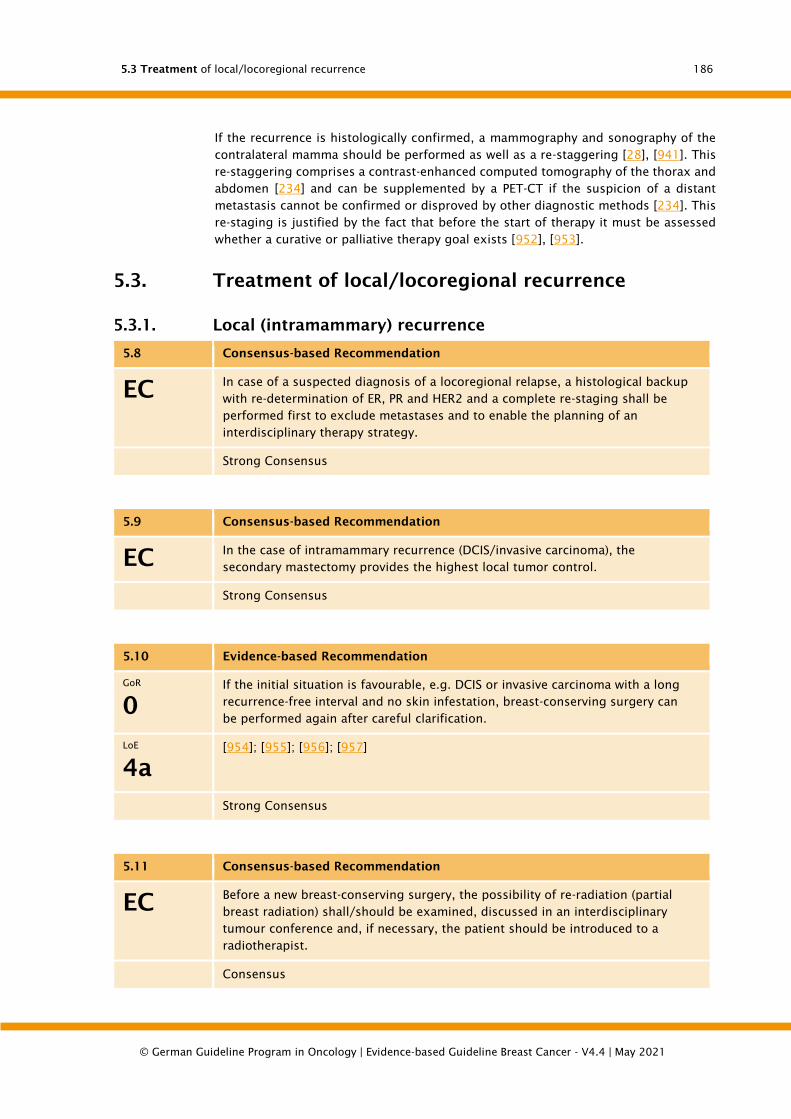
5.3 Treatment of local/locoregional recurrence
© German Guideline Program in Oncology | Evidence-based Guideline Breast Cancer - V4.4 | May 2021
186
If the recurrence is histologically confirmed, a mammography and sonography of the
contralateral mamma should be performed as well as a re-staggering [28], [941]. This
re-staggering comprises a contrast-enhanced computed tomography of the thorax and
abdomen [234] and can be supplemented by a PET-CT if the suspicion of a distant
metastasis cannot be confirmed or disproved by other diagnostic methods [234]. This
re-staging is justified by the fact that before the start of therapy it must be assessed
whether a curative or palliative therapy goal exists [952], [953].
5.3. Treatment of local/locoregional recurrence
5.3.1. Local (intramammary) recurrence
5.8 Consensus-based Recommendation
EC In case of a suspected diagnosis of a locoregional relapse, a histological backup
with re-determination of ER, PR and HER2 and a complete re-staging shall be
performed first to exclude metastases and to enable the planning of an
interdisciplinary therapy strategy.
Strong Consensus
5.9 Consensus-based Recommendation
EC In the case of intramammary recurrence (DCIS/invasive carcinoma), the
secondary mastectomy provides the highest local tumor control.
Strong Consensus
5.10 Evidence-based Recommendation
GoR
0
If the initial situation is favourable, e.g. DCIS or invasive carcinoma with a long
recurrence-free interval and no skin infestation, breast-conserving surgery can
be performed again after careful clarification.
LoE
4a
[954]; [955]; [956]; [957]
Strong Consensus
5.11 Consensus-based Recommendation
EC Before a new breast-conserving surgery, the possibility of re-radiation (partial
breast radiation) shall/should be examined, discussed in an interdisciplinary
tumour conference and, if necessary, the patient should be introduced to a
radiotherapist.
Consensus

5.3 Treatment of local/locoregional recurrence
© German Guideline Program in Oncology | Evidence-based Guideline Breast Cancer - V4.4 | May 2021
187
5.12 Consensus-based Recommendation
EC In the case of breast-conserving surgery, the patient shall be advised of an
increased risk of a renewed intramammary recurrence.
Strong Consensus
Background 5.8 to 5.12
The therapy of intramammary local recurrence consists, if possible, in surgical
intervention with the aim of R0 resection. The highest tumor control is achieved by
mastectomy [932]. If organ preservation surgery is performed again, there is an
increased risk of another intramammary recurrence. A renewed breast-conserving
procedure can be performed if the initial position is favorable - DCIS or invasive
carcinoma with a long recurrence-free interval or in the absence of skin infestation or
a long distance from the primary tumor localization - [958]. The highest local control
is achieved if the disease-free interval is over 5 years and if there are clearly negative
resection margins during the renewed breast-conserving surgery [956], [959]. In the
case of renewed breast-conserving surgery, local flap plasty, such as M. latissimusdorsi
plasty, should be performed with restraint and after detailed explanation, since a
survival disadvantage cannot be excluded and reduced cosmetic results are achieved.
If the axilla is clinically unremarkable, a renewed axillary intervention after primary
axillary dissection is not recommended. If no radiotherapy has been performed as part
of the primary therapy, postoperative radiotherapy should be followed. After
radiotherapy with initial breast-conserving procedure, in the recurrence situation with
renewed local excision, repeated external radiation or local brachytherapy may be
considered if necessary to reduce recurrence and avoid salvage mastectomy [954],
[960], [961], [962].
Survival after local or locoregional relapse varies considerably. Proven independent and
significant prognostic factors for survival with or without additional therapy are the
disease-free interval and tumor mass of the relapse, multifocality, as well as the initial
tumor stage and the time to metastasis [928], [963], [964], [965], [966], [967], [968].
Further prognostic factors are grading, resectability (R0 versus R1 versus R2), hormone
receptor status and HER2 oncogene status of the recurrent tumor [928], [967], [969].
Patients should be informed and educated about the possibility of a renewed breast-
conserving procedure under appropriate initial conditions. A local recurrence indicates
the biology of the disease and the avoidance of salvage mastectomy does not
necessarily mean a deterioration in overall survival.
5.3.2. Local recurrence after mastectomy
5.13 Consensus-based Recommendation
EC If possible, an isolated recurrence of the thoracic wall shall be surgically
removed completely (R0). If the ribs/intercostal muscles are affected, the
decision on therapy should be made in interdisciplinary cooperation with
thoracic surgery.
Strong Consensus
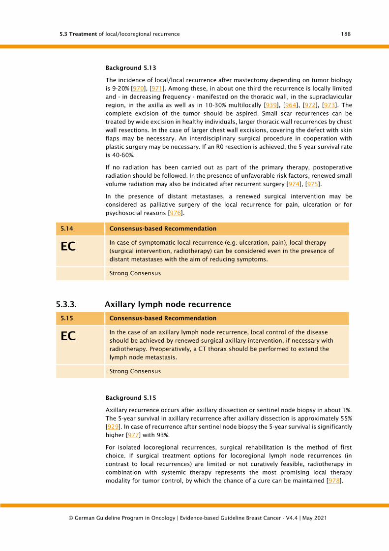
5.3 Treatment of local/locoregional recurrence
© German Guideline Program in Oncology | Evidence-based Guideline Breast Cancer - V4.4 | May 2021
188
Background 5.13
The incidence of local/local recurrence after mastectomy depending on tumor biology
is 9-20% [970], [971]. Among these, in about one third the recurrence is locally limited
and - in decreasing frequency - manifested on the thoracic wall, in the supraclavicular
region, in the axilla as well as in 10-30% multilocally [939], [964], [972], [973]. The
complete excision of the tumor should be aspired. Small scar recurrences can be
treated by wide excision in healthy individuals, larger thoracic wall recurrences by chest
wall resections. In the case of larger chest wall excisions, covering the defect with skin
flaps may be necessary. An interdisciplinary surgical procedure in cooperation with
plastic surgery may be necessary. If an R0 resection is achieved, the 5-year survival rate
is 40-60%.
If no radiation has been carried out as part of the primary therapy, postoperative
radiation should be followed. In the presence of unfavorable risk factors, renewed small
volume radiation may also be indicated after recurrent surgery [974], [975].
In the presence of distant metastases, a renewed surgical intervention may be
considered as palliative surgery of the local recurrence for pain, ulceration or for
psychosocial reasons [976].
5.14 Consensus-based Recommendation
EC In case of symptomatic local recurrence (e.g. ulceration, pain), local therapy
(surgical intervention, radiotherapy) can be considered even in the presence of
distant metastases with the aim of reducing symptoms.
Strong Consensus
5.3.3. Axillary lymph node recurrence
5.15 Consensus-based Recommendation
EC In the case of an axillary lymph node recurrence, local control of the disease
should be achieved by renewed surgical axillary intervention, if necessary with
radiotherapy. Preoperatively, a CT thorax should be performed to extend the
lymph node metastasis.
Strong Consensus
Background 5.15
Axillary recurrence occurs after axillary dissection or sentinel node biopsy in about 1%.
The 5-year survival in axillary recurrence after axillary dissection is approximately 55%
[929]. In case of recurrence after sentinel node biopsy the 5-year survival is significantly
higher [977] with 93%.
For isolated locoregional recurrences, surgical rehabilitation is the method of first
choice. If surgical treatment options for locoregional lymph node recurrences (in
contrast to local recurrences) are limited or not curatively feasible, radiotherapy in
combination with systemic therapy represents the most promising local therapy
modality for tumor control, by which the chance of a cure can be maintained [978].

5.3 Treatment of local/locoregional recurrence
© German Guideline Program in Oncology | Evidence-based Guideline Breast Cancer - V4.4 | May 2021
189
5.3.4. Pharmacological therapy
5.16 Consensus-based Recommendation
EC A systemic therapy after R0 resection of a locoregional recurrence shall be
considered for a prolonged disease-free interval as well as a prolonged overall
survival.
Strong Consensus
Background 5.16
In principle, a locoregional recurrence like the primary disease is also to be considered
a systemic disease and requires systemic therapy.
An additional systemic endocrine therapy after surgical therapy and R0 situation can
extend the disease-free interval in patients with a hormone receptor-positive
recurrence; however, the improvement of the survival rate is not proven [979], [980],
[981], [982], [983], [984]. In case of hormone sensitive recurrences a postoperative
endocrine therapy should be started or the current endocrine therapy should be
changed. Possibly this will improve disease-free and overall survival.
The data on chemotherapy after isolated locoregional recurrence and surgical therapy
is weak. However, the results of a prospective randomized study [985] are available. In
the CALOR study patients with surgically free tumor margins after mastectomy or
breast-conserving therapy and free margins were randomized to chemotherapy (n=85)
or no chemotherapy (n=77) (1:1). The choice of chemotherapy was in the hands of the
study centres, but should include at least two therapeutic agents and be carried out
over 3-6 months. In case of positive estrogen receptors, adjuvant endocrine therapy
was used. Radiation therapy was carried out on microscopically affected tumour
margins. An anti-HER2 therapy was optionally available in case of positive HER2
receptor. The primary study objective was disease-free survival (DFS). Patients with
metastases were excluded. After a median follow-up of 4.9 years, a 5-year DFS of 69%
(95% CI 56-79) was observed in the group with chemotherapy and 57% (95% CI 44-67)
without chemotherapy (HR 0.59, 95% CI 0.35-0.99), p=0.046). The benefit was
significant with negative estrogen receptor and greater than with positive estrogen
receptor of the local recurrence [ER negative: DFS 67% versus 35%, HR 0.32 (95% CI
0.14-0.73); ER positive: 70% versus 69%, HR 0.94 (95% CI 0.47-1.89)]. In terms of overall
survival, the use of chemotherapy also showed a significant benefit for the overall
collective. 5-year survival was 88% with chemotherapy versus 76% without
chemotherapy [HR 0.41 (95% CI 0.19-0.89), p=0.024)], but there was no significant
difference between the ER positive and negative subgroups. Even though the groups
are small overall, the study shows a significant benefit from the use of systemic therapy
after surgical rehabilitation. Thus, patients should be informed about the data
situation, advantages and disadvantages should be weighed and the use of
chemotherapy should be considered.
The data on chemotherapy after isolated locoregional recurrence and surgical therapy
is clearly arranged. This is particularly true for patients with inadequate adjuvant
primary therapy (e.g. trastuzumab-naive, HER2-positive patients, triple-negative
patients). In case of a HER2-positive local recurrence a combination of chemotherapy
and HER2-targeted therapy can be considered [985], [986].
If, in the course of a locoregional recurrence, R0 resection is unlikely and therefore the
local findings are not operable, endocrine therapy should be initiated for endocrine
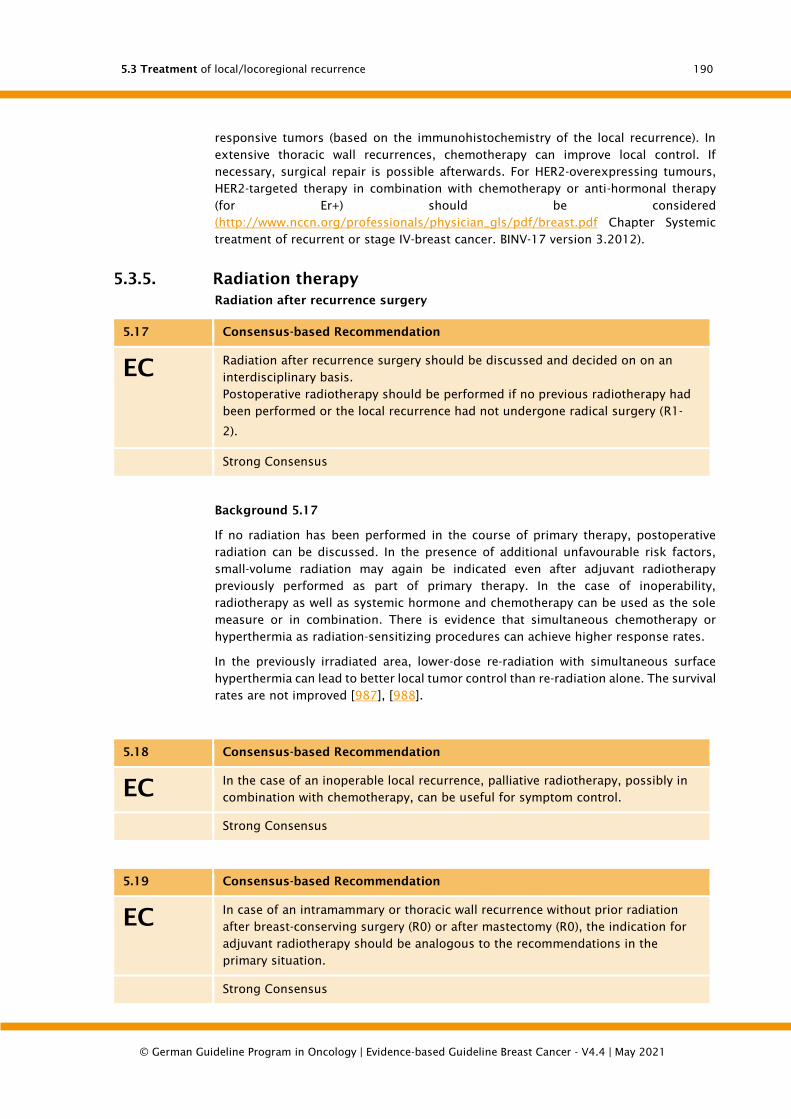
5.3 Treatment of local/locoregional recurrence
© German Guideline Program in Oncology | Evidence-based Guideline Breast Cancer - V4.4 | May 2021
190
responsive tumors (based on the immunohistochemistry of the local recurrence). In
extensive thoracic wall recurrences, chemotherapy can improve local control. If
necessary, surgical repair is possible afterwards. For HER2-overexpressing tumours,
HER2-targeted therapy in combination with chemotherapy or anti-hormonal therapy
(for Er+) should be considered
(http://www.nccn.org/professionals/physician_gls/pdf/breast.pdf Chapter Systemic
treatment of recurrent or stage IV-breast cancer. BINV-17 version 3.2012).
5.3.5. Radiation therapy
Radiation after recurrence surgery
5.17 Consensus-based Recommendation
EC Radiation after recurrence surgery should be discussed and decided on on an
interdisciplinary basis.
Postoperative radiotherapy should be performed if no previous radiotherapy had
been performed or the local recurrence had not undergone radical surgery (R1-
2).
Strong Consensus
Background 5.17
If no radiation has been performed in the course of primary therapy, postoperative
radiation can be discussed. In the presence of additional unfavourable risk factors,
small-volume radiation may again be indicated even after adjuvant radiotherapy
previously performed as part of primary therapy. In the case of inoperability,
radiotherapy as well as systemic hormone and chemotherapy can be used as the sole
measure or in combination. There is evidence that simultaneous chemotherapy or
hyperthermia as radiation-sensitizing procedures can achieve higher response rates.
In the previously irradiated area, lower-dose re-radiation with simultaneous surface
hyperthermia can lead to better local tumor control than re-radiation alone. The survival
rates are not improved [987], [988].
5.18 Consensus-based Recommendation
EC In the case of an inoperable local recurrence, palliative radiotherapy, possibly in
combination with chemotherapy, can be useful for symptom control.
Strong Consensus
5.19 Consensus-based Recommendation
EC In case of an intramammary or thoracic wall recurrence without prior radiation
after breast-conserving surgery (R0) or after mastectomy (R0), the indication for
adjuvant radiotherapy should be analogous to the recommendations in the
primary situation.
Strong Consensus
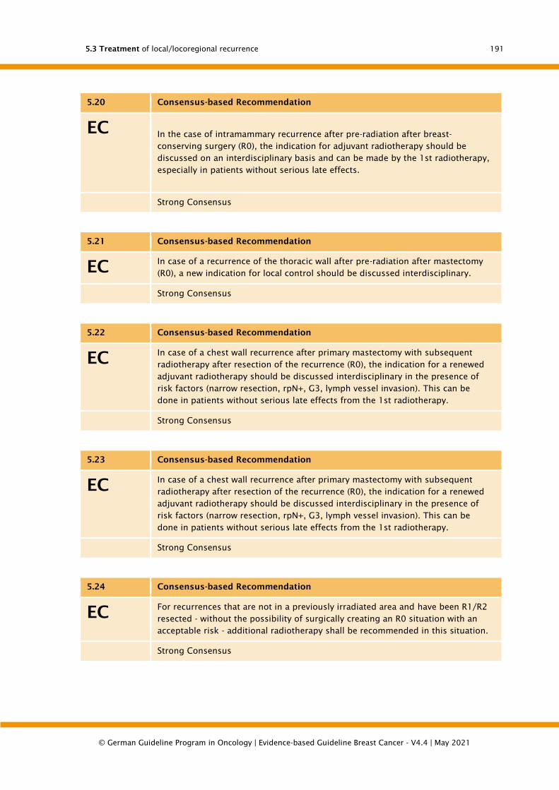
5.3 Treatment of local/locoregional recurrence
© German Guideline Program in Oncology | Evidence-based Guideline Breast Cancer - V4.4 | May 2021
191
5.20 Consensus-based Recommendation
EC
In the case of intramammary recurrence after pre-radiation after breast-
conserving surgery (R0), the indication for adjuvant radiotherapy should be
discussed on an interdisciplinary basis and can be made by the 1st radiotherapy,
especially in patients without serious late effects.
Strong Consensus
5.21 Consensus-based Recommendation
EC In case of a recurrence of the thoracic wall after pre-radiation after mastectomy
(R0), a new indication for local control should be discussed interdisciplinary.
Strong Consensus
5.22 Consensus-based Recommendation
EC In case of a chest wall recurrence after primary mastectomy with subsequent
radiotherapy after resection of the recurrence (R0), the indication for a renewed
adjuvant radiotherapy should be discussed interdisciplinary in the presence of
risk factors (narrow resection, rpN+, G3, lymph vessel invasion). This can be
done in patients without serious late effects from the 1st radiotherapy.
Strong Consensus
5.23 Consensus-based Recommendation
EC In case of a chest wall recurrence after primary mastectomy with subsequent
radiotherapy after resection of the recurrence (R0), the indication for a renewed
adjuvant radiotherapy should be discussed interdisciplinary in the presence of
risk factors (narrow resection, rpN+, G3, lymph vessel invasion). This can be
done in patients without serious late effects from the 1st radiotherapy.
Strong Consensus
5.24 Consensus-based Recommendation
EC For recurrences that are not in a previously irradiated area and have been R1/R2
resected - without the possibility of surgically creating an R0 situation with an
acceptable risk - additional radiotherapy shall be recommended in this situation.
Strong Consensus

5.4 Distant metastases
© German Guideline Program in Oncology | Evidence-based Guideline Breast Cancer - V4.4 | May 2021
192
5.25 Consensus-based Recommendation
EC In the presence of recurrences after R1/R2 resection and prior radiation without
the possibility of creating an R0 situation surgically with an acceptable risk, the
indication for renewed radiotherapy should be discussed on an interdisciplinary
basis.
This can be provided by the 1st radiotherapy in patients without serious late
effects.
Strong Consensus
5.4. Distant metastases
5.4.1. Systemic therapy in pre- and perimenopausal patients and
positive hormone receptor status and negative HER2 status.
5.26 Evidence-based Recommendation modified 2021
GoR
A
In pre- and perimenopausal patients, endocrine therapy, possibly combined with
targeted therapy, shall be offered if hormone receptor status is positive and
HER2 status is negative.
Endocrine-only monotherapy is not indicated in patients with the need to
achieve rapid remission to avert marked symptoms of the affected organ.
LoE
1b
[28]; [989]; [990]; [991]; [992]; [993]; [994]
Strong Consensus
Background 5.26
Endocrine therapy is less toxic than chemotherapy and should therefore always be used
as first-line therapy. Especially those patients who have had a long disease-free interval,
who have responded to previous anti-hormonal therapy measures, and who do not
belong to the small group of patients in whom a very rapid onset of action is necessary
(e.g., in cases of shortness of breath in diffuse lung metastasis or threatened liver
failure in liver metastasis or possible ileus in peritoneal carcinomatosis), benefit from
endocrine therapy. With a positive hormone receptor status, remission is expected in
60% of patients, with a negative hormone receptor status in less than 10%. Endocrine
therapy should therefore only be used in exceptional cases of negative hormone
receptor status. In rare cases with unknown hormone receptor status, however, the
indication for endocrine therapy can be made dependent on the clinical course.
If a patient responds to endocrine therapy, it is continued until progression is achieved.
In case of progression, the use of alternative endocrine substances is indicated and
justified. Only after all endocrine treatment measures have been exhausted or in case
of non-response to endocrine therapy should a switch to cytostatic therapy be made.
In the presence of HER2 overexpression, a worse response to endocrine therapy is to
be expected. Studies combining endocrine therapy with HER2-targeted therapy could
not show a survival benefit from additional HER2-targeted therapy. Therefore,
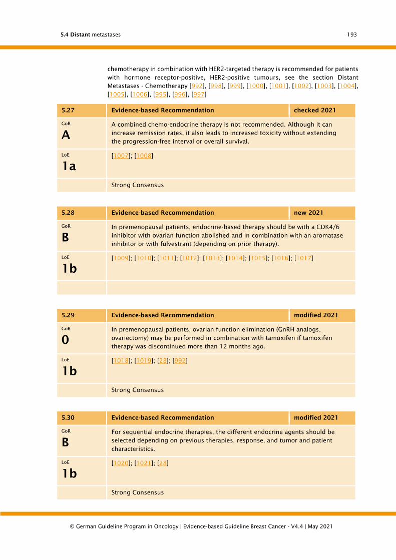
5.4 Distant metastases
© German Guideline Program in Oncology | Evidence-based Guideline Breast Cancer - V4.4 | May 2021
193
chemotherapy in combination with HER2-targeted therapy is recommended for patients
with hormone receptor-positive, HER2-positive tumours, see the section Distant
Metastases - Chemotherapy [992], [998], [999], [1000], [1001], [1002], [1003], [1004],
[1005], [1006], [995], [996], [997]
5.27 Evidence-based Recommendation checked 2021
GoR
A
A combined chemo-endocrine therapy is not recommended. Although it can
increase remission rates, it also leads to increased toxicity without extending
the progression-free interval or overall survival.
LoE
1a
[1007]; [1008]
Strong Consensus
5.28 Evidence-based Recommendation new 2021
GoR
B
In premenopausal patients, endocrine-based therapy should be with a CDK4/6
inhibitor with ovarian function abolished and in combination with an aromatase
inhibitor or with fulvestrant (depending on prior therapy).
LoE
1b
[1009]; [1010]; [1011]; [1012]; [1013]; [1014]; [1015]; [1016]; [1017]
5.29 Evidence-based Recommendation modified 2021
GoR
0
In premenopausal patients, ovarian function elimination (GnRH analogs,
ovariectomy) may be performed in combination with tamoxifen if tamoxifen
therapy was discontinued more than 12 months ago.
LoE
1b
[1018]; [1019]; [28]; [992]
Strong Consensus
5.30 Evidence-based Recommendation modified 2021
GoR
B
For sequential endocrine therapies, the different endocrine agents should be
selected depending on previous therapies, response, and tumor and patient
characteristics.
LoE
1b
[1020]; [1021]; [28]
Strong Consensus
5.4 Distant metastases
© German Guideline Program in Oncology | Evidence-based Guideline Breast Cancer - V4.4 | May 2021
194
Background 5.27.
In a meta-analysis of 26 trials involving 3,606 patients with advanced breast carcinoma,
Fossati et al [1040] demonstrated that the combination of chemotherapy and endocrine
therapy resulted in increased remission rates but not prolonged survival compared with
chemotherapy alone. Under combined chemoendocrine therapy, adverse effects such
as edema tendency and cardiovascular complications were significantly increased.
Background 5.28.
Data from AMNOG benefit assessments are available on therapy with CDK4/6 inhibitors
in addition to endocrine therapy in pre- or perimenopausal patients for a total of three
randomized controlled trials (MONARCH-2 [1009], PALOMA-3 [1013] and MONALEESA-
7 [1015]) on three substances (abemaciclib, palbociclib and ribociclib) are available.
These studies are randomized, controlled, blinded clinical trials (see guideline report
for full study assessment details). PALOMA-3 and MONARCH-2 included patients with
failure of a prior line of therapy regardless of menopausal status, while MONALEESA-7
included only pre- and perimenopausal patients. Stratification by menopausal status
was performed post hoc for PALOMA-3 and MONARCH-3 to meet the requirements of
the benefit assessment for the G-BA. The data on abemaciclib and palbociclib presented
below are therefore results of subgroup analyses. With non-significant effect estimates,
these do not have sufficient test strength to allow a statistically sound interpretation
and should therefore be regarded as exploratory results.
The following results were taken from the corresponding benefit assessment
procedures, in each case from modules 4 and/or the relevant publications. At the time
of the amendment, the active substances abemaciclib and ribociclib are still in
additional, ongoing benefit assessment procedures without decisions on the additional
benefit by the G BA. The status of the procedure can be viewed on the pages of the G-
BA (Abemaciclib: https://www.g-
ba.de/bewertungsverfahren/nutzenbewertung/535/; Ribociclib: https://www.g-
ba.de/bewertungsverfahren/nutzenbewertung/526/ und https://www.g-
ba.de/bewertungsverfahren/nutzenbewertung/527/).
Overall survival
Regarding overall survival, there were no consistent benefits of treatment with CDK4/6
inhibitors for the group of pre- or perimenopausal patients. There was no significant
survival benefit with treatment with abemaciclib or palbociclib compared with placebo
(abemaciclib HR: 0.69 [0.38; 1.25], palbociclib HR: 1.07 [0.61; 1.86]) [1009], [1013].
There was a significant survival benefit for premenopausal or perimenopausal patients
when treated with ribociclib (HR: 0.71 [0.54; 0.95]), but there were also significant
effect differences with respect to ethnicity, lineage of therapy, and age: patients with
age <40 years: HR 0.79 [0.48; 1.30] vs. age ≥40 years: HR 0.68 [0.48; 0.98]. Asian
female patients: HR 0.40 [0.22; 0.72]) vs. non-Asian female patients: HR 0.91 [0.64;
1.30]) [1015]. Stratified by line of therapy, the first-line HR was 0.68 [0.45; 1.00] and
the second- and follow-up-line HR was 0.78 [0.50; 1.21] [1028].
Progression-free survival
In all three studies, there was a clear advantage under therapy with CDK4/6 inhibitors
vs. placebo in terms of progression-free survival: HR 0.41 [0.25; 0.70] with abemaciclib
(second- and subsequent-line) [1010], HR 0.44 [0.23; 0.83] with palbociclib (second-
and subsequent-line) [1011], and HR 0.52 [0.38; 0.70] with ribociclib in the first-line
and HR 0.62 [0.44; 0.89] in the second- and subsequent-line, respectively [1016]. No
persistent effect modifications in subgroups emerged for any of the three agents
5.4 Distant metastases
© German Guideline Program in Oncology | Evidence-based Guideline Breast Cancer - V4.4 | May 2021
195
[1010], [1011], [1016], [1027], [1029], [1030], i.e., the advantage of CDK4/6 inhibitors
was also evident in the subgroup strata studied.
Health-related quality of life
A generalizing statement on the effect of CDK4/6 inhibitors on health-related quality
of life was made for all studies using the European Organization for Research and
Treatment of Cancer Quality of Life Questionnaire (EORTC QLQ-C30) and the Global
Health Status scale. For this purpose, a minimum clinically relevant difference of 10
points was assumed [1031], [1032]. There was no significant, clinically relevant
improvement (HR: 0.63 [0.33; 1.20]) for patients treated with abemaciclib [1012].
Under palbociclib, there were also no statistically significant differences compared with
placebo (HR: 0.81 [0.47; 1.41]) [1014]. However, clinically relevant benefits with
respect to global health status versus placebo were identified under second- and follow-
up ribociclib (HR: 0.70 [0.53; 0.92] [1017]. Given the heterogeneous results for the
CDK4/6 inhibitors evaluated, no robust evidence of improvement in quality of life can
be derived for them.
Adverse events
Treatment with CDK4/6 inhibitors was generally associated with a significantly higher
incidence of adverse events. Expressed as hazard ratios for the occurrence of adverse
events with Common Terminology Criteria for Adverse Events (CTCAE) grade ≥3,
comparable effect estimates were obtained for all agents: HR 5.64 [2.54; 12.55] among
abemaciclib, HR 5.90 [2.91; 11.95] among palbociclib, and HR 4.14 [3.28; 5.23] among
ribociclib [1012], [1014], [1017]. Among all agents, patients discontinued treatment
significantly more often than on placebo: RR 4.18 [0.22; 79.00] under abemaciclib, RR
3.60 [0.19; 67.81] under palbociclib, and HR 1.66 [0.82; 3.38] under ribociclib
[1012], [1014], [1017].
Overall, the evidence particularly supports a benefit of treating pre- and
perimenopausal patients in second- and subsequent-line therapy with CDK4/6
inhibitors in terms of significant effects on progression-free survival. Results on
tolerability of CDK4/6 inhibitors were comparable for all patient populations analyzed
and indicated significantly higher event rates compared with placebo. Adverse events
can be minimized by careful management of therapy [1033].
Conclusion pre- and perimenopausal patients
CDK4/6 inhibitors in early benefit assessment
For palbociclib, ribociclib and abemaciclib, results of the early benefit assessment
according to § 35a SGB are available at the time of guideline preparation (last review:
February 2020). The Institute for Quality and Efficiency in Health Care (IQWiG) and the
Federal Joint Committee (G-BA) concluded that for palbociclib, ribociclib, and
abemaciclib there is no evidence of an added medical benefit compared with the
respective appropriate comparator therapy for any therapy line or patient
group [1034], [1035], [1036], [1037], [1038], [1039]. This conclusion was mainly
based on the observed unfavorable side effect profile in the absence of evidence for
benefits in overall survival or quality of life. Effects on progression-free survival (PFS)
are only considered in the early benefit assessment if analyses are available that show
PFS as a valid surrogate endpoint for overall survival. According to IQWiG, these were
not available. Thus, the discrepancy between the results of the early benefit assessment
and the guideline recommendation on overall survival data, which were not available at
the time of the early benefit assessment, and the consideration of progression-free
survival as a benefit parameter can be explained. For abemaciclib and riboclicib,

5.4 Distant metastases
© German Guideline Program in Oncology | Evidence-based Guideline Breast Cancer - V4.4 | May 2021
196
however, further benefit dossiers are ongoing at the time of the admendment and their
final assessments are still pending.
The adverse events shown were generally compared with the placebo or endocrine
therapy alone as defined by the G-BA. In clinical practice, however, a comparison to
chemotherapy associated with significantly more severe side effects is appropriate.
CDK4/6 inhibitors are expected to show a significantly more tolerable side effect profile
than placebo compared to available chemotherapeutic agents. In addition, side effects
are very well managed with established supportive measures [1033]. A tabular overview
in the form of evidence tables of the listed effect estimates can be found in the evidence
report for this guideline.
Background 5.29 to 5.31.
Unless CDK4/6 inhibitors are given, the initial therapeutic step is ovarian function
elimination (GnRH analogs, ovariectomy, or radiomenolysis) in combination with
tamoxifen. If tumor progression occurs or tamoxifen is contraindicated, a third-
generation aromatase inhibitor plus GnRH analog can be used, and fulvestrant plus
GnRH analog may also be considered. Despite sparse data, therapy should be largely
analogous to the treatment of postmenopausal patients while maintaining ovarian
suppression [1018], [1021], [1022], [1023], [1024], [1025], [1026].
5.31 Evidence-based Recommendation modified 2021
GoR
0
Therapy can be performed while maintaining ovarian suppression in analogy to
the treatment of postmenopausal patients. As options can be used in
combination with a GNRH analogue depending on the previous therapy:
• aromatase inhibitors
• fulvestrant
• tamoxifen
LoE
1b
[1041]; [1042]; [1043]; [1044]; [28]; [989]; [992]; [1000]
Strong Consensus
5.4.2. Systemic therapy in postmenopausal patients and positive
hormone receptor status and negative HER2 status.
5.32 Evidence-based Recommendation new 2021
GoR
A
In postmenopausal patients, endocrine therapy, possibly combined with
targeted therapy, shall be offered if hormone receptor status is positive and
HER2 status is negative.
Endocrine therapy is not indicated in patients with the need to achieve rapid
remission to avert marked symptoms of the affected organ.
LoE
1b
[1045]; [1046]; [1047]; [1048]; [1049]; [1050]; [1051]
Strong Consensus
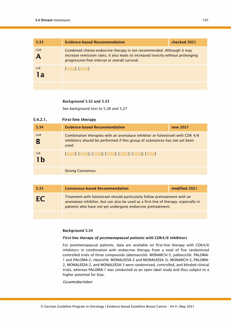
5.4 Distant metastases
© German Guideline Program in Oncology | Evidence-based Guideline Breast Cancer - V4.4 | May 2021
197
5.33 Evidence-based Recommendation checked 2021
GoR
A
Combined chemo-endocrine therapy is not recommended. Although it may
increase remission rates, it also leads to increased toxicity without prolonging
progression-free interval or overall survival.
LoE
1a
[1052]; [1053]
Background 5.32 and 5.33
See background text to 5.26 and 5.27
5.4.2.1. First-line therapy
5.34 Evidence-based Recommendation new 2021
GoR
B
Combination therapies with an aromatase inhibitor or fulvestrant with CDK 4/6
inhibitors should be performed if this group of substances has not yet been
used.
LoE
1b
[1029]; [1054]; [1055]; [1056]; [1057]; [1058]; [1059]
Strong Consensus
5.35 Consensus-based Recommendation modified 2021
EC Treatment with fulvestrant should particularly follow pretreatment with an
aromatase inhibitor, but can also be used as a first line of therapy, especially in
patients who have not yet undergone endocrine pretreatment.
Background 5.34
First-line therapy of postmenopausal patients with CDK4/6 inhibitors
For postmenopausal patients, data are available on first-line therapy with CDK4/6
inhibitors in combination with endocrine therapy from a total of five randomized
controlled trials of three compounds (abemaciclib: MONARCH-3; palbociclib: PALOMA-
1 and PALOMA-2; ribociclib: MONALEESA-2 and MONALEESA-3). MONARCH-3, PALOMA-
2, MONALEESA-2, and MONALEESA-3 were randomized, controlled, and blinded clinical
trials, whereas PALOMA-1 was conducted as an open label study and thus subject to a
higher potential for bias.
Gesamtüberleben

5.4 Distant metastases
© German Guideline Program in Oncology | Evidence-based Guideline Breast Cancer - V4.4 | May 2021
198
With regard to overall survival, there have been no significant advantages for
postmenopausal patients in first-line therapy to date (see evidence tables in the
guideline report - section 14.6).
Progressionsfreies Überleben
In contrast, treatment with CDK4/6 inhibitors showed significant advantages over
placebo in terms of progression-free survival in all studies. With abemaciclib: HR 0.54
[0.42; 0.70], with palbociclib HR 0.49 [0.32; 0.75] (PALOMA-1), resp. HR : 0.58 [0.46;
0.72] (PALOMA-2) and under ribociclib HR 0.57 [0.43; 0.74] (MONALEESA-2) and HR
0.58 [0.42; 0.80] (MONALEESA-3), respectively) [1029], [1055], [1056], [1058].
Gesundheitsbezogene Lebensqualität
There is no evidence of an effect of treatment with CDK4/6 inhibitors on health-related
quality of life in postmenopausal patients in terms of a clinically relevant difference in
scores on the EORTC QLQ-C30 or FACT-B questionnaire (see guideline report).
Adverse events
Treatment with CDK4/6 inhibitors was also generally associated with a significantly
higher incidence of adverse events for postmenopausal patients: Under abemaciclib,
the effect for UE with CTCAE grade ≥3 was HR 3.14 [2.25; 4.39] (MONARCH-3), under
palbociclib HR 5.47 [3.15; 9.51] (PALOMA-1) and 5.50 [4.14; 7.31] (PALOMA-2),
respectively, and under ribociclib HR 4.21 [3.40; 5.21] [1054], [1055], [1056], [1059].
Patients discontinued treatment significantly more often under abemaciclib and
ribociclib than under placebo. With abemaciclib, the hazard ratio was 6.04 [2.18;
16.70]; with ribociclib, first-line HR was 4.23 [2.31; 7.74]; and for any line of therapy,
it was 2.73 [1.58; 4.74] [1012], [1014], [1054], [1056], [1059].
Conclusion postmenopausal patients in first-line therapy
Overall, the evidence in postmenopausal patients in first-line therapy with CDK4/6
inhibitors shows a treatment advantage over comparator therapy only for the endpoint
progression-free survival. The results for tolerability of CDK4/6 inhibitors were
comparable for all patient populations analyzed and indicated significantly higher event
rates compared with comparator therapy. Comments on the benefit assessments by
IQWiG and the decisions of the G-BA can be found in chapter 5.4.1. A tabular overview
in the form of evidence tables of the listed effect estimates can be found in the evidence
report for this guideline
5.4.2.2. Second and follow-up line therapy
5.36 Evidence-based Recommendation modified 2021
GoR
B
If a CDK4/6 inhibitor had not yet been used in the first-line setting, it should be
used in further endocrine-based lines of therapy.
LoE
1b
[1009]; [1013]; [1057]

5.4 Distant metastases
© German Guideline Program in Oncology | Evidence-based Guideline Breast Cancer - V4.4 | May 2021
199
5.37 Consensus-based Recommendation modified 2021
EC After prior anti-hormonal therapy with a non-steroidal aromatase inhibitor as
well as CDK4/6 inhibitors, follow-up therapy with exemestane and the mTOR
inhibitor everolimus can be performed.
5.38 Consensus-based Recommendation new 2021
EC Further steps in the endocrine treatment sequence in postmenopausal patients
represent, depending on the pretreatment, the use of alpelisib (if a
corresponding PI3KA mutation is detected) or antiestrogens, estrogen receptor
antagonists, switching the aromatase inhibitor from a steroidal to a nonsteroidal
aromatase inhibitor, or vice versa.
Strong Consensus
Background text 5.36.
Second-line and follow-up therapy of postmenopausal patients with CDK4/6
inhibitors
For postmenopausal patients, data are available on second and subsequent line therapy
with CDK4/6 inhibitors in combination with endocrine therapy from a total of three
randomized controlled trials on three substances (abemaciclib: MONARCH-2,
palbociclib: PALOMA-3; ribociclib: MONALEESA-3). These studies are randomized,
controlled, blinded clinical trials. The population of postmenopausal patients in
second-line and follow-up therapy was extracted for the MONARCH-2 and PALOMA-3
studies in the form of subgroups in order to be able to represent the patient group
required by the G-BA. The data on abemaciclib and palbociclib presented below are
therefore the results of subgroup analyses. With non-significant effect estimates, these
do not have sufficient test strength to allow a statistically sound interpretation and
should therefore be regarded as exploratory results.
Overall Survival
With regard to overall survival, there were advantages for the postmenopausal patients
in the second- and follow-up-line therapy. There was a significant survival advantage
over placebo here for abemaciclib (HR 0.77 [0.61; 0.98]) and palbociclib (HR 0.73 [0.57;
0.95]) [1009], [1013]. For ribociclib, the hazard ratio for overall survival was just below
the significance threshold (HR 0.73 [0.53; 1.00]) [1057]. No subgroup analyses (e.g.,
with regard to age or ethnicity) are available for the group of postmenopausal patients
in second-line or follow-up therapy.
Progression-free Survival
There were significant advantages of treatment with CDK4/6 inhibitors over placebo in
terms of progression-free survival in all studies. With abemaciclib: HR 0.58 [0.46; 0.73],
with palbociclib: HR 0.41 [0.30; 0.56], and with ribociclib HR 0.58 [0.42;
0.80] [1060], [1061], [1058].
Health-related quality of life

5.4 Distant metastases
© German Guideline Program in Oncology | Evidence-based Guideline Breast Cancer - V4.4 | May 2021
200
There is no evidence of an effect of treatment with CDK4/6 inhibitors on health-related
quality of life in postmenopausal patients in terms of a clinically relevant difference in
EORTC QLQ-C30 score (see guideline report).
Adverse events
Treatment with CDK4/6 inhibitors was also generally associated with a significantly
higher incidence of adverse events for postmenopausal patients: For abemaciclib, the
effect estimate for UE with CTCAE grade ≥3 under any line of therapy was HR 3.34
[2.43; 4.59], for palbociclib HR 4.54 [3.22; 6.41], and for ribociclib HR 4.46 [3.45;
5.77] [1012], [1014], [1059]. Patients discontinued treatment significantly more often
with abemaciclib and ribociclib than with placebo. Under abemaciclib, the effect
estimate was HR 2.32 [1.03; 5.23] and under ribociclib, HR 2.73 [1.58; 4.74]
[1012], [1014], [1059].
Conclusion Second- and Follow-up-Line Therapy of Postmenopausal Patients with
CDK4/6 Inhibitors
Overall, the evidence supports a benefit of treating postmenopausal patients in second-
and subsequent-line therapy with CDK4/6 inhibitors in the form of significant effect
estimates for both progression-free survival and overall survival that have not been
achieved in the past. Results on tolerability of CDK4/6 inhibitors were comparable for
all patient populations analyzed and indicated significantly higher event rates
compared with placebo. Comments on the benefit assessments by IQWiG and the
decisions of the G-BA can be found in chapter 5.4.1. A tabular overview in the form of
evidence tables of the listed effect estimates can be found in the evidence report for
this guideline.
Background 5.37. and 5.38.
First-line drugs are third-generation aromatase inhibitors. Subgroup analyses for three
predominantly used aromatase inhibitors (anastrozole, exemestane, and letrozole)
show similar survival benefits [1063].
After failure of a nonsteroidal aromatase inhibitor (anastrozole or letrozole),
administration of everolimus in addition to exemestane has been shown to prolong
progression-free survival. Thus, this approach represents another treatment option.
In case of tumor re-progression, antiestrogens, estrogen receptor antagonists, and
finally high-dose progestogens or estrogens can be used [1065], [1066].
Aromatase inhibitor treatment is associated with a significant increase in side effects
induced by hormone withdrawal [1067]. During aromatase inhibitor therapy, fewer hot
flashes, thromboembolic events, and endometrial cancer occur as side effects
compared with tamoxifen, but the rate of arthralgias and myalgias is increased.
Furthermore, higher bone density loss and possibly a higher rate of osteoporotic
fractures are to be expected. However, these aspects are often not of primary concern
in the palliative treatment situation.
A combination of fulvestrant and aromatase inhibitors is not recommended due to
controversial
data [1062], [1064], [1068], [1069], [1070], [1071], [1072], [1073], [1074], [1075], [1
076].
Summary conclusion on the use of CK4/6 inhibitors in patients with breast cancer
The clinical trial data presented here include results from populations defined a priori
in the study protocol as well as results from post-hoc subgroups, some of which were

5.4 Distant metastases
© German Guideline Program in Oncology | Evidence-based Guideline Breast Cancer - V4.4 | May 2021
201
prescribed by the G-BA. They were listed to ensure a complete reporting of the available
evidence.
However, a final interpretation of the subgroup analyses presented is hampered by
statistical limitations (multiple testing, insufficient test strength (power). Therefore, the
recommendations formulated by the guideline group on the use of CK4/6 inhibitors
are based on the analyses of the respective overall populations in the studies (see
guideline report).
In these patients, advantages of the CDK4/6 inhibitors were shown with respect to
prolonged overall survival, especially for abemaciclib as well as
ribociclib [1009], [1015]. In addition, significant prolongation of progression-free
survival was observed for all agents [1060], [1061], [1016], [1029], [1058].
These survival benefits outweigh any lack of effects with respect to health-related
quality of life as well as the well-treatable side effects
5.4.3. Chemotherapy of metastatic breast cancer
Pre-chemotherapy criteria
5.39 Consensus-based Recommendation
EC Before chemotherapy is administered, the patient's general condition and
comorbidity, previous therapies and compliance shall be assessed.
Strong Consensus
Toxicity assessment
5.40 Consensus-based Recommendation
EC During the therapy, a regular toxicity assessment (subjective and objective) shall
be carried out. The dosage as well as the desired time intervals shall be carried
out according to generally accepted standard or currently published therapy
regimes. After determination of a suitable and representative measurement
parameter (symptoms, tumor markers, imaging) before the start of therapy, an
evaluation of the therapeutic effect shall be performed at least every 6-12 weeks
in accordance with clinical requirements. In the course of time, the imaging
intervals can be extended if the remission is persistent and the clinical and
laboratory chemical assessment of the disease status is good.
Strong Consensus
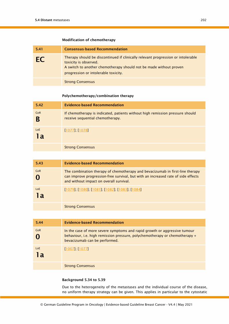
5.4 Distant metastases
© German Guideline Program in Oncology | Evidence-based Guideline Breast Cancer - V4.4 | May 2021
202
Modification of chemotherapy
5.41 Consensus-based Recommendation
EC Therapy should be discontinued if clinically relevant progression or intolerable
toxicity is observed.
A switch to another chemotherapy should not be made without proven
progression or intolerable toxicity.
Strong Consensus
Polychemotherapy/combination therapy
5.42 Evidence-based Recommendation
GoR
B
If chemotherapy is indicated, patients without high remission pressure should
receive sequential chemotherapy.
LoE
1a
[1077]; [1078]
Strong Consensus
5.43 Evidence-based Recommendation
GoR
0
The combination therapy of chemotherapy and bevacizumab in first-line therapy
can improve progression-free survival, but with an increased rate of side effects
and without impact on overall survival.
LoE
1a
[1079]; [1080]; [1081]; [1082]; [1083]; [1084]
Strong Consensus
5.44 Evidence-based Recommendation
GoR
0
In the case of more severe symptoms and rapid growth or aggressive tumour
behaviour, i.e. high remission pressure, polychemotherapy or chemotherapy +
bevacizumab can be performed.
LoE
1a
[1007]; [1077]
Strong Consensus
Background 5.34 to 5.39
Due to the heterogeneity of the metastases and the individual course of the disease,
no uniform therapy strategy can be given. This applies in particular to the cytostatic

5.4 Distant metastases
© German Guideline Program in Oncology | Evidence-based Guideline Breast Cancer - V4.4 | May 2021
203
treatment of metastasized breast carcinoma. Although monotherapy has lower
remission rates than polychemotherapy, this does not have a significant negative
impact on survival time. Monotherapies are better tolerated, so that monotherapy
should be used whenever possible. Monotherapy should be used in cases of minor
symptoms and slow tumour growth or ineffectiveness of endocrine therapy.
Polychemotherapy is only indicated for severe symptoms, rapid tumour growth and
aggressive tumour behaviour. Cytostatic therapy should be based on the therapeutic
index in the case of incurable disease, whereby the effect (e.g. symptom control) and
side effects of a therapy must be considered and weighed up. For the majority of
patients, the use of subjectively less stressful monotherapies compared to a
combination therapy is recommended. This is supported by a Cochrane meta-analysis
published in 2015, which showed that there were no significant differences in overall
survival and progression-free survival between combination therapy and sequential
monochemotherapy when compared with sequential monochemotherapy (OS HR 1.04
95% CI 0.93-1.16; p=0.45 / PFS HR1.11 95% CI 0.99-1.25; p=0.08). In comparison, the
response was significantly higher with combination chemotherapy. However,
combination chemotherapy also showed a higher toxicity with regard to the rate of
febrile neutropenia. Many mainly non-haematological side effects were not described
in this meta-analysis. In the meta-analysis, two scenarios of sequential
monochemotherapy were described, one change of monochemotherapy with
progression or fixed change of monochemotherapy without progression after a few
cycles. The results were similar for both scenarios, with the hazard ratios described
referring to the total collective. The authors conclude that the results of this meta-
analysis support the recommendations for sequential monotherapy compared to
combination chemotherapy [1077], except for the cases with rapid tumor progression
and high remission pressure.
If the patient has not yet received anthracyclines/taxanes in adjuvant therapy, they can
be used primarily.
Before and during chemotherapy the patient's general condition must be assessed
regularly. During therapy, the side effects of this treatment must also be evaluated
regularly. An evaluation of the therapeutic effect by means of imaging should be
performed every 6-12 weeks (interval depending on the spread of the disease, disease
dynamics and clinical situation). In the course of time, the imaging intervals can be
extended in case of remission and good clinical and laboratory chemical assessment
of the disease status. In case of progress or pronounced toxicity, therapy should be
discontinued. The therapeutic index (individual patient benefit versus therapy-related
side effects) should be positive in the overall assessment of the therapy.
The dosage as well as the intended time intervals of the therapy should be adapted to
general guidelines of the therapy, i.e. recognized published protocols. Dose-intensified
and high-dose therapies have not yet led to any improvement in effectiveness. Their
use is only acceptable in the context of studies (Conchrane: [800]).
A meta-analysis by Ghersi et al. showed that taxane-containing chemotherapies were
associated with improved progression-free and overall survival as well as an increased
tumor response. However, they also led to an increased risk of neuropathy and a lower
risk of nausea and vomiting compared to non-taxan-containing regimens [1085].
5.4.3.1. Bevacizumab in metastatic breast cancer (first line)
The improved efficacy of first-line treatment with paclitaxel plus bevacizumab (P/Bev)
compared to paclitaxel monotherapy was demonstrated in a randomized phase III
study: In the E2100 study, the combined use of P/Bev doubled the objective response
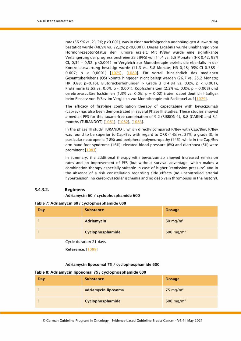
5.4 Distant metastases
© German Guideline Program in Oncology | Evidence-based Guideline Breast Cancer - V4.4 | May 2021
204
rate (36.9% vs. 21.2%; p<0.001), was in einer nachfolgenden unabhängigen Auswertung
bestätigt wurde (48,9% vs. 22,2%; p<0,0001). Dieses Ergebnis wurde unabhängig vom
Hormonrezeptor-Status der Tumore erzielt. Mit P/Bev wurde eine signifikante
Verlängerung der progressionsfreien Zeit (PFS) von 11.4 vs. 5.8 Monaten (HR 0,42; 95%
CI, 0,34 – 0,52; p<0.001) im Vergleich zur Monotherapie erzielt, die ebenfalls in der
Kontrollauswertung bestätigt wurde (11.3 vs. 5.8 Monate; HR 0,48; 95% CI 0.385 -
0.607; p < 0,0001) [1079], [1080]. Ein Vorteil hinsichtlich des medianen
Gesamtüberlebens (OS) konnte hingegen nicht belegt werden (26.7 vs. 25.2 Monate;
HR 0.88; p=0.16). Blutdruckerhöhungen > Grade 3 (14.8% vs. 0.0%, p < 0.001),
Proteinurie (3.6% vs. 0.0%, p < 0.001), Kopfschmerzen (2.2% vs. 0.0%, p = 0.008) und
cerebrovasculäre Ischämien (1.9% vs. 0.0%, p = 0.02) traten dabei deutlich häufiger
beim Einsatz von P/Bev im Vergleich zur Monotherapie mit Paclitaxel auf [1079].
The efficacy of first-line combination therapy of capecitabine with bevacizumab
(cap/ev) has also been demonstrated in several Phase III studies. These studies showed
a median PFS for this taxane-free combination of 9.2 (RIBBON-1), 8.8 (CARIN) and 8.1
months (TURANDOT) [1081], [1082], [1083].
In the phase III study TURANDOT, which directly compared P/Bev with Cap/Bev, P/Bev
was found to be superior to Cap/Bev with regard to ORR (44% vs. 27%; p grade 3), in
particular neutropenia (18%) and peripheral polyneuropathy (14%), while in the Cap/Bev
arm hand-foot syndrome (16%), elevated blood pressure (6%) and diarrhoea (5%) were
prominent [1083].
In summary, the additional therapy with bevacizumab showed increased remission
rates and an improvement of PFS (but without survival advantage, which makes a
combination therapy especially suitable in case of higher "remission pressure" and in
the absence of a risk constellation regarding side effects (no uncontrolled arterial
hypertension, no cerebrovascular ischemia and no deep vein thrombosis in the history).
5.4.3.2. Regimens
Adriamycin 60 / cyclophosphamide 600
Table 7: Adriamycin 60 / cyclophosphamide 600
Day Substance Dosage
1 Adriamycin 60 mg/m²
1 Cyclophosphamide 600 mg/m²
Cycle duration 21 days
Reference: [1089]
Adriamycin liposomal 75 / cyclophosphamide 600
Table 8: Adriamycin liposomal 75 / cyclophosphamide 600
Day Substance Dosage
1 adriamycin liposoma 75 mg/m²
1 Cyclophosphamide 600 mg/m²
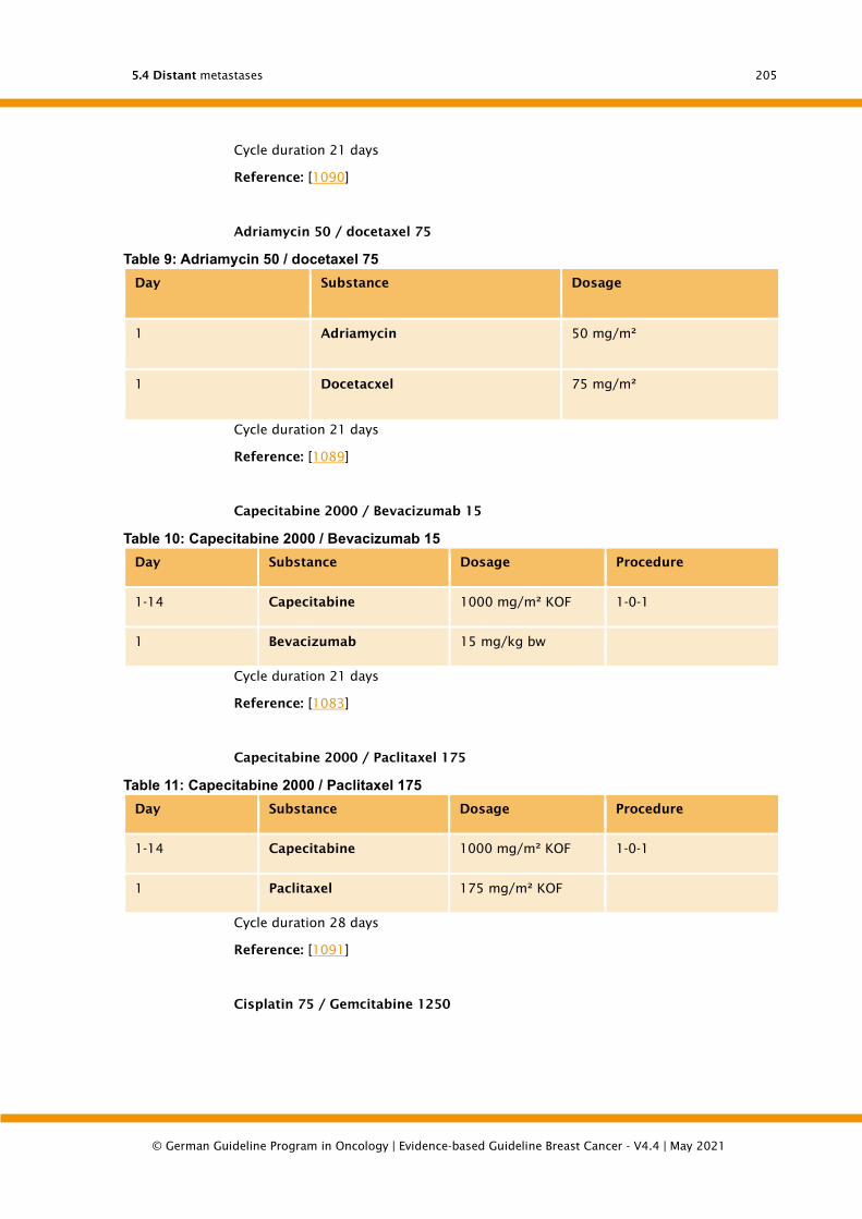
5.4 Distant metastases
© German Guideline Program in Oncology | Evidence-based Guideline Breast Cancer - V4.4 | May 2021
205
Cycle duration 21 days
Reference: [1090]
Adriamycin 50 / docetaxel 75
Table 9: Adriamycin 50 / docetaxel 75
Day Substance Dosage
1 Adriamycin 50 mg/m²
1 Docetacxel 75 mg/m²
Cycle duration 21 days
Reference: [1089]
Capecitabine 2000 / Bevacizumab 15
Table 10: Capecitabine 2000 / Bevacizumab 15
Day Substance Dosage Procedure
1-14 Capecitabine 1000 mg/m² KOF 1-0-1
1 Bevacizumab 15 mg/kg bw
Cycle duration 21 days
Reference: [1083]
Capecitabine 2000 / Paclitaxel 175
Table 11: Capecitabine 2000 / Paclitaxel 175
Day Substance Dosage Procedure
1-14 Capecitabine 1000 mg/m² KOF 1-0-1
1 Paclitaxel 175 mg/m² KOF
Cycle duration 28 days
Reference: [1091]
Cisplatin 75 / Gemcitabine 1250
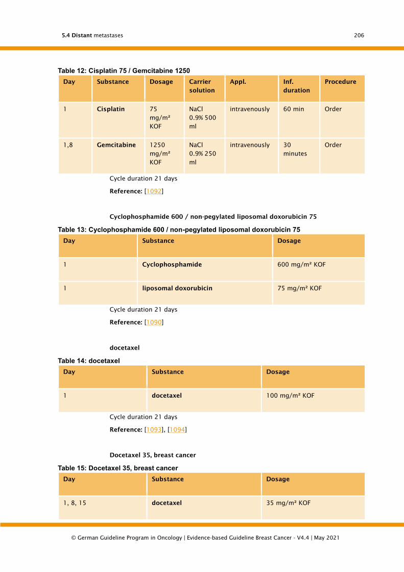
5.4 Distant metastases
© German Guideline Program in Oncology | Evidence-based Guideline Breast Cancer - V4.4 | May 2021
206
Table 12: Cisplatin 75 / Gemcitabine 1250
Day Substance Dosage Carrier
solution
Appl. Inf.
duration
Procedure
1 Cisplatin 75
mg/m²
KOF
NaCl
0.9% 500
ml
intravenously 60 min Order
1,8 Gemcitabine 1250
mg/m²
KOF
NaCl
0.9% 250
ml
intravenously 30
minutes
Order
Cycle duration 21 days
Reference: [1092]
Cyclophosphamide 600 / non-pegylated liposomal doxorubicin 75
Table 13: Cyclophosphamide 600 / non-pegylated liposomal doxorubicin 75
Day Substance Dosage
1 Cyclophosphamide 600 mg/m² KOF
1 liposomal doxorubicin 75 mg/m² KOF
Cycle duration 21 days
Reference: [1090]
docetaxel
Table 14: docetaxel
Day Substance Dosage
1 docetaxel 100 mg/m² KOF
Cycle duration 21 days
Reference: [1093], [1094]
Docetaxel 35, breast cancer
Table 15: Docetaxel 35, breast cancer
Day Substance Dosage
1, 8, 15 docetaxel 35 mg/m² KOF

5.4 Distant metastases
© German Guideline Program in Oncology | Evidence-based Guideline Breast Cancer - V4.4 | May 2021
207
Cycle duration 28 days
Reference: [1095]
Doxorubicin 50 / docetaxel 75, breast cancer
Table 16: Doxorubicin 50 / docetaxel 75, breast cancer
Day Substance Dosage
1 Doxorubicin 50 mg/m² KOF
1 docetaxel 75 mg/m² KOF
Cycle duration 21 days
Reference: [1089], [1096]
Doxorubicin 60 / cyclophosphamide 600
Table 17: Doxorubicin 60 / cyclophosphamide 600
Day Substance Dosage
1 Doxorubicin 60 mg/m² KOF
1 Cyclophosphamide 600 mg/m² KOF
Cycle duration 21 days
Reference: [1089]
Epirubicin 60 / cyclophosphamide 600
Table 18: Epirubicin 60 / cyclophosphamide 600
Day Substance Dosage
1 Epirubicin 60 mg/m²
1 Cyclophosphamide 600 mg/m²
Cycle duration 21 days
Reference: [1097]
Epirubicin 75 / cyclophosphamide 600
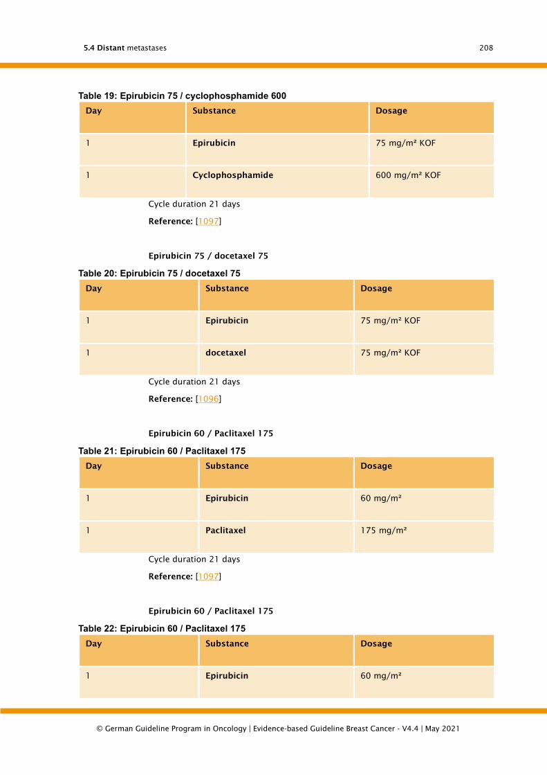
5.4 Distant metastases
© German Guideline Program in Oncology | Evidence-based Guideline Breast Cancer - V4.4 | May 2021
208
Table 19: Epirubicin 75 / cyclophosphamide 600
Day Substance Dosage
1 Epirubicin 75 mg/m² KOF
1 Cyclophosphamide 600 mg/m² KOF
Cycle duration 21 days
Reference: [1097]
Epirubicin 75 / docetaxel 75
Table 20: Epirubicin 75 / docetaxel 75
Day Substance Dosage
1 Epirubicin 75 mg/m² KOF
1 docetaxel 75 mg/m² KOF
Cycle duration 21 days
Reference: [1096]
Epirubicin 60 / Paclitaxel 175
Table 21: Epirubicin 60 / Paclitaxel 175
Day Substance Dosage
1 Epirubicin 60 mg/m²
1 Paclitaxel 175 mg/m²
Cycle duration 21 days
Reference: [1097]
Epirubicin 60 / Paclitaxel 175
Table 22: Epirubicin 60 / Paclitaxel 175
Day Substance Dosage
1 Epirubicin 60 mg/m²
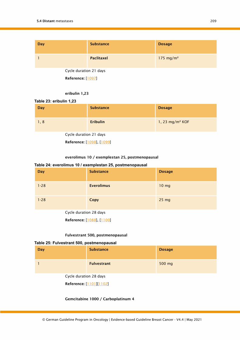
5.4 Distant metastases
© German Guideline Program in Oncology | Evidence-based Guideline Breast Cancer - V4.4 | May 2021
209
Day Substance Dosage
1 Paclitaxel 175 mg/m²
Cycle duration 21 days
Reference: [1097]
eribulin 1,23
Table 23: eribulin 1,23
Day Substance Dosage
1, 8 Eribulin 1, 23 mg/m² KOF
Cycle duration 21 days
Reference: [1098], [1099]
everolimus 10 / exemplestan 25, postmenopausal
Table 24: everolimus 10 / exemplestan 25, postmenopausal
Day Substance Dosage
1-28 Everolimus 10 mg
1-28 Copy 25 mg
Cycle duration 28 days
Reference: [1088], [1100]
Fulvestrant 500, postmenopausal
Table 25: Fulvestrant 500, postmenopausal
Day Substance Dosage
1 Fulvestrant 500 mg
Cycle duration 28 days
Reference: [1101][1102]
Gemcitabine 1000 / Carboplatinum 4
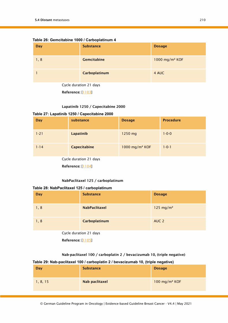
5.4 Distant metastases
© German Guideline Program in Oncology | Evidence-based Guideline Breast Cancer - V4.4 | May 2021
210
Table 26: Gemcitabine 1000 / Carboplatinum 4
Day Substance Dosage
1, 8 Gemcitabine 1000 mg/m² KOF
1 Carboplatinum 4 AUC
Cycle duration 21 days
Reference: [1103]
Lapatinib 1250 / Capecitabine 2000
Table 27: Lapatinib 1250 / Capecitabine 2000
Day substance Dosage Procedure
1-21 Lapatinib 1250 mg 1-0-0
1-14 Capecitabine 1000 mg/m² KOF 1-0-1
Cycle duration 21 days
Reference: [1104]
NabPaclitaxel 125 / carboplatinum
Table 28: NabPaclitaxel 125 / carboplatinum
Day Substance Dosage
1, 8 NabPaclitaxel 125 mg/m²
1, 8 Carboplatinum AUC 2
Cycle duration 21 days
Reference: [1105]
Nab-paclitaxel 100 / carboplatin 2 / bevacizumab 10, (triple negative)
Table 29: Nab-paclitaxel 100 / carboplatin 2 / bevacizumab 10, (triple negative)
Day Substance Dosage
1, 8, 15 Nab paclitaxel 100 mg/m² KOF

5.4 Distant metastases
© German Guideline Program in Oncology | Evidence-based Guideline Breast Cancer - V4.4 | May 2021
211
Day Substance Dosage
1, 8, 15 Carboplatinum AUC 2
1, 15 Bevacizumab 10 mg/kg bw
Cycle duration 28 days
Reference: [1106]
Nab-paclitaxel 125 / Trastuzumab (4/2)
Table 30: Nab-paclitaxel 125 / Trastuzumab (4/2)
Day Substance Dosage
1, 8, 15 Nab paclitaxel 125 mg/m² KOF
1 Trastuzumab 4 mg/kg bw
8, 15, 22 Trastuzumab 2 mg/kg bw
Cycle duration 28 days
Reference: [1107]
Nab-paclitaxel 125 weekly
Table 31: Nab-paclitaxel 125 weekly
Day Substance Dosage
1, 8, 15 Nab paclitaxel 125 mg/m² KOF
Cycle duration 28 days
Reference: [1107], [1108]
Paclitaxel 90 / Bevacizumab 10
Table 32: Paclitaxel 90 / Bevacizumab 10
Day Substance Dosage
1, 8, 15 Paclitaxel 90 mg/m² KOF

5.4 Distant metastases
© German Guideline Program in Oncology | Evidence-based Guideline Breast Cancer - V4.4 | May 2021
212
Day Substance Dosage
1, 15 Bevacizumab 10 mg/kg bw
Cycle duration 28 days
Reference: [1079], [1080], [1083], [1109], [1110]
Paclitaxel 175 / Capecitabine 2000
Table 33: Paclitaxel 175 / Capecitabine 2000
Day Substance Dosage
1 Paclitaxel 175 mg/m²
1-14 Capecitabine 2000 mg/m²
Cycle duration 21 days
Reference: [1091]
paclitaxel 175 / gemcitabine 1250
Table 34: paclitaxel 175 / gemcitabine 1250
Day Substance Dosage
1 Paclitaxel 175 mg/m² KOF
1-14 Gemcitabine 1250 mg/m² KOF
Cycle duration 21 days
Reference: [1092], [1111]
Palbociclib 125 / Fulvestrant 500
Table 35: Palbociclib 125 / Fulvestrant 500
Day Substance Dosage
1-21 Palbociclib 125 mg (every 4 weeks)
1, 15, 29 Fulvestrant 500 mg (only in 1st cycle, from 2nd cycle
every 4 weeks)
Cycle duration 28 days

5.4 Distant metastases
© German Guideline Program in Oncology | Evidence-based Guideline Breast Cancer - V4.4 | May 2021
213
Reference: [1086][1087], [1112]
Pertuzumab 840 / Trastuzumab 8 / Docetaxel 75, (HER2+) cycle 1
Table 36: Pertuzumab 840 / Trastuzumab 8 / Docetaxel 75, (HER2+) cycle 1
Day Substance Dosage
1 Pertuzumab 840 mg
1 Trastuzumab 8 mg/kg bw
1 docetaxel 75 mg/m² KOF
Cycle duration 21 days
Pertuzumab 420 / Trastuzumab 6 / Docetaxel 75, (HER2+) cycle 2+
Table 37: Pertuzumab 420 / Trastuzumab 6 / Docetaxel 75, (HER2+) cycle 2+
Day Substance Dosage
1 Pertuzumab 420 mg
1 Trastuzumab 6 mg/kg bw
1 docetaxel 75 mg/m² KOF
Cycle duration 21 days
Reference: [1113]
Trastuzumab (8) 6 / Docetaxel 100, (HER2+)
Table 38: Trastuzumab (8) 6 / Docetaxel 100, (HER2+)
Day Substance Dosage
1 Trastuzumab (8) 6 mg/kg bw
1 docetaxel 100 mg/m² KOF
Cycle duration 21 days
Reference: [1114]
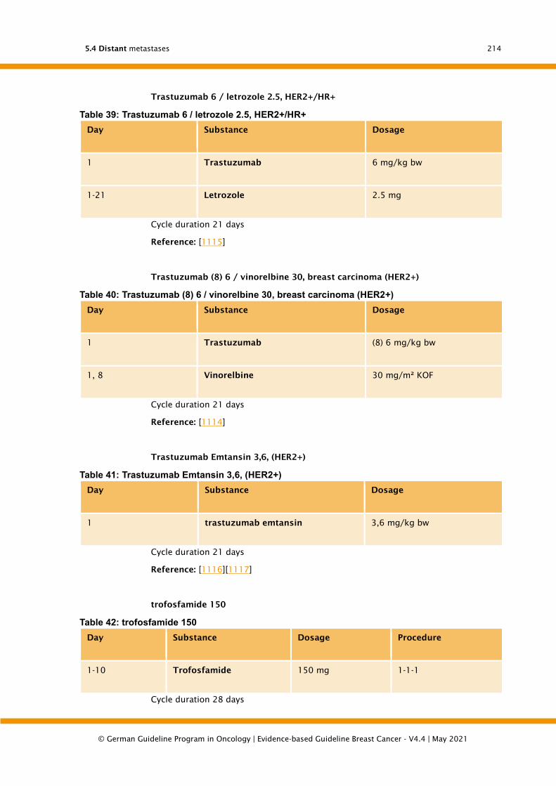
5.4 Distant metastases
© German Guideline Program in Oncology | Evidence-based Guideline Breast Cancer - V4.4 | May 2021
214
Trastuzumab 6 / letrozole 2.5, HER2+/HR+
Table 39: Trastuzumab 6 / letrozole 2.5, HER2+/HR+
Day Substance Dosage
1 Trastuzumab 6 mg/kg bw
1-21 Letrozole 2.5 mg
Cycle duration 21 days
Reference: [1115]
Trastuzumab (8) 6 / vinorelbine 30, breast carcinoma (HER2+)
Table 40: Trastuzumab (8) 6 / vinorelbine 30, breast carcinoma (HER2+)
Day Substance Dosage
1 Trastuzumab (8) 6 mg/kg bw
1, 8 Vinorelbine 30 mg/m² KOF
Cycle duration 21 days
Reference: [1114]
Trastuzumab Emtansin 3,6, (HER2+)
Table 41: Trastuzumab Emtansin 3,6, (HER2+)
Day Substance Dosage
1 trastuzumab emtansin 3,6 mg/kg bw
Cycle duration 21 days
Reference: [1116][1117]
trofosfamide 150
Table 42: trofosfamide 150
Day Substance Dosage Procedure
1-10 Trofosfamide 150 mg 1-1-1
Cycle duration 28 days
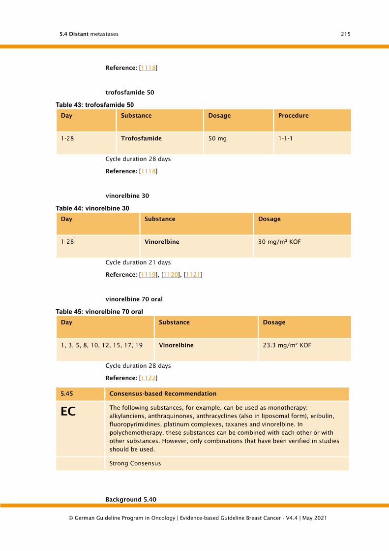
5.4 Distant metastases
© German Guideline Program in Oncology | Evidence-based Guideline Breast Cancer - V4.4 | May 2021
215
Reference: [1118]
trofosfamide 50
Table 43: trofosfamide 50
Day Substance Dosage Procedure
1-28 Trofosfamide 50 mg 1-1-1
Cycle duration 28 days
Reference: [1118]
vinorelbine 30
Table 44: vinorelbine 30
Day Substance Dosage
1-28 Vinorelbine 30 mg/m² KOF
Cycle duration 21 days
Reference: [1119], [1120], [1121]
vinorelbine 70 oral
Table 45: vinorelbine 70 oral
Day Substance Dosage
1, 3, 5, 8, 10, 12, 15, 17, 19 Vinorelbine 23.3 mg/m² KOF
Cycle duration 28 days
Reference: [1122]
5.45 Consensus-based Recommendation
EC The following substances, for example, can be used as monotherapy:
alkylanciens, anthraquinones, anthracyclines (also in liposomal form), eribulin,
fluoropyrimidines, platinum complexes, taxanes and vinorelbine. In
polychemotherapy, these substances can be combined with each other or with
other substances. However, only combinations that have been verified in studies
should be used.
Strong Consensus
Background 5.40

5.4 Distant metastases
© German Guideline Program in Oncology | Evidence-based Guideline Breast Cancer - V4.4 | May 2021
216
Due to the heterogeneity of the metastases and the individual courses of disease, no
uniform therapy strategy can be specified. This applies in particular to the cytostatic
treatment of metastasized breast carcinoma. Although monotherapy has lower
remission rates than polychemotherapy, this does not have a significant negative
impact on survival time. Monotherapies are better tolerated, so that monotherapy
should be used whenever possible. Polychemotherapy is only indicated for severe
symptoms, rapid tumor growth and aggressive tumor behavior.
If the patient has not yet received anthracyclines/taxanes in the adjuvant therapy, these
can be used primarily.
Cytostatic therapy should be based on the therapeutic index in cases of incurable
disease, whereby the effect (e.g. symptom control) and side effects of a therapy must
be considered and weighed up. The use of subjectively less stressful monotherapies or
combination therapies is recommended. This is supported by a Cochrane meta-analysis
published in 2015, which showed that there were no significant differences in
progression-free survival and overall survival between combination therapy and
sequential monochemotherapy when compared with sequential monochemotherapy.
The response was significantly higher with combination chemotherapy compared to
sequential monochemotherapy. However, combination chemotherapy also showed a
higher toxicity with regard to the rate of febrile neutropenia. Many mainly non-
haematological side effects were not described in this meta-analysis. In the meta-
analysis, two scenarios of sequential monochemotherapy were described, one change
of monochemotherapy with progression or fixed change of monochemotherapy
without progression after a few cycles. The results were similar for both scenarios, with
the described results referring to the first scenario [1077].
Before and during chemotherapy, the patient's general condition must be assessed
regularly. During therapy, the side effects of this treatment must also be evaluated
regularly. An evaluation of the therapeutic effect by means of imaging should be
performed every 6-12 weeks (interval depending on the spread of the disease, disease
dynamics and clinical situation). In the course of time, the imaging intervals can be
extended in case of remission and good clinical and laboratory chemical assessment
of the disease status. In case of progress or pronounced toxicity, therapy should be
discontinued. The duration of therapy depends on the therapeutic index, whereby
tumor response and therapy-related side effects should be included in the overall
assessment and therapy should only be continued if the assessment is positive.
The dosage as well as the intended time intervals of the therapy should be adapted to
general guidelines of the therapy, i.e. recognized published protocols. Dose-intensified
and high-dose therapies have not yet led to any improvement in effectiveness. Their
use is only acceptable in the context of studies (Cochrane: [800]).

5.4 Distant metastases
© German Guideline Program in Oncology | Evidence-based Guideline Breast Cancer - V4.4 | May 2021
217
5.4.4. Metastatic HER2-positive breast cancer
5.46 Evidence-based Recommendation
GoR
B
Metastatic HER2-positive breast carcinomas should be treated with anti-HER2
therapy, provided that there are no cardiac contraindications.
LoE
1a
[1123]; [1124]
Strong Consensus
5.47 Evidence-based Recommendation
GoR
B
In the case of a metastasized HER2-positive breast carcinoma, a dual blockade
with trastuzumab / pertuzumab and a taxane should be used in first-line
therapy.
LoE
1b
[1123]
Consensus
5.48 Evidence-based Recommendation
GoR
B
In the case of a metastasized HER2-positive breast carcinoma, therapy with T-
DM1 should be used in second-line therapy.
LoE
1b
[1123]
Consensus
5.4.5. Specific metastatic localization
5.4.5.1. Basic management of distance metastases
5.49 Consensus-based Recommendation
EC The indication for surgical or local ablative therapy of distant metastases should
be determined individually and in an interdisciplinary consultation.
Strong Consensus
Background 5.44

5.4 Distant metastases
© German Guideline Program in Oncology | Evidence-based Guideline Breast Cancer - V4.4 | May 2021
218
In individual cases, patients with oligometastasis (staging), good performance status
and a long interval between initial therapy can benefit from local therapy [1125].
However, the prognostic relevance compared to an adequate systemic therapy is
unclear, since only retrospective studies with strictly preselected patients are available.
In the following chapter, recommendations of the S3 guideline Supportive Therapy of
Oncology Patients (Version 1, 2016) are listed at several points. The guideline group is
of the opinion that these recommendations can be applied directly to patients with
breast cancer. A renewed consensus on the recommendations by the breast cancer
guideline group was not reached.
Caution: If this recommendation in the S3 guideline Supportive Therapy of Oncological
Patients (current version at http://leitlinienprogramm-onkologie.de/Supportive-
Therapie.95.0.html) is changed, for example in the context of an update, these
recommendations also lose their validity at this point.
5.4.5.2. Specific management of bone metastases
For the diagnosis and therapy of skeletal metastases, please refer to the S3 guideline
Supportive Therapy in Oncology Patients (http://leitlinienprogramm-
onkologie.de/Supportive-Therapie.95.0.html).
Osseous metastases occur very frequently (> 50%) in patients with breast cancer.
Associated skeletal complications / skeletal related events (SREs) such as pathological
fractures, pain with increased analgesic consumption, spinal compression syndromes
or hypercalcemia require effective and often interdisciplinary therapy. In symptomatic
tumor patients, a bone scintigraphy is suitable for diagnosis to assess the spread of
osseous involvement. If there is a risk of fractures in particular, an x-ray-based
procedure (native x-ray / CT) should be performed. An MRI should be performed if soft
tissue is involved or in the case of neurological deficits, especially myelocompression.
In case of detection of a newly occurring osseous manifestation, a new spread diagnosis
is necessary. These different imaging techniques have different sensitivities and
specificities in the diagnosis of bone metastases [1126].
Depending on the urgency and the aim of the therapy, the proposal of a therapy should
be determined on an interdisciplinary basis by the surgeon, radio-oncologist, nuclear
medicine specialist, internal oncologist, oncology specialist, pain therapist and, if
necessary, representatives of other specialist disciplines.
The following treatment options are available for patients with osseous manifestations:
• Medicated pain therapy
• Local radiation
• Surgical intervention
• Systemic tumour therapy
o Cytostatic drugs, hormone therapy, targeted substances,
immunomodulating therapy, etc.
o Radionuclides
o Bisphosphonates or RANK ligand antibodies
In the case of painful bone metastases, the first priority is consistent medicinal
analgesia. For stable osseous manifestations including stable vertebral body
manifestations without evidence of myelon compression, conservative therapy (e.g.
systemic tumor therapy, radiotherapy, radionuclide therapy, bisphosphonates/RANK
ligand antibodies) is indicated. For patients with myelon compression and neurological
symptoms, surgery followed by radiotherapy or radiotherapy alone is available.
Therapy recommendations should be made on an interdisciplinary basis, with special
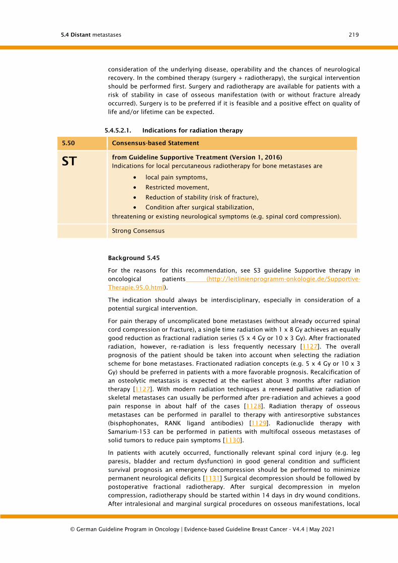
5.4 Distant metastases
© German Guideline Program in Oncology | Evidence-based Guideline Breast Cancer - V4.4 | May 2021
219
consideration of the underlying disease, operability and the chances of neurological
recovery. In the combined therapy (surgery + radiotherapy), the surgical intervention
should be performed first. Surgery and radiotherapy are available for patients with a
risk of stability in case of osseous manifestation (with or without fracture already
occurred). Surgery is to be preferred if it is feasible and a positive effect on quality of
life and/or lifetime can be expected.
5.4.5.2.1. Indications for radiation therapy
5.50 Consensus-based Statement
ST from Guideline Supportive Treatment (Version 1, 2016)
Indications for local percutaneous radiotherapy for bone metastases are
• local pain symptoms,
• Restricted movement,
• Reduction of stability (risk of fracture),
• Condition after surgical stabilization,
threatening or existing neurological symptoms (e.g. spinal cord compression).
Strong Consensus
Background 5.45
For the reasons for this recommendation, see S3 guideline Supportive therapy in
oncological patients (http://leitlinienprogramm-onkologie.de/Supportive-
Therapie.95.0.html).
The indication should always be interdisciplinary, especially in consideration of a
potential surgical intervention.
For pain therapy of uncomplicated bone metastases (without already occurred spinal
cord compression or fracture), a single time radiation with 1 x 8 Gy achieves an equally
good reduction as fractional radiation series (5 x 4 Gy or 10 x 3 Gy). After fractionated
radiation, however, re-radiation is less frequently necessary [1127]. The overall
prognosis of the patient should be taken into account when selecting the radiation
scheme for bone metastases. Fractionated radiation concepts (e.g. 5 x 4 Gy or 10 x 3
Gy) should be preferred in patients with a more favorable prognosis. Recalcification of
an osteolytic metastasis is expected at the earliest about 3 months after radiation
therapy [1127]. With modern radiation techniques a renewed palliative radiation of
skeletal metastases can usually be performed after pre-radiation and achieves a good
pain response in about half of the cases [1128]. Radiation therapy of osseous
metastases can be performed in parallel to therapy with antiresorptive substances
(bisphophonates, RANK ligand antibodies) [1129]. Radionuclide therapy with
Samarium-153 can be performed in patients with multifocal osseous metastases of
solid tumors to reduce pain symptoms [1130].
In patients with acutely occurred, functionally relevant spinal cord injury (e.g. leg
paresis, bladder and rectum dysfunction) in good general condition and sufficient
survival prognosis an emergency decompression should be performed to minimize
permanent neurological deficits [1131] Surgical decompression should be followed by
postoperative fractional radiotherapy. After surgical decompression in myelon
compression, radiotherapy should be started within 14 days in dry wound conditions.
After intralesional and marginal surgical procedures on osseous manifestations, local

5.4 Distant metastases
© German Guideline Program in Oncology | Evidence-based Guideline Breast Cancer - V4.4 | May 2021
220
radiation therapy should be performed. Postoperative radiotherapy after stabilising
procedures should be carried out after completion of wound healing. No preoperative
radiotherapy should be performed on the extremities or the spinal column in the
surgical area.
5.4.5.2.2. Indications for surgical tretment
5.51 Consensus-based Recommendation
EC from Guideline Supportive Treatment (Version 1, 2016)
Indications for surgical therapy of osseous manifestations may be:
• Myelon compression with neurological symptoms,
• pathological fracture,
• imminent fracture (fracture risk e.g. via Mirel Score, Spinal Instability
Neoplastic Scale (SINS)),
• solitary late metastasis,
• radiation-resistant osteolysis,
• therapy-resistant pain.
Strong Consensus
Background 5.46
For the justification of this recommendation, see S3 guideline Supportive therapy in
oncological patients (http://leitlinienprogramm-onkologie.de/Supportive-
Therapie.95.0.html).
A biopsy under established conditions [1132] is to be performed in cases of osseous
manifestations:
• First-time imaging evidence of possible (also multiple) skeletal metastasis in a
long-past primary tumour
• Isolated bone lesion with potential curative therapy approach without further
metastases
• Radiological and/or clinical findings are not consistent with the diagnosis of
bone metastasis (differential diagnoses must be considered)
The indication for a surgical intervention should be interdisciplinary, in particular in
consideration of a potential radiotherapeutic intervention.
Pathological fractures that have already occurred or are imminent represent the main
indication for surgical therapy in the area of the extremities. In the presence of a
pathological fracture of the lower extremities of a previously mobile patient, there is
an indication for surgical therapy. When choosing the appropriate surgical procedure
for osseous manifestations in the extremities, the patient's prognosis must be taken
into account in addition to the entity and radiation sensitivity of the tumor.
In stable vertebral body metastases or manifestations without evidence of myelon
compression, conservative therapy (systemic tumor therapy, radiotherapy, radionuclide
therapy, bisphosphonate / RANK ligand antibodies) is preferable to surgery.
In the case of therapy-refractory pain with imminent or actual compression fracture of
one or more vertebral bodies in the thoracic spinal canal and lumbar spine without
invasion of the tumor into the spinal canal, rapid pain reduction can be achieved by
percutaneous cement augmentation with vertebroplasty or kyphoplasty [1133], [1134].

5.4 Distant metastases
© German Guideline Program in Oncology | Evidence-based Guideline Breast Cancer - V4.4 | May 2021
221
Patients with acutely occurred, functionally relevant compression-related spinal cord
injury (e.g. proximal leg paresis, bladder and rectum dysfunction) in good general
condition and sufficient survival prognosis should undergo emergency decompression
to minimize permanent neurological deficits. Surgical decompression should be
followed by postoperative radiotherapy with 10 x 3 Gy. After surgical decompression
in myelon compression, radiotherapy should be started within 14 days in dry wound
conditions [1131].
5.4.5.2.3. Bone protective therapy
5.52 Evidence-based Recommendation
GoR
B
from Supportive Guideline (Version 1, 2016)
To prevent complications in osseous manifestations, an osteoprotective therapy
with bisphosphonates/denosumab should be performed.
LoE
1a
[871]
Strong Consensus
Background 5.47
For the reasons for this recommendation see S3 guideline Supportive Therapy in
Oncological Patients (http://leitlinienprogramm-onkologie.de/Supportive-
Therapie.95.0.html).
Therapy with bisophosphonates/RANK ligand antibodies in osseous manifestations can
delay or prevent the occurrence of skeletal-related events (SREs) [1135], [1136]. To
prevent complications in osseous manifestations an osteoprotective therapy with
bisphosphonates/denosumab should be performed. In patients with osseous
metastases in breast cancer and after one year of zoledronate therapy in 4-weekly
intervals the subsequent administration of zoledronate should be every 12 weeks
[1137].
In patients with osseous metastases of breast cancer, the administration of denosumab
compared to zoledronate leads to a numerically small, statistically significant reduction
of SREs [1138]. For other outcome parameters (e.g. pain, QoL, spinal compression,
mortality, AE maxillary osteonecrosis) there is no evidence for a difference. Data on the
comparison of Denosumab versus the other bisphosphonates are not available. In order
to prevent osteonecrosis of the jaw, a dental examination and any necessary dental
rehabilitation should be carried out before the administration of bisphosphonates or
Denosumab, and the patient should be instructed and motivated to maintain above-
average (careful and regular) oral hygiene. In addition, regular risk-adapted dental
examinations should be performed [1139].
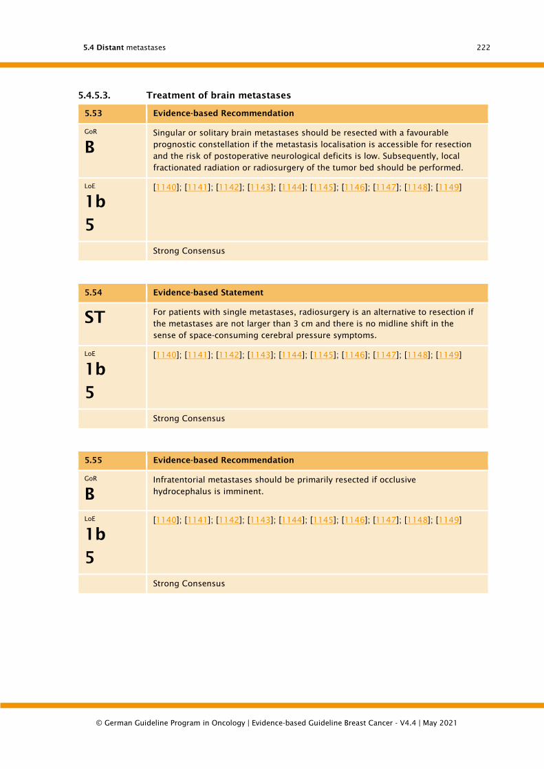
5.4 Distant metastases
© German Guideline Program in Oncology | Evidence-based Guideline Breast Cancer - V4.4 | May 2021
222
5.4.5.3. Treatment of brain metastases
5.53 Evidence-based Recommendation
GoR
B
Singular or solitary brain metastases should be resected with a favourable
prognostic constellation if the metastasis localisation is accessible for resection
and the risk of postoperative neurological deficits is low. Subsequently, local
fractionated radiation or radiosurgery of the tumor bed should be performed.
LoE
1b
5
[1140]; [1141]; [1142]; [1143]; [1144]; [1145]; [1146]; [1147]; [1148]; [1149]
Strong Consensus
5.54 Evidence-based Statement
ST For patients with single metastases, radiosurgery is an alternative to resection if
the metastases are not larger than 3 cm and there is no midline shift in the
sense of space-consuming cerebral pressure symptoms.
LoE
1b
5
[1140]; [1141]; [1142]; [1143]; [1144]; [1145]; [1146]; [1147]; [1148]; [1149]
Strong Consensus
5.55 Evidence-based Recommendation
GoR
B
Infratentorial metastases should be primarily resected if occlusive
hydrocephalus is imminent.
LoE
1b
5
[1140]; [1141]; [1142]; [1143]; [1144]; [1145]; [1146]; [1147]; [1148]; [1149]
Strong Consensus
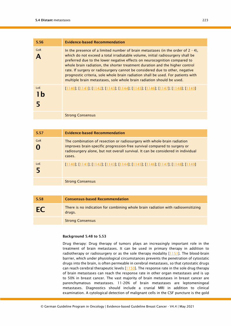
5.4 Distant metastases
© German Guideline Program in Oncology | Evidence-based Guideline Breast Cancer - V4.4 | May 2021
223
5.56 Evidence-based Recommendation
GoR
A
In the presence of a limited number of brain metastases (in the order of 2 - 4),
which do not exceed a total irradiatable volume, initial radiosurgery shall be
preferred due to the lower negative effects on neurocognition compared to
whole brain radiation, the shorter treatment duration and the higher control
rate. If surgery or radiosurgery cannot be considered due to other, negative
prognostic criteria, sole whole brain radiation shall be used. For patients with
multiple brain metastases, sole whole brain radiation should be used.
LoE
1b
5
[1140]; [1141]; [1142]; [1143]; [1144]; [1145]; [1146]; [1147]; [1148]; [1149]
Strong Consensus
5.57 Evidence-based Recommendation
GoR
0
The combination of resection or radiosurgery with whole-brain radiation
improves brain-specific progression-free survival compared to surgery or
radiosurgery alone, but not overall survival. It can be considered in individual
cases.
LoE
5
[1140]; [1141]; [1142]; [1143]; [1144]; [1145]; [1146]; [1147]; [1148]; [1149]
Strong Consensus
5.58 Consensus-based Recommendation
EC There is no indication for combining whole brain radiation with radiosensitizing
drugs.
Strong Consensus
Background 5.48 to 5.53
Drug therapy: Drug therapy of tumors plays an increasingly important role in the
treatment of brain metastases. It can be used in primary therapy in addition to
radiotherapy or radiosurgery or as the sole therapy modality [1151]. The blood-brain
barrier, which under physiological circumstances prevents the penetration of cytostatic
drugs into the brain, is often permeable in cerebral metastases, so that cytostatic drugs
can reach cerebral therapeutic levels [1150]. The response rate in the sole drug therapy
of brain metastases can reach the response rate in other organ metastases and is up
to 50% in breast cancer. The vast majority of brain metastases in breast cancer are
parenchymatous metastases. 11-20% of brain metastases are leptomeningeal
metastases. Diagnostics should include a cranial MRI in addition to clinical
examination. A cytological detection of malignant cells in the CSF puncture is the gold
5.4 Distant metastases
© German Guideline Program in Oncology | Evidence-based Guideline Breast Cancer - V4.4 | May 2021
224
standard for the diagnosis of a leptomeningeal metastasis. In the case of a negative
CSF puncture if leptomeningeal metastasis is still suspected, the CSF puncture should
be repeated [1152]. In general, the treatment of patients with solid brain metastases
[1153] and leptomeningeal metastases [1152] is based on the premise that the
systemic therapy should be based on the primary tumor and its molecular
characteristics. In a systematic review of the therapy of leptomenin-geal metastases in
breast cancer Scott et al. could show in 36 studies with a total of 851 patients with
meningeosis carcinomatosa that 87% had been treated with intrathecal chemotherapy
and the median overall survival was 15 weeks [1154]. In addition to 18 retrospective
studies and 13 case collections, 5 prospective randomized studies were included in
this current review. In the only prospective randomized study that exclusively included
breast cancer patients, 35 patients with breast cancer and meningeosis carcinomatosa
were randomized to systemic therapy + radiotherapy with and without additional
intrathecal chemotherapy (ITC) [1155]. The systemic therapy was based on hormone
receptor status, previous therapies and general condition. Radiotherapy was performed
as whole brain radiation if clinically indicated. Systemic therapy and radiotherapy were
well balanced between both therapy arms. ITC was performed with methotrexate and
applied via an ommaya reservoir. Neurological stabilisation or improvement was
described in 59% (ITC) vs. 67% (no ITC). Median time to progression 23 (ITC) vs. 24 (no
ITC) weeks. Overall survival was 18.3 weeks vs. 30.3 weeks (no ITC) (p=0.32) for those
patients who were additionally treated with ITC. Therapy related neurological
complications were found in 47% (ITC) vs. 6% (no ITC) (p=0.0072). The authors
concluded that intrathecal chemotherapy in addition to adequate systemic therapy and
possibly radiotherapy in breast cancer patients with meningeosis carcinomatosa does
not prolong survival but leads to an increased risk of therapy-related neurological
complications.
In breast cancer, more than a third of patients with HER2-positive tumours develop
brain metastases when treated with targeted therapies. One reason could be that these
tumours preferentially metastasise to the brain, where they are protected from the
action of antibodies by the blood-brain barrier. Antibody-based therapy is also effective
for existing brain metastases in HER2-positive patients, and trastuzumab has been
shown to penetrate brain metastases. The EGFR/HER2 inhibitor lapatinib is also
effective in the treatment of brain metastases, but cannot prevent the occurrence of
brain metastases. In monotherapy the substance has an objective response rate of only
6% [1156]. In combination with capecitabine the response rates are up to 38% [1157],
[1158], [1159]. Even higher response rates of more than 60% were reported in non-
irradiated patients treated with lapatinib and capecitabine [1160]. The occurrence of
brain metastases as the first progression event in metastatic HER2-positive breast
cancer was comparable between lapatinib / capecitabine vs. trastuzumab /
capecitabine (3% vs. 5%) - while progression-free survival (HR 1.30) was prolonged
under trastuzumab / capecitabine [1161]. In a retrospective analysis of the EMILIA
study, the cerebral progression rate between trastuzumab emtansine (T-DM1) vs.
capecitabine / lapatinib was comparable (without cerebral metastases at baseline 2%
vs. 0.7%; with previously known cerebral metastases 22.2% vs. 16.0%). Overall survival
in patients with cerebral metastases at baseline was significantly extended by T-DM1
compared to capecitabine / lapatinib (HR 0.38; 26.8 vs. 12.9 months) [1162].
Overall, it must be emphasized that even after the occurrence of brain metastases, a
continuation of the HER2 blockade may be useful to keep systemic metastases under
control [1163], [1164].
Breast carcinoma is the second most common cause of CNS metastases (after lung
carcinomas). Data on the actual incidence vary in the literature between 15-20%; in
5.4 Distant metastases
© German Guideline Program in Oncology | Evidence-based Guideline Breast Cancer - V4.4 | May 2021
225
autopsy collectives up to 40% [1150], [1165]. In recent years an increasing incidence
of brain metastasis in patients with breast cancer has been observed. The increasing
incidence is attributed to longer survival due to better control of the underlying disease
and extracranial metastases as well as improved diagnostic measures (MRI) and their
greater use. Patients with triple-negative histology and HER2-positive status were
identified as risk factors in subgroup analyses [1166]. A single metastasis in the brain
with simultaneously detectable metastases in other organs is described as singular,
while singular cerebral metastasis is described as the only (detected) metastasis in the
organism.
The overall prognosis is limited with a median survival time of 2 to 25 months.
However, individual patients survive several years without relapse. Particularly in the
case of isolated brain metastases occurring late in the course of the disease, intensified
therapy seems justified. By means of a recursive partition analysis of the Radiation
Therapy Oncology Group (RTOG) on a collective with brain metastases (predominantly
from lung cancer) in the USA, 3 prognosis classes (RPA Class I - III) based on whole
brain radiation were defined as therapeutic measures according to simple prognostic
criteria (age 70, systemic disease controlled). With the help of these three classes, the
median survival between less than 2 or longer than 7 months can be estimated [1167].
Based on more recent data, a new score, the so-called Graded Prognostic Assessment
(GPA), was proposed in 2012 [1168], again based on whole brain radiation, which was
supplemented by primary tumor-specific indices specifically for breast cancer. Using
the breast carcinoma-specific score (allocation of points for Karnofsky index,
histological subtype, age), the median survival times for GPA 0-1: 3.4 months, for GPA
1.5-2: 7.7 months, for GPA 3: 15 months and for GPA 3.5-4: 25.3 months.
In addition to the above-mentioned prognosis factors, the therapy strategy depends on
the number, location and size of the brain metastases. The main therapy modalities
include resection, radiosurgery, fractional radiotherapy, e.g. as stereotactically guided,
localized radiotherapy or as whole brain radiation, and drug therapy of tumors. When
assessing the effect of local therapies in clinical trials, it should be noted that median
survival time is not only influenced by the effectiveness of the treatment of brain
metastases, but also by the consequences of systemic tumor progression.
Surgery: Surgery has a safe value in the therapy of brain metastases [1140]. The median
survival improves by 6-9 months compared to whole brain radiation alone when
resecting single metastases and subsequent whole brain radiation. In symptomatic
large metastases the palliative effect of the resection is important. Two of 3
randomized studies concluded that resection of single or solitary metastases followed
by whole brain radiation is superior to sole whole brain radiation [1141], [1142],
[1143]. A third study did not prove the value of surgery [1169]. However, this
discrepancy can be explained by later surgery in the radiotherapy arm and an overall
prognostically unfavorable patient population. Numerous retrospective analyses also
speak in favor of resection of single or solitary metastases [1170]. However, the local
recurrence rates after sole resection are relatively high at 60% [1148]. Local as well as
distant recurrences are reduced if whole-brain radiation is followed by surgery [1143],
[1148]. Whether whole-brain radiation can be replaced by a hypofractionated,
circumscribed postoperative follow-up radiation of the postoperative tumor bed to
avoid the negative effects of whole-brain radiation is the subject of ongoing studies. In
all the studies cited, the group of breast cancer patients represented a relevant
subgroup; data exclusively on the therapy of brain metastases in breast cancer do not
exist.
Radiosurgery: The percutaneous stereotactic application of single high radiation doses
(radiosurgery) is a standard therapy in the treatment of brain metastases. The high
5.4 Distant metastases
© German Guideline Program in Oncology | Evidence-based Guideline Breast Cancer - V4.4 | May 2021
226
radiation dose often leads to good tumor control, while the surrounding healthy tissue
is well protected by the steep dose drop to the periphery. Radiosurgery thus represents
an alternative to neurosurgical resection. At present, radiosurgery is mostly used as
primary treatment of single or multiple lesions with a diameter of up to 30 mm (or 15
ml) or as recurrence treatment in patients who show a recurrence in a previously
conventionally irradiated region. Local control rates range from 73-94%. Retrospective
cohort studies indicate an equivalence of radiosurgery and neurosurgical resection.
Comparative studies have not yet been published. However, it must be emphasized
that local control seems to be better after radiosurgery. In EORTC study 22952-26001
the cumulative incidence of local progression was 59% after resection compared to 31%
after radiosurgery [1148].
The decision depends on clinical circumstances (per surgery: histological backup, mass
effect, per radiosurgery: applicability in any localization, even in functionally non-
resectable lesions such as brainstem).
Radiation therapy for limited (1-4 brain metastases) and multiple metastases,
significance of whole brain radiation: On the question of the combination of
radiosurgery alone or with additional whole-brain radiation, there are now data from 6
randomized studies [1144], [1145], [1146], [1147], [1148], [1149]. Here, too, the size
of the randomized breast cancer patients was almost 20%, in addition to patients with
lung cancer as the largest group. The first two studies unanimously showed an
increased local metastasis control and "in-brain-control" (prevention of new intracranial
metastases) in combination with radiosurgery and whole brain radiation [1144], [1145].
In the three subsequent studies [1146], [1147], [1148], higher control rates were also
shown due to the resulting dose escalation through the combination of radiosurgery
and whole-brain radiation, but this was not accompanied by a survival advantage of the
whole collective and the neurocognitive endpoints showed a substantial deterioration
through the addition of whole-brain radiation. In two more recent studies,
neurocognitive endpoints were placed in the foreground of the evaluation [1147],
[1149]. Due to the deterioration of neurocognition with simultaneous lack of survival
advantage, current recommendations of the American professional societies are to
initially withhold or postpone whole brain radiation in favor of radiosurgery, despite
the improved tumor control proven in all studies (using regular follow-up MRIs). Since
modern radiosurgical techniques are now available to perform this also in the case of
multiple brain metastases (up to 10), the side effects of whole-brain radiation are
increasingly questioned even in the case of multiple metastases [1171]. In this respect,
initial radiosurgery may be preferred in the case of more than 4 lesions (taking into
account the pattern of infection and localization) in order to avoid the negative effects
of whole-brain radiation on neurocognition or to provide whole-brain radiation as a
salvage option.
Which patients will benefit from additional whole brain radiation in the future due to
prognosis or infestation pattern, also with the help of the technically possible
hippocampal sparing in order to avoid negative effects on neurocognition, is the
subject of currently ongoing studies.
Whole brain radiation: The sole fractionated whole brain radiation (usually fractionated
with 10 x 3 Gy) remains the therapy of choice for patients with multiple brain
metastases, limited prognosis group and reduced general condition, in addition to the
alternative of refraining from therapy (best supportive care). In this clinical
constellation, whole brain radiation improves the median survival time from 2 months
with purely supportive therapy to 3-6 months. It leads to an improvement in tumour-
related neurological symptoms and quality of life. If favourable prognostic factors are
present, treatment should be performed normo-fractionated with 5 fractions per week,
5.4 Distant metastases
© German Guideline Program in Oncology | Evidence-based Guideline Breast Cancer - V4.4 | May 2021
227
2 Gy per fraction up to a total dose of 36-44 Gy in order to avoid neurotoxic late effects
of radiation therapy and to achieve an extension of the neurological remission time.
Since the hippocampus is considered a sensitive region with regard to the occurrence
of neurocognitive late effects, it is currently being investigated whether modern
radiation techniques (intensity-modulated radiotherapy, IMRT) allow bilateral sparing
of the hippocampus ("hippocampal sparing") without compromising cerebral tumor
control.
5.59 Consensus-based Recommendation
EC In the case of cerebral metastases, systemic therapy (chemotherapy / endocrine
therapy / anti-HER2 therapy) should be used in addition to local therapy (surgery
/ radiotherapy).
Strong Consensus
5.4.5.4. Treatment of liver metastases
5.60 Evidence-based Recommendation
GoR
0
If liver metastases are present, in individual cases a resection or possibly
another local therapy (RFA, TACE, SBRT, SIRT) may be indicated:
• no disseminated metastases
• controlled extrahepatic metastasis
LoE
3b
[1172]; [1173]; [1174]; [1175]; [1176]; [1177]; [1178]; [1179]; [1180]; [1181];
[1182]; [1183]
Strong Consensus
Background 5.55
If a limited number, especially isolated liver metastases occur in the liver, a metastasis
resection can be performed. Alternatively, radiofrequency ablation, transarterial
chemoembolization (TACE) [1184] or stereotactic radiation (SBRT) may be considered,
although even less data are available. Factors that positively influence the decision to
resect are Time interval after primary treatment > 12 months, good response to
systemic therapy and estrogen-receptor-positive disease. Systemic therapy should be
followed after surgical therapy. A prerequisite for local liver metastasis therapy is
usually the exclusion of extrahepatic metastases as well as a local/local/regional
recurrence and secondary carcinomas. As a locoregional therapy method, the Yttrium-
90-Radioembolization (SIRT) [1185] can be considered for hepatic metastases that
exceed the possibilities of local procedures in terms of number and size. All in all, the
data required for SIRT are lacking to prove a patient-relevant benefit.
In individual cases, liver resection or interventional liver therapy may be indicated
within the framework of the overall oncological concept even in the case of multiple or
bilateral liver metastases or also in the case of limited but stable extrahepatic tumors
[1186], [1187], [1188], [1189]. Even if a curative approach cannot always be pursued
with the mentioned procedures, a progression-free time may be achieved under certain
circumstances.
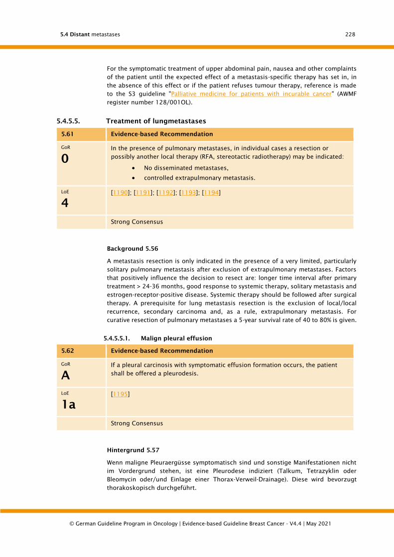
5.4 Distant metastases
© German Guideline Program in Oncology | Evidence-based Guideline Breast Cancer - V4.4 | May 2021
228
For the symptomatic treatment of upper abdominal pain, nausea and other complaints
of the patient until the expected effect of a metastasis-specific therapy has set in, in
the absence of this effect or if the patient refuses tumour therapy, reference is made
to the S3 guideline "Palliative medicine for patients with incurable cancer" (AWMF
register number 128/001OL).
5.4.5.5. Treatment of lungmetastases
5.61 Evidence-based Recommendation
GoR
0
In the presence of pulmonary metastases, in individual cases a resection or
possibly another local therapy (RFA, stereotactic radiotherapy) may be indicated:
• No disseminated metastases,
• controlled extrapulmonary metastasis.
LoE
4
[1190]; [1191]; [1192]; [1193]; [1194]
Strong Consensus
Background 5.56
A metastasis resection is only indicated in the presence of a very limited, particularly
solitary pulmonary metastasis after exclusion of extrapulmonary metastases. Factors
that positively influence the decision to resect are: longer time interval after primary
treatment > 24-36 months, good response to systemic therapy, solitary metastasis and
estrogen-receptor-positive disease. Systemic therapy should be followed after surgical
therapy. A prerequisite for lung metastasis resection is the exclusion of local/local
recurrence, secondary carcinoma and, as a rule, extrapulmonary metastasis. For
curative resection of pulmonary metastases a 5-year survival rate of 40 to 80% is given.
5.4.5.5.1. Malign pleural effusion
5.62 Evidence-based Recommendation
GoR
A
If a pleural carcinosis with symptomatic effusion formation occurs, the patient
shall be offered a pleurodesis.
LoE
1a
[1195]
Strong Consensus
Hintergrund 5.57
Wenn maligne Pleuraergüsse symptomatisch sind und sonstige Manifestationen nicht
im Vordergrund stehen, ist eine Pleurodese indiziert (Talkum, Tetrazyklin oder
Bleomycin oder/und Einlage einer Thorax-Verweil-Drainage). Diese wird bevorzugt
thorakoskopisch durchgeführt.

5.5 Palliative medicine
© German Guideline Program in Oncology | Evidence-based Guideline Breast Cancer - V4.4 | May 2021
229
Zur symptomatischen Therapie von Dyspnoe, Schmerzen u. a. Beschwerden der
Patientin bis zum Einsetzen der erwarteten Wirkung einer metastasenspezifischen
Therapie, bei Ausbleiben dieser Wirkung oder wenn die Patientin eine Tumortherapie
ablehnt, wird auf die S3-Leitlinie „Palliativmedizin für Patienten mit einer nicht heilbaren
Krebserkrankung" (AWMF-Registernummer 128/001OL) verwiesen.
5.4.5.6. Cutaneous and soft tissue metastases
5.63 Consensus-based Recommendation
EC If skin and soft tissue metastases occur, surgical excision or other local therapy
(e.g. radiotherapy) may be considered.
Strong Consensus
Background 5.58
Circumscribed skin metastases and metastases of soft tissue can be excised in healthy
individuals or locally irradiated percutaneously. If the findings are not very extensive,
topical procedures can be discussed [1196], [1197], [1198]. If necessary,
electrochemotherapy can also be used [1199], [1200], [1201].
Exulcerative tumor growth / Cancer en Cuirasse:
Breast carcinoma is the malignant tumor which most frequently develops skin
metastases, which already in curable stages of the disease lead to considerable
symptom burden with impairment of the patient's quality of life [1202] Cancer en
Cuirasse is a distinct, aggressive, nodular plate-like, partly exulcerated manifestation
with consecutive lymphedema. It is often accompanied by pain, itching, bleeding and
foetal odor. Due to the thick fibrotic manifestation with reduced vascularization of the
carapace-like skin metastases, the causal therapy options are limited. As a rule,
exudative skin metastases or Cancer en Cuirasse are non-curative stages of the disease.
Patients are severely affected by the physical disfigurement, limited movement, pain,
weeping of the wound and the sometimes very unpleasant odour. Palliative care aims
to relieve all these symptoms. This requires complex interventions (wound care plus
psychosocial support) [1198], [1202], [1203], [1204], [1205].
5.5. Palliative medicine
5.64 Consensus-based Recommendation
EC In the palliative situation, all necessary measures shall be geared to the patient's
individual therapy and life goals.
Strong Consensus
5.65 Consensus-based Statement
ST The right time to supplement regular care with palliative care depends primarily
on the patient's needs and individual disease-related requirements.
Strong Consensus

5.5 Palliative medicine
© German Guideline Program in Oncology | Evidence-based Guideline Breast Cancer - V4.4 | May 2021
230
5.66 Consensus-based Statement
ST Palliative care is characterised by a multi-professional and interdisciplinary
approach.
Strong Consensus
5.67 Consensus-based Recommendation
EC The following principles shall be applied in the palliative care of patients with an
incurable breast cancer disease:
• consideration of and response to the patient's needs in all four
dimensions (physical, psychological, social, spiritual)
• the consideration of patients' preferences
• the determination of realistic therapy goals
• Knowledge of the organizational forms of palliative care
• the creation of conditions that respect the intimacy of the patient
Strong Consensus
5.68 Consensus-based Statement
ST Palliative care includes medical symptom control, palliative care and
psychosocial support until death. It is provided as general or specialised
palliative care according to need.
Strong Consensus
Background 5.59 to 5.63
Palliative medicine/palliative care (synonym: palliative care) pursues the goal of
improving the quality of life of patients with a life-threatening disease and their
relatives. This is achieved by prevention and alleviation of suffering, by early
recognition and treatment of physical, psychological, social and spiritual problems
[1207], [1208], [1209], [1210], [1211], [1206], [1212], [1213], [1214], [1215], [1216],
[1217], [1218], [1219], [1220]. This is done by preventing and alleviating suffering, by
early recognition, careful assessment and treatment of pain and other distressing
symptoms of a physical, psychosocial and spiritual nature [1207], [1208], [1209],
[1210], [1211], [1206], [1212], [1213], [1214], [1215], [1216], [1217], [1218], [1219],
[1220], [1221].
The recommendation is based on the consensus of the technical experts involved.
Otherwise, the guideline group refers to the overarching interdisciplinary S3 cross-
sectional guidelines "Palliative Care for Patients with Non-Curable Cancer" (AWMF
Registry Number 128/001OL), the guideline "Supportive Therapy for Oncological
Patients" (AWMF Registry Number 032/054OL) and the guideline "Psycho-oncological
Diagnosis, Counselling and Treatment of Cancer Patients" (AWMF Registry Number
032/051OL) as well as the corresponding chapters in this guideline.
5.5 Palliative medicine
© German Guideline Program in Oncology | Evidence-based Guideline Breast Cancer - V4.4 | May 2021
231
Palliative care
Palliative care includes medical symptom control, palliative care and psychosocial
support until death [1208], [1209], [1210], [1211], [1206], [1212], [1213], [1214],
[1215], [1216], [1217], [1218], [1219], [1220], [1222]. In the palliative situation, all
necessary measures are oriented towards the individual therapy and life goals of the
patient [1208], [1214]. Graduated palliative care is the necessary prerequisite for
varying the intensity of treatment according to the symptom burden and at the same
time ensuring continuity of palliative support [1223], [1224]. Symptom control,
palliative care and psychosocial support stand for three dimensions of palliative
treatment which must be included in every qualified palliative care offer, regardless of
the level of specialization of the offer [1209]. Palliative care is provided in addition to
(a) disease-modifying therapies with the primary therapeutic goal of prolonging life
(palliative therapy), (b) prophylaxis or treatment of side effects associated with the
disease or with these therapies (supportive therapy) [1207], [1214], [1216], [1220],
[1225], [1226], [1227], [1228] and (c) the needs-based psychosocial and psycho-
oncological care. Specialised co-treatment is advisable for patients in whom a high level
of physical, psychosocial or spiritual stress persists despite general palliative medical
measures. This is characterised by the fact that a team (a) with specialised training (e.g.
palliative care nursing training or additional medical designation palliative medicine),
(b) which (b) is mainly and primarily entrusted with palliative care, (c) is involved in the
treatment of the patient in addition to routine care (S3 guideline AWMF register number
128/001OL). Routine integration of specialized palliative care into local therapy
standards is aimed at in accordance with current international guideline
recommendations [1208], [1214], [1229] in the sense that palliative care is integrated
into treatment and no longer whether this should be done. In Germany, Gärtner et al.
have developed disease-specific indicators for palliative medical counselling and, if
necessary, co-care. These include (1.) metastasized and inoperable breast cancer, (2.)
locally advanced and inoperable breast cancer, or (3.) recurrent disease situations in
which intravenous chemotherapy is administered [1230].
For further information on the question of care structures, reference is made to the S3
guideline on oncological palliative care in the Oncology guideline program (AWMF
register number 128/001OL).
5.5.1. Patients' needs
The highest therapeutic goal in palliative care - the individual quality of life - can only
be evaluated and defined by the patient herself [1231], [1232]. The therapy goal must
be measured against the patient's ideas, priorities and wishes [1233]. The patient's
physical, psychosocial, spiritual and existential burdens can be [1208], [1209], [1210],
[1211], [1206], [1212], [1213], [1214], [1215], [1216], [1217], [1218], [1219], [1220].
Patients with breast cancer have a high need for medical information and psychological
support [1234], [1235], [1236]. It should be noted that the interindividual needs may
be the same, but priorities may be weighted differently. In order to identify
unconsidered needs, a short assessment is routinely performed when palliative care is
called in, which includes the multidimensional needs of the patients according to their
self-assessment [1237]. A validated single-item questionnaire is the "NCCN Distress
Thermometer", while the "Palliative Outcome Scale (POS)" asks for special stresses and
needs in more detail [1238], [1239], [1240]. A structured recording of the physical
symptoms (e.g. Edmonton Symptom Assessments Scale (ESAS)) may also be helpful
[1241]. For all three assessment instruments there are validated versions for Germany
[1242], [1243], [1244]. Recording the patient load improves the need orientation in the
care of patients [1245].
6.1 General concept
© German Guideline Program in Oncology | Evidence-based Guideline Breast Cancer - V4.4 | May 2021
232
A prerequisite for the treatment of patients and a component of palliative basic values
is the high appreciation of patient autonomy and participation [1206], [1215], which,
in addition to the above-mentioned routine recording of the patient's self-assessment
of quality of life and symptom burden, also includes the active inclusion and
comprehensive/adequate information/education of the patient in the decisions for
certain therapy options [1208], [1214]. In principle, the patient has the possibility to
include close relatives in the discussions according to her wishes. In order to enable
the patient to have her confidants be able to represent the presumed will of the patient
as well as possible in the case of possible disturbances of consciousness in the course
of the disease, there is the offer of consultation for writing a "power of attorney for
medical matters" [1233]. Regarding further aspects of palliative care independent of
the underlying diagnosis, reference is made to the S3 guideline "Palliative Care for
Patients with Non-Curable Cancer" of the Oncology Guidelines Program (AWMF Register
Number 128/001OL). It discusses questions of symptom control, palliative care,
psychosocial support and care structures in detail and across all organs.
5.5.2. Family carers' needs
A central point of palliative care is the involvement of those close to the patient. In
addition to family members, this means above all those persons who are important for
the patient in the current situation, regardless of the degree of kinship. In principle,
the patient has the possibility to include the persons she wishes in the discussions.
However, the wishes and fears of the close relatives themselves as well as their needs,
for example for information, also play an important role. With regard to further aspects
of care for close relatives, reference is made to the S3 guideline "Palliative care for
patients with incurable cancer" of the Oncology Guidelines Programme (AWMF register
number 128-001OL).
6. Treatment, support and continuing care
6.1. General concept
The chapters in Section 6 - Treatment, Care, Accompaniment of our patients - have
been redefined and restructured due to the significant development of diagnostic and
therapeutic options in recent years. For example, the start of aftercare has so far
referred to the completion of primary treatment (if necessary, no later than 6 months
after surgery). The definition of this period has changed with the introduction of
neoadjuvant therapies, adjuvant long-term therapies with antibodies and (anti-
)hormones and the modified forms of radiation therapy. Consequently, the content and
timing of the term "aftercare" must be adapted. In addition, there are overlaps between
the various subject areas that exist at different points in time in the care chain for
patients with breast cancer.
The chapter "Treatment, care and support" covers the psychosocial aspects and psycho-
oncology and supportive therapies. Both chapters deal thematically with treatment
situations that begin in the context of primary treatment, but which also seamlessly
move on to long-term support. The chapter on rehabilitation covers the period
following the completion of primary local therapy. This point in time is usually the end
of primary treatment by means of surgery (also after previous neoadjuvant
chemotherapy) or radiotherapy (also after previously carried out adjuvant
chemotherapy).
6.2 Psycho-oncological aspects
© German Guideline Program in Oncology | Evidence-based Guideline Breast Cancer - V4.4 | May 2021
233
Following the completion of the primary local therapy, in particular the completed
radiotherapy, the newly defined aftercare will then follow, focusing on the earliest
possible detection of locoregional or intramammary recurrences, contralateral breast
carcinoma, the targeted search for metastases in the event of symptoms or justified
suspicion, as well as the diagnosis and therapy of side effects and late effects of the
primary and long-term therapies.
The final sections in this restructured chapter are palliative care, which will be
discussed due to its increasing importance, and the not unimportant complementary
medicine.
The tasks of the treating physicians have changed due to the increasing complexity of
the diagnosis and therapy options or the time periods. Although data from prospective
randomised trials with regard to individual aspects such as adverse effects, long-term
toxicity or the use of supportive measures are available, the definition of the
investigations, both in terms of the interval and the type of investigations, is defined
on a study-specific basis. Summarising overall data from prospective randomised
studies for the corresponding evidence-based recommendations for action within the
framework of this changed range of tasks are not available. A generalisation for
everyday clinical practice can therefore only be made after an evaluation of the study
data and the combination of individual aspects. It must also be taken into account that
since 2004, no new prospective randomized studies have been published to test new
methods or time intervals for the diagnosis of locoregional recurrence or distant
metastases. Short-term, almost annual variations in therapy recommendations
therefore make it impossible to change the recommendations on intervals and the type
of diagnosis. Prospective randomized studies to test this have been initiated, so that
the data situation may improve in the near future.
6.2. Psycho-oncological aspects
6.2.1. Basic principles of psycho-oncological care
Today, psycho-oncology is a separate discipline whose task is to scientifically research
the various psychosocial aspects in the development, treatment and course of cancer
in children, adolescents and adults and to apply the relevant findings to the care and
treatment of patients [29], [1246], [1247], [1248]. Psycho-oncology is an integral part
of the care of patients with breast cancer. In Germany, further education and training
curricula are offered by corresponding professional associations to ensure professional
qualification. The addressees of these further training courses are doctors,
psychologists and social pedagogues, who are referred to in the following as psycho-
oncology specialists. An additional psycho-oncological qualification is a prerequisite
for recognition as a psycho-oncological specialist [1249]. The psycho-oncological care
of breast cancer patients should be implemented in an interdisciplinary manner
between all professional groups involved in the treatment. This implies that a psycho-
oncological specialist is integrated into the treatment team in the respective care
setting (inpatient and outpatient treatment, inpatient rehabilitation, outpatient
aftercare) and is in regular contact with the medical staff. This exchange should be
regulated and structured in the form of case discussions or ward conferences [1250],
[1251], [1252] Detailed aspects of psycho-oncological care can be found in the S3
guideline for psycho-oncological diagnostics, counselling and treatment of cancer
patients [29].
6.2 Psycho-oncological aspects
© German Guideline Program in Oncology | Evidence-based Guideline Breast Cancer - V4.4 | May 2021
234
Psycho-oncological support
6.1 Evidence-based Statement
ST Psycho-oncological measures are part of the overall concept of oncological
therapy.
LoE
1b
[1253]; [28]; [29]; [873]
Strong Consensus
6.2 Consensus-based Recommendation
EC All patients and their relatives shall be informed early on about the possibilities
of psycho-oncological support.
Strong Consensus
6.2.2. Psycho-oncological care strategies and interventions
Psycho-oncological care of patients with breast cancer includes patient-oriented
information and counselling [37], [1254], [1255], [1256], [1257], [1258], [1259], a
qualified psychological diagnosis and determination of needs [29], [1260], [1261],
[1262] as well as a targeted psycho-oncological treatment to support the management
of the disease and treatment consequences [1263]. The relatives must be included in
the psycho-oncological care [1264], [1265].
6.3 Consensus-based Recommendation
EC All patients shall receive screening for psychosocial stress. Psycho-oncological
screening should be carried out as early as possible, at appropriate intervals if
clinically indicated, or repeatedly as the disease progresses if the patient's
condition changes (e.g. recurrence or progression of the disease).
Strong Consensus
Background 6.3
The diversity and complexity of possible psychological impairments in breast cancer in
different disease and treatment phases require that the need for psychosocial
treatment be determined individually and that a psycho-oncological specialist be
involved as needed, as well as a reference to the support services offered by self-help.
This is the only way to adequately address the different problems and burdens of breast
cancer patients.
• Target areas of psycho-oncological interventions in breast cancer are:
• Anxiety, depression, stress experience [1267], [1268] [873]
• Disease processing, disease settings [1265], [1269], [1270], [1271]
• Health-related quality of life and functional status [1253], [1272]
• Body image and self-concept [1273], [1274], [1275] [873]
• social relations, communication [1276], [1277], [1278]
• Sexuality [873], [1255], [1273], [1274], [1275]
6.2 Psycho-oncological aspects
© German Guideline Program in Oncology | Evidence-based Guideline Breast Cancer - V4.4 | May 2021
235
• Fatigue [873], [1239], [1279], [1280], [1281], [1282], [1283]
• Pain [1284], [1285]
• neuropsychological impairments (attention, memory, concentration) [873],
[1286]; see also Chapter 7.5.
Within the framework of a systematic literature review the following psycho-oncological
interventions for breast cancer patients could be identified as evidence-based [1266]:
Individual psychotherapeutic interventions [1255], [1259], [1266], [1287], [1288]
• psychoeducative individual or group intervention as well as supportive
expressive group therapy [1249], [1289], [1290], [1291], [1292]
• Relaxation method [1293], [1294], [1295], [1296], [1297]
• Couple Counselling or Couple Therapy [1274], [1298], [1299], [1300], [1301]
The publication [1266] summarizes the results for all diagnosis groups. Since studies
with breast cancer patients accounted for about 50% of all included publications, a
subanalysis was conducted for the target group of breast cancer patients, which yielded
an identical evaluation of the evidence base.
Without systematic research, randomised studies, in some cases meta-analyses, are
available for the following interventions:
• Neurocognitive Training [300], [1286], [1302], [1303] ; see also Chapter 7.5
• artistic therapy methods (art, music and dance therapy) [1304], [1305], [1306],
[1307], [1308]
A systematic literature search has shown that relaxation techniques, psychoeducational
interventions, individual psychotherapeutic interventions and psychotherapeutic group
interventions show significant improvements with regard to the target variables
anxiety, depression, psychological well-being and quality of life. In some studies, only
significant effects on depressiveness could be achieved for the couple interventions,
while no effects could be demonstrated for the other target parameters.
6.4 Evidence-based Recommendation
GoR
A
The psycho-oncological interventions listed below shall be offered to patients
after the individual needs have been determined using validated measuring
instruments:
• Relaxation method
• psychoeducational interventions
• individual psychotherapeutic interventions
• psychotherapeutic group interventions
• psychotherapeutic couple interventions
LoE
1a
[1266]
Strong Consensus
Background 6.4
In addition to the clinical picture, validated measuring instruments such as the psycho-
oncological basic documentation (PO BaDo), the German version of the Hospital Anxiety

6.2 Psycho-oncological aspects
© German Guideline Program in Oncology | Evidence-based Guideline Breast Cancer - V4.4 | May 2021
236
and Depression Scale (HADS), the Hornheider questionnaire or the distress
thermometer [1309] can be helpful for individual assessment of needs.
6.5 Consensus-based Recommendation
EC In order to ensure continuity of psycho-oncological care after inpatient
treatment, patients shall be informed about further outpatient and aftercare
offers by professional helpers and self-help.
Strong Consensus
Background 6.5
It has proven to be helpful to include quality of life in addition to the classical
parameters for the assessment and planning of diagnostics and therapeutic measures.
For the assessment of quality of life, structured and standardized questionnaires (such
as the EORTC QlQ C30 or FACT G) [1311], [1312], [1310], [1316] can be used in addition
to the medical consultation. These questionnaires can be used to evaluate the patients'
well-being in somatic (intensity and frequency of physical symptoms, functional
limitations), psychological (anxiety, depression, cognitive limitations) and social (family
life, work, sexuality) areas [1313]. They have been tested in elaborate studies with
regard to their measurement quality (reliability, validity and sensitivity) [1263].
Randomized studies on the use of quality of life instruments in routine care are
available [1314], [1315].
6.6 Consensus-based Recommendation
EC The patient's quality of life should be assessed using validated methods (e.g.
EORTC QlQ C30) at appropriate intervals when clinically indicated or when there
are changes in the disease status.
Consensus
Background 6.6
For a sufficient supply, the validated recording of the quality of life is necessary for the
identification of previously unrecognized secondary problems such as fatigue or
insufficiently adjusted pain. In the case of identification of quality of life problems,
appropriate measures for their treatment or alleviation must be initiated.
Necessary for adequate therapy is the establishment of a regional network of all health
care professions in the inpatient and outpatient sector and specific treatment according
to agreed recommendations for each area of intervention.
The evaluation of validated questionnaires and the visualisation of existing deficits (e.g.
by means of a profile showing inroads in the above options) is a prerequisite for
individual therapy and a helpful instrument for improved communication between
doctor and patient [1319]. The EORTC QlQ C30 [1318], for example, is a suitable
questionnaire for recording quality of life. The distress thermometer [1317] can also
be considered with the additional questions as an instrument for recording general
stress.
6.3 Supportive therapy
© German Guideline Program in Oncology | Evidence-based Guideline Breast Cancer - V4.4 | May 2021
237
6.3. Supportive therapy
This chapter with the respective marked statements and background texts is partly
based on the S3 guideline "Supportive therapy in oncological patients" [871].
The following sections with background texts are partly taken literally from the S3
guideline "Supportive therapy in oncological patients":
• Definition
• Drug-induced nausea and vomiting
• Radiotherapy induced nausea and vomiting
• Neutropenia, febrile neutropenia (FN), infections
• Anemia
• Neurotoxicity
Some of the background texts have been significantly shortened and focused on
relevant content for the care of patients with breast cancer. For more detailed
information on the background and data, please refer to the S3 guideline "Supportive
Therapy for Oncological Patients" [871].
The remaining sections were updated by the chapter authors based on the previous
version.
6.3.1. Definition
Supportive therapy is understood to be supportive measures that optimize the safety
and tolerability of cytostatic therapies and other drug treatments, surgical interventions
or radiotherapy for the treatment of the underlying malignant disease. Supportive
measures are an indispensable part of the oncological treatment concept, the
prevention and treatment of complications and side effects of cancer therapy. A major
goal of supportive therapy is to maintain or improve the patient's quality of life and to
be able to implement modern therapy strategies without abortion, dose reduction or
extension of intervals.
Supportive therapy includes all supporting measures to avoid or treat side effects of
the cancer disease or therapy. These can relate to the management of physical,
psychological symptoms or to side effects throughout the entire treatment process and
course of disease, starting with diagnosis, through tumor therapy, to aftercare (from
S3 guideline "Supportive therapy in oncological patients" as defined by the international
supportive organization MASCC; www.mascc.org/about-mascc, accessed 02-10-2016).
Supportive therapy and palliative medicine or palliative care are not synonymous.
Whether "side effects of cancer" are part of supportive or palliative medical therapy is
disputed (from AWMF S3 guideline Palliative Medicine for Patients with Non-Curable
Cancer; [1320]).
6.3.2. Significance and qualification of side effects
In the case of concomitant symptoms of cytostatic therapies, a distinction must be
made between objectively measurable side effects or damage on the one hand and
subjectively perceived impairment on the other. These effects are often evaluated
differently by doctors and patients - for example, nausea and alopecia are experienced
by the patient as very stressful, while the doctor's attention is directed more towards
objectively measurable organ toxicities (e.g. myelosuppression) that may be life-
threatening.
For better understanding and documentation, it is recommended to indicate the
severity of adverse effects according to a generally accepted classification, e.g.

6.3 Supportive therapy
© German Guideline Program in Oncology | Evidence-based Guideline Breast Cancer - V4.4 | May 2021
238
according to the Common Terminology Criteria for Adverse Events of the American
National Cancer Institute - currently version 4.03 (NCI-CTCAE) [1321] - or the toxicity
scales of the WHO.
6.3.3. Principle of supportive therapy
Supportive therapy in oncology or senology comprises an interdisciplinary spectrum of
measures that serve to improve the conditions for the feasibility of a therapy, reduce
side effects, achieve treatment results that meet the objectives and maintain or improve
the patient's quality of life.
In principle, foreseeable toxicities should be anticipated and, if possible, primarily
avoided. The individual situation and comorbidities of the patients must always be
taken into account when selecting and dosing e.g. drug-based cancer therapy. If side
effects cannot be avoided, prophylaxis is generally more advantageous than treatment
of the already manifest toxicity. In everyday clinical practice, therefore, concomitant
diseases and risk factors for side effects should be systematically recorded before the
start of cytostatic treatment and the therapy protocol should be adapted accordingly.
The patient should be informed early and in detail about expected side effects, general
measures to avoid them and treatment options. Preventive medication should be
prescribed. The drug prophylaxis of nausea and vomiting as well as the avoidance of
neutropenia in certain chemotherapies are oncological standards which must be
adhered to according to the current guidelines listed below.
In the course of therapy, undesirable effects must be specifically and promptly queried
and documented in order to be able to react accordingly with modifications of the
treatment protocol (e.g. dose reduction) as well as indicated measures for the
treatment of any toxicities that have occurred. This includes among others
• Detection and treatment of infections,
• Detection and treatment of symptomatic anaemia,
• Detection and treatment of skin toxicities,
• Detection and treatment of lymphedema.
6.7 Evidence-based Recommendation
GoR
A
Patients shall be advised to be physically active during oncological therapy, as
this has a positive effect on the physical fitness of the patient and thus
facilitates the performance of daily activities (ADL).
LoE
1a
[1322]; [892]
Strong Consensus
6.3.4. Medication-induced nausea and vomiting
Nausea and vomiting induced by chemotherapy and/or targeted therapy are among the
most stressful side effects of drug treatment, whereby vomiting can be effectively
prevented in the vast majority of cases with adequate antiemetic prophylaxis. More
problematic is the nausea, which is often still subjectively impairing.

6.3 Supportive therapy
© German Guideline Program in Oncology | Evidence-based Guideline Breast Cancer - V4.4 | May 2021
239
Avoiding nausea and vomiting, which can be caused by chemotherapy or other
oncological preparations, is an essential supportive measure in oncology and thus for
the doctors in charge.
The symptom complex includes nausea, gagging and vomiting. The intensity of nausea
and vomiting is classified internationally according to the Common Terminology
Criteria for Adverse Events (CTCAE) into 4 degrees of severity [1321].
Table 46: Common Terminology Critera for Adverse Events (CTCAE, Version 4.03)
Criterion Grade 0 Grade 1
Mild
Grade 2
Moderate
Grade 3
Mainly
Grade 4
Life-
threatening
Nausea No nausea Something,
food intake
not restricted
Moderate,
food intake
restricted
Strong, no
food
intake
Vomiting No
vomiting
1-2x/day 3-5x/day ≥ 6x/day Life-
threatening
Nausea and vomiting are divided into 3 forms according to the time of symptomatology
in accordance with the S3 guideline "Supportive therapy in oncological patients":
• Acute: Occurs within 24 hours after the start of tumour therapy,
• delayed: Occurs later than 24 hours after the start of tumour therapy and lasts
up to 5 days,
• anticipatory: consequence of a classical conditioning triggered by external
factors such as smell, taste and visual impressions, by psychological factors
such as fear and tension or characterized by nausea and vomiting during a
previous tumor therapy
6.3.4.1. Diagnostics
The general occurrence, frequency and intensity are recorded in a conversation before
and after each therapy cycle, before the start of a new therapy cycle and at regular
intervals in the case of long-term therapy. It is also possible to use patient diaries to
document the occurrence between two cycles.
In addition, numerous other causes of nausea and vomiting are possible in oncological
patients, which should be considered and clarified by differential diagnosis, especially
if there is no temporal connection between the occurrence and application of the
tumour therapy or a new occurrence during ongoing therapy. These are described in
detail in the S3 guideline "Supportive therapy in oncological patients" [871].
6.3.4.2. Prophylactic pharmacotherapy
In principle, antiemetic prophylaxis is based on the emetogenic potential of the drugs
(see Table 10). Table 10 shows the substances which are of importance for patients
with breast cancer. An overview of all antineoplastic substances can be found in the
current S3 guideline "Supportive Therapy in Oncological Patients" [871].
With regard to the emetogenic risk of drug therapy, the following factors play an
essential role according to the S3 guideline "Supportive Therapy in Oncological
Patients":

6.3 Supportive therapy
© German Guideline Program in Oncology | Evidence-based Guideline Breast Cancer - V4.4 | May 2021
240
• Type of therapy: higher for cytostatic drugs than for anti-hormonal therapy and
targeted therapies (e.g. antibodies or kinase inhibitors),
• Dosage: higher with increasing dosages,
• Therapy regimen: higher for dose-dense therapy regimens,
• Combination: higher in combination with other emetogenic drugs and in
combination with radiotherapy.
The cytostatic drug with the highest emetogenic potential determines the classification
in the group "high, moderate, low or minimal". Here, the emetic risk in the absence of
antiemetic prophylaxis is considered. For oral therapies the emetogenic potential is
given for a complete therapy cycle. No additive effect on the emetogenic potential is to
be expected from other cytostatic drugs, e.g. in combination chemotherapy. An
exception is the anthracycline/cyclophosphamide-based chemotherapy for patients
with breast cancer [1323], [1324], [1325].
Table 47: Emetogenes Potenzial der beim Mammakarzinom verwendeten einzelnen Zytostatika, aktualisiert 5/2012 [1323], [1326]
High: Risk of vomiting without antiemetic prophylaxis > 90 %
Anthracycline in combination with cyclophosphamide Cyclophosphamide (> 1500 mg/m2)
Cisplatin
Moderate: risk of vomiting without antiemetic prophylaxis 30-90 %
Carboplatin*\footnote {* Cave: Carboplatin is a special
group; see recommendations below.}
Doxorubicin
Cyclophosphamide ( Epirubicin
Cyclophosphamide, per os Mitoxantrone (> 12 mg/m2)
Low: risk of vomiting without antiemetic prophylaxis 10-30 %.
Capecitabine Methotrexate
docetaxel Mitoxantrone (
Eribulin Nab paclitaxel
everolimus Paclitaxel
5-fluorouracil Pertuzumab
Gemcitabine
Ixabepilon Topotecan
Lapatinib Trastuzumab emtansin

6.3 Supportive therapy
© German Guideline Program in Oncology | Evidence-based Guideline Breast Cancer - V4.4 | May 2021
241
High: Risk of vomiting without antiemetic prophylaxis > 90 %
liposomal doxorubicin
Minimal: risk of vomiting without antiemetic prophylaxis
Anastrozole Letrozole
Bevacizumab Methotrexate, per os
Bleomycin Tamoxifen
Copy Trastuzumab
Fulvestrant Vinorelbine
GnRH Analogues
According to the S3 guideline "Supportive Therapy in Oncological Patients" [871]
belongs to the patient-specific risk factors:
• Gender: higher for women,
• Age: higher in younger patients,
• Travel sickness / morning sickness: higher in patients with this condition,
• History of nausea and vomiting: higher in patients with previous exposure to
chemotherapy,
• Alcohol consumption: lower in patients with chronic heavy alcohol
consumption,
• Anxiety: higher in anxious patients,
• and the negative expectation
If necessary, the antiemetic prophylaxis should be adjusted if patient-specific risk
factors are present.
Other factors may include the time of day of treatment, the environment/ environment
and other emetogenic drugs, e.g. opiates.
In order to keep the side effects of the therapy low, to reduce the strain on the patient
and to optimise the feasibility and acceptance of the therapy, antiemesis must always
be carried out as prophylaxis during tumour treatment. Before each new therapy cycle,
the effectiveness of the previous prophylaxis must be evaluated and adjusted if
necessary. For the planning and implementation of supportive antiemetic therapy, the
following aspects must be considered:
• antiemetic prophylaxis before the start of therapy according to the cytostatic
drug protocol,
• detailed knowledge of the emetogenic risk of the respective therapy,
• Knowledge of individual patient risk,
• Reserve medication for the case of need,
• Education of the patient before therapy about prophylaxis and emergency
medication,
6.3 Supportive therapy
© German Guideline Program in Oncology | Evidence-based Guideline Breast Cancer - V4.4 | May 2021
242
• Radiation of peace and security,
• regular airing of the room,
• sufficient hydrogenation,
• Provide for distraction/relaxation.
The administration of antiemetics must always be carried out as a prophylaxis before
the start of chemotherapy application. The oral administration of antiemetics is
equivalent to the i.v. administration. Prophylaxis must be taken on the 1st day of
chemotherapy application (acute phase) and on days 2 to 3 or 4 (delayed phase). In the
case of chemotherapy lasting several days, prophylaxis for the acute phase should be
repeated every day and prophylaxis for the delayed phase should be continued for two
subsequent days.
The American Society of Clinical Oncology (ASCO) and the Multinational Association of
Supportive Care in Cancer (MASCC) have developed guidelines for the prevention and
control of nausea and vomiting which, together with the current data situation, form
the basis of the recommendations of the S3 guideline "Supportive Therapy in Oncology
Patients", which are presented in the following [1324] [1325] Background and detailed
information on the data and literature of the individual drugs as well as dosages can
be found in the current S3 guideline "Supportive Therapy in Oncology Patients" [871].
6.3.4.3. Highly emetic cancer chemotherapy
The following statement was taken from the S3 guideline "Supportive therapy in
oncological patients":
6.8 Evidence-based Recommendation
GoR
A
Acute phase:
In the case of a one-day drug therapy with a risk of vomiting > 90%, prophylaxis
with 5-HT3-RA, NK1-RA and dexamethasone shall be administered before the
drug therapy.
Delayed phase:
In a one-day drug therapy with a risk of emesis > 90%, prophylaxis with
dexamethasone shall be continued for another 2-4 days after the end of the
highly emetogenic therapy.
If the NK1- RA aprepitant was part of the primary prophylaxis, it shall be
administered for 2 further days with 80 mg daily. Fosaprepitant or
netupitant/palonosetron is only administered on day 1 of the drug therapy.
Abbreviation: 5-HT3-RA - 5-hydroxytryptamin3-receptor antagonist, NK1- RA -
neurokinin-1-receptor antagonist
LoE
1a
[871]
Strong Consensus

6.3 Supportive therapy
© German Guideline Program in Oncology | Evidence-based Guideline Breast Cancer - V4.4 | May 2021
243
6.3.4.4. Anthracycline/Cyclophosphamide (AC)-based chemotherapy specifically
for patients with breast cancer
In all international guidelines as well as in the S3 guideline "Supportive Therapy in
Oncological Patients" AC-based chemotherapy is now classified as highly emetogenic
[1323], [1324], [1325]. In analogy to the approval studies of highly emetogenic
chemotherapy, aprepitant was applied for 3 days in the so-called Warr study of AC-
based chemotherapy, but the combination with ondansetron and dexamethasone was
only given on day 1. Patients in the control arm received ondansetron and
dexamethasone on day 1 and ondansetron for 2 additional days. The primary endpoint
was also the complete response. In this study, the additional administration of
aprepitant in the overall phase of AC-based chemotherapy resulted in a 9% higher CR
(p = 0.015) [1327]. In contrast to the highly emetogenic chemotherapy no significant
effect of aprepitant in the delayed phase (day 2-5) of vomiting could be achieved. It is
worth mentioning that the response rate in the delayed phase of 55% (control arm 49%)
was significantly lower than in phase III studies with cisplatin-containing chemotherapy
(complete response 75-76%), so that a reclassification of AC-based chemotherapy as
highly emetogenic chemotherapy is logical.
In another large phase III study (n = 1449) in AC-containing chemotherapy, the efficacy
of NEPA (netupitant/palonosetron) and dexamethasone day 1 versus palonosetron and
dexamethasone day 1 was tested [1328]. No further antiemetic prophylaxis was given
beyond day 1. The complete response on days 1-5 was 66.6% in the control arm and
74.3% in the experimental arm (p = 0.001).
6.3.4.5. Moderately emetic cancer chemotherapy
The following statement was taken from the S3 guideline "Supportive therapy in
oncological patients":
6.9 Evidence-based Recommendation
GoR
A/B/0
Acute phase:
In the case of drug therapy of tumours with an emesis risk > 30-90% (except
carboplatin, see next recommendation), prophylaxis with 5-HT3- RA and
dexamethasone shall be carried out before chemotherapy.
Delayed phase:
In the case of a drug therapy with known emetogenic potential in the delayed
phase (oxaliplatin, doxorubicin, cyclophosphamide and bendamustin (for
bendamustin: EC recommendation)), dexamethasone should be administered on
days 2 to 3.
Other drug therapies with a moderate risk of vomiting do not require antiemetic
prophylaxis on days 2-3. (recommendation grade 0)
Abbreviation: 5-HT3-RA - 5-hydroxytryptamine3 receptor antagonist
LoE
1a/5
[871]
Strong Consensus

6.3 Supportive therapy
© German Guideline Program in Oncology | Evidence-based Guideline Breast Cancer - V4.4 | May 2021
244
Background 6.9
Since the use of carboplatin is increasing in patients with breast cancer, especially in
triple-negative carcinomas in neoadjuvant and BRCA mutation-associated metastatic
carcinomas, the corresponding recommendation from the S3 guideline "Supportive
Therapy in Oncological Patients" is presented below
6.10 Evidence-based Recommendation
GoR
A/B/0
Acute phase:
In the case of carboplatin-containing chemotherapy (from AUC ≥ 4), prophylaxis
with a 5-HT3 receptor antagonist and dexamethasone shall be carried out before
application. In addition, an NK1-RA can be administered.
Delayed phase:
In the case of carboplatin-containing chemotherapy (from AUC ≥ 4), antiemetic
prophylaxis with dexamethasone should be administered on days 2-3.
If the NK1- RA aprepitant was part of the primary prophylaxis, it shall be
administered for another 2 days with 80 mg daily. Fosaprepitant or
netupitant/palonosetron is administered only on day 1 of carboplatin therapy
Abbreviation: 5-HT3-RA - 5-hydroxytryptamin3 receptor antagonist, NK1- RA -
neurokinin-1 receptor antagonist
LoE
1a
[871]
Strong Consensus
6.3.4.6. Low emetic cancer chemotherapy
The following statement was taken from the S3 guideline "Supportive therapy in
oncological patients" [871]:
6.11 Consensus-based Recommendation
EC Acute phase:
In the case of drug therapy of tumours with a risk of vomiting 10-30%,
antiemetic prophylaxis can be omitted or carried out with dexamethasone, 5-
HT3- RA or metoclopramide. (recommendation grade 0)
Delayed phase:
In case of tumor therapy with a risk of vomiting 10-30%, no primary antiemetic
prophylaxis shall be taken on days 2-3. (recommendation grade A)
Strong Consensus
6.3.4.7. Minimally emetic cancer chemotherapy
In accordance with the S3 guideline "Supportive Therapy in Oncological Patients", no
prophylaxis is recommended for minimal risk, analogous to MASCC/ESMO and ASCO
6.3 Supportive therapy
© German Guideline Program in Oncology | Evidence-based Guideline Breast Cancer - V4.4 | May 2021
245
LL in first-line therapy [1324], [1325]. Randomized studies do not exist in this setting.
In case of nausea and vomiting prophylaxis is indicated from the next therapy course
on.
6.3.4.8. Anticipatory nausea ad vomiting
Anticipatory ("learned") vomiting is triggered by classical conditioning after nausea and
vomiting during previous therapies, as a consequence of patient-specific and therapy-
related factors and fear and negative expectations, and is difficult to control with
classical antiemetics [1329]. In cases of anticipatory nausea and vomiting a behavioral
therapy including desensitization and hypnosis and benzodiazepines can be used.
6.3.4.9. Nausea and vomiting despite optimal prophylaxis
The emetic risk of the therapy is to be re-evaluated, as well as disease status,
concomitant diseases and medication. Differential diagnoses of nausea and vomiting
should be excluded. It should be ensured that the optimal regime is used. According
to the S3 guideline "Supportive Therapy in Oncological Patients" [871], an alternative
antiemetic regimen should be applied in the follow-up cycle. There should be no dose
increase of 5HT3-RA/NK1-RA beyond the recommended daily dose. Furthermore, the
administration of an antiemetic of the same substance class should not be performed.
In cases of nausea and/or vomiting despite optimal antiemesis, the following drugs can
be used as rescue antiemesis with a strong consensus of the S3 guideline "Supportive
therapy in oncological patients":
• Neuroleptics and other dopamine receptor antagonists:
o Olanzapine, initial 1 x 5 mg p.o.,
o Haloperidol, initial 1-3 x 1 milligrams p. o,
o ...metoclopramide, 3 x 10 mg p. o. (maximum daily dose 0.5 mg/kg
bw to a maximum of 30 mg) over 5 days,
o Levomepromazine, initial 3 x 1-5 mg p.o. Alizapride, initial 3 x 50 mg.
• benzodiazepines:
o Lorazepam, initially 1 x 1-2 mg p.o,
o Alprazolam, initial 1 x 0.25-1.0 mg p.o.,
• H1 blocker:
o Dimenhydrinate, initially 3 x 50-100 mg p.o. or 1-2 x 150 mg rectally.
The antiemetic effect of neuroleptics (e.g. haloperidol) is much less pronounced than
with metoclopramide. As with benzodiazepines, the desired psychological distancing
is more important. Phenothia-zines, the so-called low-potency neuroleptics (e.g.
levopromazine) have a stronger sedative effect than butyrophenones (high-potency
neuroleptics: haloperidol). On the other hand, the extrapyramidal side effects
(parkinsonoid) of phenothiazines are much less pronounced than those of
butyrophenones.
Olanzapine should be preferred to metoclopramide as a rescue antiemesis. This is an
off-label use. In addition, the sedating component [1330] should be considered.
In exceptional cases cannabinoids may be considered.
6.3.4.10. Nonpharmacological treatment options
Non-drug interventions for the treatment of chemotherapy-induced nausea and
vomiting are recommended in the guideline of the Oncology Nursing Society in
combination with pharmaceutical measures [1331]. Various procedures such as
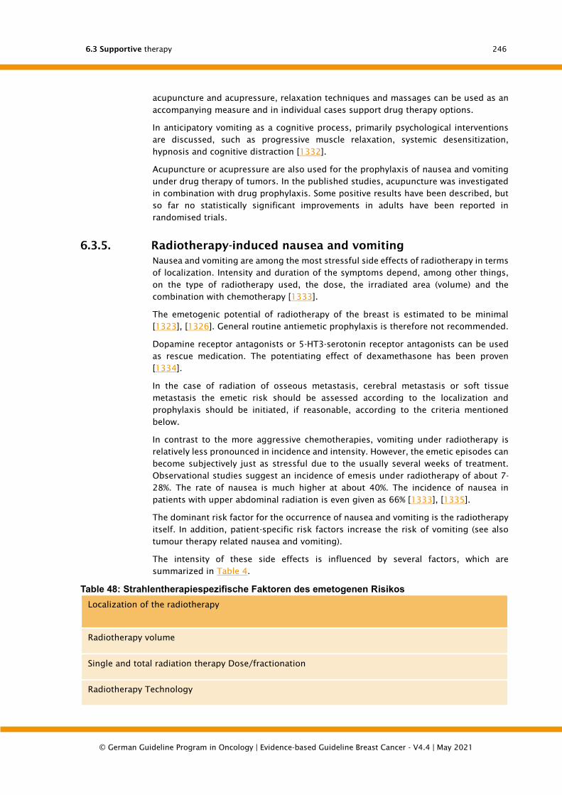
6.3 Supportive therapy
© German Guideline Program in Oncology | Evidence-based Guideline Breast Cancer - V4.4 | May 2021
246
acupuncture and acupressure, relaxation techniques and massages can be used as an
accompanying measure and in individual cases support drug therapy options.
In anticipatory vomiting as a cognitive process, primarily psychological interventions
are discussed, such as progressive muscle relaxation, systemic desensitization,
hypnosis and cognitive distraction [1332].
Acupuncture or acupressure are also used for the prophylaxis of nausea and vomiting
under drug therapy of tumors. In the published studies, acupuncture was investigated
in combination with drug prophylaxis. Some positive results have been described, but
so far no statistically significant improvements in adults have been reported in
randomised trials.
6.3.5. Radiotherapy-induced nausea and vomiting
Nausea and vomiting are among the most stressful side effects of radiotherapy in terms
of localization. Intensity and duration of the symptoms depend, among other things,
on the type of radiotherapy used, the dose, the irradiated area (volume) and the
combination with chemotherapy [1333].
The emetogenic potential of radiotherapy of the breast is estimated to be minimal
[1323], [1326]. General routine antiemetic prophylaxis is therefore not recommended.
Dopamine receptor antagonists or 5-HT3-serotonin receptor antagonists can be used
as rescue medication. The potentiating effect of dexamethasone has been proven
[1334].
In the case of radiation of osseous metastasis, cerebral metastasis or soft tissue
metastasis the emetic risk should be assessed according to the localization and
prophylaxis should be initiated, if reasonable, according to the criteria mentioned
below.
In contrast to the more aggressive chemotherapies, vomiting under radiotherapy is
relatively less pronounced in incidence and intensity. However, the emetic episodes can
become subjectively just as stressful due to the usually several weeks of treatment.
Observational studies suggest an incidence of emesis under radiotherapy of about 7-
28%. The rate of nausea is much higher at about 40%. The incidence of nausea in
patients with upper abdominal radiation is even given as 66% [1333], [1335].
The dominant risk factor for the occurrence of nausea and vomiting is the radiotherapy
itself. In addition, patient-specific risk factors increase the risk of vomiting (see also
tumour therapy related nausea and vomiting).
The intensity of these side effects is influenced by several factors, which are
summarized in Table 4.
Table 48: Strahlentherapiespezifische Faktoren des emetogenen Risikos
Localization of the radiotherapy
Radiotherapy volume
Single and total radiation therapy Dose/fractionation
Radiotherapy Technology

6.3 Supportive therapy
© German Guideline Program in Oncology | Evidence-based Guideline Breast Cancer - V4.4 | May 2021
247
Table 49: Zusammenfassung der antiemetischen Prophylaxe
Emesis risk Irradiated body region Antiemetic prophylaxis
High Full body radiation 5-HT3-RA and dexamethasone
Moderate Upper abdomen, BWS/LWS ,
neuro axis depending on the
technique
5-HT3-RA and dexamethasone can
be used for dexamethasone
Low Pelvis, cranium, ENT 5-HT3-RA or rescue therapy
Minimal Extremities, chest No routine prophylaxis
In the case of combined radiochemotherapy, antiemetic prophylaxis is based on the
highest achieved risk class of radiotherapy or chemotherapy. For example, in the case
of combined radiochemotherapy with cisplatin, the emetogenic potency of cisplatin is
decisive for antiemetic prophylaxis. In this case prophylaxis with a 5-HT3-RA,
dexamethasone and NK1-RA is recommended [1336], [1337].
6.3.6. Neutropenia, febrile neutropenia (FN), infections
Febrile neutropenia (FN) and neutropenia-associated infections are a significant factor
in morbidity and mortality after cytotoxic therapy. In addition, they can lead to a dose
reduction of chemotherapy and/or cycle delays [1338]. Granulocyte colony stimulating
factors (G-CSF) are approved for the reduction of the incidence of febrile neutropenia
and duration of neutropenia in malignant diseases with cytotoxic chemotherapy.
6.12 Consensus-based Statement
ST The sole presence of afebrile neutropenia after tumor therapy does not justify
the administration of G-CSF.
Strong Consensus
6.13 Evidence-based Statement
ST The prophylactic administration of G-CSF depends on the risk of developing
febrile neutropenia according to the individual risk factors and the cytotoxic
therapy used.
LoE
1b
[871]
Strong Consensus
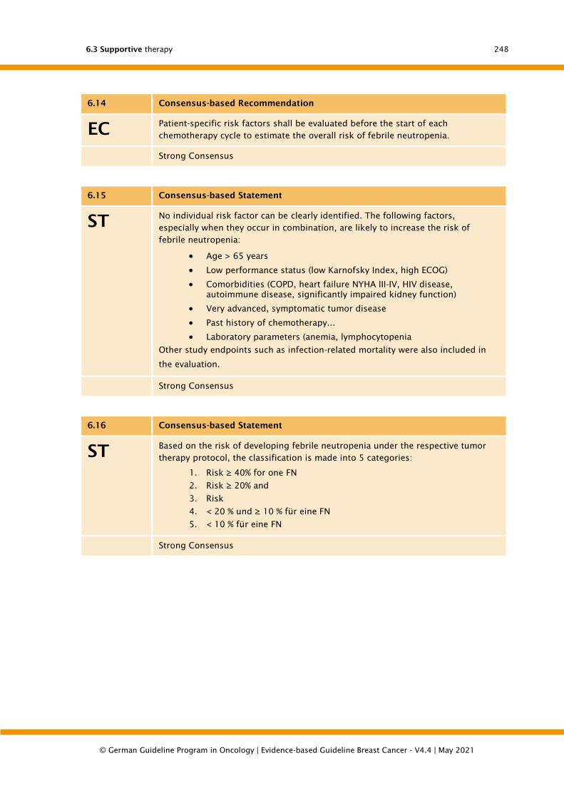
6.3 Supportive therapy
© German Guideline Program in Oncology | Evidence-based Guideline Breast Cancer - V4.4 | May 2021
248
6.14 Consensus-based Recommendation
EC Patient-specific risk factors shall be evaluated before the start of each
chemotherapy cycle to estimate the overall risk of febrile neutropenia.
Strong Consensus
6.15 Consensus-based Statement
ST No individual risk factor can be clearly identified. The following factors,
especially when they occur in combination, are likely to increase the risk of
febrile neutropenia:
• Age > 65 years
• Low performance status (low Karnofsky Index, high ECOG)
• Comorbidities (COPD, heart failure NYHA III-IV, HIV disease,
autoimmune disease, significantly impaired kidney function)
• Very advanced, symptomatic tumor disease
• Past history of chemotherapy...
• Laboratory parameters (anemia, lymphocytopenia
Other study endpoints such as infection-related mortality were also included in
the evaluation.
Strong Consensus
6.16 Consensus-based Statement
ST Based on the risk of developing febrile neutropenia under the respective tumor
therapy protocol, the classification is made into 5 categories:
1. Risk ≥ 40% for one FN
2. Risk ≥ 20% and
3. Risk
4. < 20 % und ≥ 10 % für eine FN
5. < 10 % für eine FN
Strong Consensus
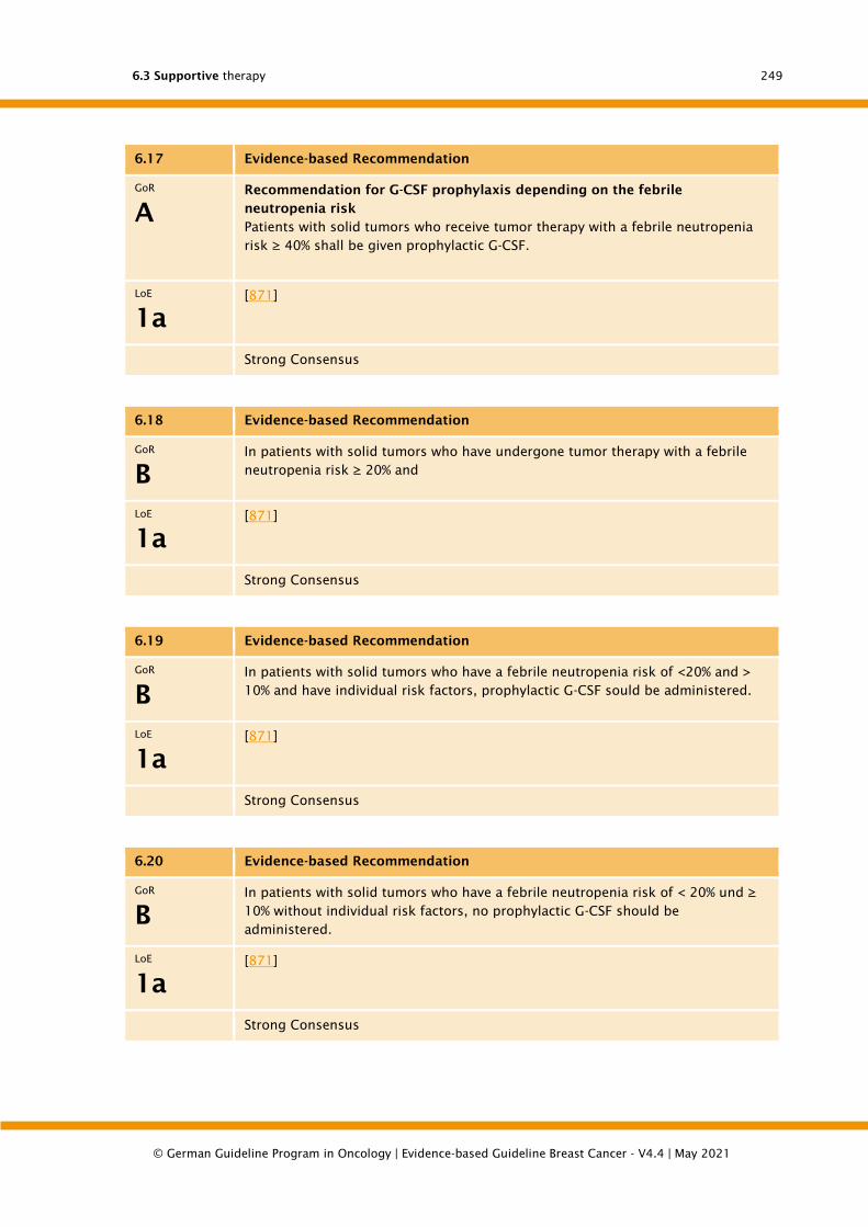
6.3 Supportive therapy
© German Guideline Program in Oncology | Evidence-based Guideline Breast Cancer - V4.4 | May 2021
249
6.17 Evidence-based Recommendation
GoR
A
Recommendation for G-CSF prophylaxis depending on the febrile
neutropenia risk
Patients with solid tumors who receive tumor therapy with a febrile neutropenia
risk ≥ 40% shall be given prophylactic G-CSF.
LoE
1a
[871]
Strong Consensus
6.18 Evidence-based Recommendation
GoR
B
In patients with solid tumors who have undergone tumor therapy with a febrile
neutropenia risk ≥ 20% and
LoE
1a
[871]
Strong Consensus
6.19 Evidence-based Recommendation
GoR
B
In patients with solid tumors who have a febrile neutropenia risk of <20% and >
10% and have individual risk factors, prophylactic G-CSF sould be administered.
LoE
1a
[871]
Strong Consensus
6.20 Evidence-based Recommendation
GoR
B
In patients with solid tumors who have a febrile neutropenia risk of < 20% und ≥
10% without individual risk factors, no prophylactic G-CSF should be
administered.
LoE
1a
[871]
Strong Consensus
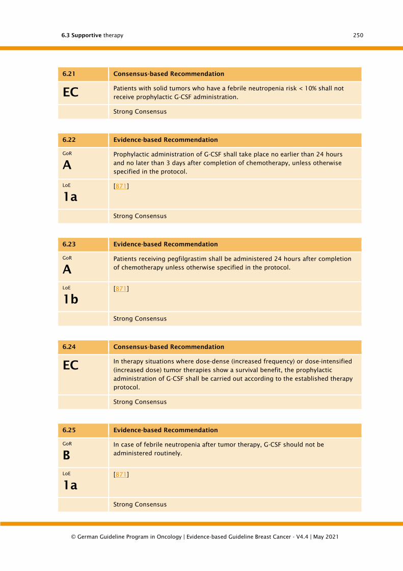
6.3 Supportive therapy
© German Guideline Program in Oncology | Evidence-based Guideline Breast Cancer - V4.4 | May 2021
250
6.21 Consensus-based Recommendation
EC Patients with solid tumors who have a febrile neutropenia risk < 10% shall not
receive prophylactic G-CSF administration.
Strong Consensus
6.22 Evidence-based Recommendation
GoR
A
Prophylactic administration of G-CSF shall take place no earlier than 24 hours
and no later than 3 days after completion of chemotherapy, unless otherwise
specified in the protocol.
LoE
1a
[871]
Strong Consensus
6.23 Evidence-based Recommendation
GoR
A
Patients receiving pegfilgrastim shall be administered 24 hours after completion
of chemotherapy unless otherwise specified in the protocol.
LoE
1b
[871]
Strong Consensus
6.24 Consensus-based Recommendation
EC In therapy situations where dose-dense (increased frequency) or dose-intensified
(increased dose) tumor therapies show a survival benefit, the prophylactic
administration of G-CSF shall be carried out according to the established therapy
protocol.
Strong Consensus
6.25 Evidence-based Recommendation
GoR
B
In case of febrile neutropenia after tumor therapy, G-CSF should not be
administered routinely.
LoE
1a
[871]
Strong Consensus
6.3 Supportive therapy
© German Guideline Program in Oncology | Evidence-based Guideline Breast Cancer - V4.4 | May 2021
251
6.3.6.1. Infections in neutropenia
Fever in chemotherapy-associated neutropenia is due to infection in over 95% of cases.
Nevertheless, no pathogen can be detected in 50 - 70% of patients. The immediate use
of broad-spectrum antibiotics is therefore necessary in order to prevent further
development into a potentially life-threatening infection or to treat [1339], [1340],
[1341], [1342] immediately and effectively. Infections are the most frequent therapy-
related causes of death in cancer patients. The risk of febrile neutropenia or life-
threatening infections correlates with the severity and duration of neutropenia [1343].
The mortality due to infections in chemotherapy-induced neutropenia is 2.8% and early
mortality is 5.7% [1344]. Documented infections in neutropenia have a significantly
worse prognosis than febrile neutropenia [1339], [1344], [1345]. Multivariate analysis
revealed the following risk factors for a fatal course of FN: gram-negative sepsis
(relative risk: 4.92), invasive aspergillosis 3.48, invasive candidiasis 2.55, lung disease
3.94, cerebrovascular disease 3.26, kidney disease 3.16, liver disease 2.89, pneumonia
2,23, gram-positive sepsis 2.29, hyptension 2.12, pulmonary artery embolism 1.94,
heart disease 1.58, leukemia 1.48, lung cancer 1.18, age > 65 years 1.12 [1344].
The diagnosis and therapy of infections in neutropenia are presented in a differentiated
manner in special recommendations and guidelines, which are explicitly referred to
here.
1. German Society for Hematology and Medical Oncology (DGHO), Onkopedia
Guidelines,
https://www.onkopedia.com/de/onkopedia/guidelines/infektionen-bei-
haematologischen-und-onkologischen-patienten-
uebersicht/@@view/html/index.html
2. European Society of Medical Oncology (ESMO):
http://www.esmo.org/Guidelines/Supportive-Care/Management-of-Febrile-
Neutropaenia
3. National Comprehensive Cancer Network (NCCN)
https://www.nccn.org/professionals/physician_gls/pdf/infections.pdf
6.3.7. Cancer therapy-induced anemia
Patients with a tumor disease often suffer from anemia that can cause clinical
symptoms. The cause can be the tumor disease itself as well as chemotherapy or
radiotherapy/radiochemotherapy.
Chemotherapy can trigger or intensify anemia. When treating anaemia, it is therefore
necessary to consider the possible causes, including multiple causes. Depending on
the clinical constellation, blood transfusions, erythropoiesis-stimulating agents (ESA)
or, in the case of functional iron deficiency, the substitution of intravenous iron, also
in combination with ESA, can be considered. In these cases, an accurate and individual
risk-benefit assessment is necessary.
In this S3 guideline, the forms of anaemia are defined as follows:
Tumour anaemia (anaemia in chronic disease): This form of anaemia results from
the activation of the immune system (by tumour, infection, autoimmune disease) with
complex effects on haematopoiesis, iron metabolism and its regulation.
Tumor therapy induced anemia: Anemia caused by chemotherapy (including "new
substances") and/or radiotherapy and/or radiochemotherapy.
Chemotherapy-induced anaemia: anaemia caused by chemotherapy
6.3 Supportive therapy
© German Guideline Program in Oncology | Evidence-based Guideline Breast Cancer - V4.4 | May 2021
252
6.3.7.1. Definition of anemia
Anaemia is a reduction in the number of erythrocytes, characterized by a decrease in
hemoglobin concentration (Hb) and/or hematocrit in peripheral blood.
The lower reference value of haemoglobin is defined (WHO) as 12 g/dl (7.45 mmol/l)
in central European adults, depending on age, in non-pregnant women and 13 g/dl
(8.07 mmol/l) in men.
6.3.7.2. Anemia in cancer, anemia of chronic disease (ACD)
Symptomatic anemia is frequent in cancer patients and, depending on the type and
stage of tumor, amounts to approx. 31-50% already at diagnosis of solid tumors
without therapy. The prevalence is even higher in hematological neoplasias [1346],
[1347].
Anemia in cancer without therapeutic influence is caused by the activated immune
system. This form of anaemia is called anaemia of chronic disease (ACD) [1348]. In the
foreground are disorders mediated by inflammatory cytokines (tumor necrosis factor-
α, interleukin-1-α and -ß, interleukin-6, interferon-γ). They concern homeostasis of iron
metabolism, inhibited proliferation of erythroid precursor cells, insufficient synthesis
of and reduced response to erythropoietin (EPO) in relation to anemia, and a shortening
of erythrocyte survival [1348], [1349], [1350].
Hepcidin, a type II acute phase peptide produced in the liver, inhibits intestinal iron
absorption, iron release from enterocytes, iron mobilization from the reticulohistiocyte
system (RHS) and iron uptake into the erythropoietic progenitor cells [1351]. Hepcidin
binds to the single cellular iron exporter ferroportin and causes its degradation. There
is a reduced haemoglobin synthesis in the case of iron overload of the organism.
Despite increased storage iron, haematopoiesis results in insufficient iron being
available, corresponding to a functional iron deficiency.
With increased erythropoietic activity, the precursor cells of erythropoiesis in the bone
marrow, the proerythroblasts, form the molecule erythroferrone, which inhibits the
formation of hepcidin in the liver and thereby promotes the absorption and distribution
of iron from the storage cells [1352]. In reduced or suppressed hematopoiesis,
erythroferrone production is therefore reduced and hepcidin production is increased.
6.3.7.3. Incidence of cancer therapy-induced anemia
Under tumor therapy anemia occurs in about 75% of all patients, most frequently in
gynecological tumors (81-88%) and in patients with lung cancer (77-83%) [1347].
With radiotherapy alone, anemia is reported in about 38% of patients, with rates of 51%
in patients with lung cancer, 35% in patients with breast cancer, 49% in tumors of the
gastrointestinal tract, 54% in gynecological tumors and 42% in malignant lymphomas
[1347], [1353]. In prostate carcinoma the anemia rate is about 32% [1346].
In combined radio- and chemotherapy the overall anemia rate is about 62% [1347].
6.3.7.4. Diagnostics
6.3.7.4.1. Laboratory parameters
The following values may be elevated in chronic anemia: Ferritin, free transferrin-iron
binding capacity (transferrin saturation decreased), BSG, fibrinogen, CRP and
haptoglobin, zinc protoporphyrin (ZPP), soluble transferrin receptor in serum,
erythropoietin in serum (but not sufficient, i.e. inadequately increased).
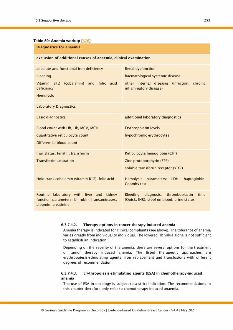
6.3 Supportive therapy
© German Guideline Program in Oncology | Evidence-based Guideline Breast Cancer - V4.4 | May 2021
253
Table 50: Anemia workup [676]
Diagnostics for anaemia
exclusion of additional causes of anaemia, clinical examination
absolute and functional iron deficiency
Bleeding
Vitamin B12 (cobalamin) and folic acid
deficiency
Hemolysis
Renal dysfunction
haematological systemic disease
other internal diseases (infection, chronic
inflammatory disease)
Laboratory Diagnostics
Basic diagnostics additional laboratory diagnostics
Blood count with Hb, Hk, MCV, MCH
quantitative reticulocyte count
Differential blood count
Erythropoietin levels
hypochromic erythrocytes
Iron status: ferritin, transferrin
Transferrin saturation
Reticulocyte hemoglobin (CHr)
Zinc protoporphyrin (ZPP),
soluble transferrin receptor (sTfR)
Holo-trans-cobalamin (vitamin B12), folic acid Hemolysis parameters: LDH, haptoglobin,
Coombs test
Routine laboratory with liver and kidney
function parameters: bilirubin, transaminases,
albumin, creatinine
Bleeding diagnosis: thromboplastin time
(Quick, INR), stool on blood, urine status
6.3.7.4.2. Therapy options in cancer therapy-induced anemia
Anemia therapy is indicated for clinical complaints (see above). The tolerance of anemia
varies greatly from individual to individual. The lowered Hb value alone is not sufficient
to establish an indication.
Depending on the severity of the anemia, there are several options for the treatment
of tumor therapy induced anemia. The listed therapeutic approaches are
erythropoiesis-stimulating agents, iron replacement and transfusions with different
degrees of recommendation.
6.3.7.4.3. Erythropoiesis-stimulating agents (ESA) in chemotherapy-induced
anemia
The use of ESA in oncology is subject to a strict indication. The recommendations in
this chapter therefore only refer to chemotherapy-induced anaemia.
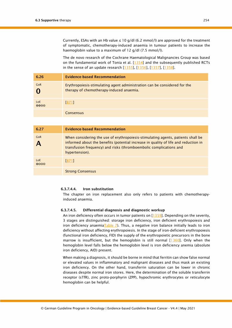
6.3 Supportive therapy
© German Guideline Program in Oncology | Evidence-based Guideline Breast Cancer - V4.4 | May 2021
254
Currently, ESAs with an Hb value ≤ 10 g/dl (6.2 mmol/l) are approved for the treatment
of symptomatic, chemotherapy-induced anaemia in tumour patients to increase the
haemoglobin value to a maximum of 12 g/dl (7.5 mmol/l).
The de novo research of the Cochrane Haematological Malignancies Group was based
on the fundamental work of Tonia et al. [1354] and the subsequently published RCTs
in the sense of an update research [1355], [1356], [1357], [1358].
6.26 Evidence-based Recommendation
GoR
0
Erythropoiesis-stimulating agent administration can be considered for the
therapy of chemotherapy-induced anaemia.
LoE
⊕⊕⊝⊝
[871]
Consensus
6.27 Evidence-based Recommendation
GoR
A
When considering the use of erythropoiesis-stimulating agents, patients shall be
informed about the benefits (potential increase in quality of life and reduction in
transfusion frequency) and risks (thromboembolic complications and
hypertension).
LoE
⊕⊝⊝⊝
[871]
Strong Consensus
6.3.7.4.4. Iron substitution
The chapter on iron replacement also only refers to patients with chemotherapy-
induced anaemia.
6.3.7.4.5. Differential diagnosis and diagnostic workup
An iron deficiency often occurs in tumor patients on [1359]. Depending on the severity,
3 stages are distinguished: storage iron deficiency, iron deficient erythropoiesis and
iron deficiency anaemiaTable 7). Thus, a negative iron balance initially leads to iron
deficiency without affecting erythropoiesis. In the stage of iron-deficient erythropoiesis
(functional iron deficiency, FID) the supply of the erythropoietic precursors in the bone
marrow is insufficient, but the hemoglobin is still normal [1360]. Only when the
hemoglobin level falls below the hemoglobin level is iron deficiency anemia (absolute
iron deficiency, AID) present.
When making a diagnosis, it should be borne in mind that ferritin can show false normal
or elevated values in inflammatory and malignant diseases and thus mask an existing
iron deficiency. On the other hand, transferrin saturation can be lower in chronic
diseases despite normal iron stores. Here, the determination of the soluble transferrin
receptor (sTfR), zinc proto-porphyrin (ZPP), hypochromic erythrocytes or reticulocyte
hemoglobin can be helpful.
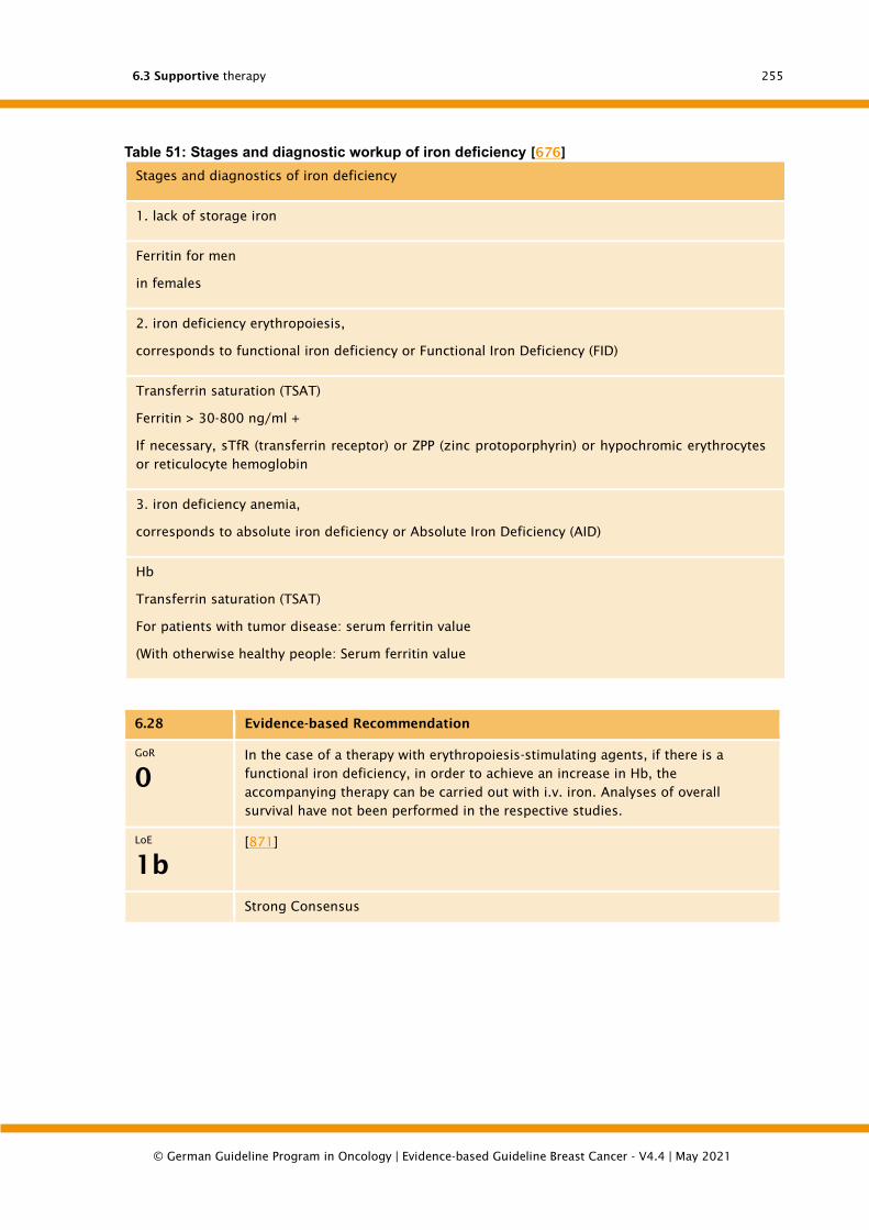
6.3 Supportive therapy
© German Guideline Program in Oncology | Evidence-based Guideline Breast Cancer - V4.4 | May 2021
255
Table 51: Stages and diagnostic workup of iron deficiency [676]
Stages and diagnostics of iron deficiency
1. lack of storage iron
Ferritin for men
in females
2. iron deficiency erythropoiesis,
corresponds to functional iron deficiency or Functional Iron Deficiency (FID)
Transferrin saturation (TSAT)
Ferritin > 30-800 ng/ml +
If necessary, sTfR (transferrin receptor) or ZPP (zinc protoporphyrin) or hypochromic erythrocytes
or reticulocyte hemoglobin
3. iron deficiency anemia,
corresponds to absolute iron deficiency or Absolute Iron Deficiency (AID)
Hb
Transferrin saturation (TSAT)
For patients with tumor disease: serum ferritin value
(With otherwise healthy people: Serum ferritin value
6.28 Evidence-based Recommendation
GoR
0
In the case of a therapy with erythropoiesis-stimulating agents, if there is a
functional iron deficiency, in order to achieve an increase in Hb, the
accompanying therapy can be carried out with i.v. iron. Analyses of overall
survival have not been performed in the respective studies.
LoE
1b
[871]
Strong Consensus
6.3 Supportive therapy
© German Guideline Program in Oncology | Evidence-based Guideline Breast Cancer - V4.4 | May 2021
256
6.29 Evidence-based Statement
ST The available evidence from the RCTs is not sufficient to make a
recommendation for or against i.v. iron therapy alone due to methodological
deficiencies.
LoE
1b
[871]
Strong Consensus
6.3.7.4.6. Transfusion of packed erythrocytes
Evidence-based recommendations on the indication can be found in greater detail in
the guidelines of the AABB (formerly "American Association of Blood Banks"), last
published in 2012 [1361], especially for the intensive care sector also from Great Britain
[1362], and in detail in the "Cross-sectional guidelines (BÄK) for therapy with blood
components and plasma derivatives", (last 4th edition, updated 2014, [1363]).
According to the cross-sectional guideline of the BÄK, transfusion is not indicated in
chronically anaemic patients without cardiovascular diseases, even at haemoglobin
concentrations of up to 8.0-7.0 g/dl (HK 24-21 % = 5.0-4.3 mmol/l), as long as no
symptoms attributable to the anaemia occur. Exceptions allow even deeper triggers,
on the one hand if the symptom burden is low or tolerable, on the other hand in special
therapeutic situations.
Aspects of quality of life must be weighed against the risks of transfusions (especially
iron overload), which are usually necessary in chronic anaemia.
In addition to the BÄK cross-sectional guidelines, it should be mentioned that in the
presence of cardiovascular risk factors only triggers of 8 g/dl versus 10 g/dl have been
randomly compared to [1364], without finding disadvantages for the restrictive
strategy. Patients with infarcts within the last 30 days were not included. Due to the
occasional coincidence of neoplastic and cardiovascular diseases at an older age, it
should be taken into account that randomized studies on the range of < 7g/dl as a
possible trigger.
6.30 Consensus-based Statement
ST In a variety of clinical contexts, a restrictive indication for transfusion is not
associated with clinical disadvantages for patients with acute anemia. By
analogy, a similar situation can also be assumed in the absence of data on
tumour therapy-induced anaemia.
Strong Consensus
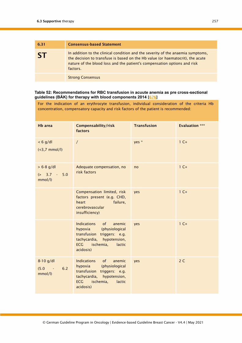
6.3 Supportive therapy
© German Guideline Program in Oncology | Evidence-based Guideline Breast Cancer - V4.4 | May 2021
257
6.31 Consensus-based Statement
ST In addition to the clinical condition and the severity of the anaemia symptoms,
the decision to transfuse is based on the Hb value (or haematocrit), the acute
nature of the blood loss and the patient's compensation options and risk
factors.
Strong Consensus
Table 52: Recommendations for RBC transfusion in acuute anemia as pre cross-sectional guidelines (BÄK) for therapy with blood components 2014 [676]
For the indication of an erythrocyte transfusion, individual consideration of the criteria Hb
concentration, compensatory capacity and risk factors of the patient is recommended:
Hb area Compensability/risk
factors
Transfusion Evaluation ***
< 6 g/dl
(<3,7 mmol/l)
/ yes * 1 C+
> 6-8 g/dl
(> 3.7 - 5.0
mmol/l)
Adequate compensation, no
risk factors
no 1 C+
Compensation limited, risk
factors present (e.g. CHD,
heart failure,
cerebrovascular
insufficiency)
yes 1 C+
Indications of anemic
hypoxia (physiological
transfusion triggers: e.g.
tachycardia, hypotension,
ECG ischemia, lactic
acidosis)
yes 1 C+
8-10 g/dl
(5.0 - 6.2
mmol/l)
Indications of anemic
hypoxia (physiological
transfusion triggers: e.g.
tachycardia, hypotension,
ECG ischemia, lactic
acidosis)
yes 2 C

6.3 Supportive therapy
© German Guideline Program in Oncology | Evidence-based Guideline Breast Cancer - V4.4 | May 2021
258
For the indication of an erythrocyte transfusion, individual consideration of the criteria Hb
concentration, compensatory capacity and risk factors of the patient is recommended:
> 10 g/dl
(> 6.2 mmol/l)
/ no ** 1 A
Notice!
Haemoglobin concentration alone is not an adequate measure of O2 supply.
In hypovolaemia, the hematocrit does not correctly reflect erythrocyte deficiency.
Individual factors may require indications that deviate from the recommendations.
* In individual cases, with adequate compensation and without risk factors, lower Hb values can be
tolerated without transfusion.
** In individual cases a transfusion to Hb values > 10 g/dl may be indicated.
*** Assessment level of the BÄK
1A: Strong recommendation that applies to most patients, based on randomized controlled trials
without significant methodological limitations
1C +: Strong recommendation that applies to most patients, no randomized controlled trials, but
clear data
2C: Very weak recommendation, depending on the individual case, a different procedure may be
indicated; based on observational studies, case descriptions
6.32 Consensus-based Statement
ST In patients with long-term tumour therapy-induced anaemia, a transfusion is
recommended if the Hk is below 24 - 21% or the haemoglobin concentration is
less than 8 - 7 g/dl (< 5,0 – 4,3 mmol/l), after considering and weighing the
overall situation.
Strong Consensus
6.33 Consensus-based Recommendation
EC In hospitalized patients with chronic anemia, whose clinical condition and
laboratory parameters are closely monitored, only one red cell concentrate
should be administered if the trigger is slightly undershot.
Strong Consensus
Background 6.32 and 6.33
The recommendation to always give only one red cell concentrate and only in
exceptional cases two preparations, because more transfusions would not be beneficial
6.3 Supportive therapy
© German Guideline Program in Oncology | Evidence-based Guideline Breast Cancer - V4.4 | May 2021
259
for the patient, comes from a setting with intensive monitoring of patients and daily
controls of the blood count [1365], [1366], The strategy is not evaluated in terms of
safety for a setting with longer control intervals.
The reduction of the transfusion frequency is part of the North American initiative
"Choosing wisely" to optimize the medical measures [1367]. There the
recommendation was chosen to "transfuse the minimum number of units necessary to
relieve the symptoms of anemia or to bring the patient into a safe Hb range (7-8 g/dL
in stable, non-cardiological inpatients)". The background text also recommends
avoiding transfusion of two units where one unit would be sufficient. On the other
hand, in order to improve the quality of life (reduced fatigue, better mobility in everyday
life and sports, reduction in the frequency of visits to the doctor), it may be useful, for
example in outpatients, to transfuse two red cell concentrates even at Hb values around
8 g/dl, depending on the planned interval until follow-up. The aspects of transfusion
medical care (blood supply time and logistics) must also be taken into account.
6.3.8. Neurotoxicity
The neurotoxicity associated with the treatment of the patient with a breast cancer is
usually chemotherapy-induced peripheral neuropathy (CIPN). This is a peripheral
neuropathy which can be motor or sensory. The symptoms are manifold. Often it is
limited to mild paresthesia, hypaesthesia and hyperaesthesia, mostly affecting hands
and feet, sometimes a superficial, burning or stabbing neuropathic pain may occur. An
impairment of the sensation of vibration and the sense of position (depth sensitivity)
can also occur. Furthermore, the neuropathic pain can significantly impair the patient's
quality of life. Motor neuropathy of the proximal muscle group is rarely seen.
After completion of the therapy, symptoms usually disappear within a few months.
Risk factors include an existing neuropathy (e.g. diabetic, alcohol toxic, renal
insufficiency), increased age, combination therapies with platinum derivatives (e.g.
cisplatin > 300 mg/m², grade I-II 14-63%, grade III-IV 7-21%), eribulin (grade I-IV up to
35%, grade III-IV up to 8%) and the taxanes, which are of fixed importance in the therapy
of breast cancer [1368]. With regard to the taxanes docetaxel, paclitaxel and nab-
paclitaxel are used. Thus, the supportive therapy of neurotoxicity is particularly
relevant for breast cancer patients.
6.3.8.1. Taxane-associated neuropathy
Typically, neuropathy is more frequent under paclitaxel than under docetaxel (sensory
neuropathy grade III/ IV: 2-33% versus 1-9%; motor neuropathy grade III/ IV: 0-14%
versus 1-9%) [1369]. It occurs less frequently under the weekly administration of
paclitaxel than under the three-weekly administration (grade III/ IV: 19% versus 12%,
p=0.001) [1370]. Sensory neuropathy occurs more frequently under nab-paclitaxel than
under standard paclitaxel. In a study by Gradishar et al. a third degree sensory
neuropathy was observed significantly more frequently with 10% than under paclitaxel
with 2% (p<0,001) [1371]. Es zeigte sich jedoch auch eine rasche Erholung der
Neuropathie auf Grad I oder II mit einem Median von 22 Tagen. Nach 28 Tagen war die
Anzahl der Patientin mit einer sensorischen Neuropathie dritten Grades in beiden
Armen identisch. Motorische Neuropathien und sensorische Neuropathien vierten
Grades wurden nicht beobachtet. In Bezug auf das Auftreten der sensorischen
Neuropathie ist auch die hö-here Dosierung des nab-Paclitaxels im Vergleich zum
konventionellen Palcitaxel zu berücksichtigen. In einer Phase-II-Studie wurde die
Dosierung von 175 m/m² nab-Paclitaxel bei 43 Patientinnen mit einem metastasierten
Mammakarzinom untersucht [1372]. Es trat keine Neuropathie Grad III oder IV auf. In

6.3 Supportive therapy
© German Guideline Program in Oncology | Evidence-based Guideline Breast Cancer - V4.4 | May 2021
260
Bezug auf die wöchentliche Therapie mit nab-Paclitaxel untersuchten Gradishar und
Kollegen 302 nicht vorbehandelte Patientinnen mit einem metastasierten
Mammakarzinom mit Docetaxel 100 mg/m², q21d, nab-Paclitaxel 300 mg/m², q21d,
oder nab-Paclitaxel 150 mg/m² oder 100 mg/m², q7d [1373]. Die Inzidenz der
sensorischen Neuropathie zeigte sich zwischen Docetaxel und allen drei nab-Paclitaxel-
Armen gleich (p > 0.1). Within the nab-paclitaxel arms, the rate of sensory neuropathy
was higher at the dosages of 300 mg/m², q21d, and 150 mg/m², q7d. Sensory
neuropathy recovered more rapidly under nab-paclitaxel than under docetaxel. The
time to recovery on ≤ 2nd degree was 22, 22 and 19 days for the doses 300 mg/m²,
100 mg/m² and 150 mg/m². In the docetaxel arm this took 37 days. In the publication
of the study with the final analysis of overall survival no significant difference in the
incidence of neuropathies was observed between the four arms (p=0.83) [1374].
Recovery time to grade II and below was 20-22 days for nab-paclitaxel and 41 days for
docetaxel. In further studies, patients with progression under taxane therapy in the
metastatic situation or metastasis within 12 months after adjuvant taxane therapy were
treated with 100 mg/m² (n=106) or 125 mg/m² nab-paclitaxel, d1, 8, 15, q28d, [1108].
The median number of cycles was 5.3 and 4.7. In the cohort with 100 mg/m², 9 patients
(8%) developed a grade III sensory neuropathy, whereas three patients already had a
pre-existing grade I neuropathy. Under the 125 mg/m² dose, 19% (n=14) of the patients
showed a grade III sensory neuropathy, with three patients also having a grade I
neuropathy before the start of therapy. Of the 23 patients with a grade III neuropathy,
15 patients were able to continue the therapy with a reduced dosage after a 1-2 week
therapy break. The study did not show any grade IV neuropathy, while 17% (n=18)
developed grade II neuropathy under the 100 mg/m² dose and 32% (n=24) under the
125 mg/m² dose.
6.3.8.2. Diagnostics
According to the S3 guideline "Supportive Therapy for Oncological Patients" [871], CIPN
is diagnosed on the basis of the patient's medical history and clinical findings. In case
of special questions electrophysiological examinations may be necessary. It is essential
that these are carried out before the start of therapy and before each cycle. To prevent
a higher degree of chemotherapy-induced polyneuropathy, its early detection is crucial.
In order to ensure an early diagnosis of CIPN, patients need to be instructed in the
documentation of neurotoxic symptoms and reliable methods of CIPN recording, which
are presented in detail in the S3 guideline "Supportive therapy in oncological patients"
[871]. The world's leading scale for quantitative assessment of peripheral neurotoxicity
is based on the National Cancer Institute-Common Toxicity Criteria (NCI-CTC).
The following statement was taken from the S3 guideline "Supportive therapy in
oncological patients":

6.3 Supportive therapy
© German Guideline Program in Oncology | Evidence-based Guideline Breast Cancer - V4.4 | May 2021
261
6.34 Consensus-based Recommendation
EC An examination of the neurological status shall be carried out before initiating a
potentially neurotoxic tumour therapy to determine the initial findings and
identify patients at risk.
Before each cycle, a detailed anamnesis shall be taken with special consideration
of possible neurotoxicities, including a repetition of the neuro status if
necessary.
Strong Consensus
6.3.8.3. Patient education
It is essential to inform the patient in detail about the background of neurotoxicity, the
risks and behavioural measures before therapy. As a result of possible sensory-motor
failures, there is a risk of burns and frostbite (e.g. use thermometers when bathing and
rinsing), falls and injuries with subsequent wound infections and in some cases limited
fitness to drive. Patients should have regular examinations for pressure points and
injuries, good skin care on hands and feet and regular gripping exercises with hands
and feet.
6.3.8.4. Prophylaxis of CIPN
There is currently insufficient data from RCTs for the prophylaxis of function loss
through CIPN using non-drug methods. Nevertheless, the expert consensus of the S3-
guideline "Supportive Therapy in Oncological Patients" recommends regular exercise
training, especially of finger and toe functions, because in the context of CIPN-related
sensory limitations, consecutive additional motor disorders are to be expected.
With regard to drug therapy, there is no effective prophylaxis of chemotherapy-induced
polyneuropathy. Standardized prophylactic measures or causal therapy approaches are
not yet available for CIPN according to the S3 guideline "Supportive Therapy in
Oncological Patients" [871]. Therefore, prevention and early detection of CIPN have the
highest priority [1375]. According to the S3 guideline "Supportive Therapy in
Oncological Patients", prophylaxis of chemotherapy-induced polyneuropathy with the
following substances is not recommended:
• Alpha lipoic acid (recommendation level A, level of evidence 1b),
• Amifostine (recommendation grade B, level of evidence 1a),
• Calcium and magnesium (recommendation grade B, level of evidence 1a),
• Carbamazepine (recommendation level B, level of evidence 1b),
• diethyldithiocarbamate (DDTC),
• Glutathione (recommendation level A, level of evidence 1a),
• and vitamin E (recommendation level B, level of evidence 1a).
6.3.8.5. Therapy of CIPN
In principle, a dose reduction can be considered in patients with known diabetic or
alcohol-induced neuropathy or in patients with the development of severe neuropathy,
but this must be weighed against the potential loss of efficacy of oncological therapy.
The data available on the treatment of CIPN with non-drug interventions are limited to
date. The effectiveness of non-drug interventions has been evaluated in a review by
Streckmann et al. [1376]. In this review 18 studies on "exercise interventions for
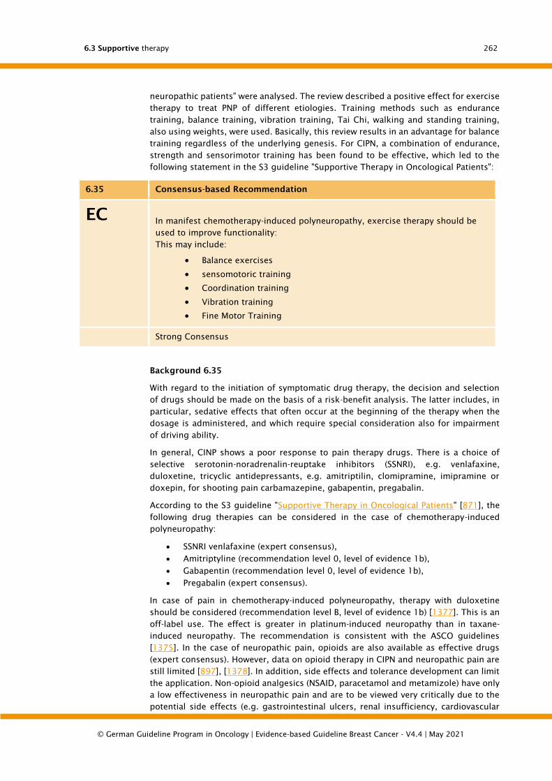
6.3 Supportive therapy
© German Guideline Program in Oncology | Evidence-based Guideline Breast Cancer - V4.4 | May 2021
262
neuropathic patients" were analysed. The review described a positive effect for exercise
therapy to treat PNP of different etiologies. Training methods such as endurance
training, balance training, vibration training, Tai Chi, walking and standing training,
also using weights, were used. Basically, this review results in an advantage for balance
training regardless of the underlying genesis. For CIPN, a combination of endurance,
strength and sensorimotor training has been found to be effective, which led to the
following statement in the S3 guideline "Supportive Therapy in Oncological Patients":
6.35 Consensus-based Recommendation
EC
In manifest chemotherapy-induced polyneuropathy, exercise therapy should be
used to improve functionality:
This may include:
• Balance exercises
• sensomotoric training
• Coordination training
• Vibration training
• Fine Motor Training
Strong Consensus
Background 6.35
With regard to the initiation of symptomatic drug therapy, the decision and selection
of drugs should be made on the basis of a risk-benefit analysis. The latter includes, in
particular, sedative effects that often occur at the beginning of the therapy when the
dosage is administered, and which require special consideration also for impairment
of driving ability.
In general, CINP shows a poor response to pain therapy drugs. There is a choice of
selective serotonin-noradrenalin-reuptake inhibitors (SSNRI), e.g. venlafaxine,
duloxetine, tricyclic antidepressants, e.g. amitriptilin, clomipramine, imipramine or
doxepin, for shooting pain carbamazepine, gabapentin, pregabalin.
According to the S3 guideline "Supportive Therapy in Oncological Patients" [871], the
following drug therapies can be considered in the case of chemotherapy-induced
polyneuropathy:
• SSNRI venlafaxine (expert consensus),
• Amitriptyline (recommendation level 0, level of evidence 1b),
• Gabapentin (recommendation level 0, level of evidence 1b),
• Pregabalin (expert consensus).
In case of pain in chemotherapy-induced polyneuropathy, therapy with duloxetine
should be considered (recommendation level B, level of evidence 1b) [1377]. This is an
off-label use. The effect is greater in platinum-induced neuropathy than in taxane-
induced neuropathy. The recommendation is consistent with the ASCO guidelines
[1375]. In the case of neuropathic pain, opioids are also available as effective drugs
(expert consensus). However, data on opioid therapy in CIPN and neuropathic pain are
still limited [897], [1378]. In addition, side effects and tolerance development can limit
the application. Non-opioid analgesics (NSAID, paracetamol and metamizole) have only
a low effectiveness in neuropathic pain and are to be viewed very critically due to the
potential side effects (e.g. gastrointestinal ulcers, renal insufficiency, cardiovascular
6.3 Supportive therapy
© German Guideline Program in Oncology | Evidence-based Guideline Breast Cancer - V4.4 | May 2021
263
side effects, very rarely agranulocytosis in metamizole) in accordance with the S3
guideline "Supportive therapy in oncological patients".
6.3.8.6. Other toxicities
Other toxicities associated with different systemic therapies of the patients and with
different frequencies depending on the therapy are diarrhoea, mucositis and stomatitis
and skin toxicities. Since these can significantly reduce the quality of life of a patient
with a therapy for breast cancer, prophylactic as well as therapeutic measures are
essential if they occur. Extensive supportive therapies are available for these side
effects which, together with the handling of extravasations, are reflected in the S3
guideline "Supportive Therapy in Oncological Patients" [871].

6.4 Follow-up and long-term care
© German Guideline Program in Oncology | Evidence-based Guideline Breast Cancer - V4.4 | May 2021
264
6.4. Follow-up and long-term care
6.4.1. Objectives
Aftercare in the narrower sense includes structured examinations of locoregional or
intramammary recurrence and contralateral breast carcinoma, examinations for distant
metastases and the monitoring of long-term therapies with diagnosis and therapy of
consequences and side effects. It begins due to the variation of the therapy regimen
following the completion of the primary locoregional therapy [1379].
Patients with a completely different starting position are treated within the scope of
aftercare. These include, for example, patients after neoadjuvant or adjuvant
chemotherapy, targeted therapy, endocrine system therapy or complementary and
alternative therapy procedures (CAM). They also include patients who have been treated
within the framework of studies. Patients who have received radiation therapy must be
regularly and specifically examined for radiogenic late effects.
An individualized risk-adapted aftercare would be necessary. While therapy decisions
are made according to risk classifications (TNM stage, steroid hormone receptors,
growth factor receptors, age, etc.), there are no larger valid studies that have
investigated individualized risk-adapted follow-up. Survival comparisons of the
different tumour stages show that survival rates are stage-dependent, so that stage-
adapted risk stratification could be carried out. There are no criteria for modifying the
structured aftercare established to date. Thus, patients with a high risk of locoregional
recurrence and a risk of distant metastasis are accompanied and treated in the same
way in structured follow-up as those with a low risk of recurrence.
At the same time, it is also apparent that a time limit of 5 years for aftercare is not
sufficient given the different risk constellations of the patients. Thus, even without
direct study funding, the time frame for follow-up care has been extended from
currently 5 years to a period of 10 years [1380]. It should be noted that therapy
monitoring should be continued for at least 10 years.
There are no new prospective randomised studies that take into account different risk
constellations, adapted aftercare schedules or integration of newer diagnostic
methods. The currently practiced aftercare concept supported by prospective
randomized studies is to be seen as an orientation, although it should be adapted to
the individual situation of the affected woman due to the symptoms.
6.36 Consensus-based Statement
ST Aftercare for patients with breast cancer begins with the completion of
locoregional primary treatment. It consists of an anamnesis, physical
examination, medical consultation, care and support as well as imaging
diagnostics to detect local and locoregional recurrence and contralateral breast
carcinoma.
In case of abnormal findings, the follow-up care should be designed in a
symptom-oriented manner.
Strong Consensus
Background 6.36
In contrast to the situation in metastatic breast carcinoma patients with an
intramammary or locoregional recurrence have a curative therapy chance.

6.4 Follow-up and long-term care
© German Guideline Program in Oncology | Evidence-based Guideline Breast Cancer - V4.4 | May 2021
265
For the early detection of distant metastases and their treatment there are no large
prospective randomized studies available that have shown a significant survival benefit.
A further distinction must be made here between patients who are undergoing long-
term therapy and those who have metastasized after a therapy-free interval [1384],
[1386].
The basis of the aftercare is the attention and the conversation. Central concerns are
the reduction of anxiety and the improvement of the patient's quality of life. This is
supplemented by a physical examination, which includes in particular the local findings
and the contralateral mamma. The invitation to participate in the recommended early
cancer detection examinations, especially in the genital area, should be made [1379].
6.37 Consensus-based Statement
ST If necessary, the individual follow-up care of breast cancer patients should
include oncologically experienced specialists and other professional groups, for
example psycho-oncologists, physiotherapists, lymphologists, oncological
nurses, breast care nurses, etc. Depending on individual needs, the patient
should be provided with information about the possibilities of further
counselling and care, including offers of self-help.
Strong Consensus
6.4.2. Examinations to detect locoregional and in-breast
recurrences or contralateral breast cancer
A local/local recurrence after mastectomy and/or axilla dissection can usually be
diagnosed by clinical examination. Palpation of the thoracic wall and the lymph
drainage areas is therefore a central part of the follow-up examinations [1390].
Local/locoregional or intramammary recurrences in patients with breast-conserving
surgery are in most cases curatively treatable. They should therefore be diagnosed as
early as possible. Aftercare should therefore include a mammography at least once a
year and, if possible, a complementary mammary sonography of the affected breast.
6.38 Evidence-based Recommendation
GoR
B
Imaging diagnostics for the detection of local and locoregional recurrences and
contralateral carcinomas should include annual mammography and quality-
assured sonography.
LoE
2c
[1391]; [1392]
Strong Consensus
Background 6.38
The time of the start of mammography in the context of follow-up care must also
depend, among other things, on the type of radiation (e.g. intraoperative radiation,
postoperative brachytherapy, etc.) and the local findings of the breast.

6.4 Follow-up and long-term care
© German Guideline Program in Oncology | Evidence-based Guideline Breast Cancer - V4.4 | May 2021
266
Since the scar region frequently changes postoperatively and the differential diagnosis
between scarring changes and recurrence is difficult, mammography and sonography
of the affected side may be necessary at shorter intervals during the first 3 years after
surgery [929]. If the findings are difficult to assess (scar, DD recurrence) an MRI is
necessary for further diagnosis [1393], [1394]. The patient with breast carcinoma
should therefore not be integrated into the mammography screening with a 2-year
examination interval.
A previous breast carcinoma is a strong risk factor for contralateral breast carcinoma.
The contralateral breast and axilla must be palpated at every follow-up examination
and sonographic checks must be performed. Mammography controls and sonography
are to be performed [1395].
MRI examinations can provide additional information for high-risk patients
[1293][1395], [1396].
6.39 Evidence-based Recommendation
GoR
B
The established quality-assured ultrasound examination in follow-up care, the
recall and biopsy rates increase. Most commonly, patients report (82%) that they
derive positive psychological aspects from the increased attentiveness and the
associated safety; rarely, some (< 6%) feel a psychological burden due to
insecurity and anxiety. Imaging diagnostics should therefore only be performed
to supplement mammography
LoE
2c
[1391]; [1392]
Consensus
Background 6.39
The question of the significance of ultrasound in aftercare was addressed in the context
of the S3 guidelines (see evidence report SF 4.6-1 Ultrasound Aftercare). A total of 54
publications were identified for the period 2005 to 2016. According to methodological
inclusion and exclusion criteria, only 2 studies could be included in the evaluation.
According to the study by Riebe et al. 2007 (LoE 3a), the clinical examination and
mammography alone show a sensitivity of 81.8% and a PPV of 37.5%. The addition of
the ultrasound examination (thoracic wall + axilla bds.) increased the sensitivity to
100% and the PPV to 41%. In the prospective cohort study by Wojcinski et al. 2011 (LoE
2c) with n = 735 patients, the addition of ultrasound (according to DEGUM Level I)
showed a significantly increased local recurrence detection rate from 3.7% (95%CI: 2.3-
5.0) to 4.5% (95%CI: 3.0-6.0) p=0.041, accompanied by an increase in the recruitment
rate from 3.3% to 5.9% (p<0.0001) und der Biopsierate von 9.0% auf 11.8% (p<0.0001).
Alle Patientinnen mit Lokalrezidiv wiesen keine Fernmetastasierung auf. Die
Patientinnen bewerteten die psychischen Aspekte der zusätzlichen Untersuchungen in
82% (95%CI: 79.3-84.8) positiv als „sicherer und aufmerksam kontrolliert“, psychisch
belastet als „verunsichert und ängstlich“ nur 5.9% (95% CI: 4.2-7.6).

6.4 Follow-up and long-term care
© German Guideline Program in Oncology | Evidence-based Guideline Breast Cancer - V4.4 | May 2021
267
6.4.3. Men with breast cancer
6.40 Consensus-based Recommendation
EC Men with breast cancer, like women, shall receive annual imaging diagnostics,
especially since there is a higher risk of contralateral carcinoma.
Consensus
Background 6.40
Men with breast cancer have a higher risk than women with breast cancer for
contralateral breast cancer. A study with data from the Surveillance, Epidemiology, and
End Results database with follow-up of n=1,788 patients showed a 30-fold increased
risk for contralateral breast cancer (SIR [standardized incidence ratio] 29.64, 95% CI:
15-52) compared to the general male population, while women with breast cancer have
only a 2-4-fold increased risk [1397].
6.4.4. Examination for metastases
The 3 most frequent metastasis sites in patients with breast cancer are the lung, liver
and bones. Within the framework of the primary therapy, a stage-dependent diagnosis
of the spread of the disease is carried out. The currently available prospective
randomized studies have shown that an intensified follow-up at fixed intervals with
lung x-ray, bone scintigraphy, upper abdomen sonography, tumor marker or CT
diagnostics in symptom-free patients does not result in a survival advantage [1384],
[1386], but rather shortens recurrence-free survival. However, the studies in question
were conducted so long ago that it is currently impossible to assess, against the
background of new therapeutic procedures, whether a corresponding survival
advantage can be achieved today. Prospective randomized studies with risk adaptation
of a diagnostic aftercare program or the integration of procedures such as PET,
SPECT/CT, short-term tumor marker controls, examination for circulating tumor cells,
risk determination according to gene chips or tissue micro arrays, etc. are currently not
available. In addition, it is possible that metastases in patients with breast cancer may
be detected between the intervals of follow-up visits due to their symptoms. Therefore,
it is all the more important to inform the affected person about the self-observation of
persistent symptoms or the self-examination of the operated region.
6.41 Evidence-based Recommendation
GoR
A
An intensified apparatus and laboratory diagnostics with x-ray thorax, bone
scintigraphy, CT, PET or MRT as well as blood count, serum biochemistry or
tumor marker determination are part of metastasis diagnostics, not of standard
aftercare, and are only indicated in case of clinical abnormalities.
LoE
1a
[1381]; [1389]; [1398]; [1399]; [1400]; [1401]
Strong Consensus
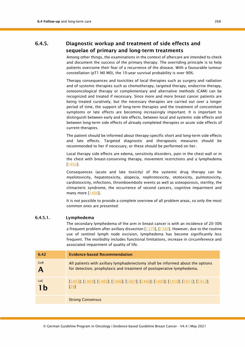
6.4 Follow-up and long-term care
© German Guideline Program in Oncology | Evidence-based Guideline Breast Cancer - V4.4 | May 2021
268
6.4.5. Diagnostic workup and treatment of side effects and
sequelae of primary and long-term treatments
Among other things, the examinations in the context of aftercare are intended to check
and document the success of the primary therapy. The overriding principle is to help
patients overcome their fear of a recurrence of the disease. With a favourable tumour
constellation (pT1 N0 M0), the 10-year survival probability is over 90%.
Therapy consequences and toxicities of local therapies such as surgery and radiation
and of systemic therapies such as chemotherapy, targeted therapy, endocrine therapy,
osteooncological therapy or complementary and alternative methods (CAM) can be
recognized and treated if necessary. Since more and more breast cancer patients are
being treated curatively, but the necessary therapies are carried out over a longer
period of time, the support of long-term therapies and the treatment of concomitant
symptoms or late effects are becoming increasingly important. It is important to
distinguish between early and late effects, between local and systemic side effects and
between long-term side effects of already completed therapies or acute side effects of
current therapies.
The patient should be informed about therapy-specific short and long-term side effects
and late effects. Targeted diagnostic and therapeutic measures should be
recommended to her if necessary, or these should be performed on her.
Local therapy side effects are edema, sensitivity disorders, pain in the chest wall or in
the chest with breast-conserving therapy, movement restrictions and a lymphedema
[1402].
Consequences (acute and late toxicity) of the systemic drug therapy can be
myelotoxicity, hepatotoxicity, alopecia, nephrotoxicity, ototoxicity, pulmotoxicity,
cardiotoxicity, infections, thromboembolic events as well as osteoporosis, sterility, the
climacteric syndrome, the occurrence of second cancers, cognitive impairment and
many more [1400].
It is not possible to provide a complete overview of all problem areas, so only the most
common ones are presented:
6.4.5.1. Lymphedema
The secondary lymphedema of the arm in breast cancer is with an incidence of 20-30%
a frequent problem after axillary dissection [1379], [1380]. However, due to the routine
use of sentinel lymph node excision, lymphedema has become significantly less
frequent. The morbidity includes functional limitations, increase in circumference and
associated impairment of quality of life.
6.42 Evidence-based Recommendation
GoR
A
All patients with axillary lymphadenectomy shall be informed about the options
for detection, prophylaxis and treatment of postoperative lymphedema.
LoE
1b
[1403]; [1404]; [1405]; [1406]; [1407]; [1408]; [1409]; [1410]; [1411]; [1412];
[28]
Strong Consensus
6.4 Follow-up and long-term care
© German Guideline Program in Oncology | Evidence-based Guideline Breast Cancer - V4.4 | May 2021
269
Background 6.42
The main influencing factors are:
• the extent of the surgical intervention in the armpit
• the number of lymph nodes removed correlates significantly with the
occurrence of lymphedema (p
• radiation of the axillary lymph drainage area (RR 1.35; 95 % CI 1.00-1.83).
3 randomized studies prove the individual benefit of morbidity reduction with reduced
surgical radicality by sentinel lymph node biopsy (SLN): absolute risk reduction for loss
of sensitivity 8% (5% SLN versus 11% ALND), for arm lymphedema symptoms 20% (11%
SLN versus 31% ALND) [1413], [1414], [1415], [1416].
Sentinel lymph node biopsy without further axillary lymphadenectomy is a primary
prophylaxis of arm lymphedema for breast cancer patients. These patients must be
informed about normal use of the arm postoperatively and should consult their
specialist or family doctor if they experience functional disorders or signs of
lymphedema.
Primary prophylaxis of lymphedema by lymphatic drainage in asymptomatic patients is
not recommended.
Physiotherapeutic exercises can improve the mobility of the arm [1417], [1418], [1419].
6.4.5.2. Cardiotoxicity
Cardiotoxicity must be considered when using anthracyclines and trastuzumab [1420].
The simultaneous combination of both substance classes significantly increases the
risk and is not recommended. Predisposing factors are age, obesity, pre-existing heart
failure, arterial hypertension, diabetes mellitus, condition after myocarditis or
infarction and left-sided radiation. In the development of acute and chronic myopathies
with heart failure, a distinction is made between the acute and sub-acute dose-
independent early form, the chronic form (within one year) and the late form. The
extent ranges from a reduction of the left ventricular ejection fraction (LVEF) to clinically
relevant heart failure (CHF). General reduction in performance or reduction in the
physical capacity of the affected person should be clarified. Early clarification of cardiac
damage is necessary to initiate appropriate supportive measures such as targeted
therapy of heart failure etc., to improve the patient's quality of life and not to worsen
the life prognosis [843], [1421], [1422].
6.4.5.3. Leukemia
Leukaemia is the most common chemotherapy-induced second malignancy. The
highest risk for secondary leukaemias is in the first ten years. The most frequent type
of leukaemia is acute myeloid leukaemia when using the anthracyclines [1423], [1424].
6.4.5.4. Menopausal syndrome
The climacteric syndrome comprises the vegetative (hot flushes, sweating, dizziness,
headache, tachycardia etc.), the psychological (insomnia, depression, abandonment
anxiety, neurotic behavior, irritability, nervousness, lack of drive, lack of concentration
etc.) and the organic climacteric syndrome (organ involution, metabolic changes etc.)
[1425]. These physiological changes can be intensified by the therapies or by therapy-
specific side effects. These include vaginal bleeding, thromboembolic events, muscle
and joint pain, dry mucous membranes, etc. The climacteric syndrome can be induced
6.4 Follow-up and long-term care
© German Guideline Program in Oncology | Evidence-based Guideline Breast Cancer - V4.4 | May 2021
270
in pre/perimenopausal patients or in postmenopausal patients by chemotherapy or
endocrine system therapy [1426].
The perception of symptoms varies subjectively and depends, among other things, on
the onset and duration of amenorrhoea or the duration of therapy, especially endocrine
therapy. The treatment of the symptoms of climacteric syndrome is symptom-oriented.
Hormone therapy after breast cancer is contraindicated. Therefore, it can only be
discussed in extremely exceptional cases, with the greatest restraint, and only be
considered if the quality of life is seriously impaired. In hormone receptor-positive
breast cancer patients, hormone therapy is contraindicated in the current data situation
[1427].
6.4.5.5. Antibody therpay
Reference can be made here to Chapter 5.7.5.
6.4.5.6. Thromboembolic events
Thromboembolic events can occur as a paraneoplastic syndrome during primary
therapy. They are often indications of a more extensive tumor or a metastasis [1428].
In endocrine systemic therapies, thromboembolic events are possible, especially in the
context of long-term therapies [1429]. The diagnosis and therapy of thrombosis or
pulmonary artery embolism and its prophylaxis are defined in interdisciplinary S2 or
S3 guidelines of other professional societies (AWMF 065/002).
6.4.5.7. Osteoporosis
One of the main factors regulating bone metabolism is oestrogen. Physiologically, the
reduction of bone substance at the beginning of the menopause is physiological. This
can be intensified by triggering the premature menopause in premenopausal patients
by chemotherapy or endocrine system therapy or in postmenopausal patients by the
use of aromatase inhibitors. In patients with a significantly increased risk of developing
osteoporosis or with known osteoporosis, appropriate medication should be
recommended. In patients who are not yet ill, attention should be drawn to behavioural
measures such as physical activity, modification of the diet or substitution with vitamin
D and calcium [1400], [1430], [1431]. Detailed information about osteo-oncological
medication options should be given.
In any case, it is important to diagnose bone stability at an early stage with a bone
densitometry before and during a possibly necessary anti-hormonal therapy and a
planned chemotherapy.

6.4 Follow-up and long-term care
© German Guideline Program in Oncology | Evidence-based Guideline Breast Cancer - V4.4 | May 2021
271
6.43 Evidence-based Recommendation
GoR
B
Bone densitometry should be recommended for breast cancer patients under
aromatase inhibitor therapy, premenopausal patients under tamoxifen and/or
GnRH therapy and patients with chemotherapy-induced premature menopause.
Depending on the result and other risk factors, the bone density measurement
should be repeated at regular intervals.
Under prophylactic osteoprotective therapy, regular monitoring is not
necessary.
LoE
2a
[873]
Consensus
6.4.5.8. Fatigue
Patients with chronic exhaustion and fatigue syndrome (Fatigue) after treatment of
breast cancer should be informed about physical training strategies and psychosocial
support [1432], [1433].
6.4.5.9. Reprodction
Premenopausal breast cancer patients who wish to have children should be informed
about the possibilities of further family planning after successful completion of the
primary therapy of breast cancer [1434]. The originally anticipated increase of the risk
of recurrence due to endocrine changes during pregnancy has not been confirmed in
any study [1435]. However, the survival advantage of patients who became pregnant in
the years following successful treatment of breast cancer, as postulated in some
studies, is probably based on a "healthy mother effect" [1434], [1436]. As a general
rule, the decision for or against the realisation of the desire to have children after
completion of primary therapy for breast cancer should follow personal considerations
of lifestyle and less vague medical hypotheses.
If there is an indication for contraception, either for medical reasons such as endocrine
therapy, or because of personal lifestyle considerations, it should generally not be
performed hormonally. The risks of hormonal contraception must be carefully weighed
up.
6.4.6. Frequency of follow-up examinations
Follow-up examinations should be performed quarterly for the first 3 years, semi-
annually for the 4th and 5th year and annually from the 6th year onwards.
Due to the tumor biology of the breast carcinoma, a follow-up phase of at least ten
years should be considered [1379], [1437]. The therapy monitoring should be
continued for at least 10 years.
A patient with breast cancer can no longer be included in normal mammography
screening. However, it is possible to extend the aftercare intervals after a longer period
of time, depending on the risk, and if necessary, to perform imaging at longer intervals.
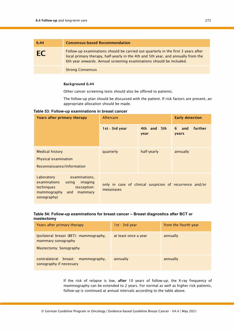
6.4 Follow-up and long-term care
© German Guideline Program in Oncology | Evidence-based Guideline Breast Cancer - V4.4 | May 2021
272
6.44 Consensus-based Recommendation
EC Follow-up examinations should be carried out quarterly in the first 3 years after
local primary therapy, half-yearly in the 4th and 5th year, and annually from the
6th year onwards. Annual screening examinations should be included.
Strong Consensus
Background 6.44
Other cancer screening tests should also be offered to patients.
The follow-up plan should be discussed with the patient. If risk factors are present, an
appropriate allocation should be made.
Table 53: Follow-up examinations in breast cancer
Years after primary therapy Aftercare Early detection
1st - 3rd year 4th and 5th
year
6 and further
years
Medical history
Physical examination
Reconnaissance/Information
quarterly half-yearly annually
Laboratory examinations,
examinations using imaging
techniques (exception:
mammography and mammary
sonography)
only in case of clinical suspicion of recurrence and/or
metastases
Table 54: Follow-up examinations for breast cancer – Breast diagnostics after BCT or mastectomy
Years after primary therapy 1st - 3rd year from the fourth year
Ipsilateral breast (BET): mammography,
mammary sonography
Mastectomy: Sonography
at least once a year annually
contralateral breast: mammography,
sonography if necessary
annually annually
If the risk of relapse is low, after 10 years of follow-up, the X-ray frequency of
mammography can be extended to 2 years. For normal as well as higher risk patients,
follow-up is continued at annual intervals according to the table above.
6.4 Follow-up and long-term care
© German Guideline Program in Oncology | Evidence-based Guideline Breast Cancer - V4.4 | May 2021
273
Patients often ask for or request more intensive aftercare. They expect detailed
information about their own risk and want to comply with this risk by intensifying
aftercare [1438]. It turns out that the belief in the effectiveness of aftercare
examinations is high and often unrealistic [1438].
More than two thirds of the patients believe that early diagnosis of metastasis and the
associated earlier use of therapy is more likely to lead to healing. Most patients
therefore wish for additional diagnostic measures. About 50% of patients also prefer a
lifelong follow-up [1438] without a time limit. On the other hand, only 67% of patients
actually use the annual mammography after the primary surgery [1439], [1440],
[1441], [1442]. While 80% of patients have a mammography in the first year of follow-
up, only 63% do so in the fifth year of follow-up. Only 33% of patients use a
mammography in each of the first five years after surgery [1439]. Since the detection
of recurrence is the central task of aftercare, it is therefore necessary to inform patients
specifically about using the established examination measures (see above) at the
recommended intervals.
6.45 Consensus-based Recommendation
EC Patients shall be motivated to be physically active (> 2-3 h/week) and to
normalize their body weight (in the case of an increased BMI) as part of
aftercare. Support should be provided.
Strong Consensus
Background 6.45
Follow-up care should not only focus on the detection of a relapse of the disease, but
also on general health maintenance with training and counselling. This includes
information about lifestyle, e.g. exercise and nutrition, especially in the case of obesity
with a BMI ≥ 30 kg/m². The increasing body weight is related to the mortality due to a
breast cancer disease. Retrospective studies have shown that patients with a BMI ≥ 30
kg/m² have a significant 4% increased risk of developing distant metastases within the
first ten years compared with those with a BMI < 25 kg/m², and a 38% higher risk of
death from breast cancer 10 or more years after primary diagnosis [877]. Continued
physical activity and the maintenance of body weight within the normal range produce
a significant improvement in quality of life, less fatigue and greater physical function
[874]. There is indeed evidence that regular exercise confers a survival benefit [878].
Notwithstanding, an improved outcome as a result of exercise interventions as part of
follow-up care has not yet been demonstrated in prospective studies. [879].
6.46 Consensus-based Statement
ST An essential part of the aftercare is the constant motivation of the patient to
regularly take the drugs prescribed for adjuvant therapy, especially the
endocrine therapy (e.g. tamoxifen or aromatase inhibitors).
The patient must be asked in detail about tolerability or side effects of the
therapy. Complaints are to be treated with suitable measures. A premature
discontinuation of therapy can be prevented by changing the endocrine
treatment.
Strong Consensus
6.5 Rehabilitation
© German Guideline Program in Oncology | Evidence-based Guideline Breast Cancer - V4.4 | May 2021
274
Background 6.46
Endocrine therapies in the adjuvant situation of patients with hormone receptor-
positive breast cancer (e.g. tamoxifen or aromatase inhibitors) are highly effective. The
disadvantage is that these preparations must be taken continuously for at least five
years. Since these therapies have side effects on the one hand, and the necessity of
long-term continuous intake is not adequately accepted by patients on the other, long-
term compliance is not sufficient for this therapy. After the first year of use, 40-50% of
patients stop taking the prescribed medication. This reduces the disease-free and
survival rates, so that it is necessary to repeatedly point out the compliance with the
therapy in the follow-up care.
Predictors for a discontinuation of endocrine therapy are a younger (< 50 years) and
older age (≥ 75 years), breast-conserving treatment (versus mastectomy), the presence
of comorbidities (≥ 2), the prescription of smaller pack sizes, and higher top-up
payments for medications (particularly among the elderly) [1394]. Predictors of reliable
adherence are married life and previous administration of chemotherapy and
radiotherapy. The aftercare consultation should address in detail the need for
compliance with endocrine therapies, taking into account the predictors of compliance,
and the possible reasons for discontinuation, such as side effects.
For the documentation of adjuvant therapy, it is important that not only the planning
of the therapy is documented, but also the actual start and end as well as the adequate
implementation.
6.5. Rehabilitation
The multimodal therapy of patients with breast cancer can lead to somatic and
psychosocial secondary disorders that result in functional impairment in the sense of
participation in everyday life. Medical rehabilitation measures to reduce or eliminate
functional disorders are available to patients according to SGB IX if there is a need for
and ability to rehabilitate and a positive rehabilitation prognosis can be given. The ICF
(International Classification of Functioning, Disability and Health) is used to assess the
functional disorders.
Pursuant to § 4 SGB IX, benefits for participation include social benefits (i.e. in this
context, in particular medical rehabilitation benefits) in order, irrespective of the cause
of the disability
• to avert or remove the disability, to prevent its aggravation or to mitigate its
consequences,
• To avoid restrictions of the earning capacity or a need for care,
• to ensure participation in working life according to aptitudes and skills,
• to promote personal development in a holistic way to enable or facilitate
independent participation in social life.
The cost units for rehabilitation measures are in particular the statutory health
insurance funds, the statutory pension insurance funds and the social administration.
According to § 19 SGB IX, rehabilitation can be provided in inpatient or outpatient form,
taking into account personal circumstances. According to § 26 SGB IX, medical
rehabilitation services include medical and nursing treatment, drug therapy,
physiotherapy and use of aids, functional occupational therapy and psychosocial
services.
For the medical rehabilitation of patients with breast cancer, the German Pension
Insurance Association (DRV Bund) has drawn up rehabilitation therapy standards and

6.5 Rehabilitation
© German Guideline Program in Oncology | Evidence-based Guideline Breast Cancer - V4.4 | May 2021
275
updated them in 2016, in which evidence-based therapy modules are summarised (see
http://www.deutsche-rentenversicherung-bund.de).
6.47 Consensus-based Recommendation
EC The tumor disease and its treatment through surgery, radiotherapy and systemic
therapy can lead to disorders of varying severity, which require specific
rehabilitative measures in the somatic and psychosocial field. Patients shall be
informed at an early stage about the possibilities of outpatient and inpatient
rehabilitation measures as well as other claims arising from social law. The
wishes of the patients should be taken into account when determining the
indication and recommending the type of rehabilitation.
Strong Consensus
6.48 Evidence-based Recommendation
GoR
A
Exercise programmes with strength and endurance training shall be offered with
the aim of reducing therapy-related limitations in physical performance,
reducing fatigue and improving the quality of life of breast cancer patients.
LoE
1a
[1443]; [1444]; [873]
Strong Consensus
Background 6.47 and 6.48
Fatigue in tumour diseases describes a persistent, multidimensional appearance, which
includes subjectively perceived increased fatigue and physical exhaustion and can be
associated with psychological stress. The symptoms often represent the greatest
burden for the patient and restrict the quality of life and the ability to cope with stress
in everyday life in the long term [1445], [1446]. In order to diagnose a tumor-associated
fatigue and to assess the degree of its severity, a multidimensional approach is
required in accordance with the clinical picture: a fatigue-related anamnesis, recording
with the support of validated questionnaires, and somatic diagnostics. Since 1998, the
American Fatigue Coalition has had a catalogue of criteria for the diagnosis of tumour-
associated fatigue, which records the various dimensions of fatigue [1447]. A tumor-
associated fatigue is considered to be accepted if at least 6 of the 11 criteria apply
[1448]. The indication for treatment should be based on the National Comprehensive
Cancer Network (NCCN) algorithm for the assessment of tumor-associated fatigue
[1449].
An improvement of the symptoms of fatigue is mainly achieved by an exercise therapy
individually adapted to the patient's capacity. There is a benefit for strength and
endurance training. Five meta-analyses and one Cochrane analysis are available. 16-72
studies were included in the meta-analyses. With regard to the reduction of fatigue, the
meta-analyses showed a moderate, significant improvement [1450], [1451], [1452],
[1453], [1454]. The Cochrane analysis included 56 randomised trials and also showed
a significant benefit for exercise therapy with regard to the improvement of fatigue.
Both the NCCN Guideline and the Guideline of the American Society of Clinical Oncology
(ASCO) therefore recommend exercise therapy for the treatment of fatigue [894],
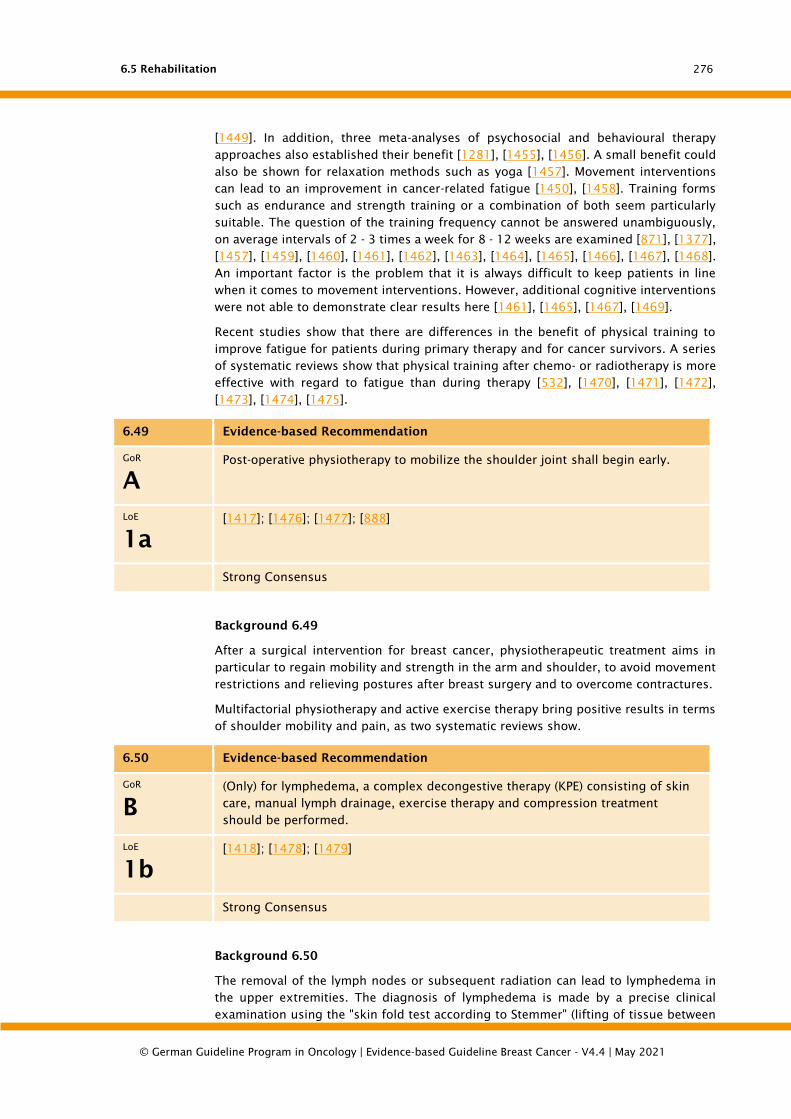
6.5 Rehabilitation
© German Guideline Program in Oncology | Evidence-based Guideline Breast Cancer - V4.4 | May 2021
276
[1449]. In addition, three meta-analyses of psychosocial and behavioural therapy
approaches also established their benefit [1281], [1455], [1456]. A small benefit could
also be shown for relaxation methods such as yoga [1457]. Movement interventions
can lead to an improvement in cancer-related fatigue [1450], [1458]. Training forms
such as endurance and strength training or a combination of both seem particularly
suitable. The question of the training frequency cannot be answered unambiguously,
on average intervals of 2 - 3 times a week for 8 - 12 weeks are examined [871], [1377],
[1457], [1459], [1460], [1461], [1462], [1463], [1464], [1465], [1466], [1467], [1468].
An important factor is the problem that it is always difficult to keep patients in line
when it comes to movement interventions. However, additional cognitive interventions
were not able to demonstrate clear results here [1461], [1465], [1467], [1469].
Recent studies show that there are differences in the benefit of physical training to
improve fatigue for patients during primary therapy and for cancer survivors. A series
of systematic reviews show that physical training after chemo- or radiotherapy is more
effective with regard to fatigue than during therapy [532], [1470], [1471], [1472],
[1473], [1474], [1475].
6.49 Evidence-based Recommendation
GoR
A
Post-operative physiotherapy to mobilize the shoulder joint shall begin early.
LoE
1a
[1417]; [1476]; [1477]; [888]
Strong Consensus
Background 6.49
After a surgical intervention for breast cancer, physiotherapeutic treatment aims in
particular to regain mobility and strength in the arm and shoulder, to avoid movement
restrictions and relieving postures after breast surgery and to overcome contractures.
Multifactorial physiotherapy and active exercise therapy bring positive results in terms
of shoulder mobility and pain, as two systematic reviews show.
6.50 Evidence-based Recommendation
GoR
B
(Only) for lymphedema, a complex decongestive therapy (KPE) consisting of skin
care, manual lymph drainage, exercise therapy and compression treatment
should be performed.
LoE
1b
[1418]; [1478]; [1479]
Strong Consensus
Background 6.50
The removal of the lymph nodes or subsequent radiation can lead to lymphedema in
the upper extremities. The diagnosis of lymphedema is made by a precise clinical
examination using the "skin fold test according to Stemmer" (lifting of tissue between

6.5 Rehabilitation
© German Guideline Program in Oncology | Evidence-based Guideline Breast Cancer - V4.4 | May 2021
277
thumb and forefinger), which is always carried out in a side by side comparison. With
the help of a measuring tape, the circumference of the arms can be checked over the
course of the procedure - it is always advisable to do this at the same places and without
pulling on the measuring tape. The date and time of day should also be noted, as
fluctuations in oedema occur throughout the day or depending on the season
(summer/winter). Both arms should be measured before the surgery and regularly
afterwards. Favourable measuring times are, for example, the oncological aftercare
appointments. A combined physiotherapy consisting of skin care, manual lymph
drainage, exercise therapy and compression is the most suitable treatment method
here. However, this should only be used if lymphedema really exists. The use of
treatment methods such as manual lymph drainage to prevent lymphedema has no
effect. The exercise programme (e.g. for shoulder mobilisation) does not represent an
increased risk for the occurrence of lymphoedema, but has a positive effect on mobility
and quality of life. Compression with bandages appears to be more effective than
pneumatic compression, although kinesio-tape may be an alternative to conventional
bandages. The results summarised here are similarly reflected in the various
international treatment recommendations.
6.51 Consensus-based Recommendation
EC In the case of chemotherapy-induced polyneuropathy (CIPN), the extent of the
damage (symptoms, localisation) should be documented and the affected
patients should be informed about the range of therapies available (pain,
physiotherapy, occupational therapy).
Strong Consensus
Background 6.51
Cytostatic drug-induced polyneuropathy (CIPN) is increasingly proving to be a
significant long-term toxicity in modern cytostatic therapy. The incidence is stated in
the literature to be about 38% and depends on the substances used, the combination
of cytostatic drugs and the type of application [1459]. CIPN manifests itself as sensory
neuropathy with paresthesia in hands and/or feet. The symptoms usually begin with
distally emphasized, stocking and glove-like dysesthesias, numbness and tingling
paresthesias which may be accompanied by considerable pain sensations and proximal
spread [1460]. Since chemotherapy in the treatment of patients with breast carcinoma
usually contains a taxane, this long-term side effect is often to be expected.
In 80% of the patients the symptoms are spontaneously completely reversible, but in
about 20% of the patients the symptoms are long-term. In addition, the symptoms can
still worsen 2-6 months after the end of chemotherapy and 40% of these patients need
another 6-8 months before a noticeable improvement occurs [1461]. In addition to
anamnesis and clinical-neurological examination, the measurement of the nerve
conduction velocity is diagnostically necessary to objectify the symptoms [1462],
[1463]. Therapeutically, neuropathic pain can be treated with the combined serotonin-
noradrenalin-reuptake inhibitor duloxetine [1377]. Therefore, the patient should be
offered a therapeutic trial with this substance. In the context of rehabilitation often
occupational therapy methods are used, but their effectiveness is unclear because no
data are available. For magnetic field therapy, positive effects were shown in a phase II
study [1464]. This was confirmed in a randomized placebo-controlled double-blind
phase III study [1465]. Other stimulation therapies such as acupuncture and
electrostimulation also show some benefit [1466], [1467]. For the stimulation

6.5 Rehabilitation
© German Guideline Program in Oncology | Evidence-based Guideline Breast Cancer - V4.4 | May 2021
278
procedures with rapeseed or hedgehog ball, which are often used in rehabilitation, no
data on the effectiveness are available. Sports and exercise therapy may also have a
positive effect on CIPN. In a randomized study, but with a small number of cases and
in lymphoma patients, a significant improvement could be achieved [896]. Further
information on cytostatic-induced polyneuropathy can also be found in the S3 guideline
supportive medicine [871].
Cognitive performance limitations
6.52 Evidence-based Recommendation
GoR
B
Patients should be asked about cognitive performance limitations
(concentration, executive functions, short-term memory) and, if necessary,
further diagnostic clarification should be initiated.
LoE
1b
[1302]; [1303]; [300]; [873]
Consensus
Background 6.52
Cognitive dysfunction (CD) in oncological diseases is a short-term, long-term or even
permanent functional disorder of the:
• Attention
• Ability to concentrate
• Thinking processes
• Memory performance (especially short-term memory)
• Learning ability
• Ability to perform complex tasks
The term KD dates back to the 1980s and was first described in patients with tumors
of the central nervous system and in pediatric tumors. It was not until the 1990s that
this clinical symptomatology received scientific attention and was perceived as an
independent functional disorder [1468]. Depending on the tumor disease investigated,
the prevalence of KD varies between 12% - 68% [1469], [1480]. According to subjective
patient data, this number can increase up to 80% [1470] 6 months after chemotherapy.
A KD after oncological therapy is described by patients up to 20 years after cancer
therapy [532]. However, these studies are mostly small inhomogeneous collectives
without control groups [1471].
Pathophysiologically, KD is most likely a multifactorial event. In addition to the actual
tumor disease itself, genetic conditions and anti-tumor therapy play an important role.
The interaction of these three components ultimately determines the severity of the
cognitive dysfunction. In particular, drug therapy of tumors is associated with
inflammatory processes and secondary changes such as vascular damage, hormonal
and metabolic changes [1472] On the other hand, the world's largest multicenter study
with 477 patients with early or locally advanced breast cancer showed that the
administration of chemotherapy has no significant influence on the development of a
KD. Rather, the symptoms of latent depression are associated with significantly
increased rates of cognitive dysfunction [1486].
Therapy- or tumor-associated KD should be distinguished from a possible pre-existing
dementia brain disorder. This can be achieved by means of a targeted collection of the
6.5 Rehabilitation
© German Guideline Program in Oncology | Evidence-based Guideline Breast Cancer - V4.4 | May 2021
279
patient's own and/or the patient's medical history. If this is not sufficient, the dementia
test DemTect can be used [1473].
A systematic diagnosis of KD is not standardized and a gold standard does not exist
yet. Neuropsychological test batteries have not found widespread application in clinical
routine, as they are associated with high personnel and time expenditure (2-3 hours
per patient, execution by psychological psychotherapist). They can therefore only be
considered if a KD needs to be further examined in individual cases and there are
consequences, e.g. for socio-medical assessment. If this is the case, a test battery
should be chosen which includes the qualities attention, reaction time, memory and
executive functions [1481]. To simplify the diagnosis of KD in oncological patients, the
d2 test, the CogPack® test or the NeuroCog FX® test can be used [1475]. The latter
computer-aided test contains all the qualities of a neuropsychological test battery and
has been validated in patients with breast cancer [see above]. A psychometric test
procedure that is also manageable in terms of time is the syndrome short test
according to Erzigkeit (SKT), which tests the qualities of memory performance and
attention [1474]. The DemTect or mini-mental test procedures commonly used in
geriatrics and dementia diagnostics are not suitable for the diagnosis of oncological
oncology patients. The classification into three degrees of severity can be made
according to the Common Terminology Criteria for Adverse Events (CTCAE Version 4.0)
of the National Cancer Institute (NCI) [1321].
There are no effective drug treatments for KD. The data on the use of Modafinil are
inconstant. Therefore, its use outside of studies cannot be recommended at present
[300]. Indications for the benefit of computer-assisted concentration and memory
training are available from one phase II and three randomized phase III studies prior to
[300], [1302], [1303], [1482]. Positive data on the benefit of behavioral therapy
approaches are available from a case-control study prior to [1483]. For sports and
exercise therapy, three randomized studies are currently available with a majority of
patients with breast cancer. While one study examined walking and fitness training
[1484], two other studies examined the use of yoga [1485], [1486]. All three studies
found a significant improvement in cognitive function. A limiting factor, however, is
that it was a heterogeneous therapy program over different therapy periods [873].
Therefore, patients with moderate and severe PD should be offered treatment with
computer-assisted training programs in combination with psychotherapeutic
interventions, exercise therapy and yoga according to the classification of NCI-CTCAE.
The application for oncological rehabilitation is usually submitted to the statutory
health or pension insurance. The statutory health insurance company bears the costs
for its insured persons (§ 40 SGB V). The pension insurance institutions provide
oncological rehabilitation services for their insured persons (according to § 15 SGB VI)
as well as for insured persons, pension recipients and their relatives (§ 31 para. 1 no.
3 SGB VI). Immediately after completion of the primary therapy, the application is
submitted via the social services of the last hospital treating the patient, otherwise via
the treating physician. Medical rehabilitation can be repeated if serious functional
disorders persist due to the disease itself or the tumour therapy.
After completion of the oncological rehabilitation (see above) a socio-medical
evaluation of the rehabilitated person is performed. On the basis of continuing
functional deficits and available resources, it is assessed which restrictions or
possibilities of participation in social life/working life arise for the affected person and
by which measures restrictions can be counteracted if necessary. On the basis of socio-
medical assessments, further benefit decisions are often made and benefits provided
by the social service providers. These can be, for example, assistance with career

6.6 Complementary medicine
© German Guideline Program in Oncology | Evidence-based Guideline Breast Cancer - V4.4 | May 2021
280
advancement, measures to secure maintenance (reduced earning capacity pensions) or
measures of secondary and tertiary prevention.
6.6. Complementary medicine
6.53 Consensus-based Recommendation
EC All patients should be asked whether they use complementary and/or alternative
therapies. Patients who use such procedures should be informed about possible
risks and, where appropriate, about interactions with standard therapies.
Strong Consensus
Background 6.53
It is important to note that there is no universally accepted definition of complementary
and alternative medicine in oncology. In most cases, it is contrasted with so-called
conventional medicine, without a clear definition of the boundaries being possible.
Complementary medicine is subject in its evaluation to the basic principles of evidence-
based therapy. It is used in addition to the standard clinical therapy [1487]. On the
other hand, alternative therapy methods are offered as a supposed substitute for an
evidence-based therapy. The resulting therapy competition represents the greatest
danger of the alternative therapy.
The integration of complementary measures to support evidence-based therapy is also
known as "integrative oncology" [1488].
According to literature data, breast cancer patients seem to use complementary therapy
methods in 50-90% of cases [1489], [1490], [1491], [1492].
Medical advice on the topic of complementary medicine can
• Strengthen the feeling of self-responsibility and control
• from dubious offers,
• Protect against damage caused by side effects or interactions,
• encourage the development of personal initiative (e.g. exercise, diet)
One risk of complementary therapies is that patients and therapists have so much
confidence in the effectiveness of these complementary measures that they abandon
conventional breast cancer therapy in favor of these measures. These are alternative
procedures and should not be used because of the obvious risks to the patient's health.
The particular problem in the evidence evaluation of complementary procedures lies in
their often less well defined effect spectra, complex mechanisms of action and
interaction possibilities, as well as the lack of conventionally conducted studies on
remedies that provide clear indications of effectiveness and benefit-risk ratio.
As part of the Oncology Guidelines Programme, complementary and alternative medical
procedures in oncology will be evaluated in a separate S3 Guidelines process in the
future (http://leitlinienprogramm-onkologie.de/Projekte.6.0.html).
In order to better describe the existing evidence at this point in time, the guideline
report lists evidence tables of an American working group [1493], which has compiled
studies on the effectiveness of complementary medicine procedures in patients with
breast cancer.

6.6 Complementary medicine
© German Guideline Program in Oncology | Evidence-based Guideline Breast Cancer - V4.4 | May 2021
281
The following Table 12 gives an overview of the most common complementary
methods and substances used by patients with breast cancer and the possible
interactions. These interactions can both attenuate and enhance (toxicity) the effect of
an existing breast cancer therapy. Some of the methods and substances listed are
discussed in further subchapters.
Table 55: Use of complementary methods, observed side effects, potential interactions
Substance/method Propagated use Side effects Interactions
beta-carotene Prevention of
recurrence,
improvement of
the tolerability of
chemotherapy
Increased
incidence of
tumours in
smokers
as antioxidant
possible
attenuation of
chemo- and
radiotherapy
vitamin C Prevention of
recurrence,
improvement of
the tolerability of
chemotherapy
in high dosage
kidney damage
possible
as antioxidant
possible
attenuation of
chemo- and
radiotherapy
High-dose vitamin C
(infusions)
Antitumoral action Vitamin C can
promote tumour
growth in vitro.
as antioxidant
possible
attenuation of
chemo- and
radiotherapy
vitamin D Prevention of
osteoporosis
Improvement of
the forecast
not known at
normal dosage
not known
generally worse
prognosis with low
serum values
vitamin E Prevention of
recurrence,
improvement of
the tolerability of
chemotherapy,
reduction of
menopausal
complaints
not known In vitro data with
indications of
attenuation of the
effects of
tamoxifen
Selenium Prevention of
recurrence,
improvement of
the tolerability of
chemotherapy
short-term, even
high-dose use
without side
effects, long-term
use only under
mirror control
No evidence of a
reduction in the
effect of anti-
tumour therapies in
preclinical or
clinical data
Zinc Prevention of
recurrence,
Zinc is important
for tumor cell
not known
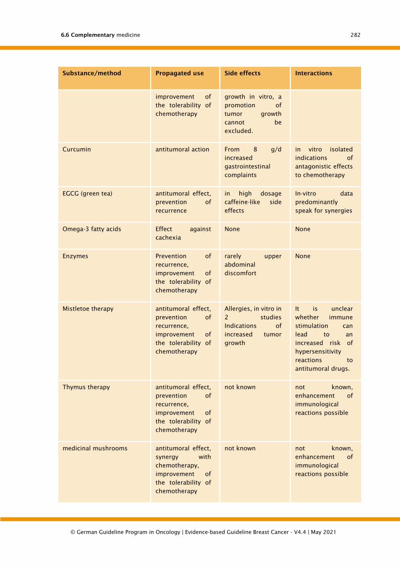
6.6 Complementary medicine
© German Guideline Program in Oncology | Evidence-based Guideline Breast Cancer - V4.4 | May 2021
282
Substance/method Propagated use Side effects Interactions
improvement of
the tolerability of
chemotherapy
growth in vitro, a
promotion of
tumor growth
cannot be
excluded.
Curcumin antitumoral action From 8 g/d
increased
gastrointestinal
complaints
in vitro isolated
indications of
antagonistic effects
to chemotherapy
EGCG (green tea) antitumoral effect,
prevention of
recurrence
in high dosage
caffeine-like side
effects
In-vitro data
predominantly
speak for synergies
Omega-3 fatty acids Effect against
cachexia
None None
Enzymes Prevention of
recurrence,
improvement of
the tolerability of
chemotherapy
rarely upper
abdominal
discomfort
None
Mistletoe therapy antitumoral effect,
prevention of
recurrence,
improvement of
the tolerability of
chemotherapy
Allergies, in vitro in
2 studies
Indications of
increased tumor
growth
It is unclear
whether immune
stimulation can
lead to an
increased risk of
hypersensitivity
reactions to
antitumoral drugs.
Thymus therapy antitumoral effect,
prevention of
recurrence,
improvement of
the tolerability of
chemotherapy
not known not known,
enhancement of
immunological
reactions possible
medicinal mushrooms antitumoral effect,
synergy with
chemotherapy,
improvement of
the tolerability of
chemotherapy
not known not known,
enhancement of
immunological
reactions possible
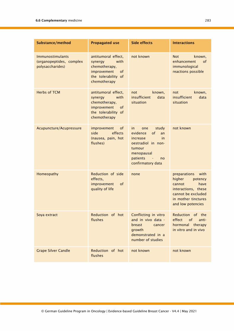
6.6 Complementary medicine
© German Guideline Program in Oncology | Evidence-based Guideline Breast Cancer - V4.4 | May 2021
283
Substance/method Propagated use Side effects Interactions
Immunostimulants
(organopeptides, complex
polysaccharides)
antitumoral effect,
synergy with
chemotherapy,
improvement of
the tolerability of
chemotherapy
not known Not known,
enhancement of
immunological
reactions possible
Herbs of TCM antitumoral effect,
synergy with
chemotherapy,
improvement of
the tolerability of
chemotherapy
not known,
insufficient data
situation
not known,
insufficient data
situation
Acupuncture/Acupressure improvement of
side effects
(nausea, pain, hot
flushes)
in one study
evidence of an
increase in
oestradiol in non-
tumour
menopausal
patients - no
confirmatory data
not known
Homeopathy Reduction of side
effects,
improvement of
quality of life
none preparations with
higher potency
cannot have
interactions, these
cannot be excluded
in mother tinctures
and low potencies
Soya extract Reduction of hot
flushes
Conflicting in vitro
and in vivo data -
breast cancer
growth
demonstrated in a
number of studies
Reduction of the
effect of anti-
hormonal therapy
in vitro and in vivo
Grape Silver Candle Reduction of hot
flushes
not known not known
6.6 Complementary medicine
© German Guideline Program in Oncology | Evidence-based Guideline Breast Cancer - V4.4 | May 2021
284
6.6.1. Diagnostics
6.54 Consensus-based Recommendation
EC The diagnostic measures offered within the framework of complementary and
alternative therapy concepts, which are based on scientifically unproven
concepts and/or misinterpretations of correlations between bodily functions,
should not be recommended.
Strong Consensus
Background 6.54
Some diagnostic measures offered within the framework of complementary or
alternative therapy concepts, such as iris diagnostics, dark field microscopy,
bioresonance, so-called allergy diagnostics, etc., are based on scientifically unproven
concepts and/or misinterpretations of interrelationships of bodily functions and should
therefore not be recommended.
6.6.2. Complementary medical interventions for anxiety/anxiety
disorders/depression
With regard to some complementary medical approaches for the treatment of anxiety
disorders, reference is made to the S3 guideline Psycho-oncological Diagnosis,
Counselling and Treatment of Adult Cancer Patients (see http://leitlinienprogramm-
onkologie.de/Psychoonkologie.59.0.html).
There, the following interventions are commented on:
• Yoga/ massages
• Artistic therapies
• Music Therapies
• Relaxation method and imaginative method
The recommendations there also apply to patients with breast cancer.
In addition, patients with anxiety disorders or depression are referred to the
corresponding S3 guidelines:
• Anxiety disorders: http://www.awmf.org/leitlinien/detail/ll/051-028.html
• Depression: http://www.versorgungsleitlinien.de/themen/depression/
In the review by Greenlee et al. 2014 [1493] a randomized study with 302 female
volunteers [1494] was identified, in which acupuncture therapy showed a reduction of
anxiety disorders (HADS [scale from 0-21]: -1.83 pts, 95% KI -2.69 -0.97) compared to
standard therapy (no sham acupuncture).
6.6.3. Complementary medical interventions for fatigue
Regarding the therapy of fatigue syndrome by means of exercise therapy in patients
with breast cancer, reference is made to the chapter on lifestyle factors that can be
influencedChapter 5.7.7) and rehabilitationChapter 7.5) as well as to the S3 guideline
Psycho-oncological Diagnosis, Counselling and Treatment of Adult Cancer Patients (see
http://leitlinienprogramm-onkologie.de/Psychoonkologie.59.0.html).
In the review by Greenlee et al. 2014 [1493], randomized studies with breast cancer
patients and partially positive effects were identified for the following interventions:
6.6 Complementary medicine
© German Guideline Program in Oncology | Evidence-based Guideline Breast Cancer - V4.4 | May 2021
285
• Hypnosis [1495] and Ginseng [1496] during therapy
• Acupuncture [1497] and Yoga [1498] after therapy
Studies on acetyl L-carnitine [1499] and guarana [1500] were also identified, but they
did not show any positive effects with regard to fatigue.
6.6.4. Complementary medical interventions for the prophylaxis of
chemotherapy-induced nausea and vomiting
The complementary use of different procedures such as acupuncture, acupressure,
relaxation techniques, massages or ginger for the prophylaxis of chemotherapy-
induced nausea and vomiting is discussed in the chapter Supportive TherapyChapter
7.3) or in the corresponding S3 guideline on Supportive Therapy in oncological
patients.
A further overview of the studies available on this subject with breast cancer patients
is provided in the review by Greenlee et al. 2014 [1493].
6.6.5. Complementary medical interventions for the prophylaxis
and treatment of oral mucositis
The S3 Guidelines for Supportive Therapy in Oncological Patients comment on
complementary medical procedures/approaches for the therapy or prophylaxis of oral
mucositis.
6.6.6. Complementary medical interventions for the treatment of
acute radiation-induced skin reactions
The S3 guideline on supportive therapy in oncological patients comments on
complementary medical procedures for the prevention and therapy of acute radiogenic
skin reactions.
6.6.7. Food supplements
6.55 Consensus-based Recommendation
EC During chemo-, hormone or radiation therapy, dietary supplements
(micronutrients), such as vitamins and trace elements, should be supplied via
the natural diet and according to the physiological requirements.
Proven deficiencies should be compensated.
Strong Consensus
Background 6.55
Dietary supplements are preparations of vitamins and trace elements, amino acids,
fatty acids and secondary plant substances, some of which are offered as single
substances or mainly as combination preparations. The composition varies
considerably, only a few preparations are based on the actual physiological
requirements.
The use of antioxidants can impair the effect of simultaneously administered chemo-
and/or radiotherapy [1501], [1502], [1503]. These include vitamin C, E and beta-
carotene. Folic acid can influence the effect of 5-fluorouracil in particular. Up to now
6.6 Complementary medicine
© German Guideline Program in Oncology | Evidence-based Guideline Breast Cancer - V4.4 | May 2021
286
there are only a few clinical studies with a sufficient number of patients which could
clearly characterize the side effects and the effects of the antioxidants, so that one
should be rather cautious with recommendations for such a therapy.
Vitamin D protects against the development of osteoporosis and should therefore be
used prophylactically in time. Patients at increased risk of osteopenia or osteoporosis
include patients on aromatase inhibitors, young patients who have become
postmenopausal as a result of chemotherapy, patients who have taken or need to take
corticosteroids long-term or repeatedly, or immobilised patients. In this context, the
recommendations of the Dachverband Osteologie DVO e.V. should be referred to.
Vitamin E has occasionally been used prophylactically to protect against the
development of neurotoxicity under cisplatin and taxol therapy.
As there are no sufficient data on efficacy and influence on survival available to date,
the use of vitamin E outside of studies is not recommended [1504] [1505].
Furthermore, there are apparently biological differences in the effects of the various
vitamin isomers and forms. In particular, alpha-tocopherol could have an inherent pro-
carcinogenic effect under certain conditions (prostate carcinoma) [1506].
The clinical studies on the administration of selenium published to date have been
presented in a review of the Cochrane Collaboration (Cochrane: Dennert et al. 2006
[1507]). While deficiencies should be compensated, the data do not show sufficient
evidence for the general supportive use of selenium (Cochrane: Dennert et al. 2006
[1507]). There are no meaningful studies specifically on the use of selenium in breast
cancer. Long-term selenium administration should only be carried out under
consecutive serum level control. Overdoses must be avoided. A Cochrane review of
2014 did not yield consistent data on cancer prevention by selenium intake [1508].
For a number of secondary plant compounds such as curcumin, quercetin, EGCG,
experimental preclinical data are available which indicate an antitumoral effect of these
substances. However, these data in no way justify the use of these substances outside
clinical studies. Since little is known about interactions of these plant substances with
standard clinical therapy, their parallel use should be avoided. In general, natural
substances have a considerable potential for interference. The uptake of secondary
plant substances via a healthy fruit and vegetable rich diet is desirable [1509].
6.6.8. Mistletoe therapy
6.56 Consensus-based Statement
ST Mistletoe therapy does not prolong the survival of patients with breast cancer,
and an improvement in quality of life is questionable according to current data.
Strong Consensus
Background 6.56
Both the Cochrane analysis by Horneber et al. (2008) [1487] and the systematic review
by Ernst et al. 2003 [1510] come to the conclusion that most of the studies on mistletoe
therapy published to date are not of sufficient quality. Methodologically robust studies
show no effect of mistletoe therapy on relevant endpoints such as survival. A review on
quality of life [1511] shows indications of an improvement in quality of life under
mistletoe therapy, but the data are based on studies of significantly lower quality.

6.6 Complementary medicine
© German Guideline Program in Oncology | Evidence-based Guideline Breast Cancer - V4.4 | May 2021
287
6.6.9. Traditional Chinese medicine (TCM)
6.6.9.1. Treatment with herbal products
The use of plant mixtures according to Traditional Chinese Medicine has been shown
to have positive effects on quality of life and individual immune functions. Although
two Cochrane reviews show positive effects for this therapy with regard to quality of
life (Cochrane: Taixiang et al. 2005 [1512]; Cochrane: Zhang et al. 2007 [1513]), it
became known after the reviews were prepared that randomised clinical trials from
China could not fulfil the internationally recognised criteria of randomisation on a large
scale. In addition, little information is available on interactions and side effects. There
are a number of reports on impurities (heavy metals, pesticides, corticoids and
coumarins) with sometimes fatal consequences. Some preparations contain
phytoestrogens which should not be used uncritically in patients with hormone-
dependent breast cancer.
Green tea
A meta-analysis of epidemiological studies suggests that green tea could reduce the
risk of disease [1514]. Likewise, a meta-analysis that took into account two non-
randomized studies (n=123, n=133) showed a lower risk of recurrence with increased
consumption of green tea [1509]. A systematic review found possible positive
interactions between green tea and tamoxifen and no negative interactions between
green tea and aromatase inhibitors or fulvestrant [1416][1515]. Thus, green tea seems
to be an interesting approach for recurrence prevention in breast cancer, especially
since problematic side effects or interactions are not known.
Due to the bias potential of previous studies on relapse prevention (retrospective
observational studies, mainly Asian collectives, partly not adjusted for known
confounders (e.g. smoking)), further (prospective) studies are necessary until clear
recommendations can be made.
Soy products
Since soya contains phytoestrogens that might interact with tamoxifen, soya could be
a healthy or harmful food for women affected by breast cancer. Accordingly, soya is
the subject of controversial discussion.
Two meta-analyses investigated the effects of soy. These included 9514 and 11206
patients from observational studies [1516], [1517]. In the analysis by Nechuta et al.
(2012) a significantly reduced risk of recurrence was found based on three cohorts (HR:
0.75; 95 % CI: 0.61, 0.92). Chi et al (2013) found a better overall survival based on 5
cohorts, especially in ER-negative, ER+/PR+ and postmenopausal patients. It remains
unclear whether this is particularly or exclusively valid for Asian women. Since the
underlying studies are observational studies in which the compared populations differ
in terms of socio-demographic parameters (ethnicity, education, age) and no data are
available on known confounders (e.g. smoking), these results are not evidence of the
effect of soy products in women after treated breast cancer. Further (prospective)
studies are also necessary until clear recommendations can be made.
Cimicifuga (grape silver candle)
According to preclinical and clinical data, Cimicifuga is not a phytoestrogen, but has a
SERM-like mechanism. Therefore, its use in breast cancer seems possible [1518],
[1519], [1520], [1521], [1522], [1523], [1524], [1525]. So far there are only two studies
on the reduction of hormone withdrawal symptoms in patients with breast cancer under

6.6 Complementary medicine
© German Guideline Program in Oncology | Evidence-based Guideline Breast Cancer - V4.4 | May 2021
288
anti-hormonal therapy. Here, an improvement of the symptoms was found with only
moderate effect strength [1493], [1526], [1527].
Homeopathy
A therapeutic benefit - of homeopathy - in terms of improving progression-free or
overall survival in breast cancer has not been proven.
6.57 Consensus-based Statement
ST There is no evidence for an improvement in progression-free and overall survival
in breast cancer through the use of homeopathic drugs.
Strong Consensus
6.6.10. Meditation and mindfulness-based stress reduction
The terms meditation, "mindfulness based meditation", as well as "mindfulness based
stress reduction" cover different therapeutic approaches. What they have in common is
the attempt to increase the (self-)attentiveness of the patient and thus to achieve a
better handling of the disease situation.
For the evaluation of such therapies, reference is made to the S3 guideline Psycho-
oncological diagnosis, counselling and treatment of adult cancer patients.
In the review by Greenlee et al. 2014 [1493] several studies with breast cancer patients
were identified, which show an improvement of quality of life through meditation.
Mostly, the so-called MBSR, mindfulness-based stress reduction, or mindfulness-based
stress reduction, is used as a meditation program. Crane-Okada et al. (2012) [1528]
and Nidich et al. (2009) [1296] showed in two randomized controlled trials with less
than 50 and 150 subjects, respectively, an improvement of quality of life in older breast
cancer patients through meditation programs. In two randomized-controlled studies by
Henderson et al. (2012 and 2013, respectively) [1529], [1530] with less than 200
subjects each, breast cancer patients in early stages between 20 and 65 years of age
benefited in terms of quality of life. After 24 months no significant benefit could be
demonstrated. A positive effect was also demonstrated in patients who received
radiotherapy treatment during the program. A randomized controlled trial with more
than 200 subjects [1531] showed the efficacy of MBSR after surgery, chemo- and
radiotherapy until 12 weeks after the intervention. Overall, the small number of cases
and the short follow-up time should be noted, long-term effects could not yet be shown.
6.6.11. Complementary medical interventions for the treatment of
sleep disorders in breast cancer patients
An updated S3 guideline on sleep disturbances/non-restful sleep is expected to be
available in October 2017. This also addresses complementary medical approaches:
http://www.awmf.org/leitlinien/detail/anmeldung/1/ll/063-003.html

6.6 Complementary medicine
© German Guideline Program in Oncology | Evidence-based Guideline Breast Cancer - V4.4 | May 2021
289
6.58 Consensus-based Recommendation
EC Stress management techniques can be considered for the treatment of sleep
disorders.
Strong Consensus
Background 6.58
The randomized studies of Garssen et al. (2013) [1532] and Andersen (2012) [1533]
with a small number of cases can demonstrate a significant effect on the improvement
of fatigue and sleep quality through psychological short intervention and MBSR.
However, the effects last only briefly in acute cases. No long-term effects could be
observed.
Light Yoga
Studies with patients with breast cancer and sleep disorders were identified in the
review by Greenlee et al. 2014 [1493] on light yoga [1498], [1534], [1535], [1536].
Two studies from 2010 and 2012 with very small case numbers n< < 100 showed an
improvement in quality of life and vitality of breast cancer patients through various
yoga regimens. The study by Danhauer et al. [1487] is not considered important due
to the small number of cases. Worth emphasizing is the work by Mustian et al. (2013)
[1488] with n=410 case numbers. A four-week yoga course with two units per week,
consisting of pranayama, hatha yoga, asanas and meditation had a significant benefit
on sleep problems. In general, it is important to note that the studies used different
yoga regimens, meaning various techniques over different times.
6.6.12. Complememntary medical interventions for the treatment of
pain in breast cancer patients
The therapy of tumor pain is addressed in the S3 guideline for palliative care:
In the review by Greenlee et al. 2014 [1493], studies with breast cancer patients were
identified for the following complementary medicine interventions:
Massage/healing touch [1537]: A randomized study from 2003 with 230 participants
who received therapeutic massages could show, among other things, a reduction of
pain.
Emotion And Symptom-focused Engagement (EASE)-Intervention [1538]: A major study
(n=292) investigated the influence of a telephone intervention/conversation with rules
of conduct by an oncologically trained nurse in patients undergoing chemotherapy.
With regard to the secondary endpoint "pain" there was as little evidence of a benefit
of the programme as for the primary endpoints fatigue, sleep quality and functional
status.
Music therapy to reduce pain associated with surgery [1539], [1540]: Two randomized
studies with small case numbers (n=120, n=30) from 2011 investigated the influence
of music on postoperative pain after mastectomy and were able to show lower pain in
the music intervention group compared to the control groups.
A Cochrane review is also available on music therapy interventions in cancer patients
in general. In a total of 7 studies (n=528), a significant reduction in pain was
determined (SMD: -0.91, 95% CI -1.46 to -0.36), although the quality of the evidence
was rated as low [1304].
6.6 Complementary medicine
© German Guideline Program in Oncology | Evidence-based Guideline Breast Cancer - V4.4 | May 2021
290
Training programs for the relief of pain associated with breast surgery [1541], [1542]:
Two small randomized studies (n=66, n=44) showed significant improvements in neck
and shoulder pain after an 8-week water sports intervention and an 8-week
multidimensional program, respectively.
Hypnosis for the relief of complaints associated with surgery [1543], [1544]: Two
studies by Montgomery et al. from 2002 and 2007 showed that patients who received
short hypnosis before a mammary PE had less propofol and lidocaine consumption and
reported lower pain intensity, less nausea and fatigue postoperatively.
Acupuncture or electro-acupuncture for short-term treatment of aromatase inhibitor-
associated musculoskeletal complaints: In two small randomized studies [890], [1545]
(n=21, n=38) there was evidence of a positive effect of acupuncture treatment on
aromatase inhibitor-associated musculoskeletal pain. In a further study (n=47) [1546]
no advantages could be determined compared to a sham acupuncture.
As for acupuncture [1547], [1548], only studies with small case numbers exist on the
influence of electroacupuncture on aromatase inhibitor-associated pain. An
improvement of pain was observed in one of the studies (n=67) [1548], but here it was
compared with a waiting list group and not with sham acupuncture.
6.6.13. Complementary medical approaches for the treatment of
taxane-induced neuropathy
6.59 Consensus-based Recommendation
EC Acetyl l-carnitine shall not be recommended for the prevention of taxane-
induced neuropathy due to the risk of damage.
Strong Consensus
6.60 Consensus-based Statement
ST There is no sufficient data base to evaluate the effectiveness of vitamin E or
omega 3 fatty acids.
Strong Consensus
Background 6.59 and 6.60
Smaller studies by Ghoreishi et al. 2012 [1549] (n=69) and Argyriou et al. 2006 [1504]
(n=37) provided evidence for a positive effect of omega-3 fatty acids and 300 mg
vitamin E (versus placebo or "standard therapy") regarding the prophylaxis of
paclitaxel-induced peripheral neuropathy in patients with breast cancer. Since there are
no larger, randomized studies that could confirm these effects, the evidence base for
these interventions is considered insufficient to make a recommendation (see also
Greenlee et al. 2014 [1493]).
Acetyl-L-carnetine (ALC) is a natural substance involved in neuronal protection. While
some previous studies had suggested that the use of ALC would be suitable for the
prevention and therapy of taxane-induced peripheral neuropathy, a prospective
placebo-controlled study by Hershman et al (2013) [1499] showed that after 12 weeks
there was no effect of ALC on neuropathy, but after 24 weeks peripheral neuropathy

6.6 Complementary medicine
© German Guideline Program in Oncology | Evidence-based Guideline Breast Cancer - V4.4 | May 2021
291
was significantly increased. Since a risk without demonstrable benefit could be
established by taking this food supplement, ALC should not be used for the purpose
of neuroprotection in chemotherapy.
6.6.14. 6.6.14. Complementary medical approaches for the
treatment of hot flushes/vasomotor symptoms
Hot flushes and other vegetative regulation disorders are a relevant problem in clinical
care. They occur particularly severely in pre- and perimenopausal patients after ovarian
failure due to chemotherapy or ovarian dysfunction in the context of endocrine therapy.
However, postmenopausal women also suffer an increased recurrence of these
symptoms in the course of anti-oestrogenic therapy, especially with tamoxifen. Since
causal therapy with estrogens is not possible, especially in hormone-responsive breast
cancer, non-hormonal interventions are of particular clinical relevance. In this context,
reference is made to the chapter "Gynaecological problems" in the collection of
recommendations of the AGO's Breast Commission.
6.61 Evidence-based Recommendation
GoR
B
Soya supplements should not be recommended for the treatment of hot flushes
in breast cancer patients due to their lack of efficacy.
LoE
2b
Guidelines adaptation : Greenlee et al. 2014
Strong Consensus
Background 6.61
Three placebo-controlled studies from the years 2000 to 2005 with an average of about
120 test subjects showed no efficacy of soy products at the selected dosage and form
of application. Due to the lack of efficacy and the fundamental problem of exposure to
a food containing phytoestrogens, the use of soy products for the purpose of hot flush
therapy should be avoided. In breast cancer a daily intake of less than 100 mg
isoflavonoids seems to be of little concern [1550]. For the interventions Cimicifuga
Racemosa, flaxseed, homeopathy, hypnosis, magnet therapy, meditation, peppermint,
vitamin E the authors around Greenlee [1493] could not find sufficient evidence to
make a recommendation.
6.6.15. Alternative medical methods
6.62 Consensus-based Recommendation
EC Alternative therapies shall not be recommended to patients. In an empathetic
counselling situation, the patient should be informed neutrally, competently and
comprehensively about the damage and benefits of such a therapy.
Consensus
6.7 Documentation, care coordination and quality management
© German Guideline Program in Oncology | Evidence-based Guideline Breast Cancer - V4.4 | May 2021
292
Background 6.62
Patients encounter many unfounded offers in their search for help. Some of these are
based on the honest efforts of doctors to help and support their patients, especially in
hopeless situations. In addition, however, there are numerous providers of alternative
therapy concepts, for which economic aspects seem to be the main focus. It becomes
dangerous if patients are deprived of effective standard therapies within the framework
of these alternative methods (therapy competition). It is therefore important to protect
patients from these offers by providing sufficient information.
Among the frequently used alternative therapy methods are Ukrain, vitamin B17
(apricot kernels, bitter almond), insulin potentiated therapy, vitamins according to Dr.
Rath®, Germanic New Medicine®, autologous blood cytokines, zapper, various cancer
diets, such as the Breuss cure and cell symbiosis therapy.
Some of the methods used are based on traditional empirical medicine. However,
adaptations of modern branches of research (e.g. hyperthermia) or our own
interpretations of carcinogenesis and immunological correlations (dendritic cells) are
also used, which are difficult to recognise as dubious even for doctors specialising in
oncology.
6.7. Documentation, care coordination and quality
management
6.7.1. Documentation
6.63 Consensus-based Recommendation
EC The course of disease of patients with breast cancer/DCIS shall be reported by
all health care providers involved in the care process in accordance with the
requirements of the German Cancer Early Detection and Registry Act.
The evaluation of the data from the cancer registries and the annual reports of
the DKG/DGS-certified breast cancer centres shall be available to service
providers, the public and health policy-makers.
Strong Consensus
Background 6.63
The recommendation is based on the knowledge and experience of the experts
involved. Today, it is possible and also necessary to present the implementation of
guidelines and the results achieved with them in a transparent manner over the long
term. Overall survival, disease-related survival, local recurrences, regional recurrences,
metastases and secondary malignancies provide evidence of success and failure of
oncological care, depending on the constellation of findings and treatment. Long-term
results (longitudinal data) are the basis for institutional, regional, national and
international comparisons. Such correlations must be made transparent on a regular
basis, both overall and for the specialist areas involved, with evaluations. A prerequisite
is the systematic collection of relevant data available in interdisciplinary and
intersectoral care. The data thus become a reflection of the patient-related network of
the doctors and clinics involved. Data quality and care-related documentation of all
service providers are two sides of the same coin and require an up-to-date information

6.7 Documentation, care coordination and quality management
© German Guideline Program in Oncology | Evidence-based Guideline Breast Cancer - V4.4 | May 2021
293
infrastructure. From the point of view of each individual health care provider, a specialty
or a single physician involved in the supply chains of various centres, this data use can
only be achieved with efficient cooperation in a regional cancer registry.
Each specialist area should make its own contribution to the cancer register. In part,
this is done by transmitting findings and treatment reports from pathologies and
radiotherapy, independently of other individual care providers. Particularly urgent is
the establishment and expansion of a valid database of drug therapy, which should at
least be provided by large centers.
The cancer registries must correctly combine the data from the various sources and
make valid and integral data available to the service providers through multi-layered
checks. The most important parameters for process and outcome quality must be
prepared for each institution and for comparison for the entire catchment area.
Prospectively collected clinical data from population-based cancer registries are
particularly valuable for science, the public and health policy-makers because they can
show changes in disease and care over time. Regional and international comparisons
are also part of transparency and can provide impetus for improvements.
In the annual reports of the DKG/DGS-certified breast cancer centres, the
implementation of the work in accordance with the guidelines is evaluated annually on
the basis of the quality indicators [1551].
The reports (see https://www.krebsgesellschaft.de/jahresberichte.html) also include
evaluations of the cooperation between the partners of the oncological network (e.g.
presentation rates in interdisciplinary tumour conferences, study activities, etc.) as well
as the expertise of the treatment partners over a period of 5 years and based on data
from over 50,000 patients per year [1552]. This allows developments of the therapies
carried out over time and between different treatment networks. The anonymised and
individualised annual reports provide the individual networks with feedback on their
results and are suitable for identifying concrete measures if certain results show
potential for improvement.
The objectives described above have received a decisive impetus from health policy.
Since April 2013, the Act on the Further Development of Cancer Early Diagnosis and
Quality Assurance through Clinical Cancer Registries Cancer Early Diagnosis and
Registry Act (KFRG) has been in force, according to which "the Länder establish clinical
cancer registries to improve the quality of oncological care" (§ 65c, SGB V). "Clinical
cancer registration is based on the nationwide uniform data set of the Association of
German Tumour Centres and the Society of Epidemiological Cancer Registries in
Germany for basic documentation for tumour patients and modules supplementing it,
covering the entire country and as complete as possible" (§ 65c, SGB V).
The range of tasks for the clinical cancer registries also provides feedback to the service
providers, which will range from differentiated evaluations to access to disease
progression with recurrence, metastasis and date of death. The aim is to ensure that
certified centres in particular and each participating specialty have access to their own
data and that it is possible to maintain and update them.
In addition, regional conferences are required in which the data of certified and non-
certified centres and the participating disciplines are analysed and discussed. The need
for action can range from completeness and comprehensiveness to the improvement
of care.
A further task that is important by law is to contribute to daily care and to fulfil the
tasks of "promoting interdisciplinary, directly patient-related cooperation in cancer
treatment" and "cooperation with centres in oncology" (§ 65c, SGB V). The "exchange
6.7 Documentation, care coordination and quality management
© German Guideline Program in Oncology | Evidence-based Guideline Breast Cancer - V4.4 | May 2021
294
of data with other regional clinical cancer registries" as well as with evaluation centres
for clinical cancer registration at the state level" (§ 65c, SGB V) is intended to support
the aforementioned care processes.
The objective of "transparency of care in a region" is to be achieved by increasingly
improving interdisciplinary and intersectoral communication.
6.7.2. Care coordination and quality management
6.7.2.1. Structural elements of good care coordination
6.64 Consensus-based Statement
ST Essential structural features for quality-assured, interdisciplinary and
intersectoral care of breast cancer patients are
• the nationwide implementation and further development of early
detection measures (such as mammography screening, recording of
genetic risk) with evidence-based and quality-assured information,
• the certification of interdisciplinary breast cancer centres according
to DKG e.V. and DGS e.V,
• the implementation of the S3 guideline "Diagnosis, therapy and
aftercare of breast cancer",
• improving communication in the supply chain for cross-sectoral
patient aftercare
• the integration of social services, psycho-oncology, rehabilitation,
physiotherapy, palliative medicine and self-help into the care
concepts.
Strong Consensus
Background 6.64
The recommendation is based on the knowledge and experience of the experts
involved. The diagnosis, therapy and aftercare of breast cancer requires a
multidisciplinary concept. Not only the experience of the individual practitioner is
important, but also the smooth organisation between the individual treatment
disciplines.
Quality-assured early detection, professional imaging diagnostics, histopathological
confirmation of findings, excellent surgical procedures, consistent radiation treatment,
drug therapy using the latest therapeutic methods and professional aftercare,
throughout the entire duration of therapy and beyond, must be part of an overall
concept. This concept can only be sustainable through interdisciplinary and cross-
sectoral cooperation.
Only the qualitative optimisation of this supply chain is suitable for reducing the
morbidity and mortality of breast cancer. Thus, all measures to improve the care of
women with breast cancer must not only relate to individual aspects, but always to the
entire care chain.
All measures must be quality-assured and transparent. Structural, process and outcome
quality must be queried and the relevant care data must be reported to cancer
registries.
6.7 Documentation, care coordination and quality management
© German Guideline Program in Oncology | Evidence-based Guideline Breast Cancer - V4.4 | May 2021
295
Annual audits and recertification at three-year intervals with presentation of patient
and referring physician satisfaction, compliance with quality objectives, fulfilment of
quality indicators and presentation of treatment quality are prerequisites for adequate
quality management.
Those responsible in our health system are called upon to overcome particular interests
and to make every effort to strive for and achieve optimal treatment conditions and
results for the benefit of our patients.
Institutions that do not operate with quality assurance should be excluded from the
care of breast cancer patients.
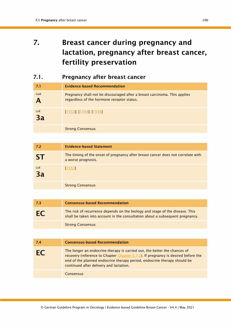
7.1 Pregnancy after breast cancer
© German Guideline Program in Oncology | Evidence-based Guideline Breast Cancer - V4.4 | May 2021
296
7. Breast cancer during pregnancy and
lactation, pregnancy after breast cancer,
fertility preservation
7.1. Pregnancy after breast cancer
7.1 Evidence-based Recommendation
GoR
A
Pregnancy shall not be discouraged after a breast carcinoma. This applies
regardless of the hormone receptor status.
LoE
3a
[1553]; [1554]; [1555]
Strong Consensus
7.2 Evidence-based Statement
ST The timing of the onset of pregnancy after breast cancer does not correlate with
a worse prognosis.
LoE
3a
[1553]
Strong Consensus
7.3 Consensus-based Recommendation
EC The risk of recurrence depends on the biology and stage of the disease. This
shall be taken into account in the consultation about a subsequent pregnancy.
Strong Consensus
7.4 Consensus-based Recommendation
EC The longer an endocrine therapy is carried out, the better the chances of
recovery (reference to Chapter Chapter 5.7.2). If pregnancy is desired before the
end of the planned endocrine therapy period, endocrine therapy should be
continued after delivery and lactation.
Consensus
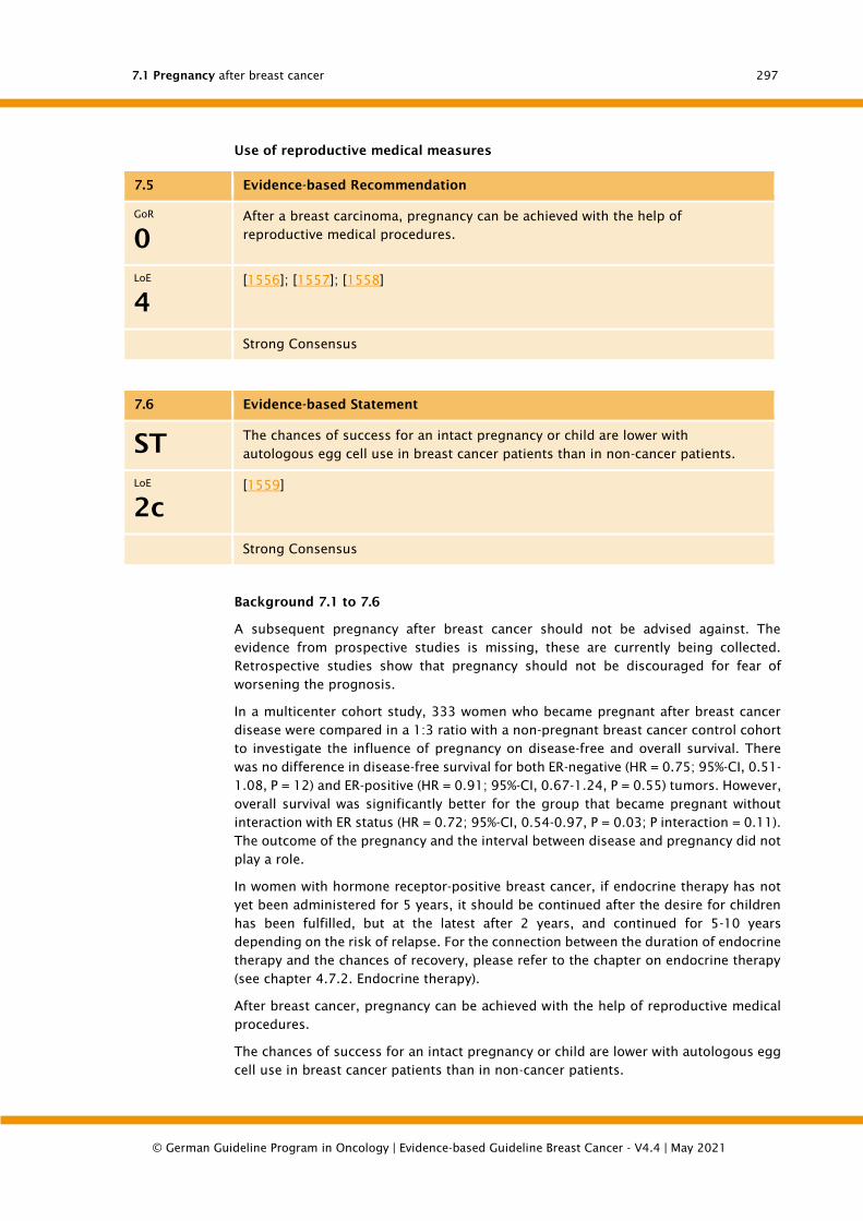
7.1 Pregnancy after breast cancer
© German Guideline Program in Oncology | Evidence-based Guideline Breast Cancer - V4.4 | May 2021
297
Use of reproductive medical measures
7.5 Evidence-based Recommendation
GoR
0
After a breast carcinoma, pregnancy can be achieved with the help of
reproductive medical procedures.
LoE
4
[1556]; [1557]; [1558]
Strong Consensus
7.6 Evidence-based Statement
ST The chances of success for an intact pregnancy or child are lower with
autologous egg cell use in breast cancer patients than in non-cancer patients.
LoE
2c
[1559]
Strong Consensus
Background 7.1 to 7.6
A subsequent pregnancy after breast cancer should not be advised against. The
evidence from prospective studies is missing, these are currently being collected.
Retrospective studies show that pregnancy should not be discouraged for fear of
worsening the prognosis.
In a multicenter cohort study, 333 women who became pregnant after breast cancer
disease were compared in a 1:3 ratio with a non-pregnant breast cancer control cohort
to investigate the influence of pregnancy on disease-free and overall survival. There
was no difference in disease-free survival for both ER-negative (HR = 0.75; 95%-CI, 0.51-
1.08, P = 12) and ER-positive (HR = 0.91; 95%-CI, 0.67-1.24, P = 0.55) tumors. However,
overall survival was significantly better for the group that became pregnant without
interaction with ER status (HR = 0.72; 95%-CI, 0.54-0.97, P = 0.03; P interaction = 0.11).
The outcome of the pregnancy and the interval between disease and pregnancy did not
play a role.
In women with hormone receptor-positive breast cancer, if endocrine therapy has not
yet been administered for 5 years, it should be continued after the desire for children
has been fulfilled, but at the latest after 2 years, and continued for 5-10 years
depending on the risk of relapse. For the connection between the duration of endocrine
therapy and the chances of recovery, please refer to the chapter on endocrine therapy
(see chapter 4.7.2. Endocrine therapy).
After breast cancer, pregnancy can be achieved with the help of reproductive medical
procedures.
The chances of success for an intact pregnancy or child are lower with autologous egg
cell use in breast cancer patients than in non-cancer patients.

7.2 Breast cancer during pregnancy
© German Guideline Program in Oncology | Evidence-based Guideline Breast Cancer - V4.4 | May 2021
298
In a retrospective study, 198 women were included, 25 of whom underwent a
reproductive examination. These women were older at diagnosis, at conception and
suffered more miscarriages. In both groups the rate of full-term pregnancies was equal
to 77% and 75%. There was no difference in prognosis between the two groups.
Necessary stimulation therapy can be used to obtain oocytes in the case of hormone
receptor-positive breast carcinoma with anti-hormonal concomitant treatment (e.g.
aromatase inhibitors or tamoxifen) (see also S3 guideline Fertility maintenance).
In a population-based register of 53,426 women, 441 women with a previous
carcinoma were identified within the last 5 years. One third of the women had a breast
carcinoma. In women with au-tologous oocytes, there were significant differences in
live birth rates between women with and without cancer (47.7% without carcinoma
versus 24.7% with previous carcinoma, p = 0.0001) and between the different tumor
types (from 53.5% for melanoma to 14.3% for breast cancer, p = 0.0001). With donor
oocytes these differences did not exist. In breast cancer patients, the probability of a
live birth was only 11.7% if the ART was performed within 6 months of the breast cancer
diagnosis, compared to 23% if it was beyond 6 months. Overall, only with melanoma
and ovarian cancer was the probability of a live birth after ART as high as without
cancer. For breast, cervical and endometrial carcinoma the probability was up to 80%
lower. However, there was no difference in the birth rate after successful conception.
7.2. Breast cancer during pregnancy
7.7 Consensus-based Recommendation
EC The treatment (system therapy, surgery, RT) of breast cancer (of pregnant
patients) during pregnancy shall be as close as possible to the standard
treatment of young, non-pregnant patients with breast cancer.
Strong Consensus
7.8 Evidence-based Recommendation
GoR
0
Standard chemotherapy with anthracyclines and taxanes can be administered in
the 2nd and 3rd trimester.
LoE
2b
[1560]; [1561]; [1562]
Strong Consensus
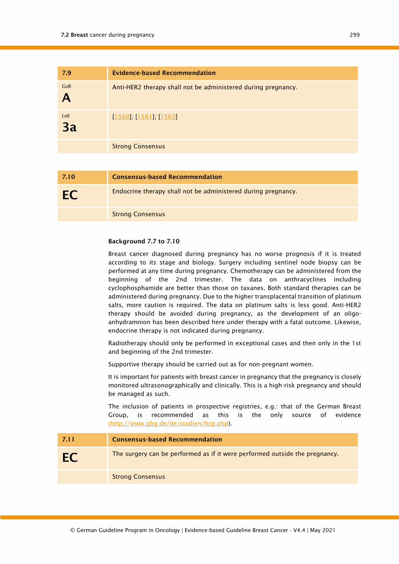
7.2 Breast cancer during pregnancy
© German Guideline Program in Oncology | Evidence-based Guideline Breast Cancer - V4.4 | May 2021
299
7.9 Evidence-based Recommendation
GoR
A
Anti-HER2 therapy shall not be administered during pregnancy.
LoE
3a
[1560]; [1561]; [1563]
Strong Consensus
7.10 Consensus-based Recommendation
EC Endocrine therapy shall not be administered during pregnancy.
Strong Consensus
Background 7.7 to 7.10
Breast cancer diagnosed during pregnancy has no worse prognosis if it is treated
according to its stage and biology. Surgery including sentinel node biopsy can be
performed at any time during pregnancy. Chemotherapy can be administered from the
beginning of the 2nd trimester. The data on anthracyclines including
cyclophosphamide are better than those on taxanes. Both standard therapies can be
administered during pregnancy. Due to the higher transplacental transition of platinum
salts, more caution is required. The data on platinum salts is less good. Anti-HER2
therapy should be avoided during pregnancy, as the development of an oligo-
anhydramnion has been described here under therapy with a fatal outcome. Likewise,
endocrine therapy is not indicated during pregnancy.
Radiotherapy should only be performed in exceptional cases and then only in the 1st
and beginning of the 2nd trimester.
Supportive therapy should be carried out as for non-pregnant women.
It is important for patients with breast cancer in pregnancy that the pregnancy is closely
monitored ultrasonographically and clinically. This is a high-risk pregnancy and should
be managed as such.
The inclusion of patients in prospective registries, e.g.: that of the German Breast
Group, is recommended as this is the only source of evidence
(http://www.gbg.de/de/studien/bcp.php).
7.11 Consensus-based Recommendation
EC The surgery can be performed as if it were performed outside the pregnancy.
Strong Consensus
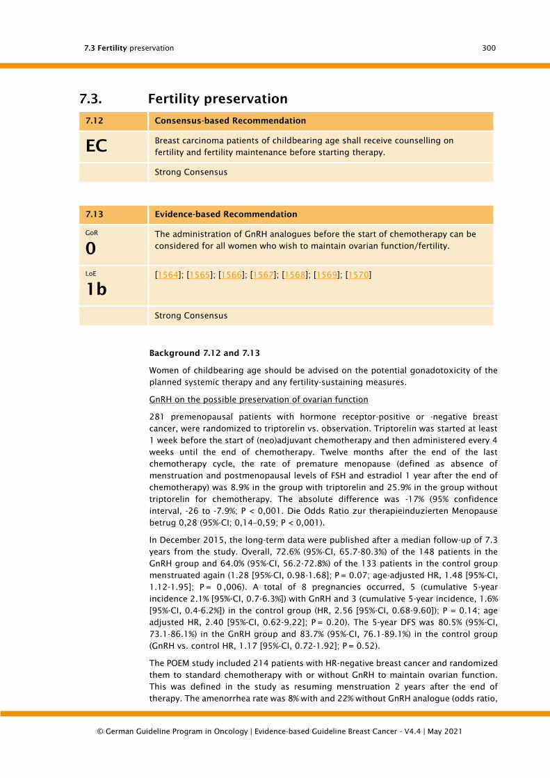
7.3 Fertility preservation
© German Guideline Program in Oncology | Evidence-based Guideline Breast Cancer - V4.4 | May 2021
300
7.3. Fertility preservation
7.12 Consensus-based Recommendation
EC Breast carcinoma patients of childbearing age shall receive counselling on
fertility and fertility maintenance before starting therapy.
Strong Consensus
7.13 Evidence-based Recommendation
GoR
0
The administration of GnRH analogues before the start of chemotherapy can be
considered for all women who wish to maintain ovarian function/fertility.
LoE
1b
[1564]; [1565]; [1566]; [1567]; [1568]; [1569]; [1570]
Strong Consensus
Background 7.12 and 7.13
Women of childbearing age should be advised on the potential gonadotoxicity of the
planned systemic therapy and any fertility-sustaining measures.
GnRH on the possible preservation of ovarian function
281 premenopausal patients with hormone receptor-positive or -negative breast
cancer, were randomized to triptorelin vs. observation. Triptorelin was started at least
1 week before the start of (neo)adjuvant chemotherapy and then administered every 4
weeks until the end of chemotherapy. Twelve months after the end of the last
chemotherapy cycle, the rate of premature menopause (defined as absence of
menstruation and postmenopausal levels of FSH and estradiol 1 year after the end of
chemotherapy) was 8.9% in the group with triptorelin and 25.9% in the group without
triptorelin for chemotherapy. The absolute difference was -17% (95% confidence
interval, -26 to -7.9%; P < 0,001. Die Odds Ratio zur therapieinduzierten Menopause
betrug 0,28 (95%-CI; 0,14–0,59; P < 0,001).
In December 2015, the long-term data were published after a median follow-up of 7.3
years from the study. Overall, 72.6% (95%-CI, 65.7-80.3%) of the 148 patients in the
GnRH group and 64.0% (95%-CI, 56.2-72.8%) of the 133 patients in the control group
menstruated again (1.28 [95%-CI, 0.98-1.68]; P = 0.07; age-adjusted HR, 1.48 [95%-CI,
1.12-1.95]; P = 0 ,006). A total of 8 pregnancies occurred, 5 (cumulative 5-year
incidence 2.1% [95%-CI, 0.7-6.3%]) with GnRH and 3 (cumulative 5-year incidence, 1.6%
[95%-CI, 0.4-6.2%]) in the control group (HR, 2.56 [95%-CI, 0.68-9.60]); P = 0.14; age
adjusted HR, 2.40 [95%-CI, 0.62-9.22]; P = 0.20). The 5-year DFS was 80.5% (95%-CI,
73.1-86.1%) in the GnRH group and 83.7% (95%-CI, 76.1-89.1%) in the control group
(GnRH vs. control HR, 1.17 [95%-CI, 0.72-1.92]; P = 0.52).
The POEM study included 214 patients with HR-negative breast cancer and randomized
them to standard chemotherapy with or without GnRH to maintain ovarian function.
This was defined in the study as resuming menstruation 2 years after the end of
therapy. The amenorrhea rate was 8% with and 22% without GnRH analogue (odds ratio,

7.3 Fertility preservation
© German Guideline Program in Oncology | Evidence-based Guideline Breast Cancer - V4.4 | May 2021
301
OR 0.3; 95%-CI 0.1-0.87, p = 0.03). Birth and pregnancy rates were significantly higher
in the group with GnRH than in the group without (21% vs. 11%; OR 2.45 p = 0.04). The
women who had a child were significantly younger than the others. They may also have
been more motivated to contribute to the success of the study. In addition, the study
showed a significantly better disease-free and overall survival for the GnRH group in
patients with HR-negative breast cancer. Overall, the primary endpoint could only be
determined in less than 50% of the patients, and the study was terminated prematurely.
The study therefore has clear deficiencies.
A meta-analysis of the various studies on the subject also comes to a positive effect of
GnRH therapy, at least as far as ovarian function is concerned.
As far as fertility maintenance is concerned, reference is made primarily to the methods
of reproductive therapy, which will not be discussed here (reference to S2/3 guideline
on this).
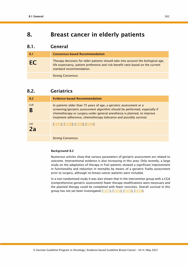
8.1 General
© German Guideline Program in Oncology | Evidence-based Guideline Breast Cancer - V4.4 | May 2021
302
8. Breast cancer in elderly patients
8.1. General
8.1 Consensus-based Recommendation
EC Therapy decisions for older patients should take into account the biological age,
life expectancy, patient preference and risk-benefit ratio based on the current
standard recommendation.
Strong Consensus
8.2. Geriatrics
8.2 Evidence-based Recommendation
GoR
B
In patients older than 75 years of age, a geriatric assessment or a
screening/geriatric assessment algorithm should be performed, especially if
chemotherapy or surgery under general anesthesia is planned, to improve
treatment adherence, chemotherapy tolerance and possibly survival.
LoE
2a
[1571]; [1572]; [1573]; [1574]
Strong Consensus
Background 8.2
Numerous articles show that various parameters of geriatric assessment are related to
outcome. Interventional evidence is also increasing in this area: Only recently, a large
study on the adaptation of therapy in frail patients showed a significant improvement
in functionality and reduction in mortality by means of a geriatric frailty assessment
prior to surgery, although no breast cancer patients were included.
In a non-randomised study it was also shown that in the intervention group with a CGA
(comprehensive geriatric assessment) fewer therapy modifications were necessary and
the planned therapy could be completed with fewer toxicities. Overall survival in this
group has not yet been investigated [1571], [1572], [1573], [1574].
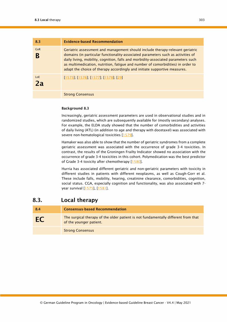
8.3 Local therapy
© German Guideline Program in Oncology | Evidence-based Guideline Breast Cancer - V4.4 | May 2021
303
8.3 Evidence-based Recommendation
GoR
B
Geriatric assessment and management should include therapy-relevant geriatric
domains (in particular functionality-associated parameters such as activities of
daily living, mobility, cognition, falls and morbidity-associated parameters such
as multimedication, nutrition, fatigue and number of comorbidities) in order to
adapt the choice of therapy accordingly and initiate supportive measures.
LoE
2a
[1575]; [1576]; [1577]; [1578]; [29]
Strong Consensus
Background 8.3
Increasingly, geriatric assessment parameters are used in observational studies and in
randomized studies, which are subsequently available for (mostly secondary) analyses.
For example, the ELDA study showed that the number of comorbidities and activities
of daily living (ATL) (in addition to age and therapy with docetaxel) was associated with
severe non-hematological toxicities [1579].
Hamaker was also able to show that the number of geriatric syndromes from a complete
geriatric assessment was associated with the occurrence of grade 3-4 toxicities. In
contrast, the results of the Groningen Frailty Indicator showed no association with the
occurrence of grade 3-4 toxicities in this cohort. Polymedication was the best predictor
of Grade 3-4 toxicity after chemotherapy [1580].
Hurria has associated different geriatric and non-geriatric parameters with toxicity in
different studies in patients with different neoplasms, as well as Cough-Gorr et al.
These include falls, mobility, hearing, creatinine clearance, comorbidities, cognition,
social status. CGA, especially cognition and functionality, was also associated with 7-
year survival [1575], [1581].
8.3. Local therapy
8.4 Consensus-based Recommendation
EC The surgical therapy of the older patient is not fundamentally different from that
of the younger patient.
Strong Consensus
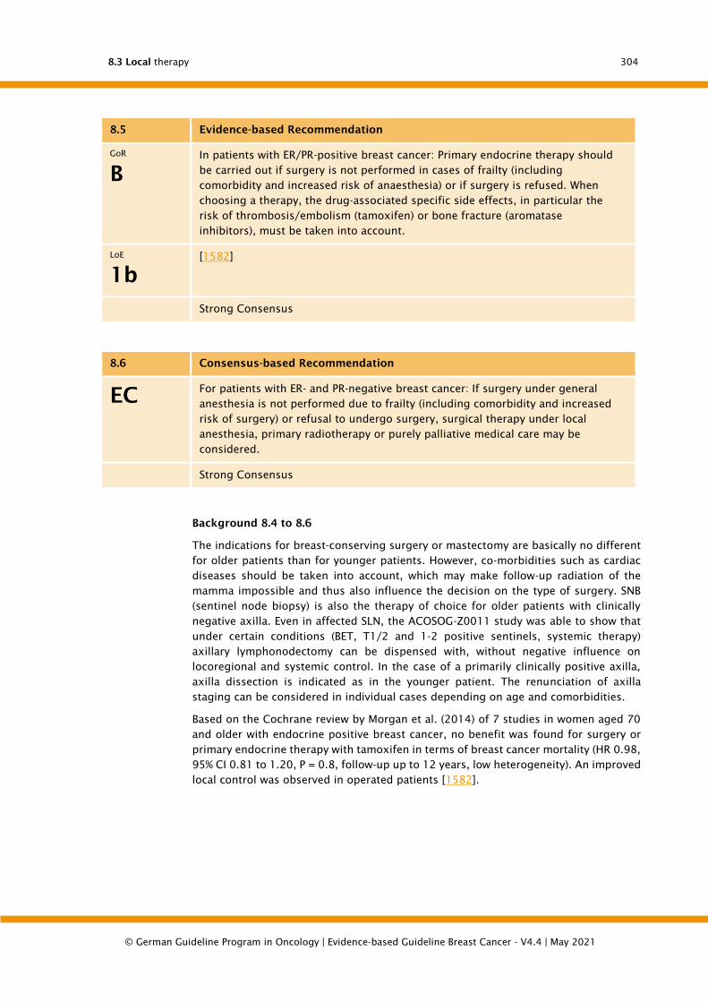
8.3 Local therapy
© German Guideline Program in Oncology | Evidence-based Guideline Breast Cancer - V4.4 | May 2021
304
8.5 Evidence-based Recommendation
GoR
B
In patients with ER/PR-positive breast cancer: Primary endocrine therapy should
be carried out if surgery is not performed in cases of frailty (including
comorbidity and increased risk of anaesthesia) or if surgery is refused. When
choosing a therapy, the drug-associated specific side effects, in particular the
risk of thrombosis/embolism (tamoxifen) or bone fracture (aromatase
inhibitors), must be taken into account.
LoE
1b
[1582]
Strong Consensus
8.6 Consensus-based Recommendation
EC For patients with ER- and PR-negative breast cancer: If surgery under general
anesthesia is not performed due to frailty (including comorbidity and increased
risk of surgery) or refusal to undergo surgery, surgical therapy under local
anesthesia, primary radiotherapy or purely palliative medical care may be
considered.
Strong Consensus
Background 8.4 to 8.6
The indications for breast-conserving surgery or mastectomy are basically no different
for older patients than for younger patients. However, co-morbidities such as cardiac
diseases should be taken into account, which may make follow-up radiation of the
mamma impossible and thus also influence the decision on the type of surgery. SNB
(sentinel node biopsy) is also the therapy of choice for older patients with clinically
negative axilla. Even in affected SLN, the ACOSOG-Z0011 study was able to show that
under certain conditions (BET, T1/2 and 1-2 positive sentinels, systemic therapy)
axillary lymphonodectomy can be dispensed with, without negative influence on
locoregional and systemic control. In the case of a primarily clinically positive axilla,
axilla dissection is indicated as in the younger patient. The renunciation of axilla
staging can be considered in individual cases depending on age and comorbidities.
Based on the Cochrane review by Morgan et al. (2014) of 7 studies in women aged 70
and older with endocrine positive breast cancer, no benefit was found for surgery or
primary endocrine therapy with tamoxifen in terms of breast cancer mortality (HR 0.98,
95% CI 0.81 to 1.20, P = 0.8, follow-up up to 12 years, low heterogeneity). An improved
local control was observed in operated patients [1582].
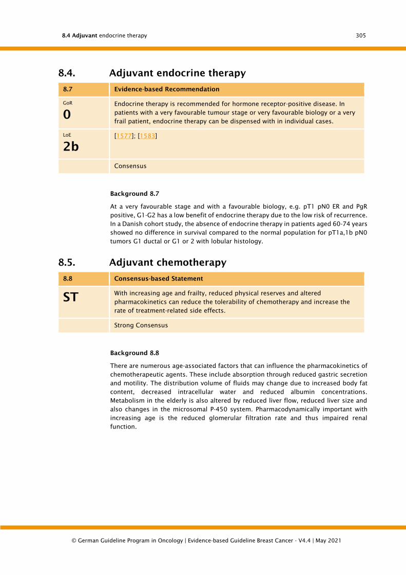
8.4 Adjuvant endocrine therapy
© German Guideline Program in Oncology | Evidence-based Guideline Breast Cancer - V4.4 | May 2021
305
8.4. Adjuvant endocrine therapy
8.7 Evidence-based Recommendation
GoR
0
Endocrine therapy is recommended for hormone receptor-positive disease. In
patients with a very favourable tumour stage or very favourable biology or a very
frail patient, endocrine therapy can be dispensed with in individual cases.
LoE
2b
[1577]; [1583]
Consensus
Background 8.7
At a very favourable stage and with a favourable biology, e.g. pT1 pN0 ER and PgR
positive, G1-G2 has a low benefit of endocrine therapy due to the low risk of recurrence.
In a Danish cohort study, the absence of endocrine therapy in patients aged 60-74 years
showed no difference in survival compared to the normal population for pT1a,1b pN0
tumors G1 ductal or G1 or 2 with lobular histology.
8.5. Adjuvant chemotherapy
8.8 Consensus-based Statement
ST With increasing age and frailty, reduced physical reserves and altered
pharmacokinetics can reduce the tolerability of chemotherapy and increase the
rate of treatment-related side effects.
Strong Consensus
Background 8.8
There are numerous age-associated factors that can influence the pharmacokinetics of
chemotherapeutic agents. These include absorption through reduced gastric secretion
and motility. The distribution volume of fluids may change due to increased body fat
content, decreased intracellular water and reduced albumin concentrations.
Metabolism in the elderly is also altered by reduced liver flow, reduced liver size and
also changes in the microsomal P-450 system. Pharmacodynamically important with
increasing age is the reduced glomerular filtration rate and thus impaired renal
function.
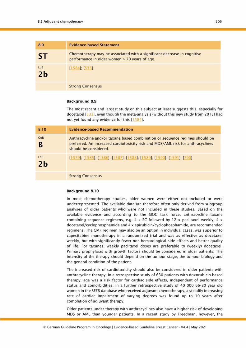
8.5 Adjuvant chemotherapy
© German Guideline Program in Oncology | Evidence-based Guideline Breast Cancer - V4.4 | May 2021
306
8.9 Evidence-based Statement
ST Chemotherapy may be associated with a significant decrease in cognitive
performance in older women > 70 years of age.
LoE
2b
[1584]; [533]
Strong Consensus
Background 8.9
The most recent and largest study on this subject at least suggests this, especially for
docetaxel [533], even though the meta-analysis (without this new study from 2015) had
not yet found any evidence for this [1584].
8.10 Evidence-based Recommendation
GoR
B
Anthracycline and/or taxane based combination or sequence regimes should be
preferred. An increased cardiotoxicity risk and MDS/AML risk for anthracyclines
should be considered.
LoE
2b
[1579]; [1585]; [1586]; [1587]; [1588]; [1589]; [1590]; [1591]; [790]
Strong Consensus
Background 8.10
In most chemotherapy studies, older women were either not included or were
underrepresented. The available data are therefore often only derived from subgroup
analyses of older patients who were not included in these studies. Based on the
available evidence and according to the SIOG task force, anthracycline taxane
containing sequence regimens, e.g. 4 x EC followed by 12 x paclitaxel weekly, 4 x
docetaxel/cyclophosphamide and 4 x epirubicin/cyclophosphamide, are recommended
regimens. The CMF regimen may also be an option in individual cases, was superior to
capecitabine monotherapy in a randomized trial and was as effective as docetaxel
weekly, but with significantly fewer non-hematological side effects and better quality
of life. For taxanes, weekly paclitaxel doses are preferable to (weekly) docetaxel.
Primary prophylaxis with growth factors should be considered in older patients. The
intensity of the therapy should depend on the tumour stage, the tumour biology and
the general condition of the patient.
The increased risk of cardiotoxicity should also be considered in older patients with
anthracycline therapy. In a retrospective study of 630 patients with doxorubicin-based
therapy, age was a risk factor for cardiac side effects, independent of performance
status and comorbidities. In a further retrospective study of 40 000 66-80 year old
women in the SEER database who received adjuvant chemotherapy, a steadily increasing
rate of cardiac impairment of varying degrees was found up to 10 years after
completion of adjuvant therapy.
Older patients under therapy with anthracyclines also have a higher risk of developing
MDS or AML than younger patients. In a recent study by Freedman, however, the
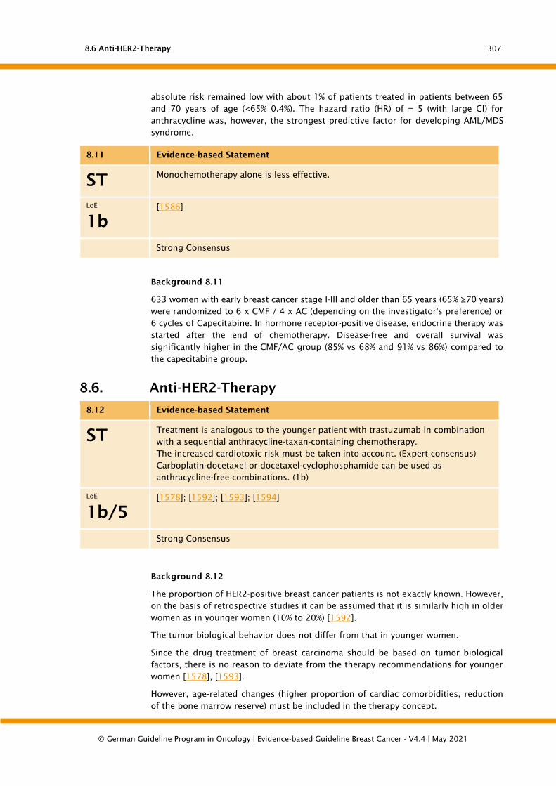
8.6 Anti-HER2-Therapy
© German Guideline Program in Oncology | Evidence-based Guideline Breast Cancer - V4.4 | May 2021
307
absolute risk remained low with about 1% of patients treated in patients between 65
and 70 years of age (<65% 0.4%). The hazard ratio (HR) of = 5 (with large Cl) for
anthracycline was, however, the strongest predictive factor for developing AML/MDS
syndrome.
8.11 Evidence-based Statement
ST Monochemotherapy alone is less effective.
LoE
1b
[1586]
Strong Consensus
Background 8.11
633 women with early breast cancer stage I-III and older than 65 years (65% ≥70 years)
were randomized to 6 x CMF / 4 x AC (depending on the investigator's preference) or
6 cycles of Capecitabine. In hormone receptor-positive disease, endocrine therapy was
started after the end of chemotherapy. Disease-free and overall survival was
significantly higher in the CMF/AC group (85% vs 68% and 91% vs 86%) compared to
the capecitabine group.
8.6. Anti-HER2-Therapy
8.12 Evidence-based Statement
ST Treatment is analogous to the younger patient with trastuzumab in combination
with a sequential anthracycline-taxan-containing chemotherapy.
The increased cardiotoxic risk must be taken into account. (Expert consensus)
Carboplatin-docetaxel or docetaxel-cyclophosphamide can be used as
anthracycline-free combinations. (1b)
LoE
1b/5
[1578]; [1592]; [1593]; [1594]
Strong Consensus
Background 8.12
The proportion of HER2-positive breast cancer patients is not exactly known. However,
on the basis of retrospective studies it can be assumed that it is similarly high in older
women as in younger women (10% to 20%) [1592].
The tumor biological behavior does not differ from that in younger women.
Since the drug treatment of breast carcinoma should be based on tumor biological
factors, there is no reason to deviate from the therapy recommendations for younger
women [1578], [1593].
However, age-related changes (higher proportion of cardiac comorbidities, reduction
of the bone marrow reserve) must be included in the therapy concept.

8.6 Anti-HER2-Therapy
© German Guideline Program in Oncology | Evidence-based Guideline Breast Cancer - V4.4 | May 2021
308
Anthracycline-free chemotherapy regimens in combination with trastuzumab showed
an identical efficacy on [1595]. In a randomized phase III study, the combination of
docetaxel-cyclophosphamide with doxorubicin-cyclophosphamide was superior to
[1594]. Due to the expected myelotoxicity, granulocyte stimulating factors (GCSF)
should be used prophylactically according to the ESMO guidelines.
8.13 Evidence-based Recommendation
GoR
0
Paclitaxel weekly (12 weeks) with trastuzumab can be used for T1-2 (up to 3cm)
pN0 tumors.
LoE
2b
[1596]; [1597]
Strong Consensus
Background 8.13
The Pase-II study of CALBG by Tolaney et al. showed excellent 3-year survival (98%
survival rate) for hormone receptor-positive, HER2-positive, nodal-negative tumors.
In this study, patients were treated for 12 weeks with weekly paclitaxel therapy in
combination with trastuzumab. After 12 weeks, treatment with trastuzumab was
continued for a total of 1 year and endocrine therapy was started. In this study one
third of the women were older than 60 years. Due to the reduced toxicity of this
combination, it appears to be particularly suitable as an adjuvant treatment option for
patients with cardiac comorbidities [1596] and HER2-positive breast cancer.
9. Breast cancer in men
The diagnosis and treatment of breast cancer in men should be interdisciplinary and
requires gynaecological and oncological expertise due to the tumor biological
characteristics and similarity to breast cancer in women. Interdisciplinary cooperation
between breast centers, practicing gynecologists, urologists and andrologists is
particularly recommended for the treatment of sexual dysfunction by tamoxifen
therapy, for men with BRCA mutations [1598] with an associated increased risk of
prostate cancer, and for men with breast cancer for whom treatment of benign prostate
syndrome should be performed [1599].
9.1 Consensus-based Recommendation
EC Early medical consultation shall be encouraged by informing men about the
disease, especially about symptoms and changes in the breast, and by
encouraging self-observation.
Strong Consensus
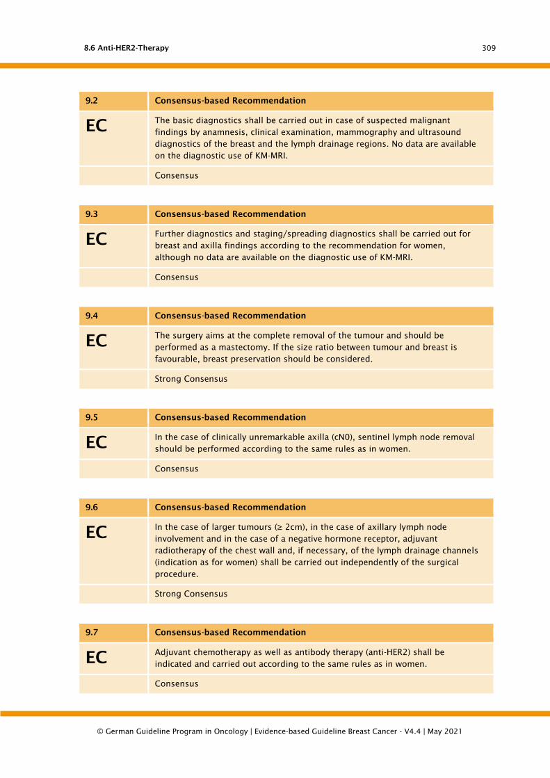
8.6 Anti-HER2-Therapy
© German Guideline Program in Oncology | Evidence-based Guideline Breast Cancer - V4.4 | May 2021
309
9.2 Consensus-based Recommendation
EC The basic diagnostics shall be carried out in case of suspected malignant
findings by anamnesis, clinical examination, mammography and ultrasound
diagnostics of the breast and the lymph drainage regions. No data are available
on the diagnostic use of KM-MRI.
Consensus
9.3 Consensus-based Recommendation
EC Further diagnostics and staging/spreading diagnostics shall be carried out for
breast and axilla findings according to the recommendation for women,
although no data are available on the diagnostic use of KM-MRI.
Consensus
9.4 Consensus-based Recommendation
EC The surgery aims at the complete removal of the tumour and should be
performed as a mastectomy. If the size ratio between tumour and breast is
favourable, breast preservation should be considered.
Strong Consensus
9.5 Consensus-based Recommendation
EC In the case of clinically unremarkable axilla (cN0), sentinel lymph node removal
should be performed according to the same rules as in women.
Consensus
9.6 Consensus-based Recommendation
EC In the case of larger tumours (≥ 2cm), in the case of axillary lymph node
involvement and in the case of a negative hormone receptor, adjuvant
radiotherapy of the chest wall and, if necessary, of the lymph drainage channels
(indication as for women) shall be carried out independently of the surgical
procedure.
Strong Consensus
9.7 Consensus-based Recommendation
EC Adjuvant chemotherapy as well as antibody therapy (anti-HER2) shall be
indicated and carried out according to the same rules as in women.
Consensus
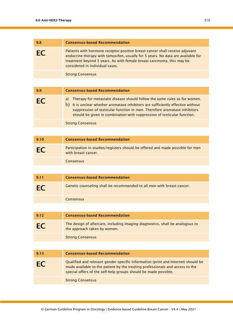
8.6 Anti-HER2-Therapy
© German Guideline Program in Oncology | Evidence-based Guideline Breast Cancer - V4.4 | May 2021
310
9.8 Consensus-based Recommendation
EC Patients with hormone receptor-positive breast cancer shall receive adjuvant
endocrine therapy with tamoxifen, usually for 5 years. No data are available for
treatment beyond 5 years. As with female breast carcinoma, this may be
considered in individual cases.
Strong Consensus
9.9 Consensus-based Recommendation
EC a) Therapy for metastatic disease should follow the same rules as for women.
b) It is unclear whether aromatase inhibitors are sufficiently effective without
suppression of testicular function in men. Therefore aromatase inhibitors
should be given in combination with suppression of testicular function.
Strong Consensus
9.10 Consensus-based Recommendation
EC Participation in studies/registers should be offered and made possible for men
with breast cancer.
Consensus
9.11 Consensus-based Recommendation
EC Genetic counseling shall be recommended to all men with breast cancer.
Consensus
9.12 Consensus-based Recommendation
EC The design of aftercare, including imaging diagnostics, shall be analogous to
the approach taken by women.
Strong Consensus
9.13 Consensus-based Recommendation
EC Qualified and relevant gender-specific information (print and Internet) should be
made available to the patient by the treating professionals and access to the
special offers of the self-help groups should be made possible.
Strong Consensus
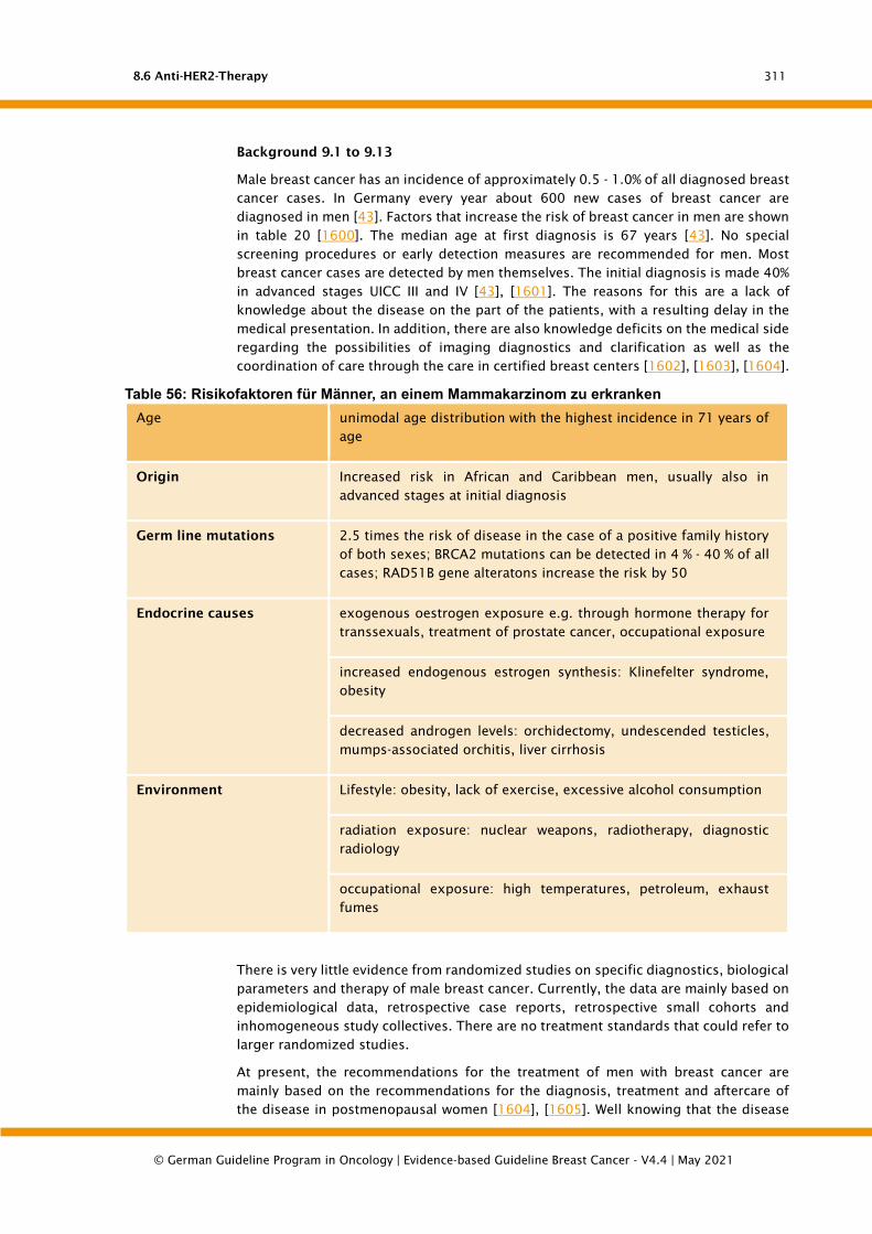
8.6 Anti-HER2-Therapy
© German Guideline Program in Oncology | Evidence-based Guideline Breast Cancer - V4.4 | May 2021
311
Background 9.1 to 9.13
Male breast cancer has an incidence of approximately 0.5 - 1.0% of all diagnosed breast
cancer cases. In Germany every year about 600 new cases of breast cancer are
diagnosed in men [43]. Factors that increase the risk of breast cancer in men are shown
in table 20 [1600]. The median age at first diagnosis is 67 years [43]. No special
screening procedures or early detection measures are recommended for men. Most
breast cancer cases are detected by men themselves. The initial diagnosis is made 40%
in advanced stages UICC III and IV [43], [1601]. The reasons for this are a lack of
knowledge about the disease on the part of the patients, with a resulting delay in the
medical presentation. In addition, there are also knowledge deficits on the medical side
regarding the possibilities of imaging diagnostics and clarification as well as the
coordination of care through the care in certified breast centers [1602], [1603], [1604].
Table 56: Risikofaktoren für Männer, an einem Mammakarzinom zu erkranken
Age unimodal age distribution with the highest incidence in 71 years of
age
Origin Increased risk in African and Caribbean men, usually also in
advanced stages at initial diagnosis
Germ line mutations 2.5 times the risk of disease in the case of a positive family history
of both sexes; BRCA2 mutations can be detected in 4 % - 40 % of all
cases; RAD51B gene alteratons increase the risk by 50
Endocrine causes
exogenous oestrogen exposure e.g. through hormone therapy for
transsexuals, treatment of prostate cancer, occupational exposure
increased endogenous estrogen synthesis: Klinefelter syndrome,
obesity
decreased androgen levels: orchidectomy, undescended testicles,
mumps-associated orchitis, liver cirrhosis
Environment
Lifestyle: obesity, lack of exercise, excessive alcohol consumption
radiation exposure: nuclear weapons, radiotherapy, diagnostic
radiology
occupational exposure: high temperatures, petroleum, exhaust
fumes
There is very little evidence from randomized studies on specific diagnostics, biological
parameters and therapy of male breast cancer. Currently, the data are mainly based on
epidemiological data, retrospective case reports, retrospective small cohorts and
inhomogeneous study collectives. There are no treatment standards that could refer to
larger randomized studies.
At present, the recommendations for the treatment of men with breast cancer are
mainly based on the recommendations for the diagnosis, treatment and aftercare of
the disease in postmenopausal women [1604], [1605]. Well knowing that the disease
8.6 Anti-HER2-Therapy
© German Guideline Program in Oncology | Evidence-based Guideline Breast Cancer - V4.4 | May 2021
312
in men has other biological potentials that have to be considered in the care of patients
[1606], [1607], [1608]. There is an international consensus to increase the knowledge
base on breast cancer in men by participating in registry studies [1605], [1609], [1610].
In male breast cancer patients a similar breast cancer mortality rate is found as in older
postmenopausal women. A genetic disposition is more frequent in men, especially
mutation BRCA1 and BRCA2 [1611]. Furthermore, men with breast cancer have an up
to 20% increased risk constellation for second malignancies [1612].
Over 90% of patients are diagnosed with ER-positive invasive ductal carcinoma. The
HER2 overexpression is inconsistently reported in the literature as 12-37%. A recent
study found 97% ER-positive and only 10% HER2-positive tumors in a unicentric cohort
of 61 invasive mammary carcinomas in men [1613]. 39-95% of the cases showed
androgen receptor expression. In contrast to the histopathological similarities to breast
cancer in women, molecular biological examinations show significant differences in
[1607], [1608], [1610], [1613].
Most men have so far been treated by mastectomy and axillary lymphonodectomy
(ALND) and possibly with chest wall radiation [1601], [1614], [1615] Current data
suggest in particular less radical surgical measures with the aim of reducing therapy-
related morbidity [1616], [1617], [1618], [1619].
Men with breast cancer with lymph node involvement benefit from adjuvant
chemotherapy with improved prognosis (disease-free survival, overall survival) [1620].
When deciding on adjuvant therapy, comorbidities and tolerance as well as patient
preferences must be considered. The substances and regimens commonly used in
women including anti-HER2-therapy are used if indicated. Tamoxifen is currently the
standard therapy for hormone receptor-positive breast cancer. The side effects such as
sexual dysfunction lead to a high therapy discontinuation rate [1621]. The use of
aromatase inhibitors in adjuvant therapy is not recommended; aromatase inhibitors
were associated with a significantly increased mortality in a retrospective analysis of
German cancer registries [1622].
There is no evidence from clinical studies on the treatment of HER2-positive breast
cancer in men; however, there is consensus that, following the successes in HER2-
positive breast cancer in women, men with HER2-positive breast cancer should also be
treated adjuvantly with trastuzumab [1623].
In metastasis, aromatase inhibitors can be used second-line, most likely in combination
with drug suppression of the gonadal function [1624], [1625], [1626], [1627], [1628].
In advanced metastatic disease, studies show treatment options for fulvestrant,
aromatase inhibitors and eribulin from [1625], [1626], [1628], [1629], [1630].
Rehabilitation and aftercare, including imaging diagnostics, are carried out in
accordance with the recommended aftercare for women. Aftercare for men focuses on
the specific risks, comorbidities, short and long-term side effects to be considered and
includes psychosocial as well as psycho-oncological aspects [1387], [1631].
8.6 Anti-HER2-Therapy
© German Guideline Program in Oncology | Evidence-based Guideline Breast Cancer - V4.4 | May 2021
313
10. Quality Indicators
Quality indicators (QI) are measured variables whose collection serves to assess the
quality of the underlying structures, processes or results [1632]. Quality indicators are
an important instrument of quality management. The aim of their use is the continuous
improvement of care by presenting the results of care, reflecting them critically and
improving them if necessary. The present selection of quality indicators was prepared
according to the methodology of the Oncology Guidelines Programme [1633]. For the
derivation process a working group "Working Group Quality Indicators" was
constituted. This group created the final set of quality indicators based on the existing
quality indicators of the 2012 guideline and the new strong recommendations (strength
of recommendation A, "should") of the updated guideline. The compilation took into
account the results of the existing quality indicators from the certified breast cancer
centers of the German Cancer Society and the German Society for Senology [1551], the
quality indicators of the external inpatient quality assurance [1634] and the results of
the research for existing national and international QIs. The results of the guideline-
based quality indicators, which are measured and evaluated in the certification
procedure, were already presented at the kick-off meeting of the present update in
order to be able to make changes to recommendations and background texts based on
the results from the care system, if necessary. Changes were made for some of the
recommendations underlying the existing indicators. The changes are mainly due to
new study results. The exact procedure for selecting potential QIs and the composition
of the working group is described in the guideline report (http://leitlinienprogramm-
onkologie.de/index.php?id=67ype=0).
After a face-to-face meeting, a written evaluation of the indicators positively prioritized
there and a concluding conference call of the working group, 2 new indicators were
adopted (supplement for QI 5 "Indication for sentinel lymph node biopsy" and QI 6
"Therapy of axillary lymph drainage areas in pN1"). The already existing QI 5 "Indication
for sentinel lymph node biopsy" will in future be calculated separately for female (5 a))
and male patients (5 b)) on the basis of the data of the QA procedure for breast surgery
of the Institute for Quality Assurance and Transparency in Health Care (IQTIG), so that
only 1 QI will be added to the already existing QI. Of the 12 QIs from Guideline 2012,
4 QIs were deleted and 2 QIs (QI 2: Intraoperative preparation radio/sonography, QI 4:
Endocrine therapy as the first therapeutic option in steroid-receptor-positive metastatic
breast cancer) were adjusted in the numerator or denominator. The justifications are
described in detail in the Methods Report. The final set thus consists of 9 quality
indicators.
Based on the recommendation "3.9 Diagnosis and care chain breast cancer early
detection", the Working Group QI has defined a QI that is intended to record and
improve the further treatment of patients secured in screening in certified breast cancer
centres.
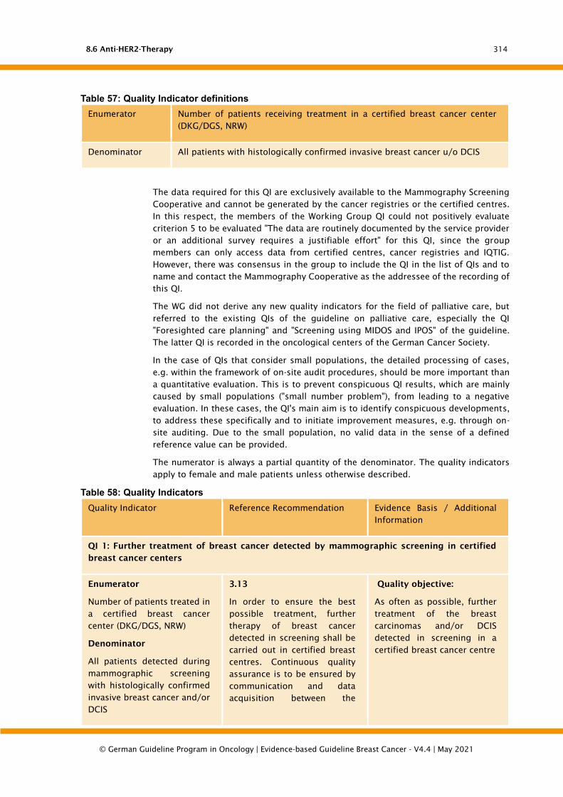
8.6 Anti-HER2-Therapy
© German Guideline Program in Oncology | Evidence-based Guideline Breast Cancer - V4.4 | May 2021
314
Table 57: Quality Indicator definitions
Enumerator Number of patients receiving treatment in a certified breast cancer center
(DKG/DGS, NRW)
Denominator All patients with histologically confirmed invasive breast cancer u/o DCIS
The data required for this QI are exclusively available to the Mammography Screening
Cooperative and cannot be generated by the cancer registries or the certified centres.
In this respect, the members of the Working Group QI could not positively evaluate
criterion 5 to be evaluated "The data are routinely documented by the service provider
or an additional survey requires a justifiable effort" for this QI, since the group
members can only access data from certified centres, cancer registries and IQTIG.
However, there was consensus in the group to include the QI in the list of QIs and to
name and contact the Mammography Cooperative as the addressee of the recording of
this QI.
The WG did not derive any new quality indicators for the field of palliative care, but
referred to the existing QIs of the guideline on palliative care, especially the QI
"Foresighted care planning" and "Screening using MIDOS and IPOS" of the guideline.
The latter QI is recorded in the oncological centers of the German Cancer Society.
In the case of QIs that consider small populations, the detailed processing of cases,
e.g. within the framework of on-site audit procedures, should be more important than
a quantitative evaluation. This is to prevent conspicuous QI results, which are mainly
caused by small populations ("small number problem"), from leading to a negative
evaluation. In these cases, the QI's main aim is to identify conspicuous developments,
to address these specifically and to initiate improvement measures, e.g. through on-
site auditing. Due to the small population, no valid data in the sense of a defined
reference value can be provided.
The numerator is always a partial quantity of the denominator. The quality indicators
apply to female and male patients unless otherwise described.
Table 58: Quality Indicators
Quality Indicator Reference Recommendation Evidence Basis / Additional
Information
QI 1: Further treatment of breast cancer detected by mammographic screening in certified
breast cancer centers
Enumerator
Number of patients treated in
a certified breast cancer
center (DKG/DGS, NRW)
Denominator
All patients detected during
mammographic screening
with histologically confirmed
invasive breast cancer and/or
DCIS
3.13
In order to ensure the best
possible treatment, further
therapy of breast cancer
detected in screening shall be
carried out in certified breast
centres. Continuous quality
assurance is to be ensured by
communication and data
acquisition between the
Quality objective:
As often as possible, further
treatment of the breast
carcinomas and/or DCIS
detected in screening in a
certified breast cancer centre

8.6 Anti-HER2-Therapy
© German Guideline Program in Oncology | Evidence-based Guideline Breast Cancer - V4.4 | May 2021
315
Quality Indicator Reference Recommendation Evidence Basis / Additional
Information
The QI can be evaluated with
data from the Mammography
Cooperation Group.
screening centre and the
certified breast centre.
QI 2: Pretherapeutic histological confirmation (since 2012, formerly: Breast CA 1)
Enumerator
Patients with pretherapeutic
histological confirmation of
diagnosis by core needle
biopsy or vacuum-assisted
biopsy
Denominator
Patients with primary
intervention and histology
“invasive breast cancer
(primary tumor) or DCIS" as
primary disease
4.14
The histological clarification of
findings shall be carried out by
punch biopsy, vacuum biopsy
and, in exceptional cases
which must be justified, by
open excision biopsy.
LOE 3a, recommendation
level A
Quality objective:
As many patients as possible
with pre-therapeutic
histological confirmation by
punch or vacuum biopsy in
case of initial intervention
and primary disease invasive
breast cancer and/or DCIS
QI 3: Intraoperative specimen radiography/sonography (since 2012, formerly: Breast CA 2)
Enumerator
Surgeries using
intraoperative specimen X-ray
or intraoperative specimen
ultrasound
Denominator
Surgeries with preoperative
wire marking guided by
mammography or ultrasound
4.25
Pre-operative or intraoperative
marking shall be carried out
using the method that allows
the findings to be clearly
visualized, especially in the
case of non-palpable changes.
Proof of adequate resection
must be provided
intraoperatively by means of
specimen radiography or
specimen sonography. If MR-
guided marking has been
performed, an MR control
should be performed within 6
months in case of
histologically unspecific
benign findings.
Quality objective:
As often as possible
intraoperative preparation
sonography or radiography
after preoperative marking
QI 4: Axillary lymph node biopsy in DCIS (since 2012, formerly: Breast CA 3)
Enumerator
Patients who have undergone
axillary lymphadenectomy
4.23
An axillary dissection shall not
be performed in DCIS. A
LOE 1b, recommendation
level A
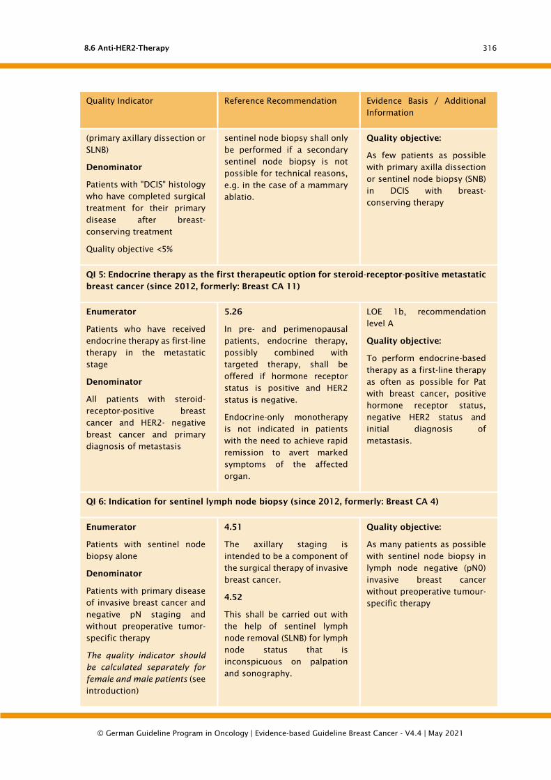
8.6 Anti-HER2-Therapy
© German Guideline Program in Oncology | Evidence-based Guideline Breast Cancer - V4.4 | May 2021
316
Quality Indicator Reference Recommendation Evidence Basis / Additional
Information
(primary axillary dissection or
SLNB)
Denominator
Patients with "DCIS" histology
who have completed surgical
treatment for their primary
disease after breast-
conserving treatment
Quality objective <5%
sentinel node biopsy shall only
be performed if a secondary
sentinel node biopsy is not
possible for technical reasons,
e.g. in the case of a mammary
ablatio.
Quality objective:
As few patients as possible
with primary axilla dissection
or sentinel node biopsy (SNB)
in DCIS with breast-
conserving therapy
QI 5: Endocrine therapy as the first therapeutic option for steroid-receptor-positive metastatic
breast cancer (since 2012, formerly: Breast CA 11)
Enumerator
Patients who have received
endocrine therapy as first-line
therapy in the metastatic
stage
Denominator
All patients with steroid-
receptor-positive breast
cancer and HER2- negative
breast cancer and primary
diagnosis of metastasis
5.26
In pre- and perimenopausal
patients, endocrine therapy,
possibly combined with
targeted therapy, shall be
offered if hormone receptor
status is positive and HER2
status is negative.
Endocrine-only monotherapy
is not indicated in patients
with the need to achieve rapid
remission to avert marked
symptoms of the affected
organ.
LOE 1b, recommendation
level A
Quality objective:
To perform endocrine-based
therapy as a first-line therapy
as often as possible for Pat
with breast cancer, positive
hormone receptor status,
negative HER2 status and
initial diagnosis of
metastasis.
QI 6: Indication for sentinel lymph node biopsy (since 2012, formerly: Breast CA 4)
Enumerator
Patients with sentinel node
biopsy alone
Denominator
Patients with primary disease
of invasive breast cancer and
negative pN staging and
without preoperative tumor-
specific therapy
The quality indicator should
be calculated separately for
female and male patients (see
introduction)
4.51
The axillary staging is
intended to be a component of
the surgical therapy of invasive
breast cancer.
4.52
This shall be carried out with
the help of sentinel lymph
node removal (SLNB) for lymph
node status that is
inconspicuous on palpation
and sonography.
Quality objective:
As many patients as possible
with sentinel node biopsy in
lymph node negative (pN0)
invasive breast cancer
without preoperative tumour-
specific therapy
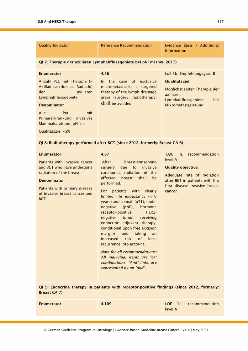
8.6 Anti-HER2-Therapy
© German Guideline Program in Oncology | Evidence-based Guideline Breast Cancer - V4.4 | May 2021
317
Quality Indicator Reference Recommendation Evidence Basis / Additional
Information
QI 7: Therapie der axillären Lymphabflussgebiete bei pN1mi (neu 2017)
Enumerator
Anzahl Pat. mit Therapie (=
Axilladissektion o. Radiatio)
der axillären
Lymphabflussgebiete
Denominator
Alle Pat. mit
Primärerkrankung invasives
Mammakarzinom, pN1mi
Qualitätsziel <5%
4.56
In the case of exclusive
micrometastasis, a targeted
therapy of the lymph drainage
areas (surgery, radiotherapy)
shall be avoided.
LoE 1b, Empfehlungsgrad B
Qualitätsziel:
Möglichst selten Therapie der
axillären
Lymphabflussgebiete bei
Mikrometastasierung
QI 8: Radiotherapy performed after BCT (since 2012, formerly: Breast CA 6)
Enumerator
Patients with invasive cancer
and BCT who have undergone
radiation of the breast
Denominator
Patients with primary disease
of invasive breast cancer and
BCT
4.87
After breast-conserving
surgery due to invasive
carcinoma, radiation of the
affected breast shall be
performed.
For patients with clearly
limited life expectancy (<10
years) and a small (pT1), node-
negative (pN0), hormone
receptor-positive HER2-
negative tumor receiving
endocrine adjuvant therapy,
conditional upon free excision
margins and taking an
increased risk of local
recurrence into account.
Note for all recommendations:
All individual items are "or"
combinations. "And" links are
represented by an "and".
LOE 1a, recommendation
level A
Quality objective:
Adequate rate of radiation
after BET in patients with the
first disease invasive breast
cancer.
QI 9: Endocrine therapy in patients with receptor-positive findings (since 2012, formerly:
Breast CA 7)
Enumerator 4.109 LOE 1a, recommendation
level A

8.6 Anti-HER2-Therapy
© German Guideline Program in Oncology | Evidence-based Guideline Breast Cancer - V4.4 | May 2021
318
Quality Indicator Reference Recommendation Evidence Basis / Additional
Information
Patients who have received
adjuvant endocrine therapy.
Denominator
Steroid-receptor-positive
patients with invasive breast
cancer as primary disease
Patients with estrogen- and/or
progesterone receptor-
positive (°) invasive tumors
shall receive endocrine
therapy.
° (>/=10% progesterone
receptor-positive tumor cell
nuclei)
Quality objective:
Endocrine therapy should be
carried out as often as
possible in receptor-positive
patients with the first disease
invasive mammary carcinoma
QI 10: Trastuzumab therapy in patients with HER2-positive findings (since 2012, formerly:
Breast CA 8)
Enumerator
All patients who have
received (neo)adjuvant
treatment with trastuzumab
for one year
Denominator
All HER2-positive (immuno-
histochemical score 3+
and/or ISH-positive) patients
with invasive breast cancer as
primary disease ≥ pT1c
4.133
Patients with HER2-
overexpressing tumours with a
diameter of ≥ 1 cm
(immunohistochemical score
3+ and/or ISH-positive) should
receive (neo-)adjuvant
treatment with anthracycline
followed by a taxane in
combination with
trastuzumab. Trastuzumab
should be administered over a
total period of one year.
LOE 1b, recommendation
level A
Quality objective:
Trastuzumab therapy as
often as possible over 1 year
in HER2-positive patients with
first diagnosis of invasive
breast cancer >= pT1c
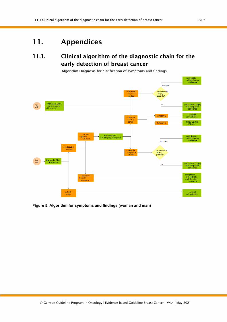
11.1 Clinical algorithm of the diagnostic chain for the early detection of breast cancer
© German Guideline Program in Oncology | Evidence-based Guideline Breast Cancer - V4.4 | May 2021
319
11. Appendices
11.1. Clinical algorithm of the diagnostic chain for the
early detection of breast cancer
Algorithm Diagnosis for clarification of symptoms and findings
Figure 5: Algorithm for symptoms and findings (woman and man)

11.1 Clinical algorithm of the diagnostic chain for the early detection of breast cancer
© German Guideline Program in Oncology | Evidence-based Guideline Breast Cancer - V4.4 | May 2021
320
Figure 6: Algorithm for early detection of breast cancer in asymptomatic women

11.1 Clinical algorithm of the diagnostic chain for the early detection of breast cancer
© German Guideline Program in Oncology | Evidence-based Guideline Breast Cancer - V4.4 | May 2021
321
11.1.1. Options and indications for plastic reconstruction
Figure 7: Options and indications for breast reconstruction.
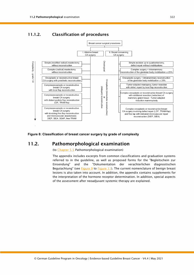
11.2 Pathomorphological examination
© German Guideline Program in Oncology | Evidence-based Guideline Breast Cancer - V4.4 | May 2021
322
11.1.2. Classification of procedures
Figure 8: Classification of breast cancer surgery by grade of complexity
11.2. Pathomorphological examination
(to Chapter 5.5 Pathomorphological examination)
The appendix includes excerpts from common classifications and graduation systems
referred to in the guideline, as well as proposed forms for the "Begleitschein zur
Einsendung" and the "Dokumentation der verachterlichen diagnostischen
Begutachtung" (see Figure 9 to Figure 11). The current nomenclature of benign breast
lesions is also taken into account. In addition, the appendix contains supplements for
the interpretation of the hormone receptor determination. In addition, special aspects
of the assessment after neoadjuvant systemic therapy are explained.
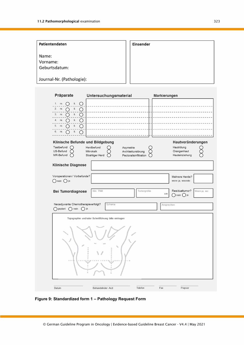
11.2 Pathomorphological examination
© German Guideline Program in Oncology | Evidence-based Guideline Breast Cancer - V4.4 | May 2021
323
Figure 9: Standardized form 1 – Pathology Request Form
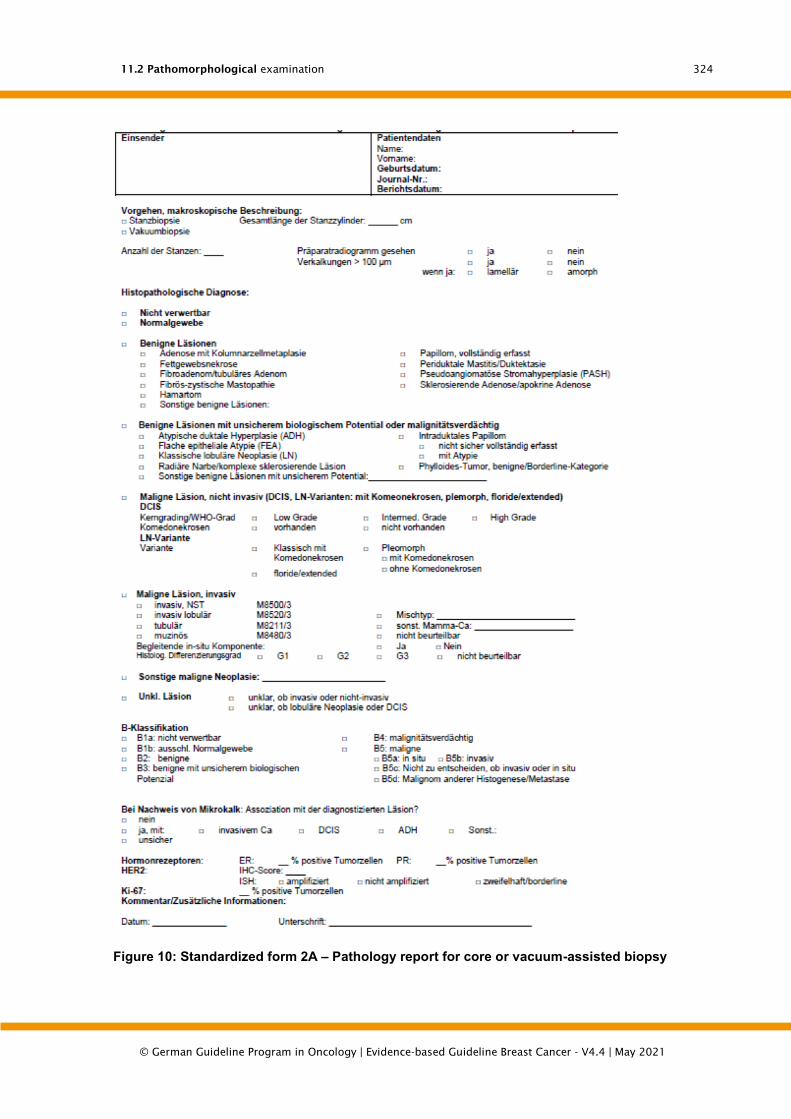
11.2 Pathomorphological examination
© German Guideline Program in Oncology | Evidence-based Guideline Breast Cancer - V4.4 | May 2021
324
Figure 10: Standardized form 2A – Pathology report for core or vacuum-assisted biopsy

11.2 Pathomorphological examination
© German Guideline Program in Oncology | Evidence-based Guideline Breast Cancer - V4.4 | May 2021
325
Figure 11: Standardized form 2B – Pathology report on surgical specimen

11.2 Pathomorphological examination
© German Guideline Program in Oncology | Evidence-based Guideline Breast Cancer - V4.4 | May 2021
326
Figure 12: Standardized form 2B – Pathology report on surgical specimen

11.2 Pathomorphological examination
© German Guideline Program in Oncology | Evidence-based Guideline Breast Cancer - V4.4 | May 2021
327
Histological classification
The nomenclature and grouping of lesions of the breast is based on the WHO
classification of tumors of the breast [118] and additionally on the "European
Guidelines for Quality Assurance in Pathology in Mammography Screening" [420], [428]
as well as recommendations from other internationally recognized guidelines [424],
[425].
In the following, only selected points are presented which are of particular importance
in terms of differential diagnosis, pathological-radiological correlation or clinical
significance. In addition, individual terms of the current WHO classification are
explained and commented on.
Normal findings and benign lesions (varia)
"Normal tissue" includes minor age-related changes such as fibrosis, lobular
involution, microscopic dilatation of azini and ducts, and mild microcystic adenosis.
These minimal changes are usually not sufficient to explain conspicuous clinical or
radiological findings.
Fibrocystic mastopathy refers to changes associated with pronounced,
macroscopically visible cyst formation, apocrine metaplasia and fibrosis.
This is distinguished from the solitary cyst, which is usually larger than 1 cm and is
lined by a flat or apocrine epithelium.
Periductal mastitis (ductectasia, plasma cell mastitis) affects larger and mid-sized
ducts, which are usually subareolar. The chronic inflammatory reaction in the vicinity
of the ducts, which is often rich in plasma cells, may contain a large number of
histiocytes and have a granulomatous aspect. It can be accompanied by pronounced
periductal fibrosis. Calcifications are possible.
Benign epithelial proliferations
The sclerosing adenosis is rather cell-rich in the early phase. The fibre content
increases with time. It is recommended that sclerosing adenosis is only mentioned in
the documentation of the expert opinion if it is a prominent finding [420], [425].
Calcifications may be present.
Sclerosing adenosis must be distinguished from tubular carcinoma, microglandular
adenosis and radial scarring by differential diagnosis.
If neighbouring lobules are affected, a mammographically or palpationally conspicuous
tumour may develop, which is called an adenosetumour.
Adenoses with pronounced apocrine metaplasia, which occupies at least 50% of the
adenosis, are documented as apocrine adenoses.
In contrast to sclerosing adenosis, microglandular adenosis does not show a
lobulocentric organoid pattern. The round glandular structures are lined by a single-
row epithelium without atypia. Myoepithelia are missing. However, an intact basal
membrane can be shown.
The radial scar consists of a central, fibro-elastoid zone with ducts proliferating
radially outwards. The lesion is rarely larger than 1 cm. The epithelium is bilayered or
11.2 Pathomorphological examination
© German Guideline Program in Oncology | Evidence-based Guideline Breast Cancer - V4.4 | May 2021
328
has ductal hyperplasia. Tubules may be trapped, distorted and tilted in the central
hyaline fibrosis.
The complex sclerosing lesion simulates an invasion. It has all the characteristics of
the radial scar, but is larger than 1 cm and more irregularly structured, often with
nodular proliferations in the periphery. The lesion may be accompanied by changes
such as papillomas, apocrine metaplasia or sclerosing adenosis. The most important
differential diagnosis for both radial sclerosing lesions is tubular carcinoma, in which
myoepithelium and an intact basement membrane in the border of the tubules are
missing.
The diagnosis of a radial scar or complex sclerosing lesion in the punch and vacuum
biopsy usually means a classification of the alteration in the B3 category [420] and the
indication for a surgical excision. Excluded from this are small radial scars, which are
completely recorded in a vacuum biopsy and represent a histological random finding
(without mammographic correlate). These do not require excision and can be classified
as B2 according to the European guidelines for mammography screening.
The reason for the assignment to the B3 category is that atypical ductal hyperplasia
and carcinomas (in situ and invasive) are relatively often detectable in the periphery of
mammographically detected radial scars and complex sclerosing lesions, especially in
lesions with a size > 0.6 cm and in women > 50 years [118]. DCIS or invasive carcinoma
is present in 4-32% of excidates after punch biopsy diagnosis of a radial scar, especially
if ADH has already been registered in the punch biopsy.
The changes, which are called "ductal adenoma", have a variable appearance.
Characteristic is a well defined benign glandular proliferation, which at least partially
expands intraductally. There is overlap with other benign changes such as the
papilloma or the complex sclerosing lesion, which is why such lesions are also called
sclerosing papillomas.
Papillomas
The papilloma can occur solitary and multiple. The solitary papilloma is usually located
centrally (central papilloma), in subareolar ducts, while the multiple papillomas are
found more peripherally in the area of the terminal ductulo-lobular units (TDLE)
(peripheral papillomas). Peripheral papillomas are not only more common with
common ductal hyperplasia (UDH), but also with atypical ductal hyperplasia (ADH), DCIS
or invasive carcinoma. Therefore, the presence of this change requires extensive tissue
embedding.
The term "papillomatosis" should be avoided, as it has been used for both UDH and
multiple papillomas.
Papillomas with atypical ductal hyperplasia (ADH) or ductal carcinoma in si-tu
(DCIS), low-grade, are characterized by the occurrence of a focal proliferation of
uniform cells with the cytological and architectural features of a low-grade neoplasia
[118] Myopithelia may be reduced. Epithelial proliferation usually does not express
basal cytokeratins and is homogeneously ER-positive. In the past, both the extent and
the proportion of atypical epithelial proliferation have been used as quantitative criteria
to distinguish between a papilloma with ADH and a DCIS in the papilloma. On the
one hand, atypical epithelial proliferations of less than 3 mm in size were classified as
ADH [1635], on the other hand, atypical epithelial proliferations that occupy less than
30% or 90% of the papilloma cross-section [361], [1636]. Flocks above this were
classified as DCIS in a papilloma.
11.2 Pathomorphological examination
© German Guideline Program in Oncology | Evidence-based Guideline Breast Cancer - V4.4 | May 2021
329
For pragmatic reasons, the WHO expert group recommends the use of the size criterion
(limit value 3 mm) to differentiate between a papilloma with ADH and a low-grade DCIS
in the papilloma and also points out that in the case of epithelial proliferation with
intermediate or high core degree, the diagnosis of a DCIS in the papilloma should be
made independent of the extent [118].
The B classification of papillary lesions is based on the epithelial component. Since the
epithelial changes within the papillary lesion can be heterogeneous and the punch
biopsy usually does not completely capture the lesion, most papillary lesions fall into
the B3 category (uncertain biological potential). This excludes small papillomas which
have been extensively sampled and are presumably completely removed by the
punch/vacuum biopsy. These can be classified as B2. A higher B category is appropriate
if the epithelial proliferates have atypia that justify either the suspicion of malignancy
(B4) or the reliable diagnosis of a DCIS in the papilloma or an intraductal papillary
carcinoma (B5).
Myoepithelial lesions
While myoepitheliosis is a mostly multifocal, microscopic process,
adenomyoepitheliomas impress as circumscribed nodal formations, which can be
multilobulated. They are characterized by their biphasic cellular differentiation: The
myoepithelial, partly clear cell compartment usually surrounds the tubular component
with a luminal epithelial lining. There are essentially 3 different morphological variants:
lobulated, tubular and spindle cell/myoid. The differential diagnosis includes not only
papilloma and adenosis (each with a fluid transition) but also the tubular adenoma
(sharply defined in contrast to the tubular variant of adenomyoepithelioma) and the
invasive carcinoma (no biphasic cellular structure).
The majority of adenomyoepitheliomas behave benignly. However, they are considered
to have a low malignant potential and should therefore be classified as B3 in the
punch/vacuum biopsy and a complete excision is recommended.
Significantly less frequent than benign adenomyoepithelioma are malignant forms
(malignant adenomyoepitheliomas) in which the epithelial and/or myoepithelial
component may be degenerated.
Fibroepithelial tumours
Fibroadenomas are benign biphasic tumours that are mostly diagnosed in women of
childbearing age. Epithelial (ductal) hyperplasia is not unusual in fibroadenoma. In any
case, ADH or DCIS in a fibroadenoma must be reported separately. In this case, when
punch biopsy is used for diagnosis, the B category is increased from B2 to B3, B4 or B5
depending on the degree and extent of the atypia.
The fibroadenoma must be distinguished from the phylloides tumour, which has a
stroma richer in cells. For its dignity assessment on the resected tissue, a sufficient
number of tissue sections is necessary (rule of thumb: 1 tissue block per cm tumor
diameter) to representatively record the characteristic stromal features (cellularity,
pleomorphy, mitotic activity, distribution pattern) and the relationship to the
surrounding tissue.
In principle, fibroepithelial tumors that suggest the presence of a phylloidal tumor (PT)
in the punch or vacuum biopsy due to their cell-rich stroma, the predominance of the
stromal component or an increased mitotic activity of the stromal cells are classified

11.2 Pathomorphological examination
© German Guideline Program in Oncology | Evidence-based Guideline Breast Cancer - V4.4 | May 2021
330
as B3. Cell-rich fibroepithelial lesions in which a phylloides tumour cannot be excluded
should also be classified as B3.
Intraductal proliferative lesions
In the current WHO classification [118], this group includes various int-raductal
proliferations that have a common point of origin: the terminal ductulo-lobular unit
(TDLUs). They are associated with an increased risk for the development of invasive
breast carcinoma, although to a significantly different extent.
The common ductal hyperplasia (UDH) comprises all cases of intraluminal epithelial
proliferation without atypia. A colorful, flowing cell picture is common. If secondary
lumina are formed, they are slit-shaped, rounded and irregularly shaped with tangential
alignment of the nuclei in the limiting epithelial cells. One of the most important
indicators of UDH is the presence of a mixture of at least 2 cell types (luminal and
basal/myoepithelial and/or metaplastic apocrine cells). It is associated with only a very
slightly increased risk (1.5-fold) of developing breast cancer. It is usually accompanied
by diffuse or mosaic-like expression of the basal cytokeratins (including CK5, CK14).
The term flat epithelial atypia (FEA) was introduced into the WHO classification in
2003. This rather descriptive category includes lesions that are neoplastic by current
standards and are also known as "clinging carcinoma" of the monomorphic type,
atypical cystic lobules, atypical lobules type A, columnar cell metaplasia with atypia or
columnar cell hyperplasia with atypia. Characteristic is the replacement of the original
epithelium by a single layer of a slightly atypical epithelium, which often shows apical
"snouts" or 3-5 layers of a monotonously atypical cell population of prismatic cells or
columnar cells. The cell nuclei are round, relatively uniform and contain small nucleoli.
Micropapillae or more complex structures like arcades are missing. The affected
extended lobules often contain secreted material and microcalcifications.
If an FEA is diagnosed in the punch or vacuum biopsy, this change must be assigned
to category B3 (see also Chapter 5.3.3 Risk lesions).
As described above, FEA is usually a columnar cell alteration with or without
hyperplasia, with low to moderate cytological atypia. This should be distinguished from
columnar cell hyperplasia with architectural atypia, in which micropapillae or
bridges are preferably formed, with only slight cytological atypia. These changes are
now classified as ADH or low-grade DCIS depending on the type and extent of the
cytological and structural atypias.
It should also be noted that columnar cell proliferations are homogeneously ER-positive
and usually CK5-negative. Immunohistochemical imaging of basal cytokeratins is often
not helpful in the differential diagnostic differentiation between columnar cell
hyperplasia without and with atypia, since the characteristic mosaic-like reaction
pattern of UDH in columnar cell hyperplasia without atypia may be absent.
Atypical ductal hyperplasia (ADH) is also considered neoplastic intraductal epithelial
proliferation. It is characterized by an intraductal proliferation of evenly distributed,
uniform cells which can form micropapillae, arches, solid or cribriform patterns [118].
Roundish, rigid secondary lumina appear together with irregularly shaped ones.
Cytologically, the cells of an ADH thus correspond to a low-grade DCIS. However, in an
ADH the characteristic cells mix with non-uniform cells within a TDLU (s) or only a
limited number of corridors of a TDLU are colonized.
In individual cases, the differentiation between ADH and low-grade DCIS can be
difficult. Immunohistochemical imaging of basal cytokeratins does not help in
11.2 Pathomorphological examination
© German Guideline Program in Oncology | Evidence-based Guideline Breast Cancer - V4.4 | May 2021
331
differential diagnosis, since the proliferating epithelia in both lesions do not express
them.
Nowadays, quantitative criteria are primarily used to distinguish ADH from low-grade
DCIS. The most commonly used quantitative criteria are those that are used to diagnose
a low-grade DCIS when at least two duct structures are completely and homogeneously
colonized by neoplastic epithelial proliferation or the lesion is > 2 mm. The expert
group of the current WHO classification was not in a position to favor either of the two
criteria [118]. It was rather pointed out that these quantitative criteria are primarily
helpful to avoid overtherapy of very small neoplastic lesions by not diagnosing these
lesions as DCIS. Therefore, a conservative approach is recommended in the differential
diagnosis between ADH and low-grade DCIS, especially in punch and vacuum biopsies.
The diagnosis of ADH or an atypical intraductal proliferative lesion should be sufficient
to induce surgical excision of the change. Depending on the degree of atypia and the
extent of the lesion, the result is a B3 or B4 category.
The final diagnostic classification is then made on the surgical specimen (see also
Chapter 5.3.3 Risk lesions).
Lobular neoplasia (LN)
According to the current WHO classification [118], the term lobular neoplasia (LN) is
used to describe the entire spectrum of atypical epithelial proliferations originating
from TDLUs and characterized by the proliferation of mostly small and non-cohesive
cells - with or without pagetoid involvement of the terminal ducts. The terms atypical
lobular hyperplasia (ALH) and lobular carcinoma in situ (LCIS) reflect the extent of the
change. A classic LCIS is diagnosed when more than half of the azini of a TDLU are
colonized and dilated by the characteristic neoplastic proliferation. A less pronounced
infestation corresponds to an ALH. The determination of the extension and thus the
differentiation between ALH and LCIS is usually only possible on surgical specimens
but not on punch biopsies.
As a special feature of the LN, its frequent multicentric (46-85 %) and bilateral
occurrence (30-67 %) must be noted. In principle, the LN is, according to current
opinion, an indicator lesion for an increased carcinoma risk. The relative risk of a
patient is bilaterally increased by a factor of 4-12 after the diagnosis of an LN, whereas
the risk after the diagnosis of ALH is half as high as after an LCIS.
According to the WHO, different variants of the LCIS are increasingly frequently
diagnosed due to their association with microcalcification:
Classical LCIS with comedonecroses
Pleomorphic LCIS with/without apocrine properties and comedonecroses
Even though individual reports suggest that these variants may have a different course
than the classic LCIS, the clinical significance and adequate therapy is not yet clear
[118].
If a classic LN is diagnosed in the punch or vacuum biopsy as part of mammography
screening, this corresponds to a B3 category. If it is not possible to decide on the punch
or vacuum biopsy material whether small cell epithelial proliferation in TDLUs and/or
ducts should be classified as LN or DCIS, a higher B category: B4 or B5 is recommended.
The variants of the LN (especially pleomorphic variant and classical LN with
comedonecroses) are classified as B5a. With regard to the management of the LN, see
also Chapter 5.3.3 Risk lesions.
Ductal carcinoma in situ (DCIS)
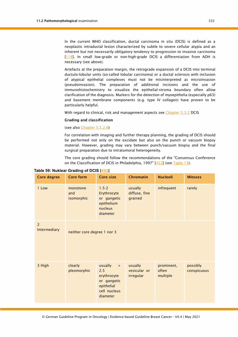
11.2 Pathomorphological examination
© German Guideline Program in Oncology | Evidence-based Guideline Breast Cancer - V4.4 | May 2021
332
In the current WHO classification, ductal carcinoma in situ (DCIS) is defined as a
neoplastic intraductal lesion characterized by subtle to severe cellular atypia and an
inherent but not necessarily obligatory tendency to progression to invasive carcinoma
[118]. In small low-grade or non-high-grade DCIS a differentiation from ADH is
necessary (see above).
Artefacts at the preparation margin, the retrograde expansion of a DCIS into terminal
ductulo-lobular units (so-called lobular carcinoma) or a ductal sclerosis with inclusion
of atypical epithelial complexes must not be misinterpreted as microinvasion
(pseudoinvasion). The preparation of additional incisions and the use of
immunohistochemistry to visualize the epithelial-stroma boundary often allow
clarification of the diagnosis. Markers for the detection of myoepithelia (especially p63)
and basement membrane components (e.g. type IV collagen) have proven to be
particularly helpful.
With regard to clinical, risk and management aspects see Chapter 5.3.2 DCIS
Grading and classification
(see also Chapter 5.5.2.6)
For correlation with imaging and further therapy planning, the grading of DCIS should
be performed not only on the excidate but also on the punch or vacuum biopsy
material. However, grading may vary between punch/vacuum biopsy and the final
surgical preparation due to intratumoral heterogeneity.
The core grading should follow the recommendations of the "Consensus Conference
on the Classification of DCIS in Philadelphia, 1997" [453] (see Table 15).
Table 59: Nuklear Grading of DCIS [455]
Core degree Core form Core size Chromatin Nucleoli Mitoses
1 Low monotone
and
isomorphic
1.5-2
Erythrocyte
or gangetic
epithelium
nucleus
diameter
usually
diffuse, fine
grained
infrequent rarely
2
Intermediary
neither core degree 1 nor 3
3 High clearly
pleomorphic
usually >
2.5
erythrocyte
or gangetic
epithelial
cell nucleus
diameter
usually
vesicular or
irregular
prominent,
often
multiple
possibly
conspicuous

11.2 Pathomorphological examination
© German Guideline Program in Oncology | Evidence-based Guideline Breast Cancer - V4.4 | May 2021
333
The grading is based on the WHO [118] grading scheme (see Table 16).
Table 60: WHO grading of DCIS [118]
Degree Cytology/nuclear
degree (KG)
Necroses Calcifications Architecture
low grade small,
monomorphic
cells, low nucleus
degree (KG 1) with
uniform nuclei,
regular chromatin
pattern,
inconspicuous
nucleols
-/+ often
psammomatous
arches,
cribriform, solid
and/or
micropapillary
intermediate
degrees
low to moderate
cell size and shape
variability,
intermediate
nucleus degree
(KG 2) with
variable coarse
chromatin,
prominent
nucleols
-/+ psammomatic or
amorphous
solid,
cribriform,
micropapillary
high grade high-grade cell
atypes, high
nucleus degree
(KG 3) with
pleomorphic
nuclei, coarse,
clumped
chromatin and
prominent
nucleoli
-/+ amorphous a cell layer,
micropapillary,
cribriform or
solid
Determination of hormone receptor expression
The evaluation and interpretation of immunohistochemistry is based on the guidelines
for invasive breast cancer (see below).
Invasive breast carcinomas
Histological typing
(see also Statement 4.26.)
For all invasive breast carcinomas a histological typing according to the current WHO
classification must be performed (see Table 17 [118] ). In the case of preoperative
diagnostics, this should be done on the punch or vacuum biopsy material. This serves
both for correlation with imaging and for therapy planning. As a result of intratumoral
heterogeneity there can rarely be deviations between the punch/vacuum biopsy and
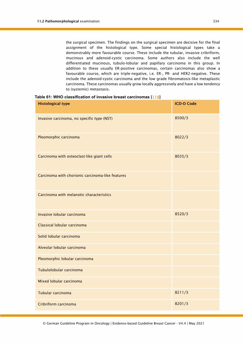
11.2 Pathomorphological examination
© German Guideline Program in Oncology | Evidence-based Guideline Breast Cancer - V4.4 | May 2021
334
the surgical specimen. The findings on the surgical specimen are decisive for the final
assignment of the histological type. Some special histological types take a
demonstrably more favourable course. These include the tubular, invasive cribriform,
mucinous and adenoid-cystic carcinoma. Some authors also include the well
differentiated mucinous, tubulo-lobular and papillary carcinoma in this group. In
addition to these usually ER-positive carcinomas, certain carcinomas also show a
favourable course, which are triple-negative, i.e. ER-, PR- and HER2-negative. These
include the adenoid-cystic carcinoma and the low grade fibromatosis-like metaplastic
carcinoma. These carcinomas usually grow locally aggressively and have a low tendency
to (systemic) metastasis.
Table 61: WHO classification of invasive breast carcinomas [118]
Histological type ICD-O Code
• Invasive carcinoma, no specific type (NST) 8500/3
o Pleomorphic carcinoma 8022/3
o Carcinoma with osteoclast-like giant cells 8035/3
o Carcinoma with chorionic carcinoma-like features
o Carcinoma with melanotic characteristics
• Invasive lobular carcinoma 8520/3
o Classical lobular carcinoma
o Solid lobular carcinoma
o Alveolar lobular carcinoma
o Pleomorphic lobular carcinoma
o Tubulolobular carcinoma
o Mixed lobular carcinoma
• Tubular carcinoma 8211/3
• Cribriform carcinoma 8201/3

11.2 Pathomorphological examination
© German Guideline Program in Oncology | Evidence-based Guideline Breast Cancer - V4.4 | May 2021
335
Histological type ICD-O Code
• Mucinous carcinoma 8480/3
• Carcinoma with medullary properties
o Medullary carcinoma 8510/3
o Atypical medullary carcinoma 8513/3
o Invasive carcinoma NST with medullary properties 8500/3
• Carcinoma with apocrine differentiation #
• Carcinoma with sigmoid ring cell differentiation #
• Invasive micropapillary carcinoma 8507/3*
• Metaplastic carcinoma, no specific type 8575/3
• Low-grade adenosquamous carcinoma 8570/3
• Fibromatosis-like metaplastic carcinoma 8572/3
• Squamous cell carcinoma 8070/3
• Spindle cell carcinoma 8032/3
• Metaplastic carcinoma with mesenchymal differentiation
o Chondroid differentiation 8571/3
o Osseous differentiation 8571/3
o Other mesenchymal differentiation 8575/3
• Mixed metaplastic carcinoma 8575/3
• myoepithelial carcinoma 8982/3
Rare types:
• Carcinomas with neuroendocrine properties:
o Neuroendocrine tumor, well differentiated 8246/3
o Neuroendocrine carcinoma, poorly differentiated (small cell carcinoma) 8041/3
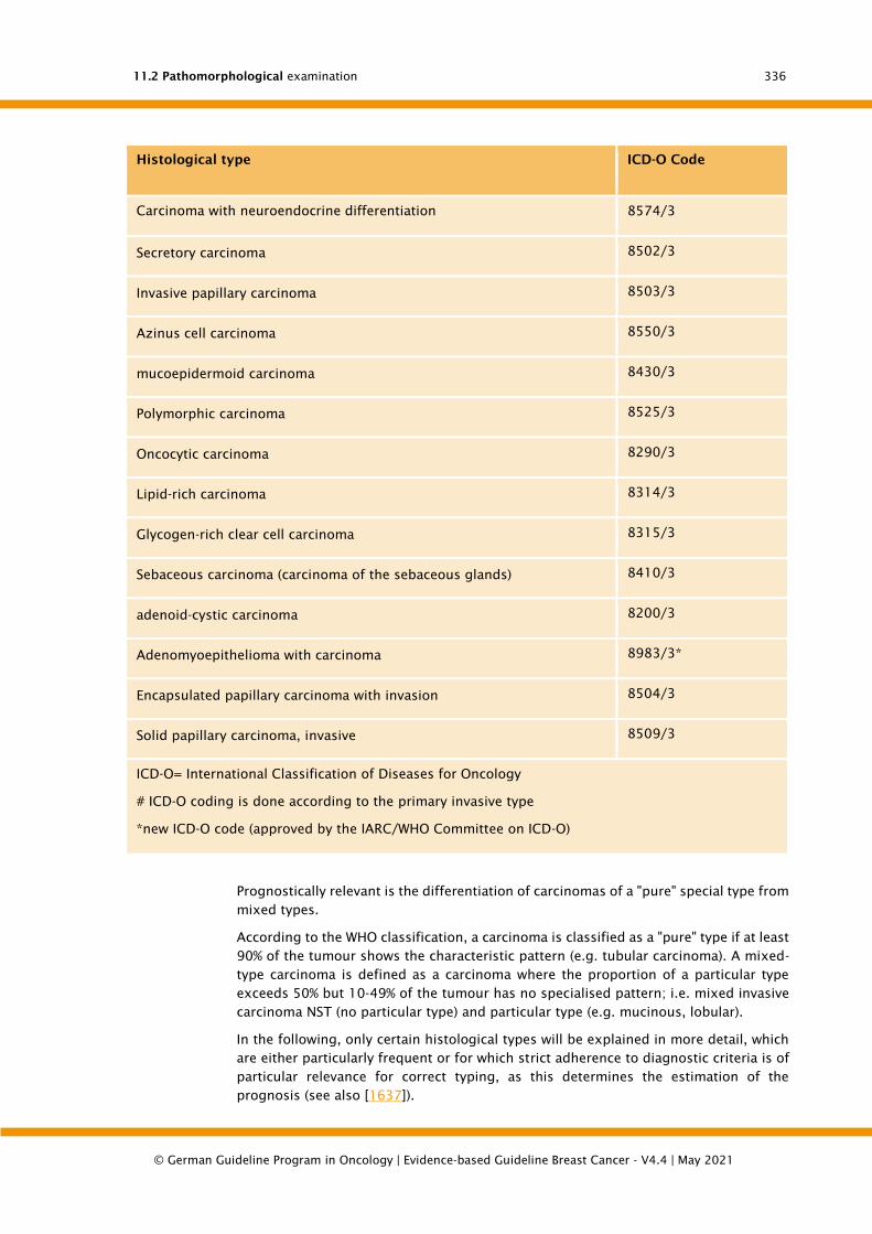
11.2 Pathomorphological examination
© German Guideline Program in Oncology | Evidence-based Guideline Breast Cancer - V4.4 | May 2021
336
Histological type ICD-O Code
o Carcinoma with neuroendocrine differentiation 8574/3
• Secretory carcinoma 8502/3
• Invasive papillary carcinoma 8503/3
• Azinus cell carcinoma 8550/3
• mucoepidermoid carcinoma 8430/3
• Polymorphic carcinoma 8525/3
• Oncocytic carcinoma 8290/3
• Lipid-rich carcinoma 8314/3
• Glycogen-rich clear cell carcinoma 8315/3
• Sebaceous carcinoma (carcinoma of the sebaceous glands) 8410/3
• adenoid-cystic carcinoma 8200/3
• Adenomyoepithelioma with carcinoma 8983/3*
• Encapsulated papillary carcinoma with invasion 8504/3
• Solid papillary carcinoma, invasive 8509/3
ICD-O= International Classification of Diseases for Oncology
# ICD-O coding is done according to the primary invasive type
*new ICD-O code (approved by the IARC/WHO Committee on ICD-O)
Prognostically relevant is the differentiation of carcinomas of a "pure" special type from
mixed types.
According to the WHO classification, a carcinoma is classified as a "pure" type if at least
90% of the tumour shows the characteristic pattern (e.g. tubular carcinoma). A mixed-
type carcinoma is defined as a carcinoma where the proportion of a particular type
exceeds 50% but 10-49% of the tumour has no specialised pattern; i.e. mixed invasive
carcinoma NST (no particular type) and particular type (e.g. mucinous, lobular).
In the following, only certain histological types will be explained in more detail, which
are either particularly frequent or for which strict adherence to diagnostic criteria is of
particular relevance for correct typing, as this determines the estimation of the
prognosis (see also [1637]).

11.2 Pathomorphological examination
© German Guideline Program in Oncology | Evidence-based Guideline Breast Cancer - V4.4 | May 2021
337
Invasive carcinoma, no special type (NST) is by far the most common type of tumour,
accounting for 50-80 % of all tumours. It comprises a heterogeneous group of tumours
that do not have sufficient characteristics to be assigned to a specific histological type
(e.g. lobular or tubular carcinoma). In order to be classified as invasive carcinoma NST,
a tumour must, after careful examination of representative sections, show a non-
specialised pattern in more than 50 % of its mass (to distinguish "pure" invasive ductal
carcinoma from mixed forms, see also above).
The prognosis of invasive carcinoma NST is equal or slightly worse compared to the
prognosis of the total group of all breast cancers. The prognosis is mainly influenced
by the established parameters: nodal status, tumor size, grading, etc. Therapeutically
relevant is the steroid hormone receptor expression in about 70-80% of invasive ductal
carcinomas. HER2 overexpression or amplification is present in about 15 % of cases.
Invasive lobular carcinomas account for about 5-15% of invasive breast carcinomas.
The classic appearance of invasive lobular carcinoma is mainly characterized by its
small cell size, its dissociated infiltrating growth. From the classic type, different
morphological variants are distinguished (solid, alveolar, pleomorphic, tubulo-lobular
and mixed) with a partly better (alveolar and tubulo-lobular variants), partly worse
prognosis (solid and pleomorphic). The classic form of invasive lobular carcinoma is in
most cases associated with lobular neoplasia, but occasionally also with DCIS or
carcinoma in situ with ductal and lobular phenotype.
A comparison of the long-term prognosis of ILC and NST carcinomas shows a lower
risk of progression of ILC in the first years, but an overlap of the survival curves after
about 10 years with a worse long-term survival of ILC. However, invasive lobular
carcinomas are characterized by multifocality (9-31%), bilateralism (5-19%) and a
different pattern of metastasis. Bones, meninges, gastrointestinal tract and peritoneum
are more frequently affected by distant metastases. Lung metastases, on the other
hand, are observed less frequently than in the ductal type.
The invasive lobular carcinomas usually express steroid hormone receptors. Among
the variants, the ER-positivity rate is highest for the alveolar variant and lowest for the
pleomorphic variant. HER2 overexpression or amplification is very rare in classic
invasive lobular carcinoma. The pleomorphic variant (G3) is more likely to show HER2
overexpression and gene amplification.
The tubular carcinoma is characterized by the presence of neoplastic rounded-oval
tubules with a single-row cubic epithelial lining in a dense collagenous connective
tissue. The tubules may be angled or folded with a drop-like shape. The epithelium
often exhibits "apical snouts". The nuclei are slightly hyperchromatic and should have
only small inconspicuous nucleoli. Mitoses are rare. The diagnosis of a (pure) tubular
carcinoma requires that the tumour consists of > 90% tubular structures with the
described structural and cytological criteria. The (pure) tubular carcinoma accounts for
about 2% of all breast carcinomas, but seems to be found more frequently with subtle
radiological diagnosis. Its proportion of tumours less than 1 cm in diameter is at least
8%, in pure screening populations even 8-27%.
Strict adherence to the above criteria is crucial for the assessment of the prognosis.
Pure tubular carcinoma has an excellent prognosis. The disease-free 10-year survival
rate is well over 90%. Even the rare presence of axillary lymph node metastases (6-19%)
has no influence on the survival rate.
The tubular carcinomas are usually ER- and PR-positive and HER2-negative.
In mucinous carcinomas, islands of relatively uniform cells lie in lakes of extracellular
mucus. The classification as (pure) mucinous carcinoma requires, as with the other
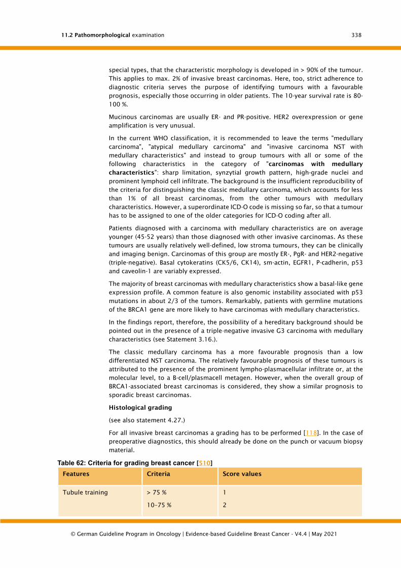
11.2 Pathomorphological examination
© German Guideline Program in Oncology | Evidence-based Guideline Breast Cancer - V4.4 | May 2021
338
special types, that the characteristic morphology is developed in > 90% of the tumour.
This applies to max. 2% of invasive breast carcinomas. Here, too, strict adherence to
diagnostic criteria serves the purpose of identifying tumours with a favourable
prognosis, especially those occurring in older patients. The 10-year survival rate is 80-
100 %.
Mucinous carcinomas are usually ER- and PR-positive. HER2 overexpression or gene
amplification is very unusual.
In the current WHO classification, it is recommended to leave the terms "medullary
carcinoma", "atypical medullary carcinoma" and "invasive carcinoma NST with
medullary characteristics" and instead to group tumours with all or some of the
following characteristics in the category of "carcinomas with medullary
characteristics": sharp limitation, synzytial growth pattern, high-grade nuclei and
prominent lymphoid cell infiltrate. The background is the insufficient reproducibility of
the criteria for distinguishing the classic medullary carcinoma, which accounts for less
than 1% of all breast carcinomas, from the other tumours with medullary
characteristics. However, a superordinate ICD-O code is missing so far, so that a tumour
has to be assigned to one of the older categories for ICD-O coding after all.
Patients diagnosed with a carcinoma with medullary characteristics are on average
younger (45-52 years) than those diagnosed with other invasive carcinomas. As these
tumours are usually relatively well-defined, low stroma tumours, they can be clinically
and imaging benign. Carcinomas of this group are mostly ER-, PgR- and HER2-negative
(triple-negative). Basal cytokeratins (CK5/6, CK14), sm-actin, EGFR1, P-cadherin, p53
and caveolin-1 are variably expressed.
The majority of breast carcinomas with medullary characteristics show a basal-like gene
expression profile. A common feature is also genomic instability associated with p53
mutations in about 2/3 of the tumors. Remarkably, patients with germline mutations
of the BRCA1 gene are more likely to have carcinomas with medullary characteristics.
In the findings report, therefore, the possibility of a hereditary background should be
pointed out in the presence of a triple-negative invasive G3 carcinoma with medullary
characteristics (see Statement 3.16.).
The classic medullary carcinoma has a more favourable prognosis than a low
differentiated NST carcinoma. The relatively favourable prognosis of these tumours is
attributed to the presence of the prominent lympho-plasmacellular infiltrate or, at the
molecular level, to a B-cell/plasmacell metagen. However, when the overall group of
BRCA1-associated breast carcinomas is considered, they show a similar prognosis to
sporadic breast carcinomas.
Histological grading
(see also statement 4.27.)
For all invasive breast carcinomas a grading has to be performed [118]. In the case of
preoperative diagnostics, this should already be done on the punch or vacuum biopsy
material.
Table 62: Criteria for grading breast cancer [510]
Features Criteria Score values
Tubule training > 75 %
10–75 %
1
2
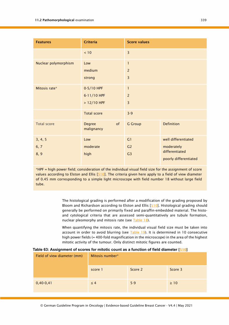
11.2 Pathomorphological examination
© German Guideline Program in Oncology | Evidence-based Guideline Breast Cancer - V4.4 | May 2021
339
Features Criteria Score values
< 10 3
Nuclear polymorphism Low
medium
strong
1
2
3
Mitosis rate* 0-5/10 HPF
6-11/10 HPF
> 12/10 HPF
1
2
3
Total score 3-9
Total score Degree of
malignancy
G Group Definition
3, 4, 5
6, 7
8, 9
Low
moderate
high
G1
G2
G3
well differentiated
moderately
differentiated
poorly differentiated
*HPF = high power field; consideration of the individual visual field size for the assignment of score
values according to Elston and Ellis [510]. The criteria given here apply to a field of view diameter
of 0.45 mm corresponding to a simple light microscope with field number 18 without large field
tube.
The histological grading is performed after a modification of the grading proposed by
Bloom and Richardson according to Elston and Ellis [510]. Histological grading should
generally be performed on primarily fixed and paraffin-embedded material. The histo-
and cytological criteria that are assessed semi-quantitatively are tubule formation,
nuclear pleomorphy and mitosis rate (see Table 18).
When quantifying the mitosis rate, the individual visual field size must be taken into
account in order to avoid blurring (see Table 19). It is determined in 10 consecutive
high power fields (= 400-fold magnification in the microscope) in the area of the highest
mitotic activity of the tumour. Only distinct mitotic figures are counted.
Table 63: Assignment of scores for mitotic count as a function of field diameter [510]
Field of view diameter (mm) Mitosis number*
score 1 Score 2 Score 3
0,40-0,41 ≤ 4 5-9 ≥ 10

11.2 Pathomorphological examination
© German Guideline Program in Oncology | Evidence-based Guideline Breast Cancer - V4.4 | May 2021
340
Field of view diameter (mm) Mitosis number*
score 1 Score 2 Score 3
0,42-0,43 ≤ 5 6-10 ≥ 11
0,44-0,45 ≤ 5 6-11 ≥ 12
0,46-0,47 ≤ 6 7-12 ≥ 13
0,48-0,49 ≤ 6 7-13 ≥ 14
0,50-0,51 ≤ 7 8-14 ≥ 15
0,52 ≤ 7 8-15 ≥ 16
0,53-0,54 ≤ 8 9-16 ≥ 17
0,55-0,56 ≤ 8 9-17 ≥ 18
0,57 ≤ 9 10-18 ≥ 19
0,58-0,59 ≤ 9 10-19 ≥ 20
0,60 ≤ 10 11-20 ≥ 21
0,61 ≤ 10 11-21 ≥ 22
0,62-0,63 ≤ 11 12-22 ≥ 23
0,64 ≤ 11 12-23 ≥ 24
0,65-0,66 ≤ 12 13-24 ≥ 25
0,67 ≤ 12 13-25 ≥ 26
0,68 ≤ 13 14-26 ≥ 27
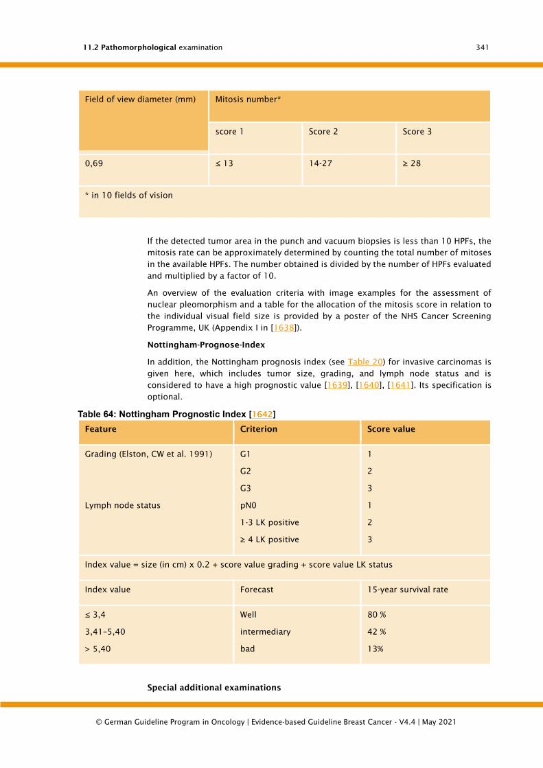
11.2 Pathomorphological examination
© German Guideline Program in Oncology | Evidence-based Guideline Breast Cancer - V4.4 | May 2021
341
Field of view diameter (mm) Mitosis number*
score 1 Score 2 Score 3
0,69 ≤ 13 14-27 ≥ 28
* in 10 fields of vision
If the detected tumor area in the punch and vacuum biopsies is less than 10 HPFs, the
mitosis rate can be approximately determined by counting the total number of mitoses
in the available HPFs. The number obtained is divided by the number of HPFs evaluated
and multiplied by a factor of 10.
An overview of the evaluation criteria with image examples for the assessment of
nuclear pleomorphism and a table for the allocation of the mitosis score in relation to
the individual visual field size is provided by a poster of the NHS Cancer Screening
Programme, UK (Appendix I in [1638]).
Nottingham-Prognose-Index
In addition, the Nottingham prognosis index (see Table 20) for invasive carcinomas is
given here, which includes tumor size, grading, and lymph node status and is
considered to have a high prognostic value [1639], [1640], [1641]. Its specification is
optional.
Table 64: Nottingham Prognostic Index [1642]
Feature Criterion Score value
Grading (Elston, CW et al. 1991)
Lymph node status
G1
G2
G3
pN0
1-3 LK positive
≥ 4 LK positive
1
2
3
1
2
3
Index value = size (in cm) x 0.2 + score value grading + score value LK status
Index value Forecast 15-year survival rate
≤ 3,4
3,41–5,40
> 5,40
Well
intermediary
bad
80 %
42 %
13%
Special additional examinations

11.2 Pathomorphological examination
© German Guideline Program in Oncology | Evidence-based Guideline Breast Cancer - V4.4 | May 2021
342
(see also Statements 4.28. with text)
In the case of invasive breast cancer, the estrogen and progesterone receptor status
and the HER2 status must be determined in the primary diagnosis, preferably already
on the punch biopsies [185], [265], [421], [426].
Hormone receptor status: allred score and immunoreactive score
For the immunohistochemical determination of the estrogen and progesterone
receptor status, the percentage of positive tumor cell nuclei and the average staining
intensity must be indicated in each case. The evaluation as ER- or PR-positive requires
at least 1% positive tumor cell nuclei.
In addition, scores can be given that take into account the percentage of positive cells
and the staining intensity: Allred-Score [479] or the immunoreactive score (IRS)
according to Remmele and Stegner [480]. Their calculation bases are shown in Table
21.
Table 65: Immunohistochemistry scores for hormone receptor assessment
Percentage of positive cell nuclei
(PP)
dyeing intensity (FI) Score
Immunoreactive score (IRS) [480]
No positive
nuclei
< 10 % positive
Kerne
10-50 % positive
cores
51-80 % positive
cores
> 80 % positive
cores
0 points
1 point
2 points
3 points
4 points
no colour
reaction
weak dye reaction
moderate dye
reaction
strong colouring
reaction
0 points
1 point
2 points
PP x FI= IRS (0-
12 points)
Allred-Score (AS) [479]
No positive
nuclei
< 1 % positive
Kerne
1-10 % positive
cores
11-33 % positive
cores
34-66 % positive
cores
> 66 % positive
cores
0 points
1 point
2 points
3 points
4 points
5 points
no colour
reaction
weak dye reaction
moderate dye
reaction
strong colouring
reaction
0 points
1 point
2 points
3 points
PP + FI= AS (0-8
points)

11.2 Pathomorphological examination
© German Guideline Program in Oncology | Evidence-based Guideline Breast Cancer - V4.4 | May 2021
343
Further special examinations
Fresh material for additional molecular investigations or for tissue agerivation in a
tumour bank can only be taken if it is ensured that sufficient and representative
material is available for an adequate histopathological examination. The removal of
fresh material from surgical specimens for such examinations is exclusively under the
control of the pathologist (see Statement 4.24. and text).
Special aspects in the processing and reporting of surgical specimens after
primary (neoadjuvant) chemotherapy
The pathomorphological examination of surgical preparations after neoadjuvant
chemotherapy (NACT) provides objective information on the effect of the therapy and
the prognosis. The recommendations of an international working group, which
emerged from the cooperation of the Breast International Group (BIG) and the North
American Breast Cancer Group (NABCG) [1643], provide support for the standardization
of the pathological processing and characterization of tumor residuals.
For histological diagnosis the following information should be sent from the clinic to
the pathologist:
The information that a NACT has occurred
Whether this was done in a study and whether a specific grading system is
recommended in the study for the response to therapy
Results of a pre-therapeutic punch biopsy, especially if it was performed externally
Lymph node status pre-NACT and method of determination
The localization of the tumor/tumor bed, whether a clip marker was used
The clinically determined size of the tumor pretherapeutic and posttherapeutic
The processing of the surgical specimens is essentially analogous to the procedure for
primary surgical therapy (see sections 4.5.6-4.5.8). Special features result from the
varying degrees of tumor regression during cutting and the assessment of the size of
the residual tumor and the resection margins.
In the case of a tumour that can be clearly delimited macroscopically, the
embedding is carried out in the same way as for palpable tumours in primary surgical
therapy.
If macroscopically no clear tumor focus or tumor bed can be delimited, a systematic
examination of the former tumor bed should be performed to document the response
to preoperative chemotherapy. For this purpose, the extent and location/quadrant of
the original tumor should be clinically indicated or marked. If the tumor bed is then
recognizable as a blurred fibrosis area in this clinically indicated area, the extent of the
cut is based on the clinically indicated preoperative tumor size. At least one cross-
section of the largest tumour diameter should be embedded, taking into account the
resection margins (as a guideline, at least one block per cm of the pretherapeutic
tumour size). In addition, fibrosed areas suspected of being tumour-suspicious should
be examined from the vicinity of the tumour bed and from the area of the resection
edges.
If macroscopically no clear tumor bed can be identified even when taking into account
the clinical information, it is advisable to embed smaller diagnostic excidates primarily
completely. In the case of larger resectates, an orienting cut from fibrosed areas should
11.2 Pathomorphological examination
© German Guideline Program in Oncology | Evidence-based Guideline Breast Cancer - V4.4 | May 2021
344
be made first. The extent of the cutting depends on the pre-therapeutic tumor size and
the size of the preparation. As a rule of thumb it is recommended to take at least one
block per 2 cm of the largest preparation diameter as well as samples from the
resection margins (at least one block per dimension). In the case of microscopic
detection of residual tumours or inflammatory/regressive changes, tissue from this
area should then be examined in an expanded section, taking into account the
resection margins, and if necessary the entire former tumour bed.
In the case of a pronounced resorptive inflammatory reaction, the differentiation
between regressively altered tumor cells and histiocytes can be difficult. This is where
immunohistological examinations with pancytokeratin antibodies help.
In order to be able to diagnose pathological complete remission (pCR) with sufficient
certainty, it is recommended to embed one complete cross-section of the tumour bed
per cm of the former tumour size or, in the case of very large tumours, 5 representative
blocks of a cross-section per 1-2 cm of the former tumour size (max. 25 blocks) [1643].
In the pTNM classification after primary systemic therapy the prefix y should be
prefixed [429].
The determination of residual tumor size post-NACT is often difficult. Tumor residuals
can be scattered as small tumor foci in the area of the former tumor bed. Since the 7th
edition of the TNM Classification (AJCC/UICC), the ypT classification should be based
on the largest coherent tumor focus without inclusion of fibrosis areas. If several tumor
foci are present, this is indicated by the suffix "m" (e.g. ypT1b(m)). However, it is also
pointed out by the BIG-NABCG working group that this recommendation of the
AJCC/UICC leads to a systematic artifactual downgrading of those tumors that respond
to therapy with tumor thinning and multifocal scattered tumor residuals. In the view of
the experts of the BIG-NABCG working group, the largest extension of tumor residuals
(including possible intermediate fibrosis areas) is a better indicator of tumor response
than the largest diameter of a contiguous tumor focus and should therefore also be
reported in the findings report.
Various histopathological classification systems are under discussion for the
graduation of tumor regression. The system recommended by the BIG-NABCG working
group for quantification of the residual tumor burden, the Residual Cancer Burden
(RCB) System, takes into account not only the maximum tumor extent (2-dimensional)
but also the tumor cellularity as well as the DCIS fraction and the number and maximum
size of lymph node metastases [1644]. A calculator for the calculation of the 4-stage
RCB system is freely available on the Internet on the website of the MD Anderson Cancer
Center: http://www3.mdanderson.org/app/medcalc/index.cfm?pagename=jsconvert3
In various studies, the CPS+EG score [1645], [1646], [1647] has also proven to be
prognostically relevant in the meantime. This takes into account the clinical stage
(according to AJCC) before NACT (CS), the post-therapeutic pathological stage (PS) as
well as the oestrogen receptor status (E) and the core grading (G) at the pre-therapeutic
biopsy (see Table 22). The level of the resulting score correlates with the disease-
specific survival of patients after neoadjuvant therapy. It is therefore recommended to
document the pre- and post-therapeutic relevant pathomorphological parameters in the
findings report.

11.3 TNM and pTNM Classification and UICC Staging
© German Guideline Program in Oncology | Evidence-based Guideline Breast Cancer - V4.4 | May 2021
345
Table 66: CPS+EG-Score [1648]
Clinical stage (pre-NACT;
AJCC)
Pathological stage (post-
NACT; AJCC)
Tumor marker (biopsy pre-
NACT)
I 0 0 0 ER-negative* 1
IIA 0 I 0 Core degree 3 1
IIB 1 IIA 1
IIIA 1 IIB 1
IIIB 2 IIIA 1
IIIC 2 IIIB 1
IIIC 2
CPS+EG score (0-6) = score values for pre-therapeutic clinical stage (CS) + pathological stage post-
NACT (PS) + ER status (E) + core degree (G)
*ER-positive: >10% ER-positive tumor cells
11.3. TNM and pTNM Classification and UICC Staging
(8th ed. [1649], [1650],[1651], [1652], [1653], [1654])
Rules for classification
The classification only applies to carcinomas of both the male and female breast.
In case of multiple simultaneous breast tumours, the tumour with the highest T-
category is classified. Simultaneous bilateral breast carcinomas should be classified
separately in order to allow for the possible assignment of tumors to different
histological types.
TNM: Clinical classification
T-primary tumor
Table 67: T-primary tumor
TX Primary tumor cannot be assessed
T0 No evidence of primary tumor
Tis Carcinoma in situ
Tis (DCIS) Ductal carcinoma in situ
Tis (LCIS) Lobular carcinoma in situ
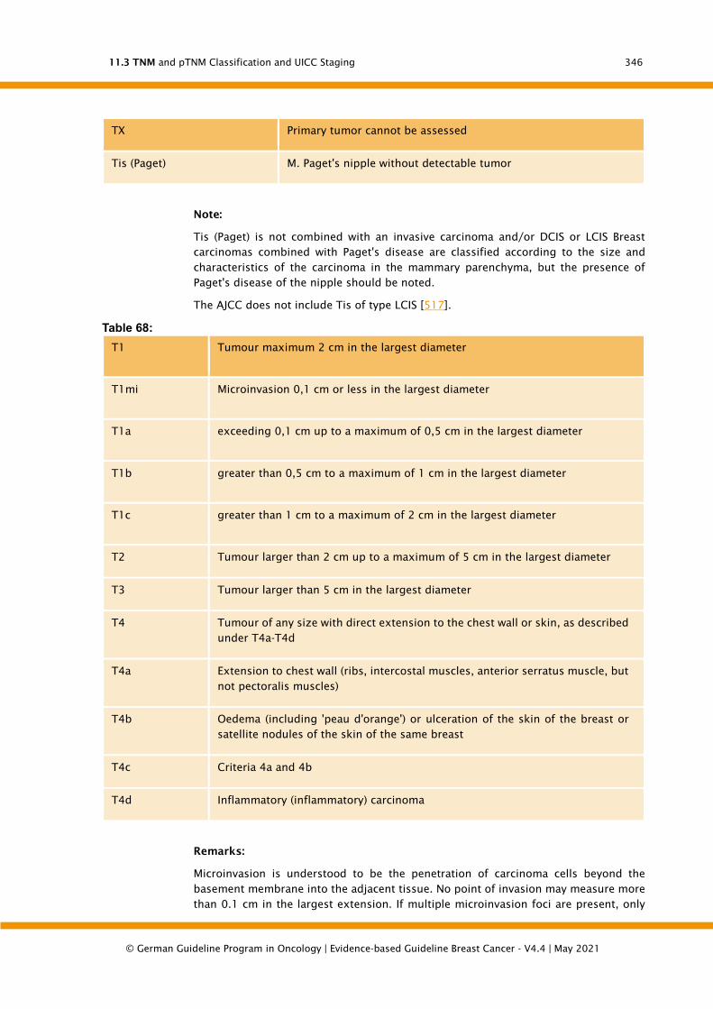
11.3 TNM and pTNM Classification and UICC Staging
© German Guideline Program in Oncology | Evidence-based Guideline Breast Cancer - V4.4 | May 2021
346
TX Primary tumor cannot be assessed
Tis (Paget) M. Paget's nipple without detectable tumor
Note:
Tis (Paget) is not combined with an invasive carcinoma and/or DCIS or LCIS Breast
carcinomas combined with Paget's disease are classified according to the size and
characteristics of the carcinoma in the mammary parenchyma, but the presence of
Paget's disease of the nipple should be noted.
The AJCC does not include Tis of type LCIS [517].
Table 68:
T1 Tumour maximum 2 cm in the largest diameter
T1mi Microinvasion 0,1 cm or less in the largest diameter
T1a exceeding 0,1 cm up to a maximum of 0,5 cm in the largest diameter
T1b greater than 0,5 cm to a maximum of 1 cm in the largest diameter
T1c greater than 1 cm to a maximum of 2 cm in the largest diameter
T2 Tumour larger than 2 cm up to a maximum of 5 cm in the largest diameter
T3 Tumour larger than 5 cm in the largest diameter
T4 Tumour of any size with direct extension to the chest wall or skin, as described
under T4a-T4d
T4a Extension to chest wall (ribs, intercostal muscles, anterior serratus muscle, but
not pectoralis muscles)
T4b Oedema (including 'peau d'orange') or ulceration of the skin of the breast or
satellite nodules of the skin of the same breast
T4c Criteria 4a and 4b
T4d Inflammatory (inflammatory) carcinoma
Remarks:
Microinvasion is understood to be the penetration of carcinoma cells beyond the
basement membrane into the adjacent tissue. No point of invasion may measure more
than 0.1 cm in the largest extension. If multiple microinvasion foci are present, only
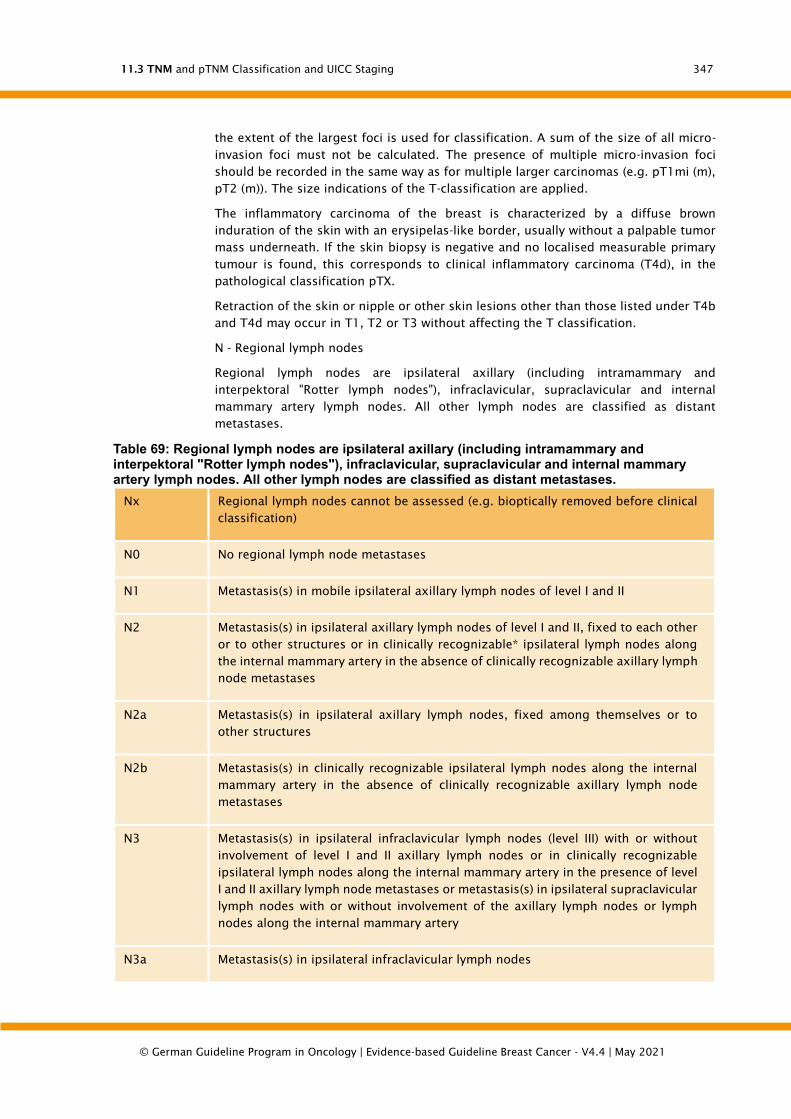
11.3 TNM and pTNM Classification and UICC Staging
© German Guideline Program in Oncology | Evidence-based Guideline Breast Cancer - V4.4 | May 2021
347
the extent of the largest foci is used for classification. A sum of the size of all micro-
invasion foci must not be calculated. The presence of multiple micro-invasion foci
should be recorded in the same way as for multiple larger carcinomas (e.g. pT1mi (m),
pT2 (m)). The size indications of the T-classification are applied.
The inflammatory carcinoma of the breast is characterized by a diffuse brown
induration of the skin with an erysipelas-like border, usually without a palpable tumor
mass underneath. If the skin biopsy is negative and no localised measurable primary
tumour is found, this corresponds to clinical inflammatory carcinoma (T4d), in the
pathological classification pTX.
Retraction of the skin or nipple or other skin lesions other than those listed under T4b
and T4d may occur in T1, T2 or T3 without affecting the T classification.
N - Regional lymph nodes
Regional lymph nodes are ipsilateral axillary (including intramammary and
interpektoral "Rotter lymph nodes"), infraclavicular, supraclavicular and internal
mammary artery lymph nodes. All other lymph nodes are classified as distant
metastases.
Table 69: Regional lymph nodes are ipsilateral axillary (including intramammary and interpektoral "Rotter lymph nodes"), infraclavicular, supraclavicular and internal mammary artery lymph nodes. All other lymph nodes are classified as distant metastases.
Nx Regional lymph nodes cannot be assessed (e.g. bioptically removed before clinical
classification)
N0 No regional lymph node metastases
N1 Metastasis(s) in mobile ipsilateral axillary lymph nodes of level I and II
N2 Metastasis(s) in ipsilateral axillary lymph nodes of level I and II, fixed to each other
or to other structures or in clinically recognizable* ipsilateral lymph nodes along
the internal mammary artery in the absence of clinically recognizable axillary lymph
node metastases
N2a Metastasis(s) in ipsilateral axillary lymph nodes, fixed among themselves or to
other structures
N2b Metastasis(s) in clinically recognizable ipsilateral lymph nodes along the internal
mammary artery in the absence of clinically recognizable axillary lymph node
metastases
N3 Metastasis(s) in ipsilateral infraclavicular lymph nodes (level III) with or without
involvement of level I and II axillary lymph nodes or in clinically recognizable
ipsilateral lymph nodes along the internal mammary artery in the presence of level
I and II axillary lymph node metastases or metastasis(s) in ipsilateral supraclavicular
lymph nodes with or without involvement of the axillary lymph nodes or lymph
nodes along the internal mammary artery
N3a Metastasis(s) in ipsilateral infraclavicular lymph nodes
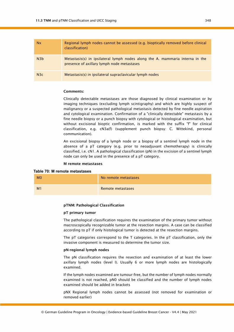
11.3 TNM and pTNM Classification and UICC Staging
© German Guideline Program in Oncology | Evidence-based Guideline Breast Cancer - V4.4 | May 2021
348
Nx Regional lymph nodes cannot be assessed (e.g. bioptically removed before clinical
classification)
N3b Metastasis(s) in ipsilateral lymph nodes along the A. mammaria interna in the
presence of axillary lymph node metastases
N3c Metastasis(s) in ipsilateral supraclavicular lymph nodes
Comments:
Clinically detectable metastases are those diagnosed by clinical examination or by
imaging techniques (excluding lymph scintigraphy) and which are highly suspect of
malignancy or a suspected pathological metastasis detected by fine needle aspiration
and cytological examination. Confirmation of a "clinically detectable" metastasis by a
fine needle biopsy or a punch biopsy with cytological or histological examination, but
without excisional bioptic confirmation, is marked with the suffix "f" for clinical
classification, e.g. cN3a(f) (supplement punch biopsy: C. Wittekind, personal
communication).
An excisional biopsy of a lymph node or a biopsy of a sentinel lymph node in the
absence of a pT category (e.g. prior to neoadjuvant chemotherapy) is clinically
classified, i.e. cN1. A pathological classification (pN) in the excision of a sentinel lymph
node can only be used in the presence of a pT category.
M remote metastases
Table 70: M remote metastases
M0 No remote metastases
M1 Remote metastases
pTNM: Pathological Classification
pT primary tumor
The pathological classification requires the examination of the primary tumor without
macroscopically recognizable tumor at the resection margins. A case can be classified
according to pT if only histological tumor is detected at the resection margins.
The pT categories correspond to the T categories. In the pT classification, only the
invasive component is measured to determine the tumor size.
pN-regional lymph nodes
The pN classification requires the resection and examination of at least the lower
axillary lymph nodes (level I). Usually 6 or more lymph nodes are histologically
examined.
If the lymph nodes examined are tumour-free, but the number of lymph nodes normally
examined is not reached, pN0 should be classified and the number of lymph nodes
examined should be added in brackets
pNX Regional lymph nodes cannot be assessed (not removed for examination or
removed earlier)
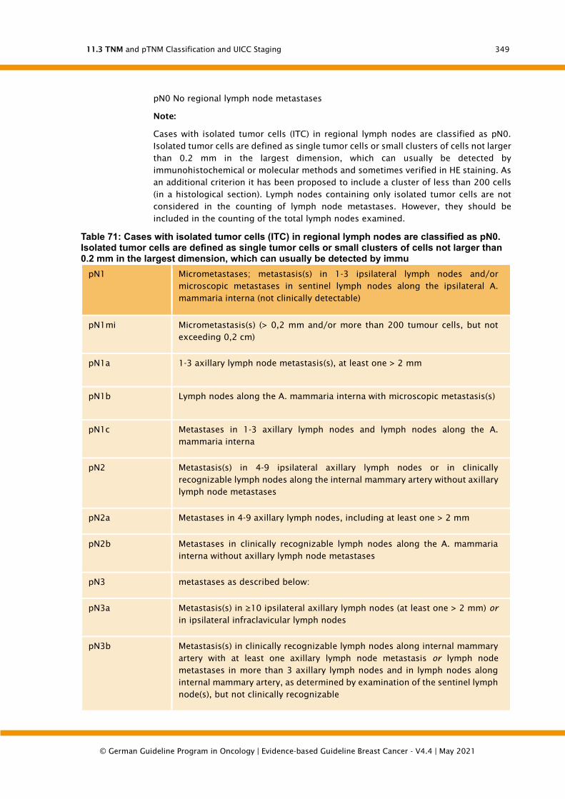
11.3 TNM and pTNM Classification and UICC Staging
© German Guideline Program in Oncology | Evidence-based Guideline Breast Cancer - V4.4 | May 2021
349
pN0 No regional lymph node metastases
Note:
Cases with isolated tumor cells (ITC) in regional lymph nodes are classified as pN0.
Isolated tumor cells are defined as single tumor cells or small clusters of cells not larger
than 0.2 mm in the largest dimension, which can usually be detected by
immunohistochemical or molecular methods and sometimes verified in HE staining. As
an additional criterion it has been proposed to include a cluster of less than 200 cells
(in a histological section). Lymph nodes containing only isolated tumor cells are not
considered in the counting of lymph node metastases. However, they should be
included in the counting of the total lymph nodes examined.
Table 71: Cases with isolated tumor cells (ITC) in regional lymph nodes are classified as pN0. Isolated tumor cells are defined as single tumor cells or small clusters of cells not larger than 0.2 mm in the largest dimension, which can usually be detected by immu
pN1 Micrometastases; metastasis(s) in 1-3 ipsilateral lymph nodes and/or
microscopic metastases in sentinel lymph nodes along the ipsilateral A.
mammaria interna (not clinically detectable)
pN1mi Micrometastasis(s) (> 0,2 mm and/or more than 200 tumour cells, but not
exceeding 0,2 cm)
pN1a 1-3 axillary lymph node metastasis(s), at least one > 2 mm
pN1b Lymph nodes along the A. mammaria interna with microscopic metastasis(s)
pN1c Metastases in 1-3 axillary lymph nodes and lymph nodes along the A.
mammaria interna
pN2 Metastasis(s) in 4-9 ipsilateral axillary lymph nodes or in clinically
recognizable lymph nodes along the internal mammary artery without axillary
lymph node metastases
pN2a Metastases in 4-9 axillary lymph nodes, including at least one > 2 mm
pN2b Metastases in clinically recognizable lymph nodes along the A. mammaria
interna without axillary lymph node metastases
pN3 metastases as described below:
pN3a Metastasis(s) in ≥10 ipsilateral axillary lymph nodes (at least one > 2 mm) or
in ipsilateral infraclavicular lymph nodes
pN3b Metastasis(s) in clinically recognizable lymph nodes along internal mammary
artery with at least one axillary lymph node metastasis or lymph node
metastases in more than 3 axillary lymph nodes and in lymph nodes along
internal mammary artery, as determined by examination of the sentinel lymph
node(s), but not clinically recognizable
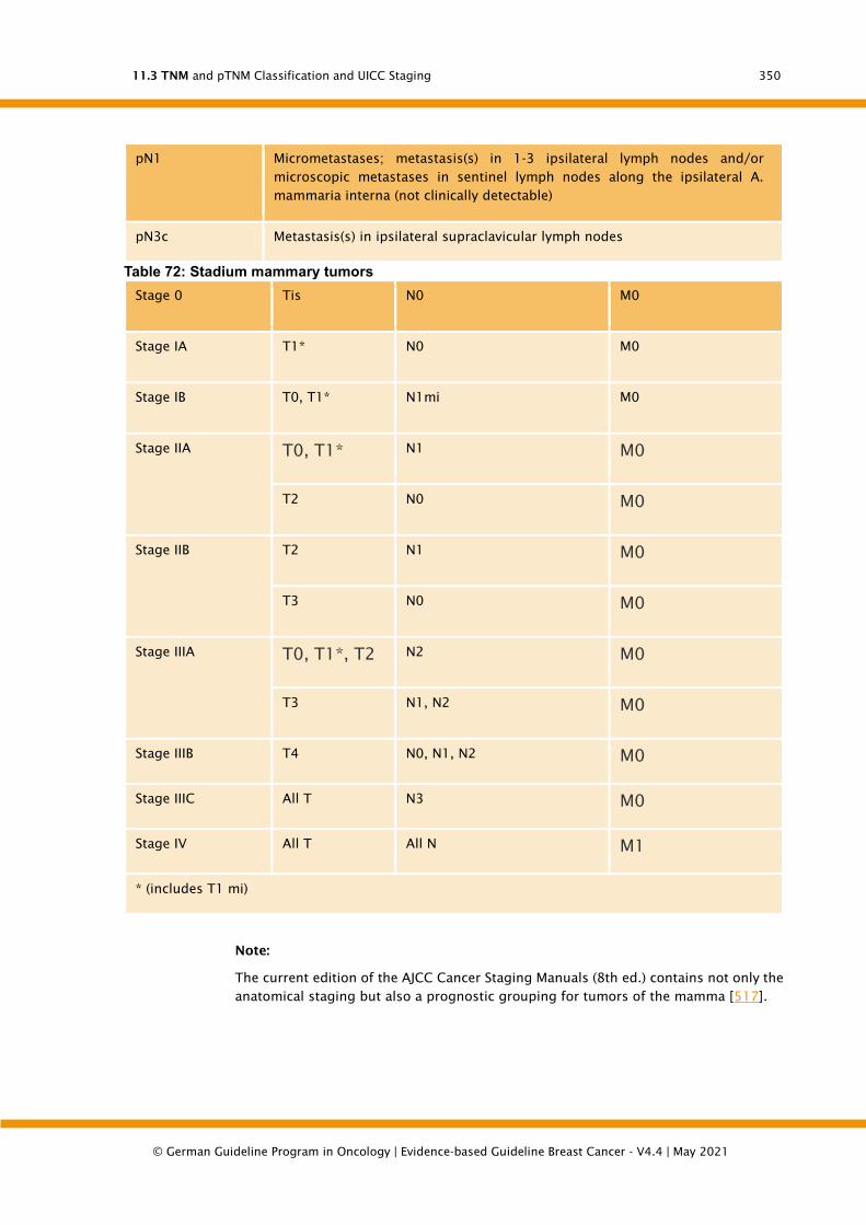
11.3 TNM and pTNM Classification and UICC Staging
© German Guideline Program in Oncology | Evidence-based Guideline Breast Cancer - V4.4 | May 2021
350
pN1 Micrometastases; metastasis(s) in 1-3 ipsilateral lymph nodes and/or
microscopic metastases in sentinel lymph nodes along the ipsilateral A.
mammaria interna (not clinically detectable)
pN3c Metastasis(s) in ipsilateral supraclavicular lymph nodes
Table 72: Stadium mammary tumors
Stage 0 Tis N0 M0
Stage IA T1* N0 M0
Stage IB T0, T1* N1mi M0
Stage IIA
T0, T1* N1 M0
T2 N0 M0
Stage IIB
T2 N1 M0
T3 N0 M0
Stage IIIA
T0, T1*, T2 N2 M0
T3 N1, N2 M0
Stage IIIB T4 N0, N1, N2 M0
Stage IIIC All T N3 M0
Stage IV All T All N M1
* (includes T1 mi)
Note:
The current edition of the AJCC Cancer Staging Manuals (8th ed.) contains not only the
anatomical staging but also a prognostic grouping for tumors of the mamma [517].
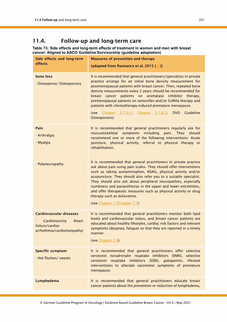
11.4 Follow-up and long-term care
© German Guideline Program in Oncology | Evidence-based Guideline Breast Cancer - V4.4 | May 2021
351
11.4. Follow-up and long-term care
Table 73: Side effects and long-term effects of treatment in women and men with breast cancer: Aligned to ASCO Guideline Survivorship (guideline adaptation)
Side effects and long-term
effects
Measures of prevention and therapy
(adapted from Runowicz et al. 2015 [31])
bone loss
- Osteopenia/ Osteoporosis
It is recommended that general practitioners/specialists in private
practice arrange for an initial bone density measurement for
postmenopausal patients with breast cancer. Then, repeated bone
density measurements every 2 years should be recommended for
breast cancer patients on aromatase inhibitor therapy,
premenopausal patients on tamoxifen and/or GnRHa therapy and
patients with chemotherapy-induced premature menopause.
(see Chapter 5.7.6.1; Chapter 5.7.6.2; DVO Guideline
Osteoporosis)
Pain
- Arthralgia
- Myalgia
- Polyneuropathy
It is recommended that general practitioners regularly ask for
musculoskeletal symptoms including pain. They should
recommend one or more of the following interventions: Acute
puncture, physical activity, referral to physical therapy or
rehabilitation.
It is recommended that general practitioners in private practice
ask about pain using pain scales. They should offer interventions
such as taking acetaminophen, NSAIs, physical activity and/or
acupuncture. They should also refer you to a suitable specialist.
They should also ask about peripheral neuropathies, especially
numbness and paraesthesias in the upper and lower extremities,
and offer therapeutic measures such as physical activity or drug
therapy such as duloxetine.
(see Chapter 7.5Chapter 7.5)
Cardiovascular diseases
- Cardiotoxicity (heart
failure/cardiac
arrhythmia/cardiomyopathy)
It is recommended that general practitioners monitor both lipid
levels and cardiovascular status, and breast cancer patients are
educated about healthy lifestyles, cardiac risk factors and relevant
symptoms (dyspnea, fatigue) so that they are reported in a timely
manner.
(see Chapter 7.4)
Specific symptom
- Hot flushes/ sweats
It is recommended that general practitioners offer selective
serotonin noradrenalin reuptake inhibitors (SNRI), selective
serotonin reuptake inhibitors (SSRI), gabapentin, lifestyle
interventions to alleviate vasomotor symptoms of premature
menopause.
Lymphedema It is recommended that general practitioners educate breast
cancer patients about the prevention or reduction of lymphedema,

11.4 Follow-up and long-term care
© German Guideline Program in Oncology | Evidence-based Guideline Breast Cancer - V4.4 | May 2021
352
Side effects and long-term
effects
Measures of prevention and therapy
(adapted from Runowicz et al. 2015 [31])
including weight reduction in obese patients. Patients with clinical
symptoms or swelling should be referred to a therapist who is
familiar with the diagnosis and treatment of lymphedema, e.g.
physiotherapists, lymphedema specialists. (see Chapter 7.5)
Infertility It is recommended that general practitioners refer breast cancer
patients of reproductive age with unfulfilled desire for children to
a specialist in endocrinology and reproductive medicine as soon
as possible.
Sexual health:
Dysfunction
- Urogenital Postmenopausal
Syndrome
- Partnership
It is recommended that general practitioners
a) assess signs and symptoms of sexual dysfunction or problems
of sexual intimacy
(b) should assess and treat risk factors for sexual dysfunction,
where appropriate
c) non-hormonal, water-based lubricants and moisturisers for
vaginal dryness should be offered
(d) refer patients to psychoeducational support, group therapy,
sexual counselling, marriage guidance or intensive
psychotherapy, as appropriate
Cognitive Dysfunction General practitioners should ask patients about cognitive
impairment. Reversible factors that can affect cognitive
performance should be identified and treated as well as possible.
Patients with signs of cognitive dysfunction should receive
neurocognitive assessment and rehabilitation with group training,
if available.
(see Chapter 7.5)
Fatigue General practitioners should ask for signs of fatigue and treat any
causative factors (anaemia, thyroid dysfunction, cardiac causes).
In patients where no underlying cause can be found, other factors
that promote fatigue, such as mood swings, sleep disturbances
and pain, should be treated. Patients should be advised on regular
physical activity and referred to cognitive behavioural therapy if
necessary.
Psychosocial stress
- Anxiety/depression/
distress
- Body image
- Emotional perception
General practitioners should evaluate patients for signs of
distress, depression or anxiety. Patients with a higher risk of
depression (young patients, those with psychiatric illness, low
socioeconomic status) should be assessed more precisely. Clinical
signs of distress, depression or anxiety should be assessed by
counselling and/or pharmacotherapy and/or referral to psycho-
oncologists and appropriate mental health facilities.

11.4 Follow-up and long-term care
© German Guideline Program in Oncology | Evidence-based Guideline Breast Cancer - V4.4 | May 2021
353
Side effects and long-term
effects
Measures of prevention and therapy
(adapted from Runowicz et al. 2015 [31])
- Social role perception
- Financial burdens
(see Chapter 7.2)
Secondary malignancies
- intestine, skin
- (Gynaecological/prostate)
- Leukemia
Gynaecologists in private practice should offer the regular check-
ups of other departments and perform an annual gynaecological
examination in postmenopausal patients under SERM therapy.
Health behaviour
- Overweight
- Physical mobility
- Nutrition/alcohol
- Smoking cessation
Aftercare physicians should advise patients in their aftercare
regarding weight loss and maintaining a normal body weight.
Overweight or obese patients should be advised as follows: high-
calorie foods and beverages should be avoided and physical
activity increased.
Aftercare physicians should advise patients to be physically active
on a regular basis (according to ACS guidelines). More specifically,
patients should avoid physical inactivity and resume normal daily
activities as soon as possible after diagnosis. Patients should be
physically active for at least 150 minutes per week at moderate
levels or 75 minutes at high levels. Training should include
strength training at least 2 days per week. Patients who have
received adjuvant chemotherapy or hormone therapy should pay
particular attention to strength training.
After-care physicians should advise patients on a healthy diet. This
should include plenty of vegetables, fruit, wholemeal products,
legumes and low saturated fats and alcohol.
Aftercare physicians should advise patients to stop smoking and
offer smokers supportive measures and programmes.
Aftercare plan, family and
relatives, need for
information
Aftercare physicians should be in contact with the treating
oncological colleagues and receive information about the therapy
carried out and other planned measures.
Aftercare physicians should support the involvement of (spouses)
partners and caregivers in aftercare.
Follow-up physicians should ask about patients' needs for
information regarding breast cancer, therapies, side effects, other
health aspects and support services and should try to meet these
needs.
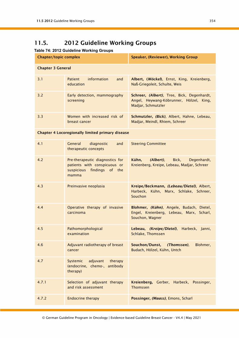
11.5 2012 Guideline Working Groups
© German Guideline Program in Oncology | Evidence-based Guideline Breast Cancer - V4.4 | May 2021
354
11.5. 2012 Guideline Working Groups
Table 74: 2012 Guideline Working Groups
Chapter/topic complex Speaker, (Reviewer), Working Group
Chapter 3 General
3.1 Patient information and
education
Albert, (Wöckel), Ernst, King, Kreienberg,
Naß-Griegoleit, Schulte, Weis
3.2 Early detection, mammography
screening
Schreer, (Albert), Tree, Bick, Degenhardt,
Angel, Heywang-Köbrunner, Hölzel, King,
Madjar, Schmutzler
3.3 Women with increased risk of
breast cancer
Schmutzler, (Bick), Albert, Hahne, Lebeau,
Madjar, Meindl, Rhiem, Schreer
Chapter 4 Locoregionally limited primary disease
4.1 General diagnostic and
therapeutic concepts
Steering Committee
4.2 Pre-therapeutic diagnostics for
patients with conspicuous or
suspicious findings of the
mamma
Kühn, (Albert), Bick, Degenhardt,
Kreienberg, Kreipe, Lebeau, Madjar, Schreer
4.3 Preinvasive neoplasia Kreipe/Beckmann, (Lebeau/Dietel), Albert,
Harbeck, Kühn, Marx, Schlake, Schreer,
Souchon
4.4 Operative therapy of invasive
carcinoma
Blohmer, (Kühn), Angele, Budach, Dietel,
Engel, Kreienberg, Lebeau, Marx, Scharl,
Souchon, Wagner
4.5 Pathomorphological
examination
Lebeau, (Kreipe/Dietel), Harbeck, Janni,
Schlake, Thomssen
4.6 Adjuvant radiotherapy of breast
cancer
Souchon/Dunst, (Thomssen), Blohmer,
Budach, Hölzel, Kühn, Untch
4.7 Systemic adjuvant therapy
(endocrine, chemo-, antibody
therapy)
4.7.1 Selection of adjuvant therapy
and risk assessment
Kreienberg, Gerber, Harbeck, Possinger,
Thomssen
4.7.2 Endocrine therapy Possinger, (Maass), Emons, Scharl

11.5 2012 Guideline Working Groups
© German Guideline Program in Oncology | Evidence-based Guideline Breast Cancer - V4.4 | May 2021
355
Chapter/topic complex Speaker, (Reviewer), Working Group
4.7.3 Chemotherapy Harbeck, (Möbus), Janni, Possinger
4.7.4 Neoadjuvant (primarily
systemic) therapy (NACT or PST)
Gerber, (v. Minckwitz), Marschner, Untch
4.7.5 Antibody Therapy Thomssen, (Snow White), Jackisch
4.7.6 Bisphosphonates Thomssen, (Snow White), Jackisch
4.8 Management of primarily
local/local/regional advanced
tumours
Steering Committee
Chapter 5 Recurrent or metastatic breast cancer
5.1 Definition and forecast Steering Committee
5.2 Diagnosis of local/local
recurrence
Bick, (Sharl), Blohmer, Buck, Degenhardt,
Madjar
5.3 Therapy of local/local
recurrence
Dunst, (Kühn), Angele, Blohmer, Dietel,
Heitmann, Marx, Gerber
5.4 Remote Metastases Marschner, (Emons), Angele, Dunst,
Harbeck, Possinger, Thomssen
Chapter 6 Treatment, care, support
6.1 General concept Steering Committee
6.2 Psychosocial aspects and
psycho-oncology
Weis/Beckmann, (Scharl), Albert, Bartsch,
Ernst, Faller, King, Naß-Griegoleit, Schulte
6.3 Supportive therapy Link, (Follmann), tree, Emons, Henscher,
Rup-pert, Skoetz
6.4 Rehabilitation Bartsch, (Schulte), Baum, Henscher, Knauth,
Ruppert
6.5 Aftercare with recurrence and
metastasis diagnostics and
therapy support
Janni, (Beckmann), Hölzel, King, Wet-
Griegoleit, Paradise, Schulte, Souchon,
Thomssen, Weis
6.6 Palliative Care Gardener, (Schulte), Beckmann, Gerlach,
Naß-Griegoleit
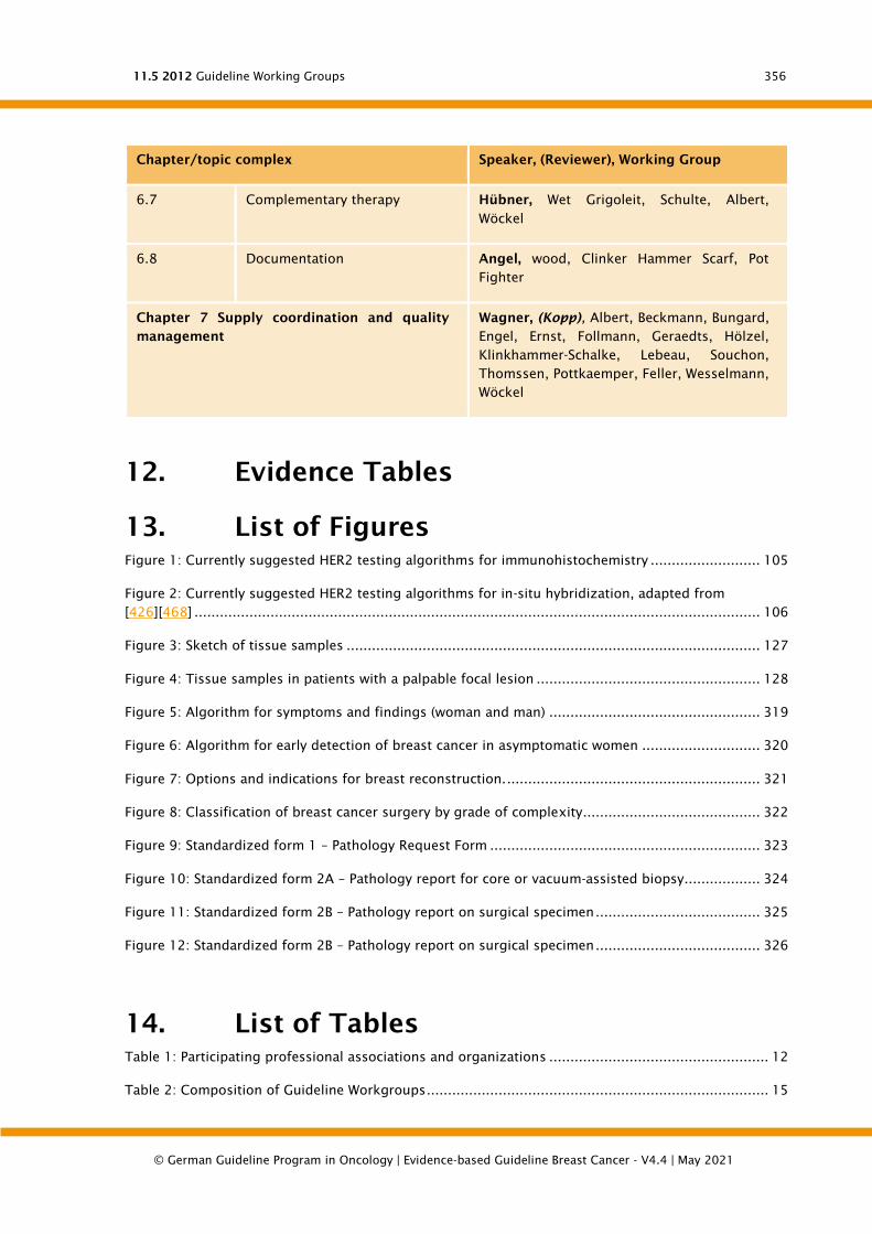
11.5 2012 Guideline Working Groups
© German Guideline Program in Oncology | Evidence-based Guideline Breast Cancer - V4.4 | May 2021
356
Chapter/topic complex Speaker, (Reviewer), Working Group
6.7 Complementary therapy Hübner, Wet Grigoleit, Schulte, Albert,
Wöckel
6.8 Documentation Angel, wood, Clinker Hammer Scarf, Pot
Fighter
Chapter 7 Supply coordination and quality
management
Wagner, (Kopp), Albert, Beckmann, Bungard,
Engel, Ernst, Follmann, Geraedts, Hölzel,
Klinkhammer-Schalke, Lebeau, Souchon,
Thomssen, Pottkaemper, Feller, Wesselmann,
Wöckel
12. Evidence Tables
13. List of Figures
Figure 1: Currently suggested HER2 testing algorithms for immunohistochemistry .......................... 105
Figure 2: Currently suggested HER2 testing algorithms for in-situ hybridization, adapted from
[426][468] ...................................................................................................................................... 106
Figure 3: Sketch of tissue samples .................................................................................................. 127
Figure 4: Tissue samples in patients with a palpable focal lesion ..................................................... 128
Figure 5: Algorithm for symptoms and findings (woman and man) .................................................. 319
Figure 6: Algorithm for early detection of breast cancer in asymptomatic women ............................ 320
Figure 7: Options and indications for breast reconstruction. ............................................................ 321
Figure 8: Classification of breast cancer surgery by grade of complexity .......................................... 322
Figure 9: Standardized form 1 – Pathology Request Form ................................................................ 323
Figure 10: Standardized form 2A – Pathology report for core or vacuum-assisted biopsy.................. 324
Figure 11: Standardized form 2B – Pathology report on surgical specimen ....................................... 325
Figure 12: Standardized form 2B – Pathology report on surgical specimen ....................................... 326
14. List of Tables
Table 1: Participating professional associations and organizations .................................................... 12
Table 2: Composition of Guideline Workgroups ................................................................................. 15
11.5 2012 Guideline Working Groups
© German Guideline Program in Oncology | Evidence-based Guideline Breast Cancer - V4.4 | May 2021
357
Table 3: Abbreviations Used ............................................................................................................. 24
Table 4: B-classification for punch and vacuum biopsies [420], [424] ............................................... 122
Table 5: Indication algorithm for radiation therapy after neoadjuvant therapy .................................. 142
Table 6: Definitions of weight categories according to body-mass-index .......................................... 175
Table 7: Adriamycin 60 / cyclophosphamide 600 ............................................................................ 204
Table 8: Adriamycin liposomal 75 / cyclophosphamide 600 ............................................................ 204
Table 9: Adriamycin 50 / docetaxel 75 ............................................................................................ 205
Table 10: Capecitabine 2000 / Bevacizumab 15 .............................................................................. 205
Table 11: Capecitabine 2000 / Paclitaxel 175.................................................................................. 205
Table 12: Cisplatin 75 / Gemcitabine 1250 ..................................................................................... 206
Table 13: Cyclophosphamide 600 / non-pegylated liposomal doxorubicin 75 .................................. 206
Table 14: docetaxel ........................................................................................................................ 206
Table 15: Docetaxel 35, breast cancer ............................................................................................ 206
Table 16: Doxorubicin 50 / docetaxel 75, breast cancer .................................................................. 207
Table 17: Doxorubicin 60 / cyclophosphamide 600 ........................................................................ 207
Table 18: Epirubicin 60 / cyclophosphamide 600 ............................................................................ 207
Table 19: Epirubicin 75 / cyclophosphamide 600 ............................................................................ 208
Table 20: Epirubicin 75 / docetaxel 75 ............................................................................................ 208
Table 21: Epirubicin 60 / Paclitaxel 175 .......................................................................................... 208
Table 22: Epirubicin 60 / Paclitaxel 175 .......................................................................................... 208
Table 23: eribulin 1,23 ................................................................................................................... 209
Table 24: everolimus 10 / exemplestan 25, postmenopausal .......................................................... 209
Table 25: Fulvestrant 500, postmenopausal .................................................................................... 209
Table 26: Gemcitabine 1000 / Carboplatinum 4 .............................................................................. 210
Table 27: Lapatinib 1250 / Capecitabine 2000 ................................................................................ 210
Table 28: NabPaclitaxel 125 / carboplatinum .................................................................................. 210
Table 29: Nab-paclitaxel 100 / carboplatin 2 / bevacizumab 10, (triple negative) ............................. 210
Table 30: Nab-paclitaxel 125 / Trastuzumab (4/2) .......................................................................... 211
Table 31: Nab-paclitaxel 125 weekly ............................................................................................... 211
Table 32: Paclitaxel 90 / Bevacizumab 10 ....................................................................................... 211
11.5 2012 Guideline Working Groups
© German Guideline Program in Oncology | Evidence-based Guideline Breast Cancer - V4.4 | May 2021
358
Table 33: Paclitaxel 175 / Capecitabine 2000.................................................................................. 212
Table 34: paclitaxel 175 / gemcitabine 1250 .................................................................................. 212
Table 35: Palbociclib 125 / Fulvestrant 500..................................................................................... 212
Table 36: Pertuzumab 840 / Trastuzumab 8 / Docetaxel 75, (HER2+) cycle 1 .................................. 213
Table 37: Pertuzumab 420 / Trastuzumab 6 / Docetaxel 75, (HER2+) cycle 2+ ................................ 213
Table 38: Trastuzumab (8) 6 / Docetaxel 100, (HER2+) ................................................................... 213
Table 39: Trastuzumab 6 / letrozole 2.5, HER2+/HR+ ..................................................................... 214
Table 40: Trastuzumab (8) 6 / vinorelbine 30, breast carcinoma (HER2+) ........................................ 214
Table 41: Trastuzumab Emtansin 3,6, (HER2+) ................................................................................ 214
Table 42: trofosfamide 150 ............................................................................................................ 214
Table 43: trofosfamide 50 .............................................................................................................. 215
Table 44: vinorelbine 30 ................................................................................................................. 215
Table 45: vinorelbine 70 oral .......................................................................................................... 215
Table 46: Common Terminology Critera for Adverse Events (CTCAE, Version 4.03) .......................... 239
Table 47: Emetogenes Potenzial der beim Mammakarzinom verwendeten einzelnen Zytostatika,
aktualisiert 5/2012 [1323], [1326] .................................................................................................. 240
Table 48: Strahlentherapiespezifische Faktoren des emetogenen Risikos ......................................... 246
Table 49: Zusammenfassung der antiemetischen Prophylaxe .......................................................... 247
Table 50: Anemia workup [676] ...................................................................................................... 253
Table 51: Stages and diagnostic workup of iron deficiency [676] ..................................................... 255
Table 52: Recommendations for RBC transfusion in acuute anemia as pre cross-sectional guidelines
(BÄK) for therapy with blood components 2014 [676] ...................................................................... 257
Table 53: Follow-up examinations in breast cancer .......................................................................... 272
Table 54: Follow-up examinations for breast cancer – Breast diagnostics after BCT or mastectomy .. 272
Table 55: Use of complementary methods, observed side effects, potential interactions .................. 281
Table 56: Risikofaktoren für Männer, an einem Mammakarzinom zu erkranken ............................... 311
Table 57: Quality Indicator definitions............................................................................................. 314
Table 58: Quality Indicators ............................................................................................................ 314
Table 59: Nuklear Grading of DCIS [455] ......................................................................................... 332
Table 60: WHO grading of DCIS [118] .............................................................................................. 333
11.5 2012 Guideline Working Groups
© German Guideline Program in Oncology | Evidence-based Guideline Breast Cancer - V4.4 | May 2021
359
Table 61: WHO classification of invasive breast carcinomas [118] .................................................... 334
Table 62: Criteria for grading breast cancer [510] ........................................................................... 338
Table 63: Assignment of scores for mitotic count as a function of field diameter [510] .................... 339
Table 64: Nottingham Prognostic Index [1642] ............................................................................... 341
Table 65: Immunohistochemistry scores for hormone receptor assessment ..................................... 342
Table 66: CPS+EG-Score [1648] ....................................................................................................... 345
Table 67: T-primary tumor .............................................................................................................. 345
Table 68: ........................................................................................................................................ 346
Table 69: Regional lymph nodes are ipsilateral axillary (including intramammary and interpektoral
"Rotter lymph nodes"), infraclavicular, supraclavicular and internal mammary artery lymph nodes. All
other lymph nodes are classified as distant metastases. .................................................................. 347
Table 70: M remote metastases ...................................................................................................... 348
Table 71: Cases with isolated tumor cells (ITC) in regional lymph nodes are classified as pN0. Isolated
tumor cells are defined as single tumor cells or small clusters of cells not larger than 0.2 mm in the
largest dimension, which can usually be detected by immu ............................................................. 349
Table 72: Stadium mammary tumors............................................................................................... 350
Table 73: Side effects and long-term effects of treatment in women and men with breast cancer:
Aligned to ASCO Guideline Survivorship (guideline adaptation) ........................................................ 351
Table 74: 2012 Guideline Working Groups ...................................................................................... 354

11.5 2012 Guideline Working Groups
© German Guideline Program in Oncology | Evidence-based Guideline Breast Cancer - V4.4 | May 2021
360
11.5 2012 Guideline Working Groups
© German Guideline Program in Oncology | Evidence-based Guideline Breast Cancer - V4.4 | May 2021
361
15. Bibliography
1. Jansen, S. J., Otten, W., Baas-Thijssen, M. C., van de Velde, C. J., Nortier, J. W., Stiggelbout, A.
M., Explaining differences in attitude toward adjuvant chemotherapy between experienced and
inexperienced breast cancer patients. J Clin Oncol, 2005. 23(27): p. 6623-30.
2. Katz, S. J., Lantz, P. M., Janz, N. K., Fagerlin, A., Schwartz, K., Liu, L., et.al. Patient involvement
in surgery treatment decisions for breast cancer. J Clin Oncol, 2005. 23(24): p. 5526-33.
3. Wagner, Katja, Koller, M, Keil, Astrid, Trott, Diana, Engenhart-Cabillic, Rita, Pfab, R, et.al.
Strahlentherapie bei chirurgischen und nichtchirurgischen Patienten Therapieerwartungen,
Lebensqualität und Arzteinschätzungen. Der Chirurg, 1998. 69(3): p. 252-258.
4. Weis, J., Giesler, J. M., Subjective dimensions of patient competence: relationships with
selected healthcare usage behaviors and general features of self-rated competence. Patient
Educ Couns, 2008. 73(3): p. 511-8.
5. Steinbach, K., van Oorschot, B. Anselm, R. Leppert, K. Schweitzer, S. Hausmann, C. Köhler, N.,
Wer soll entscheiden?. Deutsches Ärzteblatt, 2004. 101(41): p. A-2741.
6. Albert, U-S, Koller, M, Wagner, U, Schulz, K-D, Survival chances and psychological aspects of
quality of life in patients with localized early stage breast cancer. Inflammation Research,
2004. 53: p. S136-S141.
7. Chouliara, Z., Kearney, N., Stott, D., Molassiotis, A., Miller, M., Perceptions of older people with
cancer of information, decision making and treatment: a systematic review of selected
literature. Ann Oncol, 2004. 15(11): p. 1596-602.
8. Hagerty, R. G., Butow, P. N., Ellis, P. M., Dimitry, S., Tattersall, M. H., Communicating prognosis
in cancer care: a systematic review of the literature. Ann Oncol, 2005. 16(7): p. 1005-53.
9. Nilsen, E. S., Myrhaug, H. T., Johansen, M., Oliver, S., Oxman, A. D., Methods of consumer
involvement in developing healthcare policy and research, clinical practice guidelines and
patient information material. Cochrane Database Syst Rev, 2006. p. Cd004563.
10. Gysels, M., Higginson, I. J., Interactive technologies and videotapes for patient education in
cancer care: systematic review and meta-analysis of randomised trials. Support Care Cancer,
2007. 15(1): p. 7-20.
11. Wofford, J. L., Smith, E. D., Miller, D. P., The multimedia computer for office-based patient
education: a systematic review. Patient Educ Couns, 2005. 59(2): p. 148-57.
12. Beauchamp, Tom L, Childress, James F, Principles of biomedical ethicsOxford University Press,
USA, 2001.
13. Sieber, W. J., Kaplan, R. M., Informed adherence: the need for shared medical decision making.
Control Clin Trials, 2000. 21(5 Suppl): p. 233s-40s.
14. Weinstein, James N, Editorial: The missing piece: Embracing shared decision making to reform
health careLWW, 2000.
15. Wright, E. B., Holcombe, C., Salmon, P., Doctors‘ communication of trust, care, and respect in
breast cancer: qualitative study. Bmj, 2004. 328(7444): p. 864.
16. Dissemination, NHS Centre for Reviews and, Effective Health Care: Informing, communicating
and sharing decision with people who have cancer.Latimer Trend & Company Ltd., 2000.
17. Center, D.-H.M., Available from: http://www.dartmouth-
hitchcock.org/about_dh/project_search.html, 2016., Available, from:, , ,
http://www.dartmouth-hitchcock.org/about_dh/project_search.html, , .
18. Berger-Hoger, B., Liethmann, K., Muhlhauser, I., Haastert, B., Steckelberg, A., Informed shared
decision-making supported by decision coaches for women with ductal carcinoma in situ:
study protocol for a cluster randomized controlled trial. Trials, 2015. 16: p. 452.
19. Rahn, A. C., Kopke, S., Kasper, J., Vettorazzi, E., Muhlhauser, I., Heesen, C., Evaluator-blinded
trial evaluating nurse-led immunotherapy DEcision Coaching In persons with relapsing-
remitting Multiple Sclerosis (DECIMS) and accompanying process evaluation: study protocol for
a cluster randomised controlled trial. Trials, 2015. 16: p. 106.

11.5 2012 Guideline Working Groups
© German Guideline Program in Oncology | Evidence-based Guideline Breast Cancer - V4.4 | May 2021
362
20. Albert, U-S, Schulz, K-D, Alt, D, Beck, V, Doherty, J, Holsteg, K, et.al. Eine Leitlinie für Leitlinien:
die methodische Entwicklung und Anwendung der Leitlinie Fraueninformation. Zentralblatt für
Gynäkologie, 2003. 125(12): p. 484-493.
21. O‘connor, Annette M, Rostom, Alaa, Fiset, Valerie, Tetroe, Jacqueline, Entwistle, Vikki,
Llewellyn-Thomas, Hilary, et.al. Decision aids for patients facing health treatment or screening
decisions: systematic review. Bmj, 1999. 319(7212): p. 731-734.
22. Klemperer, D, Lang, B, Koch, K, Bastian, H, Brunsmann, F, Burkhardt, M, et.al. Gute Praxis
Gesundheitsinformation. Z Evid Fortbild Qual Gesundh wesen (ZEFQ), 2010. 104: p. 66-68.
23. Bruera, E., Willey, J. S., Palmer, J. L., Rosales, M., Treatment decisions for breast carcinoma:
patient preferences and physician perceptions. Cancer, 2002. 94(7): p. 2076-80.
24. Butow, P., Harrison, J. D., Choy, E. T., Young, J. M., Spillane, A., Evans, A., Health professional
and consumer views on involving breast cancer patients in the multidisciplinary discussion of
their disease and treatment plan. Cancer, 2007. 110(9): p. 1937-44.
25. Elkin, E. B., Kim, S. H., Casper, E. S., Kissane, D. W., Schrag, D., Desire for information and
involvement in treatment decisions: elderly cancer patients‘ preferences and their physicians‘
perceptions. J Clin Oncol, 2007. 25(33): p. 5275-80.
26. Ford, S., Schofield, T., Hope, T., Observing decision-making in the general practice
consultation: who makes which decisions?. Health Expect, 2006. 9(2): p. 130-7.
27. Politi, M. C., Han, P. K., Col, N. F., Communicating the uncertainty of harms and benefits of
medical interventions. Med Decis Making, 2007. 27(5): p. 681-95.
28. NICE, The National Institute for Health and Care Excellence (NICE). Advanced breast cancer:
diagnosis and treatment, 2009 [addendum 2014].
29. Wildiers, H., Heeren, P., Puts, M., Topinkova, E., Janssen-Heijnen, M. L., Extermann, M., et.al.
International Society of Geriatric Oncology consensus on geriatric assessment in older patients
with cancer. J Clin Oncol, 2014. 32(24): p. 2595-603.
30. NICE, The National Institute for Health and Care Excellence (NICE). Advanced breast cancer:
diagnosis and treatment, 2009 (Update 2014).
31. Runowicz, Carolyn D, Leach, Corinne R, Henry, N Lynn, Henry, Karen S, Mackey, Heather T,
Cowens‐Alvarado, Rebecca L, et.al. American Cancer society/American society of clinical
oncology breast Cancer survivorship care guideline. CA: a cancer journal for clinicians, 2016.
66(1): p. 43-73.
32. Du, S., Hu, L., Dong, J., Xu, G., Jin, S., Zhang, H., et.al. Patient education programs for cancer-
related fatigue: A systematic review. Patient Educ Couns, 2015. 98(11): p. 1308-19.
33. Gesundheit, Bundesministeriumfür, Patientenrechte in Deutschland. Leitfaden für
Patientinnen/Patienten und Ärztinnen/Ärzte., 2007.
34. Schwartz, Alan, Crockett, Rachel A, Sutton, Stephen, Walter, Fiona M, Clinch, Megan, Marteau,
Theresa M, et.al. Impact on decisions to start or continue medicines of providing information
to patients about possible benefits and/or harms: a systematic review and meta-analysis.
Medical Decision Making, 2011. 31(5): p. 767-777.
35. Butow, P. N., Maclean, M., Dunn, S. M., Tattersall, M. H., Boyer, M. J., The dynamics of change:
cancer patients‘ preferences for information, involvement and support. Ann Oncol, 1997. 8(9):
p. 857-63.
36. Degner, L. F., Kristjanson, L. J., Bowman, D., Sloan, J. A., Carriere, K. C., O‘Neil, J., et.al.
Information needs and decisional preferences in women with breast cancer. Jama, 1997.
277(18): p. 1485-92.
37. Leinster, Samuel J, Ashcroft, Jennifer J, Slade, Peter, Dewey, Michael E, Mastectomy versus
conservative surgery: psychosocial effects of the patient‘s choice of treatment. Journal of
Psychosocial Oncology, 1989. 7(1-2): p. 179-192.
38. Hundertmark-Mayser, J, Thiel, W., Selbsthilfe, in Gesundheit in Deutschland.
Gesundheitsberichterstattung des Bundes Gemeinsam getragen von RKI und Destatis Berlin,
2015. p. 369-374.
11.5 2012 Guideline Working Groups
© German Guideline Program in Oncology | Evidence-based Guideline Breast Cancer - V4.4 | May 2021
363
39. Nickel, Stefan, Werner, Silke, Kofahl, Christopher, Gesundheitsbezogene Selbsthilfe in
Deutschland–Entwicklungen, Wirkungen, Perspektiven. Deskriptiver Ergebnis-Bericht zu der
Befragung von Kontaktpersonen der Selbsthilfegruppen, 2014.
40. Albert, Ute Susann, Altland, H, Duda, V, Stufe-3-Leitlinie Brustkrebs-Früherkennung in
DeutschlandZuckschwerdt München, 2008.
41. Group., Duke Evidence Synthesis, Systematic Review of Cancer Screening Literature for
Updating American Cancer Society Breast Cancer Screening Guidelines.Duke Clinical Research
Institute, Durham, NC: Guidelines Development Group, 2014.
42. Organization, World Health, WHO position paper on mammography screeningWorld Health
Organization, 2014.
43. RKI., Krebs in Deutschland 2011/2012Robert Koch Institut, 2015.
44. Broeders, M., Moss, S., Nystrom, L., Njor, S., Jonsson, H., Paap, E., et.al. The impact of
mammographic screening on breast cancer mortality in Europe: a review of observational
studies. J Med Screen, 2012. 19 Suppl 1: p. 14-25.
45. Lauby-Secretan, B., Scoccianti, C., Loomis, D., Benbrahim-Tallaa, L., Bouvard, V., Bianchini, F.,
et.al. Breast-cancer screening—viewpoint of the IARC Working Group. N Engl J Med, 2015.
372(24): p. 2353-8.
46. Oeffinger, K. C., Fontham, E. T., Etzioni, R., Herzig, A., Michaelson, J. S., Shih, Y. C., et.al.
Breast Cancer Screening for Women at Average Risk: 2015 Guideline Update From the
American Cancer Society. Jama, 2015. 314(15): p. 1599-614.
47. Tabar, L., Yen, A. M., Wu, W. Y., Chen, S. L., Chiu, S. Y., Fann, J. C., et.al. Insights from the
breast cancer screening trials: how screening affects the natural history of breast cancer and
implications for evaluating service screening programs. Breast J, 2015. 21(1): p. 13-20.
48. Simbrich, A., Wellmann, I., Heidrich, J., Heidinger, O., Hense, H. W., Trends in advanced breast
cancer incidence rates after implementation of a mammography screening program in a
German population. Cancer Epidemiol, 2016. 44: p. 44-51.
49. Malek, D, Kääb-Sanyal, V, Evaluationsbericht 2011. Zusammenfassung der Ergebnisse des
Mammographie-Screening-Programms in Deutschland., 2011.
50. Bromham, N., Schmidt-Hansen, M., Astin, M., Hasler, E., Reed, M. W., Axillary treatment for
operable primary breast cancer. Cochrane Database Syst Rev, 2017. 1: p. Cd004561.
51. Nelson, H. D., Pappas, M., Cantor, A., Griffin, J., Daeges, M., Humphrey, L., Harms of Breast
Cancer Screening: Systematic Review to Update the 2009 U.S. Preventive Services Task Force
Recommendation. Ann Intern Med, 2016. 164(4): p. 256-67.
52. Myers, E. R., Moorman, P., Gierisch, J. M., Havrilesky, L. J., Grimm, L. J., Ghate, S., et.al. Benefits
and Harms of Breast Cancer Screening: A Systematic Review. Jama, 2015. 314(15): p. 1615-34.
53. Bleyer, A., Welch, H. G., Effect of three decades of screening mammography on breast-cancer
incidence. N Engl J Med, 2012. 367(21): p. 1998-2005.
54. Helvie, Mark A, Chang, Joanne T, Hendrick, R Edward, Banerjee, Mousumi, Reduction in late‐
stage breast cancer incidence in the mammography era: Implications for overdiagnosis of
invasive cancer. Cancer, 2014. 120(17): p. 2649-2656.
55. Duffy, S. W., Dibden, A., Michalopoulos, D., Offman, J., Parmar, D., Jenkins, J., et.al. Screen
detection of ductal carcinoma in situ and subsequent incidence of invasive interval breast
cancers: a retrospective population-based study. Lancet Oncol, 2016. 17(1): p. 109-14.
56. van Luijt, P. A., Heijnsdijk, E. A., Fracheboud, J., Overbeek, L. I., Broeders, M. J., Wesseling, J.,
et.al. The distribution of ductal carcinoma in situ (DCIS) grade in 4232 women and its impact
on overdiagnosis in breast cancer screening. Breast Cancer Res, 2016. 18(1): p. 47.
57. Perry, N., Broeders, M., de Wolf, C., Tornberg, S., Holland, R., von Karsa, L., European
guidelines for quality assurance in breast cancer screening and diagnosis. Fourth edition—
summary document. Ann Oncol, 2008. 19(4): p. 614-22.
58. (ECIBC), European Commission Initiative on Breast Cancer, Evidencereport update, 2016.

11.5 2012 Guideline Working Groups
© German Guideline Program in Oncology | Evidence-based Guideline Breast Cancer - V4.4 | May 2021
364
59. Azavedo, E., Zackrisson, S., Mejare, I., Heibert Arnlind, M., Is single reading with computer-
aided detection (CAD) as good as double reading in mammography screening? A systematic
review. BMC Med Imaging, 2012. 12: p. 22.
60. Lehman, C. D., Wellman, R. D., Buist, D. S., Kerlikowske, K., Tosteson, A. N., Miglioretti, D. L.,
Diagnostic Accuracy of Digital Screening Mammography With and Without Computer-Aided
Detection. JAMA Intern Med, 2015. 175(11): p. 1828-37.
61. Swedish Council on Health Technology, Assessment, SBU Systematic Review Summaries, in
Computer-Aided Detection (CAD) in Mammography Screening, 2011, Swedish Council on
Health Technology Assessment (SBU) Copyright © 2011 by the Swedish Council on Health
Technology Assessment.: Stockholm.
62. Obi, N., Waldmann, A., Schafer, F., Schreer, I., Katalinic, A., Impact of the Quality assured
Mamma Diagnostic (QuaMaDi) programme on survival of breast cancer patients. Cancer
Epidemiol, 2011. 35(3): p. 286-92.
63. Lehman, C. D., Lee, A. Y., Lee, C. I., Imaging management of palpable breast abnormalities. AJR
Am J Roentgenol, 2014. 203(5): p. 1142-53.
64. Miglioretti, D. L., Zhu, W., Kerlikowske, K., Sprague, B. L., Onega, T., Buist, D. S., et.al. Breast
Tumor Prognostic Characteristics and Biennial vs Annual Mammography, Age, and Menopausal
Status. JAMA Oncol, 2015. 1(8): p. 1069-77.
65. Siu, A. L., Screening for Breast Cancer: U.S. Preventive Services Task Force Recommendation
Statement. Ann Intern Med, 2016. 164(4): p. 279-96.
66. Fügemann, H, Kääb-Sanyal, V, Mammographie-Screening: Nutzen-Schaden-Abwägung im
internationalen Vergleich. Dtsch Arztebl, 2016. 113(3): p. A74-A78.
67. Erstellung von Patientenleitlinien zu S3-Leitlinien/NVL im Rahmen der Leitlinienprogramme.,
2017.
68. (IARC)., International Agency for Research on Cancer, Breast Cancer Screening. IARC Handbook
of Cancer Prevention., 2016. 15:
69. Pace, L. E., Keating, N. L., A systematic assessment of benefits and risks to guide breast cancer
screening decisions. Jama, 2014. 311(13): p. 1327-35.
70. Nelson, H. D., Fu, R., Cantor, A., Pappas, M., Daeges, M., Humphrey, L., Effectiveness of Breast
Cancer Screening: Systematic Review and Meta-analysis to Update the 2009 U.S. Preventive
Services Task Force Recommendation. Ann Intern Med, 2016. 164(4): p. 244-55.
71. Moss, S. M., Wale, C., Smith, R., Evans, A., Cuckle, H., Duffy, S. W., Effect of mammographic
screening from age 40 years on breast cancer mortality in the UK Age trial at 17 years‘ follow-
up: a randomised controlled trial. Lancet Oncol, 2015. 16(9): p. 1123-32.
72. Hodgson, Robert, Heywang-Köbrunner, Sylvia H, Harvey, Susan C, Edwards, Mary, Shaikh,
Javed, Arber, Mick, et.al. Systematic review of 3D mammography for breast cancer screening.
The Breast, 2016. 27: p. 52-61.
73. Melnikow, J., Fenton, J. J., Whitlock, E. P., Miglioretti, D. L., Weyrich, M. S., Thompson, J. H.,
et.al. Supplemental Screening for Breast Cancer in Women With Dense Breasts: A Systematic
Review for the U.S. Preventive Services Task Force. Ann Intern Med, 2016. 164(4): p. 268-78.
74. Skaane, P., Bandos, A. I., Eben, E. B., Jebsen, I. N., Krager, M., Haakenaasen, U., et.al. Two-view
digital breast tomosynthesis screening with synthetically reconstructed projection images:
comparison with digital breast tomosynthesis with full-field digital mammographic images.
Radiology, 2014. 271(3): p. 655-63.
75. Lang, K., Andersson, I., Rosso, A., Tingberg, A., Timberg, P., Zackrisson, S., Performance of
one-view breast tomosynthesis as a stand-alone breast cancer screening modality: results from
the Malmo Breast Tomosynthesis Screening Trial, a population-based study. Eur Radiol, 2016.
26(1): p. 184-90.
76. Group., Duke Evidence Synthesis, Systematic Review of Cancer Screening Literature for
Updating American Cancer Society Breast Cancer Screening Guidelines.Duke Clinical Research
Institute, Durham, NC: Guidelines Development Group, 2014.

11.5 2012 Guideline Working Groups
© German Guideline Program in Oncology | Evidence-based Guideline Breast Cancer - V4.4 | May 2021
365
77. Siu, A. L., Screening for Breast Cancer: U.S. Preventive Services Task Force Recommendation
Statement. Ann Intern Med, 2016. 164(4): p. 279-96.
78. (IARC)., International Agency for Research on Cancer, Breast Cancer Screening. IARC Handbook
of Cancer Prevention., 2016. 15:
79. Melnikow, J., Fenton, J. J., Whitlock, E. P., Miglioretti, D. L., Weyrich, M. S., Thompson, J. H.,
et.al. Supplemental Screening for Breast Cancer in Women With Dense Breasts: A Systematic
Review for the U.S. Preventive Services Task Force. Ann Intern Med, 2016. 164(4): p. 268-78.
80. Golatta, M., Franz, D., Harcos, A., Junkermann, H., Rauch, G., Scharf, A., et.al. Interobserver
reliability of automated breast volume scanner (ABVS) interpretation and agreement of ABVS
findings with hand held breast ultrasound (HHUS), mammography and pathology results. Eur J
Radiol, 2013. 82(8): p. e332-6.
81. Golatta, Michael, Baggs, Christina, Schweitzer-Martin, Mirjam, Domschke, Christoph, Schott,
Sarah, Harcos, Aba, et.al. Evaluation of an automated breast 3D-ultrasound system (ABUS) by
comparing it with hand-held ultrasound (HHUS) and mammography. Archives of gynecology
and obstetrics, 2014. 291:
82. Wojcinski, S., Gyapong, S., Farrokh, A., Soergel, P., Hillemanns, P., Degenhardt, F., Diagnostic
performance and inter-observer concordance in lesion detection with the automated breast
volume scanner (ABVS). BMC Med Imaging, 2013. 13: p. 36.
83. Choi, W. J., Cha, J. H., Kim, H. H., Shin, H. J., Kim, H., Chae, E. Y., et.al. Comparison of
automated breast volume scanning and hand- held ultrasound in the detection of breast
cancer: an analysis of 5,566 patient evaluations. Asian Pac J Cancer Prev, 2014. 15(21): p.
9101-5.
84. Shin, H. J., Kim, H. H., Cha, J. H., Current status of automated breast ultrasonography.
Ultrasonography, 2015. 34(3): p. 165-72.
85. Brem, R. F., Tabar, L., Duffy, S. W., Inciardi, M. F., Guingrich, J. A., Hashimoto, B. E., et.al.
Assessing improvement in detection of breast cancer with three-dimensional automated breast
US in women with dense breast tissue: the SomoInsight Study. Radiology, 2015. 274(3): p.
663-73.
86. Hellgren, Roxanna, Dickman, Paul, Leifland, Karin, Saracco, Ariel, Hall, Per, Celebioglu, Fuat,
Comparison of handheld ultrasound and automated breast ultrasound in women recalled after
mammography screening. Acta Radiologica, 2016. p. 0284185116665421.
87. Wilczek, B., Wilczek, H. E., Rasouliyan, L., Leifland, K., Adding 3D automated breast ultrasound
to mammography screening in women with heterogeneously and extremely dense breasts:
Report from a hospital-based, high-volume, single-center breast cancer screening program. Eur
J Radiol, 2016. 85(9): p. 1554-63.
88. Giger, M. L., Inciardi, M. F., Edwards, A., Papaioannou, J., Drukker, K., Jiang, Y., et.al.
Automated Breast Ultrasound in Breast Cancer Screening of Women With Dense Breasts: Reader
Study of Mammography-Negative and Mammography-Positive Cancers. AJR Am J Roentgenol,
2016. 206(6): p. 1341-50.
89. Houssami, N., Abraham, L. A., Kerlikowske, K., Buist, D. S., Irwig, L., Lee, J., et.al. Risk factors
for second screen-detected or interval breast cancers in women with a personal history of
breast cancer participating in mammography screening. Cancer Epidemiol Biomarkers Prev,
2013. 22(5): p. 946-61.
90. Kerlikowske, Karla, Zhu, Weiwei, Tosteson, Anna NA, Sprague, Brian L, Tice, Jeffrey A, Lehman,
Constance D, et.al. Identifying women with dense breasts at high risk for interval cancer: a
cohort study. Annals of internal medicine, 2015. 162(10): p. 673-681.
91. Brentnall, A. R., Harkness, E. F., Astley, S. M., Donnelly, L. S., Stavrinos, P., Sampson, S., et.al.
Mammographic density adds accuracy to both the Tyrer-Cuzick and Gail breast cancer risk
models in a prospective UK screening cohort. Breast Cancer Res, 2015. 17(1): p. 147.
92. Ohuchi, N., Suzuki, A., Sobue, T., Kawai, M., Yamamoto, S., Zheng, Y. F., et.al. Sensitivity and
specificity of mammography and adjunctive ultrasonography to screen for breast cancer in the
11.5 2012 Guideline Working Groups
© German Guideline Program in Oncology | Evidence-based Guideline Breast Cancer - V4.4 | May 2021
366
Japan Strategic Anti-cancer Randomized Trial (J-START): a randomised controlled trial. Lancet,
2016. 387(10016): p. 341-8.
93. Tagliafico, Alberto S, Calabrese, Massimo, Mariscotti, Giovanna, Durando, Manuela, Tosto,
Simona, Monetti, Francesco, et.al. Adjunct screening with tomosynthesis or ultrasound in
women with mammography-negative dense breasts: interim report of a prospective
comparative trial. Journal of Clinical Oncology, 2016. 34(16): p. 1882-1888.
94. Caumo, Francesca, Bernardi, Daniela, Ciatto, Stefano, Macaskill, Petra, Pellegrini, Marco,
Brunelli, Silvia, et.al. Incremental effect from integrating 3D-mammography (tomosynthesis)
with 2D-mammography: increased breast cancer detection evident for screening centres in a
population-based trial. The Breast, 2014. 23(1): p. 76-80.
95. McCormack, V. A., dos Santos Silva, I., Breast density and parenchymal patterns as markers of
breast cancer risk: a meta-analysis. Cancer Epidemiol Biomarkers Prev, 2006. 15(6): p. 1159-
69.
96. Gail, M. H., Mai, P. L., Comparing breast cancer risk assessment models. J Natl Cancer Inst,
2010. 102(10): p. 665-8.
97. D‘Orsi, CJ, Bassett, LW, Berg, WA, al., et, BIRADS: Mammography, 4th edition. American College
of Radiology, 2003.
98. Radiology, American College of, Breast imaging reporting and data system atlas, Breast
Imaging Atlas.. Reston, VA: American College of Radiology, 2013.
99. Carney, P. A., Miglioretti, D. L., Yankaskas, B. C., Kerlikowske, K., Rosenberg, R., Rutter, C. M.,
et.al. Individual and combined effects of age, breast density, and hormone replacement
therapy use on the accuracy of screening mammography. Ann Intern Med, 2003. 138(3): p.
168-75.
100. NICE, Familial Breast Cancer: classification, care and managing breast cancer and
related risks in people with a family history of breast cancer., 2015.
101. Kast, K., Rhiem, K., Wappenschmidt, B., Hahnen, E., Hauke, J., Bluemcke, B., et.al.
Prevalence of BRCA1/2 germline mutations in 21 401 families with breast and ovarian cancer. J
Med Genet, 2016. 53(7): p. 465-71.
102. Moyer, V. A., Risk assessment, genetic counseling, and genetic testing for BRCA-
related cancer in women: U.S. Preventive Services Task Force recommendation statement. Ann
Intern Med, 2014. 160(4): p. 271-81.
103. Mavaddat, N., Rebbeck, T. R., Lakhani, S. R., Easton, D. F., Antoniou, A. C.,
Incorporating tumour pathology information into breast cancer risk prediction algorithms.
Breast Cancer Res, 2010. 12(3): p. R28.
104. Couch, F. J., Hart, S. N., Sharma, P., Toland, A. E., Wang, X., Miron, P., et.al. Inherited
mutations in 17 breast cancer susceptibility genes among a large triple-negative breast cancer
cohort unselected for family history of breast cancer. J Clin Oncol, 2015. 33(4): p. 304-11.
105. Schmidt, M., Thomssen, C., Untch, M., Intrinsic Subtypes of Primary Breast Cancer—
Gene Expression Analysis. Oncol Res Treat, 2016. 39(3): p. 102-10.
106. Antoniou, A. C., Casadei, S., Heikkinen, T., Barrowdale, D., Pylkas, K., Roberts, J., et.al.
Breast-cancer risk in families with mutations in PALB2. N Engl J Med, 2014. 371(6): p. 497-506.
107. Meindl, A., Hellebrand, H., Wiek, C., Erven, V., Wappenschmidt, B., Niederacher, D.,
et.al. Germline mutations in breast and ovarian cancer pedigrees establish RAD51C as a human
cancer susceptibility gene. Nat Genet, 2010. 42(5): p. 410-4.
108. Legare, F., Stacey, D., Turcotte, S., Cossi, M. J., Kryworuchko, J., Graham, I. D., et.al.
Interventions for improving the adoption of shared decision making by healthcare
professionals. Cochrane Database Syst Rev, 2014. p. Cd006732.
109. Stacey, D., Legare, F., Col, N. F., Bennett, C. L., Barry, M. J., Eden, K. B., et.al. Decision
aids for people facing health treatment or screening decisions. Cochrane Database Syst Rev,
2014. p. Cd001431.
110. Stacey, D., Samant, R., Bennett, C., Decision making in oncology: a review of patient
decision aids to support patient participation. CA Cancer J Clin, 2008. 58(5): p. 293-304.
11.5 2012 Guideline Working Groups
© German Guideline Program in Oncology | Evidence-based Guideline Breast Cancer - V4.4 | May 2021
367
111. Kopke, S., Gerlach, A., [Informed decisions]. Pflege Z, 2012. 65(4): p. 220-3.
112. Mühlhauser, I., Steckelberg, A., Evidenzbasierte Patienteninformation: Wünsche der
Betroffenen.. Deutsches Ärzteblatt, 2009. 106(51-52): p. A-2554–A-2556.
113. Lühnen J, Albrecht M, Mühlhauser I, A, Steckelberg, Leitlinie evidenzbasierte
Gesundheitsinformation., 2017.
114. Lühnen J, Albrecht M, Mühlhauser I, A, Steckelberg, Leitlinie evidenzbasierte
Gesundheitsinformation., 2017.
115. Stacey, D., Kryworuchko, J., Bennett, C., Murray, M. A., Mullan, S., Legare, F., Decision
coaching to prepare patients for making health decisions: a systematic review of decision
coaching in trials of patient decision AIDS. Med Decis Making, 2012. 32(3): p. E22-33.
116. Arbeitsgruppe, GPGI, Gute Praxis Gesundheitsinformation. Zeitschrift für Evidenz,
Fortbildung und Qualität im Gesundheitswesen, 2016. 110: p. 85-92.
117. Neumeyer-Gromen, Angela, Bodemer, Nicolai, Müller, Stephanie M, Gigerenzer, Gerd,
Ermöglichen Medienberichte und Broschüren informierte Entscheidungen zur
Gebärmutterhalskrebsprävention?. Bundesgesundheitsblatt-Gesundheitsforschung-
Gesundheitsschutz, 2011. 54(11): p. 1197-1210.
118. Lakhani, S. R., Ellis, I., Schnitt, S., Tan, P. H., Van de Vijver, M., WHO Classification of
Tumours of the BreastIARC Press, 2012.
119. Honrado, E., Osorio, A., Palacios, J., Benitez, J., Pathology and gene expression of
hereditary breast tumors associated with BRCA1, BRCA2 and CHEK2 gene mutations.
Oncogene, 2006. 25(43): p. 5837-45.
120. Stacey, D., Kryworuchko, J., Bennett, C., Murray, M. A., Mullan, S., Legare, F., Decision
coaching to prepare patients for making health decisions: a systematic review of decision
coaching in trials of patient decision AIDS. Med Decis Making, 2012. 32(3): p. E22-33.
121. Honrado, E., Osorio, A., Palacios, J., Benitez, J., Pathology and gene expression of
hereditary breast tumors associated with BRCA1, BRCA2 and CHEK2 gene mutations.
Oncogene, 2006. 25(43): p. 5837-45.
122. Atchley, D. P., Albarracin, C. T., Lopez, A., Valero, V., Amos, C. I., Gonzalez-Angulo, A.
M., et.al. Clinical and pathologic characteristics of patients with BRCA-positive and BRCA-
negative breast cancer. J Clin Oncol, 2008. 26(26): p. 4282-8.
123. Evans, D. G., Clayton, R., Donnai, P., Shenton, A., Lalloo, F., Risk-reducing surgery for
ovarian cancer: outcomes in 300 surgeries suggest a low peritoneal primary risk. Eur J Hum
Genet, 2009. 17(11): p. 1381-5.
124. Gadzicki, D., Schubert, A., Fischer, C., Milde, S., Lehmann, U., Steinemann, D., et.al.
Histopathological criteria and selection algorithms for BRCA1 genetic testing. Cancer Genet
Cytogenet, 2009. 189(2): p. 105-11.
125. Young, S. R., Pilarski, R. T., Donenberg, T., Shapiro, C., Hammond, L. S., Miller, J., et.al.
The prevalence of BRCA1 mutations among young women with triple-negative breast cancer.
BMC Cancer, 2009. 9: p. 86.
126. Evans, D. G., Lennard, F., Pointon, L. J., Ramus, S. J., Gayther, S. A., Sodha, N., et.al.
Eligibility for magnetic resonance imaging screening in the United Kingdom: effect of strict
selection criteria and anonymous DNA testing on breast cancer incidence in the MARIBS Study.
Cancer Epidemiol Biomarkers Prev, 2009. 18(7): p. 2123-31.
127. Evans, D. G., Kesavan, N., Lim, Y., Gadde, S., Hurley, E., Massat, N. J., et.al. MRI breast
screening in high-risk women: cancer detection and survival analysis. Breast Cancer Res Treat,
2014. 145(3): p. 663-72.
128. Phi, X. A., Saadatmand, S., De Bock, G. H., Warner, E., Sardanelli, F., Leach, M. O., et.al.
Contribution of mammography to MRI screening in BRCA mutation carriers by BRCA status and
age: individual patient data meta-analysis. Br J Cancer, 2016. 114(6): p. 631-7.
129. (ASC), American Cancer Society, Breast cancer early detection and diagnosis. American
Cancer Society screenings recommendation for women at higher than average risk., 2016.
11.5 2012 Guideline Working Groups
© German Guideline Program in Oncology | Evidence-based Guideline Breast Cancer - V4.4 | May 2021
368
130. Robays, Jo, Stordeur, Sabine, Hulstaert, Frank, Van Maerken, Tom, Claes, Kathleen,
Janin, Nicolas, et.al. Oncogenetic testing and follow-up for women with familial breast/ovarian
cancer, Li-Fraumeni syndrome and Cowden syndrome. KCE Report, 2015. 236:
131. Passaperuma, K., Warner, E., Causer, P. A., Hill, K. A., Messner, S., Wong, J. W., et.al.
Long-term results of screening with magnetic resonance imaging in women with BRCA
mutations. Br J Cancer, 2012. 107(1): p. 24-30.
132. Paluch-Shimon, S., Cardoso, F., Sessa, C., Balmana, J., Cardoso, M. J., Gilbert, F., et.al.
Prevention and screening in BRCA mutation carriers and other breast/ovarian hereditary cancer
syndromes: ESMO Clinical Practice Guidelines for cancer prevention and screening. Ann Oncol,
2016. 27(suppl 5): p. v103-v110.
133. Obdeijn, I. M., Winter-Warnars, G. A., Mann, R. M., Hooning, M. J., Hunink, M. G.,
Tilanus-Linthorst, M. M., Should we screen BRCA1 mutation carriers only with MRI? A
multicenter study. Breast Cancer Res Treat, 2014. 144(3): p. 577-82.
134. Saadatmand, S., Obdeijn, I. M., Rutgers, E. J., Oosterwijk, J. C., Tollenaar, R. A.,
Woldringh, G. H., et.al. Survival benefit in women with BRCA1 mutation or familial risk in the
MRI screening study (MRISC). Int J Cancer, 2015. 137(7): p. 1729-38.
135. Audeh, M. W., Novel treatment strategies in triple-negative breast cancer: specific role
of poly(adenosine diphosphate-ribose) polymerase inhibition. Pharmgenomics Pers Med, 2014.
7: p. 307-16.
136. Byrski, T., Huzarski, T., Dent, R., Marczyk, E., Jasiowka, M., Gronwald, J., et.al.
Pathologic complete response to neoadjuvant cisplatin in BRCA1-positive breast cancer
patients. Breast Cancer Res Treat, 2014. 147(2): p. 401-5.
137. Byrski, T., Gronwald, J., Huzarski, T., Grzybowska, E., Budryk, M., Stawicka, M., et.al.
Pathologic complete response rates in young women with BRCA1-positive breast cancers after
neoadjuvant chemotherapy. J Clin Oncol, 2010. 28(3): p. 375-9.
138. Liu, M., Mo, Q. G., Wei, C. Y., Qin, Q. H., Huang, Z., He, J., Platinum-based
chemotherapy in triple-negative breast cancer: A meta-analysis. Oncol Lett, 2013. 5(3): p. 983-
991.
139. Telli, M., Optimizing chemotherapy in triple-negative breast cancer: the role of
platinum. Am Soc Clin Oncol Educ Book, 2014. p. e37-42.
140. Turner, N. C., Tutt, A. N., Platinum chemotherapy for BRCA1-related breast cancer: do
we need more evidence?. Breast Cancer Res, 2012. 14(6): p. 115.
141. Lafarge, S., Sylvain, V., Ferrara, M., Bignon, Y. J., Inhibition of BRCA1 leads to increased
chemoresistance to microtubule-interfering agents, an effect that involves the JNK pathway.
Oncogene, 2001. 20(45): p. 6597-606.
142. Quinn, J. E., Kennedy, R. D., Mullan, P. B., Gilmore, P. M., Carty, M., Johnston, P. G.,
et.al. BRCA1 functions as a differential modulator of chemotherapy-induced apoptosis. Cancer
Res, 2003. 63(19): p. 6221-8.
143. Hahnen, E., Lederer, B., Hauke, J., Loibl, S., Kröber, S., Schneeweiss, A, et.al. Germline
mutation status, pathological complete response and disease-free survival rates in triple-
negative breast cancer (GeparSixto trial), A Randomized Clinical Trial.. JAMA Oncology in press,
2017.
144. Tutt, Andrew, Ellis, Paul, Kilburn, Lucy, Gilett, Cheryl, Pinder, Sarah, Abraham, Jacinta,
et.al. Abstract S3-01: the TNT trial: a randomized phase III trial of carboplatin © compared with
docetaxel (D) for patients with metastatic or recurrent locally advanced triple negative or
BRCA1/2 breast cancer (CRUK/07/012)AACR, 2015.
145. Bryant, H. E., Schultz, N., Thomas, H. D., Parker, K. M., Flower, D., Lopez, E., et.al.
Specific killing of BRCA2-deficient tumours with inhibitors of poly(ADP-ribose) polymerase.
Nature, 2005. 434(7035): p. 913-7.
146. Farmer, H., McCabe, N., Lord, C. J., Tutt, A. N., Johnson, D. A., Richardson, T. B., et.al.
Targeting the DNA repair defect in BRCA mutant cells as a therapeutic strategy. Nature, 2005.
434(7035): p. 917-21.

11.5 2012 Guideline Working Groups
© German Guideline Program in Oncology | Evidence-based Guideline Breast Cancer - V4.4 | May 2021
369
147. Li, X., You, R., Wang, X., Liu, C., Xu, Z., Zhou, J., et.al. Effectiveness of Prophylactic
Surgeries in BRCA1 or BRCA2 Mutation Carriers: A Meta-analysis and Systematic Review. Clin
Cancer Res, 2016. 22(15): p. 3971-81.
148. De Felice, F., Marchetti, C., Musella, A., Palaia, I., Perniola, G., Musio, D., et.al. Bilateral
risk-reduction mastectomy in BRCA1 and BRCA2 mutation carriers: a meta-analysis. Ann Surg
Oncol, 2015. 22(9): p. 2876-80.
149. Domchek, S. M., Friebel, T. M., Singer, C. F., Evans, D. G., Lynch, H. T., Isaacs, C., et.al.
Association of risk-reducing surgery in BRCA1 or BRCA2 mutation carriers with cancer risk and
mortality. Jama, 2010. 304(9): p. 967-75.
150. Evans, D. G., Ingham, S. L., Baildam, A., Ross, G. L., Lalloo, F., Buchan, I., et.al.
Contralateral mastectomy improves survival in women with BRCA1/2-associated breast cancer.
Breast Cancer Res Treat, 2013. 140(1): p. 135-42.
151. Lindor, N. M., Goldgar, D. E., Tavtigian, S. V., Plon, S. E., Couch, F. J., BRCA1/2
sequence variants of uncertain significance: a primer for providers to assist in discussions and
in medical management. Oncologist, 2013. 18(5): p. 518-24.
152. Heemskerk-Gerritsen, B. A., Menke-Pluijmers, M. B., Jager, A., Tilanus-Linthorst, M. M.,
Koppert, L. B., Obdeijn, I. M., et.al. Substantial breast cancer risk reduction and potential
survival benefit after bilateral mastectomy when compared with surveillance in healthy BRCA1
and BRCA2 mutation carriers: a prospective analysis. Ann Oncol, 2013. 24(8): p. 2029-35.
153. Lostumbo, L., Carbine, N. E., Wallace, J., Prophylactic mastectomy for the prevention of
breast cancer. Cochrane Database Syst Rev, 2010. p. Cd002748.
154. Meijers-Heijboer, H., van Geel, B., van Putten, W. L., Henzen-Logmans, S. C., Seynaeve,
C., Menke-Pluymers, M. B., et.al. Breast cancer after prophylactic bilateral mastectomy in
women with a BRCA1 or BRCA2 mutation. N Engl J Med, 2001. 345(3): p. 159-64.
155. Rebbeck, T. R., Friebel, T., Lynch, H. T., Neuhausen, S. L., van ‚t Veer, L., Garber, J. E.,
et.al. Bilateral prophylactic mastectomy reduces breast cancer risk in BRCA1 and BRCA2
mutation carriers: the PROSE Study Group. J Clin Oncol, 2004. 22(6): p. 1055-62.
156. Kauff, N. D., Domchek, S. M., Friebel, T. M., Robson, M. E., Lee, J., Garber, J. E., et.al.
Risk-reducing salpingo-oophorectomy for the prevention of BRCA1- and BRCA2-associated
breast and gynecologic cancer: a multicenter, prospective study. J Clin Oncol, 2008. 26(8): p.
1331-7.
157. Metcalfe, K., Lynch, H. T., Ghadirian, P., Tung, N., Olivotto, I., Warner, E., et.al.
Contralateral breast cancer in BRCA1 and BRCA2 mutation carriers. J Clin Oncol, 2004. 22(12):
p. 2328-35.
158. Domchek, S. M., Friebel, T. M., Neuhausen, S. L., Wagner, T., Evans, G., Isaacs, C., et.al.
Mortality after bilateral salpingo-oophorectomy in BRCA1 and BRCA2 mutation carriers: a
prospective cohort study. Lancet Oncol, 2006. 7(3): p. 223-9.
159. Kotsopoulos, Joanne, Huzarski, Tomasz, Gronwald, Jacek, Singer, Christian F, Moller,
Pal, Lynch, Henry T, et.al. Bilateral oophorectomy and breast cancer risk in BRCA1 and BRCA2
mutation carriers. JNCI: Journal of the National Cancer Institute, 2017. 109(1):
160. Heemskerk-Gerritsen, B. A., Seynaeve, C., van Asperen, C. J., Ausems, M. G., Collee, J.
M., van Doorn, H. C., et.al. Breast cancer risk after salpingo-oophorectomy in healthy BRCA1/2
mutation carriers: revisiting the evidence for risk reduction. J Natl Cancer Inst, 2015. 107(5):
161. Robson, M., Svahn, T., McCormick, B., Borgen, P., Hudis, C. A., Norton, L., et.al.
Appropriateness of breast-conserving treatment of breast carcinoma in women with germline
mutations in BRCA1 or BRCA2: a clinic-based series. Cancer, 2005. 103(1): p. 44-51.
162. Graeser, M. K., Engel, C., Rhiem, K., Gadzicki, D., Bick, U., Kast, K., et.al. Contralateral
breast cancer risk in BRCA1 and BRCA2 mutation carriers. J Clin Oncol, 2009. 27(35): p. 5887-
92.
163. Metcalfe, K. A., Lynch, H. T., Ghadirian, P., Tung, N., Olivotto, I. A., Foulkes, W. D.,
et.al. The risk of ovarian cancer after breast cancer in BRCA1 and BRCA2 carriers. Gynecol
Oncol, 2005. 96(1): p. 222-6.
11.5 2012 Guideline Working Groups
© German Guideline Program in Oncology | Evidence-based Guideline Breast Cancer - V4.4 | May 2021
370
164. Rhiem, K., Engel, C., Graeser, M., Zachariae, S., Kast, K., Kiechle, M., et.al. The risk of
contralateral breast cancer in patients from BRCA1/2 negative high risk families as compared
to patients from BRCA1 or BRCA2 positive families: a retrospective cohort study. Breast Cancer
Res, 2012. 14(6): p. R156.
165. van Sprundel, T. C., Schmidt, M. K., Rookus, M. A., Brohet, R., van Asperen, C. J.,
Rutgers, E. J., et.al. Risk reduction of contralateral breast cancer and survival after contralateral
prophylactic mastectomy in BRCA1 or BRCA2 mutation carriers. Br J Cancer, 2005. 93(3): p.
287-92.
166. Heemskerk-Gerritsen, B. A., Rookus, M. A., Aalfs, C. M., Ausems, M. G., Collee, J. M.,
Jansen, L., et.al. Improved overall survival after contralateral risk-reducing mastectomy in
BRCA1/2 mutation carriers with a history of unilateral breast cancer: a prospective analysis. Int
J Cancer, 2015. 136(3): p. 668-77.
167. Pierce, L. J., Levin, A. M., Rebbeck, T. R., Ben-David, M. A., Friedman, E., Solin, L. J.,
et.al. Ten-year multi-institutional results of breast-conserving surgery and radiotherapy in
BRCA1/2-associated stage I/II breast cancer. J Clin Oncol, 2006. 24(16): p. 2437-43.
168. Fakkert, I. E., Mourits, M. J., Jansen, L., van der Kolk, D. M., Meijer, K., Oosterwijk, J. C.,
et.al. Breast Cancer Incidence After Risk-Reducing Salpingo-Oophorectomy in BRCA1 and
BRCA2 Mutation Carriers. Cancer Prev Res (Phila), 2012. 5(11): p. 1291-7.
169. van den Broek, A. J., van ‚t Veer, L. J., Hooning, M. J., Cornelissen, S., Broeks, A.,
Rutgers, E. J., et.al. Impact of Age at Primary Breast Cancer on Contralateral Breast Cancer Risk
in BRCA1/2 Mutation Carriers. J Clin Oncol, 2016. 34(5): p. 409-18.
170. Marchetti, C., De Felice, F., Palaia, I., Perniola, G., Musella, A., Musio, D., et.al. Risk-
reducing salpingo-oophorectomy: a meta-analysis on impact on ovarian cancer risk and all
cause mortality in BRCA 1 and BRCA 2 mutation carriers. BMC Womens Health, 2014. 14: p.
150.
171. Metcalfe, K., Lynch, H. T., Foulkes, W. D., Tung, N., Kim-Sing, C., Olopade, O. I., et.al.
Effect of Oophorectomy on Survival After Breast Cancer in BRCA1 and BRCA2 Mutation Carriers.
JAMA Oncol, 2015. 1(3): p. 306-13.
172. Plon, S. E., Eccles, D. M., Easton, D., Foulkes, W. D., Genuardi, M., Greenblatt, M. S.,
et.al. Sequence variant classification and reporting: recommendations for improving the
interpretation of cancer susceptibility genetic test results. Hum Mutat, 2008. 29(11): p. 1282-
91.
173. Boughey, J. C., Hoskin, T. L., Degnim, A. C., Sellers, T. A., Johnson, J. L., Kasner, M. J.,
et.al. Contralateral prophylactic mastectomy is associated with a survival advantage in high-
risk women with a personal history of breast cancer. Ann Surg Oncol, 2010. 17(10): p. 2702-9.
174. Fayanju, O. M., Stoll, C. R., Fowler, S., Colditz, G. A., Margenthaler, J. A., Contralateral
prophylactic mastectomy after unilateral breast cancer: a systematic review and meta-analysis.
Ann Surg, 2014. 260(6): p. 1000-10.
175. Speroff, L., The meaning of mammographic breast density in users of postmenopausal
hormone therapy. Maturitas, 2002. 41(3): p. 171-5.
176. Morrow, M., Chatterton, R. T., Jr., Rademaker, A. W., Hou, N., Jordan, V. C., Hendrick,
R. E., et.al. A prospective study of variability in mammographic density during the menstrual
cycle. Breast Cancer Res Treat, 2010. 121(3): p. 565-74.
177. Scaranelo, A. M., Carrillo, M. C., Fleming, R., Jacks, L. M., Kulkarni, S. R., Crystal, P.,
Pilot study of quantitative analysis of background enhancement on breast MR images:
association with menstrual cycle and mammographic breast density. Radiology, 2013. 267(3):
p. 692-700.
178. Chiarelli, A. M., Prummel, M. V., Muradali, D., Shumak, R. S., Majpruz, V., Brown, P.,
et.al. Digital versus screen-film mammography: impact of mammographic density and
hormone therapy on breast cancer detection. Breast Cancer Res Treat, 2015. 154(2): p. 377-
87.

11.5 2012 Guideline Working Groups
© German Guideline Program in Oncology | Evidence-based Guideline Breast Cancer - V4.4 | May 2021
371
179. Houssami, N., Hayes, D. F., Review of preoperative magnetic resonance imaging (MRI)
in breast cancer: should MRI be performed on all women with newly diagnosed, early stage
breast cancer?. CA Cancer J Clin, 2009. 59(5): p. 290-302.
180. Kerlikowske, K., Zhu, W., Hubbard, R. A., Geller, B., Dittus, K., Braithwaite, D., et.al.
Outcomes of screening mammography by frequency, breast density, and postmenopausal
hormone therapy. JAMA Intern Med, 2013. 173(9): p. 807-16.
181. Banks, E., Reeves, G., Beral, V., Bull, D., Crossley, B., Simmonds, M., et.al. Hormone
replacement therapy and false positive recall in the Million Women Study: patterns of use,
hormonal constituents and consistency of effect. Breast Cancer Res, 2006. 8(1): p. R8.
182. Holm, J., Humphreys, K., Li, J., Ploner, A., Cheddad, A., Eriksson, M., et.al. Risk factors
and tumor characteristics of interval cancers by mammographic density. J Clin Oncol, 2015.
33(9): p. 1030-7.
183. Nothacker, M., Duda, V., Hahn, M., Warm, M., Degenhardt, F., Madjar, H., et.al. Early
detection of breast cancer: benefits and risks of supplemental breast ultrasound in
asymptomatic women with mammographically dense breast tissue. A systematic review. BMC
Cancer, 2009. 9: p. 335.
184. Hendrick, R. E., Radiation doses and cancer risks from breast imaging studies.
Radiology, 2010. 257(1): p. 246-53.
185. NZGG, Management of Early Breast Cancer - Evidence-based Best Practice Guideline.
New Zealand Guidelines Group, 2009.
186. Zuley, M. L., Bandos, A. I., Ganott, M. A., Sumkin, J. H., Kelly, A. E., Catullo, V. J., et.al.
Digital breast tomosynthesis versus supplemental diagnostic mammographic views for
evaluation of noncalcified breast lesions. Radiology, 2013. 266(1): p. 89-95.
187. Morel, J. C., Iqbal, A., Wasan, R. K., Peacock, C., Evans, D. R., Rahim, R., et.al. The
accuracy of digital breast tomosynthesis compared with coned compression magnification
mammography in the assessment of abnormalities found on mammography. Clin Radiol, 2014.
69(11): p. 1112-6.
188. Cornford, E. J., Turnbull, A. E., James, J. J., Tsang, R., Akram, T., Burrell, H. C., et.al.
Accuracy of GE digital breast tomosynthesis vs supplementary mammographic views for
diagnosis of screen-detected soft-tissue breast lesions. Br J Radiol, 2016. 89(1058): p.
20150735.
189. Whelehan, P., Heywang-Kobrunner, S. H., Vinnicombe, S. J., Hacker, A., Jansch, A.,
Hapca, A., et.al. Clinical performance of Siemens digital breast tomosynthesis versus standard
supplementary mammography for the assessment of screen-detected soft-tissue abnormalities:
a multi-reader study. Clin Radiol, 2017. 72(1): p. 95.e9-95.e15.
190. Houssami, N., Skaane, P., Overview of the evidence on digital breast tomosynthesis in
breast cancer detection. Breast, 2013. 22(2): p. 101-8.
191. Garcia-Leon, F. J., Llanos-Mendez, A., Isabel-Gomez, R., Digital tomosynthesis in breast
cancer: A systematic review. Radiologia, 2015. 57(4): p. 333-43.
192. Bernardi, D., Macaskill, P., Pellegrini, M., Valentini, M., Fanto, C., Ostillio, L., et.al.
Breast cancer screening with tomosynthesis (3D mammography) with acquired or synthetic 2D
mammography compared with 2D mammography alone (STORM-2): a population-based
prospective study. Lancet Oncol, 2016. 17(8): p. 1105-13.
193. Fallenberg, E. M., Dromain, C., Diekmann, F., Renz, D. M., Amer, H., Ingold-Heppner,
B., et.al. Contrast-enhanced spectral mammography: Does mammography provide additional
clinical benefits or can some radiation exposure be avoided?. Breast Cancer Res Treat, 2014.
146(2): p. 371-81.
194. Fallenberg, E. M., Schmitzberger, F. F., Amer, H., Ingold-Heppner, B., Balleyguier, C.,
Diekmann, F., et.al. Contrast-enhanced spectral mammography vs. mammography and MRI -
clinical performance in a multi-reader evaluation. Eur Radiol, 2017. 27(7): p. 2752-2764.

11.5 2012 Guideline Working Groups
© German Guideline Program in Oncology | Evidence-based Guideline Breast Cancer - V4.4 | May 2021
372
195. Lobbes, M. B., Lalji, U., Houwers, J., Nijssen, E. C., Nelemans, P. J., van Roozendaal, L.,
et.al. Contrast-enhanced spectral mammography in patients referred from the breast cancer
screening programme. Eur Radiol, 2014. 24(7): p. 1668-76.
196. Lobbes, M. B., Lalji, U. C., Nelemans, P. J., Houben, I., Smidt, M. L., Heuts, E., et.al. The
quality of tumor size assessment by contrast-enhanced spectral mammography and the benefit
of additional breast MRI. J Cancer, 2015. 6(2): p. 144-50.
197. Luczynska, E., Heinze-Paluchowska, S., Hendrick, E., Dyczek, S., Rys, J., Herman, K.,
et.al. Comparison between breast MRI and contrast-enhanced spectral mammography. Med Sci
Monit, 2015. 21: p. 1358-67.
198. Tagliafico, A. S., Bignotti, B., Rossi, F., Signori, A., Sormani, M. P., Valdora, F., et.al.
Diagnostic performance of contrast-enhanced spectral mammography: Systematic review and
meta-analysis. Breast, 2016. 28: p. 13-9.
199. Tennant, S. L., James, J. J., Cornford, E. J., Chen, Y., Burrell, H. C., Hamilton, L. J., et.al.
Contrast-enhanced spectral mammography improves diagnostic accuracy in the symptomatic
setting. Clin Radiol, 2016. 71(11): p. 1148-55.
200. Mueller-Schimpfle, M. P., Brandenbusch, V. C., Degenhardt, F., Duda, V., Madjar, H.,
Mundinger, A., et.al. The Problem of Mammographic Breast Density - The Position of the
DEGUM Working Group on Breast Ultrasound. Ultraschall Med, 2016. 37(2): p. 170-5.
201. Berg, W. A., Bandos, A. I., Mendelson, E. B., Lehrer, D., Jong, R. A., Pisano, E. D.,
Ultrasound as the Primary Screening Test for Breast Cancer: Analysis From ACRIN 6666. J Natl
Cancer Inst, 2016. 108(4):
202. Houssami, N., Irwig, L., Simpson, J. M., McKessar, M., Blome, S., Noakes, J., Sydney
Breast Imaging Accuracy Study: Comparative sensitivity and specificity of mammography and
sonography in young women with symptoms. AJR Am J Roentgenol, 2003. 180(4): p. 935-40.
203. Kolb, T. M., Lichy, J., Newhouse, J. H., Comparison of the performance of screening
mammography, physical examination, and breast US and evaluation of factors that influence
them: an analysis of 27,825 patient evaluations. Radiology, 2002. 225(1): p. 165-75.
204. Müller-Schimpfle, M, Graf, O, Madjar, H, al., et, Diskussionspapier - BI-RADS die 5. -
eine Kurzmitteilung aus deutsch- /österreichischer Sicht. Rofo, 2016. p. 346-352.
205. Berg, W. A., Cosgrove, D. O., Dore, C. J., Schafer, F. K., Svensson, W. E., Hooley, R. J.,
et.al. Shear-wave elastography improves the specificity of breast US: the BE1 multinational
study of 939 masses. Radiology, 2012. 262(2): p. 435-49.
206. Madjar, H., Prompeler, H., Wolfahrt, R., Bauknecht, T., Pfleiderer, A., [Color Doppler
flow data of breast tumors]. Ultraschall Med, 1994. 15(2): p. 69-73.
207. Svensson, W. E., Pandian, A. J., Hashimoto, H., The use of breast ultrasound color
Doppler vascular pattern morphology improves diagnostic sensitivity with minimal change in
specificity. Ultraschall Med, 2010. 31(5): p. 466-74.
208. Giuliano, V., Giuliano, C., Improved breast cancer detection in asymptomatic women
using 3D-automated breast ultrasound in mammographically dense breasts. Clin Imaging,
2013. 37(3): p. 480-6.
209. Krekel, N. M., Haloua, M. H., Lopes Cardozo, A. M., de Wit, R. H., Bosch, A. M., de Widt-
Levert, L. M., et.al. Intraoperative ultrasound guidance for palpable breast cancer excision
(COBALT trial): a multicentre, randomised controlled trial. Lancet Oncol, 2013. 14(1): p. 48-54.
210. Eggemann, H., Ignatov, T., Beni, A., Costa, S. D., Ignatov, A., Ultrasonography-guided
breast-conserving surgery is superior to palpation-guided surgery for palpable breast cancer.
Clin Breast Cancer, 2014. 14(1): p. 40-5.
211. Eggemann, H., Ignatov, T., Beni, A., Costa, S. D., Ortmann, O., Ignatov, A.,
Intraoperative Ultrasound in the Treatment of Breast Cancer. Geburtshilfe Frauenheilkd, 2013.
73(10): p. 1028-1034.
212. Wurstlein, R., Degenhardt, F., Duda, V., Madjar, H., Merz, E., Mundinger, A., et.al.
[Evaluation of the nationwide DEGUM breast ultrasound training program]. Ultraschall Med,
2014. 35(4): p. 345-9.

11.5 2012 Guideline Working Groups
© German Guideline Program in Oncology | Evidence-based Guideline Breast Cancer - V4.4 | May 2021
373
213. Wilczek, B., Wilczek, H. E., Rasouliyan, L., Leifland, K., Adding 3D automated breast
ultrasound to mammography screening in women with heterogeneously and extremely dense
breasts: Report from a hospital-based, high-volume, single-center breast cancer screening
program. Eur J Radiol, 2016. 85(9): p. 1554-63.
214. Giger, M. L., Inciardi, M. F., Edwards, A., Papaioannou, J., Drukker, K., Jiang, Y., et.al.
Automated Breast Ultrasound in Breast Cancer Screening of Women With Dense Breasts: Reader
Study of Mammography-Negative and Mammography-Positive Cancers. AJR Am J Roentgenol,
2016. 206(6): p. 1341-50.
215. Bennani-Baiti, B., Bennani-Baiti, N., Baltzer, P. A., Diagnostic Performance of Breast
Magnetic Resonance Imaging in Non-Calcified Equivocal Breast Findings: Results from a
Systematic Review and Meta-Analysis. PLoS One, 2016. 11(8): p. e0160346.
216. Fancellu, A., Turner, R. M., Dixon, J. M., Pinna, A., Cottu, P., Houssami, N., Meta-
analysis of the effect of preoperative breast MRI on the surgical management of ductal
carcinoma in situ. Br J Surg, 2015. 102(8): p. 883-93.
217. Houssami, N., Turner, R., Morrow, M., Preoperative magnetic resonance imaging in
breast cancer: meta-analysis of surgical outcomes. Ann Surg, 2013. 257(2): p. 249-55.
218. Plana, M. N., Carreira, C., Muriel, A., Chiva, M., Abraira, V., Emparanza, J. I., et.al.
Magnetic resonance imaging in the preoperative assessment of patients with primary breast
cancer: systematic review of diagnostic accuracy and meta-analysis. Eur Radiol, 2012. 22(1): p.
26-38.
219. Di Leo, G., Trimboli, R. M., Benedek, A., Jereczek-Fossa, B. A., Fossati, P., Leonardi, M.
C., et.al. MR Imaging for Selection of Patients for Partial Breast Radiation: A Systematic Review
and Meta-Analysis. Radiology, 2015. 277(3): p. 716-26.
220. Fortune-Greeley, A. K., Wheeler, S. B., Meyer, A. M., Reeder-Hayes, K. E., Biddle, A. K.,
Muss, H. B., et.al. Preoperative breast MRI and surgical outcomes in elderly women with
invasive ductal and lobular carcinoma: a population-based study. Breast Cancer Res Treat,
2014. 143(1): p. 203-12.
221. Gonzalez, V., Sandelin, K., Karlsson, A., Aberg, W., Lofgren, L., Iliescu, G., et.al.
Preoperative MRI of the breast (POMB) influences primary treatment in breast cancer: a
prospective, randomized, multicenter study. World J Surg, 2014. 38(7): p. 1685-93.
222. Mann, R. M., Balleyguier, C., Baltzer, P. A., Bick, U., Colin, C., Cornford, E., et.al. Breast
MRI: EUSOBI recommendations for women‘s information. Eur Radiol, 2015. 25(12): p. 3669-78.
223. Dahabreh, I. J., Wieland, L. S., Adam, G. P., Halladay, C., Lau, J., Trikalinos, T. A., AHRQ
Comparative Effectiveness Reviews, in Core Needle and Open Surgical Biopsy for Diagnosis of
Breast Lesions: An Update to the 2009 Report, 2014, Agency for Healthcare Research and
Quality (US): Rockville (MD).
224. Ahn, H. S., Kim, S. M., Jang, M., La Yun, B., Kim, S. W., Kang, E., et.al. Comparison of
sonography with sonographically guided fine-needle aspiration biopsy and core-needle biopsy
for initial axillary staging of breast cancer. J Ultrasound Med, 2013. 32(12): p. 2177-84.
225. Ganott, M. A., Zuley, M. L., Abrams, G. S., Lu, A. H., Kelly, A. E., Sumkin, J. H., et.al.
Ultrasound Guided Core Biopsy versus Fine Needle Aspiration for Evaluation of Axillary
Lymphadenopathy in Patients with Breast Cancer. ISRN Oncol, 2014. 2014: p. 703160.
226. Rao, R., Lilley, L., Andrews, V., Radford, L., Ulissey, M., Axillary staging by
percutaneous biopsy: sensitivity of fine-needle aspiration versus core needle biopsy. Ann Surg
Oncol, 2009. 16(5): p. 1170-5.
227. Rautiainen, S., Masarwah, A., Sudah, M., Sutela, A., Pelkonen, O., Joukainen, S., et.al.
Axillary lymph node biopsy in newly diagnosed invasive breast cancer: comparative accuracy of
fine-needle aspiration biopsy versus core-needle biopsy. Radiology, 2013. 269(1): p. 54-60.
228. Bolivar, A. V., Alonso-Bartolome, P., Garcia, E. O., Ayensa, F. G., Ultrasound-guided core
needle biopsy of non-palpable breast lesions: a prospective analysis in 204 cases. Acta Radiol,
2005. 46(7): p. 690-5.
11.5 2012 Guideline Working Groups
© German Guideline Program in Oncology | Evidence-based Guideline Breast Cancer - V4.4 | May 2021
374
229. Fishman, J. E., Milikowski, C., Ramsinghani, R., Velasquez, M. V., Aviram, G., US-guided
core-needle biopsy of the breast: how many specimens are necessary?. Radiology, 2003.
226(3): p. 779-82.
230. Schulz-Wendtland, R., Aichinger, U., Kramer, S., Tartsch, M., Kuchar, I., Magener, A.,
et.al. [Sonographical breast biopsy: how many core biopsy specimens are needed?]. Rofo,
2003. 175(1): p. 94-8.
231. Bruening, W., Fontanarosa, J., Tipton, K., Treadwell, J. R., Launders, J., Schoelles, K.,
Systematic review: comparative effectiveness of core-needle and open surgical biopsy to
diagnose breast lesions. Ann Intern Med, 2010. 152(4): p. 238-46.
232. Diaz, L. K., Wiley, E. L., Venta, L. A., Are malignant cells displaced by large-gauge
needle core biopsy of the breast?. AJR Am J Roentgenol, 1999. 173(5): p. 1303-13.
233. Knight, R., Horiuchi, K., Parker, S. H., Ratzer, E. R., Fenoglio, M. E., Risk of needle-track
seeding after diagnostic image-guided core needle biopsy in breast cancer. Jsls, 2002. 6(3): p.
207-9.
234. Department of Health, National Clinical Guideline - Diagnosis, staging and treatment of
patients with Breast Cancer. National Clinical Guideline No. 7., 2015.
235. Department of Health, National Clinical Guideline - Diagnosis, staging and treatment of
patients with Breast Cancer. National Clinical Guideline No. 7., 2015.
236. Brennan, M., Houssami, N., Newly diagnosed early breast cancer - an update on pre-
operative assessment and staging. Aust Fam Physician, 2012. 41(11): p. 871-4.
237. Virnig, B. A., Tuttle, T. M., Shamliyan, T., Kane, R. L., Ductal carcinoma in situ of the
breast: a systematic review of incidence, treatment, and outcomes. J Natl Cancer Inst, 2010.
102(3): p. 170-8.
238. Narod, S. A., Iqbal, J., Giannakeas, V., Sopik, V., Sun, P., Breast Cancer Mortality After a
Diagnosis of Ductal Carcinoma In Situ. JAMA Oncol, 2015. 1(7): p. 888-96.
239. Morrow, M., Katz, S. J., Addressing Overtreatment in DCIS: What Should Physicians Do
Now?. J Natl Cancer Inst, 2015. 107(12): p. djv290.
240. Lebeau, A., Kuhn, T., Updates in the treatment of ductal carcinoma in situ of the
breast. Curr Opin Obstet Gynecol, 2016. 28(1): p. 49-58.
241. Van Zee, K. J., Barrio, A. V., Tchou, J., Treatment and Long-Term Risks for Patients With
a Diagnosis of Ductal Carcinoma In Situ. JAMA Oncol, 2016. 2(3): p. 397-8.
242. Sanders, M. E., Schuyler, P. A., Simpson, J. F., Page, D. L., Dupont, W. D., Continued
observation of the natural history of low-grade ductal carcinoma in situ reaffirms proclivity for
local recurrence even after more than 30 years of follow-up. Mod Pathol, 2015. 28(5): p. 662-9.
243. Collins, L. C., Tamimi, R. M., Baer, H. J., Connolly, J. L., Colditz, G. A., Schnitt, S. J.,
Outcome of patients with ductal carcinoma in situ untreated after diagnostic biopsy: results
from the Nurses‘ Health Study. Cancer, 2005. 103(9): p. 1778-84.
244. Shamliyan, T., Wang, S. Y., Virnig, B. A., Tuttle, T. M., Kane, R. L., Association between
patient and tumor characteristics with clinical outcomes in women with ductal carcinoma in
situ. J Natl Cancer Inst Monogr, 2010. 2010(41): p. 121-9.
245. Tunon-de-Lara, C., Lemanski, C., Cohen-Solal-Le-Nir, C., de Lafontan, B., Charra-
Brunaud, C., Gonzague-Casabianca, L., et.al. Ductal carcinoma in situ of the breast in younger
women: a subgroup of patients at high risk. Eur J Surg Oncol, 2010. 36(12): p. 1165-71.
246. Wang, S. Y., Shamliyan, T., Virnig, B. A., Kane, R., Tumor characteristics as predictors
of local recurrence after treatment of ductal carcinoma in situ: a meta-analysis. Breast Cancer
Res Treat, 2011. 127(1): p. 1-14.
247. Lagios, MD, Pathologic procedures for mammographically-detected ductal carcinomain
situ, in Ductal carcinoma in situ of the breast., Silverstein, Mel E. Recht Abram E. Lagios
Michael D. , Editor. 2002, Lippincott Williams & Wilkins: Philadelphia. p. 189–193.
248. Lakhani, S. R.%A Ellis, I%A Schnitt, S%A Tan, P. H.%A Vijver, MJ van de, WHO
Classification of Tumours of the BreastIARC Press. World Health Organization classification of
tumours, 2012. p. 240 p..
11.5 2012 Guideline Working Groups
© German Guideline Program in Oncology | Evidence-based Guideline Breast Cancer - V4.4 | May 2021
375
249. Rakovitch, E., Nofech-Mozes, S., Hanna, W., Baehner, F. L., Saskin, R., Butler, S. M.,
et.al. A population-based validation study of the DCIS Score predicting recurrence risk in
individuals treated by breast-conserving surgery alone. Breast Cancer Res Treat, 2015. 152(2):
p. 389-98.
250. Pinder, S. E., Duggan, C., Ellis, I. O., Cuzick, J., Forbes, J. F., Bishop, H., et.al. A new
pathological system for grading DCIS with improved prediction of local recurrence: results
from the UKCCCR/ANZ DCIS trial. Br J Cancer, 2010. 103(1): p. 94-100.
251. Solin, L. J., Gray, R., Baehner, F. L., Butler, S. M., Hughes, L. L., Yoshizawa, C., et.al. A
multigene expression assay to predict local recurrence risk for ductal carcinoma in situ of the
breast. J Natl Cancer Inst, 2013. 105(10): p. 701-10.
252. McCormick, B., Winter, K., Hudis, C., Kuerer, H. M., Rakovitch, E., Smith, B. L., et.al.
RTOG 9804: a prospective randomized trial for good-risk ductal carcinoma in situ comparing
radiotherapy with observation. J Clin Oncol, 2015. 33(7): p. 709-15.
253. Wehner, P., Lagios, M. D., Silverstein, M. J., DCIS treated with excision alone using the
National Comprehensive Cancer Network (NCCN) guidelines. Ann Surg Oncol, 2013. 20(10): p.
3175-9.
254. Pang, J. M., Gorringe, K. L., Fox, S. B., Ductal carcinoma in situ - update on risk
assessment and management. Histopathology, 2016. 68(1): p. 96-109.
255. Dillon, M. F., Mc Dermott, E. W., O‘Doherty, A., Quinn, C. M., Hill, A. D., O‘Higgins, N.,
Factors affecting successful breast conservation for ductal carcinoma in situ. Ann Surg Oncol,
2007. 14(5): p. 1618-28.
256. Maffuz, A., Barroso-Bravo, S., Najera, I., Zarco, G., Alvarado-Cabrero, I., Rodriguez-
Cuevas, S. A., Tumor size as predictor of microinvasion, invasion, and axillary metastasis in
ductal carcinoma in situ. J Exp Clin Cancer Res, 2006. 25(2): p. 223-7.
257. Sigal-Zafrani, B., Lewis, J. S., Clough, K. B., Vincent-Salomon, A., Fourquet, A., Meunier,
M., et.al. Histological margin assessment for breast ductal carcinoma in situ: precision and
implications. Mod Pathol, 2004. 17(1): p. 81-8.
258. Kantor, O., Winchester, D. J., Breast conserving therapy for DCIS—does size matter?. J
Surg Oncol, 2014. 110(1): p. 75-81.
259. MacDonald, H. R., Silverstein, M. J., Mabry, H., Moorthy, B., Ye, W., Epstein, M. S., et.al.
Local control in ductal carcinoma in situ treated by excision alone: incremental benefit of
larger margins. Am J Surg, 2005. 190(4): p. 521-5.
260. Asjoe, F. T., Altintas, S., Huizing, M. T., Colpaert, C., Marck, E. V., Vermorken, J. B.,
et.al. The value of the Van Nuys Prognostic Index in ductal carcinoma in situ of the breast: a
retrospective analysis. Breast J, 2007. 13(4): p. 359-67.
261. Lagios, M. D., Margolin, F. R., Westdahl, P. R., Rose, M. R., Mammographically detected
duct carcinoma in situ. Frequency of local recurrence following tylectomy and prognostic effect
of nuclear grade on local recurrence. Cancer, 1989. 63(4): p. 618-24.
262. Pinder, S. E., Ductal carcinoma in situ (DCIS): pathological features, differential
diagnosis, prognostic factors and specimen evaluation. Mod Pathol, 2010. 23 Suppl 2: p. S8-
13.
263. Lester, S. C., Bose, S., Chen, Y. Y., Connolly, J. L., de Baca, M. E., Fitzgibbons, P. L.,
et.al. Protocol for the examination of specimens from patients with ductal carcinoma in situ of
the breast. Arch Pathol Lab Med, 2009. 133(1): p. 15-25.
264. Marinovich, M. L., Azizi, L., Macaskill, P., Irwig, L., Morrow, M., Solin, L. J., et.al. The
Association of Surgical Margins and Local Recurrence in Women with Ductal Carcinoma In Situ
Treated with Breast-Conserving Therapy: A Meta-Analysis. Ann Surg Oncol, 2016. 23(12): p.
3811-3821.
265. NICE, Early and locally advanced breast cancer overview. National Institute for Health
and Care Excellence, 2016.
266. SIGN, Treatment of primary breast cancer. SIGN 134. Scottish Intercollegiate Guidelines
Network, 2013.
11.5 2012 Guideline Working Groups
© German Guideline Program in Oncology | Evidence-based Guideline Breast Cancer - V4.4 | May 2021
376
267. , National Comprehensive Cancer Network, Breast cancer. Version 2.2016, 2016.
268. Morrow, M., Van Zee, K. J., Solin, L. J., Houssami, N., Chavez-MacGregor, M., Harris, J.
R., et.al. Society of Surgical Oncology-American Society for Radiation Oncology-American
Society of Clinical Oncology Consensus Guideline on Margins for Breast-Conserving Surgery
With Whole-Breast Radiation in Ductal Carcinoma In Situ. J Clin Oncol, 2016. 34(33): p. 4040-
4046.
269. Macdonald, H. R., Silverstein, M. J., Lee, L. A., Ye, W., Sanghavi, P., Holmes, D. R., et.al.
Margin width as the sole determinant of local recurrence after breast conservation in patients
with ductal carcinoma in situ of the breast. Am J Surg, 2006. 192(4): p. 420-2.
270. Hughes, L. L., Wang, M., Page, D. L., Gray, R., Solin, L. J., Davidson, N. E., et.al. Local
excision alone without radiation for ductal carcinoma in situ of the breast: a trial of the Eastern
Cooperative Oncology Group. J Clin Oncol, 2009. 27(32): p. 5319-24.
271. Rudloff, U., Brogi, E., Reiner, A. S., Goldberg, J. I., Brockway, J. P., Wynveen, C. A., et.al.
The influence of margin width and volume of disease near margin on benefit of radiation
therapy for women with DCIS treated with breast-conserving therapy. Ann Surg, 2010. 251(4):
p. 583-91.
272. Silverstein, M. J., Lagios, M. D., Groshen, S., Waisman, J. R., Lewinsky, B. S., Martino, S.,
et.al. The influence of margin width on local control of ductal carcinoma in situ of the breast. N
Engl J Med, 1999. 340(19): p. 1455-61.
273. Dunne, C., Burke, J. P., Morrow, M., Kell, M. R., Effect of margin status on local
recurrence after breast conservation and radiation therapy for ductal carcinoma in situ. J Clin
Oncol, 2009. 27(10): p. 1615-20.
274. Gradishar, W. J., Anderson, B. O., Balassanian, R., Blair, S. L., Burstein, H. J., Cyr, A.,
et.al. Invasive Breast Cancer Version 1.2016, NCCN Clinical Practice Guidelines in Oncology. J
Natl Compr Canc Netw, 2016. 14(3): p. 324-54.
275. Lyman, G. H., Temin, S., Edge, S. B., Newman, L. A., Turner, R. R., Weaver, D. L., et.al.
Sentinel lymph node biopsy for patients with early-stage breast cancer: American Society of
Clinical Oncology clinical practice guideline update. J Clin Oncol, 2014. 32(13): p. 1365-83.
276. Shapiro-Wright, H. M., Julian, T. B., Sentinel lymph node biopsy and management of the
axilla in ductal carcinoma in situ. J Natl Cancer Inst Monogr, 2010. 2010(41): p. 145-9.
277. Tunon-de-Lara, C., Chauvet, M. P., Baranzelli, M. C., Baron, M., Piquenot, J., Le-Bouedec,
G., et.al. The Role of Sentinel Lymph Node Biopsy and Factors Associated with Invasion in
Extensive DCIS of the Breast Treated by Mastectomy: The Cinnamome Prospective Multicenter
Study. Ann Surg Oncol, 2015. 22(12): p. 3853-60.
278. Trentin, C., Dominelli, V., Maisonneuve, P., Menna, S., Bazolli, B., Luini, A., et.al.
Predictors of invasive breast cancer and lymph node involvement in ductal carcinoma in situ
initially diagnosed by vacuum-assisted breast biopsy: experience of 733 cases. Breast, 2012.
21(5): p. 635-40.
279. Chin-Lenn, L., Mack, L. A., Temple, W., Cherniak, W., Quinn, R. R., Ravani, P., et.al.
Predictors of treatment with mastectomy, use of sentinel lymph node biopsy and upstaging to
invasive cancer in patients diagnosed with breast ductal carcinoma in situ (DCIS) on core
biopsy. Ann Surg Oncol, 2014. 21(1): p. 66-73.
280. Schulz, S., Sinn, P., Golatta, M., Rauch, G., Junkermann, H., Schuetz, F., et.al. Prediction
of underestimated invasiveness in patients with ductal carcinoma in situ of the breast on
percutaneous biopsy as rationale for recommending concurrent sentinel lymph node biopsy.
Breast, 2013. 22(4): p. 537-42.
281. Yen, T. W., Hunt, K. K., Ross, M. I., Mirza, N. Q., Babiera, G. V., Meric-Bernstam, F.,
et.al. Predictors of invasive breast cancer in patients with an initial diagnosis of ductal
carcinoma in situ: a guide to selective use of sentinel lymph node biopsy in management of
ductal carcinoma in situ. J Am Coll Surg, 2005. 200(4): p. 516-26.

11.5 2012 Guideline Working Groups
© German Guideline Program in Oncology | Evidence-based Guideline Breast Cancer - V4.4 | May 2021
377
282. Kim, J., Han, W., Lee, J. W., You, J. M., Shin, H. C., Ahn, S. K., et.al. Factors associated
with upstaging from ductal carcinoma in situ following core needle biopsy to invasive cancer in
subsequent surgical excision. Breast, 2012. 21(5): p. 641-5.
283. Prendeville, S., Ryan, C., Feeley, L., O‘Connell, F., Browne, T. J., O‘Sullivan, M. J., et.al.
Sentinel lymph node biopsy is not warranted following a core needle biopsy diagnosis of ductal
carcinoma in situ (DCIS) of the breast. Breast, 2015. 24(3): p. 197-200.
284. Nicholson, S., Hanby, A., Clements, K., Kearins, O., Lawrence, G., Dodwell, D., et.al.
Variations in the management of the axilla in screen-detected ductal carcinoma in situ:
evidence from the UK NHS breast screening programme audit of screen detected DCIS. Eur J
Surg Oncol, 2015. 41(1): p. 86-93.
285. Goodwin, A., Parker, S., Ghersi, D., Wilcken, N., Post-operative radiotherapy for ductal
carcinoma in situ of the breast. Cochrane Database Syst Rev, 2013. p. Cd000563.
286. Warnberg, F., Garmo, H., Emdin, S., Hedberg, V., Adwall, L., Sandelin, K., et.al. Effect of
radiotherapy after breast-conserving surgery for ductal carcinoma in situ: 20 years follow-up in
the randomized SweDCIS Trial. J Clin Oncol, 2014. 32(32): p. 3613-8.
287. Sagara, Y., Freedman, R. A., Vaz-Luis, I., Mallory, M. A., Wong, S. M., Aydogan, F., et.al.
Patient Prognostic Score and Associations With Survival Improvement Offered by Radiotherapy
After Breast-Conserving Surgery for Ductal Carcinoma In Situ: A Population-Based Longitudinal
Cohort Study. J Clin Oncol, 2016. 34(11): p. 1190-6.
288. Early Breast Cancer Trialists‘ Collaborative, Group%A Correa, C.%A McGale, P.%A Taylor,
C.%A Wang, Y.%A Clarke, M.%A Davies, C.%A Peto, R.%A Bijker, N.%A Solin, L.%A Darby, S.,
Overview of the randomized trials of radiotherapy in ductal carcinoma in situ of the breast. J
Natl Cancer Inst Monogr, 2010. 2010(41): p. 162-77.
289. Stuart, K. E., Houssami, N., Taylor, R., Hayen, A., Boyages, J., Long-term outcomes of
ductal carcinoma in situ of the breast: a systematic review, meta-analysis and meta-regression
analysis. BMC Cancer, 2015. 15: p. 890.
290. Subhedar, P., Olcese, C., Patil, S., Morrow, M., Van Zee, K. J., Decreasing Recurrence
Rates for Ductal Carcinoma In Situ: Analysis of 2996 Women Treated with Breast-Conserving
Surgery Over 30 Years. Ann Surg Oncol, 2015. 22(10): p. 3273-81.
291. Miller, K. L., Marks, L. B., Barrier, R. C., Jr., Leight, G. S., Clough, R. W., Prosnitz, R. G.,
et.al. Increased sectioning of pathologic specimens with ductal carcinoma in situ of the breast:
are there clinical consequences?. Clin Breast Cancer, 2003. 4(3): p. 198-202.
292. Rutgers, E. J., Quality control in the locoregional treatment of breast cancer. Eur J
Cancer, 2001. 37(4): p. 447-53.
293. Sagara, Y., Mallory, M. A., Wong, S., Aydogan, F., DeSantis, S., Barry, W. T., et.al.
Survival Benefit of Breast Surgery for Low-Grade Ductal Carcinoma In Situ: A Population-Based
Cohort Study. JAMA Surg, 2015. 150(8): p. 739-45.
294. Cante, D., Franco, P., Sciacero, P., Girelli, G., Marra, A. M., Pasquino, M., et.al.
Hypofractionation and concomitant boost to deliver adjuvant whole-breast radiation in ductal
carcinoma in situ (DCIS): a subgroup analysis of a prospective case series. Med Oncol, 2014.
31(2): p. 838.
295. Nilsson, C., Valachis, A., The role of boost and hypofractionation as adjuvant
radiotherapy in patients with DCIS: a meta-analysis of observational studies. Radiother Oncol,
2015. 114(1): p. 50-5.
296. Allred, D. C., Anderson, S. J., Paik, S., Wickerham, D. L., Nagtegaal, I. D., Swain, S. M.,
et.al. Adjuvant tamoxifen reduces subsequent breast cancer in women with estrogen receptor-
positive ductal carcinoma in situ: a study based on NSABP protocol B-24. J Clin Oncol, 2012.
30(12): p. 1268-73.
297. Staley, H., McCallum, I., Bruce, J., Postoperative tamoxifen for ductal carcinoma in situ.
Cochrane Database Syst Rev, 2012. 10: p. Cd007847.
298. Wapnir, I. L., Dignam, J. J., Fisher, B., Mamounas, E. P., Anderson, S. J., Julian, T. B.,
et.al. Long-term outcomes of invasive ipsilateral breast tumor recurrences after lumpectomy in
11.5 2012 Guideline Working Groups
© German Guideline Program in Oncology | Evidence-based Guideline Breast Cancer - V4.4 | May 2021
378
NSABP B-17 and B-24 randomized clinical trials for DCIS. J Natl Cancer Inst, 2011. 103(6): p.
478-88.
299. Cuzick, J., Sestak, I., Pinder, S. E., Ellis, I. O., Forsyth, S., Bundred, N. J., et.al. Effect of
tamoxifen and radiotherapy in women with locally excised ductal carcinoma in situ: long-term
results from the UK/ANZ DCIS trial. Lancet Oncol, 2011. 12(1): p. 21-9.
300. Forbes, J. F., Sestak, I., Howell, A., Bonanni, B., Bundred, N., Levy, C., et.al. Anastrozole
versus tamoxifen for the prevention of locoregional and contralateral breast cancer in
postmenopausal women with locally excised ductal carcinoma in situ (IBIS-II DCIS): a double-
blind, randomised controlled trial. Lancet, 2016. 387(10021): p. 866-73.
301. Morrow, M., Refining the use of endocrine therapy for ductal carcinoma in situ. J Clin
Oncol, 2012. 30(12): p. 1249-51.
302. Guerrieri-Gonzaga, A., Lazzeroni, M., Botteri, E., Serrano, D., Rotmensz, N., Varricchio,
M. C., et.al. Effect of low-dose tamoxifen after surgical excision of ductal intraepithelial
neoplasia: results of a large retrospective monoinstitutional cohort study. Ann Oncol, 2013.
24(7): p. 1859-66.
303. Calhoun, B. C., Collins, L. C., Recommendations for excision following core needle
biopsy of the breast: a contemporary evaluation of the literature. Histopathology, 2016. 68(1):
p. 138-51.
304. Ellis, I. O., Intraductal proliferative lesions of the breast: morphology, associated risk
and molecular biology. Mod Pathol, 2010. 23 Suppl 2: p. S1-7.
305. (EUREF), European Reference Organisation for Quality Assured Breast Screening and
Diagnostic Service, European guidelines for quality assurance in breast cancer screening and
diagnosis.Wells, C. A., 2017.
306. Collins, L. C., Aroner, S. A., Connolly, J. L., Colditz, G. A., Schnitt, S. J., Tamimi, R. M.,
Breast cancer risk by extent and type of atypical hyperplasia: An update from the Nurses‘
Health Studies. Cancer, 2016. 122(4): p. 515-20.
307. Buckley, E., Sullivan, T., Farshid, G., Hiller, J., Roder, D., Risk profile of breast cancer
following atypical hyperplasia detected through organized screening. Breast, 2015. 24(3): p.
208-12.
308. Hartmann, L. C., Radisky, D. C., Frost, M. H., Santen, R. J., Vierkant, R. A., Benetti, L. L.,
et.al. Understanding the premalignant potential of atypical hyperplasia through its natural
history: a longitudinal cohort study. Cancer Prev Res (Phila), 2014. 7(2): p. 211-7.
309. Degnim, A. C., Visscher, D. W., Berman, H. K., Frost, M. H., Sellers, T. A., Vierkant, R.
A., et.al. Stratification of breast cancer risk in women with atypia: a Mayo cohort study. J Clin
Oncol, 2007. 25(19): p. 2671-7.
310. Page, D. L., Dupont, W. D., Rogers, L. W., Rados, M. S., Atypical hyperplastic lesions of
the female breast. A long-term follow-up study. Cancer, 1985. 55(11): p. 2698-708.
311. Youn, I., Kim, M. J., Moon, H. J., Kim, E. K., Absence of Residual Microcalcifications in
Atypical Ductal Hyperplasia Diagnosed via Stereotactic Vacuum-Assisted Breast Biopsy: Is
Surgical Excision Obviated?. J Breast Cancer, 2014. 17(3): p. 265-9.
312. Mesurolle, B., Perez, J. C., Azzumea, F., Lemercier, E., Xie, X., Aldis, A., et.al. Atypical
ductal hyperplasia diagnosed at sonographically guided core needle biopsy: frequency, final
surgical outcome, and factors associated with underestimation. AJR Am J Roentgenol, 2014.
202(6): p. 1389-94.
313. Khoury, T., Chen, X., Wang, D., Kumar, P., Qin, M., Liu, S., et.al. Nomogram to predict
the likelihood of upgrade of atypical ductal hyperplasia diagnosed on a core needle biopsy in
mammographically detected lesions. Histopathology, 2015. 67(1): p. 106-20.
314. Caplain, A., Drouet, Y., Peyron, M., Peix, M., Faure, C., Chassagne-Clement, C., et.al.
Management of patients diagnosed with atypical ductal hyperplasia by vacuum-assisted core
biopsy: a prospective assessment of the guidelines used at our institution. Am J Surg, 2014.
208(2): p. 260-7.

11.5 2012 Guideline Working Groups
© German Guideline Program in Oncology | Evidence-based Guideline Breast Cancer - V4.4 | May 2021
379
315. Eby, P. R., Ochsner, J. E., DeMartini, W. B., Allison, K. H., Peacock, S., Lehman, C. D.,
Frequency and upgrade rates of atypical ductal hyperplasia diagnosed at stereotactic vacuum-
assisted breast biopsy: 9-versus 11-gauge. AJR Am J Roentgenol, 2009. 192(1): p. 229-34.
316. Degnim, A. C., King, T. A., Surgical management of high-risk breast lesions. Surg Clin
North Am, 2013. 93(2): p. 329-40.
317. Yu, Y. H.A Liang, C.A Yuan, X. Z., Diagnostic value of vacuum-assisted breast biopsy
for breast carcinoma: a meta-analysis and systematic review. Breast Cancer Res Treat, 2010.
120(2): p. 469-79.
318. Sneige, N., Lim, S. C., Whitman, G. J., Krishnamurthy, S., Sahin, A. A., Smith, T. L., et.al.
Atypical ductal hyperplasia diagnosis by directional vacuum-assisted stereotactic biopsy of
breast microcalcifications. Considerations for surgical excision. Am J Clin Pathol, 2003. 119(2):
p. 248-53.
319. Ely, K. A., Carter, B. A., Jensen, R. A., Simpson, J. F., Page, D. L., Core biopsy of the
breast with atypical ductal hyperplasia: a probabilistic approach to reporting. Am J Surg Pathol,
2001. 25(8): p. 1017-21.
320. Nguyen, C. V., Albarracin, C. T., Whitman, G. J., Lopez, A., Sneige, N., Atypical ductal
hyperplasia in directional vacuum-assisted biopsy of breast microcalcifications: considerations
for surgical excision. Ann Surg Oncol, 2011. 18(3): p. 752-61.
321. Allison, K. H., Eby, P. R., Kohr, J., DeMartini, W. B., Lehman, C. D., Atypical ductal
hyperplasia on vacuum-assisted breast biopsy: suspicion for ductal carcinoma in situ can
stratify patients at high risk for upgrade. Hum Pathol, 2011. 42(1): p. 41-50.
322. Wagoner, M. J., Laronga, C., Acs, G., Extent and histologic pattern of atypical ductal
hyperplasia present on core needle biopsy specimens of the breast can predict ductal
carcinoma in situ in subsequent excision. Am J Clin Pathol, 2009. 131(1): p. 112-21.
323. Kohr, J. R., Eby, P. R., Allison, K. H., DeMartini, W. B., Gutierrez, R. L., Peacock, S., et.al.
Risk of upgrade of atypical ductal hyperplasia after stereotactic breast biopsy: effects of
number of foci and complete removal of calcifications. Radiology, 2010. 255(3): p. 723-30.
324. Morrow, M., Schnitt, S. J., Norton, L., Current management of lesions associated with
an increased risk of breast cancer. Nat Rev Clin Oncol, 2015. 12(4): p. 227-38.
325. Page, D. L., Kidd, T. E., Jr., Dupont, W. D., Simpson, J. F., Rogers, L. W., Lobular
neoplasia of the breast: higher risk for subsequent invasive cancer predicted by more
extensive disease. Hum Pathol, 1991. 22(12): p. 1232-9.
326. Dabbs, D. J., Schnitt, S. J., Geyer, F. C., Weigelt, B., Baehner, F. L., Decker, T., et.al.
Lobular neoplasia of the breast revisited with emphasis on the role of E-cadherin
immunohistochemistry. Am J Surg Pathol, 2013. 37(7): p. e1-11.
327. Ottesen, G. L., Graversen, H. P., Blichert-Toft, M., Zedeler, K., Andersen, J. A., Lobular
carcinoma in situ of the female breast. Short-term results of a prospective nationwide study.
The Danish Breast Cancer Cooperative Group. Am J Surg Pathol, 1993. 17(1): p. 14-21.
328. Andersen, J. A., Lobular carcinoma in situ. A long-term follow-up in 52 cases. Acta
Pathol Microbiol Scand A, 1974. 82(4): p. 519-33.
329. Middleton, L. P., Palacios, D. M., Bryant, B. R., Krebs, P., Otis, C. N., Merino, M. J.,
Pleomorphic lobular carcinoma: morphology, immunohistochemistry, and molecular analysis.
Am J Surg Pathol, 2000. 24(12): p. 1650-6.
330. Cancer_Australia, Clinical guidance for the management of lobular carcinoma in situ,
2016.
331. Nakhlis, F., Gilmore, L., Gelman, R., Bedrosian, I., Ludwig, K., Hwang, E. S., et.al.
Incidence of Adjacent Synchronous Invasive Carcinoma and/or Ductal Carcinoma In-situ in
Patients with Lobular Neoplasia on Core Biopsy: Results from a Prospective Multi-Institutional
Registry (TBCRC 020). Ann Surg Oncol, 2016. 23(3): p. 722-8.
332. Bagaria, S. P., Shamonki, J., Kinnaird, M., Ray, P. S., Giuliano, A. E., The florid subtype
of lobular carcinoma in situ: marker or precursor for invasive lobular carcinoma?. Ann Surg
Oncol, 2011. 18(7): p. 1845-51.

11.5 2012 Guideline Working Groups
© German Guideline Program in Oncology | Evidence-based Guideline Breast Cancer - V4.4 | May 2021
380
333. Ross, D. S., Hoda, S. A., Microinvasive (T1mic) lobular carcinoma of the breast:
clinicopathologic profile of 16 cases. Am J Surg Pathol, 2011. 35(5): p. 750-6.
334. Reis-Filho, J. S., Simpson, P. T., Jones, C., Steele, D., Mackay, A., Iravani, M., et.al.
Pleomorphic lobular carcinoma of the breast: role of comprehensive molecular pathology in
characterization of an entity. J Pathol, 2005. 207(1): p. 1-13.
335. Masannat, Y. A., Bains, S. K., Pinder, S. E., Purushotham, A. D., Challenges in the
management of pleomorphic lobular carcinoma in situ of the breast. Breast, 2013. 22(2): p.
194-6.
336. Brogi, E., Murray, M. P., Corben, A. D., Lobular carcinoma, not only a classic. Breast J,
2010. 16 Suppl 1: p. S10-4.
337. Sinn, H. P., Helmchen, B., Heil, J., Aulmann, S., [Lobular neoplasms and invasive lobular
breast cancer]. Pathologe, 2014. 35(1): p. 45-53.
338. Schnitt, S. J., Vincent-Salomon, A., Columnar cell lesions of the breast. Adv Anat Pathol,
2003. 10(3): p. 113-24.
339. Abdel-Fatah, T. M., Powe, D. G., Hodi, Z., Reis-Filho, J. S., Lee, A. H., Ellis, I. O.,
Morphologic and molecular evolutionary pathways of low nuclear grade invasive breast cancers
and their putative precursor lesions: further evidence to support the concept of low nuclear
grade breast neoplasia family. Am J Surg Pathol, 2008. 32(4): p. 513-23.
340. Brandt, S. M., Young, G. Q., Hoda, S. A., The „Rosen Triad“: tubular carcinoma, lobular
carcinoma in situ, and columnar cell lesions. Adv Anat Pathol, 2008. 15(3): p. 140-6.
341. Aulmann, S., Braun, L., Mietzsch, F., Longerich, T., Penzel, R., Schirmacher, P., et.al.
Transitions between flat epithelial atypia and low-grade ductal carcinoma in situ of the breast.
Am J Surg Pathol, 2012. 36(8): p. 1247-52.
342. Sinn, H. P., Breast cancer precursors: lessons learned from molecular genetics. J Mol
Med (Berl), 2009. 87(2): p. 113-5.
343. Lopez-Garcia, M. A., Geyer, F. C., Lacroix-Triki, M., Marchio, C., Reis-Filho, J. S., Breast
cancer precursors revisited: molecular features and progression pathways. Histopathology,
2010. 57(2): p. 171-92.
344. Becker, A. K., Gordon, P. B., Harrison, D. A., Hassell, P. R., Hayes, M. M., van Niekerk,
D., et.al. Flat ductal intraepithelial neoplasia 1A diagnosed at stereotactic core needle biopsy:
is excisional biopsy indicated?. AJR Am J Roentgenol, 2013. 200(3): p. 682-8.
345. Dialani, V., Venkataraman, S., Frieling, G., Schnitt, S. J., Mehta, T. S., Does isolated flat
epithelial atypia on vacuum-assisted breast core biopsy require surgical excision?. Breast J,
2014. 20(6): p. 606-14.
346. Calhoun, B. C., Sobel, A., White, R. L., Gromet, M., Flippo, T., Sarantou, T., et.al.
Management of flat epithelial atypia on breast core biopsy may be individualized based on
correlation with imaging studies. Mod Pathol, 2015. 28(5): p. 670-6.
347. Yu, C. C., Ueng, S. H., Cheung, Y. C., Shen, S. C., Kuo, W. L., Tsai, H. P., et.al. Predictors
of underestimation of malignancy after image-guided core needle biopsy diagnosis of flat
epithelial atypia or atypical ductal hyperplasia. Breast J, 2015. 21(3): p. 224-32.
348. Lerwill, M. F., Flat epithelial atypia of the breast. Arch Pathol Lab Med, 2008. 132(4): p.
615-21.
349. Pinder, S. E., Provenzano, E., Reis-Filho, J. S., Lobular in situ neoplasia and columnar
cell lesions: diagnosis in breast core biopsies and implications for management. Pathology,
2007. 39(2): p. 208-16.
350. Villa, A., Chiesa, F., Massa, T., Friedman, D., Canavese, G., Baccini, P., et.al. Flat
epithelial atypia: comparison between 9-gauge and 11-gauge devices. Clin Breast Cancer,
2013. 13(6): p. 450-4.
351. Chester, R., Bokinni, O., Ahmed, I., Kasem, A., UK national survey of management of
breast lobular carcinoma in situ. Ann R Coll Surg Engl, 2015. 97(8): p. 574-7.

11.5 2012 Guideline Working Groups
© German Guideline Program in Oncology | Evidence-based Guideline Breast Cancer - V4.4 | May 2021
381
352. Shin, S. J., Lal, A., De Vries, S., Suzuki, J., Roy, R., Hwang, E. S., et.al. Florid lobular
carcinoma in situ: molecular profiling and comparison to classic lobular carcinoma in situ and
pleomorphic lobular carcinoma in situ. Hum Pathol, 2013. 44(10): p. 1998-2009.
353. Wen, X., Cheng, W., Nonmalignant breast papillary lesions at core-needle biopsy: a
meta-analysis of underestimation and influencing factors. Ann Surg Oncol, 2013. 20(1): p. 94-
101.
354. Liberman, L., Tornos, C., Huzjan, R., Bartella, L., Morris, E. A., Dershaw, D. D., Is
surgical excision warranted after benign, concordant diagnosis of papilloma at percutaneous
breast biopsy?. AJR Am J Roentgenol, 2006. 186(5): p. 1328-34.
355. Skandarajah, A. R., Field, L., Yuen Larn Mou, A., Buchanan, M., Evans, J., Hart, S., et.al.
Benign papilloma on core biopsy requires surgical excision. Ann Surg Oncol, 2008. 15(8): p.
2272-7.
356. Shouhed, D., Amersi, F. F., Spurrier, R., Dang, C., Astvatsaturyan, K., Bose, S., et.al.
Intraductal papillary lesions of the breast: clinical and pathological correlation. Am Surg, 2012.
78(10): p. 1161-5.
357. Holley, S. O., Appleton, C. M., Farria, D. M., Reichert, V. C., Warrick, J., Allred, D. C.,
et.al. Pathologic outcomes of nonmalignant papillary breast lesions diagnosed at imaging-
guided core needle biopsy. Radiology, 2012. 265(2): p. 379-84.
358. Mulligan, A. M., O‘Malley, F. P., Papillary lesions of the breast: a review. Adv Anat
Pathol, 2007. 14(2): p. 108-19.
359. Rageth, C. J., O‘Flynn, E. A., Comstock, C., Kurtz, C., Kubik, R., Madjar, H., et.al. First
International Consensus Conference on lesions of uncertain malignant potential in the breast
(B3 lesions). Breast Cancer Res Treat, 2016. 159(2): p. 203-13.
360. Ni, Y. B., Tse, G. M., Pathological criteria and practical issues in papillary lesions of the
breast - a review. Histopathology, 2016. 68(1): p. 22-32.
361. Ueng, S. H., Mezzetti, T., Tavassoli, F. A., Papillary neoplasms of the breast: a review.
Arch Pathol Lab Med, 2009. 133(6): p. 893-907.
362. Moran, M. S., Schnitt, S. J., Giuliano, A. E., Harris, J. R., Khan, S. A., Horton, J., et.al.
Society of Surgical Oncology-American Society for Radiation Oncology consensus guideline on
margins for breast-conserving surgery with whole-breast radiation in stages I and II invasive
breast cancer. J Clin Oncol, 2014. 32(14): p. 1507-15.
363. Committee, National Clinical Effectiveness, Diagnosis, staging and treatment of
patients with breast cancer: national clinical guideline no. 7, 2015.
364. Houssami, N., Macaskill, P., Marinovich, M. L., Morrow, M., The association of surgical
margins and local recurrence in women with early-stage invasive breast cancer treated with
breast-conserving therapy: a meta-analysis. Ann Surg Oncol, 2014. 21(3): p. 717-30.
365. Buchholz, T. A., Somerfield, M. R., Griggs, J. J., El-Eid, S., Hammond, M. E., Lyman, G.
H., et.al. Margins for breast-conserving surgery with whole-breast radiation in stage I and II
invasive breast cancer: American Society of Clinical Oncology endorsement of the Society of
Surgical Oncology/American Society for Radiation Oncology consensus guideline. J Clin Oncol,
2014. 32(14): p. 1502-6.
366. Jones, H. A., Antonini, N., Hart, A. A., Peterse, J. L., Horiot, J. C., Collin, F., et.al. Impact
of pathological characteristics on local relapse after breast-conserving therapy: a subgroup
analysis of the EORTC boost versus no boost trial. J Clin Oncol, 2009. 27(30): p. 4939-47.
367. Effects of chemotherapy and hormonal therapy for early breast cancer on recurrence
and 15-year survival: an overview of the randomised trials. Lancet, 2005. 365(9472): p. 1687-
717.
368. Fisher, B., Anderson, S., Tan-Chiu, E., Wolmark, N., Wickerham, D. L., Fisher, E. R., et.al.
Tamoxifen and chemotherapy for axillary node-negative, estrogen receptor-negative breast
cancer: findings from National Surgical Adjuvant Breast and Bowel Project B-23. J Clin Oncol,
2001. 19(4): p. 931-42.
11.5 2012 Guideline Working Groups
© German Guideline Program in Oncology | Evidence-based Guideline Breast Cancer - V4.4 | May 2021
382
369. Veronesi, U., Cascinelli, N., Mariani, L., Greco, M., Saccozzi, R., Luini, A., et.al. Twenty-
year follow-up of a randomized study comparing breast-conserving surgery with radical
mastectomy for early breast cancer. N Engl J Med, 2002. 347(16): p. 1227-32.
370. Fisher, B., Anderson, S., Bryant, J., Margolese, R. G., Deutsch, M., Fisher, E. R., et.al.
Twenty-year follow-up of a randomized trial comparing total mastectomy, lumpectomy, and
lumpectomy plus radiation for the treatment of invasive breast cancer. N Engl J Med, 2002.
347(16): p. 1233-41.
371. Wald, N. J., Murphy, P., Major, P., Parkes, C., Townsend, J., Frost, C., UKCCCR
multicentre randomised controlled trial of one and two view mammography in breast cancer
screening. Bmj, 1995. 311(7014): p. 1189-93.
372. Weaver, D. L., Krag, D. N., Ashikaga, T., Harlow, S. P., O‘Connell, M., Pathologic
analysis of sentinel and nonsentinel lymph nodes in breast carcinoma: a multicenter study.
Cancer, 2000. 88(5): p. 1099-107.
373. McCahill, L. E., Single, R. M., Aiello Bowles, E. J., Feigelson, H. S., James, T. A., Barney,
T., et.al. Variability in reexcision following breast conservation surgery. Jama, 2012. 307(5): p.
467-75.
374. Brackstone, M., Fletcher, G. G., Dayes, I. S., Madarnas, Y., SenGupta, S. K., Verma, S.,
Locoregional therapy of locally advanced breast cancer: a clinical practice guideline. Curr
Oncol, 2014. 22(Suppl 1): p. S54-66.
375. Fisher, B., Anderson, S., Conservative surgery for the management of invasive and
noninvasive carcinoma of the breast: NSABP trials. National Surgical Adjuvant Breast and Bowel
Project. World J Surg, 1994. 18(1): p. 63-9.
376. Voogd, A. C., Nielsen, M., Peterse, J. L., Blichert-Toft, M., Bartelink, H., Overgaard, M.,
et.al. Differences in risk factors for local and distant recurrence after breast-conserving therapy
or mastectomy for stage I and II breast cancer: pooled results of two large European
randomized trials. J Clin Oncol, 2001. 19(6): p. 1688-97.
377. Jaeger, K, Rudde-Teufel, C, Ladra, J, Fehler und Gefahren bei der brusterhaltenden
Therapie. Chir AZ, 2000. 1: p. 130-134.
378. Gentilini, O., Botteri, E., Rotmensz, N., Da Lima, L., Caliskan, M., Garcia-Etienne, C. A.,
et.al. Conservative surgery in patients with multifocal/multicentric breast cancer. Breast Cancer
Res Treat, 2009. 113(3): p. 577-83.
379. Lynch, S. P., Lei, X., Hsu, L., Meric-Bernstam, F., Buchholz, T. A., Zhang, H., et.al. Breast
cancer multifocality and multicentricity and locoregional recurrence. Oncologist, 2013. 18(11):
p. 1167-73.
380. Neri, A., Marrelli, D., Megha, T., Bettarini, F., Tacchini, D., De Franco, L., et.al. „Clinical
significance of multifocal and multicentric breast cancers and choice of surgical treatment: a
retrospective study on a series of 1158 cases“. BMC Surg, 2015. 15: p. 1.
381. Patani, N., Carpenter, R., Oncological and aesthetic considerations of conservational
surgery for multifocal/multicentric breast cancer. Breast J, 2010. 16(3): p. 222-32.
382. Shaikh, T., Tam, T. Y., Li, T., Hayes, S. B., Goldstein, L., Bleicher, R., et.al. Multifocal
and multicentric breast cancer is associated with increased local recurrence regardless of
surgery type. Breast J, 2015. 21(2): p. 121-6.
383. Tan, M. P., Sitoh, N. Y., Sim, A. S., Breast conservation treatment for multifocal and
multicentric breast cancers in women with small-volume breast tissue. ANZ J Surg, 2014.
384. Wolters, R., Wockel, A., Janni, W., Novopashenny, I., Ebner, F., Kreienberg, R., et.al.
Comparing the outcome between multicentric and multifocal breast cancer: what is the impact
on survival, and is there a role for guideline-adherent adjuvant therapy? A retrospective
multicenter cohort study of 8,935 patients. Breast Cancer Res Treat, 2013. 142(3): p. 579-90.
385. Yerushalmi, R., Tyldesley, S., Woods, R., Kennecke, H. F., Speers, C., Gelmon, K. A., Is
breast-conserving therapy a safe option for patients with tumor multicentricity and
multifocality?. Ann Oncol, 2012. 23(4): p. 876-81.
11.5 2012 Guideline Working Groups
© German Guideline Program in Oncology | Evidence-based Guideline Breast Cancer - V4.4 | May 2021
383
386. Blamey, R. W., The British Association of Surgical Oncology Guidelines for surgeons in
the management of symptomatic breast disease in the UK (1998 revision). BASO Breast
Specialty Group. Eur J Surg Oncol, 1998. 24(6): p. 464-76.
387. Blichert-Toft, M., Smola, M. G., Cataliotti, L., O‘Higgins, N., Principles and guidelines
for surgeons—management of symptomatic breast cancer. On behalf of the European Society
of Surgical Oncology. Ann Chir Gynaecol, 1998. 87(1): p. 101-9.
388. O‘Higgins, N., Linos, D. A., Blichert-Toft, M., Cataliotti, L., de Wolf, C., Rochard, F.,
et.al. European guidelines for quality assurance in the surgical management of
mammographically detected lesions. Eur J Surg Oncol, 1998. 24(2): p. 96-8.
389. Bartelink, H., Horiot, J. C., Poortmans, P. M., Struikmans, H., Van den Bogaert, W.,
Fourquet, A., et.al. Impact of a higher radiation dose on local control and survival in breast-
conserving therapy of early breast cancer: 10-year results of the randomized boost versus no
boost EORTC 22881-10882 trial. J Clin Oncol, 2007. 25(22): p. 3259-65.
390. Hoffmann, J., Wallwiener, D., Classifying breast cancer surgery: a novel, complexity-
based system for oncological, oncoplastic and reconstructive procedures, and proof of
principle by analysis of 1225 operations in 1166 patients. BMC Cancer, 2009. 9: p. 108.
391. De La Cruz, L., Moody, A. M., Tappy, E. E., Blankenship, S. A., Hecht, E. M., Overall
Survival, Disease-Free Survival, Local Recurrence, and Nipple-Areolar Recurrence in the Setting
of Nipple-Sparing Mastectomy: A Meta-Analysis and Systematic Review. Ann Surg Oncol, 2015.
22(10): p. 3241-9.
392. Endara, M., Chen, D., Verma, K., Nahabedian, M. Y., Spear, S. L., Breast reconstruction
following nipple-sparing mastectomy: a systematic review of the literature with pooled
analysis. Plast Reconstr Surg, 2013. 132(5): p. 1043-54.
393. Lanitis, S., Tekkis, P. P., Sgourakis, G., Dimopoulos, N., Al Mufti, R., Hadjiminas, D. J.,
Comparison of skin-sparing mastectomy versus non-skin-sparing mastectomy for breast
cancer: a meta-analysis of observational studies. Ann Surg, 2010. 251(4): p. 632-9.
394. Piper, M., Peled, A. W., Foster, R. D., Moore, D. H., Esserman, L. J., Total skin-sparing
mastectomy: a systematic review of oncologic outcomes and postoperative complications. Ann
Plast Surg, 2013. 70(4): p. 435-7.
395. Kurian, A. W., Lichtensztajn, D. Y., Keegan, T. H., Nelson, D. O., Clarke, C. A., Gomez,
S. L., Use of and mortality after bilateral mastectomy compared with other surgical treatments
for breast cancer in California, 1998-2011. Jama, 2014. 312(9): p. 902-14.
396. Potter, S., Brigic, A., Whiting, P. F., Cawthorn, S. J., Avery, K. N., Donovan, J. L., et.al.
Reporting clinical outcomes of breast reconstruction: a systematic review. J Natl Cancer Inst,
2011. 103(1): p. 31-46.
397. SIGN 2013, Scottish Intercollegiate Guidelines Network. Treatment of primary breast
cancer – a national clinical guideline.. SIGN publication no. 134
398. Audretsch, Werner P, Rezai, Mahdi, Kolotas, Christos, Zamboglou, Nikolaos, Schnabel,
Thomas, Bojar, Hans, Tumor-specific immediate reconstruction in breast cancer patients.
Perspectives in plastic surgery, 1998. 11(01): p. 71-100.
399. Calabrese, C, Distante, V, Orzalesi, L, Immediate reconstruction with mammaplasty in
conservative breast cancer treatment: Long-term results. Focus Rec Breast Cancer Surg. Osp
Ital Chir, 2001. 7: p. 38-46.
400. Kroll, S. S., Khoo, A., Singletary, S. E., Ames, F. C., Wang, B. G., Reece, G. P., et.al. Local
recurrence risk after skin-sparing and conventional mastectomy: a 6-year follow-up. Plast
Reconstr Surg, 1999. 104(2): p. 421-5.
401. Bohmert, H., Gabka, CJ., Plastic and reconstructive surgery of the breast, Thieme.
402. Plastische ChirurgieKrupp, S., 1994.
403. Krag, D. N., Anderson, S. J., Julian, T. B., Brown, A. M., Harlow, S. P., Costantino, J. P.,
et.al. Sentinel-lymph-node resection compared with conventional axillary-lymph-node
dissection in clinically node-negative patients with breast cancer: overall survival findings from
the NSABP B-32 randomised phase 3 trial. Lancet Oncol, 2010. 11(10): p. 927-33.
11.5 2012 Guideline Working Groups
© German Guideline Program in Oncology | Evidence-based Guideline Breast Cancer - V4.4 | May 2021
384
404. Houssami, N., Ciatto, S., Turner, R. M., Cody, H. S., 3rd, Macaskill, P., Preoperative
ultrasound-guided needle biopsy of axillary nodes in invasive breast cancer: meta-analysis of
its accuracy and utility in staging the axilla. Ann Surg, 2011. 254(2): p. 243-51.
405. Straver, M. E., Meijnen, P., van Tienhoven, G., van de Velde, C. J., Mansel, R. E.,
Bogaerts, J., et.al. Role of axillary clearance after a tumor-positive sentinel node in the
administration of adjuvant therapy in early breast cancer. J Clin Oncol, 2010. 28(5): p. 731-7.
406. Giuliano, A. E., Hunt, K. K., Ballman, K. V., Beitsch, P. D., Whitworth, P. W.,
Blumencranz, P. W., et.al. Axillary dissection vs no axillary dissection in women with invasive
breast cancer and sentinel node metastasis: a randomized clinical trial. Jama, 2011. 305(6): p.
569-75.
407. Galimberti, V., Cole, B. F., Zurrida, S., Viale, G., Luini, A., Veronesi, P., et.al. Axillary
dissection versus no axillary dissection in patients with sentinel-node micrometastases (IBCSG
23-01): a phase 3 randomised controlled trial. Lancet Oncol, 2013. 14(4): p. 297-305.
408. Classe, J. M., Bordes, V., Campion, L., Mignotte, H., Dravet, F., Leveque, J., et.al.
Sentinel lymph node biopsy after neoadjuvant chemotherapy for advanced breast cancer:
results of Ganglion Sentinelle et Chimiotherapie Neoadjuvante, a French prospective
multicentric study. J Clin Oncol, 2009. 27(5): p. 726-32.
409. Xing, Y., Foy, M., Cox, D. D., Kuerer, H. M., Hunt, K. K., Cormier, J. N., Meta-analysis of
sentinel lymph node biopsy after preoperative chemotherapy in patients with breast cancer. Br
J Surg, 2006. 93(5): p. 539-46.
410. Kuehn, Thorsten, Bauerfeind, Ingo, Fehm, Tanja, Fleige, Barbara, Hausschild, Maik,
Helms, Gisela, et.al. Sentinel-lymph-node biopsy in patients with breast cancer before and after
neoadjuvant chemotherapy (SENTINA): a prospective, multicentre cohort study. The lancet
oncology, 2013. 14(7): p. 609-618.
411. Boughey, J. C., Suman, V. J., Mittendorf, E. A., Ahrendt, G. M., Wilke, L. G., Taback, B.,
et.al. Sentinel lymph node surgery after neoadjuvant chemotherapy in patients with node-
positive breast cancer: the ACOSOG Z1071 (Alliance) clinical trial. Jama, 2013. 310(14): p.
1455-61.
412. Giuliano, A. E., Ballman, K., McCall, L., Beitsch, P., Whitworth, P. W., Blumencranz, P.,
et.al. Locoregional Recurrence After Sentinel Lymph Node Dissection With or Without Axillary
Dissection in Patients With Sentinel Lymph Node Metastases: Long-term Follow-up From the
American College of Surgeons Oncology Group (Alliance) ACOSOG Z0011 Randomized Trial.
Ann Surg, 2016. 264(3): p. 413-20.
413. Gartlehner, G., Chapman, A., Strobelberger, M., Kerschner, B., Thaler, K., Griebler, U.,
et.al. Vergleichende Wirksamkeit und Sicherheit von alleiniger Sentinel-Lymphknoten-Biopsie
oder kompletter Axilladissektion bei Sentinel-positivem Mammakarzinom: Systematische
Übersichtsarbeit., 2011.
414. Martelli, G., Boracchi, P., De Palo, M., Pilotti, S., Oriana, S., Zucali, R., et.al. A
randomized trial comparing axillary dissection to no axillary dissection in older patients with
T1N0 breast cancer: results after 5 years of follow-up. Ann Surg, 2005. 242(1): p. 1-6;
discussion 7-9.
415. Rudenstam, C. M., Zahrieh, D., Forbes, J. F., Crivellari, D., Holmberg, S. B., Rey, P.,
et.al. Randomized trial comparing axillary clearance versus no axillary clearance in older
patients with breast cancer: first results of International Breast Cancer Study Group Trial 10-93.
J Clin Oncol, 2006. 24(3): p. 337-44.
416. Veronesi, U., Marubini, E., Mariani, L., Valagussa, P., Zucali, R., The dissection of
internal mammary nodes does not improve the survival of breast cancer patients. 30-year
results of a randomised trial. Eur J Cancer, 1999. 35(9): p. 1320-5.
417. Veronesi, U., Orecchia, R., Zurrida, S., Galimberti, V., Luini, A., Veronesi, P., et.al.
Avoiding axillary dissection in breast cancer surgery: a randomized trial to assess the role of
axillary radiotherapy. Ann Oncol, 2005. 16(3): p. 383-8.
11.5 2012 Guideline Working Groups
© German Guideline Program in Oncology | Evidence-based Guideline Breast Cancer - V4.4 | May 2021
385
418. Vogt, H., Schmidt, M., Bares, R., Brenner, W., Grunwald, F., Kopp, J., et.al. [Procedure
guideline for sentinel lymph node diagnosis]. Nuklearmedizin, 2010. 49(4): p. 167-72; quiz
N19.
419. Caudle, A. S., Yang, W. T., Krishnamurthy, S., Mittendorf, E. A., Black, D. M., Gilcrease,
M. Z., et.al. Improved Axillary Evaluation Following Neoadjuvant Therapy for Patients With
Node-Positive Breast Cancer Using Selective Evaluation of Clipped Nodes: Implementation of
Targeted Axillary Dissection. J Clin Oncol, 2016. 34(10): p. 1072-8.
420. Amendoeira, I., Apostolikas, N., Bellocq, J. P., Bianchi, S., Boecker, W., Borisch, B., et.al.
Quality assurance guidelines for pathology: Cytological and histological non-operative
procedures, in European guidelines for quality assurance in breast cancer screening and
diagnosis, Perry, N.Broeders, M.de Wolf, C.Störnberg, S.Holland, R. and von Karsa, L., Editors.
2006, European Communities. p. 221-256.
421. Hammond, M. E., Hayes, D. F., Dowsett, M., Allred, D. C., Hagerty, K. L., Badve, S., et.al.
American Society of Clinical Oncology/College Of American Pathologists guideline
recommendations for immunohistochemical testing of estrogen and progesterone receptors in
breast cancer. J Clin Oncol, 2010. 28(16): p. 2784-95.
422. Lester, S., Bose, S., Chen, Y. Y., Connolly, J. L., De Baca, M. E., Fitzgibbons, P., et.al.
Protocol for the Examination of Specimens from Patients with Invasive Carcinoma of the Breast.
InvasiveBreast 3.2.0.0College of American Pathologists, 2013. p. 1-37.
423. Lester, S. E., Bose, S., Chen, Y. Y., Connolly, J. L., De Baca, M. E., Fitzgibbons, P., et.al.
Protocol for the Examination of Specimens from Patients with Ductal Carcinoma In Situ (DCIS)
of the Breast. DCIS 3.2.0.0College of American Pathologists, 2013. p. 1-20.
424. Ellis, I. O., Al-Sam, S., Anderson, N., Carder, P., Deb, R., Girling, A., et.al. Pathology
reporting of breast disease in surgical excision specimens incorporating the dataset for
histological reporting of breast cancer. G 148 LRThe Royal College of Pathologists, 2016. p. 1-
160.
425. Lee, A. H., Anderson, N., Carder, P., Cooke, J., Deb, R., Ellis, I. O., et.al. Guidelines for
non-operative diagnostic procedures and reporting in breast cancer screening. G 150The Royal
College of Pathologists, 2016. p. 1-74.
426. Wolff, A. C., Hammond, M. E., Hicks, D. G., Dowsett, M., McShane, L. M., Allison, K. H.,
et.al. Recommendations for human epidermal growth factor receptor 2 testing in breast
cancer: American Society of Clinical Oncology/College of American Pathologists clinical
practice guideline update. J Clin Oncol, 2013. 31(31): p. 3997-4013.
427. Lyman, G. H., Temin, S., Edge, S. B., Newman, L. A., Turner, R. R., Weaver, D. L., et.al.
Sentinel lymph node biopsy for patients with early-stage breast cancer: American Society of
Clinical Oncology clinical practice guideline update. J Clin Oncol, 2014. 32(13): p. 1365-83.
428. Wells, C. A., Bellocq, J. P., Boecker, W., Borisch, B., Bruun Rasmussen, B., Callagy, G.,
et.al. Pathology update. Quality assurance guidelines for pathology., in European guidelines for
quality assurance in breast cancer screening and diagnosis. 4th ed.-Supplements., Perry,
N.Broeders, M.De Wolf, C.Törnberg, S.Holland, R. and Von Karsa, L., Editors. 2013, European
Commission, Office for Official Publications of the European Union.: Luxembourg.
429. Brierley, James, Gospodarowicz, M. K., Wittekind, Ch (Hrsg.), TNM classification of
malignant tumoursJohn Wiley & Sons, Inc., 2017.
430. Jorns, J. M., Visscher, D., Sabel, M., Breslin, T., Healy, P., Daignaut, S., et.al.
Intraoperative frozen section analysis of margins in breast conserving surgery significantly
decreases reoperative rates: one-year experience at an ambulatory surgical center. Am J Clin
Pathol, 2012. 138(5): p. 657-69.
431. Schnitt, S. J., Morrow, M., Should intraoperative frozen section evaluation of breast
lumpectomy margins become routine practice?. Am J Clin Pathol, 2012. 138(5): p. 635-8.
432. Cserni, G., Amendoeira, I., Apostolikas, N., Bellocq, J. P., Bianchi, S., Bussolati, G., et.al.
Pathological work-up of sentinel lymph nodes in breast cancer. Review of current data to be
considered for the formulation of guidelines. Eur J Cancer, 2003. 39(12): p. 1654-67.
11.5 2012 Guideline Working Groups
© German Guideline Program in Oncology | Evidence-based Guideline Breast Cancer - V4.4 | May 2021
386
433. Langer, I., Guller, U., Berclaz, G., Koechli, O. R., Moch, H., Schaer, G., et.al. Accuracy of
frozen section of sentinel lymph nodes: a prospective analysis of 659 breast cancer patients of
the Swiss multicenter study. Breast Cancer Res Treat, 2009. 113(1): p. 129-36.
434. Rakha, E. A., Ellis, I. O., An overview of assessment of prognostic and predictive factors
in breast cancer needle core biopsy specimens. J Clin Pathol, 2007. 60(12): p. 1300-6.
435. Moran, M. S., Schnitt, S. J., Giuliano, A. E., Harris, J. R., Khan, S. A., Horton, J., et.al.
Society of Surgical Oncology-American Society for Radiation Oncology consensus guideline on
margins for breast-conserving surgery with whole-breast radiation in stages I and II invasive
breast cancer. J Clin Oncol, 2014. 32(14): p. 1507-15.
436. Schnitt, S. J., Abner, A., Gelman, R., Connolly, J. L., Recht, A., Duda, R. B., et.al. The
relationship between microscopic margins of resection and the risk of local recurrence in
patients with breast cancer treated with breast-conserving surgery and radiation therapy.
Cancer, 1994. 74(6): p. 1746-51.
437. Sinn, H. P., Anton, H. W., Magener, A., von Fournier, D., Bastert, G., Otto, H. F.,
Extensive and predominant in situ component in breast carcinoma: their influence on
treatment results after breast-conserving therapy. Eur J Cancer, 1998. 34(5): p. 646-53.
438. Schnitt, S. J., Connolly, J. L., Khettry, U., Mazoujian, G., Brenner, M., Silver, B., et.al.
Pathologic findings on re-excision of the primary site in breast cancer patients considered for
treatment by primary radiation therapy. Cancer, 1987. 59(4): p. 675-81.
439. Elston, C. W., Ellis, I. O., Pathological prognostic factors in breast cancer. I. The value
of histological grade in breast cancer: experience from a large study with long-term follow-up.
Histopathology, 1991. 19(5): p. 403-10.
440. Andrade, V. P., Gobbi, H., Accuracy of typing and grading invasive mammary
carcinomas on core needle biopsy compared with the excisional specimen. Virchows Arch,
2004. 445(6): p. 597-602.
441. Badoual, C., Maruani, A., Ghorra, C., Lebas, P., Avigdor, S., Michenet, P., Pathological
prognostic factors of invasive breast carcinoma in ultrasound-guided large core biopsies-
correlation with subsequent surgical excisions. Breast, 2005. 14(1): p. 22-7.
442. Burge, C. N., Chang, H. R., Apple, S. K., Do the histologic features and results of breast
cancer biomarker studies differ between core biopsy and surgical excision specimens?. Breast,
2006. 15(2): p. 167-72.
443. Cahill, R. A., Walsh, D., Landers, R. J., Watson, R. G., Preoperative profiling of
symptomatic breast cancer by diagnostic core biopsy. Ann Surg Oncol, 2006. 13(1): p. 45-51.
444. Di Loreto, C., Puglisi, F., Rimondi, G., Zuiani, C., Anania, G., Della Mea, V., et.al. Large
core biopsy for diagnostic and prognostic evaluation of invasive breast carcinomas. Eur J
Cancer, 1996. 32A(10): p. 1693-700.
445. Harris, G. C., Denley, H. E., Pinder, S. E., Lee, A. H., Ellis, I. O., Elston, C. W., et.al.
Correlation of histologic prognostic factors in core biopsies and therapeutic excisions of
invasive breast carcinoma. Am J Surg Pathol, 2003. 27(1): p. 11-5.
446. Ough, M., Velasco, J., Hieken, T. J., A comparative analysis of core needle biopsy and
final excision for breast cancer: histology and marker expression. Am J Surg, 2011. 201(5): p.
692-4.
447. Park, S. Y., Kim, K. S., Lee, T. G., Park, S. S., Kim, S. M., Han, W., et.al. The accuracy of
preoperative core biopsy in determining histologic grade, hormone receptors, and human
epidermal growth factor receptor 2 status in invasive breast cancer. Am J Surg, 2009. 197(2):
p. 266-9.
448. Richter-Ehrenstein, C., Muller, S., Noske, A., Schneider, A., Diagnostic accuracy and
prognostic value of core biopsy in the management of breast cancer: a series of 542 patients.
Int J Surg Pathol, 2009. 17(4): p. 323-6.
449. Sharifi, S., Peterson, M. K., Baum, J. K., Raza, S., Schnitt, S. J., Assessment of pathologic
prognostic factors in breast core needle biopsies. Mod Pathol, 1999. 12(10): p. 941-5.
11.5 2012 Guideline Working Groups
© German Guideline Program in Oncology | Evidence-based Guideline Breast Cancer - V4.4 | May 2021
387
450. Usami, S., Moriya, T., Amari, M., Suzuki, A., Ishida, T., Sasano, H., et.al. Reliability of
prognostic factors in breast carcinoma determined by core needle biopsy. Jpn J Clin Oncol,
2007. 37(4): p. 250-5.
451. Kwok, T. C., Rakha, E. A., Lee, A. H., Grainge, M., Green, A. R., Ellis, I. O., et.al.
Histological grading of breast cancer on needle core biopsy: the role of immunohistochemical
assessment of proliferation. Histopathology, 2010. 57(2): p. 212-9.
452. O‘Shea, A. M., Rakha, E. A., Hodi, Z., Ellis, I. O., Lee, A. H., Histological grade of
invasive carcinoma of the breast assessed on needle core biopsy - modifications to mitotic
count assessment to improve agreement with surgical specimens. Histopathology, 2011. 59(3):
p. 543-8.
453. Lee, A. H., Rakha, E. A., Hodi, Z., Ellis, I. O., Re-audit of revised method for assessing
the mitotic component of histological grade in needle core biopsies of invasive carcinoma of
the breast. Histopathology, 2012. 60(7): p. 1166-7.
454. Christgen, M., Langer, F., Kreipe, H., [Histological grading of breast cancer]. Pathologe,
2016. 37(4): p. 328-36.
455. Committee, The Consensus Conference, Consensus Conference on the classification of
ductal carcinoma in situ.. Cancer, 1997. 80(9): p. 1798-802.
456. Cserni, G., Bori, R., Sejben, I., Voros, A., Kaiser, L., Hamar, S., et.al. Unifocal, multifocal
and diffuse carcinomas: a reproducibility study of breast cancer distribution. Breast, 2013.
22(1): p. 34-8.
457. Tot, T., Gere, M., Pekar, G., Tarjan, M., Hofmeyer, S., Hellberg, D., et.al. Breast cancer
multifocality, disease extent, and survival. Hum Pathol, 2011. 42(11): p. 1761-9.
458. Faverly, D. R., Burgers, L., Bult, P., Holland, R., Three dimensional imaging of mammary
ductal carcinoma in situ: clinical implications. Semin Diagn Pathol, 1994. 11(3): p. 193-8.
459. Gujam, F. J., Going, J. J., Edwards, J., Mohammed, Z. M., McMillan, D. C., The role of
lymphatic and blood vessel invasion in predicting survival and methods of detection in patients
with primary operable breast cancer. Crit Rev Oncol Hematol, 2014. 89(2): p. 231-41.
460. Rakha, E. A., Martin, S., Lee, A. H., Morgan, D., Pharoah, P. D., Hodi, Z., et.al. The
prognostic significance of lymphovascular invasion in invasive breast carcinoma. Cancer, 2012.
118(15): p. 3670-80.
461. Lee, A. K., Loda, M., Mackarem, G., Bosari, S., DeLellis, R. A., Heatley, G. J., et.al. Lymph
node negative invasive breast carcinoma 1 centimeter or less in size (T1a,bNOMO):
clinicopathologic features and outcome. Cancer, 1997. 79(4): p. 761-71.
462. Leitner, S. P., Swern, A. S., Weinberger, D., Duncan, L. J., Hutter, R. V., Predictors of
recurrence for patients with small (one centimeter or less) localized breast cancer (T1a,b N0
M0). Cancer, 1995. 76(11): p. 2266-74.
463. Schnitt, Stuart J., Collins, Laura C., Biopsy interpretation of the breastWolters Kluwer
Health/Lippincott Williams & Wilkins. Biopsy interpretation series, 2013. p. xi, 540 p..
464. Zaorsky, N. G., Patil, N., Freedman, G. M., Tuluc, M., Differentiating lymphovascular
invasion from retraction artifact on histological specimen of breast carcinoma and their
implications on prognosis. J Breast Cancer, 2012. 15(4): p. 478-80.
465. NICE, Early and locally advanced breast cancer: diagnosis and treatment. CG80National
Institute for Health and Care Excellence (NICE), 2009 (last update 2017). p. 1-26.
466. NZGG, Management of Early Breast CancerNew Zealand Guidelines Group (NZGG).
Evidence-based Best Practice Guideline, 2009. p. 1-255.
467. Nothacker, M., Lelgemann, M., Giersiepen, K., S, Weinbrenner, Evidenzbericht 2007 zur
S3-Leitlinie Brustkrebsfrüherkennung in Deutschland.Ärztliches Zentrum für Qualität in der der
Medizin (ÄZQ), 2007. p. 1-281.
468. Wolff, A. C., Hammond, M. E., Hicks, D. G., Allison, K. H., Bartlett, J. M., Bilous, M.,
et.al. Reply to E.A. Rakha et al. J Clin Oncol, 2015. 33(11): p. 1302-4.
11.5 2012 Guideline Working Groups
© German Guideline Program in Oncology | Evidence-based Guideline Breast Cancer - V4.4 | May 2021
388
469. Rüschoff, J., Lebeau, A., Kreipe, H., Sinn, P., Gerharz, C. D., Koch, W., et.al. Assessing
HER2 testing quality in breast cancer: variables that influence HER2 positivity rate from a large,
multicenter, observational study in Germany. Mod Pathol, 2017. 30(2): p. 217-226.
470. Coates, A. S., Winer, E. P., Goldhirsch, A., Gelber, R. D., Gnant, M., Piccart-Gebhart, M.,
et.al. Tailoring therapies—improving the management of early breast cancer: St Gallen
International Expert Consensus on the Primary Therapy of Early Breast Cancer 2015. Ann
Oncol, 2015. 26(8): p. 1533-46.
471. Curigliano, G., Burstein, H. J., E, P. Winer, Gnant, M., Dubsky, P., Loibl, S., et.al. De-
escalating and escalating treatments for early-stage breast cancer: the St. Gallen International
Expert Consensus Conference on the Primary Therapy of Early Breast Cancer 2017. Ann Oncol,
2017. 28(8): p. 1700-1712.
472. Nofech-Mozes, S., Vella, E. T., Dhesy-Thind, S., Hagerty, K. L., Mangu, P. B., Temin, S.,
et.al. Systematic review on hormone receptor testing in breast cancer. Appl Immunohistochem
Mol Morphol, 2012. 20(3): p. 214-63.
473. Deyarmin, B., Kane, J. L., Valente, A. L., van Laar, R., Gallagher, C., Shriver, C. D., et.al.
Effect of ASCO/CAP guidelines for determining ER status on molecular subtype. Ann Surg
Oncol, 2013. 20(1): p. 87-93.
474. Iwamoto, T., Booser, D., Valero, V., Murray, J. L., Koenig, K., Esteva, F. J., et.al. Estrogen
receptor (ER) mRNA and ER-related gene expression in breast cancers that are 1% to 10% ER-
positive by immunohistochemistry. J Clin Oncol, 2012. 30(7): p. 729-34.
475. Prabhu, J. S., Korlimarla, A., Desai, K., Alexander, A., Raghavan, R., Anupama, C., et.al.
A Majority of Low (1-10%) ER Positive Breast Cancers Behave Like Hormone Receptor Negative
Tumors. J Cancer, 2014. 5(2): p. 156-65.
476. Sanford, R. A., Song, J., Gutierrez-Barrera, A. M., Profato, J., Woodson, A., Litton, J. K.,
et.al. High incidence of germline BRCA mutation in patients with ER low-positive/PR low-
positive/HER-2 neu negative tumors. Cancer, 2015. 121(19): p. 3422-7.
477. Yi, M., Huo, L., Koenig, K. B., Mittendorf, E. A., Meric-Bernstam, F., Kuerer, H. M., et.al.
Which threshold for ER positivity? a retrospective study based on 9639 patients. Ann Oncol,
2014. 25(5): p. 1004-11.
478. Nofech-Mozes, S., Vella, E. T., Dhesy-Thind, S., Hanna, W. M., Cancer care Ontario
guideline recommendations for hormone receptor testing in breast cancer. Clin Oncol (R Coll
Radiol), 2012. 24(10): p. 684-96.
479. Harvey, J. M., Clark, G. M., Osborne, C. K., Allred, D. C., Estrogen receptor status by
immunohistochemistry is superior to the ligand-binding assay for predicting response to
adjuvant endocrine therapy in breast cancer. J Clin Oncol, 1999. 17(5): p. 1474-81.
480. Remmele, W., Stegner, H. E., [Recommendation for uniform definition of an
immunoreactive score (IRS) for immunohistochemical estrogen receptor detection (ER-ICA) in
breast cancer tissue]. Pathologe, 1987. 8(3): p. 138-40.
481. Wolff, A. C., Hammond, M. E., Schwartz, J. N., Hagerty, K. L., Allred, D. C., Cote, R. J.,
et.al. American Society of Clinical Oncology/College of American Pathologists guideline
recommendations for human epidermal growth factor receptor 2 testing in breast cancer. J Clin
Oncol, 2007. 25(1): p. 118-45.
482. Wolff, A. C., Hammond, M. E., Hayes, D. F., Re: predictability of adjuvant trastuzumab
benefit in N9831 patients using the ASCO/CAP HER2-positivity criteria. J Natl Cancer Inst,
2012. 104(12): p. 957-8.
483. Rakha, E. A., Pigera, M., Shin, S. J., D‘Alfonso, T., Ellis, I. O., Lee, A. H., Human
epidermal growth factor receptor 2 testing in invasive breast cancer: should histological grade,
type and oestrogen receptor status influence the decision to repeat testing?. Histopathology,
2016. 69(1): p. 20-4.
484. Petrelli, F., Viale, G., Cabiddu, M., Barni, S., Prognostic value of different cut-off levels
of Ki-67 in breast cancer: a systematic review and meta-analysis of 64,196 patients. Breast
Cancer Res Treat, 2015. 153(3): p. 477-91.
11.5 2012 Guideline Working Groups
© German Guideline Program in Oncology | Evidence-based Guideline Breast Cancer - V4.4 | May 2021
389
485. Harris, L. N., Ismaila, N., McShane, L. M., Andre, F., Collyar, D. E., Gonzalez-Angulo, A.
M., et.al. Use of Biomarkers to Guide Decisions on Adjuvant Systemic Therapy for Women With
Early-Stage Invasive Breast Cancer: American Society of Clinical Oncology Clinical Practice
Guideline. J Clin Oncol, 2016. 34(10): p. 1134-50.
486. Mengel, M., von Wasielewski, R., Wiese, B., Rudiger, T., Muller-Hermelink, H. K., Kreipe,
H., Inter-laboratory and inter-observer reproducibility of immunohistochemical assessment of
the Ki-67 labelling index in a large multi-centre trial. J Pathol, 2002. 198(3): p. 292-9.
487. Varga, Z., Diebold, J., Dommann-Scherrer, C., Frick, H., Kaup, D., Noske, A., et.al. How
reliable is Ki-67 immunohistochemistry in grade 2 breast carcinomas? A QA study of the Swiss
Working Group of Breast- and Gynecopathologists. PLoS One, 2012. 7(5): p. e37379.
488. Dowsett, M., Nielsen, T. O., A‘Hern, R., Bartlett, J., Coombes, R. C., Cuzick, J., et.al.
Assessment of Ki67 in breast cancer: recommendations from the International Ki67 in Breast
Cancer working group. J Natl Cancer Inst, 2011. 103(22): p. 1656-64.
489. Leung, Samuel C. Y., Nielsen, Torsten O., Zabaglo, Lila, Arun, Indu, Badve, Sunil S.,
Bane, Anita L., et.al. Analytical validation of a standardized scoring protocol for Ki67: phase 3
of an international multicenter collaboration. Npj Breast Cancer, 2016. 2: p. 16014.
490. Polley, M. Y., Leung, S. C., Gao, D., Mastropasqua, M. G., Zabaglo, L. A., Bartlett, J. M.,
et.al. An international study to increase concordance in Ki67 scoring. Mod Pathol, 2015. 28(6):
p. 778-86.
491. Varga, Z., Cassoly, E., Li, Q., Oehlschlegel, C., Tapia, C., Lehr, H. A., et.al.
Standardization for Ki-67 assessment in moderately differentiated breast cancer. A
retrospective analysis of the SAKK 28/12 study. PLoS One, 2015. 10(4): p. e0123435.
492. Gluz, O., Nitz, U. A., Christgen, M., Kates, R. E., Shak, S., Clemens, M., et.al. West
German Study Group Phase III PlanB Trial: First Prospective Outcome Data for the 21-Gene
Recurrence Score Assay and Concordance of Prognostic Markers by Central and Local
Pathology Assessment. J Clin Oncol, 2016. 34(20): p. 2341-9.
493. Nitz, U., Gluz, O., Huober, J., Kreipe, H. H., Kates, R. E., Hartmann, A., et.al. Final
analysis of the prospective WSG-AGO EC-Doc versus FEC phase III trial in intermediate-risk
(pN1) early breast cancer: efficacy and predictive value of Ki67 expression. Ann Oncol, 2014.
25(8): p. 1551-7.
494. Inwald, E. C., Klinkhammer-Schalke, M., Hofstadter, F., Zeman, F., Koller, M.,
Gerstenhauer, M., et.al. Ki-67 is a prognostic parameter in breast cancer patients: results of a
large population-based cohort of a cancer registry. Breast Cancer Res Treat, 2013. 139(2): p.
539-52.
495. O‘Shaughnessy, J., Koeppen, H., Xiao, Y., Lackner, M. R., Paul, D., Stokoe, C., et.al.
Patients with Slowly Proliferative Early Breast Cancer Have Low Five-Year Recurrence Rates in a
Phase III Adjuvant Trial of Capecitabine. Clin Cancer Res, 2015. 21(19): p. 4305-11.
496. Sonnenblick, A., Francis, P. A., Azim, H. A., Jr., de Azambuja, E., Nordenskjold, B.,
Gutierez, J., et.al. Final 10-year results of the Breast International Group 2-98 phase III trial and
the role of Ki67 in predicting benefit of adjuvant docetaxel in patients with oestrogen receptor
positive breast cancer. Eur J Cancer, 2015. 51(12): p. 1481-9.
497. Christgen, M., Winkens, W., Kreipe, H. H., [Determination of proliferation in breast
cancer by immunohistochemical detection of Ki-67]. Pathologe, 2014. 35(1): p. 54-60.
498. Denkert, C., Budczies, J., von Minckwitz, G., Wienert, S., Loibl, S., Klauschen, F.,
Strategies for developing Ki67 as a useful biomarker in breast cancer. Breast, 2015. 24 Suppl
2: p. S67-72.
499. Klauschen, F., Wienert, S., Schmitt, W. D., Loibl, S., Gerber, B., Blohmer, J. U., et.al.
Standardized Ki67 Diagnostics Using Automated Scoring—Clinical Validation in the GeparTrio
Breast Cancer Study. Clin Cancer Res, 2015. 21(16): p. 3651-7.
500. Christgen, M., von Ahsen, S., Christgen, H., Langer, F., Kreipe, H., The region-of-
interest size impacts on Ki67 quantification by computer-assisted image analysis in breast
cancer. Hum Pathol, 2015. 46(9): p. 1341-9.
11.5 2012 Guideline Working Groups
© German Guideline Program in Oncology | Evidence-based Guideline Breast Cancer - V4.4 | May 2021
390
501. Brierley, James, Gospodarowicz, M. K., Wittekind, Ch (Hrsg.), TNM classification of
malignant tumoursJohn Wiley & Sons, Inc., 2017.
502. Bundred, N. J., Prognostic and predictive factors in breast cancer. Cancer Treat Rev,
2001. 27(3): p. 137-42.
503. Carter, C. L., Allen, C., Henson, D. E., Relation of tumor size, lymph node status, and
survival in 24,740 breast cancer cases. Cancer, 1989. 63(1): p. 181-7.
504. Page, D. L., Jensen, R. A., Simpson, J. F., Routinely available indicators of prognosis in
breast cancer. Breast Cancer Res Treat, 1998. 51(3): p. 195-208.
505. Page, D. L., Rogers, L. W., Combined histologic and cytologic criteria for the diagnosis
of mammary atypical ductal hyperplasia. Hum Pathol, 1992. 23(10): p. 1095-7.
506. Rakha, E. A., Agarwal, D., Green, A. R., Ashankyty, I., Ellis, I. O., Ball, G., et.al.
Prognostic stratification of oestrogen receptor-positive HER2-negative lymph node-negative
class of breast cancer. Histopathology, 2017. 70(4): p. 622-631.
507. Rosen, P. P., Groshen, S., Kinne, D. W., Prognosis in T2N0M0 stage I breast carcinoma:
a 20-year follow-up study. J Clin Oncol, 1991. 9(9): p. 1650-61.
508. Rosen, P. P., Groshen, S., Kinne, D. W., Norton, L., Factors influencing prognosis in
node-negative breast carcinoma: analysis of 767 T1N0M0/T2N0M0 patients with long-term
follow-up. J Clin Oncol, 1993. 11(11): p. 2090-100.
509. Fisher, E. R., Redmond, C., Fisher, B., Bass, G., Pathologic findings from the National
Surgical Adjuvant Breast and Bowel Projects (NSABP). Prognostic discriminants for 8-year
survival for node-negative invasive breast cancer patients. Cancer, 1990. 65(9 Suppl): p. 2121-
8.
510. Elston, C. W., Ellis, I. O., Pathological prognostic factors in breast cancer. I. The value
of histological grade in breast cancer: experience from a large study with long-term follow-up.
Histopathology, 1991. 19(5): p. 403-10.
511. Rakha, E. A., El-Sayed, M. E., Lee, A. H., Elston, C. W., Grainge, M. J., Hodi, Z., et.al.
Prognostic significance of Nottingham histologic grade in invasive breast carcinoma. J Clin
Oncol, 2008. 26(19): p. 3153-8.
512. Schwartz, Arnold M., Henson, Donald Earl, Chen, Dechang, Rajamarthandan,
Sivasankari, Histologic Grade Remains a Prognostic Factor for Breast Cancer Regardless of the
Number of Positive Lymph Nodes and Tumor Size: A Study of 161 708 Cases of Breast Cancer
From the SEER Program. Archives of Pathology & Laboratory Medicine, 2014. 138(8): p. 1048-
1052.
513. Colleoni, M., Rotmensz, N., Maisonneuve, P., Sonzogni, A., Pruneri, G., Casadio, C.,
et.al. Prognostic role of the extent of peritumoral vascular invasion in operable breast cancer.
Ann Oncol, 2007. 18(10): p. 1632-40.
514. Buus, R., Sestak, I., Kronenwett, R., Denkert, C., Dubsky, P., Krappmann, K., et.al.
Comparison of EndoPredict and EPclin With Oncotype DX Recurrence Score for Prediction of
Risk of Distant Recurrence After Endocrine Therapy. J Natl Cancer Inst, 2016. 108(11):
515. Cardoso, F., van‘t Veer, L. J., Bogaerts, J., Slaets, L., Viale, G., Delaloge, S., et.al. 70-
Gene Signature as an Aid to Treatment Decisions in Early-Stage Breast Cancer. N Engl J Med,
2016. 375(8): p. 717-29.
516. Sparano, J. A., Gray, R. J., Makower, D. F., Pritchard, K. I., Albain, K. S., Hayes, D. F.,
et.al. Prospective Validation of a 21-Gene Expression Assay in Breast Cancer. N Engl J Med,
2015. 373(21): p. 2005-14.
517. Giuliano, A. E., Connolly, J. L., Edge, S. B., Mittendorf, E. A., Rugo, H. S., Solin, L. J.,
et.al. Breast Cancer-Major changes in the American Joint Committee on Cancer eighth edition
cancer staging manual. CA Cancer J Clin, 2017.
518. Harbeck, N., Schmitt, M., Meisner, C., Friedel, C., Untch, M., Schmidt, M., et.al. Ten-
year analysis of the prospective multicentre Chemo-N0 trial validates American Society of
Clinical Oncology (ASCO)-recommended biomarkers uPA and PAI-1 for therapy decision making
in node-negative breast cancer patients. Eur J Cancer, 2013. 49(8): p. 1825-35.
11.5 2012 Guideline Working Groups
© German Guideline Program in Oncology | Evidence-based Guideline Breast Cancer - V4.4 | May 2021
391
519. Schmidt, M., Dogan, G., Battista, M., Lenhard, H. G., Lebrecht, A., Hönig, A., et.al.
Zusammenhang zwischen urokinase-typ Plasminogen Aktivator (uPA)/Plasminogen Aktivator
Inhibitor-1 (PAI-1) und intrinsischen Subtypen beim frühen Mammakarzinom. Geburtshilfe
Frauenheilkd, 2014. 74(S 01): p. PO_Onko07_18.
520. Witzel, I., Milde-Langosch, K., Schmidt, M., Karn, T., Becker, S., Wirtz, R., et.al. Role of
urokinase plasminogen activator and plasminogen activator inhibitor mRNA expression as
prognostic factors in molecular subtypes of breast cancer. Onco Targets Ther, 2014. 7: p.
2205-13.
521. IQWiG, Bestimmung der Antigenexpressionslevel von uPA und PAI-1 beim primären
Mammakarzinom mit intermediärem Rückfallrisiko nach R0-Primäroperation. D13-02Institut für
Qualität und Wirtschaftlichkeit im Gesundheitswesen (IQWiG), 2014. p. 1-78.
522. Perou, C. M., Sorlie, T., Eisen, M. B., van de Rijn, M., Jeffrey, S. S., Rees, C. A., et.al.
Molecular portraits of human breast tumours. Nature, 2000. 406(6797): p. 747-52.
523. Sorlie, T., Perou, C. M., Tibshirani, R., Aas, T., Geisler, S., Johnsen, H., et.al. Gene
expression patterns of breast carcinomas distinguish tumor subclasses with clinical
implications. Proc Natl Acad Sci U S A, 2001. 98(19): p. 10869-74.
524. Parker, J. S., Mullins, M., Cheang, M. C., Leung, S., Voduc, D., Vickery, T., et.al.
Supervised risk predictor of breast cancer based on intrinsic subtypes. J Clin Oncol, 2009.
27(8): p. 1160-7.
525. Cheang, M. C., Chia, S. K., Voduc, D., Gao, D., Leung, S., Snider, J., et.al. Ki67 index,
HER2 status, and prognosis of patients with luminal B breast cancer. J Natl Cancer Inst, 2009.
101(10): p. 736-50.
526. Hugh, J., Hanson, J., Cheang, M. C., Nielsen, T. O., Perou, C. M., Dumontet, C., et.al.
Breast cancer subtypes and response to docetaxel in node-positive breast cancer: use of an
immunohistochemical definition in the BCIRG 001 trial. J Clin Oncol, 2009. 27(8): p. 1168-76.
527. Prat, A., Cheang, M. C., Martin, M., Parker, J. S., Carrasco, E., Caballero, R., et.al.
Prognostic significance of progesterone receptor-positive tumor cells within
immunohistochemically defined luminal A breast cancer. J Clin Oncol, 2013. 31(2): p. 203-9.
528. Goldhirsch, A., Winer, E. P., Coates, A. S., Gelber, R. D., Piccart-Gebhart, M.,
Thurlimann, B., et.al. Personalizing the treatment of women with early breast cancer: highlights
of the St Gallen International Expert Consensus on the Primary Therapy of Early Breast Cancer
2013. Ann Oncol, 2013. 24(9): p. 2206-23.
529. Goldhirsch, A., Wood, W. C., Coates, A. S., Gelber, R. D., Thurlimann, B., Senn, H. J.,
et.al. Strategies for subtypes—dealing with the diversity of breast cancer: highlights of the St.
Gallen International Expert Consensus on the Primary Therapy of Early Breast Cancer 2011.
Ann Oncol, 2011. 22(8): p. 1736-47.
530. Prat, A., Adamo, B., Cheang, M. C., Anders, C. K., Carey, L. A., Perou, C. M., Molecular
characterization of basal-like and non-basal-like triple-negative breast cancer. Oncologist,
2013. 18(2): p. 123-33.
531. Early Breast Cancer Trialists‘ Collaborative, Group, Peto, R., Davies, C., Godwin, J.,
Gray, R., Pan, H. C., et.al. Comparisons between different polychemotherapy regimens for early
breast cancer: meta-analyses of long-term outcome among 100,000 women in 123 randomised
trials. Lancet, 2012. 379(9814): p. 432-44.
532. Koppelmans, V., Breteler, M. M., Boogerd, W., Seynaeve, C., Gundy, C., Schagen, S. B.,
Neuropsychological performance in survivors of breast cancer more than 20 years after
adjuvant chemotherapy. J Clin Oncol, 2012. 30(10): p. 1080-6.
533. Lange, M., Heutte, N., Rigal, O., Noal, S., Kurtz, J. E., Levy, C., et.al. Decline in Cognitive
Function in Older Adults With Early-Stage Breast Cancer After Adjuvant Treatment. Oncologist,
2016.
534. Shapiro, C. L., Recht, A., Side effects of adjuvant treatment of breast cancer. N Engl J
Med, 2001. 344(26): p. 1997-2008.
11.5 2012 Guideline Working Groups
© German Guideline Program in Oncology | Evidence-based Guideline Breast Cancer - V4.4 | May 2021
392
535. Tao, J. J., Visvanathan, K., Wolff, A. C., Long term side effects of adjuvant
chemotherapy in patients with early breast cancer. Breast, 2015. 24 Suppl 2: p. S149-53.
536. Mayer, E. L., Early and late long-term effects of adjuvant chemotherapy. Am Soc Clin
Oncol Educ Book, 2013. p. 9-14.
537. Simon, R. M., Paik, S., Hayes, D. F., Use of archived specimens in evaluation of
prognostic and predictive biomarkers. J Natl Cancer Inst, 2009. 101(21): p. 1446-52.
538. Dubsky, P., Brase, J. C., Jakesz, R., Rudas, M., Singer, C. F., Greil, R., et.al. The
EndoPredict score provides prognostic information on late distant metastases in ER+/HER2-
breast cancer patients. Br J Cancer, 2013. 109(12): p. 2959-64.
539. Dubsky, P., Filipits, M., Jakesz, R., Rudas, M., Singer, C. F., Greil, R., et.al. EndoPredict
improves the prognostic classification derived from common clinical guidelines in ER-positive,
HER2-negative early breast cancer. Ann Oncol, 2013. 24(3): p. 640-7.
540. Filipits, M., Rudas, M., Jakesz, R., Dubsky, P., Fitzal, F., Singer, C. F., et.al. A new
molecular predictor of distant recurrence in ER-positive, HER2-negative breast cancer adds
independent information to conventional clinical risk factors. Clin Cancer Res, 2011. 17(18): p.
6012-20.
541. Martin, M., Brase, J. C., Calvo, L., Krappmann, K., Ruiz-Borrego, M., Fisch, K., et.al.
Clinical validation of the EndoPredict test in node-positive, chemotherapy-treated ER+/HER2-
breast cancer patients: results from the GEICAM 9906 trial. Breast Cancer Res, 2014. 16(2): p.
R38.
542. Paik, S., Shak, S., Tang, G., Kim, C., Baker, J., Cronin, M., et.al. A multigene assay to
predict recurrence of tamoxifen-treated, node-negative breast cancer. N Engl J Med, 2004.
351(27): p. 2817-26.
543. Paik, S., Tang, G., Shak, S., Kim, C., Baker, J., Kim, W., et.al. Gene expression and
benefit of chemotherapy in women with node-negative, estrogen receptor-positive breast
cancer. J Clin Oncol, 2006. 24(23): p. 3726-34.
544. Wolmark, N., Mamounas, E. P., Baehner, F. L., Butler, S. M., Tang, G., Jamshidian, F.,
et.al. Prognostic Impact of the Combination of Recurrence Score and Quantitative Estrogen
Receptor Expression (ESR1) on Predicting Late Distant Recurrence Risk in Estrogen Receptor-
Positive Breast Cancer After 5 Years of Tamoxifen: Results From NRG Oncology/National
Surgical Adjuvant Breast and Bowel Project B-28 and B-14. J Clin Oncol, 2016. 34(20): p. 2350-
8.
545. Albain, K. S., Barlow, W. E., Shak, S., Hortobagyi, G. N., Livingston, R. B., Yeh, I. T.,
et.al. Prognostic and predictive value of the 21-gene recurrence score assay in postmenopausal
women with node-positive, oestrogen-receptor-positive breast cancer on chemotherapy: a
retrospective analysis of a randomised trial. Lancet Oncol, 2010. 11(1): p. 55-65.
546. Sgroi, D. C., Sestak, I., Cuzick, J., Zhang, Y., Schnabel, C. A., Schroeder, B., et.al.
Prediction of late distant recurrence in patients with oestrogen-receptor-positive breast cancer:
a prospective comparison of the breast-cancer index (BCI) assay, 21-gene recurrence score,
and IHC4 in the TransATAC study population. Lancet Oncol, 2013. 14(11): p. 1067-76.
547. Filipits, M., Nielsen, T. O., Rudas, M., Greil, R., Stoger, H., Jakesz, R., et.al. The PAM50
risk-of-recurrence score predicts risk for late distant recurrence after endocrine therapy in
postmenopausal women with endocrine-responsive early breast cancer. Clin Cancer Res, 2014.
20(5): p. 1298-305.
548. Martin, M., Prat, A., Rodriguez-Lescure, A., Caballero, R., Ebbert, M. T., Munarriz, B.,
et.al. PAM50 proliferation score as a predictor of weekly paclitaxel benefit in breast cancer.
Breast Cancer Res Treat, 2013. 138(2): p. 457-66.
549. Gnant, M., Filipits, M., Greil, R., Stoeger, H., Rudas, M., Bago-Horvath, Z., et.al.
Predicting distant recurrence in receptor-positive breast cancer patients with limited
clinicopathological risk: using the PAM50 Risk of Recurrence score in 1478 postmenopausal
patients of the ABCSG-8 trial treated with adjuvant endocrine therapy alone. Ann Oncol, 2014.
25(2): p. 339-45.
11.5 2012 Guideline Working Groups
© German Guideline Program in Oncology | Evidence-based Guideline Breast Cancer - V4.4 | May 2021
393
550. Gnant, M., Sestak, I., Filipits, M., Dowsett, M., Balic, M., Lopez-Knowles, E., et.al.
Identifying clinically relevant prognostic subgroups of postmenopausal women with node-
positive hormone receptor-positive early-stage breast cancer treated with endocrine therapy: a
combined analysis of ABCSG-8 and ATAC using the PAM50 risk of recurrence score and
intrinsic subtype. Ann Oncol, 2015. 26(8): p. 1685-91.
551. Bartlett, J. M., Bayani, J., Marshall, A., Dunn, J. A., Campbell, A., Cunningham, C., et.al.
Comparing Breast Cancer Multiparameter Tests in the OPTIMA Prelim Trial: No Test Is More
Equal Than the Others. J Natl Cancer Inst, 2016. 108(9):
552. Stein, R. C., Dunn, J. A., Bartlett, J. M., Campbell, A. F., Marshall, A., Hall, P., et.al.
OPTIMA prelim: a randomised feasibility study of personalised care in the treatment of women
with early breast cancer. Health Technol Assess, 2016. 20(10): p. xxiii-xxix, 1-201.
553. Dowsett, M., Sestak, I., Lopez-Knowles, E., Sidhu, K., Dunbier, A. K., Cowens, J. W.,
et.al. Comparison of PAM50 risk of recurrence score with oncotype DX and IHC4 for predicting
risk of distant recurrence after endocrine therapy. J Clin Oncol, 2013. 31(22): p. 2783-90.
554. Martin, M., Brase, J. C., Ruiz, A., Prat, A., Kronenwett, R., Calvo, L., et.al. Prognostic
ability of EndoPredict compared to research-based versions of the PAM50 risk of recurrence
(ROR) scores in node-positive, estrogen receptor-positive, and HER2-negative breast cancer. A
GEICAM/9906 sub-study. Breast Cancer Res Treat, 2016. 156(1): p. 81-9.
555. Krop, I., Ismaila, N., Andre, F., Bast, R. C., Barlow, W., Collyar, D. E., et.al. Use of
Biomarkers to Guide Decisions on Adjuvant Systemic Therapy for Women With Early-Stage
Invasive Breast Cancer: American Society of Clinical Oncology Clinical Practice Guideline
Focused Update. J Clin Oncol, 2017. 35(24): p. 2838-2847.
556. IQWiG, Biomarkerbasierte Tests zur Entscheidung für oder gegen eine adjuvante
systemische Chemotherapie beim primären Mammakarzinom. Abschlussbericht. Version 1.0.
D14-01Institut für Qualität und Wirtschaftlichkeit im Gesundheitswesen (IQWiG), 2016. IQWIG-
Bericht Nr. 457: p. 1-225.
557. Fitzal, F., Filipits, M., Rudas, M., Greil, R., Dietze, O., Samonigg, H., et.al. The genomic
expression test EndoPredict is a prognostic tool for identifying risk of local recurrence in
postmenopausal endocrine receptor-positive, her2neu-negative breast cancer patients
randomised within the prospective ABCSG 8 trial. Br J Cancer, 2015. 112(8): p. 1405-10.
558. Sestak, I, Buus, R, Cuzick, J, Dubsky, P, Kronenwett, R, Ferree, S, et.al. Abstract S6-05:
Comprehensive comparison of prognostic signatures for breast cancer in TransATAC. Cancer
Research, 2017. 77(4 Supplement): p. S6-05-S6-05.
559. Early Breast Cancer Trialists‘ Collaborative, Group, Davies, C., Godwin, J., Gray, R.,
Clarke, M., Cutter, D., et.al. Relevance of breast cancer hormone receptors and other factors to
the efficacy of adjuvant tamoxifen: patient-level meta-analysis of randomised trials. Lancet,
2011. 378(9793): p. 771-84.
560. Freedman, O. C., Fletcher, G. G., Gandhi, S., Mates, M., Dent, S. F., Trudeau, M. E.,
et.al. Adjuvant endocrine therapy for early breast cancer: a systematic review of the evidence
for the 2014 Cancer Care Ontario systemic therapy guideline. Curr Oncol, 2015. 22(Suppl 1):
p. S95-S113.
561. Early Breast Cancer Trialists‘ Collaborative, Group, Ovarian ablation for early breast
cancer. Cochrane Database Syst Rev, 2000. p. CD000485.
562. Houssami, N., Macaskill, P., von Minckwitz, G., Marinovich, M. L., Mamounas, E., Meta-
analysis of the association of breast cancer subtype and pathologic complete response to
neoadjuvant chemotherapy. Eur J Cancer, 2012. 48(18): p. 3342-54.
563. von Minckwitz, G., Untch, M., Nuesch, E., Loibl, S., Kaufmann, M., Kummel, S., et.al.
Impact of treatment characteristics on response of different breast cancer phenotypes: pooled
analysis of the German neo-adjuvant chemotherapy trials. Breast Cancer Res Treat, 2011.
125(1): p. 145-56.
564. Amoroso, V., Generali, D., Buchholz, T., Cristofanilli, M., Pedersini, R., Curigliano, G.,
et.al. International Expert Consensus on Primary Systemic Therapy in the Management of Early
11.5 2012 Guideline Working Groups
© German Guideline Program in Oncology | Evidence-based Guideline Breast Cancer - V4.4 | May 2021
394
Breast Cancer: Highlights of the Fifth Symposium on Primary Systemic Therapy in the
Management of Operable Breast Cancer, Cremona, Italy (2013). J Natl Cancer Inst Monogr,
2015. 2015(51): p. 90-6.
565. Cortazar, P., Zhang, L., Untch, M., Mehta, K., Costantino, J. P., Wolmark, N., et.al.
Pathological complete response and long-term clinical benefit in breast cancer: the CTNeoBC
pooled analysis. Lancet, 2014. 384(9938): p. 164-72.
566. Berruti, A., Amoroso, V., Gallo, F., Bertaglia, V., Simoncini, E., Pedersini, R., et.al.
Pathologic complete response as a potential surrogate for the clinical outcome in patients with
breast cancer after neoadjuvant therapy: a meta-regression of 29 randomized prospective
studies. J Clin Oncol, 2014. 32(34): p. 3883-91.
567. Denkert, C., Loibl, S., Muller, B. M., Eidtmann, H., Schmitt, W. D., Eiermann, W., et.al.
Ki67 levels as predictive and prognostic parameters in pretherapeutic breast cancer core
biopsies: a translational investigation in the neoadjuvant GeparTrio trial. Ann Oncol, 2013.
24(11): p. 2786-93.
568. Caldarella, A., Crocetti, E., Paci, E., Ki67 in breast cancer: a useful prognostic marker?.
Ann Oncol, 2014. 25(2): p. 542.
569. Denkert, C., von Minckwitz, G., Reply to Ki67 in breast cancer: a useful prognostic
marker!. Ann Oncol, 2014. 25(2): p. 542-3.
570. Ingold Heppner, B., Untch, M., Denkert, C., Pfitzner, B. M., Lederer, B., Schmitt, W.,
et.al. Tumor-Infiltrating Lymphocytes: A Predictive and Prognostic Biomarker in Neoadjuvant-
Treated HER2-Positive Breast Cancer. Clin Cancer Res, 2016. 22(23): p. 5747-5754.
571. Pruneri, G., Gray, K. P., Vingiani, A., Viale, G., Curigliano, G., Criscitiello, C., et.al.
Tumor-infiltrating lymphocytes (TILs) are a powerful prognostic marker in patients with triple-
negative breast cancer enrolled in the IBCSG phase III randomized clinical trial 22-00. Breast
Cancer Res Treat, 2016. 158(2): p. 323-31.
572. Wang, K., Xu, J., Zhang, T., Xue, D., Tumor-infiltrating lymphocytes in breast cancer
predict the response to chemotherapy and survival outcome: A meta-analysis. Oncotarget,
2016. 7(28): p. 44288-44298.
573. Salgado, R., Denkert, C., Demaria, S., Sirtaine, N., Klauschen, F., Pruneri, G., et.al. The
evaluation of tumor-infiltrating lymphocytes (TILs) in breast cancer: recommendations by an
International TILs Working Group 2014. Ann Oncol, 2015. 26(2): p. 259-71.
574. Lee, A. H., Villena Salinas, N. M., Hodi, Z., Rakha, E. A., Ellis, I. O., The value of
examination of multiple levels of mammary needle core biopsy specimens taken for
investigation of lesions other than calcification. J Clin Pathol, 2012. 65(12): p. 1097-9.
575. Thomas, J., Evans, A., Macartney, J., Pinder, S. E., Hanby, A., Ellis, I., et.al. Radiological
and pathological size estimations of pure ductal carcinoma in situ of the breast, specimen
handling and the influence on the success of breast conservation surgery: a review of 2564
cases from the Sloane Project. Br J Cancer, 2010. 102(2): p. 285-93.
576. Lebeau, A., [Prognostic factors in ductal carcinoma in situ]. Pathologe, 2006. 27(5): p.
326-36.
577. Kühn, T., Bembenek, A., Decker, T., Munz, D. L., Sautter-Bihl, M. L., Untch, M., et.al. A
concept for the clinical implementation of sentinel lymph node biopsy in patients with breast
carcinoma with special regard to quality assurance. Cancer, 2005. 103(3): p. 451-61.
578. Cserni, G., Gregori, D., Merletti, F., Sapino, A., Mano, M. P., Ponti, A., et.al. Meta-
analysis of non-sentinel node metastases associated with micrometastatic sentinel nodes in
breast cancer. Br J Surg, 2004. 91(10): p. 1245-52.
579. Viale, G., Maiorano, E., Pruneri, G., Mastropasqua, M. G., Valentini, S., Galimberti, V.,
et.al. Predicting the risk for additional axillary metastases in patients with breast carcinoma
and positive sentinel lymph node biopsy. Ann Surg, 2005. 241(2): p. 319-25.
580. Clarke, M., Collins, R., Darby, S., Davies, C., Elphinstone, P., Evans, V., et.al. Effects of
radiotherapy and of differences in the extent of surgery for early breast cancer on local

11.5 2012 Guideline Working Groups
© German Guideline Program in Oncology | Evidence-based Guideline Breast Cancer - V4.4 | May 2021
395
recurrence and 15-year survival: an overview of the randomised trials. Lancet, 2005.
366(9503): p. 2087-106.
581. Darby, S., McGale, P., Correa, C., Taylor, C., Arriagada, R., Clarke, M., et.al. Effect of
radiotherapy after breast-conserving surgery on 10-year recurrence and 15-year breast cancer
death: meta-analysis of individual patient data for 10,801 women in 17 randomised trials.
Lancet, 2011. 378(9804): p. 1707-16.
582. Potter, R., Gnant, M., Kwasny, W., Tausch, C., Handl-Zeller, L., Pakisch, B., et.al.
Lumpectomy plus tamoxifen or anastrozole with or without whole breast radiation in women
with favorable early breast cancer. Int J Radiat Oncol Biol Phys, 2007. 68(2): p. 334-40.
583. Hughes, K. S., Schnaper, L. A., Bellon, J. R., Cirrincione, C. T., Berry, D. A., McCormick,
B., et.al. Lumpectomy plus tamoxifen with or without radiation in women age 70 years or older
with early breast cancer: long-term follow-up of CALGB 9343. J Clin Oncol, 2013. 31(19): p.
2382-7.
584. Kunkler, I. H., Williams, L. J., Jack, W. J., Cameron, D. A., Dixon, J. M., Breast-conserving
surgery with or without radiation in women aged 65 years or older with early breast cancer
(PRIME II): a randomised controlled trial. Lancet Oncol, 2015. 16(3): p. 266-73.
585. Blamey, R. W., Bates, T., Chetty, U., Duffy, S. W., Ellis, I. O., George, D., et.al.
Radiotherapy or tamoxifen after conserving surgery for breast cancers of excellent prognosis:
British Association of Surgical Oncology (BASO) II trial. Eur J Cancer, 2013. 49(10): p. 2294-
302.
586. Fyles, Anthony W, McCready, David R, Manchul, Lee A, Trudeau, Maureen E, Merante,
Patricia, Pintilie, Melania, et.al. Tamoxifen with or without breast radiation in women 50 years
of age or older with early breast cancer. New England Journal of Medicine, 2004. 351(10): p.
963-970.
587. Kauer-Dorner, D., Potter, R., Resch, A., Handl-Zeller, L., Kirchheiner, K., Meyer-Schell,
K., et.al. Partial breast radiation for locally recurrent breast cancer within a second breast
conserving treatment: alternative to mastectomy? Results from a prospective trial. Radiother
Oncol, 2012. 102(1): p. 96-101.
588. Sedlmayer, F., Sautter-Bihl, M. L., Budach, W., Dunst, J., Fastner, G., Feyer, P., et.al.
DEGRO practical guidelines: radiotherapy of breast cancer I: radiotherapy following breast
conserving therapy for invasive breast cancer. Strahlenther Onkol, 2013. 189(10): p. 825-33.
589. Bantema-Joppe, E. J., Vredeveld, E. J., de Bock, G. H., Busz, D. M., Woltman-van Iersel,
M., Dolsma, W. V., et.al. Five year outcomes of hypofractionated simultaneous integrated boost
radiation in breast conserving therapy; patterns of recurrence. Radiother Oncol, 2013. 108(2):
p. 269-72.
590. Pötter, Richard, Gnant, Michael, Kwasny, Werner, Tausch, Christoph, Handl-Zeller,
Leonore, Pakisch, Brigitte, et.al. Lumpectomy plus tamoxifen or anastrozole with or without
whole breast radiation in women with favorable early breast cancer. International Journal of
Radiation Oncology* Biology* Physics, 2007. 68(2): p. 334-340.
591. Matuschek, C., Bolke, E., Haussmann, J., Mohrmann, S., Nestle-Kramling, C., Gerber, P.
A., et.al. The benefit of adjuvant radiotherapy after breast conserving surgery in older patients
with low risk breast cancer- a meta-analysis of randomized trials. Radiat Oncol, 2017. 12(1): p.
60.
592. Hancke, K., Denkinger, M. D., Konig, J., Kurzeder, C., Wockel, A., Herr, D., et.al.
Standard treatment of female patients with breast cancer decreases substantially for women
aged 70 years and older: a German clinical cohort study. Ann Oncol, 2010. 21(4): p. 748-53.
593. Houssami, N., Macaskill, P., Marinovich, M. L., Dixon, J. M., Irwig, L., Brennan, M. E.,
et.al. Meta-analysis of the impact of surgical margins on local recurrence in women with early-
stage invasive breast cancer treated with breast-conserving therapy. Eur J Cancer, 2010.
46(18): p. 3219-32.

11.5 2012 Guideline Working Groups
© German Guideline Program in Oncology | Evidence-based Guideline Breast Cancer - V4.4 | May 2021
396
594. Darby, S. C., Ewertz, M., McGale, P., Bennet, A. M., Blom-Goldman, U., Bronnum, D.,
et.al. Risk of ischemic heart disease in women after radiotherapy for breast cancer. N Engl J
Med, 2013. 368(11): p. 987-98.
595. Poortmans, Philip M, Collette, Sandra, Kirkove, Carine, Van Limbergen, Erik, Budach,
Volker, Struikmans, Henk, et.al. Internal mammary and medial supraclavicular radiation in
breast cancer. New England Journal of Medicine, 2015. 373(4): p. 317-327.
596. Thorsen, L. B., Offersen, B. V., Dano, H., Berg, M., Jensen, I., Pedersen, A. N., et.al.
DBCG-IMN: A Population-Based Cohort Study on the Effect of Internal Mammary Node Radiation
in Early Node-Positive Breast Cancer. J Clin Oncol, 2016. 34(4): p. 314-20.
597. Whelan, Timothy J, Olivotto, Ivo A, Parulekar, Wendy R, Ackerman, Ida, Chua, Boon H,
Nabid, Abdenour, et.al. Regional nodal radiation in early-stage breast cancer. New England
Journal of Medicine, 2015. 373(4): p. 307-316.
598. Halyard, M. Y., Pisansky, T. M., Dueck, A. C., Suman, V., Pierce, L., Solin, L., et.al.
Radiotherapy and adjuvant trastuzumab in operable breast cancer: tolerability and adverse
event data from the NCCTG Phase III Trial N9831. J Clin Oncol, 2009. 27(16): p. 2638-44.
599. Donovan, E., Bleakley, N., Denholm, E., Evans, P., Gothard, L., Hanson, J., et.al.
Randomised trial of standard 2D radiotherapy (RT) versus intensity modulated radiotherapy
(IMRT) in patients prescribed breast radiotherapy. Radiother Oncol, 2007. 82(3): p. 254-64.
600. Mukesh, M. B., Barnett, G. C., Wilkinson, J. S., Moody, A. M., Wilson, C., Dorling, L.,
et.al. Randomized controlled trial of intensity-modulated radiotherapy for early breast cancer:
5-year results confirm superior overall cosmesis. J Clin Oncol, 2013. 31(36): p. 4488-95.
601. McCormick, B., Hunt, M., Intensity-modulated radiation therapy for breast: is it for
everyone?. Semin Radiat Oncol, 2011. 21(1): p. 51-4.
602. Staffurth, J, A review of the clinical evidence for intensity-modulated radiotherapy.
Clinical oncology, 2010. 22(8): p. 643-657.
603. Bartlett, F. R., Colgan, R. M., Donovan, E. M., McNair, H. A., Carr, K., Evans, P. M., et.al.
The UK HeartSpare Study (Stage IB): randomised comparison of a voluntary breath-hold
technique and prone radiotherapy after breast conserving surgery. Radiother Oncol, 2015.
114(1): p. 66-72.
604. Berrington de Gonzalez, A., Curtis, R. E., Kry, S. F., Gilbert, E., Lamart, S., Berg, C. D.,
et.al. Proportion of second cancers attributable to radiotherapy treatment in adults: a cohort
study in the US SEER cancer registries. Lancet Oncol, 2011. 12(4): p. 353-60.
605. Wiltink, Lisette M, Nout, Remi A, Fiocco, Marta, Meershoek-Klein Kranenbarg, Elma,
Jürgenliemk-Schulz, Ina M, Jobsen, Jan J, et.al. No increased risk of second cancer after
radiotherapy in patients treated for rectal or endometrial cancer in the randomized TME,
PORTEC-1, and PORTEC-2 trials. Journal of Clinical Oncology, 2014. 33(15): p. 1640-1646.
606. Owen, J. R., Ashton, A., Bliss, J. M., Homewood, J., Harper, C., Hanson, J., et.al. Effect of
radiotherapy fraction size on tumour control in patients with early-stage breast cancer after
local tumour excision: long-term results of a randomised trial. Lancet Oncol, 2006. 7(6): p.
467-71.
607. Haviland, J. S., Owen, J. R., Dewar, J. A., Agrawal, R. K., Barrett, J., Barrett-Lee, P. J.,
et.al. The UK Standardisation of Breast Radiotherapy (START) trials of radiotherapy
hypofractionation for treatment of early breast cancer: 10-year follow-up results of two
randomised controlled trials. Lancet Oncol, 2013. 14(11): p. 1086-94.
608. Whelan, Timothy J, Pignol, Jean-Philippe, Levine, Mark N, Julian, Jim A, MacKenzie,
Robert, Parpia, Sameer, et.al. Long-term results of hypofractionated radiation therapy for
breast cancer. New England Journal of Medicine, 2010. 362(6): p. 513-520.
609. Yarnold, J., Ashton, A., Bliss, J., Homewood, J., Harper, C., Hanson, J., et.al.
Fractionation sensitivity and dose response of late adverse effects in the breast after
radiotherapy for early breast cancer: long-term results of a randomised trial. Radiother Oncol,
2005. 75(1): p. 9-17.
11.5 2012 Guideline Working Groups
© German Guideline Program in Oncology | Evidence-based Guideline Breast Cancer - V4.4 | May 2021
397
610. Bentzen, S. M., Agrawal, R. K., Aird, E. G., Barrett, J. M., Barrett-Lee, P. J., Bliss, J. M.,
et.al. The UK Standardisation of Breast Radiotherapy (START) Trial A of radiotherapy
hypofractionation for treatment of early breast cancer: a randomised trial. Lancet Oncol, 2008.
9(4): p. 331-41.
611. Trialists‘ Group, The START, The UK Standardisation of Breast Radiotherapy (START)
Trial B of radiotherapy hypofractionation for treatment of early breast cancer: a randomised
trial. The Lancet, 2008. 371(9618): p. 1098-1107.
612. Shaitelman, S. F., Schlembach, P. J., Arzu, I., Ballo, M., Bloom, E. S., Buchholz, D., et.al.
Acute and Short-term Toxic Effects of Conventionally Fractionated vs Hypofractionated Whole-
Breast Radiation: A Randomized Clinical Trial. JAMA Oncol, 2015. 1(7): p. 931-41.
613. Agrawal, R. K., Alhasso, A., Barrett-Lee, P. J., Bliss, J. M., Bliss, P., Bloomfield, D., et.al.
First results of the randomised UK FAST Trial of radiotherapy hypofractionation for treatment
of early breast cancer (CRUKE/04/015). Radiother Oncol, 2011. 100(1): p. 93-100.
614. Brunt, A. M., Wheatley, D., Yarnold, J., Somaiah, N., Kelly, S., Harnett, A., et.al. Acute
skin toxicity associated with a 1-week schedule of whole breast radiotherapy compared with a
standard 3-week regimen delivered in the UK FAST-Forward Trial. Radiother Oncol, 2016.
120(1): p. 114-8.
615. Zhou, Z. R., Mei, X., Chen, X. X., Yang, Z. Z., Hou, J., Zhang, L., et.al. Systematic review
and meta-analysis comparing hypofractionated with conventional fraction radiotherapy in
treatment of early breast cancer. Surg Oncol, 2015. 24(3): p. 200-11.
616. Bane, A. L., Whelan, T. J., Pond, G. R., Parpia, S., Gohla, G., Fyles, A. W., et.al. Tumor
factors predictive of response to hypofractionated radiotherapy in a randomized trial following
breast conserving therapy. Ann Oncol, 2014. 25(5): p. 992-8.
617. Galecki, J., Hicer-Grzenkowicz, J., Grudzien-Kowalska, M., Michalska, T., Zalucki, W.,
Radiation-induced brachial plexopathy and hypofractionated regimens in adjuvant radiation of
patients with breast cancer—a review. Acta Oncol, 2006. 45(3): p. 280-4.
618. Antonini, N., Jones, H., Horiot, J. C., Poortmans, P., Struikmans, H., Van den Bogaert,
W., et.al. Effect of age and radiation dose on local control after breast conserving treatment:
EORTC trial 22881-10882. Radiother Oncol, 2007. 82(3): p. 265-71.
619. Bartelink, H., Maingon, P., Poortmans, P., Weltens, C., Fourquet, A., Jager, J., et.al.
Whole-breast radiation with or without a boost for patients treated with breast-conserving
surgery for early breast cancer: 20-year follow-up of a randomised phase 3 trial. Lancet Oncol,
2015. 16(1): p. 47-56.
620. Vrieling, C., van Werkhoven, E., Maingon, P., Poortmans, P., Weltens, C., Fourquet, A.,
et.al. Prognostic Factors for Local Control in Breast Cancer After Long-term Follow-up in the
EORTC Boost vs No Boost Trial: A Randomized Clinical Trial. JAMA Oncol, 2017. 3(1): p. 42-48.
621. Romestaing, P., Lehingue, Y., Carrie, C., Coquard, R., Montbarbon, X., Ardiet, J. M.,
et.al. Role of a 10-Gy boost in the conservative treatment of early breast cancer: results of a
randomized clinical trial in Lyon, France. J Clin Oncol, 1997. 15(3): p. 963-8.
622. Collette, S., Collette, L., Budiharto, T., Horiot, J. C., Poortmans, P. M., Struikmans, H.,
et.al. Predictors of the risk of fibrosis at 10 years after breast conserving therapy for early
breast cancer: a study based on the EORTC Trial 22881-10882 ‚boost versus no boost‘. Eur J
Cancer, 2008. 44(17): p. 2587-99.
623. Poortmans, P. M., Collette, L., Bartelink, H., Struikmans, H., Van den Bogaert, W. F.,
Fourquet, A., et.al. The addition of a boost dose on the primary tumour bed after lumpectomy
in breast conserving treatment for breast cancer. A summary of the results of EORTC 22881-
10882 „boost versus no boost“ trial. Cancer Radiother, 2008. 12(6-7): p. 565-70.
624. Wickberg, A., Holmberg, L., Adami, H. O., Magnuson, A., Villman, K., Liljegren, G.,
Sector resection with or without postoperative radiotherapy for stage I breast cancer: 20-year
results of a randomized trial. J Clin Oncol, 2014. 32(8): p. 791-7.
11.5 2012 Guideline Working Groups
© German Guideline Program in Oncology | Evidence-based Guideline Breast Cancer - V4.4 | May 2021
398
625. Aly, M. M., Abo-Madyan, Y., Jahnke, L., Wenz, F., Glatting, G., Comparison of breast
sequential and simultaneous integrated boost using the biologically effective dose volume
histogram (BEDVH). Radiat Oncol, 2016. 11: p. 16.
626. Van Parijs, Hilde, Reynders, Truus, Heuninckx, Karina, Verellen, Dirk, Storme, Guy, De
Ridder, Mark, Breast conserving treatment for breast cancer: dosimetric comparison of
sequential versus simultaneous integrated photon boost. BioMed research international, 2014.
2014:
627. McDonald, M. W., Godette, K. D., Whitaker, D. J., Davis, L. W., Johnstone, P. A., Three-
year outcomes of breast intensity-modulated radiation therapy with simultaneous integrated
boost. Int J Radiat Oncol Biol Phys, 2010. 77(2): p. 523-30.
628. (DEGRO), Deutsche Geselleschaft für Radioonkologie, Brustkrebs: Neue Techniken
ermöglichen kürzere Bestrahlungszeiten., 2013.
629. Polgar, C., Van Limbergen, E., Potter, R., Kovacs, G., Polo, A., Lyczek, J., et.al. Patient
selection for accelerated partial-breast radiation (APBI) after breast-conserving surgery:
recommendations of the Groupe Europeen de Curietherapie-European Society for Therapeutic
Radiology and Oncology (GEC-ESTRO) breast cancer working group based on clinical evidence
(2009). Radiother Oncol, 2010. 94(3): p. 264-73.
630. Polgar, C., Fodor, J., Major, T., Sulyok, Z., Kasler, M., Breast-conserving therapy with
partial or whole breast radiation: ten-year results of the Budapest randomized trial. Radiother
Oncol, 2013. 108(2): p. 197-202.
631. Veronesi, U., Orecchia, R., Maisonneuve, P., Viale, G., Rotmensz, N., Sangalli, C., et.al.
Intraoperative radiotherapy versus external radiotherapy for early breast cancer (ELIOT): a
randomised controlled equivalence trial. Lancet Oncol, 2013. 14(13): p. 1269-77.
632. Vaidya, J. S., Wenz, F., Bulsara, M., Tobias, J. S., Joseph, D. J., Keshtgar, M., et.al. Risk-
adapted targeted intraoperative radiotherapy versus whole-breast radiotherapy for breast
cancer: 5-year results for local control and overall survival from the TARGIT-A randomised trial.
Lancet, 2014. 383(9917): p. 603-13.
633. Strnad, V., Ott, O. J., Hildebrandt, G., Kauer-Dorner, D., Knauerhase, H., Major, T., et.al.
5-year results of accelerated partial breast radiation using sole interstitial multicatheter
brachytherapy versus whole-breast radiation with boost after breast-conserving surgery for
low-risk invasive and in-situ carcinoma of the female breast: a randomised, phase 3, non-
inferiority trial. Lancet, 2016. 387(10015): p. 229-38.
634. Polgar, C., Ott, O. J., Hildebrandt, G., Kauer-Dorner, D., Knauerhase, H., Major, T., et.al.
Late side-effects and cosmetic results of accelerated partial breast radiation with interstitial
brachytherapy versus whole-breast radiation after breast-conserving surgery for low-risk
invasive and in-situ carcinoma of the female breast: 5-year results of a randomised, controlled,
phase 3 trial. Lancet Oncol, 2017. 18(2): p. 259-268.
635. Chen, P. Y., Wallace, M., Mitchell, C., Grills, I., Kestin, L., Fowler, A., et.al. Four-year
efficacy, cosmesis, and toxicity using three-dimensional conformal external beam radiation
therapy to deliver accelerated partial breast radiation. Int J Radiat Oncol Biol Phys, 2010. 76(4):
p. 991-7.
636. Herskind, C., Griebel, J., Kraus-Tiefenbacher, U., Wenz, F., Sphere of equivalence—a
novel target volume concept for intraoperative radiotherapy using low-energy X rays. Int J
Radiat Oncol Biol Phys, 2008. 72(5): p. 1575-81.
637. Ivanov, O., Dickler, A., Lum, B. Y., Pellicane, J. V., Francescatti, D. S., Twelve-month
follow-up results of a trial utilizing Axxent electronic brachytherapy to deliver intraoperative
radiation therapy for early-stage breast cancer. Ann Surg Oncol, 2011. 18(2): p. 453-8.
638. Jeruss, J. S., Kuerer, H. M., Beitsch, P. D., Vicini, F. A., Keisch, M., Update on DCIS
outcomes from the American Society of Breast Surgeons accelerated partial breast radiation
registry trial. Ann Surg Oncol, 2011. 18(1): p. 65-71.
639. Livi, L., Buonamici, F. B., Simontacchi, G., Scotti, V., Fambrini, M., Compagnucci, A.,
et.al. Accelerated partial breast radiation with IMRT: new technical approach and interim
11.5 2012 Guideline Working Groups
© German Guideline Program in Oncology | Evidence-based Guideline Breast Cancer - V4.4 | May 2021
399
analysis of acute toxicity in a phase III randomized clinical trial. Int J Radiat Oncol Biol Phys,
2010. 77(2): p. 509-15.
640. Lemanski, C., Azria, D., Gourgon-Bourgade, S., Gutowski, M., Rouanet, P., Saint-Aubert,
B., et.al. Intraoperative radiotherapy in early-stage breast cancer: results of the montpellier
phase II trial. Int J Radiat Oncol Biol Phys, 2010. 76(3): p. 698-703.
641. Nelson, J. C., Beitsch, P. D., Vicini, F. A., Quiet, C. A., Garcia, D., Snider, H. C., et.al.
Four-year clinical update from the American Society of Breast Surgeons MammoSite
brachytherapy trial. Am J Surg, 2009. 198(1): p. 83-91.
642. Njeh, C. F., Saunders, M. W., Langton, C. M., Accelerated Partial Breast Radiation (APBI):
A review of available techniques. Radiat Oncol, 2010. 5: p. 90.
643. Offersen, B. V., Overgaard, M., Kroman, N., Overgaard, J., Accelerated partial breast
radiation as part of breast conserving therapy of early breast carcinoma: a systematic review.
Radiother Oncol, 2009. 90(1): p. 1-13.
644. Strauss, Jonathan B, Dickler, Adam, Accelerated partial breast radiation utilizing
balloon brachytherapy techniques. Radiotherapy and Oncology, 2009. 91(2): p. 157-165.
645. Wenz, Frederik, Welzel, Grit, Blank, Elena, Hermann, Brigitte, Steil, Volker, Sütterlin,
Marc, et.al. Intraoperative radiotherapy as a boost during breast-conserving surgery using low-
kilovoltage X-rays: the first 5 years of experience with a novel approach. International Journal
of Radiation Oncology* Biology* Physics, 2010. 77(5): p. 1309-1314.
646. Strnad, V., Hildebrandt, G., Potter, R., Hammer, J., Hindemith, M., Resch, A., et.al.
Accelerated partial breast radiation: 5-year results of the German-Austrian multicenter phase II
trial using interstitial multicatheter brachytherapy alone after breast-conserving surgery. Int J
Radiat Oncol Biol Phys, 2011. 80(1): p. 17-24.
647. Vaidya, J. S., Baum, M., Tobias, J. S., Wenz, F., Massarut, S., Keshtgar, M., et.al. Long-
term results of targeted intraoperative radiotherapy (Targit) boost during breast-conserving
surgery. Int J Radiat Oncol Biol Phys, 2011. 81(4): p. 1091-7.
648. Veronesi, U., Orecchia, R., Luini, A., Galimberti, V., Zurrida, S., Intra, M., et.al.
Intraoperative radiotherapy during breast conserving surgery: a study on 1,822 cases treated
with electrons. Breast Cancer Res Treat, 2010. 124(1): p. 141-51.
649. Vaidya, J. S., Joseph, D. J., Tobias, J. S., Bulsara, M., Wenz, F., Saunders, C., et.al.
Targeted intraoperative radiotherapy versus whole breast radiotherapy for breast cancer
(TARGIT-A trial): an international, prospective, randomised, non-inferiority phase 3 trial.
Lancet, 2010. 376(9735): p. 91-102.
650. Livi, L., Meattini, I., Marrazzo, L., Simontacchi, G., Pallotta, S., Saieva, C., et.al.
Accelerated partial breast radiation using intensity-modulated radiotherapy versus whole
breast radiation: 5-year survival analysis of a phase 3 randomised controlled trial. Eur J Cancer,
2015. 51(4): p. 451-63.
651. Meattini, I., Saieva, C., Miccinesi, G., Desideri, I., Francolini, G., Scotti, V., et.al.
Accelerated partial breast radiation using intensity modulated radiotherapy versus whole
breast radiation: Health-related quality of life final analysis from the Florence phase 3 trial. Eur
J Cancer, 2017. 76: p. 17-26.
652. Vaidya, J. S., Bulsara, M., Wenz, F., Coombs, N., Singer, J., Ebbs, S., et.al. Reduced
Mortality With Partial-Breast Radiation for Early Breast Cancer: A Meta-Analysis of Randomized
Trials. Int J Radiat Oncol Biol Phys, 2016. 96(2): p. 259-65.
653. Keshtgar, M. R., Vaidya, J. S., Tobias, J. S., Wenz, F., Joseph, D., Stacey, C., et.al.
Targeted intraoperative radiotherapy for breast cancer in patients in whom external beam
radiation is not possible. Int J Radiat Oncol Biol Phys, 2011. 80(1): p. 31-8.
654. Kraus-Tiefenbacher, U., Bauer, L., Scheda, A., Schoeber, C., Schaefer, J., Steil, V., et.al.
Intraoperative radiotherapy (IORT) is an option for patients with localized breast recurrences
after previous external-beam radiotherapy. BMC Cancer, 2007. 7: p. 178.
655. McGale, P., Taylor, C., Correa, C., Cutter, D., Duane, F., Ewertz, M., et.al. Effect of
radiotherapy after mastectomy and axillary surgery on 10-year recurrence and 20-year breast
11.5 2012 Guideline Working Groups
© German Guideline Program in Oncology | Evidence-based Guideline Breast Cancer - V4.4 | May 2021
400
cancer mortality: meta-analysis of individual patient data for 8135 women in 22 randomised
trials. Lancet, 2014. 383(9935): p. 2127-35.
656. Wang, H., Kong, L., Zhang, C., Chen, D., Zhu, H., Yu, J., Should all breast cancer
patients with four or more positive lymph nodes who underwent modified radical mastectomy
be treated with postoperative radiotherapy? A population-based study. Oncotarget, 2016.
7(46): p. 75492-75502.
657. Elmore, Leisha, Deshpande, Anjali, Daly, MacKenzie, Margenthaler, Julie A,
Postmastectomy radiation therapy in T3 node-negative breast cancer. Journal of Surgical
Research, 2015. 199(1): p. 90-96.
658. Francis, S. R., Frandsen, J., Kokeny, K. E., Gaffney, D. K., Poppe, M. M., Outcomes and
utilization of postmastectomy radiotherapy for T3N0 breast cancers. Breast, 2017. 32: p. 156-
161.
659. Karlsson, P., Cole, B. F., Chua, B. H., Price, K. N., Lindtner, J., Collins, J. P., et.al.
Patterns and risk factors for locoregional failures after mastectomy for breast cancer: an
International Breast Cancer Study Group report. Ann Oncol, 2012. 23(11): p. 2852-8.
660. Kyndi, Marianne, Overgaard, Marie, Nielsen, Hanne M, Sørensen, Flemming B, Knudsen,
Helle, Overgaard, Jens, High local recurrence risk is not associated with large survival reduction
after postmastectomy radiotherapy in high-risk breast cancer: a subgroup analysis of DBCG 82
b&c. Radiotherapy and Oncology, 2009. 90(1): p. 74-79.
661. Nagao, T., Kinoshita, T., Tamura, N., Hojo, T., Morota, M., Kagami, Y., Locoregional
recurrence risk factors in breast cancer patients with positive axillary lymph nodes and the
impact of postmastectomy radiotherapy. Int J Clin Oncol, 2013. 18(1): p. 54-61.
662. Nielsen, H. M., Overgaard, M., Grau, C., Jensen, A. R., Overgaard, J., Study of failure
pattern among high-risk breast cancer patients with or without postmastectomy radiotherapy
in addition to adjuvant systemic therapy: long-term results from the Danish Breast Cancer
Cooperative Group DBCG 82 b and c randomized studies. J Clin Oncol, 2006. 24(15): p. 2268-
75.
663. Recht, A., Comen, E. A., Fine, R. E., Fleming, G. F., Hardenbergh, P. H., Ho, A. Y., et.al.
Postmastectomy Radiotherapy: An American Society of Clinical Oncology, American Society for
Radiation Oncology, and Society of Surgical Oncology Focused Guideline Update. J Clin Oncol,
2016. 34(36): p. 4431-4442.
664. Wang, H., Zhang, C., Kong, L., Zhu, H., Yu, J., Better survival in PMRT of female breast
cancer patients with >5 negative lymph nodes: A population-based study. Medicine (Baltimore),
2017. 96(4): p. e5998.
665. Headon, Hannah, Kasem, Abdul, Almukbel, Reham, Mokbel, Kefah, Improvement of
survival with postmastectomy radiotherapy in patients with 1-3 positive axillary lymph nodes:
A systematic review and meta-analysis of the current literature. Molecular and Clinical
Oncology, 2016. 5(4): p. 429-436.
666. Valli, M. C., Controversies in loco-regional treatment: post-mastectomy radiation for
pT2-pT3N0 breast cancer arguments in favour. Crit Rev Oncol Hematol, 2012. 84 Suppl 1: p.
e70-4.
667. Overgaard, M., Hansen, P. S., Overgaard, J., Rose, C., Andersson, M., Bach, F., et.al.
Postoperative radiotherapy in high-risk premenopausal women with breast cancer who receive
adjuvant chemotherapy. Danish Breast Cancer Cooperative Group 82b Trial. N Engl J Med,
1997. 337(14): p. 949-55.
668. Overgaard, M., Jensen, M. B., Overgaard, J., Hansen, P. S., Rose, C., Andersson, M.,
et.al. Postoperative radiotherapy in high-risk postmenopausal breast-cancer patients given
adjuvant tamoxifen: Danish Breast Cancer Cooperative Group DBCG 82c randomised trial.
Lancet, 1999. 353(9165): p. 1641-8.
669. Rusthoven, C. G., Rabinovitch, R. A., Jones, B. L., Koshy, M., Amini, A., Yeh, N., et.al.
The impact of postmastectomy and regional nodal radiation after neoadjuvant chemotherapy

11.5 2012 Guideline Working Groups
© German Guideline Program in Oncology | Evidence-based Guideline Breast Cancer - V4.4 | May 2021
401
for clinically lymph node-positive breast cancer: a National Cancer Database (NCDB) analysis.
Ann Oncol, 2016. 27(5): p. 818-27.
670. Mamounas, E. P., Anderson, S. J., Dignam, J. J., Bear, H. D., Julian, T. B., Geyer, C. E., Jr.,
et.al. Predictors of locoregional recurrence after neoadjuvant chemotherapy: results from
combined analysis of National Surgical Adjuvant Breast and Bowel Project B-18 and B-27. J Clin
Oncol, 2012. 30(32): p. 3960-6.
671. Kishan, A. U., McCloskey, S. A., Postmastectomy radiation therapy after neoadjuvant
chemotherapy: review and interpretation of available data. Ther Adv Med Oncol, 2016. 8(1): p.
85-97.
672. Kantor, O., Pesce, C., Singh, P., Miller, M., Tseng, J., Wang, C. H., et.al. Post-
mastectomy radiation therapy and overall survival after neoadjuvant chemotherapy. J Surg
Oncol, 2017.
673. De Felice, F., Osti, M. F., De Sanctis, V., Musio, D., Tombolini, V., Critical decision-
making in radiotherapy for early stage breast cancer in a neo-adjuvant treatment era. Expert
Rev Anticancer Ther, 2017. 17(5): p. 481-485.
674. McGuire, S. E., Gonzalez-Angulo, A. M., Huang, E. H., Tucker, S. L., Kau, S. W., Yu, T. K.,
et.al. Postmastectomy radiation improves the outcome of patients with locally advanced breast
cancer who achieve a pathologic complete response to neoadjuvant chemotherapy. Int J Radiat
Oncol Biol Phys, 2007. 68(4): p. 1004-9.
675. Nagar, H., Mittendorf, E. A., Strom, E. A., Perkins, G. H., Oh, J. L., Tereffe, W., et.al.
Local-regional recurrence with and without radiation therapy after neoadjuvant chemotherapy
and mastectomy for clinically staged T3N0 breast cancer. Int J Radiat Oncol Biol Phys, 2011.
81(3): p. 782-7.
676. Nagar, H., Boothe, D., Ginter, P. S., Sison, C., Vahdat, L., Shin, S., et.al. Disease-free
survival according to the use of postmastectomy radiation therapy after neoadjuvant
chemotherapy. Clin Breast Cancer, 2015. 15(2): p. 128-34.
677. Huang, E. H., Tucker, S. L., Strom, E. A., McNeese, M. D., Kuerer, H. M., Hortobagyi, G.
N., et.al. Predictors of locoregional recurrence in patients with locally advanced breast cancer
treated with neoadjuvant chemotherapy, mastectomy, and radiotherapy. Int J Radiat Oncol Biol
Phys, 2005. 62(2): p. 351-7.
678. Le Scodan, R., Selz, J., Stevens, D., Bollet, M. A., de la Lande, B., Daveau, C., et.al.
Radiotherapy for stage II and stage III breast cancer patients with negative lymph nodes after
preoperative chemotherapy and mastectomy. Int J Radiat Oncol Biol Phys, 2012. 82(1): p. e1-7.
679. Shim, S. J., Park, W., Huh, S. J., Choi, D. H., Shin, K. H., Lee, N. K., et.al. The role of
postmastectomy radiation therapy after neoadjuvant chemotherapy in clinical stage II-III breast
cancer patients with pN0: a multicenter, retrospective study (KROG 12-05). Int J Radiat Oncol
Biol Phys, 2014. 88(1): p. 65-72.
680. Committee, AGO Breast, Diagnosis and Treatment of Patients with Primary and
Metastatic Breast Cancer. Recommendations 2017., 2017.
681. Conference, St. Gallen International Breast Cancer, Primary Therapy of Early Breast
Cancer.. St. Gallen international consensus session on the optimal primary treatment of breast
cancer 2017 at the 15th St. Gallen International Breast Cancer Conference 2017, 2017.
682. Selz, J., Le Scodan, R., Menard, J., Hennequin, C., Quero, L., [Indication of radiotherapy
after neoadjuvant chemotherapy in breast cancer]. Cancer Radiother, 2014. 18(3): p. 229-34.
683. Bernier, J., Post-mastectomy radiotherapy after neodjuvant chemotherapy in breast
cancer patients: A review. Crit Rev Oncol Hematol, 2015. 93(3): p. 180-9.
684. Rueth, N. M., Lin, H. Y., Bedrosian, I., Shaitelman, S. F., Ueno, N. T., Shen, Y., et.al.
Underuse of trimodality treatment affects survival for patients with inflammatory breast cancer:
an analysis of treatment and survival trends from the National Cancer Database. J Clin Oncol,
2014. 32(19): p. 2018-24.

11.5 2012 Guideline Working Groups
© German Guideline Program in Oncology | Evidence-based Guideline Breast Cancer - V4.4 | May 2021
402
685. van Uden, D. J., Bretveld, R., Siesling, S., de Wilt, J. H., Blanken-Peeters, C. F.,
Inflammatory breast cancer in the Netherlands; improved survival over the last decades. Breast
Cancer Res Treat, 2017. 162(2): p. 365-374.
686. Hennequin, C., Bossard, N., Servagi-Vernat, S., Maingon, P., Dubois, J. B., Datchary, J.,
et.al. Ten-year survival results of a randomized trial of radiation of internal mammary nodes
after mastectomy. Int J Radiat Oncol Biol Phys, 2013. 86(5): p. 860-6.
687. Budach, W., Bolke, E., Kammers, K., Gerber, P. A., Nestle-Kramling, C., Matuschek, C.,
Adjuvant radiation therapy of regional lymph nodes in breast cancer - a meta-analysis of
randomized trials- an update. Radiat Oncol, 2015. 10: p. 258.
688. Recht, A., Edge, S. B., Solin, L. J., Robinson, D. S., Estabrook, A., Fine, R. E., et.al.
Postmastectomy radiotherapy: clinical practice guidelines of the American Society of Clinical
Oncology. J Clin Oncol, 2001. 19(5): p. 1539-69.
689. Yates, L., Kirby, A., Crichton, S., Gillett, C., Cane, P., Fentiman, I., et.al. Risk factors for
regional nodal relapse in breast cancer patients with one to three positive axillary nodes. Int J
Radiat Oncol Biol Phys, 2012. 82(5): p. 2093-103.
690. Caussa, L., Kirova, Y. M., Gault, N., Pierga, J. Y., Savignoni, A., Campana, F., et.al. The
acute skin and heart toxicity of a concurrent association of trastuzumab and locoregional
breast radiotherapy including internal mammary chain: a single-institution study. Eur J Cancer,
2011. 47(1): p. 65-73.
691. Shaffer, R., Tyldesley, S., Rolles, M., Chia, S., Mohamed, I., Acute cardiotoxicity with
concurrent trastuzumab and radiotherapy including internal mammary chain nodes: a
retrospective single-institution study. Radiother Oncol, 2009. 90(1): p. 122-6.
692. Thorsen, L. B., Thomsen, M. S., Berg, M., Jensen, I., Josipovic, M., Overgaard, M., et.al.
CT-planned internal mammary node radiotherapy in the DBCG-IMN study: benefit versus
potentially harmful effects. Acta Oncol, 2014. 53(8): p. 1027-34.
693. Popescu, C. C., Olivotto, I. A., Beckham, W. A., Ansbacher, W., Zavgorodni, S., Shaffer,
R., et.al. Volumetric modulated arc therapy improves dosimetry and reduces treatment time
compared to conventional intensity-modulated radiotherapy for locoregional radiotherapy of
left-sided breast cancer and internal mammary nodes. Int J Radiat Oncol Biol Phys, 2010. 76(1):
p. 287-95.
694. Hjelstuen, Mari HB, Mjaaland, Ingvil, Vikström, Johan, Dybvik, Kjell Ivar, Radiation
during deep inspiration allows loco-regional treatment of left breast and axillary-,
supraclavicular-and internal mammary lymph nodes without compromising target coverage or
dose restrictions to organs at risk. Acta Oncologica, 2012. 51(3): p. 333-344.
695. Donker, M., van Tienhoven, G., Straver, M. E., Meijnen, P., van de Velde, C. J., Mansel,
R. E., et.al. Radiotherapy or surgery of the axilla after a positive sentinel node in breast cancer
(EORTC 10981-22023 AMAROS): a randomised, multicentre, open-label, phase 3 non-inferiority
trial. Lancet Oncol, 2014. 15(12): p. 1303-10.
696. Gruber, G., Cole, B. F., Castiglione-Gertsch, M., Holmberg, S. B., Lindtner, J., Golouh, R.,
et.al. Extracapsular tumor spread and the risk of local, axillary and supraclavicular recurrence
in node-positive, premenopausal patients with breast cancer. Ann Oncol, 2008. 19(8): p. 1393-
401.
697. Jagsi, R., Chadha, M., Moni, J., Ballman, K., Laurie, F., Buchholz, T. A., et.al. Radiation
field design in the ACOSOG Z0011 (Alliance) Trial. J Clin Oncol, 2014. 32(32): p. 3600-6.
698. van Wely, B. J., Teerenstra, S., Schinagl, D. A., Aufenacker, T. J., de Wilt, J. H., Strobbe,
L. J., Systematic review of the effect of external beam radiation therapy to the breast on axillary
recurrence after negative sentinel lymph node biopsy. Br J Surg, 2011. 98(3): p. 326-33.
699. Gentile, M. S., Usman, A. A., Neuschler, E. I., Sathiaseelan, V., Hayes, J. P., Small, W., Jr.,
Contouring Guidelines for the Axillary Lymph Nodes for the Delivery of Radiation Therapy in
Breast Cancer: Evaluation of the RTOG Breast Cancer Atlas. Int J Radiat Oncol Biol Phys, 2015.
93(2): p. 257-65.
11.5 2012 Guideline Working Groups
© German Guideline Program in Oncology | Evidence-based Guideline Breast Cancer - V4.4 | May 2021
403
700. Offersen, B. V., Boersma, L. J., Kirkove, C., Hol, S., Aznar, M. C., Sola, A. B., et.al. ESTRO
consensus guideline on target volume delineation for elective radiation therapy of early stage
breast cancer, version 1.1. Radiother Oncol, 2016. 118(1): p. 205-8.
701. Nottegar, A., Veronese, N., Senthil, M., Roumen, R. M., Stubbs, B., Choi, A. H., et.al.
Extra-nodal extension of sentinel lymph node metastasis is a marker of poor prognosis in
breast cancer patients: A systematic review and an exploratory meta-analysis. Eur J Surg Oncol,
2016. 42(7): p. 919-25.
702. Swaminathan, S., Reintgen, M., Kerivan, L., Reintgen, E., Smith, J., Reintgen, D.,
Extracapsular Extension in the Sentinel Lymph Node: Guidelines for Therapy. Clin Breast
Cancer, 2016. 16(3): p. e65-8.
703. Bartelink, H., Rubens, R. D., van der Schueren, E., Sylvester, R., Hormonal therapy
prolongs survival in irradiated locally advanced breast cancer: a European Organization for
Research and Treatment of Cancer Randomized Phase III Trial. J Clin Oncol, 1997. 15(1): p.
207-15.
704. Scotti, V., Desideri, I., Meattini, I., Di Cataldo, V., Cecchini, S., Petrucci, A., et.al.
Management of inflammatory breast cancer: focus on radiotherapy with an evidence-based
approach. Cancer Treat Rev, 2013. 39(2): p. 119-24.
705. De Lena, M., Zucali, R., Viganotti, G., Valagussa, P., Bonadonna, G., Combined
chemotherapy-radiotherapy approach in locally advanced (T3b-T4) breast cancer. Cancer
Chemother Pharmacol, 1978. 1(1): p. 53-9.
706. Ragaz, J., Olivotto, I. A., Spinelli, J. J., Phillips, N., Jackson, S. M., Wilson, K. S., et.al.
Locoregional radiation therapy in patients with high-risk breast cancer receiving adjuvant
chemotherapy: 20-year results of the British Columbia randomized trial. J Natl Cancer Inst,
2005. 97(2): p. 116-26.
707. Merajver, S. D., Weber, B. L., Cody, R., Zhang, D., Strawderman, M., Calzone, K. A.,
et.al. Breast conservation and prolonged chemotherapy for locally advanced breast cancer: the
University of Michigan experience. J Clin Oncol, 1997. 15(8): p. 2873-81.
708. Ring, A., Webb, A., Ashley, S., Allum, W. H., Ebbs, S., Gui, G., et.al. Is surgery necessary
after complete clinical remission following neoadjuvant chemotherapy for early breast cancer?.
J Clin Oncol, 2003. 21(24): p. 4540-5.
709. Daveau, C., Savignoni, A., Abrous-Anane, S., Pierga, J. Y., Reyal, F., Gautier, C., et.al. Is
radiotherapy an option for early breast cancers with complete clinical response after
neoadjuvant chemotherapy?. Int J Radiat Oncol Biol Phys, 2011. 79(5): p. 1452-9.
710. Huang, E. H., Tucker, S. L., Strom, E. A., McNeese, M. D., Kuerer, H. M., Buzdar, A. U.,
et.al. Postmastectomy radiation improves local-regional control and survival for selected
patients with locally advanced breast cancer treated with neoadjuvant chemotherapy and
mastectomy. J Clin Oncol, 2004. 22(23): p. 4691-9.
711. Garg, A. K., Oh, J. L., Oswald, M. J., Huang, E., Strom, E. A., Perkins, G. H., et.al. Effect
of postmastectomy radiotherapy in patients <35 years old with stage II-III breast cancer treated
with doxorubicin-based neoadjuvant chemotherapy and mastectomy. Int J Radiat Oncol Biol
Phys, 2007. 69(5): p. 1478-83.
712. Badwe, R., Hawaldar, R., Nair, N., Kaushik, R., Parmar, V., Siddique, S., et.al.
Locoregional treatment versus no treatment of the primary tumour in metastatic breast cancer:
an open-label randomised controlled trial. Lancet Oncol, 2015. 16(13): p. 1380-8.
713. Bellon, J. R., Come, S. E., Gelman, R. S., Henderson, I. C., Shulman, L. N., Silver, B. J.,
et.al. Sequencing of chemotherapy and radiation therapy in early-stage breast cancer: updated
results of a prospective randomized trial. J Clin Oncol, 2005. 23(9): p. 1934-40.
714. Hickey, B. E., Francis, D., Lehman, M. H., Sequencing of chemotherapy and radiation
therapy for early breast cancer. Cochrane Database Syst Rev, 2006. p. Cd005212.
715. Hickey, B. E., Francis, D. P., Lehman, M., Sequencing of chemotherapy and radiotherapy
for early breast cancer. Cochrane Database Syst Rev, 2013. p. Cd005212.

11.5 2012 Guideline Working Groups
© German Guideline Program in Oncology | Evidence-based Guideline Breast Cancer - V4.4 | May 2021
404
716. Pinnaro, P., Rambone, R., Giordano, C., Giannarelli, D., Strigari, L., Arcangeli, G., Long-
term results of a randomized trial on the sequencing of radiotherapy and chemotherapy in
breast cancer. Am J Clin Oncol, 2011. 34(3): p. 238-44.
717. Chen, Z., King, W., Pearcey, R., Kerba, M., Mackillop, W. J., The relationship between
waiting time for radiotherapy and clinical outcomes: a systematic review of the literature.
Radiother Oncol, 2008. 87(1): p. 3-16.
718. Huang, J., Barbera, L., Brouwers, M., Browman, G., Mackillop, W. J., Does delay in
starting treatment affect the outcomes of radiotherapy? A systematic review. J Clin Oncol,
2003. 21(3): p. 555-63.
719. Li, Y. F., Chang, L., Li, W. H., Xiao, M. Y., Wang, Y., He, W. J., et.al. Radiotherapy
concurrent versus sequential with endocrine therapy in breast cancer: A meta-analysis. Breast,
2016. 27: p. 93-8.
720. Balduzzi, A., Leonardi, M. C., Cardillo, A., Orecchia, R., Dellapasqua, S., Iorfida, M.,
et.al. Timing of adjuvant systemic therapy and radiotherapy after breast-conserving surgery
and mastectomy. Cancer Treat Rev, 2010. 36(6): p. 443-50.
721. Benchalal, M., Le Prise, E., de Lafontan, B., Berton-Rigaud, D., Belkacemi, Y.,
Romestaing, P., et.al. Influence of the time between surgery and radiotherapy on local
recurrence in patients with lymph node-positive, early-stage, invasive breast carcinoma
undergoing breast-conserving surgery: results of the French Adjuvant Study Group. Cancer,
2005. 104(2): p. 240-50.
722. Azria, D., Belkacemi, Y., Romieu, G., Gourgou, S., Gutowski, M., Zaman, K., et.al.
Concurrent or sequential adjuvant letrozole and radiotherapy after conservative surgery for
early-stage breast cancer (CO-HO-RT): a phase 2 randomised trial. Lancet Oncol, 2010. 11(3):
p. 258-65.
723. Bourgier, C., Kerns, S., Gourgou, S., Lemanski, C., Gutowski, M., Fenoglietto, P., et.al.
Concurrent or sequential letrozole with adjuvant breast radiotherapy: final results of the CO-
HO-RT phase II randomized trial. Ann Oncol, 2016. 27(3): p. 474-80.
724. Ishitobi, M., Komoike, Y., Motomura, K., Koyama, H., Nishiyama, K., Inaji, H.,
Retrospective analysis of concurrent vs. sequential administration of radiotherapy and
hormone therapy using aromatase inhibitor for hormone receptor-positive postmenopausal
breast cancer. Anticancer Res, 2009. 29(11): p. 4791-4.
725. Ishitobi, M., Nakahara, S., Komoike, Y., Motomura, K., Koyama, H., Inaji, H., Risk of
Ipsilateral breast tumor recurrence in patients treated with Tamoxifen or Anastrozole following
breast-conserving surgery with or without radiotherapy. Anticancer Res, 2011. 31(1): p. 367-
71.
726. Ishitobi, M., Shiba, M., Nakayama, T., Motomura, K., Koyama, H., Nishiyama, K., et.al.
Treatment sequence of aromatase inhibitors and radiotherapy and long-term outcomes of
breast cancer patients. Anticancer Res, 2014. 34(8): p. 4311-4.
727. Tsoutsou, P. G., Belkacemi, Y., Gligorov, J., Kuten, A., Boussen, H., Bese, N., et.al.
Optimal sequence of implied modalities in the adjuvant setting of breast cancer treatment: an
update on issues to consider. Oncologist, 2010. 15(11): p. 1169-78.
728. Advani, P. P., Ballman, K. V., Dockter, T. J., Colon-Otero, G., Perez, E. A., Long-Term
Cardiac Safety Analysis of NCCTG N9831 (Alliance) Adjuvant Trastuzumab Trial. J Clin Oncol,
2016. 34(6): p. 581-7.
729. Polychemotherapy for early breast cancer: an overview of the randomised trials. Early
Breast Cancer Trialists‘ Collaborative Group. Lancet, 1998. 352(9132): p. 930-42.
730. Davies, C., Godwin, J., Gray, R., Clarke, M., Cutter, D., Darby, S., et.al. Relevance of
breast cancer hormone receptors and other factors to the efficacy of adjuvant tamoxifen:
patient-level meta-analysis of randomised trials. Lancet, 2011. 378(9793): p. 771-84.
731. Fisher, B., Dignam, J., Wolmark, N., DeCillis, A., Emir, B., Wickerham, D. L., et.al.
Tamoxifen and chemotherapy for lymph node-negative, estrogen receptor-positive breast
cancer. J Natl Cancer Inst, 1997. 89(22): p. 1673-82.
11.5 2012 Guideline Working Groups
© German Guideline Program in Oncology | Evidence-based Guideline Breast Cancer - V4.4 | May 2021
405
732. Thuerlimann, B, Price, KN, Castiglione, M, Coates, AS, Goldhirsch, A, Gelber, RD, et.al.
Randomized controlled trial of ovarian function suppression plus tamoxifen versus the same
endocrine therapy plus chemotherapy: Is chemotherapy necessary for premenopausal women
with node-positive, endocrine-responsive breast cancer? First results of International Breast
Cancer Study Group Trial 11–93. The Breast, 2001. 10: p. 130-138.
733. Yi, M., Huo, L., Koenig, K. B., Mittendorf, E. A., Meric-Bernstam, F., Kuerer, H. M., et.al.
Which threshold for ER positivity? a retrospective study based on 9639 patients. Ann Oncol,
2014. 25(5): p. 1004-11.
734. Eisen, Andrea, Fletcher, Glenn G, Gandhi, Sonal, Mates, Mihaela, Freedman, Orit C,
Dent, Susan F, et.al. Optimal Systemic Therapy for Early Female Breast Cancer. Evidence-based
series, 2014. p. 1-21.
735. Gloyeske, N. C., Dabbs, D. J., Bhargava, R., Low ER+ breast cancer: Is this a distinct
group?. Am J Clin Pathol, 2014. 141(5): p. 697-701.
736. Cserni, G., Francz, M., Kalman, E., Kelemen, G., Komjathy, D. C., Kovacs, I., et.al.
Estrogen receptor negative and progesterone receptor positive breast carcinomas-how
frequent are they?. Pathol Oncol Res, 2011. 17(3): p. 663-8.
737. Hefti, M. M., Hu, R., Knoblauch, N. W., Collins, L. C., Haibe-Kains, B., Tamimi, R. M.,
et.al. Estrogen receptor negative/progesterone receptor positive breast cancer is not a
reproducible subtype. Breast Cancer Res, 2013. 15(4): p. R68.
738. Delozier, T., Switsers, O., Genot, J. Y., Ollivier, J. M., Hery, M., Namer, M., et.al. Delayed
adjuvant tamoxifen: ten-year results of a collaborative randomized controlled trial in early
breast cancer (TAM-02 trial). Ann Oncol, 2000. 11(5): p. 515-9.
739. Veronesi, A., Miolo, G., Magri, M. D., Crivellari, D., Scalone, S., Bidoli, E., et.al. Late
tamoxifen in patients previously operated for breast cancer without postoperative tamoxifen:
5-year results of a single institution randomised study. BMC Cancer, 2010. 10: p. 205.
740. Goss, P. E., Ingle, J. N., Martino, S., Robert, N. J., Muss, H. B., Piccart, M. J., et.al.
Randomized trial of letrozole following tamoxifen as extended adjuvant therapy in receptor-
positive breast cancer: updated findings from NCIC CTG MA.17. J Natl Cancer Inst, 2005.
97(17): p. 1262-71.
741. Burstein, H. J., Temin, S., Anderson, H., Buchholz, T. A., Davidson, N. E., Gelmon, K. E.,
et.al. Adjuvant endocrine therapy for women with hormone receptor-positive breast cancer:
american society of clinical oncology clinical practice guideline focused update. J Clin Oncol,
2014. 32(21): p. 2255-69.
742. Davies, C., Pan, H., Godwin, J., Gray, R., Arriagada, R., Raina, V., et.al. Long-term
effects of continuing adjuvant tamoxifen to 10 years versus stopping at 5 years after diagnosis
of oestrogen receptor-positive breast cancer: ATLAS, a randomised trial. Lancet, 2013.
381(9869): p. 805-16.
743. Gray, Richard G, Rea, Daniel, Handley, Kelly, Bowden, Sarah Jane, Perry, Philip, Earl,
Helena Margaret, et.al. aTTom: Long-term effects of continuing adjuvant tamoxifen to 10 years
versus stopping at 5 years in 6,953 women with early breast cancerAmerican Society of Clinical
Oncology, 2013.
744. Petrelli, F., Coinu, A., Cabiddu, M., Ghilardi, M., Lonati, V., Barni, S., Five or more years
of adjuvant endocrine therapy in breast cancer: a meta-analysis of published randomised trials.
Breast Cancer Res Treat, 2013. 140(2): p. 233-40.
745. Rea, DW, Gray, RG, Bowden, SJ, Handley, K, Earl, HM, Poole, CJ, et.al. Overall and
subgroup findings of the aTTom trial: A randomised comparison of continuing adjuvant
tamoxifen to 10 years compared to stopping after 5 years in 6953 women with ER positive or
ER untested early breast cancer, 2013.
746. Pagani, O., Regan, M. M., Walley, B. A., Fleming, G. F., Colleoni, M., Lang, I., et.al.
Adjuvant exemestane with ovarian suppression in premenopausal breast cancer. N Engl J Med,
2014. 371(2): p. 107-18.
11.5 2012 Guideline Working Groups
© German Guideline Program in Oncology | Evidence-based Guideline Breast Cancer - V4.4 | May 2021
406
747. Chlebowski, R. T., Pan, K., Col, N. F., Ovarian suppression in combination endocrine
adjuvant therapy in premenopausal women with early breast cancer. Breast Cancer Res Treat,
2017. 161(2): p. 185-190.
748. Hackshaw, A., Baum, M., Fornander, T., Nordenskjold, B., Nicolucci, A., Monson, K.,
et.al. Long-term effectiveness of adjuvant goserelin in premenopausal women with early breast
cancer. J Natl Cancer Inst, 2009. 101(5): p. 341-9.
749. Ryden, L., Heibert Arnlind, M., Vitols, S., Hoistad, M., Ahlgren, J., Aromatase inhibitors
alone or sequentially combined with tamoxifen in postmenopausal early breast cancer
compared with tamoxifen or placebo - Meta-analyses on efficacy and adverse events based on
randomized clinical trials. Breast, 2016. 26: p. 106-14.
750. Burstein, Harold J, Temin, Sarah, Anderson, Holly, Buchholz, Thomas A, Davidson,
Nancy E, Gelmon, Karen E, et.al. Adjuvant endocrine therapy for women with hormone
receptor–positive breast cancer: American Society of Clinical Oncology clinical practice
guideline focused update. Journal of Clinical Oncology, 2014. 32(21): p. 2255-2269.
751. Mamounas, EP, Bandos, H, Lembersky, BC, Geyer, CE, Fehrenbacher, L, Graham, ML,
et.al. Abstract S1-05: A randomized, double-blinded, placebo-controlled clinical trial of
extended adjuvant endocrine therapy (tx) with letrozole (L) in postmenopausal women with
hormone-receptor (+) breast cancer (BC) who have completed previous adjuvant tx with an
aromatase inhibitor (AI): Results from NRG Oncology/NSABP B-42AACR, 2017.
752. Van de Velde, CJH, Blok, E, Kranenbarg, E Meershoek-Klein, Putter, H, Van den Bosch, J,
Maartense, E, et.al. Optimal duration of extended letrozole treatment after 5 years of adjuvant
endocrine therapy; results of the randomized phase III IDEAL trial (BOOG 2006–05). European
Journal of Cancer, 2017. 72: p. S9.
753. Goss, P. E., Ingle, J. N., Pritchard, K. I., Robert, N. J., Muss, H., Gralow, J., et.al.
Extending Aromatase-Inhibitor Adjuvant Therapy to 10 Years. N Engl J Med, 2016. 375(3): p.
209-19.
754. Gnant, M., Discussion. San Antonio Breast Cancer Symposium, 2016. p. S1-06.
755. Ferguson, T., Wilcken, N., Vagg, R., Ghersi, D., Nowak, A. K., Taxanes for adjuvant
treatment of early breast cancer. Cochrane Database Syst Rev, 2007. p. Cd004421.
756. Sparano, J. A., Zhao, F., Martino, S., Ligibel, J. A., Perez, E. A., Saphner, T., et.al. Long-
Term Follow-Up of the E1199 Phase III Trial Evaluating the Role of Taxane and Schedule in
Operable Breast Cancer. J Clin Oncol, 2015. 33(21): p. 2353-60.
757. Peto, R., Davies, C., Godwin, J., Gray, R., Pan, H. C., Clarke, M., et.al. Comparisons
between different polychemotherapy regimens for early breast cancer: meta-analyses of long-
term outcome among 100,000 women in 123 randomised trials. Lancet, 2012. 379(9814): p.
432-44.
758. Reviews., EBM, Multi-agent chemotherapy for early breast cancer. Cochrane Database
of Systematic Review., 2003.
759. Budman, D. R., Berry, D. A., Cirrincione, C. T., Henderson, I. C., Wood, W. C., Weiss, R.
B., et.al. Dose and dose intensity as determinants of outcome in the adjuvant treatment of
breast cancer. The Cancer and Leukemia Group B. J Natl Cancer Inst, 1998. 90(16): p. 1205-11.
760. Fisher, B., Anderson, S., Wickerham, D. L., DeCillis, A., Dimitrov, N., Mamounas, E.,
et.al. Increased intensification and total dose of cyclophosphamide in a doxorubicin-
cyclophosphamide regimen for the treatment of primary breast cancer: findings from National
Surgical Adjuvant Breast and Bowel Project B-22. J Clin Oncol, 1997. 15(5): p. 1858-69.
761. Benefit of a high-dose epirubicin regimen in adjuvant chemotherapy for node-positive
breast cancer patients with poor prognostic factors: 5-year follow-up results of French
Adjuvant Study Group 05 randomized trial. J Clin Oncol, 2001. 19(3): p. 602-11.
762. Fumoleau, P., Kerbrat, P., Romestaing, P., Fargeot, P., Bremond, A., Namer, M., et.al.
Randomized trial comparing six versus three cycles of epirubicin-based adjuvant chemotherapy
in premenopausal, node-positive breast cancer patients: 10-year follow-up results of the French
Adjuvant Study Group 01 trial. J Clin Oncol, 2003. 21(2): p. 298-305.

11.5 2012 Guideline Working Groups
© German Guideline Program in Oncology | Evidence-based Guideline Breast Cancer - V4.4 | May 2021
407
763. Swain, S. M., Jeong, J. H., Geyer, C. E., Jr., Costantino, J. P., Pajon, E. R., Fehrenbacher,
L., et.al. Longer therapy, iatrogenic amenorrhea, and survival in early breast cancer. N Engl J
Med, 2010. 362(22): p. 2053-65.
764. Bonadonna, G., Zambetti, M., Valagussa, P., Sequential or alternating doxorubicin and
CMF regimens in breast cancer with more than three positive nodes. Ten-year results. Jama,
1995. 273(7): p. 542-7.
765. Citron, M. L., Berry, D. A., Cirrincione, C., Hudis, C., Winer, E. P., Gradishar, W. J., et.al.
Randomized trial of dose-dense versus conventionally scheduled and sequential versus
concurrent combination chemotherapy as postoperative adjuvant treatment of node-positive
primary breast cancer: first report of Intergroup Trial C9741/Cancer and Leukemia Group B
Trial 9741. J Clin Oncol, 2003. 21(8): p. 1431-9.
766. Eiermann, W., Pienkowski, T., Crown, J., Sadeghi, S., Martin, M., Chan, A., et.al. Phase
III study of doxorubicin/cyclophosphamide with concomitant versus sequential docetaxel as
adjuvant treatment in patients with human epidermal growth factor receptor 2-normal, node-
positive breast cancer: BCIRG-005 trial. J Clin Oncol, 2011. 29(29): p. 3877-84.
767. Francis, P., Crown, J., Di Leo, A., Buyse, M., Balil, A., Andersson, M., et.al. Adjuvant
chemotherapy with sequential or concurrent anthracycline and docetaxel: Breast International
Group 02-98 randomized trial. J Natl Cancer Inst, 2008. 100(2): p. 121-33.
768. Moebus, V., Jackisch, C., Lueck, H. J., du Bois, A., Thomssen, C., Kurbacher, C., et.al.
Intense dose-dense sequential chemotherapy with epirubicin, paclitaxel, and
cyclophosphamide compared with conventionally scheduled chemotherapy in high-risk primary
breast cancer: mature results of an AGO phase III study. J Clin Oncol, 2010. 28(17): p. 2874-
80.
769. Del Mastro, L., De Placido, S., Bruzzi, P., De Laurentiis, M., Boni, C., Cavazzini, G., et.al.
Fluorouracil and dose-dense chemotherapy in adjuvant treatment of patients with early-stage
breast cancer: an open-label, 2 x 2 factorial, randomised phase 3 trial. Lancet, 2015.
385(9980): p. 1863-72.
770. Bonadonna, G., Zambetti, M., Valagussa, P., Sequential or alternating doxorubicin and
CMF regimens in breast cancer with more than three positive nodes. Ten-year results. Jama,
1995. 273(7): p. 542-7.
771. Citron, M. L., Berry, D. A., Cirrincione, C., Hudis, C., Winer, E. P., Gradishar, W. J., et.al.
Randomized trial of dose-dense versus conventionally scheduled and sequential versus
concurrent combination chemotherapy as postoperative adjuvant treatment of node-positive
primary breast cancer: first report of Intergroup Trial C9741/Cancer and Leukemia Group B
Trial 9741. J Clin Oncol, 2003. 21(8): p. 1431-9.
772. Eiermann, W., Pienkowski, T., Crown, J., Sadeghi, S., Martin, M., Chan, A., et.al. Phase
III study of doxorubicin/cyclophosphamide with concomitant versus sequential docetaxel as
adjuvant treatment in patients with human epidermal growth factor receptor 2-normal, node-
positive breast cancer: BCIRG-005 trial. J Clin Oncol, 2011. 29(29): p. 3877-84.
773. Francis, P., Crown, J., Di Leo, A., Buyse, M., Balil, A., Andersson, M., et.al. Adjuvant
chemotherapy with sequential or concurrent anthracycline and docetaxel: Breast International
Group 02-98 randomized trial. J Natl Cancer Inst, 2008. 100(2): p. 121-33.
774. Moebus, V., Jackisch, C., Lueck, H. J., du Bois, A., Thomssen, C., Kurbacher, C., et.al.
Intense dose-dense sequential chemotherapy with epirubicin, paclitaxel, and
cyclophosphamide compared with conventionally scheduled chemotherapy in high-risk primary
breast cancer: mature results of an AGO phase III study. J Clin Oncol, 2010. 28(17): p. 2874-
80.
775. Del Mastro, L., De Placido, S., Bruzzi, P., De Laurentiis, M., Boni, C., Cavazzini, G., et.al.
Fluorouracil and dose-dense chemotherapy in adjuvant treatment of patients with early-stage
breast cancer: an open-label, 2 x 2 factorial, randomised phase 3 trial. Lancet, 2015.
385(9980): p. 1863-72.
11.5 2012 Guideline Working Groups
© German Guideline Program in Oncology | Evidence-based Guideline Breast Cancer - V4.4 | May 2021
408
776. Bria, E., Nistico, C., Cuppone, F., Carlini, P., Ciccarese, M., Milella, M., et.al. Benefit of
taxanes as adjuvant chemotherapy for early breast cancer: pooled analysis of 15,500 patients.
Cancer, 2006. 106(11): p. 2337-44.
777. Clavarezza, M., Del Mastro, L., Venturini, M., Taxane-containing chemotherapy in the
treatment of early breast cancer patients. Ann Oncol, 2006. 17 Suppl 7: p. vii22-6.
778. Estevez, L. G., Munoz, M., Alvarez, I., Fernandez, Y., Garcia-Mata, J., Ruiz-Borrego, M.,
et.al. Evidence-based use of taxanes in the adjuvant setting of breast cancer. A review of
randomized phase III trials. Cancer Treat Rev, 2007. 33(5): p. 474-83.
779. Henderson, I. C., Berry, D. A., Demetri, G. D., Cirrincione, C. T., Goldstein, L. J.,
Martino, S., et.al. Improved outcomes from adding sequential Paclitaxel but not from
escalating Doxorubicin dose in an adjuvant chemotherapy regimen for patients with node-
positive primary breast cancer. J Clin Oncol, 2003. 21(6): p. 976-83.
780. Mamounas, E. P., Bryant, J., Lembersky, B., Fehrenbacher, L., Sedlacek, S. M., Fisher, B.,
et.al. Paclitaxel after doxorubicin plus cyclophosphamide as adjuvant chemotherapy for node-
positive breast cancer: results from NSABP B-28. J Clin Oncol, 2005. 23(16): p. 3686-96.
781. Roche, H., Fumoleau, P., Spielmann, M., Canon, J. L., Delozier, T., Serin, D., et.al.
Sequential adjuvant epirubicin-based and docetaxel chemotherapy for node-positive breast
cancer patients: the FNCLCC PACS 01 Trial. J Clin Oncol, 2006. 24(36): p. 5664-71.
782. Blum, J. L., Flynn, P. J., Yothers, G., Asmar, L., Geyer, C. E., Jr., Jacobs, S. A., et.al.
Anthracyclines in Early Breast Cancer: The ABC Trials-USOR 06-090, NSABP B-46-I/USOR 07132,
and NSABP B-49 (NRG Oncology). J Clin Oncol, 2017. p. Jco2016714147.
783. Ejlertsen, B., Tuxen, M. K., Jakobsen, E. H., Jensen, M. B., Knoop, A. S., Hojris, I., et.al.
Adjuvant Cyclophosphamide and Docetaxel With or Without Epirubicin for Early TOP2A-Normal
Breast Cancer: DBCG 07-READ, an Open-Label, Phase III, Randomized Trial. J Clin Oncol, 2017.
p. Jco2017723494.
784. Harbeck, Nadia, Gluz, Oleg, Clemens, Michael R, Malter, Wolfram, Reimer, Toralf,
Nuding, Benno, et.al. Prospective WSG phase III PlanB trial: Final analysis of adjuvant 4xEC→ 4x
doc vs. 6x docetaxel/cyclophosphamide in patients with high clinical risk and intermediate-to-
high genomic risk HER2-negative, early breast cancerAmerican Society of Clinical Oncology,
2017.
785. Martin, M., Segui, M. A., Anton, A., Ruiz, A., Ramos, M., Adrover, E., et.al. Adjuvant
docetaxel for high-risk, node-negative breast cancer. N Engl J Med, 2010. 363(23): p. 2200-10.
786. Martin, M., Pienkowski, T., Mackey, J., Pawlicki, M., Guastalla, J. P., Weaver, C., et.al.
Adjuvant docetaxel for node-positive breast cancer. N Engl J Med, 2005. 352(22): p. 2302-13.
787. Martín, Miguel, Rodríguez-Lescure, Álvaro, Ruiz, Amparo, Alba, Emilio, Calvo, Lourdes,
Ruiz-Borrego, Manuel, et.al. Randomized phase 3 trial of fluorouracil, epirubicin, and
cyclophosphamide alone or followed by paclitaxel for early breast cancer. Journal of the
National Cancer Institute, 2008. 100(11): p. 805-814.
788. Sparano, J. A., Wang, M., Martino, S., Jones, V., Perez, E. A., Saphner, T., et.al. Weekly
paclitaxel in the adjuvant treatment of breast cancer. N Engl J Med, 2008. 358(16): p. 1663-71.
789. Burnell, M., Levine, M. N., Chapman, J. A., Bramwell, V., Gelmon, K., Walley, B., et.al.
Cyclophosphamide, epirubicin, and Fluorouracil versus dose-dense epirubicin and
cyclophosphamide followed by Paclitaxel versus Doxorubicin and cyclophosphamide followed
by Paclitaxel in node-positive or high-risk node-negative breast cancer. J Clin Oncol, 2010.
28(1): p. 77-82.
790. Jones, S., Holmes, F. A., O‘Shaughnessy, J., Blum, J. L., Vukelja, S. J., McIntyre, K. J.,
et.al. Docetaxel With Cyclophosphamide Is Associated With an Overall Survival Benefit
Compared With Doxorubicin and Cyclophosphamide: 7-Year Follow-Up of US Oncology
Research Trial 9735. J Clin Oncol, 2009. 27(8): p. 1177-83.
791. Poole, CJ, Hiller, L, Howard, HC, Dunn, JA, Canney, P, Wardley, AM, et.al. tAnGo: a
randomized phase III trial of gemcitabine (gem) in paclitaxel-containing,

11.5 2012 Guideline Working Groups
© German Guideline Program in Oncology | Evidence-based Guideline Breast Cancer - V4.4 | May 2021
409
epirubicin/cyclophosphamide-based, adjuvant chemotherapy (CT) for women with early-stage
breast cancer (EBC). Journal of Clinical Oncology, 2008. 26(15_suppl): p. 506-506.
792. Joensuu, H., Kellokumpu-Lehtinen, P. L., Huovinen, R., Jukkola-Vuorinen, A., Tanner,
M., Asola, R., et.al. Adjuvant capecitabine in combination with docetaxel and
cyclophosphamide plus epirubicin for breast cancer: an open-label, randomised controlled
trial. Lancet Oncol, 2009. 10(12): p. 1145-51.
793. O‘Shaughnessy, J, Paul, D, Stokoe, C, Pippen Jr, J, Blum, JL, Krekow, L, et.al. Abstract
S4-2: First Efficacy Results of a Randomized, Open-Label, Phase III Study of Adjuvant
Doxorubicin Plus Cyclophosphamide, Followed by Docetaxel with or without Capecitabine, in
High-Risk Early Breast CancerAACR, 2010.
794. Joensuu, H., Kellokumpu-Lehtinen, P. L., Huovinen, R., Jukkola-Vuorinen, A., Tanner,
M., Kokko, R., et.al. Adjuvant capecitabine, docetaxel, cyclophosphamide, and epirubicin for
early breast cancer: final analysis of the randomized FinXX trial. J Clin Oncol, 2012. 30(1): p.
11-8.
795. Foukakis, T., von Minckwitz, G., Bengtsson, N. O., Brandberg, Y., Wallberg, B.,
Fornander, T., et.al. Effect of Tailored Dose-Dense Chemotherapy vs Standard 3-Weekly
Adjuvant Chemotherapy on Recurrence-Free Survival Among Women With High-Risk Early
Breast Cancer: A Randomized Clinical Trial. Jama, 2016. 316(18): p. 1888-1896.
796. Swain, S. M., Tang, G., Geyer, C. E., Jr., Rastogi, P., Atkins, J. N., Donnellan, P. P., et.al.
Definitive results of a phase III adjuvant trial comparing three chemotherapy regimens in
women with operable, node-positive breast cancer: the NSABP B-38 trial. J Clin Oncol, 2013.
31(26): p. 3197-204.
797. Cameron, D, Barrett-Lee, P, Canney, P, Banerji, J, Bartlett, J, Bloomfield, D, et.al.
Abstract S3-3: The UK TACT2 Trial: comparison of standard vs accelerated epirubicin in
patients requiring chemotherapy for early breast cancer (EBC)(CRUK/05/019)AACR, 2012.
798. Berry, D. A., Ueno, N. T., Johnson, M. M., Lei, X., Caputo, J., Rodenhuis, S., et.al. High-
dose chemotherapy with autologous stem-cell support as adjuvant therapy in breast cancer:
overview of 15 randomized trials. J Clin Oncol, 2011. 29(24): p. 3214-23.
799. Farquhar, C. M., Marjoribanks, J., Lethaby, A., Basser, R., High dose chemotherapy for
poor prognosis breast cancer: systematic review and meta-analysis. Cancer Treat Rev, 2007.
33(4): p. 325-37.
800. Farquhar, C., Marjoribanks, J., Basser, R., Hetrick, S., Lethaby, A., High dose
chemotherapy and autologous bone marrow or stem cell transplantation versus conventional
chemotherapy for women with metastatic breast cancer. Cochrane Database Syst Rev, 2005. p.
Cd003142.
801. Kaufmann, M., Hortobagyi, G. N., Goldhirsch, A., Scholl, S., Makris, A., Valagussa, P.,
et.al. Recommendations from an international expert panel on the use of neoadjuvant
(primary) systemic treatment of operable breast cancer: an update. J Clin Oncol, 2006. 24(12):
p. 1940-9.
802. Bear, H. D., Anderson, S., Smith, R. E., Geyer, C. E., Jr., Mamounas, E. P., Fisher, B.,
et.al. Sequential preoperative or postoperative docetaxel added to preoperative doxorubicin
plus cyclophosphamide for operable breast cancer:National Surgical Adjuvant Breast and Bowel
Project Protocol B-27. J Clin Oncol, 2006. 24(13): p. 2019-27.
803. von Minckwitz, G., Blohmer, J. U., Raab, G., Lohr, A., Gerber, B., Heinrich, G., et.al. In
vivo chemosensitivity-adapted preoperative chemotherapy in patients with early-stage breast
cancer: the GEPARTRIO pilot study. Ann Oncol, 2005. 16(1): p. 56-63.
804. Mieog, J. S., van der Hage, J. A., van de Velde, C. J., Preoperative chemotherapy for
women with operable breast cancer. Cochrane Database Syst Rev, 2007. p. Cd005002.
805. Mauri, D., Pavlidis, N., Ioannidis, J. P., Neoadjuvant versus adjuvant systemic treatment
in breast cancer: a meta-analysis. J Natl Cancer Inst, 2005. 97(3): p. 188-94.
806. Buzdar, A. U., Ibrahim, N. K., Francis, D., Booser, D. J., Thomas, E. S., Theriault, R. L.,
et.al. Significantly higher pathologic complete remission rate after neoadjuvant therapy with
11.5 2012 Guideline Working Groups
© German Guideline Program in Oncology | Evidence-based Guideline Breast Cancer - V4.4 | May 2021
410
trastuzumab, paclitaxel, and epirubicin chemotherapy: results of a randomized trial in human
epidermal growth factor receptor 2-positive operable breast cancer. J Clin Oncol, 2005. 23(16):
p. 3676-85.
807. Gianni, L., Eiermann, W., Semiglazov, V., Manikhas, A., Lluch, A., Tjulandin, S., et.al.
Neoadjuvant chemotherapy with trastuzumab followed by adjuvant trastuzumab versus
neoadjuvant chemotherapy alone, in patients with HER2-positive locally advanced breast cancer
(the NOAH trial): a randomised controlled superiority trial with a parallel HER2-negative cohort.
Lancet, 2010. 375(9712): p. 377-84.
808. Untch, M., Fasching, P. A., Konecny, G. E., Hasmuller, S., Lebeau, A., Kreienberg, R.,
et.al. Pathologic complete response after neoadjuvant chemotherapy plus trastuzumab
predicts favorable survival in human epidermal growth factor receptor 2-overexpressing breast
cancer: results from the TECHNO trial of the AGO and GBG study groups. J Clin Oncol, 2011.
29(25): p. 3351-7.
809. Gianni, L., Pienkowski, T., Im, Y. H., Roman, L., Tseng, L. M., Liu, M. C., et.al. Efficacy
and safety of neoadjuvant pertuzumab and trastuzumab in women with locally advanced,
inflammatory, or early HER2-positive breast cancer (NeoSphere): a randomised multicentre,
open-label, phase 2 trial. Lancet Oncol, 2012. 13(1): p. 25-32.
810. Gianni, L., Pienkowski, T., Im, Y. H., Tseng, L. M., Liu, M. C., Lluch, A., et.al. 5-year
analysis of neoadjuvant pertuzumab and trastuzumab in patients with locally advanced,
inflammatory, or early-stage HER2-positive breast cancer (NeoSphere): a multicentre, open-
label, phase 2 randomised trial. Lancet Oncol, 2016. 17(6): p. 791-800.
811. Schneeweiss, A, Chia, S, Hickish, T, Harvey, V, Eniu, A, Hegg, R, et.al. Pertuzumab plus
trastuzumab in combination with standard neoadjuvant anthracycline-containing and
anthracycline-free chemotherapy regimens in patients with HER2-positive early breast cancer: a
randomized phase II cardiac safety study (TRYPHAENA). Annals of oncology, 2013. 24(9): p.
2278-2284.
812. Korn, E. L., Sachs, M. C., McShane, L. M., Statistical controversies in clinical research:
assessing pathologic complete response as a trial-level surrogate end point for early-stage
breast cancer. Ann Oncol, 2016. 27(1): p. 10-5.
813. Bazzola, L., Foroni, C., Andreis, D., Zanoni, V., M, R. Cappelletti, Allevi, G., et.al.
Combination of letrozole, metronomic cyclophosphamide and sorafenib is well-tolerated and
shows activity in patients with primary breast cancer. British Journal of Cancer, 2015. 112(1):
p. 52-60.
814. Ellis, M. J., Coop, A., Singh, B., Mauriac, L., Llombert-Cussac, A., Janicke, F., et.al.
Letrozole is more effective neoadjuvant endocrine therapy than tamoxifen for ErbB-1- and/or
ErbB-2-positive, estrogen receptor-positive primary breast cancer: evidence from a phase III
randomized trial. J Clin Oncol, 2001. 19(18): p. 3808-16.
815. Smith, I. E., Dowsett, M., Ebbs, S. R., Dixon, J. M., Skene, A., Blohmer, J. U., et.al.
Neoadjuvant treatment of postmenopausal breast cancer with anastrozole, tamoxifen, or both
in combination: the Immediate Preoperative Anastrozole, Tamoxifen, or Combined with
Tamoxifen (IMPACT) multicenter double-blind randomized trial. J Clin Oncol, 2005. 23(22): p.
5108-16.
816. Spring, L. M., Gupta, A., Reynolds, K. L., Gadd, M. A., Ellisen, L. W., Isakoff, S. J., et.al.
Neoadjuvant Endocrine Therapy for Estrogen Receptor-Positive Breast Cancer: A Systematic
Review and Meta-analysis. JAMA Oncol, 2016. 2(11): p. 1477-1486.
817. Moja, L., Tagliabue, L., Balduzzi, S., Parmelli, E., Pistotti, V., Guarneri, V., et.al.
Trastuzumab containing regimens for early breast cancer. Cochrane Database Syst Rev, 2012.
p. Cd006243.
818. Petrelli, Fausto, Barni, Sandro, Meta-analysis of concomitant compared to sequential
adjuvant trastuzumab in breast cancer: the sooner the better. Medical Oncology, 2012. 29(2):
p. 503-510.
11.5 2012 Guideline Working Groups
© German Guideline Program in Oncology | Evidence-based Guideline Breast Cancer - V4.4 | May 2021
411
819. Bartlett, J. M., Campbell, F. M., Ibrahim, M., O‘Grady, A., Kay, E., Faulkes, C., et.al. A UK
NEQAS ISH multicenter ring study using the Ventana HER2 dual-color ISH assay. Am J Clin
Pathol, 2011. 135(1): p. 157-62.
820. Ellis, I. O., Bartlett, J., Dowsett, M., Humphreys, S., Jasani, B., Miller, K., et.al. Best
Practice No 176: Updated recommendations for HER2 testing in the UK. J Clin Pathol, 2004.
57(3): p. 233-7.
821. Penault-Llorca, F., Bilous, M., Dowsett, M., Hanna, W., Osamura, R. Y., Ruschoff, J.,
et.al. Emerging technologies for assessing HER2 amplification. Am J Clin Pathol, 2009. 132(4):
p. 539-48.
822. Ruschoff, J., Lebeau, A., Kreipe, H., Sinn, P., Gerharz, C. D., Koch, W., et.al. Assessing
HER2 testing quality in breast cancer: variables that influence HER2 positivity rate from a large,
multicenter, observational study in Germany. Mod Pathol, 2016.
823. Choritz, H., Busche, G., Kreipe, H., Quality assessment of HER2 testing by monitoring
of positivity rates. Virchows Arch, 2011. 459(3): p. 283-9.
824. DKG, Erhebungsbogen PathologieDeutsche Krebsgesellschaft, 2017.
825. Dahabreh, I. J., Linardou, H., Siannis, F., Fountzilas, G., Murray, S., Trastuzumab in the
adjuvant treatment of early-stage breast cancer: a systematic review and meta-analysis of
randomized controlled trials. Oncologist, 2008. 13(6): p. 620-30.
826. Gianni, L., Dafni, U., Gelber, R. D., Azambuja, E., Muehlbauer, S., Goldhirsch, A., et.al.
Treatment with trastuzumab for 1 year after adjuvant chemotherapy in patients with HER2-
positive early breast cancer: a 4-year follow-up of a randomised controlled trial. Lancet Oncol,
2011. 12(3): p. 236-44.
827. Joensuu, H., Kellokumpu-Lehtinen, P. L., Bono, P., Alanko, T., Kataja, V., Asola, R., et.al.
Adjuvant docetaxel or vinorelbine with or without trastuzumab for breast cancer. N Engl J Med,
2006. 354(8): p. 809-20.
828. Madarnas, Y., Trudeau, M., Franek, J. A., McCready, D., Pritchard, K. I., Messersmith,
H., Adjuvant/neoadjuvant trastuzumab therapy in women with HER-2/neu-overexpressing
breast cancer: a systematic review. Cancer Treat Rev, 2008. 34(6): p. 539-57.
829. Perez, E. A., Romond, E. H., Suman, V. J., Jeong, J. H., Davidson, N. E., Geyer, C. E., Jr.,
et.al. Four-year follow-up of trastuzumab plus adjuvant chemotherapy for operable human
epidermal growth factor receptor 2-positive breast cancer: joint analysis of data from NCCTG
N9831 and NSABP B-31. J Clin Oncol, 2011. 29(25): p. 3366-73.
830. Piccart-Gebhart, M. J., Procter, M., Leyland-Jones, B., Goldhirsch, A., Untch, M., Smith,
I., et.al. Trastuzumab after adjuvant chemotherapy in HER2-positive breast cancer. N Engl J
Med, 2005. 353(16): p. 1659-72.
831. Romond, E. H., Perez, E. A., Bryant, J., Suman, V. J., Geyer, C. E., Jr., Davidson, N. E.,
et.al. Trastuzumab plus adjuvant chemotherapy for operable HER2-positive breast cancer. N
Engl J Med, 2005. 353(16): p. 1673-84.
832. Slamon, D. J., Romond, E. H., Perez, E. A., Advances in adjuvant therapy for breast
cancer. Clin Adv Hematol Oncol, 2006. 4(3 Suppl 7): p. suppl 1, 4-9; discussion suppl 10; quiz
2 p following suppl 10.
833. Smith, I., Procter, M., Gelber, R. D., Guillaume, S., Feyereislova, A., Dowsett, M., et.al.
2-year follow-up of trastuzumab after adjuvant chemotherapy in HER2-positive breast cancer: a
randomised controlled trial. Lancet, 2007. 369(9555): p. 29-36.
834. Spielmann, M., Roche, H., Delozier, T., Canon, J. L., Romieu, G., Bourgeois, H., et.al.
Trastuzumab for patients with axillary-node-positive breast cancer: results of the FNCLCC-PACS
04 trial. J Clin Oncol, 2009. 27(36): p. 6129-34.
835. Viani, G. A., Afonso, S. L., Stefano, E. J., De Fendi, L. I., Soares, F. V., Adjuvant
trastuzumab in the treatment of her-2-positive early breast cancer: a meta-analysis of
published randomized trials. BMC Cancer, 2007. 7: p. 153.

11.5 2012 Guideline Working Groups
© German Guideline Program in Oncology | Evidence-based Guideline Breast Cancer - V4.4 | May 2021
412
836. Yin, W., Jiang, Y., Shen, Z., Shao, Z., Lu, J., Trastuzumab in the adjuvant treatment of
HER2-positive early breast cancer patients: a meta-analysis of published randomized controlled
trials. PLoS One, 2011. 6(6): p. e21030.
837. Fehrenbacher, L., Capra, A. M., Quesenberry, C. P., Jr., Fulton, R., Shiraz, P., Habel, L.
A., Distant invasive breast cancer recurrence risk in human epidermal growth factor receptor 2-
positive T1a and T1b node-negative localized breast cancer diagnosed from 2000 to 2006: a
cohort from an integrated health care delivery system. J Clin Oncol, 2014. 32(20): p. 2151-8.
838. Gonzalez-Angulo, A. M., Litton, J. K., Broglio, K. R., Meric-Bernstam, F., Rakkhit, R.,
Cardoso, F., et.al. High risk of recurrence for patients with breast cancer who have human
epidermal growth factor receptor 2-positive, node-negative tumors 1 cm or smaller. J Clin
Oncol, 2009. 27(34): p. 5700-6.
839. Park, Yeon Hee, Kim, Seung Tae, Cho, Eun Yoon, La Choi, Yoon, Ok, Oh-Nam, Baek,
Hae Jin, et.al. A risk stratification by hormonal receptors (ER, PgR) and HER-2 status in small (≤
1 cm) invasive breast cancer: who might be possible candidates for adjuvant treatment?. Breast
cancer research and treatment, 2010. 119(3): p. 653-661.
840. Cameron, D., Piccart-Gebhart, M. J., Gelber, R. D., Procter, M., Goldhirsch, A., de
Azambuja, E., et.al. 11 years‘ follow-up of trastuzumab after adjuvant chemotherapy in HER2-
positive early breast cancer: final analysis of the HERceptin Adjuvant (HERA) trial. Lancet, 2017.
389(10075): p. 1195-1205.
841. Pivot, X., Suter, T., Nabholtz, J. M., Pierga, J. Y., Espie, M., Lortholary, A., et.al. Cardiac
toxicity events in the PHARE trial, an adjuvant trastuzumab randomised phase III study. Eur J
Cancer, 2015. 51(13): p. 1660-6.
842. Dang, C. T., Yu, A. F., Jones, L. W., Liu, J., Steingart, R. M., Argolo, D. F., et.al. Cardiac
Surveillance Guidelines for Trastuzumab-Containing Therapy in Early-Stage Breast Cancer:
Getting to the Heart of the Matter. J Clin Oncol, 2016. 34(10): p. 1030-3.
843. Tan-Chiu, E., Yothers, G., Romond, E., Geyer, C. E., Jr., Ewer, M., Keefe, D., et.al.
Assessment of cardiac dysfunction in a randomized trial comparing doxorubicin and
cyclophosphamide followed by paclitaxel, with or without trastuzumab as adjuvant therapy in
node-positive, human epidermal growth factor receptor 2-overexpressing breast cancer: NSABP
B-31. J Clin Oncol, 2005. 23(31): p. 7811-9.
844. Pfeilschifter, J., Diel, I. J., Osteoporosis due to cancer treatment: pathogenesis and
management. J Clin Oncol, 2000. 18(7): p. 1570-93.
845. Gnant, M., Mlineritsch, B., Schippinger, W., Luschin-Ebengreuth, G., Postlberger, S.,
Menzel, C., et.al. Endocrine therapy plus zoledronic acid in premenopausal breast cancer. N
Engl J Med, 2009. 360(7): p. 679-91.
846. Gnant, M., Mlineritsch, B., Stoeger, H., Luschin-Ebengreuth, G., Heck, D., Menzel, C.,
et.al. Adjuvant endocrine therapy plus zoledronic acid in premenopausal women with early-
stage breast cancer: 62-month follow-up from the ABCSG-12 randomised trial. Lancet Oncol,
2011. 12(7): p. 631-41.
847. Hadji, P., Kauka, A., Ziller, M., Birkholz, K., Baier, M., Muth, M., et.al. Effects of
zoledronic acid on bone mineral density in premenopausal women receiving neoadjuvant or
adjuvant therapies for HR+ breast cancer: the ProBONE II study. Osteoporos Int, 2014. 25(4): p.
1369-78.
848. Gnant, M., Pfeiler, G., Dubsky, P. C., Hubalek, M., Greil, R., Jakesz, R., et.al. Adjuvant
denosumab in breast cancer (ABCSG-18): a multicentre, randomised, double-blind, placebo-
controlled trial. Lancet, 2015. 386(9992): p. 433-43.
849. Kalder, M., Hans, D., Kyvernitakis, I., Lamy, O., Bauer, M., Hadji, P., Effects of
Exemestane and Tamoxifen treatment on bone texture analysis assessed by TBS in comparison
with bone mineral density assessed by DXA in women with breast cancer. J Clin Densitom,
2014. 17(1): p. 66-71.
850. Hadji, P., Asmar, L., van Nes, J. G., Menschik, T., Hasenburg, A., Kuck, J., et.al. The
effect of exemestane and tamoxifen on bone health within the Tamoxifen Exemestane

11.5 2012 Guideline Working Groups
© German Guideline Program in Oncology | Evidence-based Guideline Breast Cancer - V4.4 | May 2021
413
Adjuvant Multinational (TEAM) trial: a meta-analysis of the US, German, Netherlands, and
Belgium sub-studies. J Cancer Res Clin Oncol, 2011. 137(6): p. 1015-25.
851. Rabaglio, M., Sun, Z., Price, K. N., Castiglione-Gertsch, M., Hawle, H., Thurlimann, B.,
et.al. Bone fractures among postmenopausal patients with endocrine-responsive early breast
cancer treated with 5 years of letrozole or tamoxifen in the BIG 1-98 trial. Ann Oncol, 2009.
20(9): p. 1489-98.
852. Greep, N. C., Giuliano, A. E., Hansen, N. M., Taketani, T., Wang, H. J., Singer, F. R., The
effects of adjuvant chemotherapy on bone density in postmenopausal women with early breast
cancer. Am J Med, 2003. 114(8): p. 653-9.
853. Hadji, P., Ziller, M., Maskow, C., Albert, U., Kalder, M., The influence of chemotherapy
on bone mineral density, quantitative ultrasonometry and bone turnover in pre-menopausal
women with breast cancer. Eur J Cancer, 2009. 45(18): p. 3205-12.
854. Kanis, J. A., Oden, A., Johnell, O., Johansson, H., De Laet, C., Brown, J., et.al. The use of
clinical risk factors enhances the performance of BMD in the prediction of hip and osteoporotic
fractures in men and women. Osteoporos Int, 2007. 18(8): p. 1033-46.
855. Frost, S. A., Nguyen, N. D., Center, J. R., Eisman, J. A., Nguyen, T. V., Timing of repeat
BMD measurements: development of an absolute risk-based prognostic model. J Bone Miner
Res, 2009. 24(11): p. 1800-7.
856. Coleman, R., Cameron, D., Dodwell, D., Bell, R., Wilson, C., Rathbone, E., et.al.
Adjuvant zoledronic acid in patients with early breast cancer: final efficacy analysis of the
AZURE (BIG ¼) randomised open-label phase 3 trial. Lancet Oncol, 2014. 15(9): p. 997-1006.
857. Col, N. F., Hirota, L. K., Orr, R. K., Erban, J. K., Wong, J. B., Lau, J., Hormone
replacement therapy after breast cancer: a systematic review and quantitative assessment of
risk. J Clin Oncol, 2001. 19(8): p. 2357-63.
858. Gnant, M., Mlineritsch, B., Schippinger, W., Luschin-Ebengreuth, G., Postlberger, S.,
Menzel, C., et.al. Endocrine therapy plus zoledronic acid in premenopausal breast cancer. N
Engl J Med, 2009. 360(7): p. 679-91.
859. Hadji, P., Kauka, A., Ziller, M., Birkholz, K., Baier, M., Muth, M., et.al. Effects of
zoledronic acid on bone mineral density in premenopausal women receiving neoadjuvant or
adjuvant therapies for HR+ breast cancer: the ProBONE II study. Osteoporos Int, 2014. 25(4): p.
1369-78.
860. Gnant, M., Pfeiler, G., Dubsky, P. C., Hubalek, M., Greil, R., Jakesz, R., et.al. Adjuvant
denosumab in breast cancer (ABCSG-18): a multicentre, randomised, double-blind, placebo-
controlled trial. Lancet, 2015. 386(9992): p. 433-43.
861. Coleman, R. E., Marshall, H., Cameron, D., Dodwell, D., Burkinshaw, R., Keane, M.,
et.al. Breast-cancer adjuvant therapy with zoledronic acid. N Engl J Med, 2011. 365(15): p.
1396-405.
862. Eidtmann, H., de Boer, R., Bundred, N., Llombart-Cussac, A., Davidson, N., Neven, P.,
et.al. Efficacy of zoledronic acid in postmenopausal women with early breast cancer receiving
adjuvant letrozole: 36-month results of the ZO-FAST Study. Ann Oncol, 2010. 21(11): p. 2188-
94.
863. Brufsky, A. M., Harker, W. G., Beck, J. T., Bosserman, L., Vogel, C., Seidler, C., et.al.
Final 5-year results of Z-FAST trial: adjuvant zoledronic acid maintains bone mass in
postmenopausal breast cancer patients receiving letrozole. Cancer, 2012. 118(5): p. 1192-201.
864. Pantel, K., Alix-Panabieres, C., Riethdorf, S., Cancer micrometastases. Nat Rev Clin
Oncol, 2009. 6(6): p. 339-51.
865. Wilson, C., Holen, I., Coleman, R. E., Seed, soil and secreted hormones: potential
interactions of breast cancer cells with their endocrine/paracrine microenvironment and
implications for treatment with bisphosphonates. Cancer Treat Rev, 2012. 38(7): p. 877-89.
866. Domschke, C., Diel, I. J., Englert, S., Kalteisen, S., Mayer, L., Rom, J., et.al. Prognostic
value of disseminated tumor cells in the bone marrow of patients with operable primary breast
cancer: a long-term follow-up study. Ann Surg Oncol, 2013. 20(6): p. 1865-71.

11.5 2012 Guideline Working Groups
© German Guideline Program in Oncology | Evidence-based Guideline Breast Cancer - V4.4 | May 2021
414
867. Banys, M., Solomayer, E. F., Gebauer, G., Janni, W., Krawczyk, N., Lueck, H. J., et.al.
Influence of zoledronic acid on disseminated tumor cells in bone marrow and survival: results
of a prospective clinical trial. BMC Cancer, 2013. 13: p. 480.
868. Ben-Aharon, I., Vidal, L., Rizel, S., Yerushalmi, R., Shpilberg, O., Sulkes, A., et.al.
Bisphosphonates in the adjuvant setting of breast cancer therapy—effect on survival: a
systematic review and meta-analysis. PLoS One, 2013. 8(8): p. e70044.
869. Coleman, R., Powles, T., Paterson, A., Gnant, M., Anderson, S., Diel, I., et.al. Adjuvant
bisphosphonate treatment in early breast cancer: meta-analyses of individual patient data from
randomised trials. Lancet, 2015. 386(10001): p. 1353-61.
870. Coleman, R., Body, J. J., Aapro, M., Hadji, P., Herrstedt, J., Bone health in cancer
patients: ESMO Clinical Practice Guidelines. Ann Oncol, 2014. 25 Suppl 3: p. iii124-37.
871. Onkologie, Leitlinienprogramm, Supportive Therapie bei onkologischen PatientInnen-
Konsultationsfassung, LangversionDeutsche Krebsgesellschaft, D.K., AWMF, 2016.
872. Coleman, R. E., Marshall, H., Cameron, D., Dodwell, D., Burkinshaw, R., Keane, M.,
et.al. Breast-cancer adjuvant therapy with zoledronic acid. N Engl J Med, 2011. 365(15): p.
1396-405.
873. Runowicz, C. D., Leach, C. R., Henry, N. L., Henry, K. S., Mackey, H. T., Cowens-
Alvarado, R. L., et.al. American Cancer Society/American Society of Clinical Oncology Breast
Cancer Survivorship Care Guideline. J Clin Oncol, 2016. 34(6): p. 611-35.
874. Grunfeld, E., Dhesy-Thind, S., Levine, M., Clinical practice guidelines for the care and
treatment of breast cancer: follow-up after treatment for breast cancer (summary of the 2005
update). Cmaj, 2005. 172(10): p. 1319-20.
875. Hauner, D., Janni, W., Rack, B., Hauner, H., The effect of overweight and nutrition on
prognosis in breast cancer. Dtsch Arztebl Int, 2011. 108(47): p. 795-801.
876. Rijnsburger, A. J., Obdeijn, I. M., Kaas, R., Tilanus-Linthorst, M. M., Boetes, C., Loo, C.
E., et.al. BRCA1-associated breast cancers present differently from BRCA2-associated and
familial cases: long-term follow-up of the Dutch MRISC Screening Study. Journal of Clinical
Oncology, 2010. 28(36): p. 5265-73.
877. Voskuil, D. W., van Nes, J. G., Junggeburt, J. M., van de Velde, C. J., van Leeuwen, F. E.,
de Haes, J. C., Maintenance of physical activity and body weight in relation to subsequent
quality of life in postmenopausal breast cancer patients. Ann Oncol, 2010. 21(10): p. 2094-
101.
878. Rock, C. L., Doyle, C., Demark-Wahnefried, W., Meyerhardt, J., Courneya, K. S.,
Schwartz, A. L., et.al. Nutrition and physical activity guidelines for cancer survivors. CA Cancer
J Clin, 2012. 62(4): p. 243-74.
879. Calle, E. E., Rodriguez, C., Walker-Thurmond, K., Thun, M. J., Overweight, obesity, and
mortality from cancer in a prospectively studied cohort of U.S. adults. N Engl J Med, 2003.
348(17): p. 1625-38.
880. Ewertz, M., Jensen, M. B., Gunnarsdottir, K. A., Hojris, I., Jakobsen, E. H., Nielsen, D.,
et.al. Effect of obesity on prognosis after early-stage breast cancer. J Clin Oncol, 2011. 29(1):
p. 25-31.
881. Gralow, J. R., Biermann, J. S., Farooki, A., Fornier, M. N., Gagel, R. F., Kumar, R., et.al.
NCCN Task Force Report: Bone Health In Cancer Care. J Natl Compr Canc Netw, 2013. 11 Suppl
3: p. S1-50; quiz S51.
882. Underwood, J. M., Townsend, J. S., Stewart, S. L., Buchannan, N., Ekwueme, D. U.,
Hawkins, N. A., et.al. Surveillance of demographic characteristics and health behaviors among
adult cancer survivors—Behavioral Risk Factor Surveillance System, United States, 2009. MMWR
Surveill Summ, 2012. 61(1): p. 1-23.
883. Ballard-Barbash, R., Friedenreich, C. M., Courneya, K. S., Siddiqi, S. M., McTiernan, A.,
Alfano, C. M., Physical activity, biomarkers, and disease outcomes in cancer survivors: a
systematic review. J Natl Cancer Inst, 2012. 104(11): p. 815-40.
11.5 2012 Guideline Working Groups
© German Guideline Program in Oncology | Evidence-based Guideline Breast Cancer - V4.4 | May 2021
415
884. Cheema, B. S., Kilbreath, S. L., Fahey, P. P., Delaney, G. P., Atlantis, E., Safety and
efficacy of progressive resistance training in breast cancer: a systematic review and meta-
analysis. Breast Cancer Res Treat, 2014. 148(2): p. 249-68.
885. Courneya, K. S., McKenzie, D. C., Mackey, J. R., Gelmon, K., Friedenreich, C. M., Yasui,
Y., et.al. Subgroup effects in a randomised trial of different types and doses of exercise during
breast cancer chemotherapy. Br J Cancer, 2014. 111(9): p. 1718-25.
886. Irwin, M. L., Cartmel, B., Gross, C. P., Ercolano, E., Li, F., Yao, X., et.al. Randomized
exercise trial of aromatase inhibitor-induced arthralgia in breast cancer survivors. J Clin Oncol,
2015. 33(10): p. 1104-11.
887. Steindorf, K., Schmidt, M. E., Klassen, O., Ulrich, C. M., Oelmann, J., Habermann, N.,
et.al. Randomized, controlled trial of resistance training in breast cancer patients receiving
adjuvant radiotherapy: results on cancer-related fatigue and quality of life. Ann Oncol, 2014.
25(11): p. 2237-43.
888. De Groef, A., Van Kampen, M., Dieltjens, E., Christiaens, M. R., Neven, P., Geraerts, I.,
et.al. Effectiveness of postoperative physical therapy for upper-limb impairments after breast
cancer treatment: a systematic review. Arch Phys Med Rehabil, 2015. 96(6): p. 1140-53.
889. Loh, S. Y., Musa, A. N., Methods to improve rehabilitation of patients following breast
cancer surgery: a review of systematic reviews. Breast Cancer (Dove Med Press), 2015. 7: p. 81-
98.
890. Crew, K. D., Capodice, J. L., Greenlee, H., Brafman, L., Fuentes, D., Awad, D., et.al.
Randomized, blinded, sham-controlled trial of acupuncture for the management of aromatase
inhibitor-associated joint symptoms in women with early-stage breast cancer. J Clin Oncol,
2010. 28(7): p. 1154-60.
891. Mishra, S. I., Scherer, R. W., Geigle, P. M., Berlanstein, D. R., Topaloglu, O., Gotay, C.
C., et.al. Exercise interventions on health-related quality of life for cancer survivors. Cochrane
Database Syst Rev, 2012. p. Cd007566.
892. Furmaniak, A. C., Menig, M., Markes, M. H., Exercise for women receiving adjuvant
therapy for breast cancer. Cochrane Database Syst Rev, 2016. 9: p. Cd005001.
893. Meneses-Echavez, J. F., Gonzalez-Jimenez, E., Ramirez-Velez, R., Effects of supervised
exercise on cancer-related fatigue in breast cancer survivors: a systematic review and meta-
analysis. BMC Cancer, 2015. 15: p. 77.
894. Bower, J. E., Bak, K., Berger, A., Breitbart, W., Escalante, C. P., Ganz, P. A., et.al.
Screening, assessment, and management of fatigue in adult survivors of cancer: an American
Society of Clinical oncology clinical practice guideline adaptation. J Clin Oncol, 2014. 32(17): p.
1840-50.
895. Carayol, M., Bernard, P., Boiche, J., Riou, F., Mercier, B., Cousson-Gelie, F., et.al.
Psychological effect of exercise in women with breast cancer receiving adjuvant therapy: what
is the optimal dose needed?. Ann Oncol, 2013. 24(2): p. 291-300.
896. Streckmann, F., Kneis, S., Leifert, J. A., Baumann, F. T., Kleber, M., Ihorst, G., et.al.
Exercise program improves therapy-related side-effects and quality of life in lymphoma
patients undergoing therapy. Ann Oncol, 2014. 25(2): p. 493-9.
897. Cartoni, C., Brunetti, G. A., Federico, V., Efficace, F., Grammatico, S., Tendas, A., et.al.
Controlled-release oxycodone for the treatment of bortezomib-induced neuropathic pain in
patients with multiple myeloma. Support Care Cancer, 2012. 20(10): p. 2621-6.
898. Gollhofer, A, Granacher, U, Taube, W, Melnyk, M, Gruber, M, Motor control and injury
prevention. DEUTSCHE ZEITSCHRIFT FUR SPORTMEDIZIN, 2006. 57(11-12): p. 266-270.
899. Steimann, M, Kerschgens, C, Barth, J, Rehabilitation bei chemotherapieinduzierter
Polyneuropathie. Der Onkologe, 2011. 17(10): p. 940.
900. Khan, F., Amatya, B., Rehabilitation interventions in patients with acute demyelinating
inflammatory polyneuropathy: a systematic review. Eur J Phys Rehabil Med, 2012. 48(3): p.
507-22.

11.5 2012 Guideline Working Groups
© German Guideline Program in Oncology | Evidence-based Guideline Breast Cancer - V4.4 | May 2021
416
901. McLeod, H. L., Precision medicine to improve the risk and benefit of cancer care:
genetic factors in vincristine-related neuropathy. Jama, 2015. 313(8): p. 803-4.
902. Keilani, M., Hasenoehrl, T., Neubauer, M., Crevenna, R., Resistance exercise and
secondary lymphedema in breast cancer survivors-a systematic review. Support Care Cancer,
2016. 24(4): p. 1907-16.
903. Nelson, N. L., Breast Cancer-Related Lymphedema and Resistance Exercise: A
Systematic Review. J Strength Cond Res, 2016. 30(9): p. 2656-65.
904. Bok, S. K., Jeon, Y., Hwang, P. S., Ultrasonographic Evaluation of the Effects of
Progressive Resistive Exercise in Breast Cancer-Related Lymphedema. Lymphat Res Biol, 2016.
14(1): p. 18-24.
905. Letellier, M. E., Towers, A., Shimony, A., Tidhar, D., Breast cancer-related lymphedema:
a randomized controlled pilot and feasibility study. Am J Phys Med Rehabil, 2014. 93(9): p.
751-9; quiz 760-1.
906. Cormie, P., Galvao, D. A., Spry, N., Newton, R. U., Neither heavy nor light load
resistance exercise acutely exacerbates lymphedema in breast cancer survivor. Integr Cancer
Ther, 2013. 12(5): p. 423-32.
907. Cormie, P., Pumpa, K., Galvao, D. A., Turner, E., Spry, N., Saunders, C., et.al. Is it safe
and efficacious for women with lymphedema secondary to breast cancer to lift heavy weights
during exercise: a randomised controlled trial. J Cancer Surviv, 2013. 7(3): p. 413-24.
908. Global, B. M. I. Mortality Collaboration, Di Angelantonio, E., Bhupathiraju Sh, N.,
Wormser, D., Gao, P., Kaptoge, S., et.al. Body-mass index and all-cause mortality: individual-
participant-data meta-analysis of 239 prospective studies in four continents. Lancet, 2016.
388(10046): p. 776-86.
909. Lauby-Secretan, B., Scoccianti, C., Loomis, D., Grosse, Y., Bianchini, F., Straif, K., Body
Fatness and Cancer—Viewpoint of the IARC Working Group. N Engl J Med, 2016. 375(8): p.
794-8.
910. Arnold, M., Pandeya, N., Byrnes, G., Renehan, A. G., Stevens, G. A., Ezzati, M., et.al.
Global burden of cancer attributable to high body-mass index in 2012: a population-based
study. Lancet Oncol, 2015. 16(1): p. 36-46.
911. Chan, D. S., Vieira, A. R., Aune, D., Bandera, E. V., Greenwood, D. C., McTiernan, A.,
et.al. Body mass index and survival in women with breast cancer-systematic literature review
and meta-analysis of 82 follow-up studies. Ann Oncol, 2014. 25(10): p. 1901-14.
912. Reeves, M. M., Terranova, C. O., Eakin, E. G., Demark-Wahnefried, W., Weight loss
intervention trials in women with breast cancer: a systematic review. Obes Rev, 2014. 15(9): p.
749-68.
913. Demark-Wahnefried, W., Colditz, G. A., Rock, C. L., Sedjo, R. L., Liu, J., Wolin, K. Y.,
et.al. Quality of life outcomes from the Exercise and Nutrition Enhance Recovery and Good
Health for You (ENERGY)-randomized weight loss trial among breast cancer survivors. Breast
Cancer Res Treat, 2015. 154(2): p. 329-37.
914. Batsis, J. A., Gill, L. E., Masutani, R. K., Adachi-Mejia, A. M., Blunt, H. B., Bagley, P. J.,
et.al. Weight Loss Interventions in Older Adults with Obesity: A Systematic Review of
Randomized Controlled Trials Since 2005. J Am Geriatr Soc, 2017. 65(2): p. 257-268.
915. Chlebowski, R. T., Blackburn, G. L., Thomson, C. A., Nixon, D. W., Shapiro, A., Hoy, M.
K., et.al. Dietary fat reduction and breast cancer outcome: interim efficacy results from the
Women‘s Intervention Nutrition Study. J Natl Cancer Inst, 2006. 98(24): p. 1767-76.
916. Pierce, J. P., Natarajan, L., Caan, B. J., Parker, B. A., Greenberg, E. R., Flatt, S. W., et.al.
Influence of a diet very high in vegetables, fruit, and fiber and low in fat on prognosis
following treatment for breast cancer: the Women‘s Healthy Eating and Living (WHEL)
randomized trial. Jama, 2007. 298(3): p. 289-98.
917. Toledo, E., Salas-Salvado, J., Donat-Vargas, C., Buil-Cosiales, P., Estruch, R., Ros, E.,
et.al. Mediterranean Diet and Invasive Breast Cancer Risk Among Women at High

11.5 2012 Guideline Working Groups
© German Guideline Program in Oncology | Evidence-based Guideline Breast Cancer - V4.4 | May 2021
417
Cardiovascular Risk in the PREDIMED Trial: A Randomized Clinical Trial. JAMA Intern Med,
2015. 175(11): p. 1752-60.
918. Aune, D., Chan, D. S., Greenwood, D. C., Vieira, A. R., Rosenblatt, D. A., Vieira, R., et.al.
Dietary fiber and breast cancer risk: a systematic review and meta-analysis of prospective
studies. Ann Oncol, 2012. 23(6): p. 1394-402.
919. Guo, J., Wei, W., Zhan, L., Red and processed meat intake and risk of breast cancer: a
meta-analysis of prospective studies. Breast Cancer Res Treat, 2015. 151(1): p. 191-8.
920. Dong, J. Y., Zhang, L., He, K., Qin, L. Q., Dairy consumption and risk of breast cancer: a
meta-analysis of prospective cohort studies. Breast Cancer Res Treat, 2011. 127(1): p. 23-31.
921. Oberritter, H, Schäbethal, K, von Ruesten, A, Boeing, H, The DGE nutrition circle—
Presentation and basis of the food-related recommendations from the German Nutrition
Society (DGE). Ernaehrungs Umschau international, 2013. 60(2): p. 24-29.
922. Scoccianti, C., Lauby-Secretan, B., Bello, P. Y., Chajes, V., Romieu, I., Female breast
cancer and alcohol consumption: a review of the literature. Am J Prev Med, 2014. 46(3 Suppl
1): p. S16-25.
923. Force, US Preventive Services Task, Obesity in adults: screening and management.
Rockville, MD: US Preventive Services Task Force, 2012.
924. Berube, S., Lemieux, J., Moore, L., Maunsell, E., Brisson, J., Smoking at time of
diagnosis and breast cancer-specific survival: new findings and systematic review with meta-
analysis. Breast Cancer Res, 2014. 16(2): p. R42.
925. Nechuta, S., Chen, W. Y., Cai, H., Poole, E. M., Kwan, M. L., Flatt, S. W., et.al. A pooled
analysis of post-diagnosis lifestyle factors in association with late estrogen-receptor-positive
breast cancer prognosis. Int J Cancer, 2016. 138(9): p. 2088-97.
926. Bruce, J., Carter, D. C., Fraser, J., Patterns of recurrent disease in breast cancer. Lancet,
1970. 1(7644): p. 433-5.
927. Kurtz, J. M., Amalric, R., Brandone, H., Ayme, Y., Jacquemier, J., Pietra, J. C., et.al. Local
recurrence after breast-conserving surgery and radiotherapy. Frequency, time course, and
prognosis. Cancer, 1989. 63(10): p. 1912-7.
928. Dunst, J., Steil, B., Furch, S., Fach, A., Lautenschlager, C., Diestelhorst, A., et.al.
Prognostic significance of local recurrence in breast cancer after postmastectomy radiotherapy.
Strahlenther Onkol, 2001. 177(10): p. 504-10.
929. Haffty, B. G., Fischer, D., Beinfield, M., McKhann, C., Prognosis following local
recurrence in the conservatively treated breast cancer patient. Int J Radiat Oncol Biol Phys,
1991. 21(2): p. 293-8.
930. Karabali-Dalamaga, S., Souhami, R. L., O‘Higgins, N. J., Soumilas, A., Clark, C. G.,
Natural history and prognosis of recurrent breast cancer. Br Med J, 1978. 2(6139): p. 730-3.
931. Halverson, K. J., Perez, C. A., Kuske, R. R., Garcia, D. M., Simpson, J. R., Fineberg, B.,
Survival following locoregional recurrence of breast cancer: univariate and multivariate
analysis. Int J Radiat Oncol Biol Phys, 1992. 23(2): p. 285-91.
932. Jobsen, J. J., van der Palen, J., Meerwaldt, J. H., The impact of age on local control in
women with pT1 breast cancer treated with conservative surgery and radiation therapy. Eur J
Cancer, 2001. 37(15): p. 1820-7.
933. Katz, A., Strom, E. A., Buchholz, T. A., Theriault, R., Singletary, S. E., McNeese, M. D.,
The influence of pathologic tumor characteristics on locoregional recurrence rates following
mastectomy. Int J Radiat Oncol Biol Phys, 2001. 50(3): p. 735-42.
934. van Tienhoven, G., Voogd, A. C., Peterse, J. L., Nielsen, M., Andersen, K. W., Mignolet,
F., et.al. Prognosis after treatment for loco-regional recurrence after mastectomy or breast
conserving therapy in two randomised trials (EORTC 10801 and DBCG-82TM). EORTC Breast
Cancer Cooperative Group and the Danish Breast Cancer Cooperative Group. Eur J Cancer,
1999. 35(1): p. 32-8.
935. Tamoxifen for early breast cancer: an overview of the randomised trials. Early Breast
Cancer Trialists‘ Collaborative Group. Lancet, 1998. 351(9114): p. 1451-67.
11.5 2012 Guideline Working Groups
© German Guideline Program in Oncology | Evidence-based Guideline Breast Cancer - V4.4 | May 2021
418
936. Haylock, B. J., Coppin, C. M., Jackson, J., Basco, V. E., Wilson, K. S., Locoregional first
recurrence after mastectomy: prospective cohort studies with and without immediate
chemotherapy. Int J Radiat Oncol Biol Phys, 2000. 46(2): p. 355-62.
937. Huang, E., Buchholz, T. A., Meric, F., Krishnamurthy, S., Mirza, N. Q., Ames, F. C., et.al.
Classifying local disease recurrences after breast conservation therapy based on location and
histology: new primary tumors have more favorable outcomes than true local disease
recurrences. Cancer, 2002. 95(10): p. 2059-67.
938. Newman, L. A., Kuerer, H. M., Hunt, K. K., Kroll, S. S., Ames, F. C., Ross, M. I., et.al.
Presentation, treatment, and outcome of local recurrence afterskin-sparing mastectomy and
immediate breast reconstruction. Ann Surg Oncol, 1998. 5(7): p. 620-6.
939. Taylor, M. E., Perez, C. A., Halverson, K. J., Kuske, R. R., Philpott, G. W., Garcia, D. M.,
et.al. Factors influencing cosmetic results after conservation therapy for breast cancer. Int J
Radiat Oncol Biol Phys, 1995. 31(4): p. 753-64.
940. Schwaibold, F., Fowble, B. L., Solin, L. J., Schultz, D. J., Goodman, R. L., The results of
radiation therapy for isolated local regional recurrence after mastectomy. Int J Radiat Oncol Biol
Phys, 1991. 21(2): p. 299-310.
941. Moy, L., Newell, M. S., Mahoney, M. C., Bailey, L., Barke, L. D., Carkaci, S., et.al. ACR
Appropriateness Criteria stage I breast cancer: initial workup and surveillance for local
recurrence and distant metastases in asymptomatic women. J Am Coll Radiol, 2014. 11(12 Pt
A): p. 1160-8.
942. Shah, C., Ahlawat, S., Khan, A., Tendulkar, R. D., Wazer, D. E., Shah, S. S., et.al. The
Role of MRI in the Follow-up of Women Undergoing Breast-conserving Therapy. Am J Clin
Oncol, 2016. 39(3): p. 314-9.
943. Moy, L., Newell, M. S., Mahoney, M. C., Bailey, L., Barke, L. D., Carkaci, S., et.al. ACR
Appropriateness Criteria stage I breast cancer: initial workup and surveillance for local
recurrence and distant metastases in asymptomatic women. J Am Coll Radiol, 2014. 11(12 Pt
A): p. 1160-8.
944. Shah, C., Ahlawat, S., Khan, A., Tendulkar, R. D., Wazer, D. E., Shah, S. S., et.al. The
Role of MRI in the Follow-up of Women Undergoing Breast-conserving Therapy. Am J Clin
Oncol, 2016. 39(3): p. 314-9.
945. Moossdorff, M., van Roozendaal, L. M., Strobbe, L. J., Aebi, S., Cameron, D. A., Dixon, J.
M., et.al. Maastricht Delphi consensus on event definitions for classification of recurrence in
breast cancer research. J Natl Cancer Inst, 2014. 106(12):
946. Witteveen, Annemieke, Kwast, Annemiek BG, Sonke, Gabe S, IJzerman, Maarten J,
Siesling, Sabine, Survival after locoregional recurrence or second primary breast cancer: impact
of the disease-free interval. PloS one, 2015. 10(4): p. e0120832.
947. Houssami, N, Ciatto, S, Martinelli, F, Bonardi, R, Duffy, SW, Early detection of second
breast cancers improves prognosis in breast cancer survivors. Annals of oncology, 2009. 20(9):
p. 1505-1510.
948. Lu, W. L., Jansen, L., Post, W. J., Bonnema, J., Van de Velde, J. C., De Bock, G. H., Impact
on survival of early detection of isolated breast recurrences after the primary treatment for
breast cancer: a meta-analysis. Breast Cancer Res Treat, 2009. 114(3): p. 403-12.
949. Houssami, N., Abraham, L. A., Miglioretti, D. L., Sickles, E. A., Kerlikowske, K., Buist, D.
S., et.al. Accuracy and outcomes of screening mammography in women with a personal history
of early-stage breast cancer. Jama, 2011. 305(8): p. 790-9.
950. Robertson, C., Ragupathy, S. K., Boachie, C., Fraser, C., Heys, S. D., Maclennan, G.,
et.al. Surveillance mammography for detecting ipsilateral breast tumour recurrence and
metachronous contralateral breast cancer: a systematic review. Eur Radiol, 2011. 21(12): p.
2484-91.
951. Robertson, C., Arcot Ragupathy, S. K., Boachie, C., Dixon, J. M., Fraser, C., Hernandez,
R., et.al. The clinical effectiveness and cost-effectiveness of different surveillance
mammography regimens after the treatment for primary breast cancer: systematic reviews
11.5 2012 Guideline Working Groups
© German Guideline Program in Oncology | Evidence-based Guideline Breast Cancer - V4.4 | May 2021
419
registry database analyses and economic evaluation. Health Technol Assess, 2011. 15(34): p.
v-vi, 1-322.
952. Hölzel, Dieter, Engel, Jutta, Schmidt, Michael, Sauer, Hansjörg, Modell zur primären
und sekundären Metastasierung beim Mammakarzinom und dessen klinische Bedeutung.
Strahlentherapie und Onkologie, 2001. 177(1): p. 10-24.
953. Veronesi, Umberto, Marubini, Ettore, Del Vecchio, Marcella, Manzari, Antonia,
Andreola, Salvatore, Greco, Marco, et.al. Local recurrences and distant metastases after
conservative breast cancer treatments: partly independent events. Journal of the National
Cancer Institute, 1995. 87(1): p. 19-27.
954. Deutsch, M., Repeat high-dose external beam radiation for in-breast tumor recurrence
after previous lumpectomy and whole breast radiation. Int J Radiat Oncol Biol Phys, 2002.
53(3): p. 687-91.
955. Haffty, B. G., Reiss, M., Beinfield, M., Fischer, D., Ward, B., McKhann, C., Ipsilateral
breast tumor recurrence as a predictor of distant disease: implications for systemic therapy at
the time of local relapse. J Clin Oncol, 1996. 14(1): p. 52-7.
956. Kurtz, J. M., Jacquemier, J., Amalric, R., Brandone, H., Ayme, Y., Hans, D., et.al. Is
breast conservation after local recurrence feasible?. Eur J Cancer, 1991. 27(3): p. 240-4.
957. Whelan, T., Clark, R., Roberts, R., Levine, M., Foster, G., Ipsilateral breast tumor
recurrence postlumpectomy is predictive of subsequent mortality: results from a randomized
trial. Investigators of the Ontario Clinical Oncology Group. Int J Radiat Oncol Biol Phys, 1994.
30(1): p. 11-6.
958. Alpert, T. E., Kuerer, H. M., Arthur, D. W., Lannin, D. R., Haffty, B. G., Ipsilateral breast
tumor recurrence after breast conservation therapy: outcomes of salvage mastectomy vs.
salvage breast-conserving surgery and prognostic factors for salvage breast preservation. Int J
Radiat Oncol Biol Phys, 2005. 63(3): p. 845-51.
959. van der Sangen, M. J., van de Poll-Franse, L. V., Roumen, R. M., Rutten, H. J., Coebergh,
J. W., Vreugdenhil, G., et.al. The prognosis of patients with local recurrence more than five
years after breast conservation therapy for invasive breast carcinoma. Eur J Surg Oncol, 2006.
32(1): p. 34-8.
960. Maulard, C., Housset, M., Brunel, P., Delanian, S., Taurelle, R., Baillet, F., Use of
perioperative or split-course interstitial brachytherapy techniques for salvage radiation of
isolated local recurrences after conservative management of breast cancer. Am J Clin Oncol,
1995. 18(4): p. 348-52.
961. Newman, L. A., Kuerer, H. M., Advances in breast conservation therapy. J Clin Oncol,
2005. 23(8): p. 1685-97.
962. Resch, A., Fellner, C., Mock, U., Handl-Zeller, L., Biber, E., Seitz, W., et.al. Locally
recurrent breast cancer: pulse dose rate brachytherapy for repeat radiation following
lumpectomy—a second chance to preserve the breast. Radiology, 2002. 225(3): p. 713-8.
963. Engel, J., Eckel, R., Aydemir, U., Aydemir, S., Kerr, J., Schlesinger-Raab, A., et.al.
Determinants and prognoses of locoregional and distant progression in breast cancer. Int J
Radiat Oncol Biol Phys, 2003. 55(5): p. 1186-95.
964. Perez, Carlos A, Taylor, ME, Bradley, J, Mansur, D, Sanchez-Aragon, MM, Breast: Stage
T1 and T2 tumors. Principles and practice of radiation oncology, 1992. 4: p. 1331-1501.
965. Feigenberg, S. J., Price Mendenhall, N., Benda, R. K., Morris, C. G., Postmastectomy
radiotherapy: patterns of recurrence and long-term disease control using electrons. Int J Radiat
Oncol Biol Phys, 2003. 56(3): p. 716-25.
966. Moran, M. S., Haffty, B. G., Local-regional breast cancer recurrence: prognostic groups
based on patterns of failure. Breast J, 2002. 8(2): p. 81-7.
967. Schmoor, C., Sauerbrei, W., Bastert, G., Schumacher, M., Role of isolated locoregional
recurrence of breast cancer: results of four prospective studies. J Clin Oncol, 2000. 18(8): p.
1696-708.
11.5 2012 Guideline Working Groups
© German Guideline Program in Oncology | Evidence-based Guideline Breast Cancer - V4.4 | May 2021
420
968. Wapnir, I. L., Aebi, S., Gelber, S., Anderson, S. J., Lang, I., Robidoux, A., et.al. Progress
on BIG 1-02/IBCSG 27-02/NSABP B-37, a prospective randomized trial evaluating chemotherapy
after local therapy for isolated locoregional recurrences of breast cancer. Ann Surg Oncol,
2008. 15(11): p. 3227-31.
969. Borner, M., Bacchi, M., Goldhirsch, A., Greiner, R., Harder, F., Castiglione, M., et.al.
First isolated locoregional recurrence following mastectomy for breast cancer: results of a
phase III multicenter study comparing systemic treatment with observation after excision and
radiation. Swiss Group for Clinical Cancer Research. J Clin Oncol, 1994. 12(10): p. 2071-7.
970. Voduc, K. D., Cheang, M. C., Tyldesley, S., Gelmon, K., Nielsen, T. O., Kennecke, H.,
Breast cancer subtypes and the risk of local and regional relapse. J Clin Oncol, 2010. 28(10): p.
1684-91.
971. Metzger-Filho, O., Sun, Z., Viale, G., Price, K. N., Crivellari, D., Snyder, R. D., et.al.
Patterns of Recurrence and outcome according to breast cancer subtypes in lymph node-
negative disease: results from international breast cancer study group trials VIII and IX. J Clin
Oncol, 2013. 31(25): p. 3083-90.
972. Buchanan, C. L., Dorn, P. L., Fey, J., Giron, G., Naik, A., Mendez, J., et.al. Locoregional
recurrence after mastectomy: incidence and outcomes. J Am Coll Surg, 2006. 203(4): p. 469-
74.
973. van der Pol, C. C., van Geel, A. N., Menke-Pluymers, M. B., Schmitz, P. I., Lans, T. E.,
Prognostic factors in 77 curative chest wall resections for isolated breast cancer recurrence.
Ann Surg Oncol, 2009. 16(12): p. 3414-21.
974. Aberizk, W. J., Silver, B., Henderson, I. C., Cady, B., Harris, J. R., The use of
radiotherapy for treatment of isolated locoregional recurrence of breast carcinoma after
mastectomy. Cancer, 1986. 58(6): p. 1214-8.
975. van Dongen, J. A., Bartelink, H., Fentiman, I. S., Lerut, T., Mignolet, F., Olthuis, G., et.al.
Factors influencing local relapse and survival and results of salvage treatment after breast-
conserving therapy in operable breast cancer: EORTC trial 10801, breast conservation
compared with mastectomy in TNM stage I and II breast cancer. Eur J Cancer, 1992. 28a(4-5):
p. 801-5.
976. Rapiti, E., Verkooijen, H. M., Vlastos, G., Fioretta, G., Neyroud-Caspar, I., Sappino, A. P.,
et.al. Complete excision of primary breast tumor improves survival of patients with metastatic
breast cancer at diagnosis. J Clin Oncol, 2006. 24(18): p. 2743-9.
977. Andersson, Y., de Boniface, J., Jonsson, P. E., Ingvar, C., Liljegren, G., Bergkvist, L.,
et.al. Axillary recurrence rate 5 years after negative sentinel node biopsy for breast cancer. Br J
Surg, 2012. 99(2): p. 226-31.
978. Newman, L. A., Hunt, K. K., Buchholz, T., Kuerer, H. M., Vlastos, G., Mirza, N., et.al.
Presentation, management and outcome of axillary recurrence from breast cancer. Am J Surg,
2000. 180(4): p. 252-6.
979. Feyerabend, T., Wiedemann, G. J., Jager, B., Vesely, H., Mahlmann, B., Richter, E., Local
hyperthermia, radiation, and chemotherapy in recurrent breast cancer is feasible and effective
except for inflammatory disease. Int J Radiat Oncol Biol Phys, 2001. 49(5): p. 1317-25.
980. Sherar, M., Liu, F. F., Pintilie, M., Levin, W., Hunt, J., Hill, R., et.al. Relationship between
thermal dose and outcome in thermoradiotherapy treatments for superficial recurrences of
breast cancer: data from a phase III trial. Int J Radiat Oncol Biol Phys, 1997. 39(2): p. 371-80.
981. van der Zee, J., van der Holt, B., Rietveld, P. J., Helle, P. A., Wijnmaalen, A. J., van
Putten, W. L., et.al. Reradiation combined with hyperthermia in recurrent breast cancer results
in a worthwhile local palliation. Br J Cancer, 1999. 79(3-4): p. 483-90.
982. Vernon, C. C., Hand, J. W., Field, S. B., Machin, D., Whaley, J. B., van der Zee, J., et.al.
Radiotherapy with or without hyperthermia in the treatment of superficial localized breast
cancer: results from five randomized controlled trials. International Collaborative Hyperthermia
Group. Int J Radiat Oncol Biol Phys, 1996. 35(4): p. 731-44.

11.5 2012 Guideline Working Groups
© German Guideline Program in Oncology | Evidence-based Guideline Breast Cancer - V4.4 | May 2021
421
983. Waeber, M., Castiglione-Gertsch, M., Dietrich, D., Thurlimann, B., Goldhirsch, A.,
Brunner, K. W., et.al. Adjuvant therapy after excision and radiation of isolated postmastectomy
locoregional breast cancer recurrence: definitive results of a phase III randomized trial (SAKK
23/82) comparing tamoxifen with observation. Ann Oncol, 2003. 14(8): p. 1215-21.
984. Yarbro, J. W., Page, D. L., Fielding, L. P., Partridge, E. E., Murphy, G. P., American Joint
Committee on Cancer prognostic factors consensus conference. Cancer, 1999. 86(11): p.
2436-46.
985. Aebi, S., Gelber, S., Anderson, S. J., Lang, I., Robidoux, A., Martin, M., et.al.
Chemotherapy for isolated locoregional recurrence of breast cancer (CALOR): a randomised
trial. Lancet Oncol, 2014. 15(2): p. 156-63.
986. Cardoso, F., Harbeck, N., Fallowfield, L., Kyriakides, S., Senkus, E., Locally recurrent or
metastatic breast cancer: ESMO Clinical Practice Guidelines for diagnosis, treatment and follow-
up. Ann Oncol, 2012. 23 Suppl 7: p. vii11-9.
987. McCormick, B., Counterpoint: Hyperthermia with radiation therapy for chest wall
recurrences. J Natl Compr Canc Netw, 2007. 5(3): p. 345-8.
988. Nederland., Nationaal Borstkanker Overleg, Richtlijn Mammacarcinoom
(Niederländische Leitlinie)., 2011.
989. Fossati, R., Confalonieri, C., Torri, V., Ghislandi, E., Penna, A., Pistotti, V., et.al.
Cytotoxic and hormonal treatment for metastatic breast cancer: a systematic review of
published randomized trials involving 31,510 women. J Clin Oncol, 1998. 16(10): p. 3439-60.
990. Stockler, M, Wilcken, N, Ghersi, D, Simes, RJ, The management of advanced breast
cancer: systemic reviews of randomised controlled trials regarding the use of cytotoxic
chemotherapy and endocrine therapy. Woolloomooloo, NHMRC National Breast Cancer Centre,
1997.
991. Stockler, M., Wilcken, N. R., Ghersi, D., Simes, R. J., Systematic reviews of
chemotherapy and endocrine therapy in metastatic breast cancer. Cancer Treat Rev, 2000.
26(3): p. 151-68.
992. Rugo, H. S., Rumble, R. B., Macrae, E., Barton, D. L., Connolly, H. K., Dickler, M. N.,
et.al. Endocrine Therapy for Hormone Receptor-Positive Metastatic Breast Cancer: American
Society of Clinical Oncology Guideline. J Clin Oncol, 2016. 34(25): p. 3069-103.
993. Cancer Australia. Recommendations for the management of early breast cancer in
women with an identified BRCA1 or BRCA2 gene mutation or at high risk of a gene mutation,
2014.
994. Partridge, A. H., Rumble, R. B., Carey, L. A., Come, S. E., Davidson, N. E., Di Leo, A.,
et.al. Chemotherapy and targeted therapy for women with human epidermal growth factor
receptor 2-negative (or unknown) advanced breast cancer: American Society of Clinical
Oncology Clinical Practice Guideline. J Clin Oncol, 2014. 32(29): p. 3307-29.
995. Kaufmann, M., Bajetta, E., Dirix, L. Y., Fein, L. E., Jones, S. E., Cervek, J., et.al.
Exemestane improves survival compared with megoestrol acetate in postmenopausal patients
with advanced breast cancer who have failed on tamoxifen. results Of a double-blind
randomised phase III trial. Eur J Cancer, 2000. 36 Suppl 4: p. S86-7.
996. Paridaens, R., Dirix, L., Lohrisch, C., Beex, L., Nooij, M., Cameron, D., et.al. Mature
results of a randomized phase II multicenter study of exemestane versus tamoxifen as first-line
hormone therapy for postmenopausal women with metastatic breast cancer. Ann Oncol, 2003.
14(9): p. 1391-8.
997. Mauri, D., Pavlidis, N., Polyzos, N. P., Ioannidis, J. P., Survival with aromatase inhibitors
and inactivators versus standard hormonal therapy in advanced breast cancer: meta-analysis. J
Natl Cancer Inst, 2006. 98(18): p. 1285-91.
998. Wilcken, N., Hornbuckle, J., Ghersi, D., Chemotherapy alone versus endocrine therapy
alone for metastatic breast cancer. Cochrane Database Syst Rev, 2003. p. Cd002747.
11.5 2012 Guideline Working Groups
© German Guideline Program in Oncology | Evidence-based Guideline Breast Cancer - V4.4 | May 2021
422
999. De Laurentiis, M., Arpino, G., Massarelli, E., Ruggiero, A., Carlomagno, C., Ciardiello,
F., et.al. A meta-analysis on the interaction between HER-2 expression and response to
endocrine treatment in advanced breast cancer. Clin Cancer Res, 2005. 11(13): p. 4741-8.
1000. Gibson, L., Lawrence, D., Dawson, C., Bliss, J., Aromatase inhibitors for treatment of
advanced breast cancer in postmenopausal women. Cochrane Database Syst Rev, 2009. p.
Cd003370.
1001. Ferretti, G., Bria, E., Giannarelli, D., Felici, A., Papaldo, P., Fabi, A., et.al. Second- and
third-generation aromatase inhibitors as first-line endocrine therapy in postmenopausal
metastatic breast cancer patients: a pooled analysis of the randomised trials. Br J Cancer,
2006. 94(12): p. 1789-96.
1002. Nabholtz, J. M., Buzdar, A., Pollak, M., Harwin, W., Burton, G., Mangalik, A., et.al.
Anastrozole is superior to tamoxifen as first-line therapy for advanced breast cancer in
postmenopausal women: results of a North American multicenter randomized trial. Arimidex
Study Group. J Clin Oncol, 2000. 18(22): p. 3758-67.
1003. Thurlimann, B., Robertson, J. F., Nabholtz, J. M., Buzdar, A., Bonneterre, J., Efficacy of
tamoxifen following anastrozole (‚Arimidex‘) compared with anastrozole following tamoxifen
as first-line treatment for advanced breast cancer in postmenopausal women. Eur J Cancer,
2003. 39(16): p. 2310-7.
1004. Bonneterre, J., Buzdar, A., Nabholtz, J. M., Robertson, J. F., Thurlimann, B., von Euler,
M., et.al. Anastrozole is superior to tamoxifen as first-line therapy in hormone receptor
positive advanced breast carcinoma. Cancer, 2001. 92(9): p. 2247-58.
1005. Buzdar, A., Douma, J., Davidson, N., Elledge, R., Morgan, M., Smith, R., et.al. Phase III,
multicenter, double-blind, randomized study of letrozole, an aromatase inhibitor, for advanced
breast cancer versus megestrol acetate. J Clin Oncol, 2001. 19(14): p. 3357-66.
1006. Mouridsen, H., Gershanovich, M., Sun, Y., Perez-Carrion, R., Boni, C., Monnier, A., et.al.
Phase III study of letrozole versus tamoxifen as first-line therapy of advanced breast cancer in
postmenopausal women: analysis of survival and update of efficacy from the International
Letrozole Breast Cancer Group. J Clin Oncol, 2003. 21(11): p. 2101-9.
1007. Carrick, S., Parker, S., Wilcken, N., Ghersi, D., Marzo, M., Simes, J., Single agent versus
combination chemotherapy for metastatic breast cancer. Cochrane Database Syst Rev, 2005. p.
Cd003372.
1008. Sledge, G. W., Jr., Hu, P., Falkson, G., Tormey, D., Abeloff, M., Comparison of
chemotherapy with chemohormonal therapy as first-line therapy for metastatic, hormone-
sensitive breast cancer: An Eastern Cooperative Oncology Group study. J Clin Oncol, 2000.
18(2): p. 262-6.
1009. Sledge, G W, Toi, M, Neven, P, Sohn, J, Inoue, K, Pivot, X, et.al. LBA6_PRMONARCH 2:
Overall survival of abemaciclib plus fulvestrant in patients with HR+, HER2- advanced breast
cancer. Annals of Oncology, 2019. 30(Supplement_5):,
https://doi.org/10.1093/annonc/mdz394.006
1010. Sledge, G. W., Jr., Toi, M., Neven, P., Sohn, J., Inoue, K., Pivot, X., et.al. MONARCH 2:
Abemaciclib in Combination With Fulvestrant in Women With HR+/HER2- Advanced Breast
Cancer Who Had Progressed While Receiving Endocrine Therapy. J Clin Oncol, 2017. 35(25): p.
2875-2884.
1011. Turner, N. C., Ro, J., Andre, F., Loi, S., Verma, S., Iwata, H., et.al. Palbociclib in
Hormone-Receptor-Positive Advanced Breast Cancer. N Engl J Med, 2015. 373(3): p. 209-19.
1012. Lilly Deutschland GmbH, Abemaciclib (Verzenios®) - Dossier zur Nutzenbewertung
gemäß § 35a SGB V. Modul 4B, 2019., https://www.g-ba.de/downloads/92-975-2707/2018-10-
26_Modul4B_Abemaciclib.pdf
1013. Turner, N. C., Slamon, D. J., Ro, J., Bondarenko, I., Im, S. A., Masuda, N., et.al. Overall
Survival with Palbociclib and Fulvestrant in Advanced Breast Cancer. N Engl J Med, 2018.
379(20): p. 1926-1936., https://www.ncbi.nlm.nih.gov/pubmed/30345905
11.5 2012 Guideline Working Groups
© German Guideline Program in Oncology | Evidence-based Guideline Breast Cancer - V4.4 | May 2021
423
1014. Pfizer Pharma GmbH, Palbociclib (IBRANCE®) - Dossier zur Nutzenbewertung gemäß §
35a SGB V. Modul 4 B, 2018., https://www.g-ba.de/downloads/92-975-2609/2018-09-
28_Modul4B_Palbociclib.pdf
1015. Im, S. A., Lu, Y. S., Bardia, A., Harbeck, N., Colleoni, M., Franke, F., et.al. Overall
Survival with Ribociclib plus Endocrine Therapy in Breast Cancer. N Engl J Med, 2019. 381(4): p.
307-316.
1016. Tripathy, D., Im, S. A., Colleoni, M., Franke, F., Bardia, A., Harbeck, N., et.al. Ribociclib
plus endocrine therapy for premenopausal women with hormone-receptor-positive, advanced
breast cancer (MONALEESA-7): a randomised phase 3 trial. Lancet Oncol, 2018. 19(7): p. 904-
915.
1017. Novartis Pharma Gmbh, Ribociclib (Kisqali®) - Dossier zur Nutzenbewertung gemäß §
35a SGB V. Modul 4B, 2019., https://www.g-ba.de/downloads/92-975-2866/2019-01-
11_Modul4B_Ribociclib.pdf
1018. Klijn, J. G., Blamey, R. W., Boccardo, F., Tominaga, T., Duchateau, L., Sylvester, R.,
Combined tamoxifen and luteinizing hormone-releasing hormone (LHRH) agonist versus LHRH
agonist alone in premenopausal advanced breast cancer: a meta-analysis of four randomized
trials. J Clin Oncol, 2001. 19(2): p. 343-53.
1019. (NBOCC)., National Breast and Ovarian Cancer Centre, Recommendations for use of
Chemotherapy for the treatment of advanced breast cancer., 2010.
1020. Taylor, C. W., Green, S., Dalton, W. S., Martino, S., Rector, D., Ingle, J. N., et.al.
Multicenter randomized clinical trial of goserelin versus surgical ovariectomy in
premenopausal patients with receptor-positive metastatic breast cancer: an intergroup study. J
Clin Oncol, 1998. 16(3): p. 994-9.
1021. Loibl, S, Turner, NC, Ro, J, Cristofanilli, M, Iwata, H, Im, SA, Palbociclib (PAL) in
combination with fulvestrant (F) in pre-/peri-menopausal (PreM) women with metastatic breast
cancer (MBC) and prior progression on endocrine therapy–results from Paloma-3. J Clin Oncol,
2016. 34(suppl): p. abstr 524.
1022. Klijn, J. G., Beex, L. V., Mauriac, L., van Zijl, J. A., Veyret, C., Wildiers, J., et.al.
Combined treatment with buserelin and tamoxifen in premenopausal metastatic breast cancer:
a randomized study. J Natl Cancer Inst, 2000. 92(11): p. 903-11.
1023. Jonat, W., Kaufmann, M., Blamey, R. W., Howell, A., Collins, J. P., Coates, A., et.al. A
randomised study to compare the effect of the luteinising hormone releasing hormone (LHRH)
analogue goserelin with or without tamoxifen in pre- and perimenopausal patients with
advanced breast cancer. Eur J Cancer, 1995. 31a(2): p. 137-42.
1024. Boccardo, F., Rubagotti, A., Perrotta, A., Amoroso, D., Balestrero, M., De Matteis, A.,
et.al. Ovarian ablation versus goserelin with or without tamoxifen in pre-perimenopausal
patients with advanced breast cancer: results of a multicentric Italian study. Ann Oncol, 1994.
5(4): p. 337-42.
1025. Forward, D. P., Cheung, K. L., Jackson, L., Robertson, J. F., Clinical and endocrine data
for goserelin plus anastrozole as second-line endocrine therapy for premenopausal advanced
breast cancer. Br J Cancer, 2004. 90(3): p. 590-4.
1026. Bartsch, R., Bago-Horvath, Z., Berghoff, A., DeVries, C., Pluschnig, U., Dubsky, P., et.al.
Ovarian function suppression and fulvestrant as endocrine therapy in premenopausal women
with metastatic breast cancer. Eur J Cancer, 2012. 48(13): p. 1932-8.
1027. Finn, R. S., Martin, M., Rugo, H. S., Jones, S., Im, S. A., Gelmon, K., et.al. Palbociclib and
Letrozole in Advanced Breast Cancer. N Engl J Med, 2016. 375(20): p. 1925-1936.
1028. Institut für Qualität und Wirtschaftlichkeit im Gesundheitswesen (IQWiG), Bericht Nr.
781 - Ribociclib (Mammakarzinom; Kombination mit einem Aromatasehemmer) – Addendum
zum Auftrag A19-06, 2019., https://www.g-ba.de/downloads/92-975-3033/2019-07-
04_Addendum_Ribociclib-Aromatasehemmer_D-430.pdf
1029. Johnston, Stephen, Martin, Miguel, Di Leo, Angelo, Im, Seock-Ah, Awada, Ahmad,
Forrester, Tammy, et.al. MONARCH 3 final PFS: a randomized study of abemaciclib as initial
11.5 2012 Guideline Working Groups
© German Guideline Program in Oncology | Evidence-based Guideline Breast Cancer - V4.4 | May 2021
424
therapy for advanced breast cancer. npj Breast Cancer, 2019. 5(1): p. 5.,
https://doi.org/10.1038/s41523-018-0097-z
1030. Yardley, D. A., MONALEESA clinical program: a review of ribociclib use in different
clinical settings. Future Oncol, 2019. 15(23): p. 2673-2686.
1031. Osoba, D., Rodrigues, G., Myles, J., Zee, B., Pater, J., Interpreting the significance of
changes in health-related quality-of-life scores. J Clin Oncol, 1998. 16(1): p. 139-44.
1032. Gemeinsamer Bundesausschuss (G-BA), Tragende Gründe zum Beschluss des
Gemeinsamen Bundesausschusses über eine Änderung der Arzneimittel-Richtlinie (AM-RL):
Anlage XII - Beschlüsse über die Nutzenbewertung von Arzneimitteln mit neuen Wirkstoffen
nach § 35a SGB V - Crizotinib (neues Anwendungsgebiet), 2016., https://www.g-
ba.de/downloads/40-268-3830/2016-06-16_AM-Rl-XII_Crizotinib_nAWG_D-205_TrG.pdf?
1033. Cazzaniga, M. E., Danesi, R., Girmenia, C., Invernizzi, P., Elvevi, A., Uguccioni, M.,
Management of toxicities associated with targeted therapies for HR-positive metastatic breast
cancer: a multidisciplinary approach is the key to success. Breast Cancer Res Treat, 2019.
176(3): p. 483-494.
1034. Gemeinsamer Bundesausschuss (G-BA), Nutzenbewertungsverfahren zum Wirkstoff
Ribociclib (neues Anwendungsgebiet: Mammakarzinom, Kombination mit Fulvestrant / prä-,
perimenopausale Frauen), 2019., https://www.g-
ba.de/bewertungsverfahren/nutzenbewertung/430
1035. Gemeinsamer Bundesausschuss (G-BA), Nutzenbewertungsverfahren zum Wirkstoff
Ribociclib, 2019., https://www.g-ba.de/bewertungsverfahren/nutzenbewertung/311
1036. Gemeinsamer Bundesausschuss (G-BA), Nutzenbewertungsverfahren zum Wirkstoff
Palbociclib, 2018., https://www.g-ba.de/bewertungsverfahren/nutzenbewertung/269
1037. Gemeinsamer Bundesausschuss (G-BA), Nutzenbewertungsverfahren zum Wirkstoff
Palbociclib (Neubewertung nach Fristablauf – Patientenpopulation b1 und b2), 2019.,
https://www.g-ba.de/bewertungsverfahren/nutzenbewertung/394
1038. Gemeinsamer Bundesausschuss (G-BA), Nutzenbewertungsverfahren zum Wirkstoff
Abemaciclib (in Kombination mit einem Aromatasehemmer), 2019., https://www.g-
ba.de/bewertungsverfahren/nutzenbewertung/410
1039. Gemeinsamer Bundesausschuss (G-BA), Nutzenbewertungsverfahren zum Wirkstoff
Abemaciclib (in Kombination mit Fulvestrant), 2019., hhttps://www.g-
ba.de/bewertungsverfahren/nutzenbewertung/409
1040. Fossati, R., Confalonieri, C., Torri, V., Ghislandi, E., Penna, A., Pistotti, V., et.al.
Cytotoxic and hormonal treatment for metastatic breast cancer: a systematic review of
published randomized trials involving 31,510 women. J Clin Oncol, 1998. 16(10): p. 3439-60.
1041. Ellis, MJ, Hayes, DF, Lippman, ME, Treatment of metastatic breast cancer. Cancer,
2000. 2000: p. 749-797.
1042. Hayes, D. F., Henderson, I. C., Shapiro, C. L., Treatment of metastatic breast cancer:
present and future prospects. Semin Oncol, 1995. 22(2 Suppl 5): p. 5-19; discussion 19-21.
1043. Mouridsen, H., Gershanovich, M., Sun, Y., Perez-Carrion, R., Boni, C., Monnier, A., et.al.
Superior efficacy of letrozole versus tamoxifen as first-line therapy for postmenopausal women
with advanced breast cancer: results of a phase III study of the International Letrozole Breast
Cancer Group. J Clin Oncol, 2001. 19(10): p. 2596-606.
1044. Mouridsen, H, Sun, Y, Gershanovich, M, Perez-Carrion, R, Becquart, D, Chaudri-Ross,
HA, et.al. First-line therapy with letrozole (femara®) for advanced breast cancer prolongs time
to worsening of Karnofsky Performance Status compared with tamoxifen. Breast Cancer
Research and Treatment, 2001. 69(3): p. 291.
1045. Fossati, R., Confalonieri, C., Torri, V., Ghislandi, E., Penna, A., Pistotti, V., et.al.
Cytotoxic and hormonal treatment for metastatic breast cancer: a systematic review of
published randomized trials involving 31,510 women. J Clin Oncol, 1998. 16(10): p. 3439-60.
1046. Stockler, M, Wilcken, N, Ghersi, D, Simes, RJ, The management of advanced breast
cancer: systemic reviews of randomised controlled trials regarding the use of cytotoxic

11.5 2012 Guideline Working Groups
© German Guideline Program in Oncology | Evidence-based Guideline Breast Cancer - V4.4 | May 2021
425
chemotherapy and endocrine therapy. Woolloomooloo, NHMRC National Breast Cancer Centre,
1997.
1047. Stockler, M., Wilcken, N. R., Ghersi, D., Simes, R. J., Systematic reviews of
chemotherapy and endocrine therapy in metastatic breast cancer. Cancer Treat Rev, 2000.
26(3): p. 151-68.
1048. Rugo, H. S., Rumble, R. B., Macrae, E., Barton, D. L., Connolly, H. K., Dickler, M. N.,
et.al. Endocrine Therapy for Hormone Receptor-Positive Metastatic Breast Cancer: American
Society of Clinical Oncology Guideline. J Clin Oncol, 2016. 34(25): p. 3069-103.
1049. NICE, The National Institute for Health and Care Excellence (NICE). Advanced breast
cancer: diagnosis and treatment, 2009 [addendum 2014].,
https://www.nice.org.uk/guidance/cg81/evidence/addendum-242246990
1050. Cancer Australia. Recommendations for the management of early breast cancer in
women with an identified BRCA1 or BRCA2 gene mutation or at high risk of a gene mutation,
2014., http://guidelines.canceraustralia.gov.au/guidelines/guideline_17.pdf
1051. Partridge, A. H., Rumble, R. B., Carey, L. A., Come, S. E., Davidson, N. E., Di Leo, A.,
et.al. Chemotherapy and targeted therapy for women with human epidermal growth factor
receptor 2-negative (or unknown) advanced breast cancer: American Society of Clinical
Oncology Clinical Practice Guideline. J Clin Oncol, 2014. 32(29): p. 3307-29.
1052. Sledge, G. W., Jr., Hu, P., Falkson, G., Tormey, D., Abeloff, M., Comparison of
chemotherapy with chemohormonal therapy as first-line therapy for metastatic, hormone-
sensitive breast cancer: An Eastern Cooperative Oncology Group study. J Clin Oncol, 2000.
18(2): p. 262-6.
1053. Carrick, S., Parker, S., Wilcken, N., Ghersi, D., Marzo, M., Simes, J., Single agent versus
combination chemotherapy for metastatic breast cancer. Cochrane Database Syst Rev, 2005. p.
Cd003372.
1054. Lilly Deutschland GmbH, Abemaciclib (Verzenios®) - Dossier zur Nutzenbewertung
gemäß § 35a SGB V. Modul 4A, 2019., https://www.g-ba.de/downloads/92-975-2701/2018-
10-26_Modul4A_Abemaciclib.pdf
1055. Pfizer Pharma GmbH, Palbociclib (IBRANCE®) - Dossier zur Nutzenbewertung gemäß §
35a SGB V. Modul 4 A, 2016., https://www.g-ba.de/downloads/92-975-1744/2016-11-
22_Palbociclib_Modul4A.pdf
1056. Novartis Pharma GmbH, Ribociclib (Kisqali®) - Dossier zur Nutzenbewertung gemäß §
35a SGB V. Modul 4, 2017., https://www.g-ba.de/downloads/92-975-2078/2017-08-
29_Modul4_Ribociclib.pdf
1057. Slamon, Dennis J., Neven, Patrick, Chia, Stephen, Fasching, Peter A., De Laurentiis,
Michelino, Im, Seock-Ah, et.al. Overall Survival with Ribociclib plus Fulvestrant in Advanced
Breast Cancer. New England Journal of Medicine, 2019.,
https://doi.org/10.1056/NEJMoa1911149
1058. Slamon, D. J., Neven, P., Chia, S., Fasching, P. A., De Laurentiis, M., Im, S. A., et.al.
Phase III Randomized Study of Ribociclib and Fulvestrant in Hormone Receptor-Positive, Human
Epidermal Growth Factor Receptor 2-Negative Advanced Breast Cancer: MONALEESA-3. J Clin
Oncol, 2018. 36(24): p. 2465-2472.
1059. Novartis Pharma Gmbh, Ribociclib (Kisqali®) - Dossier zur Nutzenbewertung gemäß §
35a SGB V. Modul 4A, 2019., https://www.g-ba.de/downloads/92-975-2865/2019-01-
11_Modul4A_Ribociclib.pdf
1060. Sledge, G. W., Jr., Toi, M., Neven, P., Sohn, J., Inoue, K., Pivot, X., et.al. MONARCH 2:
Abemaciclib in Combination With Fulvestrant in Women With HR+/HER2- Advanced Breast
Cancer Who Had Progressed While Receiving Endocrine Therapy. J Clin Oncol, 2017. 35(25): p.
2875-2884.
1061. Turner, N. C., Ro, J., Andre, F., Loi, S., Verma, S., Iwata, H., et.al. Palbociclib in
Hormone-Receptor-Positive Advanced Breast Cancer. N Engl J Med, 2015. 373(3): p. 209-19.
11.5 2012 Guideline Working Groups
© German Guideline Program in Oncology | Evidence-based Guideline Breast Cancer - V4.4 | May 2021
426
1062. Loibl, S, Turner, NC, Ro, J, Cristofanilli, M, Iwata, H, Im, SA, Palbociclib (PAL) in
combination with fulvestrant (F) in pre-/peri-menopausal (PreM) women with metastatic breast
cancer (MBC) and prior progression on endocrine therapy–results from Paloma-3. J Clin Oncol,
2016. 34(suppl): p. abstr 524.
1063. Gibson, L., Lawrence, D., Dawson, C., Bliss, J., Aromatase inhibitors for treatment of
advanced breast cancer in postmenopausal women. Cochrane Database Syst Rev, 2009. p.
Cd003370.
1064. Finn, R. S., Martin, M., Rugo, H. S., Jones, S., Im, S. A., Gelmon, K., et.al. Palbociclib and
Letrozole in Advanced Breast Cancer. N Engl J Med, 2016. 375(20): p. 1925-1936.
1065. Gershanovich, M., Chaudri, H. A., Campos, D., Lurie, H., Bonaventura, A., Jeffrey, M.,
et.al. Letrozole, a new oral aromatase inhibitor: randomised trial comparing 2.5 mg daily, 0.5
mg daily and aminoglutethimide in postmenopausal women with advanced breast cancer.
Letrozole International Trial Group (AR/BC3). Ann Oncol, 1998. 9(6): p. 639-45.
1066. Robertson, J. F., Osborne, C. K., Howell, A., Jones, S. E., Mauriac, L., Ellis, M., et.al.
Fulvestrant versus anastrozole for the treatment of advanced breast carcinoma in
postmenopausal women: a prospective combined analysis of two multicenter trials. Cancer,
2003. 98(2): p. 229-38.
1067. Goss, P. E., Ingle, J. N., Martino, S., Robert, N. J., Muss, H. B., Piccart, M. J., et.al.
Randomized trial of letrozole following tamoxifen as extended adjuvant therapy in receptor-
positive breast cancer: updated findings from NCIC CTG MA.17. J Natl Cancer Inst, 2005.
97(17): p. 1262-71.
1068. Finn, R. S., Crown, J. P., Lang, I., Boer, K., Bondarenko, I. M., Kulyk, S. O., et.al. The
cyclin-dependent kinase 4/6 inhibitor palbociclib in combination with letrozole versus letrozole
alone as first-line treatment of oestrogen receptor-positive, HER2-negative, advanced breast
cancer (PALOMA-1/TRIO-18): a randomised phase 2 study. Lancet Oncol, 2015. 16(1): p. 25-35.
1069. Cristofanilli, M., Turner, N. C., Bondarenko, I., Ro, J., Im, S. A., Masuda, N., et.al.
Fulvestrant plus palbociclib versus fulvestrant plus placebo for treatment of hormone-receptor-
positive, HER2-negative metastatic breast cancer that progressed on previous endocrine
therapy (PALOMA-3): final analysis of the multicentre, double-blind, phase 3 randomised
controlled trial. Lancet Oncol, 2016. 17(4): p. 425-439.
1070. Harbeck, N., Iyer, S., Turner, N., Cristofanilli, M., Ro, J., Andre, F., et.al. Quality of life
with palbociclib plus fulvestrant in previously treated hormone receptor-positive, HER2-
negative metastatic breast cancer: patient-reported outcomes from the PALOMA-3 trial. Ann
Oncol, 2016. 27(6): p. 1047-54.
1071. Bell, T., Crown, J. P., Lang, I., Bhattacharyya, H., Zanotti, G., Randolph, S., et.al. Impact
of palbociclib plus letrozole on pain severity and pain interference with daily activities in
patients with estrogen receptor-positive/human epidermal growth factor receptor 2-negative
advanced breast cancer as first-line treatment. Curr Med Res Opin, 2016. 32(5): p. 959-65.
1072. Finn, R. S., Crown, J. P., Ettl, J., Schmidt, M., Bondarenko, I. M., Lang, I., et.al. Efficacy
and safety of palbociclib in combination with letrozole as first-line treatment of ER-positive,
HER2-negative, advanced breast cancer: expanded analyses of subgroups from the randomized
pivotal trial PALOMA-1/TRIO-18. Breast Cancer Res, 2016. 18(1): p. 67.
1073. Verma, S., Bartlett, C. H., Schnell, P., DeMichele, A. M., Loi, S., Ro, J., et.al. Palbociclib
in Combination With Fulvestrant in Women With Hormone Receptor-Positive/HER2-Negative
Advanced Metastatic Breast Cancer: Detailed Safety Analysis From a Multicenter, Randomized,
Placebo-Controlled, Phase III Study (PALOMA-3). Oncologist, 2016. 21(10): p. 1165-1175.
1074. Hortobagyi, G. N., Stemmer, S. M., Burris, H. A., Yap, Y. S., Sonke, G. S., Paluch-Shimon,
S., et.al. Ribociclib as First-Line Therapy for HR-Positive, Advanced Breast Cancer. N Engl J Med,
2016. 375(18): p. 1738-1748.
1075. Baselga, J., Campone, M., Piccart, M., Burris, H. A., 3rd, Rugo, H. S., Sahmoud, T., et.al.
Everolimus in postmenopausal hormone-receptor-positive advanced breast cancer. N Engl J
Med, 2012. 366(6): p. 520-9.
11.5 2012 Guideline Working Groups
© German Guideline Program in Oncology | Evidence-based Guideline Breast Cancer - V4.4 | May 2021
427
1076. Piccart, M., Hortobagyi, G. N., Campone, M., Pritchard, K. I., Lebrun, F., Ito, Y., et.al.
Everolimus plus exemestane for hormone-receptor-positive, human epidermal growth factor
receptor-2-negative advanced breast cancer: overall survival results from BOLERO-2dagger. Ann
Oncol, 2014. 25(12): p. 2357-62.
1077. Dear, R. F., McGeechan, K., Jenkins, M. C., Barratt, A., Tattersall, M. H., Wilcken, N.,
Combination versus sequential single agent chemotherapy for metastatic breast cancer.
Cochrane Database Syst Rev, 2013. p. Cd008792.
1078. Sledge, G. W., Neuberg, D., Bernardo, P., Ingle, J. N., Martino, S., Rowinsky, E. K., et.al.
Phase III trial of doxorubicin, paclitaxel, and the combination of doxorubicin and paclitaxel as
front-line chemotherapy for metastatic breast cancer: an intergroup trial (E1193). J Clin Oncol,
2003. 21(4): p. 588-92.
1079. Miller, K., Wang, M., Gralow, J., Dickler, M., Cobleigh, M., Perez, E. A., et.al. Paclitaxel
plus bevacizumab versus paclitaxel alone for metastatic breast cancer. N Engl J Med, 2007.
357(26): p. 2666-76.
1080. Gray, R., Bhattacharya, S., Bowden, C., Miller, K., Comis, R. L., Independent review of
E2100: a phase III trial of bevacizumab plus paclitaxel versus paclitaxel in women with
metastatic breast cancer. J Clin Oncol, 2009. 27(30): p. 4966-72.
1081. Robert, Nicholas J, Diéras, Véronique, Glaspy, John, Brufsky, Adam M, Bondarenko,
Igor, Lipatov, Oleg N, et.al. RIBBON-1: randomized, double-blind, placebo-controlled, phase III
trial of chemotherapy with or without bevacizumab for first-line treatment of human epidermal
growth factor receptor 2–negative, locally recurrent or metastatic breast cancer. Journal of
Clinical Oncology, 2011. 29(10): p. 1252-1260.
1082. Welt, A., Marschner, N., Lerchenmueller, C., Decker, T., Steffens, C. C., Koehler, A.,
et.al. Capecitabine and bevacizumab with or without vinorelbine in first-line treatment of
HER2/neu-negative metastatic or locally advanced breast cancer: final efficacy and safety data
of the randomised, open-label superiority phase 3 CARIN trial. Breast Cancer Res Treat, 2016.
156(1): p. 97-107.
1083. Lang, I., Brodowicz, T., Ryvo, L., Kahan, Z., Greil, R., Beslija, S., et.al. Bevacizumab plus
paclitaxel versus bevacizumab plus capecitabine as first-line treatment for HER2-negative
metastatic breast cancer: interim efficacy results of the randomised, open-label, non-inferiority,
phase 3 TURANDOT trial. Lancet Oncol, 2013. 14(2): p. 125-33.
1084. Zielinski, C., Lang, I., Inbar, M., Kahan, Z., Greil, R., Beslija, S., et.al. Bevacizumab plus
paclitaxel versus bevacizumab plus capecitabine as first-line treatment for HER2-negative
metastatic breast cancer (TURANDOT): primary endpoint results of a randomised, open-label,
non-inferiority, phase 3 trial. Lancet Oncol, 2016. 17(9): p. 1230-9.
1085. Ghersi, D., Willson, M. L., Chan, M. M., Simes, J., Donoghue, E., Wilcken, N., Taxane-
containing regimens for metastatic breast cancer. Cochrane Database Syst Rev, 2015. p.
Cd003366.
1086. Cristofanilli, M., Turner, N. C., Bondarenko, I., Ro, J., Im, S. A., Masuda, N., et.al.
Fulvestrant plus palbociclib versus fulvestrant plus placebo for treatment of hormone-receptor-
positive, HER2-negative metastatic breast cancer that progressed on previous endocrine
therapy (PALOMA-3): final analysis of the multicentre, double-blind, phase 3 randomised
controlled trial. Lancet Oncol, 2016. 17(4): p. 425-39.
1087. Verma, S., Bartlett, C. H., Schnell, P., DeMichele, A. M., Loi, S., Ro, J., et.al. Palbociclib
in Combination With Fulvestrant in Women With Hormone Receptor-Positive/HER2-Negative
Advanced Metastatic Breast Cancer: Detailed Safety Analysis From a Multicenter, Randomized,
Placebo-Controlled, Phase III Study (PALOMA-3). Oncologist, 2016. 21(10): p. 1165-1175.
1088. Piccart, M., Hortobagyi, G. N., Campone, M., Pritchard, K. I., Lebrun, F., Ito, Y., et.al.
Everolimus plus exemestane for hormone-receptor-positive, human epidermal growth factor
receptor-2-negative advanced breast cancer: overall survival results from BOLERO-2dagger. Ann
Oncol, 2014. 25(12): p. 2357-62.

11.5 2012 Guideline Working Groups
© German Guideline Program in Oncology | Evidence-based Guideline Breast Cancer - V4.4 | May 2021
428
1089. Nabholtz, J. M., Falkson, C., Campos, D., Szanto, J., Martin, M., Chan, S., et.al.
Docetaxel and doxorubicin compared with doxorubicin and cyclophosphamide as first-line
chemotherapy for metastatic breast cancer: results of a randomized, multicenter, phase III
trial. J Clin Oncol, 2003. 21(6): p. 968-75.
1090. Chan, S., Davidson, N., Juozaityte, E., Erdkamp, F., Pluzanska, A., Azarnia, N., et.al.
Phase III trial of liposomal doxorubicin and cyclophosphamide compared with epirubicin and
cyclophosphamide as first-line therapy for metastatic breast cancer. Ann Oncol, 2004. 15(10):
p. 1527-34.
1091. Luck, H. J., Du Bois, A., Loibl, S., Schrader, I., Huober, J., Heilmann, V., et.al.
Capecitabine plus paclitaxel versus epirubicin plus paclitaxel as first-line treatment for
metastatic breast cancer: efficacy and safety results of a randomized, phase III trial by the AGO
Breast Cancer Study Group. Breast Cancer Res Treat, 2013. 139(3): p. 779-87.
1092. Hu, X. C., Zhang, J., Xu, B. H., Cai, L., Ragaz, J., Wang, Z. H., et.al. Cisplatin plus
gemcitabine versus paclitaxel plus gemcitabine as first-line therapy for metastatic triple-
negative breast cancer (CBCSG006): a randomised, open-label, multicentre, phase 3 trial.
Lancet Oncol, 2015. 16(4): p. 436-46.
1093. Harvey, V., Mouridsen, H., Semiglazov, V., Jakobsen, E., Voznyi, E., Robinson, B. A.,
et.al. Phase III trial comparing three doses of docetaxel for second-line treatment of advanced
breast cancer. J Clin Oncol, 2006. 24(31): p. 4963-70.
1094. Chan, S., Friedrichs, K., Noel, D., Pinter, T., Van Belle, S., Vorobiof, D., et.al. Prospective
randomized trial of docetaxel versus doxorubicin in patients with metastatic breast cancer. J
Clin Oncol, 1999. 17(8): p. 2341-54.
1095. Rivera, E., Mejia, J. A., Arun, B. K., Adinin, R. B., Walters, R. S., Brewster, A., et.al. Phase
3 study comparing the use of docetaxel on an every-3-week versus weekly schedule in the
treatment of metastatic breast cancer. Cancer, 2008. 112(7): p. 1455-61.
1096. Malinovszky, K., Johnston, S., Barrett-Lee, P., Howell, A., Verrill, M., O‘Reilly, S., et.al.
TEXAS (Taxotere EXperience with Anthracyclines Study) trial: mature results of activity/toxicity
of docetaxel given with anthracyclines in a community setting, as first line therapy for MBC.
Cancer Chemother Pharmacol, 2007. 59(3): p. 413-8.
1097. Langley, R. E., Carmichael, J., Jones, A. L., Cameron, D. A., Qian, W., Uscinska, B., et.al.
Phase III trial of epirubicin plus paclitaxel compared with epirubicin plus cyclophosphamide as
first-line chemotherapy for metastatic breast cancer: United Kingdom National Cancer Research
Institute trial AB01. J Clin Oncol, 2005. 23(33): p. 8322-30.
1098. Cortes, J., O‘Shaughnessy, J., Loesch, D., Blum, J. L., Vahdat, L. T., Petrakova, K., et.al.
Eribulin monotherapy versus treatment of physician‘s choice in patients with metastatic breast
cancer (EMBRACE): a phase 3 open-label randomised study. Lancet, 2011. 377(9769): p. 914-
23.
1099. Kaufman, P. A., Awada, A., Twelves, C., Yelle, L., Perez, E. A., Velikova, G., et.al. Phase
III open-label randomized study of eribulin mesylate versus capecitabine in patients with locally
advanced or metastatic breast cancer previously treated with an anthracycline and a taxane. J
Clin Oncol, 2015. 33(6): p. 594-601.
1100. Beck, J. T., Hortobagyi, G. N., Campone, M., Lebrun, F., Deleu, I., Rugo, H. S., et.al.
Everolimus plus exemestane as first-line therapy in HR(+), HER2(-) advanced breast cancer in
BOLERO-2. Breast Cancer Res Treat, 2014. 143(3): p. 459-67.
1101. Di Leo, A., Jerusalem, G., Petruzelka, L., Torres, R., Bondarenko, I. N., Khasanov, R.,
et.al. Results of the CONFIRM phase III trial comparing fulvestrant 250 mg with fulvestrant 500
mg in postmenopausal women with estrogen receptor-positive advanced breast cancer. J Clin
Oncol, 2010. 28(30): p. 4594-600.
1102. Di Leo, A., Jerusalem, G., Petruzelka, L., Torres, R., Bondarenko, I. N., Khasanov, R.,
et.al. Final overall survival: fulvestrant 500 mg vs 250 mg in the randomized CONFIRM trial. J
Natl Cancer Inst, 2014. 106(1): p. djt337.

11.5 2012 Guideline Working Groups
© German Guideline Program in Oncology | Evidence-based Guideline Breast Cancer - V4.4 | May 2021
429
1103. Yardley, D. A., Burris, H. A., 3rd, Simons, L., Spigel, D. R., Greco, F. A., Barton, J. H.,
et.al. A phase II trial of gemcitabine/carboplatin with or without trastuzumab in the first-line
treatment of patients with metastatic breast cancer. Clin Breast Cancer, 2008. 8(5): p. 425-31.
1104. Geyer, C. E., Forster, J., Lindquist, D., Chan, S., Romieu, C. G., Pienkowski, T., et.al.
Lapatinib plus capecitabine for HER2-positive advanced breast cancer. N Engl J Med, 2006.
355(26): p. 2733-43.
1105. Yardley, D. A., Brufsky, A., Coleman, R. E., Conte, P. F., Cortes, J., Gluck, S., et.al.
Erratum to: ‚Phase II/III weekly nab-paclitaxel plus gemcitabine or carboplatin versus
gemcitabine/carboplatin as first-line treatment of patients with metastatic triple-negative
breast cancer (the tnAcity study): study protocol for a randomized controlled trial. Trials, 2016.
17: p. 63.
1106. Hamilton, E., Kimmick, G., Hopkins, J., Marcom, P. K., Rocha, G., Welch, R., et.al. Nab-
paclitaxel/bevacizumab/carboplatin chemotherapy in first-line triple negative metastatic breast
cancer. Clin Breast Cancer, 2013. 13(6): p. 416-20.
1107. Mirtsching, B., Cosgriff, T., Harker, G., Keaton, M., Chidiac, T., Min, M., A phase II study
of weekly nanoparticle albumin-bound paclitaxel with or without trastuzumab in metastatic
breast cancer. Clin Breast Cancer, 2011. 11(2): p. 121-8.
1108. Blum, J. L., Savin, M. A., Edelman, G., Pippen, J. E., Robert, N. J., Geister, B. V., et.al.
Phase II study of weekly albumin-bound paclitaxel for patients with metastatic breast cancer
heavily pretreated with taxanes. Clin Breast Cancer, 2007. 7(11): p. 850-6.
1109. Brodowicz, T., Lang, I., Kahan, Z., Greil, R., Beslija, S., Stemmer, S. M., et.al. Selecting
first-line bevacizumab-containing therapy for advanced breast cancer: TURANDOT risk factor
analyses. Br J Cancer, 2014. 111(11): p. 2051-7.
1110. Lang, I., Inbar, M. J., Kahan, Z., Greil, R., Beslija, S., Stemmer, S. M., et.al. Safety results
from a phase III study (TURANDOT trial by CECOG) of first-line bevacizumab in combination
with capecitabine or paclitaxel for HER-2-negative locally recurrent or metastatic breast cancer.
Eur J Cancer, 2012. 48(17): p. 3140-9.
1111. Albain, K. S., Nag, S. M., Calderillo-Ruiz, G., Jordaan, J. P., Llombart, A. C., Pluzanska,
A., et.al. Gemcitabine plus Paclitaxel versus Paclitaxel monotherapy in patients with metastatic
breast cancer and prior anthracycline treatment. J Clin Oncol, 2008. 26(24): p. 3950-7.
1112. Turner, N. C., Ro, J., Andre, F., Loi, S., Verma, S., Iwata, H., et.al. Palbociclib in
Hormone-Receptor-Positive Advanced Breast Cancer. N Engl J Med, 2015. 373(3): p. 209-19.
1113. Baselga, J., Cortes, J., Kim, S. B., Im, S. A., Hegg, R., Im, Y. H., et.al. Pertuzumab plus
trastuzumab plus docetaxel for metastatic breast cancer. N Engl J Med, 2012. 366(2): p. 109-
19.
1114. Andersson, M., Lidbrink, E., Bjerre, K., Wist, E., Enevoldsen, K., Jensen, A. B., et.al.
Phase III randomized study comparing docetaxel plus trastuzumab with vinorelbine plus
trastuzumab as first-line therapy of metastatic or locally advanced human epidermal growth
factor receptor 2-positive breast cancer: the HERNATA study. J Clin Oncol, 2011. 29(3): p. 264-
71.
1115. Huober, J., Fasching, P. A., Barsoum, M., Petruzelka, L., Wallwiener, D., Thomssen, C.,
et.al. Higher efficacy of letrozole in combination with trastuzumab compared to letrozole
monotherapy as first-line treatment in patients with HER2-positive, hormone-receptor-positive
metastatic breast cancer - results of the eLEcTRA trial. Breast, 2012. 21(1): p. 27-33.
1116. Verma, S., Miles, D., Gianni, L., Krop, I. E., Welslau, M., Baselga, J., et.al. Trastuzumab
emtansine for HER2-positive advanced breast cancer. N Engl J Med, 2012. 367(19): p. 1783-91.
1117. Krop, I. E., Kim, S. B., Gonzalez-Martin, A., LoRusso, P. M., Ferrero, J. M., Smitt, M.,
et.al. Trastuzumab emtansine versus treatment of physician‘s choice for pretreated HER2-
positive advanced breast cancer (TH3RESA): a randomised, open-label, phase 3 trial. Lancet
Oncol, 2014. 15(7): p. 689-99.

11.5 2012 Guideline Working Groups
© German Guideline Program in Oncology | Evidence-based Guideline Breast Cancer - V4.4 | May 2021
430
1118. Mross, K, Rüther, A, Gierlich, T, Unger, C, Tumor growth control by oral trofosfamide
in patients with metastatic breast cancer. Oncology Research and Treatment, 1998. 21(1): p.
52-56.
1119. Zelek, L., Barthier, S., Riofrio, M., Fizazi, K., Rixe, O., Delord, J. P., et.al. Weekly
vinorelbine is an effective palliative regimen after failure with anthracyclines and taxanes in
metastatic breast carcinoma. Cancer, 2001. 92(9): p. 2267-72.
1120. Weber, B. L., Vogel, C., Jones, S., Harvey, H., Hutchins, L., Bigley, J., et.al. Intravenous
vinorelbine as first-line and second-line therapy in advanced breast cancer. J Clin Oncol, 1995.
13(11): p. 2722-30.
1121. Martin, M., Ruiz, A., Munoz, M., Balil, A., Garcia-Mata, J., Calvo, L., et.al. Gemcitabine
plus vinorelbine versus vinorelbine monotherapy in patients with metastatic breast cancer
previously treated with anthracyclines and taxanes: final results of the phase III Spanish Breast
Cancer Research Group (GEICAM) trial. Lancet Oncol, 2007. 8(3): p. 219-25.
1122. Addeo, R., Sgambato, A., Cennamo, G., Montella, L., Faiola, V., Abbruzzese, A., et.al.
Low-dose metronomic oral administration of vinorelbine in the first-line treatment of elderly
patients with metastatic breast cancer. Clin Breast Cancer, 2010. 10(4): p. 301-6.
1123. Giordano, S. H., Temin, S., Kirshner, J. J., Chandarlapaty, S., Crews, J. R., Davidson, N.
E., et.al. Systemic therapy for patients with advanced human epidermal growth factor receptor
2-positive breast cancer: American Society of Clinical Oncology clinical practice guideline. J Clin
Oncol, 2014. 32(19): p. 2078-99.
1124. Balduzzi, S., Mantarro, S., Guarneri, V., Tagliabue, L., Pistotti, V., Moja, L., et.al.
Trastuzumab-containing regimens for metastatic breast cancer. Cochrane Database Syst Rev,
2014. p. Cd006242.
1125. Cardoso, F., Costa, A., Senkus, E., Aapro, M., Andre, F., Barrios, C. H., et.al. 3rd ESO-
ESMO International Consensus Guidelines for Advanced Breast Cancer (ABC 3). Ann Oncol,
2016.
1126. Heindel, Walter, Gübitz, Raphael, Vieth, Volker, Weckesser, Matthias, Schober, Otmar,
Schäfers, Michael, The diagnostic imaging of bone metastases. Deutsches Ärzteblatt
International, 2014. 111(44): p. 741.
1127. Chow, E, Zeng, L, Salvo, N, Dennis, K, Tsao, M, Lutz, S, Update on the systematic
review of palliative radiotherapy trials for bone metastases. Clinical oncology, 2012. 24(2): p.
112-124.
1128. Chow, Edward, van der Linden, Yvette M, Roos, Daniel, Hartsell, William F, Hoskin,
Peter, Wu, Jackson SY, et.al. Single versus multiple fractions of repeat radiation for painful
bone metastases: a randomised, controlled, non-inferiority trial. The Lancet Oncology, 2014.
15(2): p. 164-171.
1129. Lutz, Stephen, Berk, Lawrence, Chang, Eric, Chow, Edward, Hahn, Carol, Hoskin, Peter,
et.al. Palliative radiotherapy for bone metastases: an ASTRO evidence-based guideline.
International Journal of Radiation Oncology* Biology* Physics, 2011. 79(4): p. 965-976.
1130. Roque, I. Figuls M., Martinez-Zapata, M. J., Scott-Brown, M., Alonso-Coello, P.,
Radioisotopes for metastatic bone pain. Cochrane Database Syst Rev, 2011. p. Cd003347.
1131. Patchell, R. A., Tibbs, P. A., Regine, W. F., Payne, R., Saris, S., Kryscio, R. J., et.al. Direct
decompressive surgical resection in the treatment of spinal cord compression caused by
metastatic cancer: a randomised trial. Lancet, 2005. 366(9486): p. 643-8.
1132. Leithner, Andreas, Maurer-Ertl, Werner, Windhager, Reinhard, Biopsy of bone and soft
tissue tumours: hints and hazards. Treatment of bone and soft tissue sarcomas, 2009. p. 3-10.
1133. Berenson, J., Pflugmacher, R., Jarzem, P., Zonder, J., Schechtman, K., Tillman, J. B.,
et.al. Balloon kyphoplasty versus non-surgical fracture management for treatment of painful
vertebral body compression fractures in patients with cancer: a multicentre, randomised
controlled trial. Lancet Oncol, 2011. 12(3): p. 225-35.

11.5 2012 Guideline Working Groups
© German Guideline Program in Oncology | Evidence-based Guideline Breast Cancer - V4.4 | May 2021
431
1134. Eck, J. C., Nachtigall, D., Humphreys, S. C., Hodges, S. D., Comparison of
vertebroplasty and balloon kyphoplasty for treatment of vertebral compression fractures: a
meta-analysis of the literature. Spine J, 2008. 8(3): p. 488-97.
1135. Wong, Matthew HF, Stockler, Martin R, Pavlakis, Nick, Bisphosphonates and other bone
agents for breast cancer. The Cochrane Library, 2012.
1136. Lipton, A., Fizazi, K., Stopeck, A. T., Henry, D. H., Brown, J. E., Yardley, D. A., et.al.
Superiority of denosumab to zoledronic acid for prevention of skeletal-related events: a
combined analysis of 3 pivotal, randomised, phase 3 trials. Eur J Cancer, 2012. 48(16): p.
3082-92.
1137. Ibrahim, MFK, Mazzarello, S, Shorr, R, Vandermeer, L, Jacobs, C, Hilton, J, et.al. Should
de-escalation of bone-targeting agents be standard of care for patients with bone metastases
from breast cancer? A systematic review and meta-analysis. Annals of Oncology, 2015. 26(11):
p. 2205-2213.
1138. Stopeck, A. T., Lipton, A., Body, J. J., Steger, G. G., Tonkin, K., de Boer, R. H., et.al.
Denosumab compared with zoledronic acid for the treatment of bone metastases in patients
with advanced breast cancer: a randomized, double-blind study. J Clin Oncol, 2010. 28(35): p.
5132-9.
1139. Grötz, KA, Piesold, JU, Al-Nawas, B, Bisphosphonat-assoziierte Kiefernekrose (BP-ONJ)
und andere Medikamenten-assoziierte Kiefernekrosen. AWMF online, 2012. 4: p. 2012.
1140. Kalkanis, S. N., Kondziolka, D., Gaspar, L. E., Burri, S. H., Asher, A. L., Cobbs, C. S.,
et.al. The role of surgical resection in the management of newly diagnosed brain metastases: a
systematic review and evidence-based clinical practice guideline. J Neurooncol, 2010. 96(1): p.
33-43.
1141. Patchell, R. A., Tibbs, P. A., Walsh, J. W., Dempsey, R. J., Maruyama, Y., Kryscio, R. J.,
et.al. A randomized trial of surgery in the treatment of single metastases to the brain. N Engl J
Med, 1990. 322(8): p. 494-500.
1142. Vecht, Charles J, Haaxma‐Reiche, Hanny, Noordijk, Evert M, Padberg, George W,
Voormolen, Joan HC, Hoekstra, Foppe H, et.al. Treatment of single brain metastasis:
radiotherapy alone or combined with neurosurgery. Annals of neurology, 1993. 33(6): p. 583-
590.
1143. Patchell, R. A., Tibbs, P. A., Regine, W. F., Dempsey, R. J., Mohiuddin, M., Kryscio, R. J.,
et.al. Postoperative radiotherapy in the treatment of single metastases to the brain: a
randomized trial. Jama, 1998. 280(17): p. 1485-9.
1144. Kondziolka, D., Patel, A., Lunsford, L. D., Kassam, A., Flickinger, J. C., Stereotactic
radiosurgery plus whole brain radiotherapy versus radiotherapy alone for patients with
multiple brain metastases. Int J Radiat Oncol Biol Phys, 1999. 45(2): p. 427-34.
1145. Andrews, D. W., Scott, C. B., Sperduto, P. W., Flanders, A. E., Gaspar, L. E., Schell, M. C.,
et.al. Whole brain radiation therapy with or without stereotactic radiosurgery boost for patients
with one to three brain metastases: phase III results of the RTOG 9508 randomised trial.
Lancet, 2004. 363(9422): p. 1665-72.
1146. Aoyama, H., Shirato, H., Tago, M., Nakagawa, K., Toyoda, T., Hatano, K., et.al.
Stereotactic radiosurgery plus whole-brain radiation therapy vs stereotactic radiosurgery alone
for treatment of brain metastases: a randomized controlled trial. Jama, 2006. 295(21): p.
2483-91.
1147. Chang, Eric L, Wefel, Jeffrey S, Hess, Kenneth R, Allen, Pamela K, Lang, Frederick F,
Kornguth, David G, et.al. Neurocognition in patients with brain metastases treated with
radiosurgery or radiosurgery plus whole-brain radiation: a randomised controlled trial. The
lancet oncology, 2009. 10(11): p. 1037-1044.
1148. Kocher, M., Soffietti, R., Abacioglu, U., Villa, S., Fauchon, F., Baumert, B. G., et.al.
Adjuvant whole-brain radiotherapy versus observation after radiosurgery or surgical resection
of one to three cerebral metastases: results of the EORTC 22952-26001 study. J Clin Oncol,
2011. 29(2): p. 134-41.
11.5 2012 Guideline Working Groups
© German Guideline Program in Oncology | Evidence-based Guideline Breast Cancer - V4.4 | May 2021
432
1149. Brown, P. D., Jaeckle, K., Ballman, K. V., Farace, E., Cerhan, J. H., Anderson, S. K., et.al.
Effect of Radiosurgery Alone vs Radiosurgery With Whole Brain Radiation Therapy on Cognitive
Function in Patients With 1 to 3 Brain Metastases: A Randomized Clinical Trial. Jama, 2016.
316(4): p. 401-9.
1150. Lin, N. U., Bellon, J. R., Winer, E. P., CNS metastases in breast cancer. J Clin Oncol,
2004. 22(17): p. 3608-17.
1151. Mehta, M. P., Paleologos, N. A., Mikkelsen, T., Robinson, P. D., Ammirati, M., Andrews,
D. W., et.al. The role of chemotherapy in the management of newly diagnosed brain
metastases: a systematic review and evidence-based clinical practice guideline. J Neurooncol,
2010. 96(1): p. 71-83.
1152. Le Rhun, E., Weller, M., Brandsma, D., Van den Bent, M., de Azambuja, E., Henriksson,
R., et.al. EANO-ESMO Clinical Practice Guidelines for diagnosis, treatment and follow-up of
patients with leptomeningeal metastasis from solid tumours. Ann Oncol, 2017. 28(suppl_4): p.
iv84-iv99.
1153. Soffietti, R., Abacioglu, U., Baumert, B., Combs, S. E., Kinhult, S., Kros, J. M., et.al.
Diagnosis and treatment of brain metastases from solid tumors: guidelines from the European
Association of Neuro-Oncology (EANO). Neuro Oncol, 2017. 19(2): p. 162-174.
1154. Scott, B. J., Oberheim-Bush, N. A., Kesari, S., Leptomeningeal metastasis in breast
cancer - a systematic review. Oncotarget, 2016. 7(4): p. 3740-7.
1155. Boogerd, Willem, Van den Bent, MJ, Koehler, PJ, Heimans, JJ, Van der Sande, JJ,
Aaronson, NK, et.al. The relevance of intraventricular chemotherapy for leptomeningeal
metastasis in breast cancer: a randomised study. European Journal of Cancer, 2004. 40(18): p.
2726-2733.
1156. Lin, N. U., Dieras, V., Paul, D., Lossignol, D., Christodoulou, C., Stemmler, H. J., et.al.
Multicenter phase II study of lapatinib in patients with brain metastases from HER2-positive
breast cancer. Clin Cancer Res, 2009. 15(4): p. 1452-9.
1157. Jones, Jeremy, Takeda, Andrea, Picot, Joanna, von Keyserlingk, Camilla, Clegg, A,
Lapatinib for the treatment of HER2-overexpressing breast cancer. Health technology
assessment (Winchester, England), 2009. 13: p. 1-6.
1158. Lin, N. U., Eierman, W., Greil, R., Campone, M., Kaufman, B., Steplewski, K., et.al.
Randomized phase II study of lapatinib plus capecitabine or lapatinib plus topotecan for
patients with HER2-positive breast cancer brain metastases. J Neurooncol, 2011. 105(3): p.
613-20.
1159. Kaplan, M. A., Isikdogan, A., Koca, D., Kucukoner, M., Gumusay, O., Yildiz, R., et.al.
Clinical outcomes in patients who received lapatinib plus capecitabine combination therapy for
HER2-positive breast cancer with brain metastasis and a comparison of survival with those who
received trastuzumab-based therapy: a study by the Anatolian Society of Medical Oncology.
Breast Cancer, 2014. 21(6): p. 677-83.
1160. Bachelot, T., Romieu, G., Campone, M., Dieras, V., Cropet, C., Dalenc, F., et.al.
Lapatinib plus capecitabine in patients with previously untreated brain metastases from HER2-
positive metastatic breast cancer (LANDSCAPE): a single-group phase 2 study. Lancet Oncol,
2013. 14(1): p. 64-71.
1161. Pivot, X., Manikhas, A., Zurawski, B., Chmielowska, E., Karaszewska, B., Allerton, R.,
et.al. CEREBEL (EGF111438): A Phase III, Randomized, Open-Label Study of Lapatinib Plus
Capecitabine Versus Trastuzumab Plus Capecitabine in Patients With Human Epidermal Growth
Factor Receptor 2-Positive Metastatic Breast Cancer. J Clin Oncol, 2015. 33(14): p. 1564-73.
1162. Krop, I. E., Lin, N. U., Blackwell, K., Guardino, E., Huober, J., Lu, M., et.al. Trastuzumab
emtansine (T-DM1) versus lapatinib plus capecitabine in patients with HER2-positive metastatic
breast cancer and central nervous system metastases: a retrospective, exploratory analysis in
EMILIA. Ann Oncol, 2015. 26(1): p. 113-9.
11.5 2012 Guideline Working Groups
© German Guideline Program in Oncology | Evidence-based Guideline Breast Cancer - V4.4 | May 2021
433
1163. Larsen, P. B., Kumler, I., Nielsen, D. L., A systematic review of trastuzumab and
lapatinib in the treatment of women with brain metastases from HER2-positive breast cancer.
Cancer Treat Rev, 2013. 39(7): p. 720-7.
1164. Witzel, I., Oliveira-Ferrer, L., Pantel, K., Muller, V., Wikman, H., Breast cancer brain
metastases: biology and new clinical perspectives. Breast Cancer Res, 2016. 18(1): p. 8.
1165. Leone, J. P., Leone, B. A., Breast cancer brain metastases: the last frontier. Exp Hematol
Oncol, 2015. 4: p. 33.
1166. Aversa, C., Rossi, V., Geuna, E., Martinello, R., Milani, A., Redana, S., et.al. Metastatic
breast cancer subtypes and central nervous system metastases. Breast, 2014. 23(5): p. 623-8.
1167. Gaspar, Laurie E, Scott, Charles, Murray, Kevin, Curran, Walter, Validation of the RTOG
recursive partitioning analysis (RPA) classification for brain metastases. International Journal of
Radiation Oncology* Biology* Physics, 2000. 47(4): p. 1001-1006.
1168. Sperduto, P. W., Kased, N., Roberge, D., Xu, Z., Shanley, R., Luo, X., et.al. Summary
report on the graded prognostic assessment: an accurate and facile diagnosis-specific tool to
estimate survival for patients with brain metastases. J Clin Oncol, 2012. 30(4): p. 419-25.
1169. Mintz, A. H., Kestle, J., Rathbone, M. P., Gaspar, L., Hugenholtz, H., Fisher, B., et.al. A
randomized trial to assess the efficacy of surgery in addition to radiotherapy in patients with a
single cerebral metastasis. Cancer, 1996. 78(7): p. 1470-6.
1170. Rades, D., Kieckebusch, S., Haatanen, T., Lohynska, R., Dunst, J., Schild, S. E., Surgical
resection followed by whole brain radiotherapy versus whole brain radiotherapy alone for
single brain metastasis. Int J Radiat Oncol Biol Phys, 2008. 70(5): p. 1319-24.
1171. Yamamoto, Masaaki, Serizawa, Toru, Shuto, Takashi, Akabane, Atsuya, Higuchi,
Yoshinori, Kawagishi, Jun, et.al. Stereotactic radiosurgery for patients with multiple brain
metastases (JLGK0901): a multi-institutional prospective observational study. The Lancet
Oncology, 2014. 15(4): p. 387-395.
1172. Li, X. P., Meng, Z. Q., Guo, W. J., Li, J., Treatment for liver metastases from breast
cancer: results and prognostic factors. World J Gastroenterol, 2005. 11(24): p. 3782-7.
1173. Mariani, P, Servois, V, De Rycke, Y, Bennett, SP, Feron, JG, Almubarak, MM, et.al. Liver
metastases from breast cancer: Surgical resection or not? A case-matched control study in
highly selected patients. European Journal of Surgical Oncology (EJSO), 2013. 39(12): p. 1377-
1383.
1174. Tasci, Y., Aksoy, E., Taskin, H. E., Aliyev, S., Moore, H., Agcaoglu, O., et.al. A
comparison of laparoscopic radiofrequency ablation versus systemic therapy alone in the
treatment of breast cancer metastasis to the liver. HPB (Oxford), 2013. 15(10): p. 789-93.
1175. Fairhurst, K., Leopardi, L., Satyadas, T., Maddern, G., The safety and effectiveness of
liver resection for breast cancer liver metastases: A systematic review. Breast, 2016. 30: p.
175-184.
1176. Sadot, E., Lee, S. Y., Sofocleous, C. T., Solomon, S. B., Gonen, M., Peter Kingham, T.,
et.al. Hepatic Resection or Ablation for Isolated Breast Cancer Liver Metastasis: A Case-control
Study With Comparison to Medically Treated Patients. Ann Surg, 2016. 264(1): p. 147-54.
1177. Ruiz, A., Wicherts, D. A., Sebagh, M., Giacchetti, S., Castro-Benitez, C., van
Hillegersberg, R., et.al. Predictive Profile-Nomogram for Liver Resection for Breast Cancer
Metastases: An Aggressive Approach with Promising Results. Ann Surg Oncol, 2017. 24(2): p.
535-545.
1178. Ruiz, A., Castro-Benitez, C., Sebagh, M., Giacchetti, S., Castro-Santa, E., Wicherts, D. A.,
et.al. Repeat Hepatectomy for Breast Cancer Liver Metastases. Ann Surg Oncol, 2015. 22 Suppl
3: p. S1057-66.
1179. Zhou, J. H., Rosen, D., Andreou, A., Brouquet, A., Abbott, D., Loyer, E., et.al. Residual
tumor thickness at the tumor-normal tissue interface predicts the recurrence-free survival in
patients with liver metastasis of breast cancer. Ann Diagn Pathol, 2014. 18(5): p. 266-70.
11.5 2012 Guideline Working Groups
© German Guideline Program in Oncology | Evidence-based Guideline Breast Cancer - V4.4 | May 2021
434
1180. Polistina, F., Costantin, G., Febbraro, A., Robusto, E., Ambrosino, G., Aggressive
treatment for hepatic metastases from breast cancer: results from a single center. World J
Surg, 2013. 37(6): p. 1322-32.
1181. van Walsum, G. A., de Ridder, J. A., Verhoef, C., Bosscha, K., van Gulik, T. M.,
Hesselink, E. J., et.al. Resection of liver metastases in patients with breast cancer: survival and
prognostic factors. Eur J Surg Oncol, 2012. 38(10): p. 910-7.
1182. Abbott, D. E., Brouquet, A., Mittendorf, E. A., Andreou, A., Meric-Bernstam, F., Valero,
V., et.al. Resection of liver metastases from breast cancer: estrogen receptor status and
response to chemotherapy before metastasectomy define outcome. Surgery, 2012. 151(5): p.
710-6.
1183. Spolverato, G., Vitale, A., Bagante, F., Connolly, R., Pawlik, T. M., Liver Resection for
Breast Cancer Liver Metastases: A Cost-utility Analysis. Ann Surg, 2016.
1184. Wang, M., Zhang, J., Ji, S., Shao, G., Zhao, K., Wang, Z., et.al. Transarterial
chemoembolisation for breast cancer with liver metastasis: A systematic review. Breast, 2017.
36: p. 25-30.
1185. Gordon, A. C., Gradishar, W. J., Kaklamani, V. G., Thuluvath, A. J., Ryu, R. K., Sato, K.
T., et.al. Yttrium-90 radioembolization stops progression of targeted breast cancer liver
metastases after failed chemotherapy. J Vasc Interv Radiol, 2014. 25(10): p. 1523-32, 1532.e1-
2.
1186. Caralt, M., Bilbao, I., Cortes, J., Escartin, A., Lazaro, J. L., Dopazo, C., et.al. Hepatic
resection for liver metastases as part of the „oncosurgical“ treatment of metastatic breast
cancer. Ann Surg Oncol, 2008. 15(10): p. 2804-10.
1187. Chua, T. C., Saxena, A., Liauw, W., Chu, F., Morris, D. L., Hepatic resection for
metastatic breast cancer: a systematic review. Eur J Cancer, 2011. 47(15): p. 2282-90.
1188. Groeschl, Ryan T, Nachmany, Ido, Steel, Jennifer L, Reddy, Srinevas K, Glazer, Evan S,
De Jong, Mechteld C, et.al. Hepatectomy for noncolorectal non-neuroendocrine metastatic
cancer: a multi-institutional analysis. Journal of the American College of Surgeons, 2012.
214(5): p. 769-777.
1189. Hoffmann, Katrin, Franz, Clemens, Hinz, Ulf, Schirmacher, Peter, Herfarth, Christian,
Eichbaum, Michael, et.al. Liver resection for multimodal treatment of breast cancer metastases:
identification of prognostic factors. Annals of surgical oncology, 2010. 17(6): p. 1546-1554.
1190. Fan, J., Chen, D., Du, H., Shen, C., Che, G., Prognostic factors for resection of isolated
pulmonary metastases in breast cancer patients: a systematic review and meta-analysis. J
Thorac Dis, 2015. 7(8): p. 1441-51.
1191. Meimarakis, G., Ruttinger, D., Stemmler, J., Crispin, A., Weidenhagen, R., Angele, M.,
et.al. Prolonged overall survival after pulmonary metastasectomy in patients with breast
cancer. Ann Thorac Surg, 2013. 95(4): p. 1170-80.
1192. Kycler, W., Laski, P., Surgical approach to pulmonary metastases from breast cancer.
Breast J, 2012. 18(1): p. 52-7.
1193. García-Yuste, Mariano, Cassivi, Stephen, Paleru, Cristian, Pulmonary metastasectomy in
breast cancer. Journal of Thoracic Oncology, 2010. 5(6): p. S170-S171.
1194. Yhim, H. Y., Han, S. W., Oh, D. Y., Han, W., Im, S. A., Kim, T. Y., et.al. Prognostic factors
for recurrent breast cancer patients with an isolated, limited number of lung metastases and
implications for pulmonary metastasectomy. Cancer, 2010. 116(12): p. 2890-901.
1195. Clive, A. O., Jones, H. E., Bhatnagar, R., Preston, N. J., Maskell, N., Interventions for the
management of malignant pleural effusions: a network meta-analysis. Cochrane Database Syst
Rev, 2016. p. Cd010529.
1196. Leonard, R., Hardy, J., van Tienhoven, G., Houston, S., Simmonds, P., David, M., et.al.
Randomized, double-blind, placebo-controlled, multicenter trial of 6% miltefosine solution, a
topical chemotherapy in cutaneous metastases from breast cancer. J Clin Oncol, 2001. 19(21):
p. 4150-9.

11.5 2012 Guideline Working Groups
© German Guideline Program in Oncology | Evidence-based Guideline Breast Cancer - V4.4 | May 2021
435
1197. Spratt, D. E., Gordon Spratt, E. A., Wu, S., DeRosa, A., Lee, N. Y., Lacouture, M. E., et.al.
Efficacy of skin-directed therapy for cutaneous metastases from advanced cancer: a meta-
analysis. J Clin Oncol, 2014. 32(28): p. 3144-55.
1198. Adderley, U. J., Holt, I. G., Topical agents and dressings for fungating wounds.
Cochrane Database Syst Rev, 2014. p. Cd003948.
1199. Cabula, C., Campana, L. G., Grilz, G., Galuppo, S., Bussone, R., De Meo, L., et.al.
Electrochemotherapy in the Treatment of Cutaneous Metastases from Breast Cancer: A
Multicenter Cohort Analysis. Ann Surg Oncol, 2015. 22 Suppl 3: p. S442-50.
1200. Campana, L. G., Galuppo, S., Valpione, S., Brunello, A., Ghiotto, C., Ongaro, A., et.al.
Bleomycin electrochemotherapy in elderly metastatic breast cancer patients: clinical outcome
and management considerations. J Cancer Res Clin Oncol, 2014. 140(9): p. 1557-65.
1201. Matthiessen, L. W., Johannesen, H. H., Hendel, H. W., Moss, T., Kamby, C., Gehl, J.,
Electrochemotherapy for large cutaneous recurrence of breast cancer: a phase II clinical trial.
Acta Oncol, 2012. 51(6): p. 713-21.
1202. Jarvis, Virginia, The range and role of palliative interventions for locally advanced
breast cancer. Current opinion in supportive and palliative care, 2014. 8(1): p. 70-76.
1203. Lund-Nielsen, B., Adamsen, L., Kolmos, H. J., Rorth, M., Tolver, A., Gottrup, F., The
effect of honey-coated bandages compared with silver-coated bandages on treatment of
malignant wounds-a randomized study. Wound Repair Regen, 2011. 19(6): p. 664-70.
1204. Jull, Andrew B, Cullum, Nicky, Dumville, Jo C, Westby, Maggie J, Deshpande, Sohan,
Walker, Natalie, Honey as a topical treatment for wounds. The Cochrane Library, 2015.
1205. Tryfonidis, K., Senkus, E., Cardoso, M. J., Cardoso, F., Management of locally advanced
breast cancer-perspectives and future directions. Nat Rev Clin Oncol, 2015. 12(3): p. 147-62.
1206. Cancer Control: Knowledge into Action: WHO Guide for Effective Programmes: Module
5: Palliative CareCopyright © World Health Organization, 2007.
1207. Radbruch, Lukas, Payne, Sheila, White Paper on standards and norms for hospice and
palliative care in Europe: part 2. European journal of palliative care, 2010. 17(1): p. 22-33.
1208. Smith, T. J., Temin, S., Alesi, E. R., Abernethy, A. P., Balboni, T. A., Basch, E. M., et.al.
American Society of Clinical Oncology provisional clinical opinion: the integration of palliative
care into standard oncology care. J Clin Oncol, 2012. 30(8): p. 880-7.
1209. Radbruch, Lukas, Payne, Sheila, White paper on standards and norms for hospice and
palliative care in Europe: part 1. European journal of palliative care, 2009. 16(6): p. 278-289.
1210. Douglas, C., Murtagh, F. E., Chambers, E. J., Howse, M., Ellershaw, J., Symptom
management for the adult patient dying with advanced chronic kidney disease: a review of the
literature and development of evidence-based guidelines by a United Kingdom Expert
Consensus Group. Palliat Med, 2009. 23(2): p. 103-10.
1211. Griffin, J. P., Koch, K. A., Nelson, J. E., Cooley, M. E., Palliative care consultation,
quality-of-life measurements, and bereavement for end-of-life care in patients with lung cancer:
ACCP evidence-based clinical practice guidelines (2nd edition). Chest, 2007. 132(3 Suppl): p.
404s-422s.
1212. Supportive, Improving, Palliative Care for Adults with Cancer NICE London http://www.
nice. org. ukCSGSP, 2004.
1213. NIH State-of-the-Science Conference Statement on improving end-of-life care. NIH
Consens State Sci Statements, 2004. 21(3): p. 1-26.
1214. Peppercorn, J. M., Smith, T. J., Helft, P. R., Debono, D. J., Berry, S. R., Wollins, D. S.,
et.al. American society of clinical oncology statement: toward individualized care for patients
with advanced cancer. J Clin Oncol, 2011. 29(6): p. 755-60.
1215. Levy, M. H., Adolph, M. D., Back, A., Block, S., Codada, S. N., Dalal, S., et.al. Palliative
care. J Natl Compr Canc Netw, 2012. 10(10): p. 1284-309.
1216. (WHO)., World Health Organization, Palliative care., 2007.
1217. Improvement., Institute for Clinical Systems, Health care guidelines: palliative care,
2009.

11.5 2012 Guideline Working Groups
© German Guideline Program in Oncology | Evidence-based Guideline Breast Cancer - V4.4 | May 2021
436
1218. Committee, O.G.A., Palliative Care: Recognizing Eligible Patients and Starting the
Discussion., 2008.
1219. Committee, O.G.A., Palliative Care: Improving Palliative Care Planning for Identified
Patients., 2007.
1220. Project, N.C., Clinical Practice Guidelines for Quality Palliative Care ., 2012.
1221. NIfC, Excellence, Guidance on Cancer Services Improving Supportive and Palliative Care
for Adults with Cancer-Research Evidence. Health Do, editor. London: Department of Health,
2004.
1222. Lindenfeld, J., Albert, N. M., Boehmer, J. P., Collins, S. P., Ezekowitz, J. A., Givertz, M.
M., et.al. HFSA 2010 Comprehensive Heart Failure Practice Guideline. J Card Fail, 2010. 16(6):
p. e1-194.
1223. Gaertner, J., Frechen, S., Sladek, M., Ostgathe, C., Voltz, R., Palliative care consultation
service and palliative care unit: why do we need both?. Oncologist, 2012. 17(3): p. 428-35.
1224. Temel, J. S., Greer, J. A., Muzikansky, A., Gallagher, E. R., Admane, S., Jackson, V. A.,
et.al. Early palliative care for patients with metastatic non-small-cell lung cancer. N Engl J Med,
2010. 363(8): p. 733-42.
1225. Bruera, E., Hui, D., Integrating supportive and palliative care in the trajectory of cancer:
establishing goals and models of care. J Clin Oncol, 2010. 28(25): p. 4013-7.
1226. Bruera, E., Yennurajalingam, S., Palliative care in advanced cancer patients: how and
when?. Oncologist, 2012. 17(2): p. 267-73.
1227. Levy, M. H., Back, A., Benedetti, C., Billings, J. A., Block, S., Boston, B., et.al. NCCN
clinical practice guidelines in oncology: palliative care. J Natl Compr Canc Netw, 2009. 7(4): p.
436-73.
1228. Gaertner, J., Wuerstlein, R., Klein, U., Scheicht, D., Frechen, S., Wolf, J., et.al. Integrating
Palliative Medicine into Comprehensive Breast Cancer Therapy - a Pilot Project. Breast Care
(Basel), 2011. 6(3): p. 215-220.
1229. Partridge, A. H., Seah, D. S., King, T., Leighl, N. B., Hauke, R., Wollins, D. S., et.al.
Developing a service model that integrates palliative care throughout cancer care: the time is
now. J Clin Oncol, 2014. 32(29): p. 3330-6.
1230. Gaertner, J., Wolf, J., Hallek, M., Glossmann, J. P., Voltz, R., Standardizing integration of
palliative care into comprehensive cancer therapy—a disease specific approach. Support Care
Cancer, 2011. 19(7): p. 1037-43.
1231. Dudgeon, D., King, S., Howell, D., Green, E., Gilbert, J., Hughes, E., et.al. Cancer Care
Ontario‘s experience with implementation of routine physical and psychological symptom
distress screening. Psychooncology, 2012. 21(4): p. 357-64.
1232. Carlson, L. E., Waller, A., Groff, S. L., Zhong, L., Bultz, B. D., Online screening for
distress, the 6th vital sign, in newly diagnosed oncology outpatients: randomised controlled
trial of computerised vs personalised triage. Br J Cancer, 2012. 107(4): p. 617-25.
1233. Billings, J. A., The need for safeguards in advance care planning. J Gen Intern Med,
2012. 27(5): p. 595-600.
1234. Aranda, S., Schofield, P., Weih, L., Yates, P., Milne, D., Faulkner, R., et.al. Mapping the
quality of life and unmet needs of urban women with metastatic breast cancer. Eur J Cancer
Care (Engl), 2005. 14(3): p. 211-22.
1235. Griesser, A. C., Vlastos, G., Morel, L., Beaume, C., Sappino, A. P., Haller, G., Socio-
demographic predictors of high support needs in newly diagnosed breast cancer patients. Eur J
Cancer Care (Engl), 2011. 20(4): p. 466-74.
1236. Fiszer, C., Dolbeault, S., Sultan, S., Bredart, A., Prevalence, intensity, and predictors of
the supportive care needs of women diagnosed with breast cancer: a systematic review.
Psychooncology, 2014. 23(4): p. 361-74.
1237. Bausewein, C., Daveson, B. A., Currow, D. C., Downing, J., Deliens, L., Radbruch, L.,
et.al. EAPC White Paper on outcome measurement in palliative care: Improving practice,
attaining outcomes and delivering quality services - Recommendations from the European
11.5 2012 Guideline Working Groups
© German Guideline Program in Oncology | Evidence-based Guideline Breast Cancer - V4.4 | May 2021
437
Association for Palliative Care (EAPC) Task Force on Outcome Measurement. Palliat Med, 2016.
30(1): p. 6-22.
1238. Simon, S. T., Higginson, I. J., Harding, R., Daveson, B. A., Gysels, M., Deliens, L., et.al.
Enhancing patient-reported outcome measurement in research and practice of palliative and
end-of-life care. Support Care Cancer, 2012. 20(7): p. 1573-8.
1239. Network, National Comprehensive Cancer, Distress management. NCCN Clinical
Practice Guidelines in Oncology, 2008.
1240. NCCN, NCCN Clinical Practice Guidelines in Oncology: Palliative Care., 2012.
1241. Stiel, S., Pollok, A., Elsner, F., Lindena, G., Ostgathe, C., Nauck, F., et.al. Validation of
the Symptom and Problem Checklist of the German Hospice and Palliative Care Evaluation
(HOPE). J Pain Symptom Manage, 2012. 43(3): p. 593-605.
1242. Stiel, S., Matthes, M. E., Bertram, L., Ostgathe, C., Elsner, F., Radbruch, L., [Validation
of the new version of the minimal documentation system (MIDOS) for patients in palliative care
: the German version of the edmonton symptom assessment scale (ESAS)]. Schmerz, 2010.
24(6): p. 596-604.
1243. Mehnert, Anja, Müller, Diana, Lehmann, Claudia, Koch, Uwe, Die deutsche version des
NCCN distress-thermometers: empirische Prüfung eines screening-instruments zur erfassung
psychosozialer belastung bei krebspatienten. Zeitschrift für Psychiatrie, Psychologie und
Psychotherapie, 2006. 54(3): p. 213-223.
1244. Bausewein, C., Fegg, M., Radbruch, L., Nauck, F., von Mackensen, S., Borasio, G. D.,
et.al. Validation and clinical application of the german version of the palliative care outcome
scale. J Pain Symptom Manage, 2005. 30(1): p. 51-62.
1245. Gilbert, J. E., Howell, D., King, S., Sawka, C., Hughes, E., Angus, H., et.al. Quality
improvement in cancer symptom assessment and control: the Provincial Palliative Care
Integration Project (PPCIP). J Pain Symptom Manage, 2012. 43(4): p. 663-78.
1246. Herschbach, Peter, Heußner, Pia, Einführung in die psychoonkologische
BehandlungspraxisKlett-Cotta, 2008. 215:
1247. Holland, J. C., History of psycho-oncology: overcoming attitudinal and conceptual
barriers. Psychosom Med, 2002. 64(2): p. 206-21.
1248. Weis, Joachim, Schwarz, Reinhold, Blettner, Gabriele, Psychoonkologische Versorgung
in Deutschland: Qualität und Quantität/Psychooncological care in Germany: Quality and
quantity. Zeitschrift für Psychosomatische Medizin und Psychotherapie, 2000. 46(1): p. 4-17.
1249. Weis, J, Schumacher, A, Blettner, G, Determann, M, Reinert, E, Rüffer, JU, et.al.
Psychoonkologie. Der Onkologe, 2007. 13(2): p. 185-194.
1250. Koch, Uwe, Weis, Joachim, Krankheitsbewältigung bei Krebs und Möglichkeiten der
Unterstützung. Stuttgart: Schattauer, 1998.
1251. Turner, Jane, Zapart, Siggi, Pedersen, Karen, Rankin, Nicole, Luxford, Karen, Fletcher,
Jane, Clinical practice guidelines for the psychosocial care of adults with cancer. Psycho‐
Oncology, 2005. 14(3): p. 159-173.
1252. (NHMRC)., National Health and Medical Research Council, Psychosocial practice
guidelines: information, support and counselling for women with breast cancer., 1999.
1253. Edwards, A. G., Hailey, S., Maxwell, M., Psychological interventions for women with
metastatic breast cancer. Cochrane Database Syst Rev, 2004. p. CD004253.
1254. Burish, T. G., Snyder, S. L., Jenkins, R. A., Preparing patients for cancer chemotherapy:
effect of coping preparation and relaxation interventions. J Consult Clin Psychol, 1991. 59(4):
p. 518-25.
1255. Burton, Mary V, Parker, Ronald W, Farrell, Anita, Bailey, Dianne, Conneely, Julia, Booth,
Sue, et.al. A randomized controlled trial of preoperative psychological preparation for
mastectomy. Psycho‐Oncology, 1995. 4(1): p. 1-19.
1256. Flam, B., Spice-Cherry, P., Amsel, R., Effects of preparatory information of a myelogram
on patients‘ expectations and anxiety levels. Patient Educ Couns, 1989. 14(2): p. 115-26.

11.5 2012 Guideline Working Groups
© German Guideline Program in Oncology | Evidence-based Guideline Breast Cancer - V4.4 | May 2021
438
1257. Hathaway, D., Effect of preoperative instruction on postoperative outcomes: a meta-
analysis. Nurs Res, 1986. 35(5): p. 269-75.
1258. Johnston, Marie, Vögele, Claus, Benefits of psychological preparation for surgery: a
meta-analysis. Annals of Behavioral Medicine, 1993. 15: p. 245-245.
1259. Meyer, T. J., Mark, M. M., Effects of psychosocial interventions with adult cancer
patients: a meta-analysis of randomized experiments. Health Psychol, 1995. 14(2): p. 101-8.
1260. Hall, Angela, Fallowfield, Lesley J, A‘Hern, Roger P, When Breast Cancer Recurs: A 3‐
Year Prospective Study of Psychological Morbidity. The Breast Journal, 1996. 2(3): p. 197-203.
1261. Jenkins, P. L., May, V. E., Hughes, L. E., Psychological morbidity associated with local
recurrence of breast cancer. Int J Psychiatry Med, 1991. 21(2): p. 149-55.
1262. Pinder, K. L., Ramirez, A. J., Black, M. E., Richards, M. A., Gregory, W. M., Rubens, R. D.,
Psychiatric disorder in patients with advanced breast cancer: prevalence and associated
factors. Eur J Cancer, 1993. 29a(4): p. 524-7.
1263. McArdle, J. M., George, W. D., McArdle, C. S., Smith, D. C., Moodie, A. R., Hughson, A.
V., et.al. Psychological support for patients undergoing breast cancer surgery: a randomised
study. Bmj, 1996. 312(7034): p. 813-6.
1264. Christ, G. H., Siegel, K., Freund, B., Langosch, D., Hendersen, S., Sperber, D., et.al.
Impact of parental terminal cancer on latency-age children. Am J Orthopsychiatry, 1993. 63(3):
p. 417-25.
1265. Nelson, D. V., Friedman, L. C., Baer, P. E., Lane, M., Smith, F. E., Subtypes of
psychosocial adjustment to breast cancer. J Behav Med, 1994. 17(2): p. 127-41.
1266. Faller, H., Schuler, M., Richard, M., Heckl, U., Weis, J., Kuffner, R., Effects of psycho-
oncologic interventions on emotional distress and quality of life in adult patients with cancer:
systematic review and meta-analysis. J Clin Oncol, 2013. 31(6): p. 782-93.
1267. Devine, E. C., Westlake, S. K., The effects of psychoeducational care provided to adults
with cancer: meta-analysis of 116 studies. Oncol Nurs Forum, 1995. 22(9): p. 1369-81.
1268. Sheard, T., Maguire, P., The effect of psychological interventions on anxiety and
depression in cancer patients: results of two meta-analyses. Br J Cancer, 1999. 80(11): p. 1770-
80.
1269. Antoni, M. H., Wimberly, S. R., Lechner, S. C., Kazi, A., Sifre, T., Urcuyo, K. R., et.al.
Reduction of cancer-specific thought intrusions and anxiety symptoms with a stress
management intervention among women undergoing treatment for breast cancer. Am J
Psychiatry, 2006. 163(10): p. 1791-7.
1270. Edgar, L., Rosberger, Z., Collet, J. P., Lessons learned: Outcomes and methodology of a
coping skills intervention trial comparing individual and group formats for patients with
cancer. Int J Psychiatry Med, 2001. 31(3): p. 289-304.
1271. Gaston-Johansson, F., Fall-Dickson, J. M., Nanda, J., Ohly, K. V., Stillman, S., Krumm, S.,
et.al. The effectiveness of the comprehensive coping strategy program on clinical outcomes in
breast cancer autologous bone marrow transplantation. Cancer Nurs, 2000. 23(4): p. 277-85.
1272. Albert, U. S., Koller, M., Lorenz, W., Kopp, I., Heitmann, C., Stinner, B., et.al. Quality of
life profile: from measurement to clinical application. Breast, 2002. 11(4): p. 324-34.
1273. Burke, Susan, Kissane, David William, Psychosocial support for breast cancer patients
provided by members of the treatment team: A summary of the literature 1976-1996NHMRC
National Breast Cancer Centre, 1998.
1274. Kalaitzi, C., Papadopoulos, V. P., Michas, K., Vlasis, K., Skandalakis, P., Filippou, D.,
Combined brief psychosexual intervention after mastectomy: effects on sexuality, body image,
and psychological well-being. J Surg Oncol, 2007. 96(3): p. 235-40.
1275. Schover, L. R., Yetman, R. J., Tuason, L. J., Meisler, E., Esselstyn, C. B., Hermann, R. E.,
et.al. Partial mastectomy and breast reconstruction. A comparison of their effects on
psychosocial adjustment, body image, and sexuality. Cancer, 1995. 75(1): p. 54-64.
11.5 2012 Guideline Working Groups
© German Guideline Program in Oncology | Evidence-based Guideline Breast Cancer - V4.4 | May 2021
439
1276. Aranda, S., Schofield, P., Weih, L., Milne, D., Yates, P., Faulkner, R., Meeting the
support and information needs of women with advanced breast cancer: a randomised
controlled trial. Br J Cancer, 2006. 95(6): p. 667-73.
1277. Dowsett, S. M., Saul, J. L., Butow, P. N., Dunn, S. M., Boyer, M. J., Findlow, R., et.al.
Communication styles in the cancer consultation: preferences for a patient-centred approach.
Psychooncology, 2000. 9(2): p. 147-56.
1278. Pistrang, N., Barker, C., The partner relationship in psychological response to breast
cancer. Soc Sci Med, 1995. 40(6): p. 789-97.
1279. Goedendorp, M. M., Gielissen, M. F., Verhagen, C. A., Bleijenberg, G., Psychosocial
interventions for reducing fatigue during cancer treatment in adults. Cochrane Database Syst
Rev, 2009. p. CD006953.
1280. Fillion, L., Gagnon, P., Leblond, F., Gelinas, C., Savard, J., Dupuis, R., et.al. A brief
intervention for fatigue management in breast cancer survivors. Cancer Nurs, 2008. 31(2): p.
145-59.
1281. Jacobsen, P. B., Donovan, K. A., Vadaparampil, S. T., Small, B. J., Systematic review and
meta-analysis of psychological and activity-based interventions for cancer-related fatigue.
Health Psychol, 2007. 26(6): p. 660-7.
1282. Montgomery, G. H., Kangas, M., David, D., Hallquist, M. N., Green, S., Bovbjerg, D. H.,
et.al. Fatigue during breast cancer radiotherapy: an initial randomized study of cognitive-
behavioral therapy plus hypnosis. Health Psychol, 2009. 28(3): p. 317-22.
1283. Yates, P., Aranda, S., Hargraves, M., Mirolo, B., Clavarino, A., McLachlan, S., et.al.
Randomized controlled trial of an educational intervention for managing fatigue in women
receiving adjuvant chemotherapy for early-stage breast cancer. J Clin Oncol, 2005. 23(25): p.
6027-36.
1284. Loscalzo, M., Psychological approaches to the management of pain in patients with
advanced cancer. Hematol Oncol Clin North Am, 1996. 10(1): p. 139-55.
1285. Fields, Howard L, Core curriculum for professional education in pain, RevIASP press,
1991.
1286. Poppelreuter, M, Weis, J, Schmid, J, Bartsch, HH, Neuropsychologische Folgestörungen
nach adjuvanter Therapie des Mammakarzinoms. Der Onkologe, 2006. 12(1): p. 27-35.
1287. Moorey, Stirling, Greer, Steven, Watson, Maggie, Baruch, John DR, Robertson,
Bernadette M, Mason, Anne, et.al. Adjuvant psychological therapy for patients with cancer:
outcome at one year. Psycho‐Oncology, 1994. 3(1): p. 39-46.
1288. Ley, P. Llewelyn, S., Improving patients understanding, recall, satisfaction and
compliance., 1992.
1289. Kissane, D. W., Bloch, S., Smith, G. C., Miach, P., Clarke, D. M., Ikin, J., et.al. Cognitive-
existential group psychotherapy for women with primary breast cancer: a randomised
controlled trial. Psychooncology, 2003. 12(6): p. 532-46.
1290. Kissane, D. W., Love, A., Hatton, A., Bloch, S., Smith, G., Clarke, D. M., et.al. Effect of
cognitive-existential group therapy on survival in early-stage breast cancer. J Clin Oncol, 2004.
22(21): p. 4255-60.
1291. Kissane, D. W., Grabsch, B., Clarke, D. M., Smith, G. C., Love, A. W., Bloch, S., et.al.
Supportive-expressive group therapy for women with metastatic breast cancer: survival and
psychosocial outcome from a randomized controlled trial. Psychooncology, 2007. 16(4): p.
277-86.
1292. Dolbeault, S., Cayrou, S., Bredart, A., Viala, A. L., Desclaux, B., Saltel, P., et.al. The
effectiveness of a psycho-educational group after early-stage breast cancer treatment: results
of a randomized French study. Psychooncology, 2009. 18(6): p. 647-56.
1293. Bindemann, S., Soukop, M., Kaye, S. B., Randomised controlled study of relaxation
training. Eur J Cancer, 1991. 27(2): p. 170-4.

11.5 2012 Guideline Working Groups
© German Guideline Program in Oncology | Evidence-based Guideline Breast Cancer - V4.4 | May 2021
440
1294. Cohen, Miri, Fried, Georgeta, Comparing relaxation training and cognitive-behavioral
group therapy for women with breast cancer. Research on Social Work Practice, 2007. 17(3): p.
313-323.
1295. Lengacher, C. A., Johnson-Mallard, V., Post-White, J., Moscoso, M. S., Jacobsen, P. B.,
Klein, T. W., et.al. Randomized controlled trial of mindfulness-based stress reduction (MBSR)
for survivors of breast cancer. Psychooncology, 2009. 18(12): p. 1261-72.
1296. Nidich, S. I., Fields, J. Z., Rainforth, M. V., Pomerantz, R., Cella, D., Kristeller, J., et.al. A
randomized controlled trial of the effects of transcendental meditation on quality of life in
older breast cancer patients. Integr Cancer Ther, 2009. 8(3): p. 228-34.
1297. Yoo, H. J., Ahn, S. H., Kim, S. B., Kim, W. K., Han, O. S., Efficacy of progressive muscle
relaxation training and guided imagery in reducing chemotherapy side effects in patients with
breast cancer and in improving their quality of life. Support Care Cancer, 2005. 13(10): p. 826-
33.
1298. Baucom, D. H., Porter, L. S., Kirby, J. S., Gremore, T. M., Wiesenthal, N., Aldridge, W.,
et.al. A couple-based intervention for female breast cancer. Psychooncology, 2009. 18(3): p.
276-83.
1299. Manne, S. L., Ostroff, J. S., Winkel, G., Fox, K., Grana, G., Miller, E., et.al. Couple-
focused group intervention for women with early stage breast cancer. J Consult Clin Psychol,
2005. 73(4): p. 634-46.
1300. Northouse, L., Kershaw, T., Mood, D., Schafenacker, A., Effects of a family intervention
on the quality of life of women with recurrent breast cancer and their family caregivers.
Psychooncology, 2005. 14(6): p. 478-91.
1301. Scott, J. L., Halford, W. K., Ward, B. G., United we stand? The effects of a couple-coping
intervention on adjustment to early stage breast or gynecological cancer. J Consult Clin
Psychol, 2004. 72(6): p. 1122-35.
1302. Kesler, S., Hadi Hosseini, S. M., Heckler, C., Janelsins, M., Palesh, O., Mustian, K., et.al.
Cognitive training for improving executive function in chemotherapy-treated breast cancer
survivors. Clin Breast Cancer, 2013. 13(4): p. 299-306.
1303. Ercoli, L. M., Petersen, L., Hunter, A. M., Cognitive rehabilitation group intervention for
breast cancer survivors: results of a randomized clinical trial, 2015. 24(11): p. 1360-7.
1304. Bradt, J., Dileo, C., Magill, L., Teague, A., Music interventions for improving
psychological and physical outcomes in cancer patients. Cochrane Database Syst Rev, 2016. p.
Cd006911.
1305. Burns, D. S., The effect of the bonny method of guided imagery and music on the
mood and life quality of cancer patients. J Music Ther, 2001. 38(1): p. 51-65.
1306. Hanser, S. B., Bauer-Wu, S., Kubicek, L., Healey, M., Manola, J., Hernandez, M., et.al.
Effects of a music therapy intervention on quality of life and distress in women with metastatic
breast cancer. J Soc Integr Oncol, 2006. 4(3): p. 116-24.
1307. Puig, Ana, Lee, Sang Min, Goodwin, Linda, Sherrard, Peter AD, The efficacy of creative
arts therapies to enhance emotional expression, spirituality, and psychological well-being of
newly diagnosed Stage I and Stage II breast cancer patients: A preliminary study. The Arts in
Psychotherapy, 2006. 33(3): p. 218-228.
1308. Svensk, A. C., Oster, I., Thyme, K. E., Magnusson, E., Sjodin, M., Eisemann, M., et.al. Art
therapy improves experienced quality of life among women undergoing treatment for breast
cancer: a randomized controlled study. Eur J Cancer Care (Engl), 2009. 18(1): p. 69-77.
1309. Mehnert, A., Lehmann, C., Cao, P., Koch, U., [Assessment of psychosocial distress and
resources in oncology—a literature review about screening measures and current
developments]. Psychother Psychosom Med Psychol, 2006. 56(12): p. 462-79.
1310. Albert, U. S., Koller, M., Lorenz, W., Kopp, I., Heitmann, C., Stinner, B., et.al. Quality of
life profile: from measurement to clinical application. Breast, 2002. 11(4): p. 324-34.
1311. Aaronson, N. K., Ahmedzai, S., Bergman, B., Bullinger, M., Cull, A., Duez, N. J., et.al.
The European Organization for Research and Treatment of Cancer QLQ-C30: a quality-of-life
11.5 2012 Guideline Working Groups
© German Guideline Program in Oncology | Evidence-based Guideline Breast Cancer - V4.4 | May 2021
441
instrument for use in international clinical trials in oncology. J Natl Cancer Inst, 1993. 85(5): p.
365-76.
1312. Cella, D. F., Tulsky, D. S., Gray, G., Sarafian, B., Linn, E., Bonomi, A., et.al. The
Functional Assessment of Cancer Therapy scale: development and validation of the general
measure. J Clin Oncol, 1993. 11(3): p. 570-9.
1313. Lemieux, J., Goodwin, P. J., Bordeleau, L. J., Lauzier, S., Theberge, V., Quality-of-life
measurement in randomized clinical trials in breast cancer: an updated systematic review
(2001-2009). J Natl Cancer Inst, 2011. 103(3): p. 178-231.
1314. Velikova, G., Wright, E. P., Smith, A. B., Cull, A., Gould, A., Forman, D., et.al. Automated
collection of quality-of-life data: a comparison of paper and computer touch-screen
questionnaires. J Clin Oncol, 1999. 17(3): p. 998-1007.
1315. Velikova, G., Booth, L., Smith, A. B., Brown, P. M., Lynch, P., Brown, J. M., et.al.
Measuring quality of life in routine oncology practice improves communication and patient
well-being: a randomized controlled trial. J Clin Oncol, 2004. 22(4): p. 714-24.
1316. Engel, J., Eckel, R., Aydemir, U., Aydemir, S., Kerr, J., Schlesinger-Raab, A., et.al.
Determinants and prognoses of locoregional and distant progression in breast cancer. Int J
Radiat Oncol Biol Phys, 2003. 55(5): p. 1186-95.
1317. Mehnert, A., Lehmann, C., Cao, P., Koch, U., [Assessment of psychosocial distress and
resources in oncology—a literature review about screening measures and current
developments]. Psychother Psychosom Med Psychol, 2006. 56(12): p. 462-79.
1318. Aaronson, N. K., Ahmedzai, S., Bergman, B., Bullinger, M., Cull, A., Duez, N. J., et.al.
The European Organization for Research and Treatment of Cancer QLQ-C30: a quality-of-life
instrument for use in international clinical trials in oncology. J Natl Cancer Inst, 1993. 85(5): p.
365-76.
1319. Klinkhammer-Schalke, M., Koller, M., Steinger, B., Ehret, C., Ernst, B., Wyatt, J. C., et.al.
Direct improvement of quality of life using a tailored quality of life diagnosis and therapy
pathway: randomised trial in 200 women with breast cancer. Br J Cancer, 2012. 106(5): p. 826-
38.
1320. Onkologie, Leitlinienprogramm, S3-Leitlinie Palliativmedizin für Patienten mit einer
nicht-heilbaren Krebserkrankung. Langversion 1.1, 2015. AWMF-Registernummer: 128/001OL,
2016.
1321. (NCI)., National Cancer Institute, Common Terminology Criteria for Adverse Events
(CTCAE Version 4.03)., 2010.
1322. Markes, M., Brockow, T., Resch, K. L., Exercise for women receiving adjuvant therapy
for breast cancer. Cochrane Database Syst Rev, 2006. p. Cd005001.
1323. Basch, E., Prestrud, A. A., Hesketh, P. J., Kris, M. G., Feyer, P. C., Somerfield, M. R.,
et.al. Antiemetics: American Society of Clinical Oncology clinical practice guideline update. J
Clin Oncol, 2011. 29(31): p. 4189-98.
1324. Hesketh, P. J., Bohlke, K., Lyman, G. H., Basch, E., Chesney, M., Clark-Snow, R. A., et.al.
Antiemetics: American Society of Clinical Oncology Focused Guideline Update. J Clin Oncol,
2016. 34(4): p. 381-6.
1325. (MASCC), Mulitnational Association of Supportive Care in Cancer, Updated antiemetic
consensus guidelines by MASCC/ESMO, 2016.
1326. Roila, F, Herrstedt, Jørn, Aapro, M, Gralla, RJ, Einhorn, LH, Ballatori, E, et.al. Guideline
update for MASCC and ESMO in the prevention of chemotherapy-and radiotherapy-induced
nausea and vomiting: results of the Perugia consensus conference. Annals of Oncology, 2010.
21(suppl 5): p. v232-v243.
1327. Warr, D. G., Hesketh, P. J., Gralla, R. J., Muss, H. B., Herrstedt, J., Eisenberg, P. D., et.al.
Efficacy and tolerability of aprepitant for the prevention of chemotherapy-induced nausea and
vomiting in patients with breast cancer after moderately emetogenic chemotherapy. J Clin
Oncol, 2005. 23(12): p. 2822-30.
11.5 2012 Guideline Working Groups
© German Guideline Program in Oncology | Evidence-based Guideline Breast Cancer - V4.4 | May 2021
442
1328. Aapro, M., Rugo, H., Rossi, G., Rizzi, G., Borroni, M. E., Bondarenko, I., et.al. A
randomized phase III study evaluating the efficacy and safety of NEPA, a fixed-dose
combination of netupitant and palonosetron, for prevention of chemotherapy-induced nausea
and vomiting following moderately emetogenic chemotherapy. Ann Oncol, 2014. 25(7): p.
1328-33.
1329. Morrow, GR, Roscoe, JA, Anticipatory nausea and vomiting: models, mechanisms and
management. Medical management of cancer treatment induced emesis. London: Martin
Dunitz, 1997. p. 149-166.
1330. Navari, R. M., Nagy, C. K., Gray, S. E., The use of olanzapine versus metoclopramide for
the treatment of breakthrough chemotherapy-induced nausea and vomiting in patients
receiving highly emetogenic chemotherapy. Support Care Cancer, 2013. 21(6): p. 1655-63.
1331. Tipton, J. M., McDaniel, R. W., Barbour, L., Johnston, M. P., Kayne, M., LeRoy, P., et.al.
Putting evidence into practice: evidence-based interventions to prevent, manage, and treat
chemotherapy-induced nausea and vomiting. Clin J Oncol Nurs, 2007. 11(1): p. 69-78.
1332. Aapro, M. S., Molassiotis, A., Olver, I., Anticipatory nausea and vomiting. Support Care
Cancer, 2005. 13(2): p. 117-21.
1333. Maranzano, E., De Angelis, V., Pergolizzi, S., Lupattelli, M., Frata, P., Spagnesi, S., et.al.
A prospective observational trial on emesis in radiotherapy: analysis of 1020 patients recruited
in 45 Italian radiation oncology centres. Radiother Oncol, 2010. 94(1): p. 36-41.
1334. Wong, R. K., Paul, N., Ding, K., Whitehead, M., Brundage, M., Fyles, A., et.al. 5-
hydroxytryptamine-3 receptor antagonist with or without short-course dexamethasone in the
prophylaxis of radiation induced emesis: a placebo-controlled randomized trial of the National
Cancer Institute of Canada Clinical Trials Group (SC19). J Clin Oncol, 2006. 24(21): p. 3458-64.
1335. Enblom, A., Bergius Axelsson, B., Steineck, G., Hammar, M., Borjeson, S., One third of
patients with radiotherapy-induced nausea consider their antiemetic treatment insufficient.
Support Care Cancer, 2009. 17(1): p. 23-32.
1336. Ruhlmann, C. H., Christensen, T. B., Dohn, L. H., Paludan, M., Ronnengart, E., Halekoh,
U., et.al. Efficacy and safety of fosaprepitant for the prevention of nausea and emesis during 5
weeks of chemoradiotherapy for cervical cancer (the GAND-emesis study): a multinational,
randomised, placebo-controlled, double-blind, phase 3 trial. Lancet Oncol, 2016. 17(4): p. 509-
18.
1337. Roila, F., Molassiotis, A., Herrstedt, J., Aapro, M., Gralla, R. J., Bruera, E., et.al. 2016
MASCC and ESMO guideline update for the prevention of chemotherapy- and radiotherapy-
induced nausea and vomiting and of nausea and vomiting in advanced cancer patients. Ann
Oncol, 2016. 27(suppl 5): p. v119-v133.
1338. Lyman, G. H., Dale, D. C., Culakova, E., Poniewierski, M. S., Wolff, D. A., Kuderer, N. M.,
et.al. The impact of the granulocyte colony-stimulating factor on chemotherapy dose intensity
and cancer survival: a systematic review and meta-analysis of randomized controlled trials. Ann
Oncol, 2013. 24(10): p. 2475-84.
1339. Link, H., Maschmeyer, G., Meyer, P., Hiddemann, W., Stille, W., Helmerking, M., et.al.
Interventional antimicrobial therapy in febrile neutropenic patients. Study Group of the Paul
Ehrlich Society for Chemotherapy. Ann Hematol, 1994. 69(5): p. 231-43.
1340. Link, H., Bohme, A., Cornely, O. A., Hoffken, K., Kellner, O., Kern, W. V., et.al.
Antimicrobial therapy of unexplained fever in neutropenic patients—guidelines of the
Infectious Diseases Working Party (AGIHO) of the German Society of Hematology and Oncology
(DGHO), Study Group Interventional Therapy of Unexplained Fever, Arbeitsgemeinschaft
Supportivmassnahmen in der Onkologie (ASO) of the Deutsche Krebsgesellschaft (DKG-German
Cancer Society). Ann Hematol, 2003. 82 Suppl 2: p. S105-17.
1341. Schiel, X., Link, H., Maschmeyer, G., Glass, B., Cornely, O. A., Buchheidt, D., et.al. A
prospective, randomized multicenter trial of the empirical addition of antifungal therapy for
febrile neutropenic cancer patients: results of the Paul Ehrlich Society for Chemotherapy (PEG)
Multicenter Trial II. Infection, 2006. 34(3): p. 118-26.
11.5 2012 Guideline Working Groups
© German Guideline Program in Oncology | Evidence-based Guideline Breast Cancer - V4.4 | May 2021
443
1342. Link, H, Buchheidt, D, Maschmeyer, G, Böhme, A, Mahlberg, R, Mousset, S, et.al.
Arbeitsgemeinschaft Infektionen in der Hämatologie und Onkologie (AGIHO) dDGfHuOeVD,
Sektion Infektionen in der Hämatologie und Onkologie dP-E-GfCeVP, Arbeitsgemeinschaft
Supportivmaßnahmen in der Onkologie dDKeVA, Deutschsprachige Mykologische Gesellschaft
eV (DMykG)(2006) Infektionen bei Neutropenie–Diagnostik und Therapie 2006–Empfehlungen
für die Praxis, 2006.
1343. Bodey, G. P., Buckley, M., Sathe, Y. S., Freireich, E. J., Quantitative relationships
between circulating leukocytes and infection in patients with acute leukemia. Ann Intern Med,
1966. 64(2): p. 328-40.
1344. Kuderer, N. M., Dale, D. C., Crawford, J., Cosler, L. E., Lyman, G. H., Mortality,
morbidity, and cost associated with febrile neutropenia in adult cancer patients. Cancer, 2006.
106(10): p. 2258-66.
1345. Elting, L. S., Rubenstein, E. B., Rolston, K. V., Bodey, G. P., Outcomes of bacteremia in
patients with cancer and neutropenia: observations from two decades of epidemiological and
clinical trials. Clin Infect Dis, 1997. 25(2): p. 247-59.
1346. Knight, K., Wade, S., Balducci, L., Prevalence and outcomes of anemia in cancer: a
systematic review of the literature. Am J Med, 2004. 116 Suppl 7A: p. 11s-26s.
1347. Ludwig, H., Van Belle, S., Barrett-Lee, P., Birgegard, G., Bokemeyer, C., Gascon, P., et.al.
The European Cancer Anaemia Survey (ECAS): a large, multinational, prospective survey
defining the prevalence, incidence, and treatment of anaemia in cancer patients. Eur J Cancer,
2004. 40(15): p. 2293-306.
1348. Weiss, G., Goodnough, L. T., Anemia of chronic disease. N Engl J Med, 2005. 352(10):
p. 1011-23.
1349. Miller, C. B., Jones, R. J., Piantadosi, S., Abeloff, M. D., Spivak, J. L., Decreased
erythropoietin response in patients with the anemia of cancer. N Engl J Med, 1990. 322(24): p.
1689-92.
1350. Fleming, R. E., Bacon, B. R., Orchestration of iron homeostasis. N Engl J Med, 2005.
352(17): p. 1741-4.
1351. Ganz, T., Hepcidin and iron regulation, 10 years later. Blood, 2011. 117(17): p. 4425-
33.
1352. Kautz, L, Jung, G, Nemeth, E, Ganz, T, The Erythroid Factor Erythroferrone and Its Role
In Iron Homeostasis. Blood, 2013. 122(4):
1353. Harrison, L. B., Shasha, D., White, C., Ramdeen, B., Radiotherapy-Associated Anemia:
The Scope of the Problem. Oncologist, 2000. 5 Suppl 2: p. 1-7.
1354. Tonia, T., Mettler, A., Robert, N., Schwarzer, G., Seidenfeld, J., Weingart, O., et.al.
Erythropoietin or darbepoetin for patients with cancer. Cochrane Database Syst Rev, 2012. 12:
p. Cd003407.
1355. Machtay, M, Zhang, Q, Suntharalingam, M, Hershock, D, Lustig, R, Nabid, A, et.al.
Long-term Outcomes from RTOG 99-03: Radiation Therapy With or Without Erythropoietin for
Anemic Head-and-Neck Cancer Patients. International Journal of Radiation Oncology* Biology*
Physics, 2012. 84(3): p. S21.
1356. Moebus, V., Jackisch, C., Schneeweiss, A., Huober, J., Lueck, H. J., du Bois, A., et.al.
Adding epoetin alfa to intense dose-dense adjuvant chemotherapy for breast cancer:
randomized clinical trial. J Natl Cancer Inst, 2013. 105(14): p. 1018-26.
1357. Debus, J., Drings, P., Baurecht, W., Angermund, R., Prospective, randomized,
controlled, and open study in primarily inoperable, stage III non-small cell lung cancer (NSCLC)
patients given sequential radiochemotherapy with or without epoetin alfa. Radiother Oncol,
2014. 112(1): p. 23-9.
1358. Nitz, U., Gluz, O., Zuna, I., Oberhoff, C., Reimer, T., Schumacher, C., et.al. Final results
from the prospective phase III WSG-ARA trial: impact of adjuvant darbepoetin alfa on event-free
survival in early breast cancer. Ann Oncol, 2014. 25(1): p. 75-80.

11.5 2012 Guideline Working Groups
© German Guideline Program in Oncology | Evidence-based Guideline Breast Cancer - V4.4 | May 2021
444
1359. Ludwig, H., Muldur, E., Endler, G., Hubl, W., Prevalence of iron deficiency across
different tumors and its association with poor performance status, disease status and anemia.
Ann Oncol, 2013. 24(7): p. 1886-92.
1360. Hastka, J, Heimpel, H, Metzgeroth, G, Deutsche Gesellschaft für Hämatologie und
medizinische Onkologie (DGHO). Eisenmangel und Eisenmangelanämie–Leitlinie 2011, 2011.
1361. Carson, J. L., Carless, P. A., Hebert, P. C., Transfusion thresholds and other strategies
for guiding allogeneic red blood cell transfusion. Cochrane Database Syst Rev, 2012. p.
Cd002042.
1362. Retter, A., Wyncoll, D., Pearse, R., Carson, D., McKechnie, S., Stanworth, S., et.al.
Guidelines on the management of anaemia and red cell transfusion in adult critically ill
patients. Br J Haematol, 2013. 160(4): p. 445-64.
1363. Hoppe, J.-D., Scriba, P. C., Klüter, H., Querschnitts-Leitlinien (BÄK) zur Therapie mit
Blutkomponenten und Plasmaderivaten 4. überarbeitete und aktualisierte Auflage 2014., 2014.
1364. Carson, J. L., Sieber, F., Cook, D. R., Hoover, D. R., Noveck, H., Chaitman, B. R., et.al.
Liberal versus restrictive blood transfusion strategy: 3-year survival and cause of death results
from the FOCUS randomised controlled trial. Lancet, 2015. 385(9974): p. 1183-9.
1365. Berger, M. D., Gerber, B., Arn, K., Senn, O., Schanz, U., Stussi, G., Significant reduction
of red blood cell transfusion requirements by changing from a double-unit to a single-unit
transfusion policy in patients receiving intensive chemotherapy or stem cell transplantation.
Haematologica, 2012. 97(1): p. 116-22.
1366. Yerrabothala, S., Desrosiers, K. P., Szczepiorkowski, Z. M., Dunbar, N. M., Significant
reduction in red blood cell transfusions in a general hospital after successful implementation
of a restrictive transfusion policy supported by prospective computerized order auditing.
Transfusion, 2014. 54(10 Pt 2): p. 2640-5.
1367. Hicks, L. K., Bering, H., Carson, K. R., Kleinerman, J., Kukreti, V., Ma, A., et.al. The ASH
Choosing Wisely® campaign: five hematologic tests and treatments to question. Blood, 2013.
122(24): p. 3879-83.
1368. Park, Susanna B, Goldstein, David, Krishnan, Arun V, Lin, Cindy S‐Y, Friedlander,
Michael L, Cassidy, James, et.al. Chemotherapy‐induced peripheral neurotoxicity: A critical
analysis. CA: a cancer journal for clinicians, 2013. 63(6): p. 419-437.
1369. Lee, J. J., Swain, S. M., Peripheral neuropathy induced by microtubule-stabilizing
agents. J Clin Oncol, 2006. 24(10): p. 1633-42.
1370. Seidman, A. D., Berry, D., Cirrincione, C., Harris, L., Muss, H., Marcom, P. K., et.al.
Randomized phase III trial of weekly compared with every-3-weeks paclitaxel for metastatic
breast cancer, with trastuzumab for all HER-2 overexpressors and random assignment to
trastuzumab or not in HER-2 nonoverexpressors: final results of Cancer and Leukemia Group B
protocol 9840. J Clin Oncol, 2008. 26(10): p. 1642-9.
1371. Gradishar, W. J., Tjulandin, S., Davidson, N., Shaw, H., Desai, N., Bhar, P., et.al. Phase III
trial of nanoparticle albumin-bound paclitaxel compared with polyethylated castor oil-based
paclitaxel in women with breast cancer. J Clin Oncol, 2005. 23(31): p. 7794-803.
1372. Ibrahim, N. K., Desai, N., Legha, S., Soon-Shiong, P., Theriault, R. L., Rivera, E., et.al.
Phase I and pharmacokinetic study of ABI-007, a Cremophor-free, protein-stabilized,
nanoparticle formulation of paclitaxel. Clin Cancer Res, 2002. 8(5): p. 1038-44.
1373. Gradishar, W. J., Krasnojon, D., Cheporov, S., Makhson, A. N., Manikhas, G. M.,
Clawson, A., et.al. Significantly longer progression-free survival with nab-paclitaxel compared
with docetaxel as first-line therapy for metastatic breast cancer. J Clin Oncol, 2009. 27(22): p.
3611-9.
1374. Gradishar, W. J., Krasnojon, D., Cheporov, S., Makhson, A. N., Manikhas, G. M.,
Clawson, A., et.al. Phase II trial of nab-paclitaxel compared with docetaxel as first-line
chemotherapy in patients with metastatic breast cancer: final analysis of overall survival. Clin
Breast Cancer, 2012. 12(5): p. 313-21.

11.5 2012 Guideline Working Groups
© German Guideline Program in Oncology | Evidence-based Guideline Breast Cancer - V4.4 | May 2021
445
1375. Hershman, D. L., Lacchetti, C., Dworkin, R. H., Lavoie Smith, E. M., Bleeker, J., Cavaletti,
G., et.al. Prevention and management of chemotherapy-induced peripheral neuropathy in
survivors of adult cancers: American Society of Clinical Oncology clinical practice guideline. J
Clin Oncol, 2014. 32(18): p. 1941-67.
1376. Streckmann, F., Zopf, E. M., Lehmann, H. C., May, K., Rizza, J., Zimmer, P., et.al.
Exercise intervention studies in patients with peripheral neuropathy: a systematic review.
Sports Med, 2014. 44(9): p. 1289-304.
1377. Smith, E. M., Pang, H., Cirrincione, C., Fleishman, S., Paskett, E. D., Ahles, T., et.al.
Effect of duloxetine on pain, function, and quality of life among patients with chemotherapy-
induced painful peripheral neuropathy: a randomized clinical trial. Jama, 2013. 309(13): p.
1359-67.
1378. Garassino, M. C., Piva, S., La Verde, N., Spagnoletti, I., Iorno, V., Carbone, C., et.al.
Randomised phase II trial (NCT00637975) evaluating activity and toxicity of two different
escalating strategies for pregabalin and oxycodone combination therapy for neuropathic pain
in cancer patients. PLoS One, 2013. 8(4): p. e59981.
1379. Khatcheressian, J. L., Wolff, A. C., Smith, T. J., Grunfeld, E., Muss, H. B., Vogel, V. G.,
et.al. American Society of Clinical Oncology 2006 update of the breast cancer follow-up and
management guidelines in the adjuvant setting. J Clin Oncol, 2006. 24(31): p. 5091-7.
1380. Saphner, T., Tormey, D. C., Gray, R., Annual hazard rates of recurrence for breast
cancer after primary therapy. J Clin Oncol, 1996. 14(10): p. 2738-46.
1381. Rojas, M. P., Telaro, E., Russo, A., Moschetti, I., Coe, L., Fossati, R., et.al. Follow-up
strategies for women treated for early breast cancer. Cochrane Database Syst Rev, 2005. p.
Cd001768.
1382. Gulliford, T., Opomu, M., Wilson, E., Hanham, I., Epstein, R., Popularity of less frequent
follow up for breast cancer in randomised study: initial findings from the hotline study. Bmj,
1997. 314(7075): p. 174-7.
1383. Hurria, A., Hudis, C., Follow-up care of breast cancer survivors. Crit Rev Oncol Hematol,
2003. 48(1): p. 89-99.
1384. Palli, D., Russo, A., Saieva, C., Ciatto, S., Rosselli Del Turco, M., Distante, V., et.al.
Intensive vs clinical follow-up after treatment of primary breast cancer: 10-year update of a
randomized trial. National Research Council Project on Breast Cancer Follow-up. Jama, 1999.
281(17): p. 1586.
1385. Pestalozzi, B. C., Luporsi-Gely, E., Jost, L. M., Bergh, J., ESMO Minimum Clinical
Recommendations for diagnosis, adjuvant treatment and follow-up of primary breast cancer.
Ann Oncol, 2005. 16 Suppl 1: p. i7-9.
1386. Rosselli Del Turco, M., Palli, D., Cariddi, A., Ciatto, S., Pacini, P., Distante, V., Intensive
diagnostic follow-up after treatment of primary breast cancer. A randomized trial. National
Research Council Project on Breast Cancer follow-up. Jama, 1994. 271(20): p. 1593-7.
1387. Ferzoco, R. M., Ruddy, K. J., Optimal delivery of male breast cancer follow-up care:
improving outcomes. Breast Cancer (Dove Med Press), 2015. 7: p. 371-9.
1388. Selby, P., Gillis, C., Haward, R., Benefits from specialised cancer care. Lancet, 1996.
348(9023): p. 313-8.
1389. (NBOCC)., National Breast and Ovarian Cancer Centre, Recommendations for follow-up
of women with early breast cancer., 2010.
1390. Dalberg, K., Mattsson, A., Sandelin, K., Rutqvist, L. E., Outcome of treatment for
ipsilateral breast tumor recurrence in early-stage breast cancer. Breast Cancer Res Treat, 1998.
49(1): p. 69-78.
1391. Riebe, E., Gunther, K., Schulz, K., Kohler, G., Schimming, A., Schwesinger, G., et.al.
Recurrent disease after breast preserving therapy (BPT) and radiation therapy for breast
cancer—diagnostic yield of palpation, mammography and ultrasonography. Ultraschall Med,
2007. 28(4): p. 394-400.
11.5 2012 Guideline Working Groups
© German Guideline Program in Oncology | Evidence-based Guideline Breast Cancer - V4.4 | May 2021
446
1392. Wojcinski, S., Farrokh, A., Hille, U., Hirschauer, E., Schmidt, W., Hillemanns, P., et.al.
Optimizing breast cancer follow-up: diagnostic value and costs of additional routine breast
ultrasound. Ultrasound Med Biol, 2011. 37(2): p. 198-206.
1393. Muuller, R. D., Barkhausen, J., Sauerwein, W., Langer, R., Assessment of local
recurrence after breast-conserving therapy with MRI. J Comput Assist Tomogr, 1998. 22(3): p.
408-12.
1394. Viehweg, P., Heinig, A., Lampe, D., Buchmann, J., Heywang-Kobrunner, S. H.,
Retrospective analysis for evaluation of the value of contrast-enhanced MRI in patients treated
with breast conservative therapy. Magma, 1998. 7(3): p. 141-52.
1395. Kollias, J., Evans, A. J., Wilson, A. R., Ellis, I. O., Elston, C. W., Blamey, R. W., Value of
contralateral surveillance mammography for primary breast cancer follow-up. World J Surg,
2000. 24(8): p. 983-7; discussion 988-9.
1396. Kuhl, C., Weigel, S., Schrading, S., Arand, B., Bieling, H., Konig, R., et.al. Prospective
multicenter cohort study to refine management recommendations for women at elevated
familial risk of breast cancer: the EVA trial. J Clin Oncol, 2010. 28(9): p. 1450-7.
1397. Auvinen, A., Curtis, R. E., Ron, E., Risk of subsequent cancer following breast cancer in
men. J Natl Cancer Inst, 2002. 94(17): p. 1330-2.
1398. Aguiar-Bujanda, D., Bohn-Sarmiento, U., Aguiar-Morales, J., False elevation of serum CA
15-3 levels in patients under follow-up for breast cancer. Breast J, 2004. 10(4): p. 375-6.
1399. Bornhak, Sven, Heidemann, Else, Herschlein, H-J, Simon, Wolfgang, Merkle, Elisabeth,
Widmaier, Guenter, et.al. Symptom-oriented follow-up of early breast cancer is not inferior to
conventional control. Results of a prospective multicentre study. Oncology Research and
Treatment, 2007. 30(8-9): p. 443-449.
1400. Hayes, D. F., Clinical practice. Follow-up of patients with early breast cancer. N Engl J
Med, 2007. 356(24): p. 2505-13.
1401. Rojas, M. P., Telaro, E., Russo, A., Fossati, R., Confalonieri, C., Liberati, A., Follow-up
strategies for women treated for early breast cancer. Cochrane Database Syst Rev, 2000. p.
Cd001768.
1402. Brennan, M. J., Lymphedema following the surgical treatment of breast cancer: a review
of pathophysiology and treatment. J Pain Symptom Manage, 1992. 7(2): p. 110-6.
1403. Armer, J., Fu, M. R., Wainstock, J. M., Zagar, E., Jacobs, L. K., Lymphedema following
breast cancer treatment, including sentinel lymph node biopsy. Lymphology, 2004. 37(2): p.
73-91.
1404. Bani, H. A., Fasching, P. A., Lux, M. M., Rauh, C., Willner, M., Eder, I., et.al.
Lymphedema in breast cancer survivors: assessment and information provision in a specialized
breast unit. Patient Educ Couns, 2007. 66(3): p. 311-8.
1405. Francis, W. P., Abghari, P., Du, W., Rymal, C., Suna, M., Kosir, M. A., Improving surgical
outcomes: standardizing the reporting of incidence and severity of acute lymphedema after
sentinel lymph node biopsy and axillary lymph node dissection. Am J Surg, 2006. 192(5): p.
636-9.
1406. Golshan, M., Martin, W. J., Dowlatshahi, K., Sentinel lymph node biopsy lowers the rate
of lymphedema when compared with standard axillary lymph node dissection. Am Surg, 2003.
69(3): p. 209-11; discussion 212.
1407. Hamner, J. B., Fleming, M. D., Lymphedema therapy reduces the volume of edema and
pain in patients with breast cancer. Ann Surg Oncol, 2007. 14(6): p. 1904-8.
1408. Harris, S. R., Hugi, M. R., Olivotto, I. A., Levine, M., Clinical practice guidelines for the
care and treatment of breast cancer: 11. Lymphedema. Cmaj, 2001. 164(2): p. 191-9.
1409. Hayes, S., Cornish, B., Newman, B., Comparison of methods to diagnose lymphoedema
among breast cancer survivors: 6-month follow-up. Breast Cancer Res Treat, 2005. 89(3): p.
221-6.

11.5 2012 Guideline Working Groups
© German Guideline Program in Oncology | Evidence-based Guideline Breast Cancer - V4.4 | May 2021
447
1410. Moseley, A. L., Carati, C. J., Piller, N. B., A systematic review of common conservative
therapies for arm lymphoedema secondary to breast cancer treatment. Ann Oncol, 2007.
18(4): p. 639-46.
1411. Sanjuan, A., Vidal-Sicart, S., Zanon, G., Pahisa, J., Velasco, M., Fernandez, P. L., et.al.
Clinical axillary recurrence after sentinel node biopsy in breast cancer: a follow-up study of
220 patients. Eur J Nucl Med Mol Imaging, 2005. 32(8): p. 932-6.
1412. Torrenga, H., Fabry, H., van der Sijp, J. R., van Diest, P. J., Pijpers, R., Meijer, S.,
Omitting axillary lymph node dissection in sentinel node negative breast cancer patients is
safe: a long term follow-up analysis. J Surg Oncol, 2004. 88(1): p. 4-7; discussion 7-8.
1413. Clark, B., Sitzia, J., Harlow, W., Incidence and risk of arm oedema following treatment
for breast cancer: a three-year follow-up study. Qjm, 2005. 98(5): p. 343-8.
1414. Kokke, M. C., Jannink, I., Barneveld, P. C., van der Linden, J. C., Gelderman, W. A.,
Wissing, J. C., et.al. Incidence of axillary recurrence in 113 sentinel node negative breast
cancer patients: a 3-year follow-up study. Eur J Surg Oncol, 2005. 31(3): p. 221-5.
1415. Mansel, R. E., Fallowfield, L., Kissin, M., Goyal, A., Newcombe, R. G., Dixon, J. M., et.al.
Randomized multicenter trial of sentinel node biopsy versus standard axillary treatment in
operable breast cancer: the ALMANAC Trial. J Natl Cancer Inst, 2006. 98(9): p. 599-609.
1416. Purushotham, A. D., Upponi, S., Klevesath, M. B., Bobrow, L., Millar, K., Myles, J. P.,
et.al. Morbidity after sentinel lymph node biopsy in primary breast cancer: results from a
randomized controlled trial. J Clin Oncol, 2005. 23(19): p. 4312-21.
1417. McNeely, M. L., Campbell, K., Ospina, M., Rowe, B. H., Dabbs, K., Klassen, T. P., et.al.
Exercise interventions for upper-limb dysfunction due to breast cancer treatment. Cochrane
Database Syst Rev, 2010. p. Cd005211.
1418. Devoogdt, N., Van Kampen, M., Geraerts, I., Coremans, T., Christiaens, M. R., Different
physical treatment modalities for lymphoedema developing after axillary lymph node
dissection for breast cancer: a review. Eur J Obstet Gynecol Reprod Biol, 2010. 149(1): p. 3-9.
1419. Schmitz, K. H., Ahmed, R. L., Troxel, A., Cheville, A., Smith, R., Lewis-Grant, L., et.al.
Weight lifting in women with breast-cancer-related lymphedema. N Engl J Med, 2009. 361(7): p.
664-73.
1420. Bonneterre, J., Roche, H., Kerbrat, P., Fumoleau, P., Goudier, M. J., Fargeot, P., et.al.
Long-term cardiac follow-up in relapse-free patients after six courses of fluorouracil,
epirubicin, and cyclophosphamide, with either 50 or 100 mg of epirubicin, as adjuvant therapy
for node-positive breast cancer: French adjuvant study group. J Clin Oncol, 2004. 22(15): p.
3070-9.
1421. Jensen, B. V., Cardiotoxic consequences of anthracycline-containing therapy in patients
with breast cancer. Semin Oncol, 2006. 33(3 Suppl 8): p. S15-21.
1422. Perez, E. A., Rodeheffer, R., Clinical cardiac tolerability of trastuzumab. J Clin Oncol,
2004. 22(2): p. 322-9.
1423. Le Deley, M. C., Suzan, F., Cutuli, B., Delaloge, S., Shamsaldin, A., Linassier, C., et.al.
Anthracyclines, mitoxantrone, radiotherapy, and granulocyte colony-stimulating factor: risk
factors for leukemia and myelodysplastic syndrome after breast cancer. J Clin Oncol, 2007.
25(3): p. 292-300.
1424. Smith, R. E., Risk for the development of treatment-related acute myelocytic leukemia
and myelodysplastic syndrome among patients with breast cancer: review of the literature and
the National Surgical Adjuvant Breast and Bowel Project experience. Clin Breast Cancer, 2003.
4(4): p. 273-9.
1425. Stearns, V., Ullmer, L., Lopez, J. F., Smith, Y., Isaacs, C., Hayes, D., Hot flushes. Lancet,
2002. 360(9348): p. 1851-61.
1426. Mom, C. H., Buijs, C., Willemse, P. H., Mourits, M. J., de Vries, E. G., Hot flushes in
breast cancer patients. Crit Rev Oncol Hematol, 2006. 57(1): p. 63-77.
11.5 2012 Guideline Working Groups
© German Guideline Program in Oncology | Evidence-based Guideline Breast Cancer - V4.4 | May 2021
448
1427. Pritchard, K. I., Khan, H., Levine, M., Clinical practice guidelines for the care and
treatment of breast cancer: 14. The role of hormone replacement therapy in women with a
previous diagnosis of breast cancer. Cmaj, 2002. 166(8): p. 1017-22.
1428. Caine, G. J., Stonelake, P. S., Rea, D., Lip, G. Y., Coagulopathic complications in breast
cancer. Cancer, 2003. 98(8): p. 1578-86.
1429. Gail, M. H., Costantino, J. P., Bryant, J., Croyle, R., Freedman, L., Helzlsouer, K., et.al.
Weighing the risks and benefits of tamoxifen treatment for preventing breast cancer. J Natl
Cancer Inst, 1999. 91(21): p. 1829-46.
1430. Hillner, B. E., Ingle, J. N., Chlebowski, R. T., Gralow, J., Yee, G. C., Janjan, N. A., et.al.
American Society of Clinical Oncology 2003 update on the role of bisphosphonates and bone
health issues in women with breast cancer. J Clin Oncol, 2003. 21(21): p. 4042-57.
1431. Winer, E. P., Hudis, C., Burstein, H. J., Wolff, A. C., Pritchard, K. I., Ingle, J. N., et.al.
American Society of Clinical Oncology technology assessment on the use of aromatase
inhibitors as adjuvant therapy for postmenopausal women with hormone receptor-positive
breast cancer: status report 2004. J Clin Oncol, 2005. 23(3): p. 619-29.
1432. Edmonds, M., McGuire, H., Price, J., Exercise therapy for chronic fatigue syndrome.
Cochrane Database Syst Rev, 2004. p. Cd003200.
1433. Servaes, P., Prins, J., Verhagen, S., Bleijenberg, G., Fatigue after breast cancer and in
chronic fatigue syndrome: similarities and differences. J Psychosom Res, 2002. 52(6): p. 453-9.
1434. Petrek, J., Seltzer, V., Breast cancer in pregnant and postpartum women. J Obstet
Gynaecol Can, 2003. 25(11): p. 944-50.
1435. Velentgas, P., Daling, J. R., Malone, K. E., Weiss, N. S., Williams, M. A., Self, S. G., et.al.
Pregnancy after breast carcinoma: outcomes and influence on mortality. Cancer, 1999. 85(11):
p. 2424-32.
1436. Sankila, R., Heinavaara, S., Hakulinen, T., Survival of breast cancer patients after
subsequent term pregnancy: „healthy mother effect“. Am J Obstet Gynecol, 1994. 170(3): p.
818-23.
1437. Donnelly, J., Mack, P., Donaldson, L. A., Follow-up of breast cancer: time for a new
approach?. Int J Clin Pract, 2001. 55(7): p. 431-3.
1438. Renton, J. P., Twelves, C. J., Yuille, F. A., Follow-up in women with breast cancer: the
patients‘ perspective. Breast, 2002. 11(3): p. 257-61.
1439. Doubeni, C. A., Field, T. S., Ulcickas Yood, M., Rolnick, S. J., Quessenberry, C. P.,
Fouayzi, H., et.al. Patterns and predictors of mammography utilization among breast cancer
survivors. Cancer, 2006. 106(11): p. 2482-8.
1440. Grunfeld, E., Noorani, H., McGahan, L., Paszat, L., Coyle, D., van Walraven, C., et.al.
Surveillance mammography after treatment of primary breast cancer: a systematic review.
Breast, 2002. 11(3): p. 228-35.
1441. Hollowell, K., Olmsted, C. L., Richardson, A. S., Pittman, H. K., Bellin, L., Tafra, L., et.al.
American Society of Clinical Oncology-recommended surveillance and physician specialty
among long-term breast cancer survivors. Cancer, 2010. 116(9): p. 2090-8.
1442. Katz, M. L., Donohue, K. A., Alfano, C. M., Day, J. M., Herndon, J. E., 2nd, Paskett, E. D.,
Cancer surveillance behaviors and psychosocial factors among long-term survivors of breast
cancer. Cancer and Leukemia Group B 79804. Cancer, 2009. 115(3): p. 480-8.
1443. Scott, D. A., Mills, M., Black, A., Cantwell, M., Campbell, A., Cardwell, C. R., et.al.
Multidimensional rehabilitation programmes for adult cancer survivors. Cochrane Database
Syst Rev, 2013. p. Cd007730.
1444. Fong, D. Y., Ho, J. W., Hui, B. P., Lee, A. M., Macfarlane, D. J., Leung, S. S., et.al.
Physical activity for cancer survivors: meta-analysis of randomised controlled trials. Bmj, 2012.
344: p. e70.
1445. Berger, A. M., Mooney, K., Alvarez-Perez, A., Breitbart, W. S., Carpenter, K. M., Cella, D.,
et.al. Cancer-Related Fatigue, Version 2.2015. J Natl Compr Canc Netw, 2015. 13(8): p. 1012-
39.
11.5 2012 Guideline Working Groups
© German Guideline Program in Oncology | Evidence-based Guideline Breast Cancer - V4.4 | May 2021
449
1446. Weis, J., Cancer-related fatigue: prevalence, assessment and treatment strategies.
Expert Rev Pharmacoecon Outcomes Res, 2011. 11(4): p. 441-6.
1447. Cella, D., Peterman, A., Passik, S., Jacobsen, P., Breitbart, W., Progress toward
guidelines for the management of fatigue. Oncology (Williston Park), 1998. 12(11a): p. 369-77.
1448. Heim, Manfred E, Weis, Joachim, Fatigue bei Krebserkrankungen: Erkennen–Behandeln–
VorbeugenSchattauer Verlag, 2014.
1449. Network, National Comprehensive Cancer, NCCN GUIDELINES FOR SUPPORTIVE CARE-
Cancer Related Fatigue, Version 1, 2015
1450. Brown, J. C., Huedo-Medina, T. B., Pescatello, L. S., Pescatello, S. M., Ferrer, R. A.,
Johnson, B. T., Efficacy of exercise interventions in modulating cancer-related fatigue among
adult cancer survivors: a meta-analysis. Cancer Epidemiol Biomarkers Prev, 2011. 20(1): p.
123-33.
1451. Puetz, T. W., Herring, M. P., Differential effects of exercise on cancer-related fatigue
during and following treatment: a meta-analysis. Am J Prev Med, 2012. 43(2): p. e1-24.
1452. McMillan, E. M., Newhouse, I. J., Exercise is an effective treatment modality for
reducing cancer-related fatigue and improving physical capacity in cancer patients and
survivors: a meta-analysis. Appl Physiol Nutr Metab, 2011. 36(6): p. 892-903.
1453. Speck, R. M., Courneya, K. S., Masse, L. C., Duval, S., Schmitz, K. H., An update of
controlled physical activity trials in cancer survivors: a systematic review and meta-analysis. J
Cancer Surviv, 2010. 4(2): p. 87-100.
1454. Tomlinson, D., Diorio, C., Beyene, J., Sung, L., Effect of exercise on cancer-related
fatigue: a meta-analysis. Am J Phys Med Rehabil, 2014. 93(8): p. 675-86.
1455. Duijts, S. F., Faber, M. M., Oldenburg, H. S., van Beurden, M., Aaronson, N. K.,
Effectiveness of behavioral techniques and physical exercise on psychosocial functioning and
health-related quality of life in breast cancer patients and survivors—a meta-analysis.
Psychooncology, 2011. 20(2): p. 115-26.
1456. Kangas, M., Bovbjerg, D. H., Montgomery, G. H., Cancer-related fatigue: a systematic
and meta-analytic review of non-pharmacological therapies for cancer patients. Psychol Bull,
2008. 134(5): p. 700-41.
1457. Buffart, L. M., van Uffelen, J. G., Riphagen, II, Brug, J., van Mechelen, W., Brown, W. J.,
et.al. Physical and psychosocial benefits of yoga in cancer patients and survivors, a systematic
review and meta-analysis of randomized controlled trials. BMC Cancer, 2012. 12: p. 559.
1458. Cramp, F., Daniel, J., Exercise for the management of cancer-related fatigue in adults.
Cochrane Database Syst Rev, 2008. p. Cd006145.
1459. Cavaletti, G., Cornblath, D. R., Merkies, I. S., Postma, T. J., Rossi, E., Frigeni, B., et.al.
The chemotherapy-induced peripheral neuropathy outcome measures standardization study:
from consensus to the first validity and reliability findings. Ann Oncol, 2013. 24(2): p. 454-62.
1460. Thompson, S. W., Davis, L. E., Kornfeld, M., Hilgers, R. D., Standefer, J. C., Cisplatin
neuropathy. Clinical, electrophysiologic, morphologic, and toxicologic studies. Cancer, 1984.
54(7): p. 1269-75.
1461. Argyriou, A. A., Bruna, J., Marmiroli, P., Cavaletti, G., Chemotherapy-induced peripheral
neurotoxicity (CIPN): an update. Crit Rev Oncol Hematol, 2012. 82(1): p. 51-77.
1462. Wiederholt, W. C., Threshold and conduction velocity in human median nerve sensory
fibers. Electroencephalogr Clin Neurophysiol, 1969. 27(7): p. 718.
1463. Bock, W. J., Liesegang, J., [Comparative measurement of neural conduction velocity
using surface and needle electrodes]. Zentralbl Neurochir, 1972. 33(1): p. 45-51.
1464. Geiger, G., Mikus, E., Dertinger, H., Rick, O., Low frequency magnetic field therapy in
patients with cytostatic-induced polyneuropathy: a phase II pilot study. Bioelectromagnetics,
2015. 36(3): p. 251-4.
1465. Rick, O, Mikus, E, Dertinger, H, Geiger, G, Treatment of chemotherapy-induced
polyneuropathy with magnetic field therapy: A randomized, double-blind placebo-controlled
comparative phase Iii study. Oncology Research and Treatment, 2014. 37: p. 189.
11.5 2012 Guideline Working Groups
© German Guideline Program in Oncology | Evidence-based Guideline Breast Cancer - V4.4 | May 2021
450
1466. Franconi, G., Manni, L., Schroder, S., Marchetti, P., Robinson, N., A systematic review of
experimental and clinical acupuncture in chemotherapy-induced peripheral neuropathy. Evid
Based Complement Alternat Med, 2013. 2013: p. 516916.
1467. Pachman, D. R., Weisbrod, B. L., Seisler, D. K., Barton, D. L., Fee-Schroeder, K. C.,
Smith, T. J., et.al. Pilot evaluation of Scrambler therapy for the treatment of chemotherapy-
induced peripheral neuropathy. Support Care Cancer, 2015. 23(4): p. 943-51.
1468. oder Chemobrain, Kognitive Dysfunktion, GMS Onkologische Rehabilitation und
Sozialmedizin
1469. Shilling, V., Jenkins, V., Trapala, I. S., The (mis)classification of chemo-fog—
methodological inconsistencies in the investigation of cognitive impairment after
chemotherapy. Breast Cancer Res Treat, 2006. 95(2): p. 125-9.
1470. Kohli, S., Griggs, J. J., Roscoe, J. A., Jean-Pierre, P., Bole, C., Mustian, K. M., et.al. Self-
reported cognitive impairment in patients with cancer. J Oncol Pract, 2007. 3(2): p. 54-9.
1471. Ahles, T. A., Root, J. C., Ryan, E. L., Cancer- and cancer treatment-associated cognitive
change: an update on the state of the science. J Clin Oncol, 2012. 30(30): p. 3675-86.
1472. Vardy, J., Wefel, J. S., Ahles, T., Tannock, I. F., Schagen, S. B., Cancer and cancer-
therapy related cognitive dysfunction: an international perspective from the Venice cognitive
workshop. Ann Oncol, 2008. 19(4): p. 623-9.
1473. Kalbe, E., Kessler, J., Calabrese, P., Smith, R., Passmore, A. P., Brand, M., et.al.
DemTect: a new, sensitive cognitive screening test to support the diagnosis of mild cognitive
impairment and early dementia. Int J Geriatr Psychiatry, 2004. 19(2): p. 136-43.
1474. Erzigkeit, H, Kurztest zur Erfassung von Gedächtnis-und Aufmerksamkeitssterungen,
SKT-Manual (24. Auflage). Herzogenaurach: Geromed, 2001.
1475. König, V., Chemobrain – Was kann ich dagegen tun?. GMS Onkologische Rehabilitation
und Sozialmedizin., 2014.
1476. Chan, D. N., Lui, L. Y., So, W. K., Effectiveness of exercise programmes on shoulder
mobility and lymphoedema after axillary lymph node dissection for breast cancer: systematic
review. J Adv Nurs, 2010. 66(9): p. 1902-14.
1477. Chung, C., Lee, S., Hwang, S., Park, E., Systematic review of exercise effects on health
outcomes in women with breast cancer. Asian Nurs Res (Korean Soc Nurs Sci), 2013. 7(3): p.
149-59.
1478. Lasinski, B. B., McKillip Thrift, K., Squire, D., Austin, M. K., Smith, K. M., Wanchai, A.,
et.al. A systematic review of the evidence for complete decongestive therapy in the treatment
of lymphedema from 2004 to 2011. Pm r, 2012. 4(8): p. 580-601.
1479. Dayes, I. S., Whelan, T. J., Julian, J. A., Parpia, S., Pritchard, K. I., D‘Souza, D. P., et.al.
Randomized trial of decongestive lymphatic therapy for the treatment of lymphedema in
women with breast cancer. J Clin Oncol, 2013. 31(30): p. 3758-63.
1480. Ahles, T. A., Saykin, A. J., Noll, W. W., Furstenberg, C. T., Guerin, S., Cole, B., et.al. The
relationship of APOE genotype to neuropsychological performance in long-term cancer
survivors treated with standard dose chemotherapy. Psychooncology, 2003. 12(6): p. 612-9.
1481. Scherwath, A., Poppelreuter, M., Weis, J., Schulz-Kindermann, F., Koch, U., Mehnert, A.,
[Psychometric evaluation of a neuropsychological test battery measuring cognitive dysfunction
in cancer patients—recommendations for a screening tool]. Fortschr Neurol Psychiatr, 2008.
76(10): p. 583-93.
1482. Bray, V. J., Dhillon, H. M., Bell, M. L., Kabourakis, M., Fiero, M. H., Yip, D., et.al.
Evaluation of a Web-Based Cognitive Rehabilitation Program in Cancer Survivors Reporting
Cognitive Symptoms After Chemotherapy. J Clin Oncol, 2017. 35(2): p. 217-225.
1483. Ferguson, R. J., McDonald, B. C., Rocque, M. A., Furstenberg, C. T., Horrigan, S., Ahles,
T. A., et.al. Development of CBT for chemotherapy-related cognitive change: results of a
waitlist control trial. Psychooncology, 2012. 21(2): p. 176-86.
1484. Mustian, Karen Michelle, Janelsins, Michelle Christine, Peppone, Luke Joseph, Kamen,
Charles Stewart, Guido, Joseph John, Heckler, Charles E, EXCAP exercise effects on cognitive

11.5 2012 Guideline Working Groups
© German Guideline Program in Oncology | Evidence-based Guideline Breast Cancer - V4.4 | May 2021
451
impairment and inflammation: A URCC NCORP RCT in 479 cancer patientsAmerican Society of
Clinical Oncology, 2015.
1485. Derry, H. M., Jaremka, L. M., Bennett, J. M., Peng, J., Andridge, R., Shapiro, C., et.al.
Yoga and self-reported cognitive problems in breast cancer survivors: a randomized controlled
trial. Psychooncology, 2015. 24(8): p. 958-66.
1486. Janelsins, M. C., Peppone, L. J., Heckler, C. E., Kesler, S. R., Sprod, L. K., Atkins, J., et.al.
YOCAS©® Yoga Reduces Self-reported Memory Difficulty in Cancer Survivors in a Nationwide
Randomized Clinical Trial: Investigating Relationships Between Memory and Sleep. Integr
Cancer Ther, 2016. 15(3): p. 263-71.
1487. Horneber, M. A., Bueschel, G., Huber, R., Linde, K., Rostock, M., Mistletoe therapy in
oncology. Cochrane Database Syst Rev, 2008. p. Cd003297.
1488. Kalder, M., Muller, T., Fischer, D., Muller, A., Bader, W., Beckmann, M. W., et.al. A
Review of Integrative Medicine in Gynaecological Oncology. Geburtshilfe Frauenheilkd, 2016.
76(2): p. 150-155.
1489. Horneber, M., Bueschel, G., Dennert, G., Less, D., Ritter, E., Zwahlen, M., How many
cancer patients use complementary and alternative medicine: a systematic review and
metaanalysis. Integr Cancer Ther, 2012. 11(3): p. 187-203.
1490. Micke, Oliver, Bruns, Frank, Glatzel, Michael, Schönekaes, Klaus, Micke, Patrick, Mücke,
Ralph, et.al. Predictive factors for the use of complementary and alternative medicine (CAM) in
radiation oncology. European Journal of Integrative Medicine, 2009. 1(1): p. 19-25.
1491. Zeller, T., Muenstedt, K., Stoll, C., Schweder, J., Senf, B., Ruckhaeberle, E., et.al.
Potential interactions of complementary and alternative medicine with cancer therapy in
outpatients with gynecological cancer in a comprehensive cancer center. J Cancer Res Clin
Oncol, 2013. 139(3): p. 357-65.
1492. Huebner, J., Muenstedt, K., Prott, F. J., Stoll, C., Micke, O., Buentzel, J., et.al. Online
survey of patients with breast cancer on complementary and alternative medicine. Breast Care
(Basel), 2014. 9(1): p. 60-3.
1493. Greenlee, H., Balneaves, L. G., Carlson, L. E., Cohen, M., Deng, G., Hershman, D., et.al.
Erratum. Clinical Practice Guidelines on the Use of Integrative Therapies as Supportive Care in
Patients Treated for Breast Cancer. J Natl Cancer Inst Monogr, 2015. 2015(51): p. 98.
1494. Molassiotis, A., Bardy, J., Finnegan-John, J., Mackereth, P., Ryder, D. W., Filshie, J., et.al.
Acupuncture for cancer-related fatigue in patients with breast cancer: a pragmatic randomized
controlled trial. J Clin Oncol, 2012. 30(36): p. 4470-6.
1495. Montgomery, G. H., David, D., Kangas, M., Green, S., Sucala, M., Bovbjerg, D. H., et.al.
Randomized controlled trial of a cognitive-behavioral therapy plus hypnosis intervention to
control fatigue in patients undergoing radiotherapy for breast cancer. J Clin Oncol, 2014.
32(6): p. 557-63.
1496. Barton, D. L., Liu, H., Dakhil, S. R., Linquist, B., Sloan, J. A., Nichols, C. R., et.al.
Wisconsin Ginseng (Panax quinquefolius) to improve cancer-related fatigue: a randomized,
double-blind trial, N07C2. J Natl Cancer Inst, 2013. 105(16): p. 1230-8.
1497. Deng, G., Chan, Y., Sjoberg, D., Vickers, A., Yeung, K. S., Kris, M., et.al. Acupuncture
for the treatment of post-chemotherapy chronic fatigue: a randomized, blinded, sham-
controlled trial. Support Care Cancer, 2013. 21(6): p. 1735-41.
1498. Bower, J. E., Garet, D., Sternlieb, B., Ganz, P. A., Irwin, M. R., Olmstead, R., et.al. Yoga
for persistent fatigue in breast cancer survivors: a randomized controlled trial. Cancer, 2012.
118(15): p. 3766-75.
1499. Hershman, D. L., Unger, J. M., Crew, K. D., Minasian, L. M., Awad, D., Moinpour, C. M.,
et.al. Randomized double-blind placebo-controlled trial of acetyl-L-carnitine for the prevention
of taxane-induced neuropathy in women undergoing adjuvant breast cancer therapy. J Clin
Oncol, 2013. 31(20): p. 2627-33.
1500. da Costa Miranda, V., Trufelli, D. C., Santos, J., Campos, M. P., Nobuo, M., da Costa
Miranda, M., et.al. Effectiveness of guarana (Paullinia cupana) for postradiation fatigue and

11.5 2012 Guideline Working Groups
© German Guideline Program in Oncology | Evidence-based Guideline Breast Cancer - V4.4 | May 2021
452
depression: results of a pilot double-blind randomized study. J Altern Complement Med, 2009.
15(4): p. 431-3.
1501. Bairati, I., Meyer, F., Gelinas, M., Fortin, A., Nabid, A., Brochet, F., et.al. Randomized
trial of antioxidant vitamins to prevent acute adverse effects of radiation therapy in head and
neck cancer patients. J Clin Oncol, 2005. 23(24): p. 5805-13.
1502. Bairati, I., Meyer, F., Gelinas, M., Fortin, A., Nabid, A., Brochet, F., et.al. A randomized
trial of antioxidant vitamins to prevent second primary cancers in head and neck cancer
patients. J Natl Cancer Inst, 2005. 97(7): p. 481-8.
1503. Camphausen, K., Citrin, D., Krishna, M. C., Mitchell, J. B., Implications for tumor control
during protection of normal tissues with antioxidants. J Clin Oncol, 2005. 23(24): p. 5455-7.
1504. Argyriou, A. A., Chroni, E., Koutras, A., Iconomou, G., Papapetropoulos, S.,
Polychronopoulos, P., et.al. A randomized controlled trial evaluating the efficacy and safety of
vitamin E supplementation for protection against cisplatin-induced peripheral neuropathy: final
results. Support Care Cancer, 2006. 14(11): p. 1134-40.
1505. Argyriou, A. A., Chroni, E., Koutras, A., Iconomou, G., Papapetropoulos, S.,
Polychronopoulos, P., et.al. Preventing paclitaxel-induced peripheral neuropathy: a phase II
trial of vitamin E supplementation. J Pain Symptom Manage, 2006. 32(3): p. 237-44.
1506. Thompson, I., Jr., Kristal, A., Platz, E. A., Prevention of prostate cancer: outcomes of
clinical trials and future opportunities. Am Soc Clin Oncol Educ Book, 2014. p. e76-80.
1507. Dennert, G., Horneber, M., Selenium for alleviating the side effects of chemotherapy,
radiotherapy and surgery in cancer patients. Cochrane Database Syst Rev, 2006. p. Cd005037.
1508. Vinceti, M., Dennert, G., Crespi, C. M., Zwahlen, M., Brinkman, M., Zeegers, M. P., et.al.
Selenium for preventing cancer. Cochrane Database Syst Rev, 2014. p. Cd005195.
1509. Ogunleye, Adeyemi A, Xue, Fei, Michels, Karin B, Green tea consumption and breast
cancer risk or recurrence: a meta-analysis. Breast cancer research and treatment, 2010. 119(2):
p. 477.
1510. Ernst, E., Schmidt, K., Steuer-Vogt, M. K., Mistletoe for cancer? A systematic review of
randomised clinical trials. Int J Cancer, 2003. 107(2): p. 262-7.
1511. Kienle, G. S., Kiene, H., Review article: Influence of Viscum album L (European
mistletoe) extracts on quality of life in cancer patients: a systematic review of controlled
clinical studies. Integr Cancer Ther, 2010. 9(2): p. 142-57.
1512. Taixiang, W., Munro, A. J., Guanjian, L., Chinese medical herbs for chemotherapy side
effects in colorectal cancer patients. Cochrane Database Syst Rev, 2005. p. Cd004540.
1513. Zhang, M., Liu, X., Li, J., He, L., Tripathy, D., Chinese medicinal herbs to treat the side-
effects of chemotherapy in breast cancer patients. Cochrane Database Syst Rev, 2007. p.
Cd004921.
1514. Sun, C. L., Yuan, J. M., Koh, W. P., Yu, M. C., Green tea, black tea and breast cancer
risk: a meta-analysis of epidemiological studies. Carcinogenesis, 2006. 27(7): p. 1310-5.
1515. Yiannakopoulou, ECh, Effect of green tea catechins on breast carcinogenesis: a
systematic review of in-vitro and in-vivo experimental studies. Eur J Cancer Prev, 2014. 23(2):
p. 84-9.
1516. Chi, F., Wu, R., Zeng, Y. C., Xing, R., Liu, Y., Xu, Z. G., Post-diagnosis soy food intake
and breast cancer survival: a meta-analysis of cohort studies. Asian Pac J Cancer Prev, 2013.
14(4): p. 2407-12.
1517. Nechuta, S. J., Caan, B. J., Chen, W. Y., Lu, W., Chen, Z., Kwan, M. L., et.al. Soy food
intake after diagnosis of breast cancer and survival: an in-depth analysis of combined evidence
from cohort studies of US and Chinese women. Am J Clin Nutr, 2012. 96(1): p. 123-32.
1518. Henneicke-von Zepelin, H. H., Meden, H., Kostev, K., Schroder-Bernhardi, D.,
Stammwitz, U., Becher, H., Isopropanolic black cohosh extract and recurrence-free survival
after breast cancer. Int J Clin Pharmacol Ther, 2007. 45(3): p. 143-54.
11.5 2012 Guideline Working Groups
© German Guideline Program in Oncology | Evidence-based Guideline Breast Cancer - V4.4 | May 2021
453
1519. Hirschberg, A. L., Edlund, M., Svane, G., Azavedo, E., Skoog, L., von Schoultz, B., An
isopropanolic extract of black cohosh does not increase mammographic breast density or
breast cell proliferation in postmenopausal women. Menopause, 2007. 14(1): p. 89-96.
1520. Liske, E., Hanggi, W., Henneicke-von Zepelin, H. H., Boblitz, N., Wustenberg, P., Rahlfs,
V. W., Physiological investigation of a unique extract of black cohosh (Cimicifugae racemosae
rhizoma): a 6-month clinical study demonstrates no systemic estrogenic effect. J Womens
Health Gend Based Med, 2002. 11(2): p. 163-74.
1521. Raus, K., Brucker, C., Gorkow, C., Wuttke, W., First-time proof of endometrial safety of
the special black cohosh extract (Actaea or Cimicifuga racemosa extract) CR BNO 1055.
Menopause, 2006. 13(4): p. 678-91.
1522. Rebbeck, T. R., Troxel, A. B., Norman, S., Bunin, G. R., DeMichele, A., Baumgarten, M.,
et.al. A retrospective case-control study of the use of hormone-related supplements and
association with breast cancer. Int J Cancer, 2007. 120(7): p. 1523-8.
1523. Reed, S. D., Newton, K. M., LaCroix, A. Z., Grothaus, L. C., Grieco, V. S., Ehrlich, K.,
Vaginal, endometrial, and reproductive hormone findings: randomized, placebo-controlled trial
of black cohosh, multibotanical herbs, and dietary soy for vasomotor symptoms: the Herbal
Alternatives for Menopause (HALT) Study. Menopause, 2008. 15(1): p. 51-8.
1524. Ruhlen, R. L., Haubner, J., Tracy, J. K., Zhu, W., Ehya, H., Lamberson, W. R., et.al. Black
cohosh does not exert an estrogenic effect on the breast. Nutr Cancer, 2007. 59(2): p. 269-77.
1525. Walji, R., Boon, H., Guns, E., Oneschuk, D., Younus, J., Black cohosh (Cimicifuga
racemosa [L.] Nutt.): safety and efficacy for cancer patients. Support Care Cancer, 2007. 15(8):
p. 913-21.
1526. Hernandez Munoz, G., Pluchino, S., Cimicifuga racemosa for the treatment of hot
flushes in women surviving breast cancer. Maturitas, 2003. 44 Suppl 1: p. S59-65.
1527. Jacobson, J. S., Troxel, A. B., Evans, J., Klaus, L., Vahdat, L., Kinne, D., et.al.
Randomized trial of black cohosh for the treatment of hot flashes among women with a history
of breast cancer. J Clin Oncol, 2001. 19(10): p. 2739-45.
1528. Crane-Okada, R., Kiger, H., Sugerman, F., Uman, G. C., Shapiro, S. L., Wyman-McGinty,
W., et.al. Mindful movement program for older breast cancer survivors: a pilot study. Cancer
Nurs, 2012. 35(4): p. E1-13.
1529. Henderson, V. P., Clemow, L., Massion, A. O., Hurley, T. G., Druker, S., Hebert, J. R.,
The effects of mindfulness-based stress reduction on psychosocial outcomes and quality of life
in early-stage breast cancer patients: a randomized trial. Breast Cancer Res Treat, 2012.
131(1): p. 99-109.
1530. Henderson, V. P., Massion, A. O., Clemow, L., Hurley, T. G., Druker, S., Hebert, J. R., A
randomized controlled trial of mindfulness-based stress reduction for women with early-stage
breast cancer receiving radiotherapy. Integr Cancer Ther, 2013. 12(5): p. 404-13.
1531. Hoffman, C. J., Ersser, S. J., Hopkinson, J. B., Nicholls, P. G., Harrington, J. E., Thomas,
P. W., Effectiveness of mindfulness-based stress reduction in mood, breast- and endocrine-
related quality of life, and well-being in stage 0 to III breast cancer: a randomized, controlled
trial. J Clin Oncol, 2012. 30(12): p. 1335-42.
1532. Garssen, B., Boomsma, M. F., Meezenbroek Ede, J., Porsild, T., Berkhof, J., Berbee, M.,
et.al. Stress management training for breast cancer surgery patients. Psychooncology, 2013.
22(3): p. 572-80.
1533. Andersen, S. R., Wurtzen, H., Steding-Jessen, M., Christensen, J., Andersen, K. K.,
Flyger, H., et.al. Effect of mindfulness-based stress reduction on sleep quality: results of a
randomized trial among Danish breast cancer patients. Acta Oncol, 2013. 52(2): p. 336-44.
1534. Chandwani, K. D., Thornton, B., Perkins, G. H., Arun, B., Raghuram, N. V., Nagendra, H.
R., et.al. Yoga improves quality of life and benefit finding in women undergoing radiotherapy
for breast cancer. J Soc Integr Oncol, 2010. 8(2): p. 43-55.

11.5 2012 Guideline Working Groups
© German Guideline Program in Oncology | Evidence-based Guideline Breast Cancer - V4.4 | May 2021
454
1535. Danhauer, S. C., Mihalko, S. L., Russell, G. B., Campbell, C. R., Felder, L., Daley, K.,
et.al. Restorative yoga for women with breast cancer: findings from a randomized pilot study.
Psychooncology, 2009. 18(4): p. 360-8.
1536. Mustian, K. M., Sprod, L. K., Janelsins, M., Peppone, L. J., Palesh, O. G., Chandwani, K.,
et.al. Multicenter, randomized controlled trial of yoga for sleep quality among cancer survivors.
J Clin Oncol, 2013. 31(26): p. 3233-41.
1537. Post-White, J., Kinney, M. E., Savik, K., Gau, J. B., Wilcox, C., Lerner, I., Therapeutic
massage and healing touch improve symptoms in cancer. Integr Cancer Ther, 2003. 2(4): p.
332-44.
1538. Barsevick, A., Beck, S. L., Dudley, W. N., Wong, B., Berger, A. M., Whitmer, K., et.al.
Efficacy of an intervention for fatigue and sleep disturbance during cancer chemotherapy. J
Pain Symptom Manage, 2010. 40(2): p. 200-16.
1539. Li, X. M., Yan, H., Zhou, K. N., Dang, S. N., Wang, D. L., Zhang, Y. P., Effects of music
therapy on pain among female breast cancer patients after radical mastectomy: results from a
randomized controlled trial. Breast Cancer Res Treat, 2011. 128(2): p. 411-9.
1540. Binns-Turner, P. G., Wilson, L. L., Pryor, E. R., Boyd, G. L., Prickett, C. A., Perioperative
music and its effects on anxiety, hemodynamics, and pain in women undergoing mastectomy.
Aana j, 2011. 79(4 Suppl): p. S21-7.
1541. Cantarero-Villanueva, I., Fernandez-Lao, C., Fernandez-de-Las-Penas, C., Lopez-Barajas,
I. B., Del-Moral-Avila, R., de la-Llave-Rincon, A. I., et.al. Effectiveness of water physical therapy
on pain, pressure pain sensitivity, and myofascial trigger points in breast cancer survivors: a
randomized, controlled clinical trial. Pain Med, 2012. 13(11): p. 1509-19.
1542. Fernandez-Lao, C., Cantarero-Villanueva, I., Fernandez-de-Las-Penas, C., del Moral-
Avila, R., Castro-Sanchez, A. M., Arroyo-Morales, M., Effectiveness of a multidimensional
physical therapy program on pain, pressure hypersensitivity, and trigger points in breast
cancer survivors: a randomized controlled clinical trial. Clin J Pain, 2012. 28(2): p. 113-21.
1543. Montgomery, G. H., Bovbjerg, D. H., Schnur, J. B., David, D., Goldfarb, A., Weltz, C. R.,
et.al. A randomized clinical trial of a brief hypnosis intervention to control side effects in
breast surgery patients. J Natl Cancer Inst, 2007. 99(17): p. 1304-12.
1544. Montgomery, G. H., Weltz, C. R., Seltz, M., Bovbjerg, D. H., Brief presurgery hypnosis
reduces distress and pain in excisional breast biopsy patients. Int J Clin Exp Hypn, 2002. 50(1):
p. 17-32.
1545. Crew, K. D., Capodice, J. L., Greenlee, H., Apollo, A., Jacobson, J. S., Raptis, G., et.al.
Pilot study of acupuncture for the treatment of joint symptoms related to adjuvant aromatase
inhibitor therapy in postmenopausal breast cancer patients. J Cancer Surviv, 2007. 1(4): p.
283-91.
1546. Bao, T., Cai, L., Giles, J. T., Gould, J., Tarpinian, K., Betts, K., et.al. A dual-center
randomized controlled double blind trial assessing the effect of acupuncture in reducing
musculoskeletal symptoms in breast cancer patients taking aromatase inhibitors. Breast
Cancer Res Treat, 2013. 138(1): p. 167-74.
1547. Oh, B., Kimble, B., Costa, D. S., Davis, E., McLean, A., Orme, K., et.al. Acupuncture for
treatment of arthralgia secondary to aromatase inhibitor therapy in women with early breast
cancer: pilot study. Acupunct Med, 2013. 31(3): p. 264-71.
1548. Mao, J. J., Xie, S. X., Farrar, J. T., Stricker, C. T., Bowman, M. A., Bruner, D., et.al. A
randomised trial of electro-acupuncture for arthralgia related to aromatase inhibitor use. Eur J
Cancer, 2014. 50(2): p. 267-76.
1549. Ghoreishi, Z., Esfahani, A., Djazayeri, A., Djalali, M., Golestan, B., Ayromlou, H., et.al.
Omega-3 fatty acids are protective against paclitaxel-induced peripheral neuropathy: a
randomized double-blind placebo controlled trial. BMC Cancer, 2012. 12: p. 355.
1550. Hanf, V., Schutz, F., Liedtke, C., Thill, M., AGO Recommendations for the Diagnosis and
Treatment of Patients with Early Breast Cancer: Update 2015. Breast Care (Basel), 2015. 10(3):
p. 189-97.
11.5 2012 Guideline Working Groups
© German Guideline Program in Oncology | Evidence-based Guideline Breast Cancer - V4.4 | May 2021
455
1551. Deutsche Krebsgesellschaft, Deutsche Gesellschaft für Senologie., Jahresbericht der
zertifizierten Brustkrebszentren. Kennzahlenauswertung 2017., 2017.
1552. Deutsche Krebsgesellschaft, Deutsche Gesellschaft für Senologie., Individueller
Jahresbericht der zertifizierten Brustkrebszentren. Kennzahlenauswertung 2017., 2017.
1553. Azim, H. A., Jr., Kroman, N., Paesmans, M., Gelber, S., Rotmensz, N., Ameye, L., et.al.
Prognostic impact of pregnancy after breast cancer according to estrogen receptor status: a
multicenter retrospective study. J Clin Oncol, 2013. 31(1): p. 73-9.
1554. Azim, H. A., Jr., Santoro, L., Pavlidis, N., Gelber, S., Kroman, N., Azim, H., et.al. Safety
of pregnancy following breast cancer diagnosis: a meta-analysis of 14 studies. Eur J Cancer,
2011. 47(1): p. 74-83.
1555. Valachis, A., Tsali, L., Pesce, L. L., Polyzos, N. P., Dimitriadis, C., Tsalis, K., et.al. Safety
of pregnancy after primary breast carcinoma in young women: a meta-analysis to overcome
bias of healthy mother effect studies. Obstet Gynecol Surv, 2010. 65(12): p. 786-93.
1556. Goldrat, O., Kroman, N., Peccatori, F. A., Cordoba, O., Pistilli, B., Lidegaard, O., et.al.
Pregnancy following breast cancer using assisted reproduction and its effect on long-term
outcome. Eur J Cancer, 2015. 51(12): p. 1490-6.
1557. Lambertini, M., Del Mastro, L., Pescio, M. C., Andersen, C. Y., Azim, H. A., Jr., Peccatori,
F. A., et.al. Cancer and fertility preservation: international recommendations from an expert
meeting. BMC Med, 2016. 14: p. 1.
1558. Gennari, A., Costa, M., Puntoni, M., Paleari, L., De Censi, A., Sormani, M. P., et.al.
Breast cancer incidence after hormonal treatments for infertility: systematic review and meta-
analysis of population-based studies. Breast Cancer Res Treat, 2015. 150(2): p. 405-13.
1559. Luke, B., Brown, M. B., Missmer, S. A., Spector, L. G., Leach, R. E., Williams, M., et.al.
Assisted reproductive technology use and outcomes among women with a history of cancer.
Hum Reprod, 2016. 31(1): p. 183-9.
1560. Loibl, S., Han, S. N., von Minckwitz, G., Bontenbal, M., Ring, A., Giermek, J., et.al.
Treatment of breast cancer during pregnancy: an observational study. Lancet Oncol, 2012.
13(9): p. 887-96.
1561. Loibl, S., Schmidt, A., Gentilini, O., Kaufman, B., Kuhl, C., Denkert, C., et.al. Breast
Cancer Diagnosed During Pregnancy: Adapting Recent Advances in Breast Cancer Care for
Pregnant Patients. JAMA Oncol, 2015. 1(8): p. 1145-53.
1562. NTP Monograph: Developmental Effects and Pregnancy Outcomes Associated With
Cancer Chemotherapy Use During Pregnancy. NTP Monogr, 2013. p. i-214.
1563. Zagouri, F., Sergentanis, T. N., Chrysikos, D., Papadimitriou, C. A., Dimopoulos, M. A.,
Bartsch, R., Trastuzumab administration during pregnancy: a systematic review and meta-
analysis. Breast Cancer Res Treat, 2013. 137(2): p. 349-57.
1564. Del Mastro, L., Rossi, G., Lambertini, M., Poggio, F., Pronzato, P., New insights on the
role of luteinizing hormone releasing hormone agonists in premenopausal early breast cancer
patients. Cancer Treat Rev, 2016. 42: p. 18-23.
1565. Vitek, W. S., Shayne, M., Hoeger, K., Han, Y., Messing, S., Fung, C., Gonadotropin-
releasing hormone agonists for the preservation of ovarian function among women with breast
cancer who did not use tamoxifen after chemotherapy: a systematic review and meta-analysis.
Fertil Steril, 2014. 102(3): p. 808-815.e1.
1566. Moore, H. C., Unger, J. M., Phillips, K. A., Boyle, F., Hitre, E., Porter, D., et.al. Goserelin
for ovarian protection during breast-cancer adjuvant chemotherapy. N Engl J Med, 2015.
372(10): p. 923-32.
1567. Del Mastro, L., Boni, L., Michelotti, A., Gamucci, T., Olmeo, N., Gori, S., et.al. Effect of
the gonadotropin-releasing hormone analogue triptorelin on the occurrence of chemotherapy-
induced early menopause in premenopausal women with breast cancer: a randomized trial.
Jama, 2011. 306(3): p. 269-76.
1568. Lambertini, M., Boni, L., Michelotti, A., Gamucci, T., Scotto, T., Gori, S., et.al. Ovarian
Suppression With Triptorelin During Adjuvant Breast Cancer Chemotherapy and Long-term
11.5 2012 Guideline Working Groups
© German Guideline Program in Oncology | Evidence-based Guideline Breast Cancer - V4.4 | May 2021
456
Ovarian Function, Pregnancies, and Disease-Free Survival: A Randomized Clinical Trial. Jama,
2015. 314(24): p. 2632-40.
1569. Gerber, B., von Minckwitz, G., Stehle, H., Reimer, T., Felberbaum, R., Maass, N., et.al.
Effect of luteinizing hormone-releasing hormone agonist on ovarian function after modern
adjuvant breast cancer chemotherapy: the GBG 37 ZORO study. J Clin Oncol, 2011. 29(17): p.
2334-41.
1570. Munster, P. N., Moore, A. P., Ismail-Khan, R., Cox, C. E., Lacevic, M., Gross-King, M.,
et.al. Randomized trial using gonadotropin-releasing hormone agonist triptorelin for the
preservation of ovarian function during (neo)adjuvant chemotherapy for breast cancer. J Clin
Oncol, 2012. 30(5): p. 533-8.
1571. Kalsi, T., Babic-Illman, G., Ross, P. J., Maisey, N. R., Hughes, S., Fields, P., et.al. The
impact of comprehensive geriatric assessment interventions on tolerance to chemotherapy in
older people. Br J Cancer, 2015. 112(9): p. 1435-44.
1572. Hall, D. E., Arya, S., Schmid, K. K., Carlson, M. A., Lavedan, P., Bailey, T. L., et.al.
Association of a Frailty Screening Initiative With Postoperative Survival at 30, 180, and 365
Days. JAMA Surg, 2017. 152(3): p. 233-240.
1573. Le Saux, O., Ripamonti, B., Bruyas, A., Bonin, O., Freyer, G., Bonnefoy, M., et.al.
Optimal management of breast cancer in the elderly patient: current perspectives. Clin Interv
Aging, 2015. 10: p. 157-74.
1574. Decoster, L., Van Puyvelde, K., Mohile, S., Wedding, U., Basso, U., Colloca, G., et.al.
Screening tools for multidimensional health problems warranting a geriatric assessment in
older cancer patients: an update on SIOG recommendationsdagger. Ann Oncol, 2015. 26(2): p.
288-300.
1575. Clough-Gorr, K. M., Stuck, A. E., Thwin, S. S., Silliman, R. A., Older breast cancer
survivors: geriatric assessment domains are associated with poor tolerance of treatment
adverse effects and predict mortality over 7 years of follow-up. J Clin Oncol, 2010. 28(3): p.
380-6.
1576. Mislang, A. R., Biganzoli, L., Adjuvant Systemic Therapy in Older Breast Cancer Women:
Can We Optimize the Level of Care?. Cancers (Basel), 2015. 7(3): p. 1191-214.
1577. Biganzoli, L., Wildiers, H., Oakman, C., Marotti, L., Loibl, S., Kunkler, I., et.al.
Management of elderly patients with breast cancer: updated recommendations of the
International Society of Geriatric Oncology (SIOG) and European Society of Breast Cancer
Specialists (EUSOMA). Lancet Oncol, 2012. 13(4): p. e148-60.
1578. Thavarajah, N., Menjak, I., Trudeau, M., Mehta, R., Wright, F., Leahey, A., et.al. Towards
an optimal multidisciplinary approach to breast cancer treatment for older women. Can Oncol
Nurs J, 2015. 25(4): p. 384-408.
1579. Perrone, F., Nuzzo, F., Di Rella, F., Gravina, A., Iodice, G., Labonia, V., et.al. Weekly
docetaxel versus CMF as adjuvant chemotherapy for older women with early breast cancer:
final results of the randomized phase III ELDA trial. Ann Oncol, 2015. 26(4): p. 675-82.
1580. Hamaker, M. E., Seynaeve, C., Wymenga, A. N., van Tinteren, H., Nortier, J. W.,
Maartense, E., et.al. Baseline comprehensive geriatric assessment is associated with toxicity
and survival in elderly metastatic breast cancer patients receiving single-agent chemotherapy:
results from the OMEGA study of the Dutch breast cancer trialists‘ group. Breast, 2014. 23(1):
p. 81-7.
1581. Hurria, A., Togawa, K., Mohile, S. G., Owusu, C., Klepin, H. D., Gross, C. P., et.al.
Predicting chemotherapy toxicity in older adults with cancer: a prospective multicenter study. J
Clin Oncol, 2011. 29(25): p. 3457-65.
1582. Morgan, Jenna, Wyld, Lynda, Collins Karen, A., Reed Malcolm, W., Surgery versus
primary endocrine therapy for operable primary breast cancer in elderly women (70 years
plus). Cochrane Database of Systematic Reviews, 2014.
11.5 2012 Guideline Working Groups
© German Guideline Program in Oncology | Evidence-based Guideline Breast Cancer - V4.4 | May 2021
457
1583. Christiansen, P., Bjerre, K., Ejlertsen, B., Jensen, M. B., Rasmussen, B. B., Laenkholm, A.
V., et.al. Mortality rates among early-stage hormone receptor-positive breast cancer patients: a
population-based cohort study in Denmark. J Natl Cancer Inst, 2011. 103(18): p. 1363-72.
1584. Ono, M., Ogilvie, J. M., Wilson, J. S., Green, H. J., Chambers, S. K., Ownsworth, T., et.al.
A meta-analysis of cognitive impairment and decline associated with adjuvant chemotherapy in
women with breast cancer. Front Oncol, 2015. 5: p. 59.
1585. Biganzoli, L., Aapro, M., Loibl, S., Wildiers, H., Brain, E., Taxanes in the treatment of
breast cancer: Have we better defined their role in older patients? A position paper from a SIOG
Task Force. Cancer Treat Rev, 2016. 43: p. 19-26.
1586. Muss, H. B., Berry, D. A., Cirrincione, C. T., Theodoulou, M., Mauer, A. M., Kornblith, A.
B., et.al. Adjuvant chemotherapy in older women with early-stage breast cancer. N Engl J Med,
2009. 360(20): p. 2055-65.
1587. Freyer, G., Campone, M., Peron, J., Facchini, T., Terret, C., Berdah, J. F., et.al. Adjuvant
docetaxel/cyclophosphamide in breast cancer patients over the age of 70: results of an
observational study. Crit Rev Oncol Hematol, 2011. 80(3): p. 466-73.
1588. Loibl, S., von Minckwitz, G., Harbeck, N., Janni, W., Elling, D., Kaufmann, M., et.al.
Clinical feasibility of (neo)adjuvant taxane-based chemotherapy in older patients: analysis of
>4,500 patients from four German randomized breast cancer trials. Breast Cancer Res, 2008.
10(5): p. R77.
1589. Swain, S. M., Whaley, F. S., Ewer, M. S., Congestive heart failure in patients treated with
doxorubicin: a retrospective analysis of three trials. Cancer, 2003. 97(11): p. 2869-79.
1590. Freedman, R. A., Seisler, D. K., Foster, J. C., Sloan, J. A., Lafky, J. M., Kimmick, G. G.,
et.al. Risk of acute myeloid leukemia and myelodysplastic syndrome among older women
receiving anthracycline-based adjuvant chemotherapy for breast cancer on Modern Cooperative
Group Trials (Alliance A151511). Breast Cancer Res Treat, 2017. 161(2): p. 363-373.
1591. Pinder, M. C., Duan, Z., Goodwin, J. S., Hortobagyi, G. N., Giordano, S. H., Congestive
heart failure in older women treated with adjuvant anthracycline chemotherapy for breast
cancer. J Clin Oncol, 2007. 25(25): p. 3808-15.
1592. Dall, P., Lenzen, G., Gohler, T., Lerchenmuller, C., Feisel-Schwickardi, G., Koch, T.,
et.al. Trastuzumab in the treatment of elderly patients with early breast cancer: Results from
an observational study in Germany. J Geriatr Oncol, 2015. 6(6): p. 462-9.
1593. Brollo, J., Curigliano, G., Disalvatore, D., Marrone, B. F., Criscitiello, C., Bagnardi, V.,
et.al. Adjuvant trastuzumab in elderly with HER-2 positive breast cancer: a systematic review of
randomized controlled trials. Cancer Treat Rev, 2013. 39(1): p. 44-50.
1594. Jones, S. E., Savin, M. A., Holmes, F. A., O‘Shaughnessy, J. A., Blum, J. L., Vukelja, S.,
et.al. Phase III trial comparing doxorubicin plus cyclophosphamide with docetaxel plus
cyclophosphamide as adjuvant therapy for operable breast cancer. J Clin Oncol, 2006. 24(34):
p. 5381-7.
1595. Slamon, D., Eiermann, W., Robert, N., Pienkowski, T., Martin, M., Press, M., et.al.
Adjuvant trastuzumab in HER2-positive breast cancer. N Engl J Med, 2011. 365(14): p. 1273-
83.
1596. Dang, C., Guo, H., Najita, J., Yardley, D., Marcom, K., Albain, K., et.al. Cardiac
Outcomes of Patients Receiving Adjuvant Weekly Paclitaxel and Trastuzumab for Node-
Negative, ERBB2-Positive Breast Cancer. JAMA Oncol, 2016. 2(1): p. 29-36.
1597. Tolaney, S. M., Barry, W. T., Dang, C. T., Yardley, D. A., Moy, B., Marcom, P. K., et.al.
Adjuvant paclitaxel and trastuzumab for node-negative, HER2-positive breast cancer. N Engl J
Med, 2015. 372(2): p. 134-41.
1598. Castro, E., Goh, C., Olmos, D., Saunders, E., Leongamornlert, D., Tymrakiewicz, M.,
et.al. Germline BRCA mutations are associated with higher risk of nodal involvement, distant
metastasis, and poor survival outcomes in prostate cancer. J Clin Oncol, 2013. 31(14): p. 1748-
57.
1599. BfArM, Bulletin zur Arzneimittelsicherheit, 2010.
11.5 2012 Guideline Working Groups
© German Guideline Program in Oncology | Evidence-based Guideline Breast Cancer - V4.4 | May 2021
458
1600. Patten, D. K., Sharifi, L. K., Fazel, M., New approaches in the management of male
breast cancer. Clin Breast Cancer, 2013. 13(5): p. 309-14.
1601. Harlan, L. C., Zujewski, J. A., Goodman, M. T., Stevens, J. L., Breast cancer in men in the
United States: a population-based study of diagnosis, treatment, and survival. Cancer, 2010.
116(15): p. 3558-68.
1602. Caruso, G., Ienzi, R., Piovana, G., Ricotta, V., Cirino, A., Salvaggio, G., et.al. High-
frequency ultrasound in the study of male breast palpable masses. Radiol Med, 2004. 108(3):
p. 185-93.
1603. Hines, S. L., Tan, W. W., Yasrebi, M., DePeri, E. R., Perez, E. A., The role of
mammography in male patients with breast symptoms. Mayo Clin Proc, 2007. 82(3): p. 297-
300.
1604. Chau, A., Jafarian, N., Rosa, M., Male Breast: Clinical and Imaging Evaluations of Benign
and Malignant Entities with Histologic Correlation. Am J Med, 2016. 129(8): p. 776-91.
1605. Korde, L. A., Zujewski, J. A., Kamin, L., Giordano, S., Domchek, S., Anderson, W. F.,
et.al. Multidisciplinary meeting on male breast cancer: summary and research
recommendations. J Clin Oncol, 2010. 28(12): p. 2114-22.
1606. Cardoso, Fatima, Bartlett, John, Slaets, Leen, van Deurzen, Carolien, van Leewen-Stok,
Elise, Porter, Peggy, et.al. Abstract S6-05: Characterization of male breast cancer: First results
of the EORTC10085/TBCRC/BIG/NABCG International Male BC ProgramAACR, 2015.
1607. Javidiparsijani, S., Rosen, L. E., Gattuso, P., Male Breast Carcinoma: A Clinical and
Pathological Review. Int J Surg Pathol, 2017. 25(3): p. 200-205.
1608. Deb, S., Lakhani, S. R., Ottini, L., Fox, S. B., The cancer genetics and pathology of male
breast cancer. Histopathology, 2016. 68(1): p. 110-8.
1609. Sousa, B., Moser, E., Cardoso, F., An update on male breast cancer and future
directions for research and treatment. Eur J Pharmacol, 2013. 717(1-3): p. 71-83.
1610. Fentiman, I. S., Male breast cancer is not congruent with the female disease. Crit Rev
Oncol Hematol, 2016. 101: p. 119-24.
1611. Mitri, Z. I., Jackson, M., Garby, C., Song, J., Giordano, S. H., Hortobagyi, G. N., et.al.
BRCAPRO 6.0 Model Validation in Male Patients Presenting for BRCA Testing. Oncologist, 2015.
20(6): p. 593-7.
1612. Ruddy, K. J., Winer, E. P., Male breast cancer: risk factors, biology, diagnosis,
treatment, and survivorship. Ann Oncol, 2013. 24(6): p. 1434-43.
1613. Serdy, K. M., Leone, J. P., Dabbs, D. J., Bhargava, R., Male Breast Cancer. Am J Clin
Pathol, 2017. 147(1): p. 110-119.
1614. Cutuli, B., Le-Nir, C. C., Serin, D., Kirova, Y., Gaci, Z., Lemanski, C., et.al. Male breast
cancer. Evolution of treatment and prognostic factors. Analysis of 489 cases. Crit Rev Oncol
Hematol, 2010. 73(3): p. 246-54.
1615. Eggemann, H., Ignatov, A., Stabenow, R., von Minckwitz, G., Rohl, F. W., Hass, P., et.al.
Male breast cancer: 20-year survival data for post-mastectomy radiotherapy. Breast Care
(Basel), 2013. 8(4): p. 270-5.
1616. Flynn, L. W., Park, J., Patil, S. M., Cody, H. S., 3rd, Port, E. R., Sentinel lymph node
biopsy is successful and accurate in male breast carcinoma. J Am Coll Surg, 2008. 206(4): p.
616-21.
1617. Cloyd, J. M., Hernandez-Boussard, T., Wapnir, I. L., Outcomes of partial mastectomy in
male breast cancer patients: analysis of SEER, 1983-2009. Ann Surg Oncol, 2013. 20(5): p.
1545-50.
1618. Fogh, S., Kachnic, L. A., Goldberg, S. I., Taghian, A. G., Powell, S. N., Hirsch, A. E.,
Localized therapy for male breast cancer: functional advantages with comparable outcomes
using breast conservation. Clin Breast Cancer, 2013. 13(5): p. 344-9.
1619. Zaenger, D., Rabatic, B. M., Dasher, B., Mourad, W. F., Is Breast Conserving Therapy a
Safe Modality for Early-Stage Male Breast Cancer?. Clin Breast Cancer, 2016. 16(2): p. 101-4.
11.5 2012 Guideline Working Groups
© German Guideline Program in Oncology | Evidence-based Guideline Breast Cancer - V4.4 | May 2021
459
1620. Giordano, S. H., Perkins, G. H., Broglio, K., Garcia, S. G., Middleton, L. P., Buzdar, A. U.,
et.al. Adjuvant systemic therapy for male breast carcinoma. Cancer, 2005. 104(11): p. 2359-
64.
1621. Wibowo, E., Pollock, P. A., Hollis, N., Wassersug, R. J., Tamoxifen in men: a review of
adverse events. Andrology, 2016. 4(5): p. 776-88.
1622. Eggemann, H., Ignatov, A., Smith, B. J., Altmann, U., von Minckwitz, G., Rohl, F. W.,
et.al. Adjuvant therapy with tamoxifen compared to aromatase inhibitors for 257 male breast
cancer patients. Breast Cancer Res Treat, 2013. 137(2): p. 465-70.
1623. Board, PDQ Adult Treatment Editorial, Male Breast Cancer Treatment (PDQ®), 2016.
1624. Mauras, N., O‘Brien, K. O., Klein, K. O., Hayes, V., Estrogen suppression in males:
metabolic effects. J Clin Endocrinol Metab, 2000. 85(7): p. 2370-7.
1625. Di Lauro, L., Vici, P., Del Medico, P., Laudadio, L., Tomao, S., Giannarelli, D., et.al.
Letrozole combined with gonadotropin-releasing hormone analog for metastatic male breast
cancer. Breast Cancer Res Treat, 2013. 141(1): p. 119-23.
1626. Zagouri, F., Sergentanis, T. N., Koutoulidis, V., Sparber, C., Steger, G. G., Dubsky, P.,
et.al. Aromatase inhibitors with or without gonadotropin-releasing hormone analogue in
metastatic male breast cancer: a case series. Br J Cancer, 2013. 108(11): p. 2259-63.
1627. Khan, M. H., Allerton, R., Pettit, L., Hormone Therapy for Breast Cancer in Men. Clin
Breast Cancer, 2015. 15(4): p. 245-50.
1628. Kuba, S., Ishida, M., Oikawa, M., Nakamura, Y., Yamanouchi, K., Tokunaga, E., et.al.
Aromatase inhibitors with or without luteinizing hormone-releasing hormone agonist for
metastatic male breast cancer: report of four cases and review of the literature. Breast Cancer,
2016. 23(6): p. 945-949.
1629. Masci, G., Gandini, C., Zuradelli, M., Pedrazzoli, P., Torrisi, R., Lutman, F. R., et.al.
Fulvestrant for advanced male breast cancer patients: a case series. Ann Oncol, 2011. 22(4): p.
985.
1630. Giotta, F., Acito, L., Candeloro, G., Del Medico, P., Gadaleta-Caldarola, G., Giordano, G.,
et.al. Eribulin in Male Patients With Breast Cancer: The First Report of Clinical Outcomes.
Oncologist, 2016.
1631. Wernberg, J. A., Yap, J., Murekeyisoni, C., Mashtare, T., Wilding, G. E., Kulkarni, S. A.,
Multiple primary tumors in men with breast cancer diagnoses: a SEER database review. J Surg
Oncol, 2009. 99(1): p. 16-9.
1632. (ÄZQ), Ärztliches Zentrum für Qualität in der Medizin, E, Gramsch, JD, Hoppe, G, Jonitz,
A, Köhler, al., Ollenschläger G et, Kompendium Q-M-A. Qualitätsmanagement in der
ambulanten VersorgungDt. Ärzte-Verl., 2008.
1633. Entwicklung von Leitlinien basierten Qualitätsindikatoren. Methodenpapier für das
Leitlinienprogramm OnkologieLeitlinienprogramm Onkologie (Deutsche Krebsgesellschaft
(DKG), Deutsche Krebshilfe (DKH), Arbeitsgemeinschaft der Wissenschaftlichen Medizinischen
Fachgesellschaften (AWMF)), 2017.
1634. Gesundheitswesen, IQTIG - Institut für Qualitätssicherung und Transparenz im,
Bundesauswertung zum Erfassungsjahr 2016 Mammachirurgie. Qualitätsindikatoren., 2017.
1635. Page, D. L., Salhany, K. E., Jensen, R. A., Dupont, W. D., Subsequent breast carcinoma
risk after biopsy with atypia in a breast papilloma. Cancer, 1996. 78(2): p. 258-66.
1636. Tavassoli, F.A., Pathology of the breastAppleton & Lange, 1999.
1637. Lebeau, A., Kriegsmann, M., Burandt, E., Sinn, H. P., [Invasive breast cancer: the current
WHO classification]. Pathologe, 2014. 35(1): p. 7-17.
1638. Ellis, I. O., Al-Sam, S., Anderson, N., Carder, P., Deb, R., Girling, A., et.al. Pathology
reporting of breast disease in surgical excision specimens incorporating the dataset for
histological reporting of breast cancer. G 148 LRThe Royal College of Pathologists, 2016. p. 1-
160.
11.5 2012 Guideline Working Groups
© German Guideline Program in Oncology | Evidence-based Guideline Breast Cancer - V4.4 | May 2021
460
1639. Balslev, I., Axelsson, C. K., Zedeler, K., Rasmussen, B. B., Carstensen, B., Mouridsen, H.
T., The Nottingham Prognostic Index applied to 9,149 patients from the studies of the Danish
Breast Cancer Cooperative Group (DBCG). Breast Cancer Res Treat, 1994. 32(3): p. 281-90.
1640. D‘Eredita, G., Giardina, C., Martellotta, M., Natale, T., Ferrarese, F., Prognostic factors
in breast cancer: the predictive value of the Nottingham Prognostic Index in patients with a
long-term follow-up that were treated in a single institution. Eur J Cancer, 2001. 37(5): p. 591-
6.
1641. Galea, M. H., Blamey, R. W., Elston, C. E., Ellis, I. O., The Nottingham Prognostic Index
in primary breast cancer. Breast Cancer Res Treat, 1992. 22(3): p. 207-19.
1642. Bossuyt, V., Provenzano, E., Symmans, W. F., Boughey, J. C., Coles, C., Curigliano, G.,
et.al. Recommendations for standardized pathological characterization of residual disease for
neoadjuvant clinical trials of breast cancer by the BIG-NABCG collaboration. Ann Oncol, 2015.
26(7): p. 1280-91.
1643. Bossuyt, V., Provenzano, E., Symmans, W. F., Boughey, J. C., Coles, C., Curigliano, G.,
et.al. Recommendations for standardized pathological characterization of residual disease for
neoadjuvant clinical trials of breast cancer by the BIG-NABCG collaboration. Ann Oncol, 2015.
26(7): p. 1280-91.
1644. Symmans, W. F., Peintinger, F., Hatzis, C., Rajan, R., Kuerer, H., Valero, V., et.al.
Measurement of residual breast cancer burden to predict survival after neoadjuvant
chemotherapy. J Clin Oncol, 2007. 25(28): p. 4414-22.
1645. Marme, F., Aigner, J., Lorenzo Bermejo, J., Sinn, P., Sohn, C., Jager, D., et.al.
Neoadjuvant epirubicin, gemcitabine and docetaxel for primary breast cancer: long-term
survival data and major prognostic factors based on two consecutive neoadjuvant phase I/II
trials. Int J Cancer, 2013. 133(4): p. 1006-15.
1646. Mittendorf, E. A., Jeruss, J. S., Tucker, S. L., Kolli, A., Newman, L. A., Gonzalez-Angulo,
A. M., et.al. Validation of a novel staging system for disease-specific survival in patients with
breast cancer treated with neoadjuvant chemotherapy. J Clin Oncol, 2011. 29(15): p. 1956-62.
1647. Vila, J., Teshome, M., Tucker, S. L., Woodward, W. A., Chavez-MacGregor, M., Hunt, K.
K., et.al. Combining Clinical and Pathologic Staging Variables Has Prognostic Value in
Predicting Local-regional Recurrence Following Neoadjuvant Chemotherapy for Breast Cancer.
Ann Surg, 2017. 265(3): p. 574-580.
1648. Vila, J., Teshome, M., Tucker, S. L., Woodward, W. A., Chavez-MacGregor, M., Hunt, K.
K., et.al. Combining Clinical and Pathologic Staging Variables Has Prognostic Value in
Predicting Local-regional Recurrence Following Neoadjuvant Chemotherapy for Breast Cancer.
Ann Surg, 2017. 265(3): p. 574-580.
1649. D‘Eredita, G., Giardina, C., Martellotta, M., Natale, T., Ferrarese, F., Prognostic factors
in breast cancer: the predictive value of the Nottingham Prognostic Index in patients with a
long-term follow-up that were treated in a single institution. Eur J Cancer, 2001. 37(5): p. 591-
6.
1650. Galea, M. H., Blamey, R. W., Elston, C. E., Ellis, I. O., The Nottingham Prognostic Index
in primary breast cancer. Breast Cancer Res Treat, 1992. 22(3): p. 207-19.
1651. Vila, J., Teshome, M., Tucker, S. L., Woodward, W. A., Chavez-MacGregor, M., Hunt, K.
K., et.al. Combining Clinical and Pathologic Staging Variables Has Prognostic Value in
Predicting Local-regional Recurrence Following Neoadjuvant Chemotherapy for Breast Cancer.
Ann Surg, 2017. 265(3): p. 574-580.
1652. TNM classification of malignant tumoursJohn Wiley & Sons, Inc., 2017. p. p..
1653. TNM-Klassifikation maligner TumorenWiley-VCH, 2017.
1654. NICE, Early and locally advanced breast cancer: diagnosis and treatment. CG80National
Institute for Health and Care Excellence (NICE), 2009 (last update 2017). p. 1-26.