Embed Size (px)
Citation preview

Evasion of adaptive and innate immune response mechanisms byg-herpesvirusesPinghui Feng1, Ashlee Moses2 and Klaus Fruh2
Available online at www.sciencedirect.com
g-Herpesviral immune evasion mechanisms are optimized to
support the acute, lytic and the longterm, latent phase of
infection. During acute infection, specific immune modulatory
proteins limit, but also exploit, the antiviral activities of cell
intrinsic innate immune responses as well as those of innate
and adaptive immune cells. During latent infection, a restricted
gene expression program limits immune targeting and cis-
acting mechanisms to reduce the antigen presentation as well
as antigenicity of latency-associated proteins. Here, we will
review recent progress in our understanding of g-herpesviral
immune evasion strategies.
Addresses1 Department of Molecular Microbiology and Immunology, Keck School
of Medicine, University of Southern California, Los Angeles, CA 90089,
USA2 Vaccine and Gene Therapy Institute, Oregon Health and Science
University, 505 NW 185th Avenue, Beaverton, OR 97006, USA
Corresponding author: Fruh, Klaus ([email protected])
Current Opinion in Virology 2013, 3:285–295
This review comes from a themed issue on Viral pathogenesis
Edited by Jae U Jung and Samuel Speck
For a complete overview see the Issue and the Editorial
Available online 2nd June 2013
1879-6257/$ – see front matter, # 2013 Elsevier B.V. All rights
reserved.
http://dx.doi.org/10.1016/j.coviro.2013.05.011
g-Herpesviruses evolved elaborate strategies to evade
and exploit host immune defense mechanisms to persist
within their hosts. Innate immunity serves as the first line
of defense against viral infection. The cellular intrinsic
innate immune response is achieved through signal trans-
duction that initiates pathways to alert neighboring cells
(inflammatory response), to digest virus (autophagy), or to
induce suicide of the infected cells (apoptosis or necrosis).
The adaptive immune response, conveyed by T cells and
B cells, directly targets virion particles or virus-infected
cells for elimination by phagocytosis and cytotoxic cell-
mediated lysis. As reviewed here, every aspect of the
immune response is modulated by g-herpesviruses.
Evasion of adaptive cellular immuneresponsesThe fact that T cell depletion by HIV or by iatrogenic
treatment leads to reactivation and proliferation of g-
herpesviruses, eventually leading to cancers such as
www.sciencedirect.com
KS, strongly suggests a key role of T cell control of g-
herpesvirus latency. The switch from acute to latent
infection is reflected in the T cell response, which
initially responds broadly to antigens highly expressed
during lytic infection, but this short-lived response is then
replaced with a long-term response to latency-associated
antigens [1�,2]. Limiting antigen expression is thus the
first and foremost long-term T cell evasion mechanism
(Figure 1). Moreover, latency levels are controlled by T
cells since introduction of immunodominant epitopes
into latent antigens reduces latent viral load [3,4].
Limiting T cell recognition of latently-infected cells seems
paramount to maintaining g-herpesvirus latency. In the
complete absence of gene expression, T cells would not
notice infected cells since they depend on MHC-I display
of viral peptides. However, at least one genome mainten-
ance protein is present in all latently infected cells and g-
herpesviruses have evolved ingenious methods to protect
this Achilles heel from immune exposure [5]. The main
strategy is to limit the availability of antigenic peptides
from latency-associated proteins. On a population level,
this is achieved by selecting against MHC-I binding motifs
[6]. Within an infected organism, each virus evolved
unique strategies to avoid alerting T-cells. EBNA-1 of
EBV controls its own transcription, translation and degra-
dation to achieve minimal protein turnover [5]. Central
Gly-Ala repeats encoded by purine-rich codons are central
for biosynthetic control [5]. In mature EBNA-1 these
repeats reduce the rate of proteasomal degradation [7] in
a position-dependent manner [8]. Gly-Ala repeats
additionally inhibit EBNA-1 translation initiation thus
inhibiting production of defective ribosomal products
(DRiPs) considered the main source of MHC-I bound
peptides [9,10�,11]. EBNA-1 mRNA translation might
be further restricted by purine-biased codon usage [12].
Similar to EBNA-1, the major latency protein LANA-1 of
KSHV contains central repeat regions that enhance
LANA-1 stability and retard protein translation [13].
When fused to a heterologous antigen, the central repeat
region of LANA-1 prevents antigen presentation [14].
Interestingly, inhibition of antigen presentation maps to
the CR1 region whereas protein levels are regulated by
CR2 and CR3 regions [15]. How LANA-1 regulates
antigen processing in cis is thus still unclear. Even less
is known about how non-human g-herpesviruses escape
T cell clearance during latency, although there is evi-
dence that presentation of their respective ORF73 ortho-
logs is limited [4,16].
Current Opinion in Virology 2013, 3:285–295

286 Viral pathogenesis
Figure 1
Genomemaintenance
protein
Latency
Loss of T cellControl
Reactivationfrom Latency
Establishmentof Latency
Protein intrinsicinhibition of
Ag presentation
CD4+/CD8+T cell control
Global interference withMHC-I and MHC-II
Ag presentation
Lytic gene expressioncascade
IE E L
Virus
Current Opinion in Virology
Evasion of T cell responses by g-herpesviruses.
During lytic infection, global stealth mechanisms limit T
cell recognition of virus infected cells (Figure 1) Most g-
herpesviruses studied to date (with the notable exception
of RRV) globally prevent MHC-I antigen presentation as
part of the lytic gene expression program (for a recent
review see [17]). EBV interferes with MHC-I antigen
presentation at several levels: the protein BNLF2A inhi-
bits peptide transport across the ER membrane by TAP
[18,19,20��] and thus inhibits antigen presentation of EBV-
infected B cells to CD8+ T cells [21]. BNLF2A is a small
60AA protein that is post-translationally tail-anchored to
the ER-membrane [22,23]. In addition, MHC-I at the cell
surface is endocytosed by BILF1, a constitutively signaling
G protein coupled receptor [24,25].
KSHV, MHV68 and rhesus fibromatosis herpesvirus
(RFHV) hijack members of the MARCH transmembrane
ubiquitin-ligase family found from yeast to man (for
recent reviews see: [26,27]). MARCH proteins are trans-
membrane-spanning and locate to subcellular vesicular
compartments, including the ER, Golgi, TGN and the
plasma-membrane. They contain a RING-CH-domain
structurally and functionally related to the RING-domain
of E3 ubiquitin-ligases that catalyze the formation of
poly-ubiquitin chains in the presence of the ubiquitin-
activating and ubiquitin-conjugating enzymes. KSHV
encodes two MARCH-homologs, K3 and K5, whereas
MHV68 and RFHV encode a single homolog [28]. All of
these proteins share the ability to downregulate MHC-I
molecules suggesting that inhibition of antigen presen-
tation to CD8+ T cells is the central function of this
protein family. In addition, co-stimulatory molecules such
as ICAM-1 and B7-2 as well as NK-cell ligands and cell
adhesion molecules are targeted, particularly by K5
(reviewed in Ref. [29]). More recently, it was also shown
Current Opinion in Virology 2013, 3:285–295
that KSHV-K5 downregulates receptor tyrosine kinases
and the antiviral protein BST2/Tetherin [30,31]. In vivo,
MHC-I-downregulation by the MARCH-homolog MK3
of MHV68 did not alter the course of primary infection
whereas, in the absence of MK3, latent virus levels were
reduced in the spleen, which could be rescued by CD8+
T cell depletion [32]. Thus, MHC-I inhibition promotes
optimal seeding of latency sites but does not prevent the
induction of a robust and broad T cell response during
lytic infection [1�,2]. Moreover, under extreme T cell
pressure, the virus is able to mutate immunodominant
epitopes [33�].
In addition to CD8+ T cells, MHC-II restricted CD4+ T
cells are a major factor in controlling the establishment
and maintenance of g-herpesvirus latency [34,35�,36]. In
mixed cultures of tonsillar B and T cells, lytic replication
of KSHV was suppressed by CD4+ T cells, which were
essentially driving the virus into latency [35�]. The find-
ing that CD4+ T cells lyse latently MHV68-infected B
cells [36], suggests that CD4+ T cells directly control
latency rather than through helper T cell function. CD4+
T cells seem to be continuously stimulated by infected B
cells and dendritic cells [34]. However, g-herpesviruses
also counteract CD4+ T cell activation. The KSHV
vIRF3 protein was shown to inhibit the class II transac-
tivator (CIITA) which is essential for transcriptional
induction of MHC-II and auxiliary proteins [37]. Con-
sequently, PEL cells expressing vIRF3 are not recog-
nized by LANA-1 specific MHC-II-restricted CD4+ cells
[38]. Adding CIITA restored some recognition. In EBV-
infected B-cells, MHC-II-dependent antigen presen-
tation is limited by BZLF1-mediated downregulation
of invariant chain (CD74) [39��]. Similarly, KSHV-
infected endothelial cells were not optimally recognized
www.sciencedirect.com

Immune response mechanisms by g-herpesviruses Feng, Moses and Fruh 287
by MHC-II restricted human CD4+ T cells due to
inhibition of IFNg-induced MHC-II transcription [40].
In latently infected monocytes, KSHV additionally down-
regulates co-stimulatory molecules, which could affect T
cell priming [41]. In addition, KSHV-infected endothelial
cells were shown to secrete a factor that inhibits MHC-II
expression on neighboring cells [40]. This might aid
KSHV to prevent T cell stimulation by non-infected
APC cross-presenting viral antigens.
Evasion of cell-intrinsic innate immuneresponsesEvasion of apoptosis
Extracellular apoptosis stimuli induce the oligomeriza-
tion of death receptors whereas intracellular ‘danger’
signals perturb mitochondrial homeostasis. The death-
inducing signal complex (DISC) and the permeabiliza-
tion of mitochondrion membrane are key checkpoints of
extrinsic and intrinsic pathway, respectively. To circum-
vent apoptosis, g-herpesviruses express viral FLIP
(vFLIP) and Bcl-2 (vBcl-2) homologs during the latent
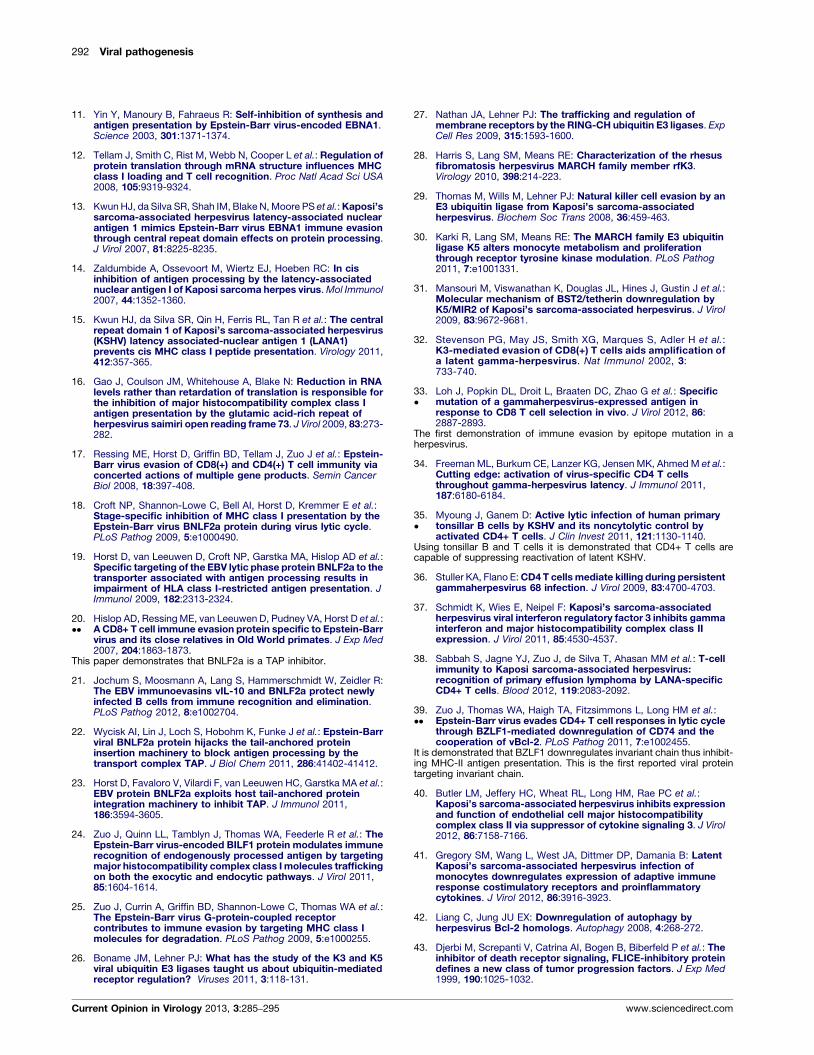
and lytic phase [42] (Figure 2). Death receptor-mediated
Figure 2
pro-caspase 8vFLIP
DeathReceptor
Fas/TNFα
EffectorCaspases, 3, 7, 9 Bid
cyto
Nucleus
CAML
vMAP
K7
Ca 2+
vBcl
APP
BaxBAK
B
Kcyto
ucleus
CAML Ca 2+
s
AML
K7 VDVcc
Inhibition of the classical apoptosis and autophagy pathway by g-herpesviru
caspase 3 for inhibition, whereas vBcl2 and MHV-68 vMAP antagonize the o
Additionally, KSHV K7 activates CAML in releasing calcium from the ER to
vBcl2 suppresses UVRAG in autophagosome formation. Viral proteins are c
www.sciencedirect.com
apoptosis (triggered by CD95 and TNFa) delivered by
cytotoxic T cells is counteracted by vFLIP via interfering
with the assembly of DISC. Counterintuitively, vFLIP
potently activates NFkB, which effectively enables the
survival of KSHV-infected lymphoma cells [43–47]. Stu-
dies using cultured cells and transgenic mouse models
demonstrated that vFLIP activated the IKK complex and
was implicated in KSHV-associated lymphoproliferative
diseases [47–51]. Surprisingly, vFLIP of herpesvirus sai-
miri was shown to be dispensable for viral replication,
transformation, and pathogenesis in a primate model [52].
It would be interesting to examine the roles of vFLIP in
the infection and pathogenesis of RRV using nonhuman
primate models.
During productive lytic infection, processes of viral
replication frequently induce stress responses that per-
turb cellular homeostasis. Cell death or survival is dic-
tated by opposing activities between pro-apoptotic and
anti-apoptotic factors, known as Bcl-2 homology
proteins, which reside within the outer membrane of
mitochondria (Figure 2). All g-herpesviruses express
cyto. c
. c
2
Autophag-osome
A
UVRAG
Beclin 1
Auto-lysosome
lysosome
AC
G
e
y
Current Opinion in Virology
s proteins. KSHV vFLIP and K7 target pro-caspase 8 and activated
ligomerization of Bax/Bak and VDAC that release cytochrome c (cyto. c).
attenuate stress-induced apoptosis. Of the classic autophagy pathway,
olored in green.
Current Opinion in Virology 2013, 3:285–295

288 Viral pathogenesis
anti-apoptotic Bcl-2 homologs during various stages of
their infection cycle [53]. Viral Bcl-2 has lost the inhibi-
tory loop that, when cleaved by an activated caspase,
converts cellular antiapoptotic Bcl-2 into a proapoptotic
product via removing the N-terminal BH4 domain
[54,55]. Thus, viral Bcl-2 proteins either are not cleaved
or, even if cleaved by caspase, do not behave as apoptosis-
inducers. Our understanding of the in vivo functions of
the antiapoptotic activity of vBcl-2 is largely derived
from studies using MHV68. Recombinant MHV68
expressing a vBcl-2 mutant that fails to suppress apop-
tosis establishes normal latent infection in splenocytes,
but is crippled to reactivate ex vivo [56�]. Thus, viral
antagonism of apoptosis is crucial to promote cell survival
during early stages of lytic infection. Similarly, vBcl-2
expression immediately after infection by EBV promotes
cell survival by enabling latent infection and transform-
ation of lymphoma cells ex vivo [57�]. The viral mito-
chondrial antiapoptotic protein (vMAP) of MHV68
displays a two-pronged mechanism to counteract intrin-
sic apoptosis [58]. vMAP binds to Bcl-2 to enhance its
recruitment to mitochondria and subsequent inhibition
of the BH3-only molecules, and impedes cytochrome c
release through physical association with VDAC, effec-
tively negating the mitochondrion apoptotic pathway.
Viral infection with recombinant MHV68 demonstrated
that this antiapoptotic activity was important for the lytic
phase of infection.
It is also necessary to inactivate p53 during active viral
replication and the growing list of such proteins includes
the KSHV latent nuclear antigen (LANA), the EBV
nuclear antigen 3C (EBNA3C), and the KSHV K7 and
vIRF3. KSHV LANA and EBV EBNA3C are major latent
gene products and both inactivate p53-mediated apopto-
sis. While KSHV LANA induces p53 degradation [59],
EBNA3C stabilizes a cellular factor that prevents p53 to
access DNA and sequestrates a co-activator key for p53-
mediated transcription [60,61]. The KSHV K7 protein is a
lytic gene product that antagonizes apoptosis by multiple
mechanisms (Figure 2). Anchored in the membrane, K7
interfaces with the mitochondrion-dependent apoptotic
pathway, the ER stress responses, and the proteasome/
ubiqutin system [62–64]. K7 expression protects cells
from apoptosis induced by intrinsic stresses, including
DNA damage and ER stress. Similar to LANA, vIRF4
targets p53 for degradation via two distinct mechanisms
governing ubiquitination [65]. These findings highlight
the importance of inhibiting intrinsic apoptosis triggered
by viral replication events, danger signals within the
infected cells.
Evasion of autophagy
Autophagy is a homeostatic process that engulfs and,
upon fusion with lysosomes, digests bulk cytoplasm
under nutrient-deprived conditions. Recent studies
indicate that host cells deploy autophagy to defeat
Current Opinion in Virology 2013, 3:285–295
pathogen infection. vBcl-2 and vFLIP can inhibit autop-
hagy, implying an inherent crosstalk between autophagy
and apoptosis. vBcl-2 targets Beclin 1, a key regulator of
autophagy, to negate autophagy [66]. Subsequently, vBcl-
2 was found to target UVRAG, an integral component of
the Beclin-1-PI3K complex, which arrests the PI3K com-
plex and suppresses autophagy [67]. The anti-autophagic
activity of viral Bcl-2 is conserved within g-2-herpes-
viruses (Figure 2). Using recombinant MHV68 carrying
mutations that differentially ablate the anti-autophagic or
anti-apoptotic activity of vBcl-2, Xiaofei et al. showed that
inhibition of autophagy by MHV68 Bcl-2 was essential for
the establishment of latent infection in splenocytes [56].
Viral and cellular Bcl-2 proteins show a difference in
affinity for Beclin 1. Structural studies illustrated that
this difference likely stems from the specific sequence of
the hydrophobic groove of Bcl-2 that directly contacts the
BH3 of Beclin 1 [68,69]. Recently, the KSHV vFLIP
protein was discovered to bind to the LC3-processing
enzyme, ATG3 [70]. As such, vFLIP and cellular FLIP
molecules potently inhibit LC3 processing and autopha-
gosome membrane elongation. Interestingly, vCyclin,
another latent protein, induces autophagy and senes-
cence, both of which are diminished by vFLIP [71].
These data suggest a complex stimulation and inhibition
of autophagy during the lytic phase of g-herpesviruses.
Evasion of interferon production
Type 1 interferon (IFN) and IFN-stimulated genes
(ISGs) represent the first line of defense against virus
infection. g-Herpesviruses interfere with the IFN path-
way at several levels as reviewed recently [72,73] (Table
1). Virus-infected cells secrete IFN upon recognition of
specific viral signatures by pattern-recognition receptors
(PRR). These PRRs include the Toll-like receptors
(TLRs), nucleotide-binding and oligomerization, leu-
cine-rich repeat (NLR) proteins, retinoic acid inducible
gene (RIG)-I-like receptors (RLRs) and C-type lectin
receptors (CLRs). Via adaptor proteins, these innate
sensors in turn activate a family of transcription factors,
the IFN regulatory factors (IRFs), which translocate to
the nucleus to initiate transcription of IFN a/b genes.
Secreted IFN binds to the type 1 IFN receptor (IFNAR)
to trigger a signaling cascade that culminates in the
expression of ISGs. g-Herpesviruses antagonize both
the expression and function of different components of
the IRF pathway.
Interferon regulatory factors
KSHV encodes four homologs of cellular IRFs, vIRF1
through 4, which interfere with the transactivating
activity of specific IRFs. vIRF-1 binds to host IRF-1 or
the coactivator protein p300, the latter action inhibiting
formation of the transcriptionally active IRF3-CBP/300
complex [74]. vIRF-2 interferes with the transactivational
activity of IRF-1 and IRF-3 [75,76] by direct binding to
IRF1 and degradation of IRF3 via a caspase 3-dependent
www.sciencedirect.com

Immune response mechanisms by g-herpesviruses Feng, Moses and Fruh 289
Table 1
Antagonism of IFN signaling by human g-herpesvirsues
Virus ORF Gene product Functional mechanism(s)
KSHV
K9 vIRF-1 Suppresses IRF-1 and IRF-3 transactivational activity; inhibits IRF3 nuclear translocation;
inhibits TLR3-driven IFN-b promoter activation and IFNb production; downregulates TLR4
K11.1/K11 vIRF-2 Suppresses IRF-1 and IRF-3 transactivational activity; inhibits TLR3-driven IFN-b promoter
activation and IFNb production; inhibits PKR function
K10.5/K10.6 vIRF-3 Inhibits IRF7 DNA binding and function; Inhibits IRF5-mediated promoter activation
ORF45 ORF45 Prevents phosphorylation and nuclear translocation of IRF7
ORF50 RTA Promotes IRF7 ubiquitination and degradation; degrades the TLR3/4 adaptor TRIF
ORF63 Disrupts formation and activity of the NLRP1 inflammasome
ORF64 Suppresses RIG-I-mediated signaling
ORF73 LANA1 Prevents IRF3-mediated induction of IFN-b during latency
ORF74 vGPCR Downregulates TLR4
ORFK8 K-bZIP Prevents IRF3-mediated induction of IFN-b during virus reactivation
miR-K9 Downregulates MyD88 and IRAK1
EBV
BGLF4 PK Binds and suppresses IRF3 transactivational activity; inhibits of IFN-b production
BGLF5 DNase Degrades TLR9 transcript
BZLF1 Zta Binds and suppresses IRF7 transactivational activity
BRLF1 Rta Inhibits IRF3 and IRF7 transcription; inhibits IFN-b production during lytic infection.
LF2 LF2 Prevents IRF7 dimerization and IFN-a production
LMP1 LMP1 Negatively regulates IFN response via induction of miR-146a; downregulates TLR9
LMP2A/2B LMP2A/2B Enhances turnover of type I/II IFN receptors
mechanism [75,77]. vIRF-2 also interacts with protein
kinase R (PKR), inhibiting its autoactivation and antiviral
function [78]. IRF7, together with IRF3, constitute the
master regulators of type 1 IFN production, and KSHV
vIRF3 antagonizes IRF 5 [79] and IRF7 [80] via direct
inhibitory interactions. vIRF-4 does not appear to
antagonize IFN-mediated signaling; instead, vIRF4
enhances the degradation of p53 (which is also targeted
by vIRF1 and 3) via a stabilizing interaction with the E3
ubiquitin ligase MDM2 [70,81]. vIRF4 also interacts with
CSL/CBF1, potentially antagonizing Notch/CBF1 signal
transduction, and cooperates with RTA to facilitate
KSHV reactivation [82,83]. Despite similar functional
activities, the vIRFs display differential expression pat-
terns, indicating that they may act at different times, and/
or in different cells [81]. Interestingly, a recent study
identified mechanistic differences between the ability of
the KSHV vIRFs to inhibit TLR3-dependent IFN sig-
naling [84].
KSHV encodes additional gene products to antagonize
host IRFs while EBV and MHV68 target host IRFs with
non-homologous viral proteins. This common strategy
underscores the importance of the IRF family in antiviral
defense. Two KSHV proteins, LANA1 (ORF73) and K-
bZIP (K8), bind competitively to the IFN-b promoter,
preventing IFN-b induction during latency and virus
reactivation [85,86]. The conserved viral kinase ORF36
inhibits IFN production. MHV68 ORF36 binds to
nuclear IRF-3 to prevent activation of the IFN-b pro-
moter [87]. Importantly, the growth and spread of an
ORF36-null MHV68 in immunocompetent mice was
attenuated, exemplifying the need to antagonize the
IFN pathway for successful infection. The EBV
www.sciencedirect.com
ORF36 homolog, BGLF4, also binds to IRF3 to suppress
IRF3-mediated signaling [88]. KSHV ORF45, a con-
served tegument and IE protein, binds to the inhibitory
domain of IRF7 to prevent its phosphorylation and
nuclear translocation by acting as an alternate substrate
for the virus-activated kinases IKKe and TBK1 [89–91].
As a virion component, ORF45 could inhibit IFN pro-
duction at the earliest stages of de novo infection, as
suggested by studies with an ORF45-null recombinant
KSHV [92]. MHV68 ORF45 is also essential for an early
stage of viral replication, suggesting a similar role in
evasion of the early IFN response [93]. KSHV RTA
(ORF50) also targets IRF7, promoting its ubiquitination
and degradation [94].
Like KSHV, RRV expresses a set of viral IRFs [95]. As
such, the RRV model provides insight into the role of the
vIRFs in regulating the host response to infection.
Accordingly, a vIRF deletion clone of RRV that inhibits
IFN release from PMC in vitro exhibits attenuated repli-
cation in vivo and induces a more robust anti-inflamma-
tory and anti-RRV T cell response [96,97]. Macaques
infected with the vIRF knockout also display diminished
B cell hyperplasia, a pathology characteristic of acute
RRV infection.
While EBV does not express IRF homologs, it encodes
several proteins that target host IRFs. The EBV IE
transactivator protein BZLF1 (Zta) binds to IRF7 and
inhibits its transactivational activity [98], while BRLF1
(Rta) inhibits transcription of IRF3 and IRF7, thus inhi-
biting IFN-b production during lytic infection [99]. In
addition, binding of the tegument protein LF2 to IRF7
prevents IRF7 dimerization and IFN-a production [100].
Current Opinion in Virology 2013, 3:285–295

290 Viral pathogenesis
Interestingly, a positive regulatory circuit exists between
IRF7 and the EBV oncoprotein LMP1 [101]. LMP1
possesses potent immune escape functions, including
induction of immunomodulatory cytokines, receptors
and exosomes [102] and induction of miR-146a, a cellular
miRNA that inhibits IFN response pathways [103,104],
and inhibition of TLR9 expression [105,106] (discussed
below). LMP2A and 2B enhance IFN receptor turnover
in epithelial cells, inciting a broad effect on ISG expres-
sion [107]. These LMP functions likely contribute to the
immune escape of EBV-positive tumors.
Recent work with MHV68 illustrates a novel mechanism
of immunomodulation whereby a host IRF is co-opted to
establish viral latency [108]. Specifically, transcription of
the M2 protein, which plays a critical role in virus reacti-
vation, is repressed by IRF2, which is in turn upregulated
by the IFN produced in response to initial virus infection.
The authors speculate that this IRF-sensing mechanism
allows MHV68 to time virus reactivation to periods of
localized immune quiescence. It is likely that other
examples of ‘cooperative subversion’ between g-herpes-
viruses and host immune effectors will be identified.
Pattern recognition receptors and adaptor proteins
KSHV modulates TLR signaling by influencing the
expression of TLRs or their adaptor molecules. Infection
of monocytes leads to an upregulation of TLR3 and
downstream innate immune effectors (CXCL10, IRF-1,
CCL2, and IFN-b), an event that may facilitate establish-
ment of latency [109]. In contrast, infection of endothelial
cells results in downregulation of TLR4 mediated by the
lytic proteins vIRF1 and ORF74 [110]. Interestingly,
stimulation of PEL cells with agonists to TLR7 and 8
reactivates virus, suggesting a viral strategy to escape from
cells targeted for immune attack [111]. Recent studies
indicate that KSHV targets other PRRs in addition to
TLRs. Inn et al. show that the tegument protein, ORF64,
specifically targets and suppresses RIG-I-mediated sig-
naling via its deubiquitinase activity [112]. Gregory et al.report that another tegument protein, ORF63, is a func-
tional homolog of NLRP1 that disrupts the formation and
activity of the NLRP1 inflammasome [113��]. While
NLRP1 is a cytoplasmic inflammasome, a study by Ker-
uer et al. shows that, during KSHV infection of endo-
thelial cells, IFN-g-inducible protein 16 (IFI16) interacts
with a caspase-1 activating complex to form a functional
nuclear inflammasome, suggesting that KSHV may
manipulate this pathway to promote latency after nuclear
delivery of the viral genome [114,115]. These recent
studies indicate a broader role for PRRs in sensing g-
herpesvirus infection and suggest that additional viral
antagonists and strategies remain to be identified.
Similar to KSHV, MHV68 can be reactivated from
latently-infected B cells with ligands for TLR3, 4, 5
and 9, and reactivation in vivo is accomplished by
Current Opinion in Virology 2013, 3:285–295
administration of LPS (TLR4) or CpG DNA (TLR9)
[116]. Interestingly, LPS/CpG-induced reactivation led
to an increase in the number of latently-infected spleno-
cytes, suggesting that TLR sensitivity contributes to
homeostatic maintenance of chronic infection. This
phenomenon could also underlie the sensitivity of KSHV
to TLR7/8 agonists. Alternatively, innate immune sig-
naling pathways are hijacked by MHV68 to enable lytic
replication that replenishes the latent pool. Indeed, it was
shown that MHV68 exploits the MAVS-IKKbeta pathway
to promote viral transcriptional activation and disable
antiviral cytokine production [117–119]. It remains
unclear how the MAVS-IKKbeta pathway is activated
during MHV68 infection.
TLR signaling is mediated through one of two adapter
proteins, myeloid differentiation primary-response
protein 88 (MyD88) or Toll-interleukin-1 receptor
(TIR) domain-containing adaptor-inducing b-interferon
protein (TRIF) [120]. KSHV targets both adaptors.
TRIF, an adaptor for TLR 3 and 4, is degraded in the
presence of RTA, via either its E3 ligase activity or an
unidentified mediator [121]. MyD88, which transmits
signals from TLRs 7, 8 and 9, is downregulated by the
viral microRNA miR-K9, which also targets a critical
kinase, interleukin-1 receptor-associated kinase 1
(IRAK1), in the same pathway [122�]. These data indicate
that KSHV may block TLR7/9-induced IFN-a pro-
duction via miRNA targeting.
In B cells, EBV infection exerts opposite effects on
expression of TLR7 and TLR9. In naıve B cells, exposure
to EBV downregulates TLR9 while inducing expression
of TLR7 and key downstream adaptor (MyD88) and
effector (IRF5) molecules, a scenario that promotes the
initial phase of B cell proliferation; IRF5 activity is
subsequently negatively regulated to allow for establish-
ment of latency [123]. EBV proteins responsible for
TLR9 downregulation have been identified: LMP1
downregulates TLR9 through NF-kB-dependent inhi-
bition of TLR9 transcription [105], while BGLF5
degrades TLR9 mRNA [106]. EBV-infected B cells are
also unresponsive to TLR7/8 and 9 agonists [124]. In
plasmacytoid dendritic cells (pDC), EBV infection
increases IFNa production by activating the TLR9 sig-
naling pathway, but the simultaneous production of IL-10
from the pDC serves to blunt the net antiviral effect
[125].
Evasion of inflammatory signalingInflammatory chemokines and cytokines produced in
response to viral infection play an important role in the
outcome of the immune response. Accordingly, the g-
herpesviruses have developed different strategies to imi-
tate or neutralize these inflammatory mediators, in-
cluding production of homologs, receptors and binding
proteins.
www.sciencedirect.com

Immune response mechanisms by g-herpesviruses Feng, Moses and Fruh 291
Virus-encoded chemokines
KSHV encodes three viral chemokines with agonist or
antagonist function against host chemokine receptors;
vCCL1 (ORF K6), vCCL2 (ORF K4), vCCL3 (ORF
K4.1). This family possess agonist function against
CCR8 (vCCL1 and 2), CCR3 (vCCL2) and CCR4
(vCCL3), indicating a role in chemoattraction of Th2
T cells, which typically downmodulate immune
responses [126]. Interestingly, vCCL2 also has broad-
spectrum antagonist activity, binding promiscuously to
several CC and CXC chemokine receptors and inhibiting
the chemotactic responses of monocytes and Th1 T cells
[126]. In combination with Th2 T cell recruitment, this
would provide an effective means of blunting an inflam-
matory cytotoxic antiviral response.
As an alternate strategy for chemokine modulation, the
EBV microRNA BHRF1, which is expressed in several
EBV+ lymphomas, suppresses expression of CXCL11/I-
TAC, an IFN-inducible T cell chemoattractant [127].
Other inflammatory modulators (vCD200 and vIL-6)
KSHV encodes a homolog of CD200, a molecule that
negatively regulates myeloid-lineage cells [128], and a
homolog of IL-6, a multifunctional inflammatory cyto-
kine [129]. KSHV vCD200 is encoded by ORF K14 and
binds with high affinity to the host receptor CD200R with
immunosuppressive consequences [130]. KSHV vIL-6 is
encoded by ORF K6 and, unlike human IL-6, can bind
and signal exclusively through gp130 without the need for
CD126 (gp80) [131,132]. In the presence of gp80 how-
ever, vIL-6 signaling is qualitatively different from that
induced by human IL-6 [133]. These properties have
relevance for immune evasion as well as oncogenesis,
since IFN-modulation of the IL-6R complex would dif-
ferentially impact virus and host IL-6 activity.
Like KSHV, RRV encodes a CD200 homolog, R15, which
inhibits macrophage production of TNF [134] and a vIL-
6, which signals through gp130 and is expressed in RRV-
infected rhesus macaques [135,136].
The IL-10 homolog encoded by the EBV BCRF1 gene
encodes a functional homolog of IL-10 that exhibits
diverse immunosuppressive properties, including inhi-
bition of T cell function, macrophage activation and
synthesis of IRN-g [137–140]. BCRF1 is functionally
expressed during the earliest phase of de novo infection
of primary B cells, a consequence of translation of virion-
delivered mRNA, or transduced viral RNA (tvRNA) [21].
Additional immunomodulatory EBV tvRNAs were
recently identified, including those encoding BGLF5,
BNLF2a and LMP1; immediate translation of tvRNAs
likely contributes to immune evasion in a crucial period
before de novo gene expression [21]. EBV BARF1 could
also contribute to immunomodulation via binding and
www.sciencedirect.com
neutralization of the pleiotropic cytokine colony-stimu-
lating factor-1 (CSF-1) [141].
The MHV68 M3 protein is a secreted viral protein that
binds selected CC and CXC chemokines with antiviral
activity [142]. While an M3-deficient virus is not impaired
in its capacity to establish latency in experimental hosts
(C57BL/6 or BALB/c mice), when tested in a natural host
(wood mice) it did attenuate infection and modulate the
host inflammatory response in a manner consistent with
its chemokine-binding properties [143,144�], indicating
that specific immunoevasins may only function effec-
tively in the appropriate host.
References and recommended readingPapers of particular interest, published within the period of review,have been highlighted as:
� of special interest�� of outstanding interest
1.�
Freeman ML, Lanzer KG, Cookenham T, Peters B, Sidney J et al.:Two kinetic patterns of epitope-specific CD8 T-cell responsesfollowing murine gammaherpesvirus 68 infection. J Virol 2010,84:2881-2892.
The authors identified a range of novel H-2(b) restricted epitopes andshowed that the T cell response to these epitopes falls into two kineticclasses based on the speed of decline. The slowly declining responsecorresponded to the latent phase since decline was accelerated whenmice were infected with a virus unable to establish latency. This suggeststhat T cell responses mirror the viral life cycle.
2. Gredmark-Russ S, Cheung EJ, Isaacson MK, Ploegh HL,Grotenbreg GM: The CD8 T-cell response against murinegammaherpesvirus 68 is directed toward a broad repertoire ofepitopes from both early and late antigens. J Virol 2008,82:12205-12212.
3. Marques S, Alenquer M, Stevenson PG, Simas JP: A single CD8+T cell epitope sets the long-term latent load of a muridherpesvirus. PLoS Pathog 2008, 4:e1000177.
4. Bennett NJ, May JS, Stevenson PG: Gamma-herpesviruslatency requires T cell evasion during episome maintenance.PLoS Biol 2005, 3:e120.
5. Blake N: Immune evasion by gammaherpesvirus genomemaintenance proteins. J Gen Virol 2010, 91:829-846.
6. Vider-Shalit T, Fishbain V, Raffaeli S, Louzoun Y: Phase-dependent immune evasion of herpesviruses. J Virol 2007,81:9536-9545.
7. Levitskaya J, Sharipo A, Leonchiks A, Ciechanover A, MasucciMG: Inhibition of ubiquitin/proteasome-dependent proteindegradation by the Gly-Ala repeat domain of the Epstein-Barrvirus nuclear antigen 1. Proc Natl Acad Sci USA 1997, 94:12616-12621.
8. Daskalogianni C, Apcher S, Candeias MM, Naski N, Calvo F et al.:Gly-Ala repeats induce position- and substrate-specificregulation of 26 S proteasome-dependent partial processing.J Biol Chem 2008, 283:30090-30100.
9. Apcher S, Komarova A, Daskalogianni C, Yin Y, Malbert-Colas Let al.: mRNA translation regulation by the Gly-Ala repeat ofEpstein-Barr virus nuclear antigen 1. J Virol 2009, 83:1289-1298.
10.�
Apcher S, Daskalogianni C, Manoury B, Fahraeus R: Epstein Barrvirus-encoded EBNA1 interference with MHC class I antigenpresentation reveals a close correlation between mRNAtranslation initiation and antigen presentation. PLoS Pathog2010, 6:e1001151.
It is shown that suppression of mRNA translation explains the inhibition ofantigen presentaion by Gly-Ala repeats in cis. These results stronglysupport the notion that MHC-I derives most of its peptides from nascentpolypeptide chains.
Current Opinion in Virology 2013, 3:285–295
292 Viral pathogenesis
11. Yin Y, Manoury B, Fahraeus R: Self-inhibition of synthesis andantigen presentation by Epstein-Barr virus-encoded EBNA1.Science 2003, 301:1371-1374.
12. Tellam J, Smith C, Rist M, Webb N, Cooper L et al.: Regulation ofprotein translation through mRNA structure influences MHCclass I loading and T cell recognition. Proc Natl Acad Sci USA2008, 105:9319-9324.
13. Kwun HJ, da Silva SR, Shah IM, Blake N, Moore PS et al.: Kaposi’ssarcoma-associated herpesvirus latency-associated nuclearantigen 1 mimics Epstein-Barr virus EBNA1 immune evasionthrough central repeat domain effects on protein processing.J Virol 2007, 81:8225-8235.
14. Zaldumbide A, Ossevoort M, Wiertz EJ, Hoeben RC: In cisinhibition of antigen processing by the latency-associatednuclear antigen I of Kaposi sarcoma herpes virus. Mol Immunol2007, 44:1352-1360.
15. Kwun HJ, da Silva SR, Qin H, Ferris RL, Tan R et al.: The centralrepeat domain 1 of Kaposi’s sarcoma-associated herpesvirus(KSHV) latency associated-nuclear antigen 1 (LANA1)prevents cis MHC class I peptide presentation. Virology 2011,412:357-365.
16. Gao J, Coulson JM, Whitehouse A, Blake N: Reduction in RNAlevels rather than retardation of translation is responsible forthe inhibition of major histocompatibility complex class Iantigen presentation by the glutamic acid-rich repeat ofherpesvirus saimiri open reading frame 73. J Virol 2009, 83:273-282.
17. Ressing ME, Horst D, Griffin BD, Tellam J, Zuo J et al.: Epstein-Barr virus evasion of CD8(+) and CD4(+) T cell immunity viaconcerted actions of multiple gene products. Semin CancerBiol 2008, 18:397-408.
18. Croft NP, Shannon-Lowe C, Bell AI, Horst D, Kremmer E et al.:Stage-specific inhibition of MHC class I presentation by theEpstein-Barr virus BNLF2a protein during virus lytic cycle.PLoS Pathog 2009, 5:e1000490.
19. Horst D, van Leeuwen D, Croft NP, Garstka MA, Hislop AD et al.:Specific targeting of the EBV lytic phase protein BNLF2a to thetransporter associated with antigen processing results inimpairment of HLA class I-restricted antigen presentation. JImmunol 2009, 182:2313-2324.
20.��
Hislop AD, Ressing ME, van Leeuwen D, Pudney VA, Horst D et al.:A CD8+ T cell immune evasion protein specific to Epstein-Barrvirus and its close relatives in Old World primates. J Exp Med2007, 204:1863-1873.
This paper demonstrates that BNLF2a is a TAP inhibitor.
21. Jochum S, Moosmann A, Lang S, Hammerschmidt W, Zeidler R:The EBV immunoevasins vIL-10 and BNLF2a protect newlyinfected B cells from immune recognition and elimination.PLoS Pathog 2012, 8:e1002704.
22. Wycisk AI, Lin J, Loch S, Hobohm K, Funke J et al.: Epstein-Barrviral BNLF2a protein hijacks the tail-anchored proteininsertion machinery to block antigen processing by thetransport complex TAP. J Biol Chem 2011, 286:41402-41412.
23. Horst D, Favaloro V, Vilardi F, van Leeuwen HC, Garstka MA et al.:EBV protein BNLF2a exploits host tail-anchored proteinintegration machinery to inhibit TAP. J Immunol 2011,186:3594-3605.
24. Zuo J, Quinn LL, Tamblyn J, Thomas WA, Feederle R et al.: TheEpstein-Barr virus-encoded BILF1 protein modulates immunerecognition of endogenously processed antigen by targetingmajor histocompatibility complex class I molecules traffickingon both the exocytic and endocytic pathways. J Virol 2011,85:1604-1614.
25. Zuo J, Currin A, Griffin BD, Shannon-Lowe C, Thomas WA et al.:The Epstein-Barr virus G-protein-coupled receptorcontributes to immune evasion by targeting MHC class Imolecules for degradation. PLoS Pathog 2009, 5:e1000255.
26. Boname JM, Lehner PJ: What has the study of the K3 and K5viral ubiquitin E3 ligases taught us about ubiquitin-mediatedreceptor regulation? Viruses 2011, 3:118-131.
Current Opinion in Virology 2013, 3:285–295
27. Nathan JA, Lehner PJ: The trafficking and regulation ofmembrane receptors by the RING-CH ubiquitin E3 ligases. ExpCell Res 2009, 315:1593-1600.
28. Harris S, Lang SM, Means RE: Characterization of the rhesusfibromatosis herpesvirus MARCH family member rfK3.Virology 2010, 398:214-223.
29. Thomas M, Wills M, Lehner PJ: Natural killer cell evasion by anE3 ubiquitin ligase from Kaposi’s sarcoma-associatedherpesvirus. Biochem Soc Trans 2008, 36:459-463.
30. Karki R, Lang SM, Means RE: The MARCH family E3 ubiquitinligase K5 alters monocyte metabolism and proliferationthrough receptor tyrosine kinase modulation. PLoS Pathog2011, 7:e1001331.
31. Mansouri M, Viswanathan K, Douglas JL, Hines J, Gustin J et al.:Molecular mechanism of BST2/tetherin downregulation byK5/MIR2 of Kaposi’s sarcoma-associated herpesvirus. J Virol2009, 83:9672-9681.
32. Stevenson PG, May JS, Smith XG, Marques S, Adler H et al.:K3-mediated evasion of CD8(+) T cells aids amplification ofa latent gamma-herpesvirus. Nat Immunol 2002, 3:733-740.
33.�
Loh J, Popkin DL, Droit L, Braaten DC, Zhao G et al.: Specificmutation of a gammaherpesvirus-expressed antigen inresponse to CD8 T cell selection in vivo. J Virol 2012, 86:2887-2893.
The first demonstration of immune evasion by epitope mutation in aherpesvirus.
34. Freeman ML, Burkum CE, Lanzer KG, Jensen MK, Ahmed M et al.:Cutting edge: activation of virus-specific CD4 T cellsthroughout gamma-herpesvirus latency. J Immunol 2011,187:6180-6184.
35.�
Myoung J, Ganem D: Active lytic infection of human primarytonsillar B cells by KSHV and its noncytolytic control byactivated CD4+ T cells. J Clin Invest 2011, 121:1130-1140.
Using tonsillar B and T cells it is demonstrated that CD4+ T cells arecapable of suppressing reactivation of latent KSHV.
36. Stuller KA, Flano E: CD4 T cells mediate killing during persistentgammaherpesvirus 68 infection. J Virol 2009, 83:4700-4703.
37. Schmidt K, Wies E, Neipel F: Kaposi’s sarcoma-associatedherpesvirus viral interferon regulatory factor 3 inhibits gammainterferon and major histocompatibility complex class IIexpression. J Virol 2011, 85:4530-4537.
38. Sabbah S, Jagne YJ, Zuo J, de Silva T, Ahasan MM et al.: T-cellimmunity to Kaposi sarcoma-associated herpesvirus:recognition of primary effusion lymphoma by LANA-specificCD4+ T cells. Blood 2012, 119:2083-2092.
39.��
Zuo J, Thomas WA, Haigh TA, Fitzsimmons L, Long HM et al.:Epstein-Barr virus evades CD4+ T cell responses in lytic cyclethrough BZLF1-mediated downregulation of CD74 and thecooperation of vBcl-2. PLoS Pathog 2011, 7:e1002455.
It is demonstrated that BZLF1 downregulates invariant chain thus inhibit-ing MHC-II antigen presentation. This is the first reported viral proteintargeting invariant chain.
40. Butler LM, Jeffery HC, Wheat RL, Long HM, Rae PC et al.:Kaposi’s sarcoma-associated herpesvirus inhibits expressionand function of endothelial cell major histocompatibilitycomplex class II via suppressor of cytokine signaling 3. J Virol2012, 86:7158-7166.
41. Gregory SM, Wang L, West JA, Dittmer DP, Damania B: LatentKaposi’s sarcoma-associated herpesvirus infection ofmonocytes downregulates expression of adaptive immuneresponse costimulatory receptors and proinflammatorycytokines. J Virol 2012, 86:3916-3923.
42. Liang C, Jung JU EX: Downregulation of autophagy byherpesvirus Bcl-2 homologs. Autophagy 2008, 4:268-272.
43. Djerbi M, Screpanti V, Catrina AI, Bogen B, Biberfeld P et al.: Theinhibitor of death receptor signaling, FLICE-inhibitory proteindefines a new class of tumor progression factors. J Exp Med1999, 190:1025-1032.
www.sciencedirect.com

Immune response mechanisms by g-herpesviruses Feng, Moses and Fruh 293
44. Chaudhary PM, Jasmin A, Eby MT, Hood L: Modulation of theNF-kappa B pathway by virally encoded death effectordomains-containing proteins. Oncogene 1999, 18:5738-5746.
45. Guasparri I, Keller SA, Cesarman E: KSHV vFLIP is essential forthe survival of infected lymphoma cells. J Exp Med 2004,199:993-1003.
46. Keller SA, Schattner EJ, Cesarman E: Inhibition of NF-kappaBinduces apoptosis of KSHV-infected primary effusionlymphoma cells. Blood 2000, 96:2537-2542.
47. Matta H, Chaudhary PM: Activation of alternative NF-kappa Bpathway by human herpes virus 8-encoded Fas-associateddeath domain-like IL-1 beta-converting enzyme inhibitoryprotein (vFLIP). Proc Natl Acad Sci USA 2004, 101:9399-9404.
48. Chugh P, Matta H, Schamus S, Zachariah S, Kumar A et al.:Constitutive NF-kappaB activation, normal Fas-inducedapoptosis, and increased incidence of lymphoma in humanherpes virus 8 K13 transgenic mice. Proc Natl Acad Sci USA2005, 102:12885-12890.
49. Sun Q, Matta H, Chaudhary PM: The human herpes virus 8-encoded viral FLICE inhibitory protein protects against growthfactor withdrawal-induced apoptosis via NF-kappa Bactivation. Blood 2003, 101:1956-1961.
50. Ballon G, Chen K, Perez R, Tam W, Cesarman E: Kaposi sarcomaherpesvirus (KSHV) vFLIP oncoprotein induces B celltransdifferentiation and tumorigenesis in mice. J Clin Invest2011, 121:1141-1153.
51. Grossmann C, Podgrabinska S, Skobe M, Ganem D: Activation ofNF-kappaB by the latent vFLIP gene of Kaposi’s sarcoma-associated herpesvirus is required for the spindle shape ofvirus-infected endothelial cells and contributes to theirproinflammatory phenotype. J Virol 2006, 80:7179-7185.
52. Glykofrydes D, Niphuis H, Kuhn EM, Rosenwirth B, Heeney JLet al.: Herpesvirus saimiri vFLIP provides an antiapoptoticfunction but is not essential for viral replication,transformation, or pathogenicity. J Virol 2000, 74:11919-11927.
53. Hardwick JM, Bellows DS: Viral versus cellular BCL-2 proteins.Cell Death Differ 2003, 10(suppl. 1):S68-S76.
54. Cheng EH, Nicholas J, Bellows DS, Hayward GS, Guo HG et al.: ABcl-2 homolog encoded by Kaposi sarcoma-associated virus,human herpesvirus 8, inhibits apoptosis but does notheterodimerize with Bax or Bak. Proc Natl Acad Sci USA 1997,94:690-694.
55. Bellows DS, Chau BN, Lee P, Lazebnik Y, Burns WH et al.:Antiapoptotic herpesvirus Bcl-2 homologs escape caspase-mediated conversion to proapoptotic proteins. J Virol 2000,74:5024-5031.
56.�
Hwang EX, Oh SS, Lee JS, Jeong JH et al.: Viral Bcl-2-mediatedevasion of autophagy aids chronic infection ofgammaherpesvirus 68. PLoS Pathog 2009, 5:e1000609.
This work provides in vivo evidence that viral Bcl-2 is important to evadeautophagy during latent infection of gamma herpesvirus 68.
57.�
Altmann M, Hammerschmidt W: Epstein-Barr virus provides anew paradigm: a requirement for the immediate inhibition ofapoptosis. PLoS Biol 2005, 3:e404.
This study shows that inhibition of apoptosis is an integral component ofthe transformation triggered by human gamma herpesviruses in B lym-phocytes.
58. Feng P, Liang C, Shin YC, Xiaofei E, Zhang W et al.: A novelinhibitory mechanism of mitochondrion-dependent apoptosisby a herpesviral protein. PLoS Pathog 2007, 3:e174.
59. Friborg J Jr, Kong W, Hottiger MO, Nabel GJ: p53 inhibition bythe LANA protein of KSHV protects against cell death. Nature1999, 402:889-894.
60. Cai Q, Guo Y, Xiao B, Banerjee S, Saha A et al.: Epstein-Barr virusnuclear antigen 3C stabilizes Gemin3 to block p53-mediatedapoptosis. PLoS Pathog 2011, 7:e1002418.
61. Saha A, Bamidele A, Murakami M, Robertson ES: EBNA3Cattenuates the function of p53 through interaction with
www.sciencedirect.com
inhibitor of growth family proteins 4 and 5. J Virol 2011,85:2079-2088.
62. Wang HW, Sharp TV, Koumi A, Koentges G, Boshoff C:Characterization of an anti-apoptotic glycoprotein encodedby Kaposi’s sarcoma-associated herpesvirus whichresembles a spliced variant of human survivin. EMBO J 2002,21:2602-2615.
63. Feng P, Park J, Lee BS, Lee SH, Bram RJ et al.: Kaposi’ssarcoma-associated herpesvirus mitochondrial K7 proteintargets a cellular calcium-modulating cyclophilin ligand tomodulate intracellular calcium concentration and inhibitapoptosis. J Virol 2002, 76:11491-11504.
64. Feng P, Scott CW, Cho NH, Nakamura H, Chung YH et al.:Kaposi’s sarcoma-associated herpesvirus K7 protein targetsa ubiquitin-like/ubiquitin-associated domain-containingprotein to promote protein degradation. Mol Cell Biol 2004,24:3938-3948.
65. Lee HR, Choi WC, Lee S, Hwang J, Hwang E et al.: Bilateralinhibition of HAUSP deubiquitinase by a viral interferonregulatory factor protein. Nat Struct Mol Biol 2011, 18:1336-1344.
66. Pattingre S, Tassa A, Qu X, Garuti R, Liang XH et al.: Bcl-2antiapoptotic proteins inhibit Beclin 1-dependent autophagy.Cell 2005, 122:927-939.
67. Liang C, Feng P, Ku B, Dotan I, Canaani D et al.: Autophagic andtumour suppressor activity of a novel Beclin1-binding proteinUVRAG. Nat Cell Biol 2006, 8:688-699.
68. Ku B, Woo JS, Liang C, Lee KH, Hong HS et al.: Structural andbiochemical bases for the inhibition of autophagy andapoptosis by viral BCL-2 of murine gamma-herpesvirus 68.PLoS Pathog 2008, 4:e25.
69. Sinha S, Colbert CL, Becker N, Wei Y, Levine B: Molecular basisof the regulation of Beclin 1-dependent autophagy by thegamma-herpesvirus 68 Bcl-2 homolog M11. Autophagy 2008,4:989-997.
70. Lee HR, Toth Z, Shin YC, Lee JS, Chang H et al.: Kaposi’ssarcoma-associated herpesvirus viral interferon regulatoryfactor 4 targets MDM2 to deregulate the p53 tumor suppressorpathway. J Virol 2009, 83:6739-6747.
71. Leidal AM, Cyr DP, Hill RJ, Lee PW, McCormick C: Subversion ofautophagy by Kaposi’s sarcoma-associated herpesvirusimpairs oncogene-induced senescence. Cell Host Microbe2012, 11:167-180.
72. de Weerd NA, Nguyen T: The interferons and their receptors —distribution and regulation. Immunol Cell Biol 2012, 90:483-491.
73. Kawai T, Akira S: The role of pattern-recognition receptors ininnate immunity: update on Toll-like receptors. Nat Immunol2010, 11:373-384.
74. West JA, Damania B: Kaposi’s sarcoma-associatedherpesvirus and innate immunity. Future Virol 2010, 5:185-196.
75. Burysek L, Yeow WS, Pitha PM: Unique properties of a secondhuman herpesvirus 8-encoded interferon regulatory factor(vIRF-2). J Hum Virol 1999, 2:19-32.
76. Fuld S, Cunningham C, Klucher K, Davison AJ, Blackbourn DJ:Inhibition of interferon signaling by the Kaposi’s sarcoma-associated herpesvirus full-length viral interferon regulatoryfactor 2 protein. J Virol 2006, 80:3092-3097.
77. Areste C, Mutocheluh M, Blackbourn DJ: Identification ofcaspase-mediated decay of interferon regulatory factor-3,exploited by a Kaposi sarcoma-associated herpesvirusimmunoregulatory protein. J Biol Chem 2009, 284:23272-23285.
78. Burysek L, Pitha PM: Latently expressed human herpesvirus 8-encoded interferon regulatory factor 2 inhibits double-stranded RNA-activated protein kinase. J Virol 2001, 75:2345-2352.
79. Wies E, Hahn AS, Schmidt K, Viebahn C, Rohland N et al.: TheKaposi’s Sarcoma-associated herpesvirus-encoded vIRF-3inhibits cellular IRF-5. J Biol Chem 2009, 284:8525-8538.
Current Opinion in Virology 2013, 3:285–295

294 Viral pathogenesis
80. Joo CH, Shin YC, Gack M, Wu L, Levy D et al.: Inhibition ofinterferon regulatory factor 7 (IRF7)-mediated interferonsignal transduction by the Kaposi’s sarcoma-associatedherpesvirus viral IRF homolog vIRF3. J Virol 2007, 81:8282-8292.
81. Jacobs SR, Damania B: The viral interferon regulatory factors ofKSHV: immunosuppressors or oncogenes? Front Immunol2011, 2:19.
82. Heinzelmann K, Scholz BA, Nowak A, Fossum E, Kremmer E et al.:Kaposi’s sarcoma-associated herpesvirus viral interferonregulatory factor 4 (vIRF4/K10) is a novel interaction partner ofCSL/CBF1, the major downstream effector of Notch signaling.J Virol 2010, 84:12255-12264.
83. Xi X, Persson LM, O’Brien MW, Mohr I, Wilson AC: Cooperationbetween viral interferon regulatory factor 4 and RTA toactivate a subset of Kaposi’s sarcoma-associatedherpesvirus lytic promoters. J Virol 2012, 86:1021-1033.
84. Jacobs SR, Gregory SM, West JA, Wollish AC, Blackbourn DJet al.: The KSHV vIRFs differ in their inhibition of interferonactivation mediated by TLR3. J Virol 2012, 87:798-806.
85. Cloutier N, Flamand L: Kaposi sarcoma-associated herpesviruslatency-associated nuclear antigen inhibits interferon (IFN)beta expression by competing with IFN regulatory factor-3 forbinding to IFNB promoter. J Biol Chem 2010, 285:7208-7221.
86. Lefort S, Soucy-Faulkner A, Grandvaux N, Flamand L: Binding ofKaposi’s sarcoma-associated herpesvirus K-bZIP tointerferon-responsive factor 3 elements modulates antiviralgene expression. J Virol 2007, 81:10950-10960.
87. Hwang S, Kim KS, Flano E, Wu TT, Tong LM et al.: Conservedherpesviral kinase promotes viral persistence by inhibiting theIRF-3-mediated type I interferon response. Cell Host Microbe2009, 5:166-178.
88. Wang JT, Doong SL, Teng SC, Lee CP, Tsai CH et al.: Epstein-Barr virus BGLF4 kinase suppresses the interferon regulatoryfactor 3 signaling pathway. J Virol 2009, 83:1856-1869.
89. Liang Q, Fu B, Wu F, Li X, Yuan Y et al.: ORF45 of Kaposi’ssarcoma-associated herpesvirus inhibits phosphorylation ofinterferon regulatory factor 7 by IKKepsilon and TBK1 as analternative substrate. J Virol 2012, 86:10162-10172.
90. Sathish N, Zhu FX, Golub EE, Liang Q, Yuan Y: Mechanisms ofautoinhibition of IRF-7 and a probable model for inactivation ofIRF-7 by Kaposi’s sarcoma-associated herpesvirus proteinORF45. J Biol Chem 2011, 286:746-756.
91. Zhu FX, King SM, Smith EJ, Levy DE, Yuan Y: A Kaposi’ssarcoma-associated herpesviral protein inhibits virus-mediated induction of type I interferon by blocking IRF-7phosphorylation and nuclear accumulation. Proc Natl Acad SciUSA 2002, 99:5573-5578.
92. Zhu FX, Sathish N, Yuan Y: Antagonism of host antiviralresponses by Kaposi’s sarcoma-associated herpesvirustegument protein ORF45. PLoS ONE 2010, 5:e10573.
93. Jia Q, Chernishof V, Bortz E, McHardy I, Wu TT et al.: Murinegammaherpesvirus 68 open reading frame 45 plays anessential role during the immediate-early phase of viralreplication. J Virol 2005, 79:5129-5141.
94. Yu Y, Wang SE, Hayward GS: The KSHV immediate-earlytranscription factor RTA encodes ubiquitin E3 ligase activitythat targets IRF7 for proteosome-mediated degradation.Immunity 2005, 22:59-70.
95. Searles RP, Bergquam EP, Axthelm MK, Wong SW: Sequenceand genomic analysis of a Rhesus macaque rhadinovirus withsimilarity to Kaposi’s sarcoma-associated herpesvirus/human herpesvirus 8. J Virol 1999, 73:3040-3053.
96. Robinson BA, Estep RD, Messaoudi I, Rogers KS, Wong SW: Viralinterferon regulatory factors decrease the induction of type Iand type II interferon during rhesus macaque rhadinovirusinfection. J Virol 2012, 86:2197-2211.
97. Robinson BA, O’Connor MA, Li H, Engelmann F, Poland B et al.:Viral interferon regulatory factors are critical for delay of the
Current Opinion in Virology 2013, 3:285–295
host immune response against rhesus macaque rhadinovirusinfection. J Virol 2012, 86:2769-2779.
98. Hahn AM, Huye LE, Ning S, Webster-Cyriaque J, Pagano JS:Interferon regulatory factor 7 is negatively regulated by theEpstein-Barr virus immediate-early gene, BZLF-1. J Virol 2005,79:10040-10052.
99. Bentz GL, Liu R, Hahn AM, Shackelford J, Pagano JS: Epstein-Barr virus BRLF1 inhibits transcription of IRF3 and IRF7 andsuppresses induction of interferon-beta. Virology 2010,402:121-128.
100. Wu L, Fossum E, Joo CH, Inn KS, Shin YC et al.: Epstein-Barrvirus LF2: an antagonist to type I interferon. J Virol 2009,83:1140-1146.
101. Ning S, Hahn AM, Huye LE, Pagano JS: Interferon regulatory factor7 regulates expression of Epstein-Barr virus latent membraneprotein 1: a regulatory circuit. J Virol 2003, 77:9359-9368.
102. Middeldorp JM, Pegtel DM: Multiple roles of LMP1 in Epstein-Barr virus induced immune escape. Semin Cancer Biol 2008,18:388-396.
103. Cameron JE, Yin Q, Fewell C, Lacey M, McBride J et al.: Epstein-Barr virus latent membrane protein 1 induces cellularmicroRNA miR-146a, a modulator of lymphocyte signalingpathways. J Virol 2008, 82:1946-1958.
104. Motsch N, Pfuhl T, Mrazek J, Barth S, Grasser FA: Epstein-Barrvirus-encoded latent membrane protein 1 (LMP1) induces theexpression of the cellular microRNA miR-146a. RNA Biol 2007,4:131-137.
105. Fathallah I, Parroche P, Gruffat H, Zannetti C, Johansson H et al.:EBV latent membrane protein 1 is a negative regulator of TLR9.J Immunol 2010, 185:6439-6447.
106. van Gent M, Griffin BD, Berkhoff EG, van Leeuwen D, Boer IG et al.:EBV lytic-phase protein BGLF5 contributes to TLR9downregulation during productive infection. J Immunol 2011,186:1694-1702.
107. Shah KM, Stewart SE, Wei W, Woodman CB, O’Neil JD et al.: TheEBV-encoded latent membrane proteins, LMP2A and LMP2B,limit the actions of interferon by targeting interferon receptorsfor degradation. Oncogene 2009, 28:3903-3914.
108. Mandal P, Krueger BE, Oldenburg D, Andry KA, Beard RS et al.: Agammaherpesvirus cooperates with interferon-alpha/beta-induced IRF2 to halt viral replication, control reactivation, andminimize host lethality. PLoS Pathog 2011, 7:e1002371.
109. West J, Damania B: Upregulation of the TLR3 pathway byKaposi’s sarcoma-associated herpesvirus during primaryinfection. J Virol 2008, 82:5440-5449.
110. Lagos D, Vart RJ, Gratrix F, Westrop SJ, Emuss V et al.: Toll-likereceptor 4 mediates innate immunity to Kaposi sarcomaherpesvirus. Cell Host Microbe 2008, 4:470-483.
111. Gregory SM, Damania B: KSHV and the toll of innate immuneactivation. Cell Cycle 2009, 8:3246-3247.
112. Inn KS, Lee SH, Rathbun JY, Wong LY, Toth Z et al.: Inhibition ofRIG-I-mediated signaling by Kaposi’s sarcoma-associatedherpesvirus-encoded deubiquitinase ORF64. J Virol 2011,85:10899-10904.
113��
Gregory SM, Davis BK, West JA, Taxman DJ, Matsuzawa S et al.:Discovery of a viral NLR homolog that inhibits theinflammasome. Science 2011, 331:330-334.
This study broadens our understanding of the role of pattern recognitionreceptors (PRR) in g-herpesvirus defense. It shows for the first time thatthe NLR family of PRRs responds to both the primary and reactivationstages of KSHV infection, and that KSHV encodes a functional homologof NLRP1 to counteract host defense.
114. Singh VV, Kerur N, Bottero V, Dutta S, Chakraborty S et al.:Kaposi’s sarcoma-associated herpesvirus latency inendothelial and B cells activates interferon gamma-inducibleprotein 16 (IFI16) mediated inflammasomes. J Virol 2013,87:4417-4431.
115. Kerur N, Veettil MV, Sharma-Walia N, Bottero V, Sadagopan Set al.: IFI16 acts as a nuclear pathogen sensor to induce the
www.sciencedirect.com

Immune response mechanisms by g-herpesviruses Feng, Moses and Fruh 295
inflammasome in response to Kaposi sarcoma-associatedherpesvirus infection. Cell Host Microbe 2011, 9:363-375.
116. Gargano LM, Forrest JC, Speck SH: Signaling through toll-likereceptors induces murine gammaherpesvirus 68 reactivationin vivo. J Virol 2009, 83:1474-1482.
117. Dong X, Feng P: Murine gamma herpesvirus 68 hijacks MAVSand IKKbeta to abrogate NFkappaB activation and antiviralcytokine production. PLoS Pathog 2011, 7:e1002336.
118. Dong X, Feng H, Sun Q, Li H, Wu TT et al.: Murine gamma-herpesvirus 68 hijacks MAVS and IKKbeta to initiate lyticreplication. PLoS Pathog 2010, 6:e1001001.
119. Dong X, He Z, Durakoglugil D, Arneson L, Shen Y et al.: Murinegammaherpesvirus 68 evades host cytokine production viareplication transactivator-induced RelA degradation. J Virol2012, 86:1930-1941.
120. Kawai T, Akira S: Innate immune recognition of viral infection.Nat Immunol 2006, 7:131-137.
121. Ahmad H, Gubbels R, Ehlers E, Meyer F, Waterbury T et al.: Kaposisarcoma-associated herpesvirus degrades cellular Toll-interleukin-1 receptor domain-containing adaptor-inducingbeta-interferon (TRIF). J Biol Chem 2011, 286:7865-7872.
122�
Abend JR, Ramalingam D, Kieffer-Kwon P, Uldrick TS, Yarchoan Ret al.: Kaposi’s sarcoma-associated herpesvirus microRNAstarget IRAK1 and MYD88, two components of the toll-likereceptor/interleukin-1R signaling cascade, to reduceinflammatory-cytokine expression. J Virol 2012, 86:11663-11674.
This study provides the first evidence that KSHV-encoded microRNAstarget the TLR/IL-1R signaling cascade.
123. Martin HJ, Lee JM, Walls D, Hayward SD: Manipulation of thetoll-like receptor 7 signaling pathway by Epstein-Barr virus. JVirol 2007, 81:9748-9758.
124. Younesi V, Nikzamir H, Yousefi M, Khoshnoodi J, Arjmand M et al.:Epstein Barr virus inhibits the stimulatory effect of TLR7/8 andTLR9 agonists but not CD40 ligand in human B lymphocytes.Microbiol Immunol 2010, 54:534-541.
125. Lim WH, Kireta S, Russ GR, Coates PT: Human plasmacytoiddendritic cells regulate immune responses to Epstein-Barrvirus (EBV) infection and delay EBV-related mortality inhumanized NOD-SCID mice. Blood 2007, 109:1043-1050.
126. Liang C, Lee JS, Jung JU: Immune evasion in Kaposi’ssarcoma-associated herpes virus associated oncogenesis.Semin Cancer Biol 2008, 18:423-436.
127. Xia T, O’Hara A, Araujo I, Barreto J, Carvalho E et al.: EBVmicroRNAs in primary lymphomas and targeting of CXCL-11by ebv-mir-BHRF1-3. Cancer Res 2008, 68:1436-1442.
128. Hoek RM, Ruuls SR, Murphy CA, Wright GJ, Goddard R et al.:Down-regulation of the macrophage lineage throughinteraction with OX2 (CD200). Science 2000, 290:1768-1771.
129. Neurath MF, Finotto S: IL-6 signaling in autoimmunity, chronicinflammation and inflammation-associated cancer. CytokineGrowth factor Rev 2011, 22:83-89.
130. Lee HR, Lee S, Chaudhary PM, Gill P, Jung JU: Immune evasionby Kaposi’s sarcoma-associated herpesvirus. Future Microbiol2010, 5:1349-1365.
www.sciencedirect.com
131. Molden J, Chang Y, You Y, Moore PS, Goldsmith MA: A Kaposi’ssarcoma-associated herpesvirus-encoded cytokine homolog(vIL-6) activates signaling through the shared gp130 receptorsubunit. J Biol Chem 1997, 272:19625-19631.
132. Wan X, Wang H, Nicholas J: Human herpesvirus 8 interleukin-6(vIL-6) signals through gp130 but has structural and receptor-binding properties distinct from those of human IL-6. J Virol1999, 73:8268-8278.
133. Hu F, Nicholas J: Signal transduction by human herpesvirus 8viral interleukin-6 (vIL-6) is modulated by the nonsignalinggp80 subunit of the IL-6 receptor complex and is distinct fromsignaling induced by human IL-6. J Virol 2006, 80:10874-10878.
134. Langlais CL, Jones JM, Estep RD, Wong SW: Rhesusrhadinovirus R15 encodes a functional homologue of humanCD200. J Virol 2006, 80:3098-3103.
135. Orzechowska BU, Manoharan M, Sprague J, Estep RD,Axthelm MK et al.: Viral interleukin-6 encoded by rhesusmacaque rhadinovirus is associated with lymphoproliferativedisorder (LPD). J Med Primatol 2009, 38(suppl. 1):2-7.
136. Kaleeba JA, Bergquam EP, Wong SW: A rhesus macaquerhadinovirus related to Kaposi’s sarcoma-associatedherpesvirus/human herpesvirus 8 encodes a functionalhomologue of interleukin-6. J Virol 1999, 73:6177-6181.
137. Swaminathan S, Hesselton R, Sullivan J, Kieff E: Epstein-Barrvirus recombinants with specifically mutated BCRF1 genes. JVirol 1993, 67:7406-7413.
138. Bejarano MT, Masucci MG: Interleukin-10 abrogates theinhibition of Epstein-Barr virus-induced B-cell transformationby memory T-cell responses. Blood 1998, 92:4256-4262.
139. Salek-Ardakani S, Arrand JR, Mackett M: Epstein-Barr virusencoded interleukin-10 inhibits HLA-class I. ICAM-1, and B7expression on human monocytes: implications for immuneevasion by EBV. Virology 2002, 304:342-351.
140. Hsu DH, de Waal Malefyt R, Fiorentino DF, Dang MN, Vieira P et al.:Expression of interleukin-10 activity by Epstein-Barr virusprotein BCRF1. Science 1990, 250:830-832.
141. Strockbine LD, Cohen JI, Farrah T, Lyman SD, Wagener F et al.:The Epstein-Barr virus BARF1 gene encodes a novel, solublecolony-stimulating factor-1 receptor. J Virol 1998, 72:4015-4021.
142. Barton E, Mandal P, Speck SH: Pathogenesis and host controlof gammaherpesviruses: lessons from the mouse. Annu RevImmunol 2011, 29:351-397.
143. van Berkel V, Levine B, Kapadia SB, Goldman JE, Speck SH et al.:Critical role for a high-affinity chemokine-binding protein ingamma-herpesvirus-induced lethal meningitis. J Clin Invest2002, 109:905-914.
144�
Hughes DJ, Kipar A, Leeming GH, Bennett E, Howarth D et al.:Chemokine binding protein M3 of murine gammaherpesvirus68 modulates the host response to infection in a natural host.PLoS Pathog 2011, 7:e1001321.
This study demonstrates that the chemokine-binding protein M3 ofMHV68 plays an important role in antagonizing the antiviral response,but that this influence is only revealed in the context of the naturalhost.
Current Opinion in Virology 2013, 3:285–295