Embed Size (px)
Citation preview

www.derechoycambiosocial.com │ ISSN: 2224-4131 │ Depósito legal: 2005-5822 1
Derecho y Cambio Social
EVALUATION OF THE GREULICH AND PYLE METHOD IN
THE DETERMINATION OF BONE AGE AND
CHRONOLOGICAL AGE IN A BRAZILIAN POPULATION
Isabelle de Sousa Dantas1
Andréa dos Anjos Pontual2
Manuella Santos Carneiro Almeida3
Mara Ilka Holanda Medeiros de Lucena4
Rejane Targino Beltrão5
Flavia Maria de Moraes Ramos-Perez2
Maria Luiza dos Anjos Pontual2
Fecha de publicación: 01/07/2015
ABSTRACT
Introduction: The aim of this study was to evaluate bone age by
means of the Greulich and Pyle method and correlate it with the
chronological age in a population sample from the Brazilian
northeastern region; sex was also considered.
1Msc in Health Science Center, Universidade Federal do Rio Grande do Norte, Brazil.
2Phd in Oral Radiology, Department of Preventive and Clinical Dentistry, Health Science
Center, Universidade Federal de Pernambuco, Brazil.
3Msc of Oral Diagnosis, Universidade Federal de Campina Grande, Brazil.
4Msc in Forensics Sciences, Faculdade de Odontologia de Pernambuco, Universidade de
Pernambuco, Brazil.
5Phd in Orthodontics, Department of Clinical and Social Dentistry, Health Science Center,
Universidade Federal da Paraíba, Brazil.
Email: [email protected]

www.derechoycambiosocial.com │ ISSN: 2224-4131 │ Depósito legal: 2005-5822 2
Methods: This study was conducted after Research Ethics
Committee approval. 150 Carpal radiographic images (150) of
patients aged 5–18, who were attending a private dental
radiology service were evaluated. The images were evaluated by
two examiners at two different time intervals. The data were
analyzed by means of correlation analyses (Pearson’s r),
Student's t tests, analysis of variance, and linear regression
analyses (p≤0.05). Results: There were strong and positive
intra-evaluator and inter-evaluator correlations. The bone and
chronological ages were well correlated in both sexes (0.85
female and 0.89 male; p<0.001). For the female sex, the
evaluation of bone age was a mean of 8 months higher than the
chronological age (p≤0.05), whereas for the male sex, there was
no significant difference (p=0.279). Conclusions: Bone age is
frequently higher than chronological age, particularly for the
female sex. The Greulich and Pyle method using correlation
factors is reliable and can be used for estimating age and
evaluating the patient's stage of development.
Key-words: Skeletal age; hand-wrist radiographs; age
determination by skeleton; bone development.
RESUMEN
Introducción: El objetivo de este estudio fue evaluar la edad
ósea mediante el método de Greulich y Pyle y correlacionarlo
con la edad cronológica en una muestra de población de la
región noreste de Brasil; También se consideró el sexo.
Métodos: Este estudio se llevó a cabo después de la aprobación
del Comité de Ética de Investigación. 150 imágenes
radiográficas carpiano (150) de los pacientes de 5-18 años, que
asistían a un servicio de radiología dental privado fueron
evaluados. Las imágenes fueron evaluadas por dos
examinadores en dos intervalos de tiempo diferentes. Los datos
fueron analizados mediante análisis de correlación (r de
Pearson), Student pruebas t, análisis de varianza y análisis de
regresión lineal (p = 0.05). Resultados: Hubo intra-evaluador e
inter-evaluador correlaciones fuertes y positivas. Las edades
óseas y cronológicos se correlacionan bien en ambos sexos (0,85
femenina y masculina 0,89; p <0,001). Para el sexo femenino, la
evaluación de la edad ósea fue una media de 8 meses superior a
la edad cronológica (p = 0.05), mientras que para el sexo
masculino, no hubo diferencia significativa (p = 0,279).
Conclusiones: La edad ósea es a menudo mayor que la edad
cronológica, en particular para el sexo femenino. El método de
Greulich y Pyle usando factores de correlación es fiable y se

www.derechoycambiosocial.com │ ISSN: 2224-4131 │ Depósito legal: 2005-5822 3
puede utilizar para la estimación de la edad y la evaluación de la
etapa de desarrollo del paciente.
Palabras-clave: Edad ósea; radiografías mano-muñeca;
determinación de la edad por el esqueleto; desarrollo de los
huesos.

www.derechoycambiosocial.com │ ISSN: 2224-4131 │ Depósito legal: 2005-5822 4
1 INTRODUCTION AND LITERATURE REVIEW
The stage of development of each individual is better estimated considering
specific levels of physiological maturity which comprise physiological or
developmental indices1,2,3. Skeletal maturity is the most reliable index and
is frequently used in clinical routine4,5,6. Skeletal age assessment is used in
Pediatric Endocrinology, Neurology, Orthodontics and functional
Orthopedics7,8 and in Forensic Dentistry7.
It is common to find different bone ages among individuals of the
same chronological age, since bone development may be influenced by
genetic, racial, climatic, socio-economic, nutritional, environmental and
hormonal factors1,5,8-12.
Bone maturity may be followed-up by means of the ossification
stages of the hand and wrist bones. This region has various ossification
centers and they are processed in parallel to the other areas of the human
body1,13,-16. Moreover, there is easy access to the region, it presents no risks,
and is not uncomfortable for the patient 8,15.
Among the various methods proposed for the determination of
skeletal age by means of carpal radiographs, the method of Greulich and
Pyle is outstanding. This method is based on an inspectional evaluation,
comparing the radiograph with the pattern presented in the atlas elaborated
in 1950, from a sample of North American children, from birth to the age
of 18 years, for the female sex and up to 19 years for the male sex17. It is
one of the most used methods for determining the skeletal age of children
and adolescents, because it is fast and easy to perform 9,16,18-21.
Many studies have evaluated the applicability of the Greulich and
Pyle method, and these have been conducted in Central Europe22, Italy23,
the USA12,24,25, Turke4,10,26, Denmark21, Taiwan5, Holland19, Pakistan7 and
in Brazil1,13,14,27-30. Brazilians form one of the most heterogeneous
populations in the world, which is the result of five centuries of interethnic
crosses of peoples from three continents. There is predominance of
Amerindian matrilineages in the Amazon (north) region, a preponderance
of African lineages in the northeast, equilibrium in the southeast, and
European dominance in the south31. Therefore, it is important to study bone
age in the different regions. However, the largest study was conducted in
the Southeastern region, particularly in the State of São Paulo13,14,27,28,30. In
the Brazilian northeast, there is only the study conducted with a sample in
one of the states1.
www.derechoycambiosocial.com │ ISSN: 2224-4131 │ Depósito legal: 2005-5822 5
Therefore, the aim of the present study was to evaluate bone age by
means of the Greulich and Pyle method and correlate it with the
chronological age, considering sex, in addition to evaluating the reliability
of the method in a population sample from the Brazilian northeastern
region.
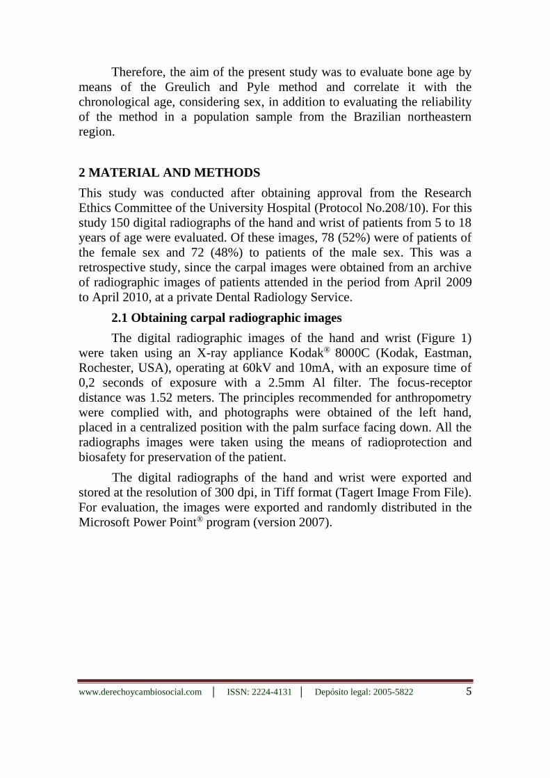
2 MATERIAL AND METHODS
This study was conducted after obtaining approval from the Research
Ethics Committee of the University Hospital (Protocol No.208/10). For this
study 150 digital radiographs of the hand and wrist of patients from 5 to 18
years of age were evaluated. Of these images, 78 (52%) were of patients of
the female sex and 72 (48%) to patients of the male sex. This was a
retrospective study, since the carpal images were obtained from an archive
of radiographic images of patients attended in the period from April 2009
to April 2010, at a private Dental Radiology Service.
2.1 Obtaining carpal radiographic images
The digital radiographic images of the hand and wrist (Figure 1)
were taken using an X-ray appliance Kodak® 8000C (Kodak, Eastman,
Rochester, USA), operating at 60kV and 10mA, with an exposure time of
0,2 seconds of exposure with a 2.5mm Al filter. The focus-receptor
distance was 1.52 meters. The principles recommended for anthropometry
were complied with, and photographs were obtained of the left hand,
placed in a centralized position with the palm surface facing down. All the
radiographs images were taken using the means of radioprotection and
biosafety for preservation of the patient.
The digital radiographs of the hand and wrist were exported and
stored at the resolution of 300 dpi, in Tiff format (Tagert Image From File).
For evaluation, the images were exported and randomly distributed in the
Microsoft Power Point® program (version 2007).

www.derechoycambiosocial.com │ ISSN: 2224-4131 │ Depósito legal: 2005-5822 6
Figure 1 - Radiographic image of the hand and wrist.
The images were individually evaluated by two examiners in a dark
room, with the aid of a 21.5" LED monitor. The examiners were previously
trained and had no knowledge about the chronological age of the patients.
The digital radiographs of the hand and wrist were compared, considering
sex, with the standard image in the atlas of Greulich and Pyle17 until there
was a coincidence of images (visual method) thus determining the bone
age. When the image presented an intermediate pattern between two
patterns, a mean was chosen, and the difference in age between some
patterns in the atlas was up to 14 months.
Each of the evaluators performed an evaluation of the same image
twice, with at least 15 days interval between evaluations. A maximum of
10 images per day were evaluated to avoid visual fatigue.
2.2 Data Analysis
The data were recorded in the form of a database in the informatics
program SPSS (Statistical Package for Social Sciences) for Windows,
version 15.0, and analyzed by means of descriptive statistics and bivariate
inferential statistics. For the descriptive procedures, central tendency
measures were presented (mean and median) and variability (Standard
Deviation and Amplitude). In the inferential statistic procedures, when the
conditions for the use of parametric statistics were satisfied, correlation
analyses (r of Pearson), Student's-t tests, analysis of variance (ANOVA)

www.derechoycambiosocial.com │ ISSN: 2224-4131 │ Depósito legal: 2005-5822 7
and linear regression analyses were performed. For all the tests a level of
significance of 0.05 was adopted.
3 RESULTS
Table 1 presents the results of intra- and inter-evaluator analyses
with regard to the bone ages. There were strong, positive and significant
relationships under all the conditions. Considering the explanatory
percentage of these correlations (over 94%), a mean bone age was
calculated for later analyses.
Table 1 - Results of analyses of intra- and inter-evaluator agreement on bone age by
means of the Pearson correlation coefficients.
Evaluators N r p
intra-evaluator 1 150 0.996 <0.001
Intra-evaluator 2 150 0.977 <0.001
Inter-evaluators 150 0.970 <0.001
p values ≤0.05 are statistically significant.
In Table 2, the results of the comparative analysis between bone age
and chronological age adjusted by sex. There was significant difference for
the female sex, in which the mean bone age was almost eight months ahead
than the mean chronological age (p≤0.05). For the male sex there was no
significant difference between the ages types (p=0,78).
Table 2 - Comparison of chronological age with bone age considering sex.
Sex
Chronological Age
In months
Bone Age
In months t p
N (SD) N (SD)
Male 134.2 (37.0)
134.8 (44.1)
-0.279 0.78
Female 122.8 (32.0)
130.4 (37.2)
-3.369 0.001

www.derechoycambiosocial.com │ ISSN: 2224-4131 │ Depósito legal: 2005-5822 8
Table 3 presents the results of the correlation between bone age and
chronological age with respect to sex. There was a correlation between
bone age and chronological age of 0.89 for the male sex and 0.84 for the
female sex. For males as females, there was significant linear relation
between both age and chronological age (p<0.001 for both sexes). Table 4
shows the results of the simple linear regression analysis in both sexes and
the Graph 1 represents the bone age as a function of chronological age for
the female sex.
Table 3 - Correlation between chronological age and bone age discriminated by sex.
Sex R p
Male 0.89 <0.001
Female 0.84 <0.001
Table 4 - Regression equations for bone age discriminated by sex.
Sex Equation R² Adjusted (%) p CI 95%*
Male y= 7.45 + 1.06x 79 <0.001 0.93 to 1.20
Female y= 9.90 + 0.98x 70 <0.001 0.84 to 1.12
*CI 95% for regression coefficient.

www.derechoycambiosocial.com │ ISSN: 2224-4131 │ Depósito legal: 2005-5822 9
Graph 1 - Representation of bone age as a function of chronological age for female sex.
Bone age (months) = 9.90 + 0.98 × chronological age (months). R2 = 0.70.
4 DISCUSSION
In the present study a sample of 150 carpal radiographs were used. This
number was adopted because it was the total number of images available in
the period of two years at a private Dental Radiology service. Moreover,
the size of the sample corresponds to a convenience sample, since the large
portion of similar studies used a similar number of carpal radiographs 13, 15,
18, 21, 24, 27, 30. However, other studies used a smaller 7, 8, 22 and larger sample
than the present study1, 4, 5, 10, 11, 14, 19, 29, 32.
As in the study of Van Rijnet al. 19, it was possible to obtain a sample
of carpal radiographs of patients with a minimum age of five years.
Although there are studies that have used a sample composed of carpal
radiographs of patients with an age lower than five years7, 11, 12, 22, the major
portion of the sample was composed of patients with a minimum age of

www.derechoycambiosocial.com │ ISSN: 2224-4131 │ Depósito legal: 2005-5822 10
over five years1, 4, 10, 13, 14, 19, 23, 27, 29, 30, 32. It is difficult to obtain a sample of
carpal radiograph images of patients under the age of five, since the carpal
radiograph is mostly used to evaluate the maturation status of a child in the
diagnosis, treatment planning and eventual outcome in Orthodontics and
functional Orthopedics. The follow-up of treatment makes it feasible to
obtain carpal radiographs of older patients. The maximum age of the
sample in the present study was similar to the majority of previous studies4,
7, 10, 11, 12, 19, 22, 23, 29, 32. However, other studies were composed of a sample of
carpal radiographs of patients with a maximum age of up to 16 years1, 13, 14,
15, 27, 30.
To determine the reliability of the method, the capacity of the same
evaluator to produce his results in a time interval and the correlation of the
evaluations performed by two evaluators of the same radiographs was
observed. The results showed strong and significant correlation of both
intra-evaluators and inter-evaluators, confirming the reproducibility of the
method. Similar results have been found by other researchers 5, 8, 11, 19, 21, 23,
30. These results emphasize that the Greulich and Pyle method is easy to
perform, and is widely used all over the world. In the present study the high
correlation found resulted from the training before the analyses of the
radiographs, when it was sought to establish a patter or sequence of
evaluation, in addition to the ease and simplicity of performing the method.
In the present study, the R2 value of 0.79 for the female sex and 0.70
for the male, indicated that 79% and 70% of the bone age can be predicted
by the chronological age respectively. This result represents good
correlation between bone age and chronological age for both sexes. The
correlation indices obtained were similar to those observed by other
authors1, 4, 8, 13, 19, 30. Furthermore, it is pointed out that in a Korean
population, of the three methods of evaluation, the Greulich-Pyle method
was the one that obtained the best correlation index. However, in an Italian
population sample with an age-range between 11 and 19 years, there was a
large margin of error in the determination of chronological age with the
Greulich-Pyle method, particularly in the determination of the ages of 14
and 18 years, so that this method is not indicated for helping to determine
chronological age in forensic researches 23.
Therefore, one must be aware of the applicability of the Greulich-
Pyle method in distinct populations, because some factors are known to
influence the development of individuals, such as genetics, race, nutrition,
hormonal, environment, socioeconomics, culture and sex1, 5, 9, 10, 11, 25.

www.derechoycambiosocial.com │ ISSN: 2224-4131 │ Depósito legal: 2005-5822 11
In the present study both sexes presented higher bone ages in relation
to chronological ages, demonstrating advanced maturity. Higher bone ages
than chronological ages for both sexes were also observed in Turkish
children4, Central European children22 and in children in the southeast of
Brazil14. On the other hand, some authors have found overestimation of
bone age only in the female sex13, 26, 30, and two of these researches13, 26
used a sample from the southeast of Brazil.
Girls usually exhibit earlier bone development than boys28. In the
present study that was more precocity for the female sex, which
demonstrates the distinct behavior between the sexes, being similar to the
data found in a study conducted in the southeast of Brazil28. Conversely, in
a study also using a sample from the southeast of Brazil, there were no
significant differences in bone age between the sexes1. Moreover, there is
disagreement with the result of the present study with regard to the data
found in a sample in the southeastern region in Brazil, 14, 27 and in
Slovenians19. In these studies there were delays in bone age in relation to
chronological age in both sexes. Furthermore, it is emphasized that delayed
bone ages before puberty were noted in children in Taiwan 5, Turkey10 and
Pakistan7.
Although the correlations between bone age and chronological age
had also been good, the use of correlation factors is recommended for
better adaptation of the method to the studied population, since it presents
different characteristics to the population for which the atlas was
developed1, 13, 30. Therefore, regression equations were created for each sex,
because as a datum of development, bone age must be referred to the
patterns of the collective society to which the individual belongs. The
regression analysis allows one to estimate by means of equations, the
extent to which bone age varies in relation to chronological age and vice
versa. As the intervals of confidence suggested different variabilities, the
regression analyses were discriminated by sex. Thus, an equation was
obtained for the male sex and another for the female sex.
The difference observed between the ages in the present study
demonstrated that children within the same age range develop in an
unequal manner. In the clinic, it allows us to reflect on the importance of
determining the correct stage of an individual's development, in which the
ideal time of treatment must consider each case in particular. The
possibility of predicting growth is an auxiliary tool in the diagnosis,
prognosis and elaboration of the treatment plan. In cases of providing
expert opinion, underestimating or overestimating age may favor or

www.derechoycambiosocial.com │ ISSN: 2224-4131 │ Depósito legal: 2005-5822 12
prejudice the individual. Therefore, it is important to use the factors of
correction to estimate the patient's age or to evaluate his/her stage of
development, and differences between the sexes must be considered.
5 CONCLUSIONS
In the studied sample the Greulich and Pyle method was shown to be
reliable, presenting correlation between the bone and chronological age for
both sexes. From the differences found with regard to the different regions
of Brazil and other countries, it is most important that this method should
be applied using the correction factors by means of previous studies in the
populations. Therefore, the applicability of the Greulich and Pyle method is
a valid tool for its application in Forensic Dentistry, determination of
chronological age, and in Orthodontics and Orthopedics for the
determination of bone maturity.
REFERENCES
1. Haiter-Neto F, Kurita LM, Menezes AV, Casanova MS.Skeletal age assessment: A
comparison of 3 methods. Am J Orthod Dentofacial Orthop.2006; 130(4): 435e15-
435e20.
2. Hassel B, Farman AG. Skeletal maturation evaluation using cervical vertebrae. Am J
Orthod Dentofacial Orthop. 1995 Jan;107(1):58-66.
3. Lai EH, Chang JZ, Jane Yao CC, Tsai SJ, Liu JP, Chen YJ, Lin CP. Relationship between
age at menarche and skeletal maturation stages in Taiwanese female orthodontic patients. J
Formos Med Assoc. 2008 Jul;107(7):527-32.
4. Büken B, Safak AA, Yazici B, Büken E, Mayda AS. Is the assessment of bone age by the
Greulich-Pyle method reliable at forensic age estimation for Turkish children? Forensic
Sci Int. 2007 Dec 20;173(2-3):146-53.
5. Chiang KH, Chou ASB, Yen PS, Ling CM, Lin CC, Lee CC, Chang PY. The reliability of
using Greulich-Pyle method to determine children's bone age in Taiwan. Tzu Chi Medical
Journal. 2005; 17(6): 417-420.
6. Vieira CL, Oliveira AEF, Ribeiro CCC, Lima AASJ. Relationship between the cervical
vertebrae maturation indicators and the dental calcification stages. R Dental Press
Ortodon Ortop Facial. 2009; 14(2): 45-53.
7. Rikhasor RM, Qureshi AM, Rathi SL, Channa NA. Skeletal maturity in Pakistani children.
J Anat. 1999 Aug;195 ( Pt 2):305-8.
8. Berst MJ, Dolan L, Bogdanowicz MM, Stevens MA, Chow S, Brandser EA. Effect of
knowledge of chronologic age on the variability of pediatric bone age determined using the
Greulich and Pyle standards. AJR Am J Roentgenol. 2001;176(2):507-10.
www.derechoycambiosocial.com │ ISSN: 2224-4131 │ Depósito legal: 2005-5822 13
9. Castriota-Scanderbeg A, Sacco MC, Emberti-Gialloreti L, Fraracci L. Skeletal age
assessment in children and young adults: comparison between a newly developed
sonographic method and conventional methods. Skeletal Radiol. 1998; 27(5):271-7.
10. Koc A, Karaoglanoglu M, Erdogan M, Kosecik M, Cesur Y. Assessment of bone ages: is
the Greulich–Pyle method sufficient for Turkish boys? Pediatrics International 2001;
43(6):662–665.
11. Mora S, Boechat MI, Pietka E, Huang HK, Gilsanz V. Skeletal age determinations in
children of European and African descent: applicability of the Greulich and Pyle standards.
Pediatr Res. 200;50(5):624-8.
12. Zhang A, Sayre JW, Vachon L, Liu BJ, Huang HK. Racial differences in growth patterns
of children assessed on the basis of bone age. Radiology. 2009;250(1):228-35.
13. Haiter Neto F, Almeida SM, Leite CC. Comparative study of the Greulich & Pyle and
Tanner & Whitehouse methods for estimating skeletal age Pesqui. Odontol. Bras.
[serial on the Internet]. 2000 [cited 2012 Sep 9];14(4):378-384. Available from:
http://www.scielo.br/scielo.php?script=sci_arttext&pid=S1517-
4912000000400013&lng=en.http://dx.doi.org/10.1590/S1517-74912000000400013.
14. Moraes MEL, Moraes LC, Medici-Filho E, Graziosi MAOC. Reliability of Greulich &
Pyle and Eklöf Ringertz methods for skeletal age evaluation in Brazilian children. Rev
Odontol UNESP 2003; 32 (1): 9-17.
15. Fishman LS. Radiographic evaluation of skeletal maturation. A clinically oriented method
based on hand-wrist films. Angle Orthod. 1982;52(2):88-112.
16. Schmidt S, Koch B, Schulz R, Reisinger W, Schmeling A. Studies in use of the Greulich-
Pyle skeletal age method to assess criminal liability. Leg Med (Tokyo). 2008
Jul;10(4):190-5.
17. Greulich WW, Pyle SI. Radiographic atlas of skeletal development of the hand and
wrist. 2. ed. Stanford: Stanford University Press, 1959. 272 p.
18. Buckler JM. How to make the most of bone ages. Arch Dis Child. 1983;58(10):761-3.
19. van Rijn RR, Lequin MH, Thodberg HH. Automatic determination of Greulich and Pyle
bone age in healthy Dutch children. Pediatr Radiol. 2009;39(6):591-7.
20. Schmeling A, Schulz R, Danner B, Rösing FW. The impact of economic progress and
modernization in medicine on the ossification of hand and wrist. International Journal of
Legal Medicine, Heidelberg, v. 120, n.2, p. 121-126, mar. 2006.
21. Lynnerup N, Belard E, Buch-Olsen K, Sejrsen B, Damgaard-Pedersen K. Intra-and
interobserver error of the Greulich-Pyle method as used on a Danish forensic sample.
Forensic Sci Int. 2008;179(2-3):242.e1-6.
22. Groell R, Lindbichler F, Riepl T, Gherra L, Roposch A, Fotter R. The reliability of bone
age determination in central European children using the Greulich and Pyle method. Br J
Radiol. 1999;72(857):461-4.
23. Tisè M, Mazzarini L, Fabrizzi G, Ferrante L, Giorgetti R, Tagliabracci A. Applicability of
Greulich and Pyle method for age assessment in forensic practice on an Italian sample. Int
J Legal Med. 2011 May;125(3):411-6.
24. Johnston FE. The use of the Greulich-Pyle method in a longitudinal growth study. Am J
Phys Anthropol. 1971;35(3):353-7.

www.derechoycambiosocial.com │ ISSN: 2224-4131 │ Depósito legal: 2005-5822 14
25. Ontell FK, Ivanovic M, Ablin DS, Barlow TW. Bone age in children of diverse ethnicity.
AJR Am J Roentgenol. 1996 Dec;167(6):1395-8.
26. Büken B, Erzengin OU, Büken E, Safak AA, Yazici B, Erkol Z. Comparison of the three
age estimation methods: which is more reliable for Turkish children? Forensic Sci Int.
2009;183(1-3):103.e1-7.
27. Carvalho AAF. Radiographic determination of growth parameters in healthy Brazilian
children with chronological age from 84 to 131 months. Rev Odontol UNESP 1993; 22(
2): 293-301.
28. Machado DRL, Barbanti VJ. Skeletal maturation and growth in children and adolescents
Rev Bras Cineantropom Desempenho Hum. 2007;9(1):12-20
29. Damian MF, Woitchunas FE, Cericato GO, Cechinato F, Moro G, Massochin ME, Castoldi
FL. Reliability and correlation analysis of two skeletal maturation evaluation indexes:
hand-wrist index and cervical vertebrae index R Dental Press Ortodon Ortop Facial
2006; 11(5):110-120.
30. Ortega AI, Haiter-Neto F, Ambrosano GM, Bóscolo FN, Almeida SM, Casanova MS.
Comparison of TW2 and TW3 skeletal age differences in a Brazilian population. J Appl
Oral Sci. 2006 Apr;14(2):142-6.
31. Parra FC, Amado RC, Lambertucci JR, Rocha J, Antunes CM, Pena SD. Color and
genomic ancestry in Brazilians. Proc Natl Acad Sci U S A. 2003 Jan 7;100(1):177-82.
32. Kim HJ, Yoon JR, Modi C, Modi H, Song HR, Song SY. Interrelationship of the Risser
sign, knee epiphysis, and bone age in determining skeletal maturity: a case-control study. J
Pediatr Orthop B. 2011 May;20(3):173-7.