Embed Size (px)
Citation preview

ORIGINAL ARTICLE
Evaluation of the effects of Global Postural Reeducationin patients with ankylosing spondylitis
Eliane Maria Silva • Sandra C. Andrade •
Maria J. Vilar
Received: 19 January 2010 / Accepted: 13 April 2011 / Published online: 5 May 2011
� Springer-Verlag 2011
Abstract The objective of this study is to assess the
effects of Global Postural Reeducation (GPR) in patients
with ankylosing spondylitis and compare GPR with group
conventional segmental self-stretching and breathing exer-
cises. This is a controlled interventional study of 38 patients
divided into 2 groups: a GPR group (n = 22) and a control
group (n = 16). Both groups were treated for more than
4 months. With the GPR group patients, positions that
stretched the shortened muscle chains were used. With the
control group patients, conventional segmental self-
stretching and breathing exercises were performed. The
variables analyzed were pain intensity, morning stiffness,
spine mobility, chest expansion, functional capacity (Health
Assessment Questionnaire–Spondyloarthropathies–HAQ-
S), quality of life (Medical Outcome Study Short Form 36
Healthy Survey–SF-36), and disease activity (Bath Anky-
losing Spondylitis Disease Activity Index–BASDAI). Sta-
tistical analysis was used with a significance level of
P \ 0.05. There was a statistically significant improvement
for all the parameters analyzed between pre-and post-
treatment in both groups. In the intergroup comparison, the
GPR group showed a significantly greater improvement in
morning stiffness (P = 0.013), spine mobility parameters,
except finger-floor distance (P = 0.118), in chest expansion
(P = 0.028), and in the physical aspect component of the
SF-36 (P = 0.001). The results of this study showed that
individual treatment with GPR (overall stretching) seems to
have better clinical outcomes than group treatment with
conventional segmental self-stretching and breathing exer-
cises for patients with ankylosing spondylitis.
Keywords Ankylosing spondylitis � Physical therapy �Stretching exercises � Spinal mobility
Introduction
Ankylosing spondylitis (AS) is a systemic chronic osteo-
articular inflammatory disease of unknown etiology that
predominantly affects the axial skeleton with pain,
impaired spinal mobility, sacroiliitis on radiographs, also
involving peripheral joints, especially shoulders and hip
joints. Efforts have been made in recent years to improve
and standardize, making an early diagnosis in AS [1].
Structural changes in the spine may begin early and when
untreated lead the patient to adopt improper postures such
as mandible protrusion, anteriorization of the head, shoul-
der protrusion, scapular protraction, thoracic hyperkypho-
sis, rectification of the lumbar lordosis, pelvic retroversion,
flexion with internal hip rotation, and flexion of the knees
[2]. Such changes can cause different degrees of physical,
social, economic, or psychological disability depending on
their activity and severity, significantly affecting the
patient’s quality of life [3].
Treatment of AS involves the use of non-steroidal anti-
inflammatory drugs and eventually also disease-modifying
anti-rheumatic drugs (DMARDs) or TNF-alpha inhibitors
in addition to physical therapy [4]. A systematic review,
conducted in 2008, addressing the evidence of the use of
physical therapy on AS [5], concluded that although
supervised group physical therapy [6–9] produces better
effects on the spine mobility and overall health of patients
than individualized exercises at home, there is no defined
protocol as to which specific exercises are appropriate for
this disease. The authors suggest that more information is
needed about various physical therapy programs regarding
E. M. Silva (&) � S. C. Andrade � M. J. Vilar
Universidade Federal do Rio Grande do Norte (UFRN),
Natal, Brazil
e-mail: [email protected]
123
Rheumatol Int (2012) 32:2155–2163
DOI 10.1007/s00296-011-1938-3

intensity, frequency, and duration to specify the most
appropriate activity for the disease [5].
In this context, we consider the role of Global Postural
Reeducation (GPR) in the physical therapy approach of
patients with AS, as it is a form of active overall muscle
stretching, which jointly works the antigravity muscle
chains and the internal and inspiratory rotators that are
often impaired in patients with AS [10–12]. In GPR, after
thorough postural assessment, eight positions are generally
used. These are associated with the contraction-relaxation
method for overall stretching of the chains before and/or
after, performed lying down or standing. It is considered
that overall stretching differs from the segmental stretching
of a muscle in an isolated manner, which can lead to
inadequate compensation and adjustments to the exercises
[10, 12].
In the literature in English language, there are few
studies on the effects of GPR in musculoskeletal diseases
[12]. Recent study suggests that a GPR intervention in
subjects with persistent low back pain induces a greater
improvement on pain and disability, as compared to a
stabilization exercise program [13]. Regarding the AS,
studies show that an exercise program based on GPR
improved significantly the functional capacity and spine
mobility when compared with the AS control group, with
the improvement parameters being maintained after
6 months and a year of follow-up [11, 14, 15]. A system-
atic review conducted in 2007 showed that GPR-based
exercise programs seem to offer a valid and promising
alternative therapy for patients with AS [16].
Thus, the objective of this study was to assess the effects
of individual global stretching through GPR and compare
them with group conventional segmental self-stretching
and breathing exercises for patients with AS.
Materials and methods
A controlled interventional study was conducted consisting
of 38 patients with a diagnosis of AS according to the
modified New York criteria [17], who were referred by
rheumatologists to the physical therapy school clinic at the
Universidade Potiguar, Natal–RN, Brazil. The inclusion
criteria were patients between 18 and 65 years of age and
the ability to perform usual self-care and vocational
activities. The study excluded patients with secondary
fracture due to osteoporosis. All patients were clinically
stable, had no other associated diseases, and were being
treated with non-steroidal anti-inflammatory drugs for the
underlying disease.
The patients were divided into two groups: an experi-
mental group (GPR group), composed of 22 patients who
underwent individual GPR sessions and a control group,
composed of 16 patients who underwent conventional
segmental self-stretching sessions associated with breath-
ing exercises in a group.
All patients were evaluated in two stages: an initial
evaluation (pre-treatment) and an evaluation at the end of
treatment (post-treatment). An independent physical ther-
apist evaluator conducted the post-treatment assessments.
We used a protocol containing demographic and clinical
data as age, sex, disease duration (defined as time between
the first symptoms and actual evaluation), spinal pain
intensity (Visual Analog Scale from 0 to 10), and duration
of morning stiffness in minutes. In the physical examina-
tion, the following sequence was observed: cervical spine
mobility (chin-to-sternum, occiput-to-wall distances, and
cervical rotation), lumbar spine mobility (finger-to-floor
distance and Schober’s modified test), and thoracic spine
flexibility (chest expansion in the 4th intercostal space)
[18]. The goniometry of the cervical rotation was per-
formed with a CARCI� goniometer, with both sides mea-
sured and a mean calculated between the two sides. The
Schober’s modified test is performed with the patient
standing with outer edges of bare feet 30 cm apart and feet
in line. Examiner marks a point midway along a line level
with the iliac crests (at the L4/5 junction). A second point
is marked 10 cm above that a third 5 cm below the first to
give a 15 cm line. Patient flexes forward from the waist
with knees fully extended. The distance between the upper
and lower 2 marks is measured. Any increase beyond
15 cm represents the amount of movement achieved [19].
All these measurements were repeated three times, with
the mean among them considered as valid. To assess the
symptoms and disease activity, the Bath Ankylosing
Spondylitis Disease Activity Index (BASDAI) was used,
validated for the Brazilian population [20], which considers
the higher the score, the greater the degree of disease
activity. Functional capacity was also measured through
the Health Assessment Questionnaire–Spondyloarthropa-
thies (HAQ-S), which corresponds to ten areas whose
scores vary from 0 to 3—the closer to zero, the greater the
individual’s functional capacity [21]. Then the patients’
quality of life was evaluated through the Medical Outcome
Study Short Form 36 Healthy Survey (SF-36), a ques-
tionnaire translated and validated for the Brazilian popu-
lation [22] and using physical and mental components. The
SF-36 provides a final score from 0 (zero) to 100 (one
hundred), where 0 (zero) corresponds to the worst overall
health status and 100 (one hundred) to the best health
status.
The physical therapy intervention was performed over
4 months (16 weeks) for both groups. The GPR group
patients underwent GPR in accordance with the postural
changes found in AS, with a frequency of once a week for
1 h. The control group patients carried out a conventional
2156 Rheumatol Int (2012) 32:2155–2163
123

segmental self-stretching program and breathing exercises,
in a group, twice a week for 40 min. Both groups were
supervised by a physical therapist with experience in
rheumatic diseases and received educational guidelines,
with clarification on positions for sitting, walking, sleeping,
and carrying out daily living activities.
The intervention of the GPR group was based on the
evaluation of the shortened muscle chains, in accordance
with the GPR treatment bases. A postural analysis of the
anterior (respiratory and anterointernal of the hip) and
posterior muscle chains, anterior of the arm, and antero-
internal of the shoulders was carried out [10, 12].
Next, five positions (Table 1) were used for the treatment
and described as [10, 12]:[10, 12] frog postures on the floor
with the upper limbs (ULs) in adduction (Fig. 1); frog in the
air with the ULs in abduction (Fig. 2); seated (Fig. 3);
standing bent forward (Fig. 4); standing against the wall,
and standing posture (Figs. 5, 6). In these postures, the
patients underwent costodiaphragmatic inspiration, fol-
lowed by expiration depressing the sternum with abdominal
protrusion. The physical therapist uses verbal commands
and manual contact to maintain the alignment and make the
necessary postural corrections, with the aim of optimizing
the stretching and discouraging compensatory movements.
In the first 8 weeks, supine positions were used (Table 1)
aiming to improve pain (especially cervical pain) and spine
mobility. In the final 8 weeks, sitting postures (Fig. 3) and
standing postures (Figs. 4, 5, 6) were introduced to improve
Table 1 Physical therapy program carried out in the GPR group
Treatment postures Objectives Duration/Evolution
POSTURE 1 (Fig. 1) 20 min
Frog on the ground with upper limbs in adduction
with open hip angle
Stretch; anterior chain (respiratory and
anterointernal of the hip); anterior chain of the
arms and upper chain of the shoulders; side chain
of the hip
Description: Decubitus position, arms in extension
and abduction, retroverted pelvis, hips in flexion,
abduction and lateral rotation, legs bent, feet
planted
Manual traction was applied to the
neck and sacral traction to align
the curves of the spinal column
Extension and adduction of lower
and upper limbs
POSTURE 2 (Fig. 2) 20 min
Frog in the air with upper limbs in abduction with
closed hip angle
Stretch respiratory chain; Posterior chain;
anterointernal of the shoulder; anterior of the arm
and side chain of the hip
Description: Dorsal decubitus Upper limbs in
adduction and extension; Sacrum supported on the
stretcher; Legs semi-flexed, hips in external
rotation and dorsiflexion of the feet
Increase hip flexion; knee extension,
dorsiflexion of the ankle and
abduction of upper limbs
POSTURE 3 (Fig. 3) 10 min
Sitting with upper limbs in adduction with closed
hip angle
Stretch the posterosuperior chain, improve the body
schema, education of the sitting position
Description: Pelvis supported in the ischiatic
tuberosities
Legs flexed, hips in abduction and external rotation;
feet planted
Extension and adduction of the legs;
Hip flexion with the trunk inclined
forward
POSTURE 4 (Fig. 4) 10 min
Standing with upper limbs in adduction, trunk
inclined forward with closed hip angle
Stretch posterior-inferior chain, improve body
schema, proprioception and balance
Description: lower limbs and hips in semi-flexion,
abduction and external rotation; abducted feet and
heels together.
Hip flexion with spine alignment
POSTURE 5 (Figs. 5, 6)
Standing against the wall and standing in the center
with open hip angle
Stretch respiratory chain; anterior
chain(anterointernal chain of the hip); anterior
chain of the arms and upper chain of the shoulders
5 min–to the end of each session
Description: Retroverted pelvis; Legs semi-flexed;
hips abducted and in external rotation; feet planted
Side chain of the hip. Improve proprioception, body
schema and balance
Leg extension
Rheumatol Int (2012) 32:2155–2163 2157
123

proprioception, body schema, balance, and strengthening of
the paraspinals and lower limbs (postures 3, 4, and 5 and 6–
Table 1). The standing posture was held at the end of each
session during the GPR program (Figs. 5, 6).
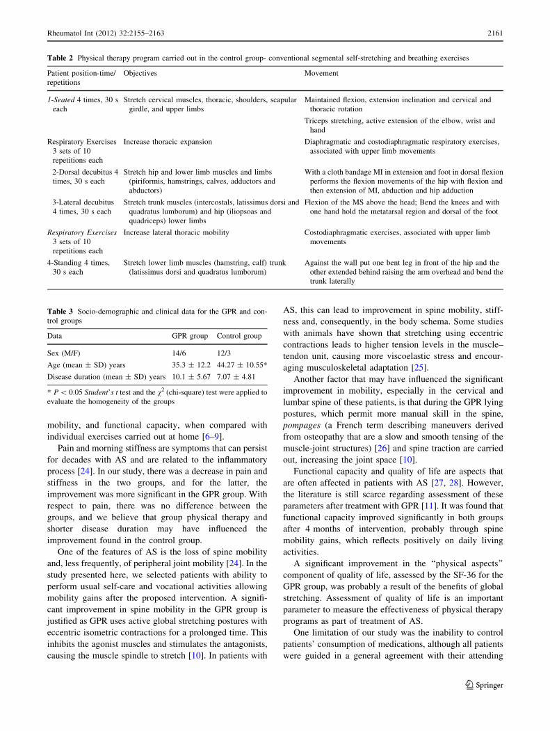
Patients in the control group carried out a program of
conventional segmental self-stretching and breathing
exercises in a group of four patients, specific to the short-
ened muscle chains (Table 2) [23]. The standing stretches
were introduced when patients reported improvement in
the amount of spinal pain felt.
This study was approved by the Research Ethics Com-
mittee of the Universidade Federal do Rio Grande do
Norte. All patients signed the Free and Informed Consent
Form, and all patients agreed not to undergo another
physical therapy treatment during the study.
Statistical analysis
In the statistical analysis, the normal distribution of all
variables was assessed using the Kolmogorov–Smirnov
nonparametric test. Student’s t test and the v2 (chi-square)
test were applied to evaluate the homogeneity of the
groups. For intra-group assessment of the quantitative
variables, we used the paired t test, and for the inter-group
analysis, the unpaired t test. For all analyses, P \ 0.05 was
considered statistically significant. Data analysis was per-
formed with SPSS, version 13.0.
Results
The socio-demographic and clinical data of the AS patients
from both groups are presented in Table 3; of the patients
studied, two from the GPR group and one from the control
group were discharged for abandoning treatment, leaving
20 patients in the GPR group and 15 in the control group. At
Fig. 1 Posture 1: Frog on the ground with upper limbs in adduction
with open hip angle. Stretch the anterior muscle chain (diaphragm,
pectoralis minor, scalene, sternocleidomastoid, intercostalis, ili-
opsoas, adductors, and anterior tibialis muscles)
Fig. 2 Posture 2: Frog in the air
with upper limbs in abduction
with closed hip angle. Stretch
the posterior chain (upper
trapezius, suboccipital, erector
spinae, subescapular, major
pectoralis and hand flexors,
gluteus maximus, ischiotibial,
triceps surae, and foot intrinsic
muscle)
2158 Rheumatol Int (2012) 32:2155–2163
123

the start of the study, there was no statistically significant
difference between the groups in the following variables:
sex (P = 0.70), disease duration (P = 0.12), morning
stiffness (P = 0.40), intensity of spinal pain (P = 0.20) and
lumbar pain (P = 0.82), disease activity–BASDAI
(P = 0.10), functional capacity–HAQ-S (P = 0.32), qual-
ity of life–SF36, and in the ‘‘physical aspects’’ (P = 0.67)
and ‘‘emotional aspects’’ (P = 0.42) components, thus
characterizing the homogeneity of the groups, except with
respect to age (P = 0.02) and cervical pain (P = 0.04).
For the pain intensity parameter, it was observed that
in the GPR group, there was a statistically significant
improvement post-treatment in the cervical (P = 0.00)
and lumbar (P = 0.00) regions. In the control group,
there was a statistically significant improvement in pain
intensity in the cervical and lumbar (P = 0.00) and dorsal
(P \ 0.02) regions. However, in the inter-group compar-
ison, there was not a statistically significant difference
(Table 4).
For the morning stiffness variable, there was a statisti-
cally significant improvement in both the GPR group and
the control group (P = 0.00; P = 0.00, respectively).
However, in the inter-group comparison, the results were
better for the GPR group post-treatment (P = 0.01).
Fig. 3 Posture 3: Sitting with
upper limbs in adduction with
closed hip angle. stretch the
posterior chain (upper trapezius,
suboccipital, erector spinae,
levator scapulae and hand
flexors; gluteus maximus,
ischiotibial, triceps surae, and
foot intrinsic muscles)
Fig. 4 Posture 4: Standing with
upper limbs in adduction, trunk
inclined forward with closed hip
angle. Stretch the posterior
chain (upper trapezius,
suboccipital, erector spinae,
levator scapulae and hand
flexors, gluteus maximus,
ischiotibial, triceps surae, and
foot intrinsic muscles)
Rheumatol Int (2012) 32:2155–2163 2159
123

For spinal mobility, there was a statistically significant
improvement in both groups after treatment in all the
measures taken in the cervical (P = 0.00) and lumbar
(P = 0.00) regions. However, in the inter-group compari-
son, there was significantly more improvement in the GPR
group in all measures, except for finger-floor distance
(P = 0.12).
For the chest-expansion measure, it was observed that in
both groups there was significantly more improvement
after treatment. Similarly, in the inter-group comparison,
there was a in the GPR group (P = 0.03).
In the post-treatment intra-group analysis, there was a
statistically significant improvement in disease activity in
both groups (P \ 0.00). In the inter-group comparison,
however, there was no statistically significant difference in
this parameter (Table 5).
In the intra-group analysis, there was a statistically
significant improvement in functional capacity in both
groups (P \ 0.00). In the inter-group comparison, how-
ever, there was no statistically significant difference in
functional capacity.
For the quality of life parameter, there was a statistically
significant improvement in both groups, for the physical as
well as the emotional aspects (P \ 0.00). However, there
was a statistically significant difference in the inter-group
analysis, with more improvement in the physical aspects of
the GPR group (P = 0.00).
Discussion
This study’s findings showed a clinical and statistically
significant improvement in all variables studied in both
groups. However, in the inter-group comparison,
improvement in the GPR group patients was higher than
the control group with respect to the following variables:
morning stiffness, spine mobility, thoracic expansion, and
quality of life (physical aspects–SF36).
This study, based on GPR, according to the muscle
chain shortening found in patients with AS, differs from
the 2005 Las Penas study [11], which in addition to using
GPR used other methods such as active mobilization of
the spine, scapular and pelvic girdle, and neural mobili-
zation. In a systematic review conducted by Cochrane in
2008, this study was considered one of the few that
compared two forms of exercises, with a detailed
description of the proposed protocol and low risk of bias,
concluding with a moderate level of evidence for GPR
use in AS [5].
In the study presented here, we used only static postures
for active global stretching, as originally described by
Souchard [10] and compared them with physical therapy
carried out in a group, considered a different therapeutic
approach for patients with AS. Group physical therapy
programs were studied as part of AS treatment and showed
better results with respect to morning stiffness, spine
Fig. 5 Posture 5: Standing against the wall with open hip angle.
Stretching anterior muscles chain (are the same muscles, Fig. 1)
Fig. 6 Posture 5: Standing posture with open hip angle. Stretching
anterior muscles chain (are the same muscles, Fig. 1)
2160 Rheumatol Int (2012) 32:2155–2163
123
mobility, and functional capacity, when compared with
individual exercises carried out at home [6–9].
Pain and morning stiffness are symptoms that can persist
for decades with AS and are related to the inflammatory
process [24]. In our study, there was a decrease in pain and
stiffness in the two groups, and for the latter, the
improvement was more significant in the GPR group. With
respect to pain, there was no difference between the
groups, and we believe that group physical therapy and
shorter disease duration may have influenced the
improvement found in the control group.
One of the features of AS is the loss of spine mobility
and, less frequently, of peripheral joint mobility [24]. In the
study presented here, we selected patients with ability to
perform usual self-care and vocational activities allowing
mobility gains after the proposed intervention. A signifi-
cant improvement in spine mobility in the GPR group is
justified as GPR uses active global stretching postures with
eccentric isometric contractions for a prolonged time. This
inhibits the agonist muscles and stimulates the antagonists,
causing the muscle spindle to stretch [10]. In patients with
AS, this can lead to improvement in spine mobility, stiff-
ness and, consequently, in the body schema. Some studies
with animals have shown that stretching using eccentric
contractions leads to higher tension levels in the muscle–
tendon unit, causing more viscoelastic stress and encour-
aging musculoskeletal adaptation [25].
Another factor that may have influenced the significant
improvement in mobility, especially in the cervical and
lumbar spine of these patients, is that during the GPR lying
postures, which permit more manual skill in the spine,
pompages (a French term describing maneuvers derived
from osteopathy that are a slow and smooth tensing of the
muscle-joint structures) [26] and spine traction are carried
out, increasing the joint space [10].
Functional capacity and quality of life are aspects that
are often affected in patients with AS [27, 28]. However,
the literature is still scarce regarding assessment of these
parameters after treatment with GPR [11]. It was found that
functional capacity improved significantly in both groups
after 4 months of intervention, probably through spine
mobility gains, which reflects positively on daily living
activities.
A significant improvement in the ‘‘physical aspects’’
component of quality of life, assessed by the SF-36 for the
GPR group, was probably a result of the benefits of global
stretching. Assessment of quality of life is an important
parameter to measure the effectiveness of physical therapy
programs as part of treatment of AS.
One limitation of our study was the inability to control
patients’ consumption of medications, although all patients
were guided in a general agreement with their attending
Table 2 Physical therapy program carried out in the control group- conventional segmental self-stretching and breathing exercises
Patient position-time/
repetitions
Objectives Movement
1-Seated 4 times, 30 s
each
Stretch cervical muscles, thoracic, shoulders, scapular
girdle, and upper limbs
Maintained flexion, extension inclination and cervical and
thoracic rotation
Triceps stretching, active extension of the elbow, wrist and
hand
Respiratory Exercises
3 sets of 10
repetitions each
Increase thoracic expansion Diaphragmatic and costodiaphragmatic respiratory exercises,
associated with upper limb movements
2-Dorsal decubitus 4
times, 30 s each
Stretch hip and lower limb muscles and limbs
(piriformis, hamstrings, calves, adductors and
abductors)
With a cloth bandage MI in extension and foot in dorsal flexion
performs the flexion movements of the hip with flexion and
then extension of MI, abduction and hip adduction
3-Lateral decubitus
4 times, 30 s each
Stretch trunk muscles (intercostals, latissimus dorsi and
quadratus lumborum) and hip (iliopsoas and
quadriceps) lower limbs
Flexion of the MS above the head; Bend the knees and with
one hand hold the metatarsal region and dorsal of the foot
Respiratory Exercises3 sets of 10
repetitions each
Increase lateral thoracic mobility Costodiaphragmatic exercises, associated with upper limb
movements
4-Standing 4 times,
30 s each
Stretch lower limb muscles (hamstring, calf) trunk
(latissimus dorsi and quadratus lumborum)
Against the wall put one bent leg in front of the hip and the
other extended behind raising the arm overhead and bend the
trunk laterally
Table 3 Socio-demographic and clinical data for the GPR and con-
trol groups
Data GPR group Control group
Sex (M/F) 14/6 12/3
Age (mean ± SD) years 35.3 ± 12.2 44.27 ± 10.55*
Disease duration (mean ± SD) years 10.1 ± 5.67 7.07 ± 4.81
* P \ 0.05 Student’s t test and the v2 (chi-square) test were applied to
evaluate the homogeneity of the groups
Rheumatol Int (2012) 32:2155–2163 2161
123

doctor not to modify their treatment during the study.
However, even considering that not all patients have this
control, there was probably no interference with the results
obtained in this study.
We emphasize that it is important to study the long-term
benefits of GPR, considering the chronic nature of AS. We
conclude that stretches carried out in a chain as proposed
by the GPR method provide more advantages with respect
to spine mobility and physical aspects of quality of life
when compared with conventional segmental stretches and
breathing exercises also carried out in a group.
Acknowledgments I would like to thank the patients and the
rheumatologists who made this study possible.
Conflict of interest The authors declare that they have no conflict
of interest.
References
1. Rudwaleit M (2010) New approaches to diagnosis and classifi-
cation of axial and peripheral spondyloarthritis. Curr Opin
Rheumatol 20:375–380
Table 4 Intragroup and intergroup differences (pre-treatment and post-treatment) in clinical outcomes in the GPR Group and Control Group of
patients with Ankylosing Spondylitis
Variables GPR group Control group
Pre-treat Post-treat Intragroup
difference
P value
Pre-treat Post-treat Intragroup
difference
P value
Intergroup
difference
P value
Pain intensity (VAS)
Cervical pain 4.5 (0.74) 1.5 (0.39) 0.00* 7.5 (0.32) 3.2 (0.55) 0.00 * 0.27
Dorsal pain 0.8 (0.35) 0.0 (0.00) 0.07 1.9 (0.63) 1.0 (0.39) 0.03* 0.61
Lumbar pain 5.5 (0.68) 1.3 (0.43) 0.00* 5.9 (0.61) 2.2 (0.67) 0.00* 0.42
Morning Stiffness (min) 38.8 (5.26) 14.5 (2.38) 0.00* 44.0 (3.09) 34.4 (2.61) 0.00* 0.01�
Cervical mobility
Chin-to-chest distance (cm) 5.5 (0.5) 2.9 (0.39) 0.00* 5.1 (0.24) 4.4 (0.19) 0.01* 0.00�
Occiput-to-wall distance (cm) 6.8 (0.58) 3.7 (0.4) 0.00* 8.1 (1.06) 6.7 (0.86) 0.00* 0.01�
Cervical rotation (degrees) 39.8 (0.8) 51.3 (0.95) 0.00* 35.7 (1.21) 40.2 (1.17) 0.00* 0.00�
Lumbar mobility
Schober’s modified test (cm) 13.3 (0.32) 14.1 (0.32) 0.00* 13.7 (0.33) 13.8 (0.33) 0.41 0.02�
Fingertip-to-floor distance(cm) 19.3 (1.91) 9.6 (1.58) 0.00* 21.9 (1.44) 14.5 (1.51) 0.00* 0.12
Chest expansion 2.9 (0.32) 5.2 (0.36) 0.00* 2.3 (0.28) 3.8 (0.26) 0.00* 0.03�
Pre-treatment and post-treatment values expressed as mean and standard deviation
* P value \ 0.05 (paired t test for intra-group analysis)� P value \ 0.05 (unpaired t test for intergroup analysis)
Table 5 Intragroup and intergroup differences (pre-treatment and post-treatment) in clinical and functional outcomes in the GPR Group and
control Group of patients with Ankylosing Spondylitis
Variables GPR group (n = 20) Control group (n = 15)
Pre-treat Post-treat Intragroup
difference
P value
Pre-treat Post-treat Intragroup
difference
P value
Intergroup
difference
P value
Disease activity (BASDAI) 6.6 (0.52) 3.1 (0.17) 0.00* 7 (0.31) 4.9 (0.19) 0.00* 0.73
Functional capacity (HAQ-S) 2.0 (0.24) 0.6 (0.1) 0.00* 2.3 (0.15) 1.5 (0.1) 0.00* 0.1
Quality of life (SF36)
Physical aspects 41.5 (3.27) 74.2 (2.84) 0.00* 47.6 (3.45) 64.8 (2.36) 0.00* 0.00�
Emotional aspects 56.5 (4.72) 78.7 (3.59) 0.00* 54.7 (4.73) 73.3 (2.94) 0.00* 0.82
Pre-treatment and post-treatment values expressed as mean and standard deviation
* P value \ 0.05 (paired t test for intragroup analysis)� P value \ 0.05 (unpaired t test for intergroup analysis)
2162 Rheumatol Int (2012) 32:2155–2163
123

2. Aydog E, Depedibi R, Bal A, Eksioglu E, Unlu E, Cakci A (2006)
Dynamic postural balance in ankylosing spondylitis patients.
Rheumatology (Oxford) 45:445–448
3. Ozgul A, Peker F, Taskaynatan MA, Tan AK, Dincer K, Kalyon
TA (2006) Effect of ankylosing spondylitis on health-related
quality of life and different aspects of social life in young
patients. Clin Rheumatol 25:168–174
4. Zochling J, Van der Heijde D, Burgos-Vargas R, Collantes E,
Davis JC Jr, Dijkmans B et al (2006) ASAS/EULAR recom-
mendations for the management of ankylosing spondylitis. Ann
Rheum Dis 65:442–452
5. Dagfinrud H, Kvien TK, Hagen KB (2008) Physiotherapy inter-
ventions for ankylosing spondylitis. Cochrane Database Syst Rev
23: (1)CD002822
6. Helliwell P, Abbott CA, Chamberlain MA (1996) A randomized
trial of three different physiotherapy regimes in Ankylosing
Spondylitis. Physiotherapy 82:85–90
7. Analay Y, Ozcan E, Karan A, Diracoglu D, Aydin R (2003) The
effectiveness of intensive group exercise on patients with
Ankylosing Spondylitis. Clin Rehabil 17:631–636
8. Hidding CA, Van Der Linden S, Boers M et al (1993) Is group
Physical therapy superior to individualized therapy in Ankyilo-
sing Spondylitis? A randomized controlled trial. Arthritis Care
Res 6:117–125
9. Santos H, Brophy S, Calin A (1998) Exercise in Ankylosing
Spondylitis: How much is optimum? J Rheumatol 25:2156–2160
10. Souchard PE (1986) Reeducacao Postural Global (Metodo do
Campo Fechado). Icone, Sao Paulo
11. Fernandez-de-Las-Penas C, Alonso-Blanco C, Morales-Cabezas
M, Miangolarra-Page JC (2005) Two exercise interventions for
the management of patients with ankylosing spondylitis: a Ran-
domized controlled trial. Am J Phys Med Rehabil 84:407–419
12. Vanti C, Generali A, Ferrari S, Nava T, Tosarelli D, Pillastrini P
(2007) Reeducation Posturale Globale in musculoskeletal dis-
eases: scientific evidence and clinical practice. Reumatismo
59:192–201
13. Bonetti F, Curti S, Mattioli S, Mugnai R, Vanti C, Violante FS,
Pillastrini P (2010) Effectiveness of a ‘Global Postural Reedu-
cation’ program for persistent low back pain: a non-randomized
controlled trial. BMC Musculoskelet Disord. doi:10.1186/1471-
2474-11-285
14. Fernandez-de-Las-Penas C, Alonso-Blanco C, Miangolarra-Page
JC, Fernandez MP (2005) Seguimiento a medio plazo de la me-
jora fısica y funcional tras tratamiento rehabilitador mediante el
trabajo de cadenas musculares en la espondilitis anquilosante.
Rehabilitacion 39:222–229
15. Fernandez-de-Las-Penas C, Alonso-Blanco C, Alguacil-Diego
IM, Miangolarra-Page JC (2006) One-year follow-up of two
exercise interventions for the management of patients with
ankylosing spondylitis: a randomized controlled trial. Am J Phys
Med Rehabil 85:559–567
16. Ribeiro F, Leite M, Silva F, Sousa O (2007) Physical exercise in
the treatment of ankylosing spondylitis: a systematic review. Acta
Reumatol Port 32:129–137
17. Van Der Linden S, Valkenburg HA, Cats A (1984) Evaluation of
diagnostic criteria for Ankylosing Spondylitis: a proposal for
modification of the New York criteria. Arthritis Rheum
27:361–368
18. Zochling J, Braun J (2005) Assessment of ankylosing spondylitis.
Clin Exp Rheumatol 23 (Suppl 39):S133–S141
19. Jenkinson TR, Mallorie PA, Whitelock HC, Kennedy LG, Garrett
SL, Calin A. (1994) Defining spinal mobility in ankylosing
spondylitis (AS). The Bath AS Metrology Index. J Rheumatol
21:1694–1698
20. Cusmanich KG (2006) Validacao para a lıngua portuguesa dos
instrumentos de avaliacao de ındice funcional e ındice de ativ-
idade de doenca em pacientes com espondilite anquilosante.
Dissertacao de Mestrado em Ciencias, Faculdade de Medicina da
Universidade de Sao Paulo
21. Daltroy LH, Larson MG, Roberts WN (1990) A modification of
the Health Assessment Questionnaire for the Spondyloarthropa-
thies. J Rheumatol 17:946–950
22. Ciconelli RM, Ferraz MB, Santos W, Meinao I, Quaresma MR
(1999) Traducao para a lıngua portuguesa e validacao do ques-
tionario generico de avaliacao de qualidade de vida SF-36 (Brasil
SF-36). Rev Bras Reumatol 39:543–550
23. Kisney C, Colby LA (2005) Exercıcios Terapeuticos: funda-
mentos e metodos. Manole, Sao Paulo
24. Braun J, Pincus T (2002) Mortality, course of disease and prog-
nosis of patients with ankylosing spondylitis. Clin Exp Rheu-
matol 20(Suppl 28):S16–S22
25. Taylor DC, Brooks DE, Ryan JB (1997) Viscoelastic character-
istics of muscle: passive stretching versos muscular contractions.
Med Sci Sport Exerc 29:1619–1624
26. Bienfait M (1999) Fascias e pompages. Summus, Sao Paulo
27. Clegg DO (2006) Treatment of ankylosing spondylitis. J Rheu-
matol (Suppl 78):S24–S31
28. Souza MC, Tutiya G, Jones A, Junior L, Natour J (2008) Ava-
liacao do equilıbrio funcional e qualidade de vida em pacientes
com espondilite anquilosante. Rev Bras Reumatol 8:2274–2277
Rheumatol Int (2012) 32:2155–2163 2163
123