Embed Size (px)
Citation preview


OPEN ACCESS Asian Journal of Plant Sciences
ISSN 1682-3974DOI: 10.3923/ajps.2021.332.343
Research ArticleEvaluation of Phytochemical, Antioxidant and Anti-inflammatoryProperties of near Endemic Aloe dhufarensis
Neelam Sherwani, Khalil Al Mahroqi and Sardar A. Farooq
Department of Biology, College of Science, Sultan Qaboos University, P.O. Box 36, Al Khoud, PC123, Oman
AbstractBackground and Objective: Aloe dhufarensis is a local endemic medicinal herb used for centuries to treat diabetes, headache, fever, skindisorders and wound healing by the local people of Oman. The present research was initiated to substantiate the ethno-medicinal usesof this plant by assessing the phytochemicals and evaluating the antioxidant, anti-inflammatory and anti-microbial activity of Aloedhufarensis. Materials and Methods: Dried, finely grinded leaves of A. dhufarensis were extracted with methanol for phytochemicalanalysis using standard protocols and several phytochemical such as saponins, alkaloids, terpenoids, flavonoids, steroids, tannins,glycosides, anthraquinones, phytosterol, coumarins and carbohydrates were detected in the extract. Results: Extracts displayed significantantioxidant property, exhibiting a potent DPPH scavenging activity with IC50 value of 83.46 µg mLG1 and a strong hydrogen peroxidescavenging activity with an IC50 value of 289.786 µg mLG1. Substantial inhibition of protein denaturation and anti-proteinase activitywere recorded. Considerable antibacterial activity was observed against all 4 studied strains with the highest antibacterial activity recordedagainst the E. coli. Conclusion: The results reveal that A. dhufarensis possess a wide spectrum of phytochemicals that have significantpharmacological properties and substantiate the support to the scientific basis for the traditional use of this ethnomedicinal herb.
Key words: Aloe dhufarensis, anti-inflammatory, anti-oxidant, total phenols, phytochemical analysis
Citation: Neelam Sherwani, Khalil Al Mahroqi and Sardar A. Farooq, 2021. Evaluation of phytochemical, antioxidant and anti-inflammatory properties ofnear endemic Aloe dhufarensis. Asian J. Plant Sci., 20: 332-343.
Corresponding Author: Sardar A. Farooq, Department of Biology, College of Science, Sultan Qaboos University, P.O. Box 36, Al Khoud, PC123, Oman
Copyright: © 2021 Neelam Sherwani et al. This is an open access article distributed under the terms of the creative commons attribution License, whichpermits unrestricted use, distribution and reproduction in any medium, provided the original author and source are credited.
Competing Interest: The authors have declared that no competing interest exists.
Data Availability: All relevant data are within the paper and its supporting information files.

Asian J. Plant Sci., 20 (2): 332-343, 2021
INTRODUCTION
Oman is a spectacular country in Arabian Peninsula interms of biodiversity and is home to 78 endemic and48 near-endemic plant species, with substantial number ofthese species being found in SouthernDhofarregion1. Thisrefugium harbors many important medicinal plants, one ofthem is Aloe dhufarensis Lavranos, a threatened species, nearendemic to Dhofar province and neighbouring EasternYemen. It is the most xerophytic of all the Aloe species foundin Oman. Locally called Subr or Sakkal, is found growing in drywater courses, gravel banks and on high plateau areas at analtitude from 1200-1400 m on Jabal al Qamar range of AlQarah Mountains in Dhofar, forming an endemic shrub landcommunity along with Sedderaglomerata and perennialsmall shrubs like Pulicariaargyrophyllum subsp. oligophyllaand Portulacadhofarica1.
This evergreen succulent perennial is a stem less solitaryrosette about 1.5 m tall with 10-20 basal upright pale greylance shaped, slightly incurved waxy leaves. The leaves arelightly spotted and mainly toothless with few whitish teethseen in young plants. Tall branched inflorescence around 90 cm, bearing striking coral red pendulous flowers inconical racemes, blooms during early spring till June. Beingxerophytic the leaves develop large thin walled water storageparenchyma cells containing a viscous gel in the inner pulpregion. The outer pulp region contains an orange yellowsap/latex.
Many medicinal uses have been ascribed to this speciesin the traditional herbal medicine, the juice extracted fromthe succulent leaves has been used to treat headaches,diabetes and for wounds healing2,3. The pharmacologicaleffects exhibited by the plants are manifestations of presenceof active secondary metabolites which impart biologicalactivity to the plant. Substantial evidence exist wheretherapeutic potential of a medicinal plant is associated withthe physiological effect of phytochemicals like flavonoids,phenols, alkaloids, anthraquinones, steroids and terpenoids4,5.Aloe genus is specifically rich in secondary metabolites likeanthraquinones, flavonoids and terpenoids3,6.
Phenolic compounds like phenolic acid and flavonoidshave bioactive effects due to their high antioxidant ability7.Phenolic compounds, antioxidant potential depends upon thedegree of hydroxylation and polymerization as the hydroxylfunctional group inhibits oxidation by neutralizing theunstable reactive free radicals by donating electrons and bychelating the metal ions8,9.
Anthraquinones, the other class of poly phenoliccompounds in polyketide group are widely used in medicinal
chemistry due to their wide range of therapeutic potentialexhibiting laxative, diuretic, antidiabetic10, antioxidant11,anticancer12, anti-inflammatory13, antibacterial14 andantifungal properties. Anthraquinones are also associated withinhibition of Advanced Glycation End-products (AGEs), whichare usually associated with chronic complications of diabetesmellitus. The main anthraquinones found throughout thegenus Aloe are emodin and aloe emodin6.
Terpenoids mainly triterpenes lupeol a pharmacologicallyvery active terpenoid and tannins which are another group ofantioxidant polyphenols are also commonly found in Aloegenus15, though alkaloids the analgesic and the cardiacstimulants are found only in few species of Aloe genus16.
Chemical composition specifically the secondarymetabolites of any plant species reflects the intricateinteraction between the species and its surroundingenvironment, geographical conditions, the type andcomposition of soil and thus helps the species to survivebetter in that particular habitat17,18. Under arid xericenvironmental conditions, soaring temperatures and extremewater stress induces oxidative stress and under suchconditions xerophytic plants elicit their adaptive survivalstrategies via production of increased amount of secondarymetabolites like flavonoids and phenolic acids19. Such aridzones are ecologically exclusive, displaying significantendemism, where species thrive under specialized ecologicalniches called “Refugia”, restricted to certain geographic rangesand these hotspots, under the influence of specializedphytoclimate accumulate some unique secondary metabolitesas their adaptive response20,21.
The A. dhufarensis is found along the xeric habitats ofNorth facing slopes of escarpment at the high plateau of JabalQamar range of Al Qarah Mountains22.
Though one of the most important medicinal plant ofDhofar region with wide implications in traditional medicine,no previous detailed quantitative analysis to authenticate thepharmacological effects of A. dhufarensis is reported. Furtherwith the other commercially important species ofAloe standing tall in terms of global trade there is ancompelling need to explore the vast commercial value oflocally used aloe species. As this species for centuries hasbeen used by locals for remarkable health benefits, theresearch aimed to validate the therapeutic potential of theimportant medicinal species Aloedhufarensis, thus the presentresearch was initiated to estimate the phytochemicals,antioxidant, anti-inflammatory and anti-microbial activity asinfluenced by the local xeric environmental conditions inAloe dhufarensis.
333

Asian J. Plant Sci., 20 (2): 332-343, 2021
MATERIALS AND METHODS
Study area: The present study was conducted from14th February-20th December, 2018 in the Department ofBiology, College of Science, Sultan Qaboos University, Muscat,Oman.
Plant material: Healthy Aloedhufarensis, plants growingwild were collected on 6th March, 2018 from Jabal Al QamarMountains, Dhofar, Oman. The temperature recorded was28.4±2EC. The collected plants were identified and comparedwith the herbarium sheets of specimen at the Herbarium, Lifescience unit, Sultan Qaboos University, Oman.
Plant extract: Fresh leaves were washed, chopped intosmaller pieces and kept under shade (25EC) for 2 weeks to drycompletely. Dried leaves were ground into a fine powder and100 g of dried leaf powder was mixed with 300 mL of puremethanol (99.9%) and kept in a temperature-controlledshaker for 24 hrs. The residues after the first extraction weresubjected to second and a third extraction and filtrateevaporated to complete dryness using a rotary evaporator.The residue was dissolved in dimethyl sulfoxide to yield100 mg mLG1 of extract.
Antibacterial activity: The following ATCC cultures ofEscherichia coli (ATCC 9637), Pseudomonas aeruginosa (ATCC10231), Bacillus subtilis (ATCC 6633) and Staphylococcusaureus (ATCC 29213) were used for the antimicrobial activity.
Disc diffusion method: Antibacterial efficacy was estimatedfollowing disk diffusion assay23. Nutrient agar plates wereinoculated with bacterium strain and cultured overnight at37EC. Sterilized filter paper discs (6 mm) soaked in 100 µL ofthe test solution were laid on the inoculated agar plates.DMSO was employed as a negative control and Ampicillin(1 mg discG1) was used as a positive control. The inoculatedagar plates with discs were incubated at 37EC for 24 hrs andlater diameter of the clear zones, measured in millimetres.Experiment analysis was done in triplicate, mean, standarddeviation and standard error were calculated.
Minimum Inhibitory Concentration (MIC): The Minimuminhibitory concentration was evaluated through themicrotiter test24. Based on preliminary screening in discdiffusion method, methanolic leaf extracts that displayedeffective anti-bacterial activity were tested for MIC. Leafextract was serially diluted, in the range of 10-0.078 mg mLG1,
then 100 µL of different concentrations (10, 5, 2.5, 1.25, 0.625,0.3125, 0.15655 and 0.078 mg mLG1) were loaded in each wellof 96-well microtiter plates and kept to incubate for a periodof 24 hrs at a temperature of 37EC. The lowest concentrationsof the A. dhufarensis leaf extract ( µg mLG1) which inhibitedthe bacterial growth, with no turbidity observed are definedas MIC.
Phytochemical screening: Phytochemicals like saponins,alkaloids, terpenoids, flavonoids, steroids, tannins, glycosides,anthraquinones and coumarins in A. dhufarensis wasevaluated using the standard test methods describedbelow25-27. All the tests were performed in triplicate.
Test for saponins (frothing test): The 0.5 mg extract and10 mL distilled water were shaken strongly in a tube. Theformation of foam or froth which persisted even after 30 minof letting the mixture to stand, indicated the presence ofsaponins25.
Tests for flavonoids (the shinoda test): In this test 5 mL leafextract, 4 strips of magnesium filings with few drops ofconcentrated HCl were mixed. The appearance of reddishcolor indicated the presence of flavonoids26.
Test for tannins (Ferric chloride test): The 0.5 g extract inwater (10 mL) was boiled and filtered to that few drops of 10%ferric chloride solution was added, a blackish-blue colourindicated gallic tannins while green-blackish colour indicatedcatechol tannins26.
Test for Terpenoids (Salkowski test): Leaf extracts(1 mL)+chloroform (0.5 mL)+few drops conc. sulphuric acid,emergence of yellow colour ring which changed into areddish-brown color after few min, establishes the presence ofterpenoids25.
Test for steroids (Liebermann-Burchard’s or Aceticanhydride test): The 0.5 g of A. dhufarensis leaf extract and10 mL anhydrous chloroform were mixed and filtered, tothis acetic anhydride (2 mL) followed by conc. sulphuric acid(2 mL) was added. Change in color from violet to blue or greenestablished the occurrence of steroids25.
Test for phytosterol: Alcoholic potassium hydroxide wasrefluxed through the leaf extract till the completesaponification, to this H2O was added and using ether as asolvent, this mixture was extracted in a water bath, ether was
334

Asian J. Plant Sci., 20 (2): 332-343, 2021
completely evaporated and to the residual aqueous layer,dilute acetic acid (few drops), acetic anhydride (3 mL) andconc. H2SO4 (few drops) were added. Formation of bluishgreen color indicated the existence of phytosterol27.
Test for glycosides (Cardiac glycosides (keller-killiani test):Glacial acetic acid (2 mL), ferric chloride solution (one drop)were mixed with A. dhufarensis leaf extract (5 mL) and to thismixture conc. sulphuric acid (1 mL) was added drop by drop.Emergence of a violet ring confirms the occurrence of cardiacglycosides25.
Test for alkaloids (The Mayer’s test): In this test mercuricchloride (1.36g) and potassium iodide (5 g) was dissolved in100 mL water to prepare fresh Mayer’s reagent. Dropwise 1 mLof the reagent was mixed with 1 mL of methanolic leaf extract.Precipitate formation (cream colored) indicated occurrence ofalkaloids26.
Test for coumarins: One gram extract in a tube wasmoistened with H2O, a filter paper moistened with diluteNaOH was firmly positioned on the rim of the tube. The tubewas heated for 15 min in a water bath and finally, filter paperwas pulled out and ultra-violet light was directed towards it,a yellowish-green luminosity confirms the occurrence ofcoumarins25.
Test for quinones: Appearance of a red color after addingconc. sulphuric acid (1 mL) to 1 mL of the leaf extractdemonstrated the occurrence of quinones26.
Determination of total phenolics (Folin-Ciocalteu reagent):Folin-Ciocalteu reagent assay was applied to ascertain thetotal phenolic content spectrophotometrically28. 0.5 mL ofmethanolic extract (100 µg mLG1) was mixed with 2.5 mL ofFolin-Ciocalteu reagent and 7.5 % Na2CO3 (2.5 mL), the testtube was covered with paraffin and then kept for 30 minunder dark conditions. Absorbance was read at 765 nm.Solution of Folin-ciocalteu, Na2CO3 and methanol acted asblank and reference standard was gallic acid. Assessment oftotal phenolic content was made using the linear equation ofthe calibration curve, expressed as milligrams of gallic acidequivalents per gram (mg GAE/gG1 extract). Analysis wasdone 3 times, mean, standard deviation and standard errorwere calculated and applied.
Determination of total flavonoids (Aluminium chloridecolorimetric method): Total flavonoid content was estimated
quantitatively using aluminium chloride colorimetric assayfollowing the protocol of Zhishen et al.29. The 1 mL extract wasadded in 2.8 mL of distilled H2O followed by 0.1 mL solution ofpotassium acetate (prepared as 1 mg mLG1) and 0.1 mLsolution of 10% aluminium chloride (AlCl3). Absorbance of thismixture was noted at 415 nm, after keeping the mixturestandstill for 30 min. The flavonoid content was estimated asQuercetin equivalent, 10-100 µg mLG1 quercetins was takenas a standard and a calibration curve was plotted. Analysis wasperformed thrice.
Determination of alkaloids: Quantitatively alkaloids wereestimated using the previously described method25. The200 mL 10% Acetic acid (prepared in ethanol) was mixed with5 g leaf extract. The solution was covered and kept to settledown for 4 hrs. After filtration, the filtrate was boiled in awater bath till to be about 1/4th of the initial volume, in theconcentrate drops of conc. ammonium hydroxide (NH4OH)was dribbled till the appearance of a precipitate. Theprecipitate formed was washed with dilute NH4OH and finallyfiltered. After filtration, the residue was dried and thenweighed. The amount of alkaloid present was calculated usingthe formula:
Final weight of the sampleAlkaloid (%) = ×100Initial weight of the extract
Determination of total antioxidant activity(Phosphomolybdenum method): The antioxidant capacitywas assessed according to method of Prieto et al.30 H2SO4(0.6 M), sodium phosphate (28 mM), ammonium molybdate(4 mM) were mixed together to form a 3 mL solution, to this0.3 mL extract was added. The solution was incubated in awater bath for 90 min, at a temperature of 95EC. Later solutioncooled off and absorbance noted at 695 nm. Methanol (0.3 mL) without extract and reagent solution (3 mL) was usedas blank. Using ascorbic acid as a standard referencecalibration curve was derived from varied concentrations ofascorbic acid, ranging from 1000, 500, 250, 125, 62.5 to31.25 µg mLG1. Analysis was done 3 times.
Determination of antioxidant efficacy (DPPH radicalscavenging activity): The DPPH radical scavenging effect wasestimated by Liyanapathirana and Shahidi method31. Asolution of DPPH (0.135 Mm solution) was prepared inmethanol, 1 mL of which was mixed with 1.0 mL aqueousextract under different concentrations (varying from0.2-1 mg mLG1). After mixing, the reaction mixture was kept
335

Asian J. Plant Sci., 20 (2): 332-343, 2021
for half an hour under dark conditions. Absorbance wasrecorded spectrophotometrically at 517 nm. DPPH scavengingcapacity was estimated using the equation:
control sample
control
Abs AbsDPPH scavenging activity (%) = 100
Abs
Where:Abscontrol = DPPH and methanol absorbanceAbssample = DPPH radical+sample extract/standard
absorbance
The IC50 value was calculated using AAT Bioquest IC50calculator32, lower values of IC50 indicates greater antioxidantcapacity.
Determination of hydrogen peroxide radical scavengingcapacity: The capacity of extracts to scavenge H2O2 wasevaluated using Ruch et al. methodology33. The 40 mMHydrogen peroxide solution was made in phosphate buffer(with pH 7.4). The 1 mL methanolic leaf extract (100 µg mLG1)was mixed with H2O2 solution (0.6 mL), after waiting for 10 minmixture’s absorbance was noted at 230 nm. Phosphate bufferalone acted as blank. Analysis was performed thrice and valueswere averaged. Percentage (%) scavenging by A. dhufarensisextract was estimated by the formula:
control sample2 2
control
Abs AbsScavenged (H O ) (%) = ×100
Abs
Evaluation of in vitro anti-inflammatory activity:Chandra et al.34 protein denaturation method were appliedto check the anti-inflammatory activity of extract. 2.8 mLsolution of phosphate buffered saline (pH of 6.4) was mixedwith 2 mL of extract (100, 200, 300, 400 and 500 µg mLG1) tothis 2 mL of egg albumin (from fresh hen's egg) was added.Mixture was incubated at 27±1EC for 15 min, thereafterheated in a water bath at 70EC for 10 min to inducedenaturation, after cooling absorbance was noted at 660 nm,with double distilled water as blank. Acetyl salicylic acid underdifferent conc. (100, 200, 300, 400 and 500 µg mLG1) was usedas standard reference. Test was performed thrice and valuesaveraged. The % inhibition of protein denaturation wasestimated as follows:
control test
control
Abs AbsInhibition (%) = ×100Abs
Abs = Absorbance
Proteinase inhibitory activity: The Proteinase Inhibitors (PI)of A. dhufarensis were quantified35. The 2 mL of reactionmixture consisting of 1 mL of leaf extract under varied conc(100, 200, 300, 400 and 500 µg mLG1), 0.06 mg trypsin, 1 mLTris-HCl buffer (20 mM, pH 7.4) was kept for incubation atroom temperature for 10 min. One mL Casein 0.8% (W/V) wasadded to this mixture and re-incubated for 20 min. To stop thereaction, 2 mL 70% perchloric acid was added at the end ofincubation and mixture centrifuged at 7830 rpm for 15 min.Absorbance was read at 210 nm with Tris-HCl buffer solutionas control, the experiment done in triplicate and meancalculated. The IC50 value was calculated using AAT BioquestIC50 calculator.
RESULTS
The phytochemicals screening of the leaf extractdepicted the presence of anthraquinones, saponins, alkaloids,glycosides, flavonoids, tannins, terpenoids, phytosterol,coumarins and quinones in Aloedhufarensis leaf extracts assummarized in Table 1. Anthraquinones, alkaloids, coumarins,saponins and glycosides were present in strong intensity whileflavonoids, terpenoids and Quinone in moderate intensity.Steroids were absent in the A. dhufarensis extract.
The total phenolic content depicted as Gallic acidequivalents/gram dry extract weight of the extract wasdetermined from the regression equation of standardGallic acid calibration curve and was recorded to be452±3.2 mg GAEgG1 showed in Table 2. Flavonoid contentwas quantified as quercetin equivalents (QE gG1) dryextract weight of extract, derived from the regressionequation of the calibration curve of quercetin and theflavonoid in extracts of A. dhufarensis was recorded to be 44.16 mg of QE gG1 of dry extract. The total alkaloid content aswas found to be 51.53±1.34 mg gG1. As the total antioxidantcapacity, is based on the reduction of Molybdenum (VI) toMo(V) by the antioxidant compounds of sample extract, itproduces green colored Phosphomolybdenum V complexand is depicted as Ascorbic Acid Equivalents (AAE). TheTAC of A. dhufarensis extracts was recorded to be256±1.4 mg AAE gG1 showed in Table 2.
DPPH radical scavenging activity: The A. dhufarensis leafextract displayed strong DPPH scavenging activityproportional to the concentration. The scavenging activitywas recorded to vary from 53-86.0% as the concentrationsprogressively increased from100-500 µg mLG1, whereasDPPH scavenging activity of BHA and "-tocopherol at
336
Asian J. Plant Sci., 20 (2): 332-343, 2021
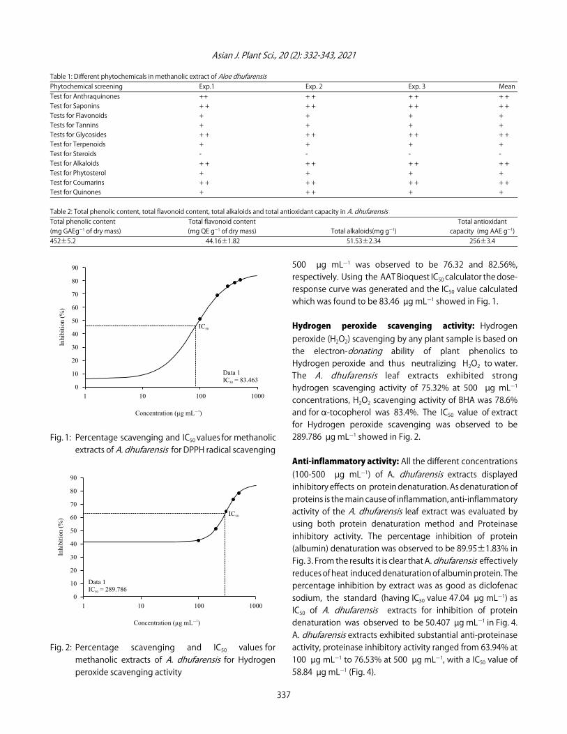
Table 1: Different phytochemicals in methanolic extract of Aloe dhufarensisPhytochemical screening Exp.1 Exp. 2 Exp. 3 MeanTest for Anthraquinones ++ + + + + + +Test for Saponins + + + + + + + +Tests for Flavonoids + + + +Tests for Tannins + + + +Tests for Glycosides + + + + + + + +Test for Terpenoids + + + +Test for Steroids - - - -Test for Alkaloids + + + + + + + +Test for Phytosterol + + + +Test for Coumarins + + + + + + + +Test for Quinones + + + + +
Table 2: Total phenolic content, total flavonoid content, total alkaloids and total antioxidant capacity in A. dhufarensisTotal phenolic content Total flavonoid content Total antioxidant(mg GAEgG1 of dry mass) (mg QE gG1 of dry mass) Total alkaloids(mg gG1) capacity (mg AAE gG1)452±5.2 44.16±1.82 51.53±2.34 256±3.4
Fig. 1: Percentage scavenging and IC50 values for methanolicextracts of A. dhufarensis for DPPH radical scavenging
Fig. 2: Percentage scavenging and IC50 values formethanolic extracts of A. dhufarensis for Hydrogenperoxide scavenging activity
500 µg mLG1 was observed to be 76.32 and 82.56%,respectively. Using the AAT Bioquest IC50 calculator the dose-response curve was generated and the IC50 value calculatedwhich was found to be 83.46 µg mLG1 showed in Fig. 1.
Hydrogen peroxide scavenging activity: Hydrogenperoxide (H2O2) scavenging by any plant sample is based onthe electron-donating ability of plant phenolics toHydrogen peroxide and thus neutralizing H2O2 to water.The A. dhufarensis leaf extracts exhibited stronghydrogen scavenging activity of 75.32% at 500 µg mLG1
concentrations, H2O2 scavenging activity of BHA was 78.6%and for "-tocopherol was 83.4%. The IC50 value of extractfor Hydrogen peroxide scavenging was observed to be289.786 µg mLG1 showed in Fig. 2.
Anti-inflammatory activity: All the different concentrations(100-500 µg mLG1) of A. dhufarensis extracts displayedinhibitory effects on protein denaturation. As denaturation ofproteins is the main cause of inflammation, anti-inflammatoryactivity of the A. dhufarensis leaf extract was evaluated byusing both protein denaturation method and Proteinaseinhibitory activity. The percentage inhibition of protein(albumin) denaturation was observed to be 89.95±1.83% inFig. 3. From the results it is clear that A. dhufarensis effectivelyreduces of heat induced denaturation of albumin protein. Thepercentage inhibition by extract was as good as diclofenacsodium, the standard (having IC50 value 47.04 µg mLG1) asIC50 of A. dhufarensis extracts for inhibition of proteindenaturation was observed to be 50.407 µg mLG1 in Fig. 4.A. dhufarensis extracts exhibited substantial anti-proteinaseactivity, proteinase inhibitory activity ranged from 63.94% at100 µg mLG1 to 76.53% at 500 µg mLG1, with a IC50 value of58.84 µg mLG1 (Fig. 4).
337
90
80
70
60
50
40
30
20
10
0
1 10 100 1000
Concentration (µg mL )�
1
Inhib
itio
n (
%)
IC50
Data 1IC = 83.46350
90
80
70
60
50
40
30
20
10
0
1 10 100 1000
Concentration (µg mL )�
1
Inhib
itio
n (
%)
IC50
Data 1IC = 289.78650

Asian J. Plant Sci., 20 (2): 332-343, 2021
Fig. 3: Percentage inhibition of protein denaturation andIC50 values for methanolic extracts of A. dhufarensis forinhibition of protein denaturation
Fig. 4: Percentage proteinase inhibitory activity and IC50values for methanolic extracts of A. dhufarensis forproteinase inhibitory activity
The Antimicrobial activity of methanolic A. dhufarensisleaf extracts of were estimated against 4 selected bacterialstrains employing disc diffusion method and the activity wasdetermined with regard to the zone of inhibition. Themethanolic leaf extracts of A. dhufarensis exhibitedconsiderable antibacterial capacity against all of the 4 testedbacteria showed in Table 3. Leaf extracts demonstratedhighest antibacterial activity against the gram -ve E. coliwith a Zone Of Inhibition (ZOI) of 13 mm, followed byS. aureus, P aeruginosa and least inhibition was observed forB. subtilis (7 mm).
Minimal inhibitory concentration observation: As minimuminhibitory concentration is the lowest concentration of any
Table 3: Inhibition zone of methanolic extract in A. dhufarensis with differentbacterial strains
Bacterial strain Inhibition zone (mm)Bacillus subtilis 7.0±0.272Staphylococcus aureus 10.5±0.404Escherichia coli 13.0±0.233Pseudomonas aeruginosa 8.0±0.360
chemical/compound at which a given microbe couldn’tdisplay any noticeable growth after 24 hrs of incubation.All 4 bacterial strains showed visible inhibition. The MICvalues for E. coli were the least at 250 µg mLG1 of the extract, for S. aureus and P aeruginosa, the MIC value was500 µg mLG1 of the extract. Leaf extract was found to be leasteffective against B. subtilis (MIC 1000 µg mLG1).
DISCUSSION
The present study was undertaken to validate thetraditional use of A. dhufarensis in wound healing, to treatdiabetes, headaches, fever, skin disorders, aching limbs andconstipation as the pharmacological effects of this importantspecies have not been fully explored.An elevated oxidative stress because of abundant
formation of reactive oxygen species and inducedinflammation has been identified are the two major factorswhich play a definitive role in development and gradualadvancement of various life-endangering ailments likeDiabetes mellitus, neurodegenerative and wound infection36.Chronic or sporadic hyperglycaemia triggers various metabolicsignalling pathways, inducing progressive inflammation invessels and nerves, overproduction of reactive oxygen species,leading to oxidative stress, cytokines secretion and cell deathconsequently leading to diabetic complications37,38.Wound healing is a complex progressive mechanism
involving a succession of overlapping events involving plateletinitiated haemostasis39,40, leading to coagulation under theinfluence of mediators and formation of fibrin clot. FurtherPlatelet degranulation influences activation and infiltrationof pro-inflammatory leukocytes, such as neutrophils,Monocytes and macrophages leading to the inflammatoryphase. These leukocytes play role in the next proliferativephase. Macrophages and neutrophils produce manyinflammatory mediators and cytokines like interleukin-1$,interleukin (IL)-6, tumor necrosis factor-" (TNF-") and NitricOxide (NO)41. Under normal wound physiology after carryingvarious functions neutrophils undergo apoptosis triggering aprogression out of the inflammatory phase. However in somecases neutrophils can generate excessive of NO and otherunstable reactive oxygen radicals which damages cell
338
100
90
80
70
60
50
40
30
20
10
0
1 10 100 1000
Concentration (µg mL )�
1
Inhib
itio
n (
%)
IC50
Data 1IC = 50.40750
90
80
70
60
50
40
30
20
10
0
1 10 100 1000
Concentration (µg mL )�
1
Inhib
itio
n (
%) IC50
Data 1IC = 58.84050
Asian J. Plant Sci., 20 (2): 332-343, 2021
membrane, destroying ECM, in normal tissues around thewound and thus triggering extra generation of pro-inflammatory mediators like IL-1 and TNF-" leading toamplified inflammation and impaired wound healing42. Theunstable reactive free radicals donates or abstracts electronsfrom DNA, proteins and fatty acids of the cell membranes, thusaltering protein structure and their functionality, fragmentingthe membranes and thus disrupting the proliferation of newcells at the site of healing. The inhibition of pro-inflammatorymediators like TNF-" may regulate the process of woundhealing43. Oxygen radicals are toxic waste products whichproduce oxidative stress at the time of the inflammatoryphase of wound healing. Overexposure to oxidative stressattributable to high levels of reactive oxygen species results inimpaired wound healing44. Thus the radical scavenging agentsand anti-inflammatory agents can be effective in woundhealing process.Several phytochemicals like phenols and flavonoids,
alkaloids and triterpenes are important natural antioxidantscapable of scavenging reactive free radicals, inhibitingoxidation and preventing cellular damages due to enhancedoxidative stress45,46. The phytochemical screening of leaves ofA. dhufarensis extracts exhibited the presence substantialamount of anthraquinones, saponins, coumarins, alkaloids,flavonoids, phenols and tannins.Anthraquinones found in latex and gel are well known for
their laxatives and potent purgative effect due to theirstimulation of mucus secretion and increased intestinalperistalsis47 and are also known for powerful analgesiceffect48,49. Presence of substantial amount of anthraquinonesin A. dhufarensis authenticates its traditional use forconstipation, headaches, fever and aching limbs.Appreciably high amount of saponins, were observed
in A. dhufarensis. Saponins exhibit substantial anti-inflammatory,50 anti-diabetic, antibacterial51 and antisepticactivity52. Saponins exert their potential anti-diabetic effect byrejuvenating insulin, resulting in lowered blood glucose level,elevated plasma insulin level, stimulating release of insulin(from pancreas) and by blocking glucose formation in thebloodstream53. Antibacterial potential of saponins is ascribedto their capability to cause excessive discharge of someenzymes and specific proteins from within the cell54.Plant polyphenols are also found to accelerate the wound
healing process by exhibiting significant initially required pro-inflammatory activity, proliferation of fibroblasts, formation ofnew blood vessels and amplification of new dermal cells55.Important flavonoids like catechin, luteolin, rutin and
apigenin are established for their wound healing properties 56
through inhibition of excessive fibroblast growth, increased
collagen synthesis, improving cross-linking of collagen andinhibiting the breakdown of soluble collagen57. Flavonoidsalong with saponins by inhibiting the enzymes involved inpro-inflammatory mediators proliferation are known to playsignificant part in wound healing and health promotion58,59.Flavonoids are widely recognized for their antibacterial,diuretic, laxative antispasmodic and cytotoxic effects. Phenolicacids along with alkaloids exhibit effective analgesicproperties60.Polyphenols are the major antioxidant compounds found
in plants, attributing their antioxidant potential to their redoxproperties. Antioxidant are substances that inhibits theaccumulation of free radicals like hydroxyl radicals (OH-),superoxide radical anion (O2CG) and other reactive oxygen H2O2and nitrogen species like NO and N2O3, respectively61 TheseROS at elevated concentrations generate oxidative stressresulting in various detrimental effects, like activation ordeactivation of enzymes, altering membrane permeability dueto lipid peroxidation, nucleic acids and altered lipid-proteininteraction62,63. Multiple OH group of phenolic compoundsespecially phenolic acids, imparts strong anti-oxidant potentialto plant phenolics by neutralizing the free radicals like O2CGand HOC and scavenging other ROS64. Other type of phenoliccompounds for example flavonoids, exert their anti-oxidantpotential by activating antioxidants enzymes, inhibitoxidases especially nitric oxide and peroxidase, deescalate"-tocopherol radicals and chelate metal catalyst65. Accordingto the present study, phenolic compounds are the present insubstantial amount in A. dhufarensis and are capable ofdonating hydrogen atom to the ROS thus contributing to thepharmacological effects associated with this species.Results verify that A. dhufarensis leaf methanolic extracts
have proton-donating abilities and potent antioxidant abilityas they stabilize the free radical of DPPH to and reducing it todiphenylpicryl hydrazine. Similar findings were observed3 in A.dhufarensis. IC50 value is negatively correlated with theantioxidant activity, the less is the IC50 value of extract, thegreater is its antioxidant potential.Within cells H2O2 is generated during the reduction of O2
to water via one-electron successive pathway. Wheremolecular oxygen is first changed into O2CG (superoxide anionradical) and this radical accepts one electron and two protons,yields hydrogen peroxide (H2O2).Hydrogen peroxide, is an unstable and a reactive oxygen
species with an important property to freely cross cellmembranes and by accepting one electron dissociate intohydroxyl radical (HOC) and (OHG) ion66. Most of the detrimentaleffects attributed to hydrogen peroxide like lipid peroxidationin biological membranes, disrupting protein structure by
339
Asian J. Plant Sci., 20 (2): 332-343, 2021
reduction of disulphide bonds are mediated via the hydroxylradical67. Antioxidant capability of a plant extract is ascribed totheir phenolics which by donating electron can reducehydroxyl radical to H2O. Low IC50 value of extract in the presentstudy indicates a strong hydroxyl radical scavenging capacityof A. dhufarensis phenolics.As discussed above Inflammation and escalated oxidative
stress is associated with a number of life-long diseases.Inflammation, a critical natural counter action of body immunesystem to injury or stress occurs via creation of certainpro-inflammatory cytokines such as IL-1$, interleukin-6 andTNF-" and their interaction with their specific receptors likeinterleukin-1 receptor and tumor necrosis factor receptor41.Denaturation of proteins is one of the prime attribute leadingto inflammation and the inhibition of protein denaturationmay help in averting inflammatory conditions68. Proteases arecatalytic enzymes hydrolysing peptide bonds of proteins andare pivotal in inflammation as the leukocytes proteinasecarries out tissue damage during an inflammatory response.Inhibition of protease activity will prevent inflammation.69
Considerable inhibition of protein denaturation and ofprotease activity by A. dhufarensis leaf extract confersconsiderable anti-inflammatory activity to the extract.Phytochemical like tannins via inhibition of inflammatory
mediators like cytokines and COX-270, Saponins by inhibitingnitric oxide (NO) by down regulation of lipopolysaccharideinduced iNOS expression50, phenolic acids by deterringTNF-" expression71, Flavonoids and coumarins by haltingcyclooxygenase and the lipoxygenase pathways ofarachidonic acid metabolism72 are effective anti-inflammatoryagents. Strong presence of these phytochemicals impartspotent anti-inflammatory potential to A. dhufarensis. Similarresults were obtained73,74 in Aloe ferox and in Aloe vera.Good antimicrobial activity of A. dhufarensis against all 4
bacterial strains can also be ascribed to the existence ofsaponins, alkaloids, tannins, flavonoids and terpenoids.Saponins and terpenoids exhibit their antimicrobial effect byimpairing bacterial cell membranes75,76, Alkaloids by increasingmembrane permeability through protein denaturation77 andflavonoids by suppressing nucleic acid synthesis and effectingthe membrane permeability78. Many previous studies onvarious species of genus Aloe has validated their antimicrobialactivity79-81.
CONCLUSION
This work reinforces the scientific basis for the establishedtraditional use of A. dhufarensis as an important healing herbas the results depicts strong radical scavenging capacity,
potent anti-inflammatory and also antibacterial potentialof leaf extracts of A. dhufarensis due to presence ofsubstantial amount of anthraquinones, saponins, phenolicacids, alkaloids and flavonoids. The encouraging findingsemphasizes on the possibility of the mainstreamcommercial exploration of the pharmaceutical potentialof A. dhufarensis in parallel to other commercial species of theAloe genus.
SIGNIFICANCE STATEMENT
This study discovered the untapped pharmaceutical andpharmacological potential of an endemic medicinal herb thatcan be commercially explored on national and internationalscale. This study will help the researchers to uncover thecritical areas of plant stress physiology under specializedrealms of microenvironment and adaptive response viaelevated generation of secondary metabolites.
ACKNOWLEDGMENT
The authors are grateful to Herbarium Staff at Life scienceunit, Sultan Qaboos University for access to herbarium plantspecimens for comparison.
REFERENCES
1. Mosti, S., M. Raffaelli and M. Tardelli, 2012. Contribution to theflora of central-southern Dhofar (Sultanate of Oman). Webbia,67: 65-91.
2. Ghazanfar, S.A., 1998. Status of the flora and plantconservation in the sultanate of Oman. Biol. Conserv.,85: 287-295.
3. Marwah, R.G., M.O. Fatope, R.A. Mahrooqi, G.B. Varma, H.A.Abai and S.A.S. Al-Burtamani, 2007. Antioxidant capacity ofsome edible and wound healing plants in Oman. Food Chem.,101: 465-470.
4. Hussain, F., Z. Rana, H. Shafique, D. Malik and Z. Hussain, 2017.Phytopharmacological potential of different species of Morusalba and their bioactive phytochemicals: A review. Asian Pac.J. Trop. Biomed., 7: 950-956.
5. Ali, S., M.R. Khan, M. Sajid and Z. Zahra, 2018. Phytochemicalinvestigation and antimicrobial appraisal of Parrotiopsisjacquemontiana (Decne) Rehder. BMC Complement. Altern.Med., 18: 43-45.
6. Cock, I.E., 2015. The genus aloe: Phytochemistry andtherapeutic uses including treatments for gastrointestinalconditions and chronic inflammation. Prog. Drug Res.,70: 179-235.
340

Asian J. Plant Sci., 20 (2): 332-343, 2021
7. Tungmunnithum, D., A. Thongboonyou, A. Pholboon andA. Yangsabai, 2018. Flavonoids and other phenoliccompounds from medicinal plants for pharmaceutical andmedical aspects: An overview. Medicines, Vol. 5, No. 3. 10.3390/medicines5030093.
8. Santos Sánchez, N., R. Salas-Coronado, C. Villanueva andB. Hernandez-Carlos, 2019. Antioxidant Compounds and Their Antioxidant Mechanism. In: Antioxidants, Shalaby, E. (Ed.).,IntechOpen, London, UK, pp: 1-28.
9. Gulcin, ¤., 2020. Antioxidants and antioxidant methods: Anupdated overview. Arch. Toxicol., 94: 651-715.
10. Jung, H.A., M.Y. Ali and J.S. Choi, 2016. Promising inhibitoryeffects of anthraquinones, naphthopyrone and naphthaleneglycosides, from Cassia obtusifolia on "-glucosidase andhuman protein tyrosine phosphatases 1B. Molecules,Vol. 22. 10.3390/molecules22010028.
11. Fouillaud, M., M. Venkatachalam, E. Girard-Valenciennes,Y. Caro and L. Dufossé, 2016. Anthraquinones andderivatives from marine-derived fungi: Structural diversityand selected biological activities. Mar. Drugs, Vol. 14. 10.3390/md14040064.
12. Tikhomirov, A.S., A.A. Shtil and A.E. Shchekotikhin, 2018.Advances in the discovery of anthraquinone-basedanticancer agents. Recent Pat. Anti-Cancer Drug Discovery,13: 159-183.
13. Park, J.G., S.C. Kim, Y.H. Kim, W.S. Yang and Y. Kim et al., 2016.Anti-inflammatory and antinociceptive activities ofanthraquinone-2-carboxylic acid. Mediators Inflamm., Vol.2016. 10.1155/2016/1903849.
14. Kemegne, G.A., P. Mkounga, J.J.E. Ngang, S.L.S. Kamdem andA.E. Nkengfack, 2017. Antimicrobial structure activityrelationship of five anthraquinones of emodine type isolatedfrom Vismia laurentii. BMC Microbiol., Vol. 17. 10.1186/s12866-017-0954-1.
15. Salehi, B., S. Albayrak, H. Antolak, D. Kregiel andE. Pawlikowska et al., 2018. Aloe genus plants: From farm tofood applications and phytopharmacotherapy. Int. J. Mol. Sci.,Vol. 19. Aloe genus plants: From farm to food applicationsand phytopharmacotherapy. Int. J. Mol. Sci., Vol. 19. 10.3390/ijms19092843.
16. Hotti, H., S.T. Häkkinen, T.S. Laakso and H. Rischer, 2017.Polyketide-derived alkaloids and anthraquinones in Aloeplants and cell cultures. J. Plant Biotechnol. Res., 1: 1-15.
17. Moore, B.D., R.L. Andrew, C. Külheim and W.J. Foley, 2013.Explaining intraspecific diversity in plant secondarymetabolites in an ecological context. New Phytol.,201: 733-750.
18. Sampaio, B.L., R. Edrada-Ebel and F.B. Da Costa 2016.Effect of the environment on the secondary metabolic profile of Tithonia diversifolia: A model for environmentalmetabolomics of plants. Sci. Rep., Vol. 6. 10.1038/srep29265.
19. Sherwani, N. and S.A. Farooq, 2019. Impact of habitatheterogeneity on growth dynamics and physiologicalresponses of Dipterygium glaucum. Asian J. Plant Sci.,18: 75-84.
20. Wink, M., 2003. Evolution of secondary metabolites from anecological molecular phylogenic perspective. Phytochem.,64: 3-19.
21. Wiens, J.J., 2011. The niche, biogeography and speciesinteractions. Philos. Trans. Royal Soc., 366: 2336-2350.
22. Hildebrandt, A. and E.A.B. Eltahir, 2006. Forest on the edge:Seasonal cloud forest in Oman creates its own ecologicalniche. Geophys. Res. Lett., Vol. 31. 10.1029/2006GL026022.
23. Bauer, A.W., W.M.M. Kirby, J.C. Sherris and M. Turck, 1966.Antibiotic susceptibility testing by a standardized single diskmethod. Am. J. Clin. Pathol., 45: 493-496.
24. Eloff, J.N., 1998. A sensitive and quick microplate method todetermine the minimal inhibitory concentration of plantextracts for bacteria. Planta Medica, 64: 711-713.
25. Sreedharan, S., A. Gothe, K. Aier, K. Shivasharanappa, K.P. Kumar and S.J. Patil, 2019. Bioactive molecules andantimicrobial studies of Indian traditional medicinal plantRhus semialata seeds. Res. J. Medic. Plants, 13: 10-17.
26. Okukpe, K.M., A.A. Adeloye, M.A. Belewu, O.I. Alli,O.A. Adeyina and A.A. Annongu, 2012. Investigation ofphytohormonal potential of some selected tropical plants.Res. J. Med. Plant, 6: 425-432.
27. Aziz, M.A., 2015. Qualitative phytochemical screening andevaluation of anti-inflammatory, analgesic and antipyreticactivities of Microcos paniculata barks and fruits. J. IntegrMed., 13: 173-184.
28. Singleton, V.L., R. Orthofer and R.M. Lamuela-Raventos, 1999.Analysis of total phenols and other oxidation substrates andantioxidants by means of Folin-Ciocalteu reagent. MethodsEnzymol., 299: 152-178.
29. Zhishen, J., T. Mengcheng and W. Jianming, 1999. Thedetermination of flavonoid contents in mulberry and theirscavenging effects on superoxide radicals. Food Chem.,64: 555-559.
30. Prieto, P., M. Pineda and M. Aguilar, 1999.Spectrophotometric quantitation of antioxidant capacitythrough the formation of a phosphomolybdenum complex:Specific application to the determination of vitamin E. Anal.Biochem., 269: 337-341.
31. Liyana-Pathirana, C.M. and F. Shahidi, 2005. Antioxidantactivity of commercial soft and hard wheat (Triticumaestivum L.) as affected by gastric pH conditions. J. Agric.Food Chem., 53: 2433-2440.
32. Lovecká, P., A. Svobodová, A. Macçrková, B. Vrchotová,K. Demnerová and Z. Wimmer, 2020. Decorative magnoliaplants: A comparison of the content of their biologicallyactive components showing antimicrobial effects. Plants,Vol. 9. 10.3390/plants9070879.
341

Asian J. Plant Sci., 20 (2): 332-343, 2021
33. Ruch, R.J., S.J. Cheng and J.E. Klaunig, 1989. Prevention ofcytotoxicity and inhibition of intercellular communication byantioxidant catechins isolated from Chinese green tea.Carcinogenesis, 10: 1003-1008.
34. Chandra, S., P. Chatterjee, P. Dey and S. Bhattacharya, 2012.Evaluation of anti-inflammatory effect of ashwagandha: Apreliminary study in vitro. Pharmacogn. J., 4: 47-49.
35. Gunathilake, K., K. Ranaweera and H. Rupasinghe, 2018. In vitro anti-inflammatory properties of selectedgreen leafy vegetables. Biomedicines, Vol. 6. 10.3390/biomedicines6040107.
36. Burgos-Morón, E., Z. Abad-Jiménez, A.M. Marañón,F. Iannantuoni, I. Escribano-Lopez, S. Lopez-Domenech andC. Salom, 2019. Relationship between oxidative stress, ERstress and inflammation in type 2 diabetes: The battlecontinues. J. Clin. Med., Vol. 8. 10.3390/jcm8091385.
37. Volpe, C.M.O., P.H. Villar-Delfino, P.M.F. dos Anjos andJ.A. Nogueira-Machado, 2018. Cellular death, reactive oxygenspecies (ROS) and diabetic complications. Cell Death Dis.,Vol. 9 10.1038/s41419-017-0135-z.
38. Dos Santos, J.M., S. Tewari and R.H. Mendes, 2019. Therole of oxidative stress in the development of diabetesmellitus and its complications. J. Diabetes Res., Vol. 2019.10.1155/2019/4189813.
39. de Oliveira Gonzalez, A.C., T.F. Costa, Z. de Araújo Andradeand A.R.A.P. Medrado, 2016. Wound healing - A literaturereview. An. Bras. Dermatol., 91: 614-620.
40. Rittié, L., 2016. Cellular mechanisms of skin repair in humansand other mammals. J. Cell Commun. Signal., 10: 103-120.
41. Kany, S., J.T. Vollrath and B. Relja, 2019. Cytokines ininflammatory disease. Int. J. Mol. Sci., Vol. 20. 10.3390/ijms20236008.
42. Nguyen, G.T., E.R. Green and J. Mecsas, 2017. Neutrophils tothe ROScue: Mechanisms of NADPH oxidase activation andbacterial resistance. Front. Cell. Infect. Microbiol., Vol. 7.10.3389/fcimb.2017.00373.
43. Tan, W.S., P. Arulselvan, S.F. Ng, C.N.M. Taib, M.N. Sarian andS. Fakurazi, 2019. Improvement of diabetic wound healing bytopical application of Vicenin-2 hydrocolloid film on SpragueDawley rats. BMC Complement. Altern. Med., Vol. 19. 10.1186/s12906-018-2427-y.
44. Mizuta, M., S. Hirano, S. Ohno, I. Tateya, S.I. Kanemaru,T. Nakamura and J. Ito, 2012. Expression of reactive oxygenspecies during wound healing of vocal folds in a rat model.Ann. Otol. Rhinol. Laryngol., 121: 804-810.
45. Upadhyay, S. and M. Dixit, 2015. Role of polyphenols andother phytochemicals on molecular signaling. Oxid. Med. Cell.Longev., Vol. 2015. 10.1155/2015/504253.
46. Forni, C., F. Facchiano, M. Bartoli, S. Pieretti andA. Facchiano et al., 2019. Beneficial role of phytochemicals onoxidative stress and age-related diseases. BioMed. Res. Int.,Vol. 2019. 10.1155/2019/8748253.
47. Gorkom, B.A.P., E.G.E. Vries and K. Karrenbeld, 1999. Reviewarticle: Anthranoid laxatives and their potential carcinogeniceffects. Aliment. Pharmacol. Ther., 13: 443-452.
48. Malik, E.M. and C.E. Muller, 2016. Anthraquinones aspharmacological tools and drugs. Med. Res. Rev., 36: 705-748.
49. Ferreira, M.A.D., O.D.R.H. Nunes, J.B. Fontenele, O.D.L. Pessoa,T.L.G. Lemos and G.S.B. Viana, 2004. Analgesic and anti-inflammatory activities of a fraction rich in oncocalyxone Aisolated from Auxemma oncocalyx. Phytomedicine, 11: 315-322.
50. Jang, K., H.K. Kim, M.H. Han, Y.N. Oh and H. Yoon et al., 2013.Anti-inflammatory effects of saponins derived from the rootsof Platycodon grandiflorus in lipopolysaccharide-stimulatedBV2 microglial cells. Int. J. Mol. Med., 31: 1357-1366.
51. Sonfack, G., C.F. Tchinda, I.K. Simo, G.T.M. Bitchagno andB.K. Nganou et al., 2019. Saponin with antibacterial activityfrom the roots of Albizia adianthifolia. Nat. Prod. Res.,10.1080/14786419.2019.1672689.
52. Surjushe, A., R. Vasani and D.G. Saple, 2008. Aloe vera: A shortreview. Indian J. Dermatol., 53: 163-166.
53. Elekofehinti, O.O., 2015. Saponins: Anti-diabetic principlesfrom medicinal plants-a review. Pathophysiology, 22: 95-103.
54. Ravi, L., V. Manasvi and B.P. Lakshmi, 2016. Antibacterial andantioxidant activity of saponin from Abutilon indicum leaves.Asian J. Pharmaceut. Clin. Res., 9: 344-347.
55. Zhao, D., J.E. Simon and Q. Wu, 2020. A critical review ongrape polyphenols for neuroprotection: Strategies toenhance bioefficacy. Crit. Rev. Food Sci. Nutr., 60: 597-625.
56. Ginwala, R., R. Bhavsar, D.I. Chigbu, P. Jain and Z.K. Khan,2019. Potential role of flavonoids in treating chronicinflammatory diseases with a special focus on the anti-inflammatory activity of Apigenin. Antioxidants, Vol. 8.10.3390/antiox8020035.
57. Galicka, A. and J. Nazaruk, 2007. Stimulation of collagenbiosynthesis by flavonoid glycosides in skin fibroblasts ofosteogenesis imperfecta type I and the potential mechanismof their action. Int. J. Mol. Med., 20: 889-895.
58. Yahfoufi, N., N. Alsadi, M. Jambi and C. Matar, 2018. Theimmunomodulatory and anti-inflammatory role ofpolyphenols. Nutrients, Vol. 10. 10.3390/nu10111618.
59. Ibrahim, N.I., S.K. Wong, I.N. Mohamed, N. Mohamed,K.Y. Chin, S. Ima-Nirwana and A.N. Shuid, 2018. Woundhealing properties of selected natural products. Int. J. Environ.Res. Public Health, Vol. 15. 10.3390/ijerph15112360.
60. Boutennoun, H., L. Boussouf, A. Rawashdeh, K. Al-Qaoud,S. Abdelhafez, M. Kebieche and K. Madani, 2017. In vitrocytotoxic and antioxidant activities of phenolic componentsof Algerian Achillea odorata leaves. Arab. J. Chem.,10: 403-409.
61. Phaniendra, A., D.B. Jestadi and L. Periyasamy, 2015. Freeradicals: Properties, sources, targets and their implication invarious diseases. Indian J. Clin. Biochem., 30: 11-26.
342

Asian J. Plant Sci., 20 (2): 332-343, 2021
62. Steven, J.F., D.S. Kikuchi, M.S. Hernandes, Q. Xu andK.K. Griendling, 2018. Reactive oxygen species in metabolicand inflammatory signalling. Circ. Res., 122: 877-902.
63. He, L., T. He, S. Farrar, L. Ji, T. Liu and X. Ma, 2017. Antioxidantsmaintain cellular redox homeostasis by elimination ofreactive oxygen species. Cell. Physiol. Biochem., 44: 532-553.
64. Tan, B.L., M.E. Norhaizan, W.P.P. Liew and H.S. Rahman, 2018.Antioxidant and oxidative stress: A mutual interplay in age-related diseases. Front. Pharmacol., Vol. 9. 10.3389/fphar.2018.01162.
65. Li, J.K., X. Liu, W.M. Zeng and G.Z. Qiu, 2016. Natural plantpolyphenols for alleviating oxidative damage in man:Current status and future perspectives. Trop. J. Pharm. Res.,15: 1089-1098.
66. Marzo, N.D., E. Chisci and R. Giovannoni, 2018. The role ofhydrogen peroxide in redox-dependent signaling:Homeostatic and pathological responses in Mammalian cells.Cells, Vol. 7. 10.3390/cells7100156.
67. Kurutas, E.B., 2016. The importance of antioxidants whichplay the role in cellular response against oxidative/nitrosativestress: Current state. Nutr. J., Vol. 15. 10.1186/s12937-016-0186-5.
68. Elisha, I.L., J.P. Dzoyem, L.J. McGaw, F.S. Botha and J.N. Eloff,2016. The anti-arthritic, anti-inflammatory, antioxidantactivity and relationships with total phenolics and totalflavonoids of nine South African plants used traditionally totreat arthritis. BMC Complement. Altern. Med., Vol. 16.10.1186/s12906-016-1301-z.
69. Shaikh, F.K., S.W. Hamad, S.W. Hamad and A.A. Shinde,2019. In vitro screening of seed extracts of medicinal plantsfor protease inhibitory activity. Cihan Univ.-Erbil Scient. J.,3: 61-65.
70. Kargutkar, S. and S. Brijesh, 2018. Anti-inflammatoryevaluation and characterization of leaf extract of Ananascomosus. Inflammopharmacol, 26: 469-477.
71. Shahidi, F. and J. Yeo, 2018. Bioactivities of phenolics byfocusing on suppression of chronic diseases: A review. Int. J.Mol. Sci., Vol. 19. 10.3390/ijms19061573.
72. Yeon, J.Y., Y.J. Bae, E.Y. Kim and E.J. Lee, 2015. Associationbetween flavonoid intake and diabetes risk among theKoreans. Clin.Chim.Acta., 439: 225-230.
73. Mwale, M. and P.J. Masika, 2010. AAnalgesic andanti-inflammatory activities of Aloe ferox Mill. aqueousextract. Afr. J. Pharm. Pharmaco., 4: 291-297.
74. Paul, S., S. Dutta, T.K. Chaudhuri and S. Bhattacharjee, 2014.Anti-inflammatory and protective properties of Aloe vera leafcrude gel in carrageenan induced acute inflammatory ratmodels. Int. J. Pharm. Pharm. Sci., 6: 368-371.
75. Arabski, M., A. W“gierek-Ciuk, G. Czerwonka, A. Lankoff andW. Kaca, 2012. Effects of saponins against clinical E. colistrains and eukaryotic cell line. J. Biomed. Biotechnol.,Vol. 2012. 10.1155/2012/286216.
76. Khan, I. M., A. Ahhmed, H.J. Shin, S.J. Baek, Y.M. Kim andD.J. Kim, 2018. Green tea seed isolated saponins exertsantibacterial effects against various strains of gram positiveand gram negative bacteria, a comprehensive study In Vitroand In Vivo. Evid. Based Complementary Altern. Med.,10.1155/2018/3486106.
77. Gurrapu, S. and E. Mamidala, 2017. In vitro antibacterialactivity of alkaloids isolated from leaves of Eclipta albaagainst human pathogenic bacteria. Pharmacog. J.,9: 573-577.
78. Górniak, I., R. Bartoszewski and J. Króliczewski, 2019.Comprehensive review of antimicrobial activities of plantflavonoids. Phytochem. Rev., 18: 241-272.
79. Athiban, P.P., B.J. Borthakur, S. Ganesan and B. Swathika, 2012.Evaluation of antimicrobial efficacy of Aloe vera and itseffectiveness in decontaminating gutta percha cones.J. Conserv. Dent., 15: 246-248.
80. Jain, S., N. Rathod, R. Nagi, J. Sur and A. Laheji et al., 2016.Antibacterial effect of Aloe vera gel against oral pathogens:An in vitro study. J. Clin. Diagnostic Res., 10: 41-44.
81. Forno-Bell, N., S.A. Bucarey, D. García, D. Iragüen, O. Chacónand B.S. Martín, 2019. Antimicrobial effects caused by Aloebarbadensis Miller on bacteria associated with mastitis indairy cattle. Nat. Prod. Commun., 14: 66-70.
343