Embed Size (px)
Citation preview

Ion Beam Centre
www.surreyibc.ac.uk1
Evaluation of non-Rutherford alpha elastic scattering cross-sections for silicon
Alexander F. GurbichAlexander F. Gurbich11 & Chris Jeynes& Chris Jeynes
University of Surrey Ion Beam Centre, Guildford, England1On leave from :-
Institute of Physics & Power Engineering, Obninsk, Russia

Ion Beam Centre
www.surreyibc.ac.uk
Acknowledgements
The The Leverhulme Trust Leverhulme Trust ::-- Visiting Professorship for A.F.Gurbich: Visiting Professorship for A.F.Gurbich: also colleagues at Surrey: also colleagues at Surrey: Jeff Tostevin Jeff Tostevin & & Ed Simpson Ed Simpson exploring exploring complementary use of SigmaCalc (Gurbich) and AZURE (Azuma++)complementary use of SigmaCalc (Gurbich) and AZURE (Azuma++)
IAEAIAEA for supporting IBANDL and SigmaCalcfor supporting IBANDL and SigmaCalcwwwwww--nds.iaea.orgnds.iaea.org//ibandl ibandl && wwwwww--nds.iaea.orgnds.iaea.org//sigmacalcsigmacalc
Engineering & Physical Sciences Research Council (Engineering & Physical Sciences Research Council (EPSRCEPSRC, UK) for , UK) for supporting the EBS measurements made in September 2005 supporting the EBS measurements made in September 2005 (grant GR/R 50097)(grant GR/R 50097)
The The Royal Society Royal Society (UK) for funding A.F.Gurbich(UK) for funding A.F.Gurbich’’s visit to Surrey in 2005s visit to Surrey in 2005
This work has been supported by the European Community as an This work has been supported by the European Community as an Integrating Activity Integrating Activity ««Support of Public and Industrial Research Using Ion Support of Public and Industrial Research Using Ion Beam Technology (Beam Technology (SPIRITSPIRIT))»» under EC contract no. 227012under EC contract no. 227012

Ion Beam Centre
www.surreyibc.ac.uk
Contents
• Introduction – Materials Analysis• The 28Si()28Si reaction• Summary

Ion Beam Centre
www.surreyibc.ac.uk
Contents
• Introduction – Materials Analysis– Surrey Ion Beam Centre
• Materials Modification by Ion Implantation• Thin Film Depth Profiling by Ion Beam Analysis
– Ion Beam Analysis Overview• Atomic Excitation – PIXE• Nuclear Excitation – RBS, EBS, ERD, NRA• Scanning microbeam, channelling, external beam
– PIXE + Backscattering – self-consistent analysis– Accurate analysis!
• The 28Si()28Si reaction• Summary

Ion Beam Centre
www.surreyibc.ac.uk
Surrey Ion Beam Centre
Controllable Materials Modification
• Facilities– 0.4-2MV High Energy Implanter (1991)– 2-200kV High Current Implanter (1997)– Implantation 2keV 4MeV (up to 10mA)– Sample size ¼mm2 to ¼m2
– Hot (1000ºC) or cold (~LN & 10K)– Sample chambers in class 100 clean room
• Applications– Ion Beam Synthesis
• Buried and surface oxides, silicides etc
– Ion Implantation– Defect Engineering– Proton beam lithography (using Tandem)
• potentially nm resolution to 10m depths

Ion Beam Centre
www.surreyibc.ac.uk
IBA equipment at Surrey
• 2 MV Tandem• Installed & commissioned 2002• up to: 4 MeV 1H+; 6 MeV 4He++; 12 MeV 197Au5+
• -ve beam injection• duoplasmatron source good for H-, He+
• For He- use Li vapour charge exchange• Nitrogen gas stripper at HV terminal• Pressure vessel SF6 at 6.5 bar
• 5 beam lines• Microbeam (1x1 m); nanobeam (30x30 nm ??)• Broad beam (100x100 m); external beam (50x50 m); PIGE/PINE line• Vertical beam line for radiation biology (500x500 nm)• 6-axis goniometer: 100 x 150 mm stage

Ion Beam Centre
www.surreyibc.ac.uk
Contents
• Introduction – Materials Analysis– Surrey Ion Beam Centre
• Materials Modification by Ion Implantation• Thin Film Depth Profiling by Ion Beam Analysis
– Ion Beam Analysis Overview• Atomic Excitation – PIXE• Nuclear Excitation – RBS, EBS, ERD, NRA• Scanning microbeam, channelling, external beam
– PIXE + Backscattering – self-consistent analysis– Accurate analysis!
• The 28Si()28Si reaction• Summary

Ion Beam Centre
www.surreyibc.ac.uk
RBSEnergy of scattered protons or He give light element composition and elemental depth profiles
Ion Beam Analysis Methods
PIXECharacteristic X-ray emissionSimultaneous part-per-million detection of trace elements from Na to U
Sample0.2 – 4 MeV protons or 0.2 – 6 MeV He++
or 0.2 – 12 MeV Au5+
or 3He (or D?) etc
PIGE/NRANuclear reactions give charac-teristic gamma rays and/or particles from light nuclei (e.g. Li, B, F)
ERDForward recoil of target atoms (particularly good for H, D profiling)
IBIL: IBIL: ion beam induced luminescenceion beam induced luminescenceIBIC: IBIC: ion beam induced current ion beam induced current STIM: STIM: scanning transmission ion microscopyscanning transmission ion microscopy

Ion Beam Centre
www.surreyibc.ac.uk
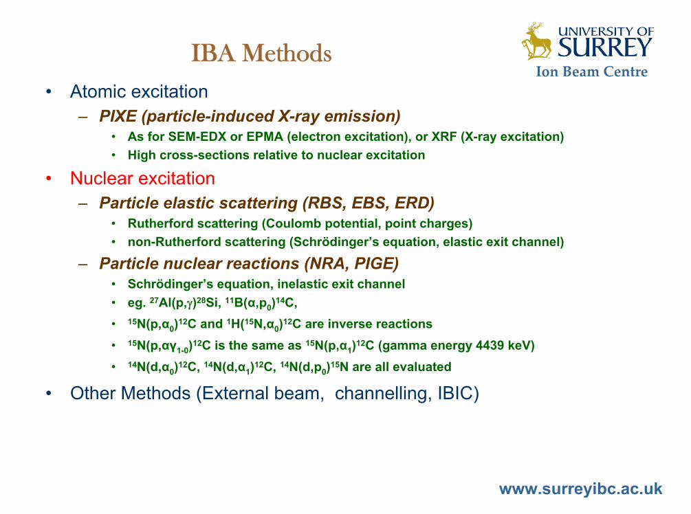
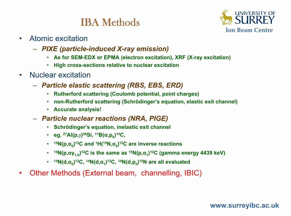
IBA Methods• Atomic excitation
– PIXE (particle-induced X-ray emission)• As for SEM-EDX or EPMA (electron excitation), XRF (X-ray excitation)• High cross-sections relative to nuclear excitation
• Nuclear excitation– Particle elastic scattering (RBS, EBS, ERD)
• Rutherford scattering (Coulomb potential, point charges)• non-Rutherford scattering (Schrödinger’s equation, elastic exit channel)
– Particle nuclear reactions (NRA, PIGE)• Schrödinger’s equation, inelastic exit channel• eg. 27Al(p,)28Si, 11B(α,p0)14C, • 15N(p,α0)12C and 1H(15N,α0)12C are inverse reactions• 15N(p,αγ1-0)12C is the same as 15N(p,α1)12C (gamma energy 4439 keV)• 14N(d,α0)12C, 14N(d,α1)12C, 14N(d,p0)15N are all evaluated
• Other Methods (External beam, channelling, IBIC)

Ion Beam Centre
www.surreyibc.ac.uk
PIXEPIXE
• Analogous to EDX using MeV protons or He• No primary electron Bremsstrahlung, so low detection limits (1-
10ppm)• Quantitative (with RBS)• Imaging resolution determined by beam size (not beam energy as in
EDX)• Very high cross-sections, so information is from femtograms of
material
SEM-EDS and PIXE spectra for a bulk bronze sample
Characteristic X-rayphoton
Ejected electron
Johansson and Campbell, PIXE, Wiley 1995
Direct comparison of SEM-EDS & PIXE
• Sensitivity• Scattering• Backscattered particles
Particle Induced X-ray Emission
Ion Beam Centre
www.surreyibc.ac.uk
IBA Methods• Atomic excitation
– PIXE (particle-induced X-ray emission)• As for SEM-EDX or EPMA (electron excitation), or XRF (X-ray excitation)• High cross-sections relative to nuclear excitation
• Nuclear excitation– Particle elastic scattering (RBS, EBS, ERD)
• Rutherford scattering (Coulomb potential, point charges)• non-Rutherford scattering (Schrödinger’s equation, elastic exit channel)
– Particle nuclear reactions (NRA, PIGE)• Schrödinger’s equation, inelastic exit channel• eg. 27Al(p,)28Si, 11B(α,p0)14C, • 15N(p,α0)12C and 1H(15N,α0)12C are inverse reactions• 15N(p,αγ1-0)12C is the same as 15N(p,α1)12C (gamma energy 4439 keV)• 14N(d,α0)12C, 14N(d,α1)12C, 14N(d,p0)15N are all evaluated
• Other Methods (External beam, channelling, IBIC)

Ion Beam Centre
www.surreyibc.ac.uk
RBSRBSRutherford BackScattering
• Energy of ions scattering from nuclear collisions depends on mass and depth
• Detection limit around 0.1%
• Depth profiling with depth resolution <20nm• Analytical cross-section (Coulomb potential)• Single scattering (cf electron backscatters in SEM)
proportional to Z2/E2
Spectrum of zirconia/silica multilayer optical coating (red), with DataFurnace fit (green)
•• Coulomb potential (accurate)potential (accurate)•• Perfect fitting of complex structures fitting of complex structures
(inverse problem solved)(inverse problem solved)
2MeV 4He
Incident Ion
To detector
Sample
C.Jeynes++ C.Jeynes++ Surface & Interface AnalysisSurface & Interface Analysis 3030 (2000) 237(2000) 237--242242

Ion Beam Centre
www.surreyibc.ac.uk
EBSEBSElastic (non-Rutherford) BackScattering
• Kinematics the same as for RBS • Scattering cross-sections not Coulomb• Can be fitted by solving Schrödinger’s equation • See SigmaCalc at www-nds.iaea.org/ibandl
Gurbich, NIM B268 (2010) 1703• Greatly enhanced cross-sections
often available for light elements• Cross-sections vary strongly with scattering angle
1752keV H+
= 1720
Mg1483keV
Mg1630keV
C O Mg
Benchmark experiment for evaluated Mg cross-sectionsGurbich & Jeynes, Nucl.Instrum.Methods B265 (2007) 447-452
Mg substrate+ 68.1015 C/cm2
+ 800.1015 MgO/cm2
Natural Mg(p,p) cross-sections
0
1
2
3
4
5
500 1000 1500 2000 2500
Proton Energy (keV)
Rel
ativ
e to
Rut
herfo
rd
172deg148deg
Natural Mg(p,p) cross-sections
0
5
10
15
20
1460 1470 1480 1490 1500Proton Energy (keV)
Rel
ativ
e to
Rut
herfo
rd
172deg148deg
SigmaCalc scattering cross-sections for natural Mg (the isotopes behave differently) at two different scattering angles
Sharp resonance at 1483keV, 2 angles (FWHM 400eV)
For sharp resonances one must correctly calculate energy straggling in depth (this effect can be LARGE)
Barradas et al, NIM B247 (2006) 381

Ion Beam Centre
www.surreyibc.ac.uk
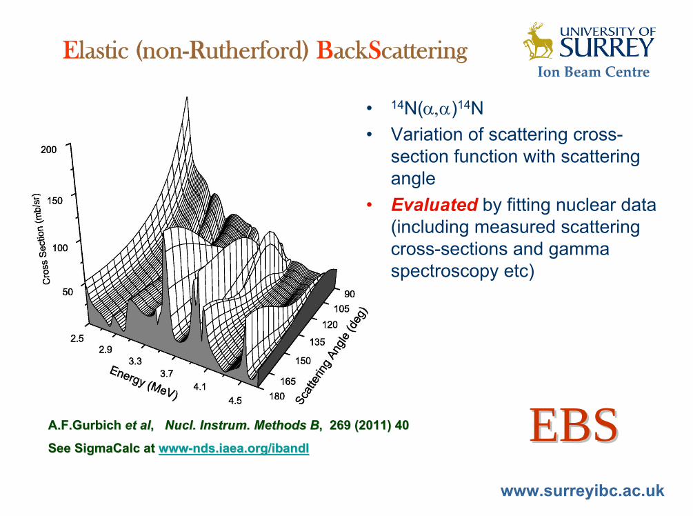
Elastic (non-Rutherford) BackScattering
EBS spectrum of 750nm TaNiC sputtered film on Si substrate
The Ta & Ni signals are Rutherford, but the C & Si signals are strongly non-Rutherford.
EBSEBSAbove: data & fit with Ta, Ni, C partial spectra shownBelow: simulation with RBS for Si & CRight: Depth profile obtained
Jeynes et al, NIM B161-163 (2000) 287
1.75 MeV H
TaNiC/Si
Si substrate
EBS signal (C & Si)
Ta
Ni
C
RBS
RBS simulation (C & Si)
• Cross-sections not analytical and must be measured• Light elements in heavy matrix can be measured (in
this case 6.6at%C in TaNiC on a silicon background signal)
• Very large cross-section enhancements, depending on target atom and beam energy
Ion Beam Centre
www.surreyibc.ac.uk
Elastic (non-Rutherford) BackScattering
• 14N()14N• Variation of scattering cross-
section function with scattering angle
• Evaluated by fitting nuclear data (including measured scattering cross-sections and gamma spectroscopy etc)
EBSEBSA.F.Gurbich A.F.Gurbich et alet al, , Nucl. Instrum. Methods BNucl. Instrum. Methods B, 269 (2011) 40, 269 (2011) 40
See SigmaCalc at See SigmaCalc at wwwwww--nds.iaea.org/ibandlnds.iaea.org/ibandl

Ion Beam Centre
www.surreyibc.ac.uk
Elastic (non-Rutherford) BackScattering
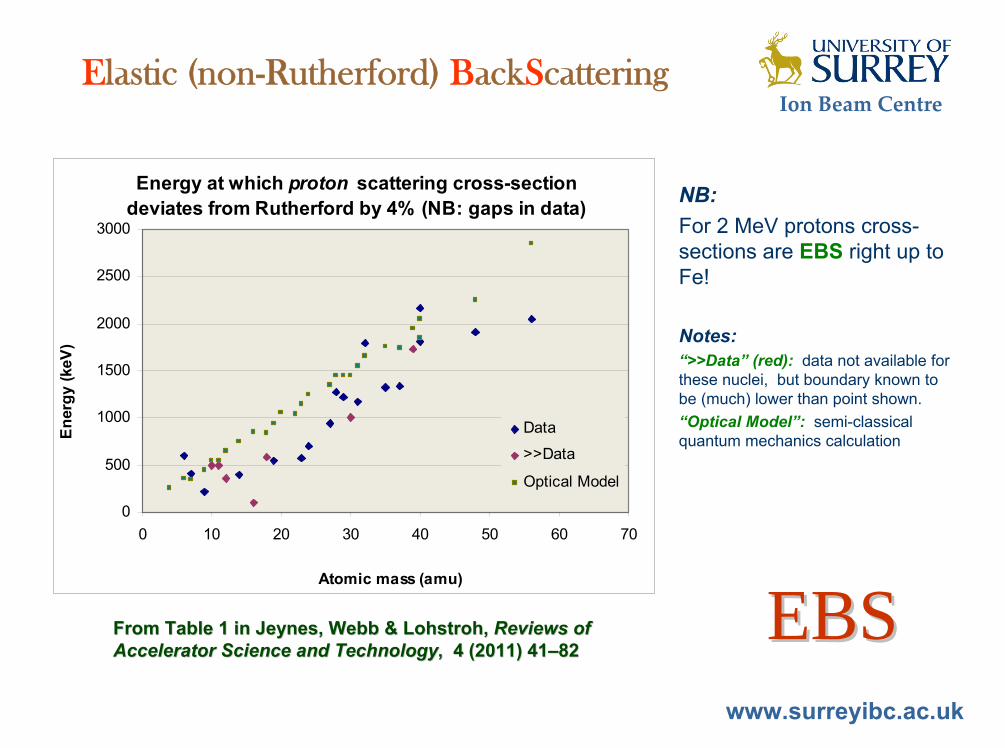
EBSEBSFrom Table 1 in Jeynes, Webb & Lohstroh, From Table 1 in Jeynes, Webb & Lohstroh, Reviews of Reviews of Accelerator Science and TechnologyAccelerator Science and Technology, 4 (2011) 41, 4 (2011) 41––8282
NB:For 2 MeV protons cross-sections are EBS right up to Fe!
Notes:“>>Data” (red): data not available for these nuclei, but boundary known to be (much) lower than point shown.“Optical Model”: semi-classical quantum mechanics calculation
Energy at which proton scattering cross-section deviates from Rutherford by 4% (NB: gaps in data)
0
500
1000
1500
2000
2500
3000
0 10 20 30 40 50 60 70
Atomic mass (amu)
Ener
gy (k
eV)
Data
>>Data
Optical Model

Ion Beam Centre
www.surreyibc.ac.uk
Elastic Recoil Detection
1.5MeV 1.5MeV 44He RBSHe RBSNormal incidenceNormal incidence
Glancing incidenceGlancing incidence
Simultaneous with:Simultaneous with:
ERDERDSiNSiNxx:H on Si:H on SiGa implant to form aGa implant to form a--GaNGaNxx??Barradas et al, NIM B148, 1999, 463
ERDERD

Ion Beam Centre
www.surreyibc.ac.uk
IBA Methods• Atomic excitation
– PIXE (particle-induced X-ray emission)• As for SEM-EDX or EPMA (electron excitation), XRF (X-ray excitation)• High cross-sections relative to nuclear excitation
• Nuclear excitation– Particle elastic scattering (RBS, EBS, ERD)
• Rutherford scattering (Coulomb potential, point charges)• non-Rutherford scattering (Schrödinger’s equation, elastic exit channel)
– Particle nuclear reactions (NRA, PIGE)• Schrödinger’s equation, inelastic exit channel• eg. 27Al(p,)28Si, 11B(α,p0)14C, • 15N(p,α0)12C and 1H(15N,α0)12C are inverse reactions• 15N(p,αγ1-0)12C is the same as 15N(p,α1)12C (gamma energy 4439 keV)• 14N(d,α0)12C, 14N(d,α1)12C, 14N(d,p0)15N are all evaluated
• Other Methods (External beam, channelling, IBIC)

Ion Beam Centre
www.surreyibc.ac.uk
1.5MeV 1.5MeV 44He RBSHe RBSNormal & glancingNormal & glancing
1.5MeV 1.5MeV 44He ERDHe ERDGlancingGlancing
Nuclear Reaction Analysis
Depth profiling of 300nm CH:DH film on SiDepth profiling of 300nm CH:DH film on Siusing simultaneous RBS/NRA/ERDusing simultaneous RBS/NRA/ERDand automatic spectrum inversion with and automatic spectrum inversion with DataFurnaceDataFurnace software software (Jeynes et al J.Phys.D 36, 2003, R97)(Jeynes et al J.Phys.D 36, 2003, R97)
0.7MeV 0.7MeV 33He RBSHe RBSNormal Normal
0.7MeV 0.7MeV 33He NRAHe NRA
d(d(33He,p)He,p)44HeHeQ=18.35MeVQ=18.35MeV
Normal & glancingNormal & glancing
CCsurfacesurface
H
D
C
D
NRANRA
Ion Beam Centre
www.surreyibc.ac.uk
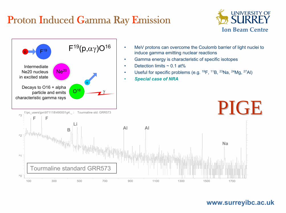
Proton Induced Gamma Ray Emission
• MeV protons can overcome the Coulomb barrier of light nuclei to induce gamma emitting nuclear reactions
• Gamma energy is characteristic of specific isotopes• Detection limits ~ 0.1 at%• Useful for specific problems (e.g. 19F, 11B, 23Na, 24Mg, 27Al)• Special case of NRA
p F19
O16
IntermediateNe20 nucleus
in excited state
Decays to O16 + alphaparticle and emits
characteristic gamma rays
Ne20
F19(p,)O16
^0
^1
^2
^3
100 300 500 700 900 1100 1300 1500 1700
f:\pc_users\jpn\971118\490001g4._ : Tourmaline std: GRR573
F F
B Al AlLi
Na
Tourmaline standard GRR573
PIGEPIGE
Ion Beam Centre
www.surreyibc.ac.uk
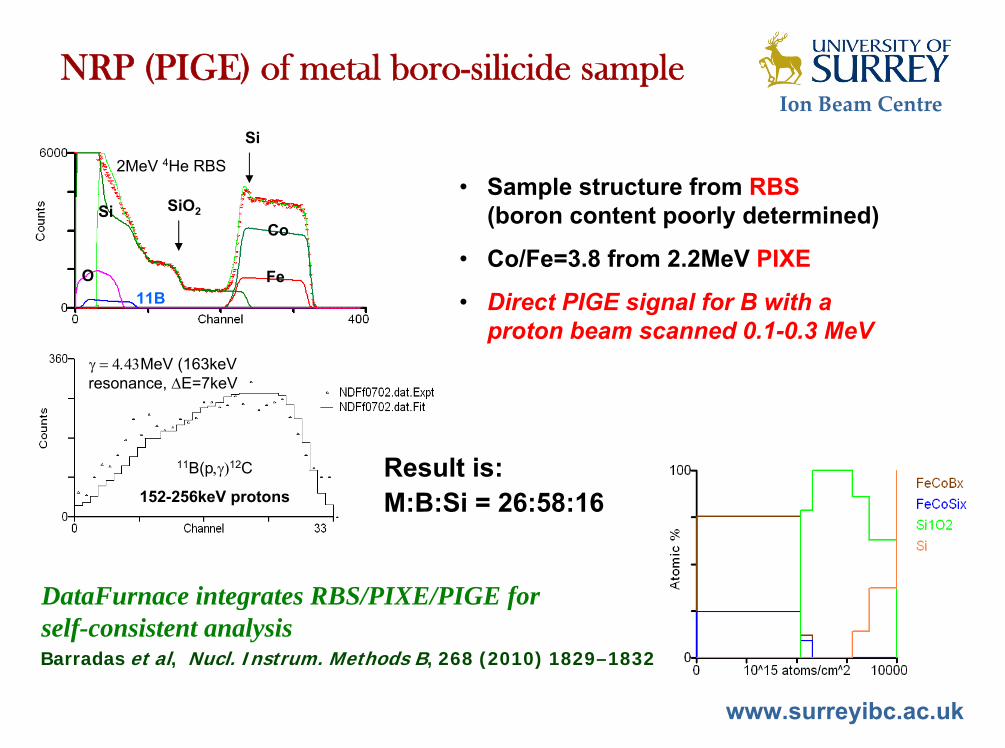
2MeV 4He RBS
Si
SiO2
O11B
SiCo
Fe
11B(p12C
152-256keV protons
MeV (163keV resonance, E=7keV
• Sample structure from RBS(boron content poorly determined)
• Co/Fe=3.8 from 2.2MeV PIXE
• Direct PIGE signal for B with a proton beam scanned 0.1-0.3 MeV
NRP (PIGE) of metal boro-silicide sample
DataFurnace integrates RBS/PIXE/PIGE for self-consistent analysis
Result is:M:B:Si = 26:58:16
Barradas et al, Nucl. Instrum. Methods B, 268 (2010) 1829–1832
Ion Beam Centre
www.surreyibc.ac.uk
IBA Methods• Atomic excitation
– PIXE (particle-induced X-ray emission)• As for SEM-EDX or EPMA (electron excitation), XRF (X-ray excitation)• High cross-sections relative to nuclear excitation
• Nuclear excitation– Particle elastic scattering (RBS, EBS, ERD)
• Rutherford scattering (Coulomb potential, point charges)• non-Rutherford scattering (Schrödinger’s equation, elastic exit channel)• Accurate analysis!
– Particle nuclear reactions (NRA, PIGE)• Schrödinger’s equation, inelastic exit channel• eg. 27Al(p,)28Si, 11B(α,p0)14C, • 15N(p,α0)12C and 1H(15N,α0)12C are inverse reactions• 15N(p,αγ1-0)12C is the same as 15N(p,α1)12C (gamma energy 4439 keV)• 14N(d,α0)12C, 14N(d,α1)12C, 14N(d,p0)15N are all evaluated
• Other Methods (External beam, channelling, IBIC)

Ion Beam Centre
www.surreyibc.ac.uk
External Beam
• 100 Reichmark bank note from Germany dated 1920
• Can we analyse the inks?
scanned area
Selection of PIXE Selection of PIXE elemental maps elemental maps (3x3 mm)(3x3 mm)
002
003
mercury
002
003
bariumRed ink (head of eagle)Red ink (head of eagle)
002
003
lead
002
003
chromium
Green ink (wavy linesGreen ink (wavy lines)
10 other elements detected10 other elements detectedAnalysis carried out in 10 minutes with no visible marking of thAnalysis carried out in 10 minutes with no visible marking of the papere paper

Ion Beam Centre
www.surreyibc.ac.uk
Channelling
Robinson & Oen, Robinson & Oen, Phys. RevPhys. Rev, 132 (1963) 2385 , 132 (1963) 2385
Projection of three calculated trajectories of1 keV Cu incident (at points marked with stars) along the (001) direction of a face centred cubic Cu crystal. The trajectories for 250 collisions are shown and the truncated Bohr potential is used.
Range in channelling can be up to 25 times “random” range

Ion Beam Centre
www.surreyibc.ac.uk
Channelling – crystalline damage example
Jeynes, Puttick, GJeynes, Puttick, Gäärtner rtner et alet al, , Nucl. Instrum. Methods BNucl. Instrum. Methods B, 118 (1996) 431 , 118 (1996) 431
Image of circular damage tracks in a silicon sample turned on an ultra-stiff lathe with a single-point diamond tool.Signal is the low-energy part of the RBS Si signal: high yield means high damage. 128×128 pixels, pixel area (23.4 μm)2, 0.15 nC/pixel.
This is plastic deformation of a brittlematerial, with an a-Si layer up to 350 nm thick over a dislocation array ~ 5.1010/cm2.
3 mm
Channelling normal1.2 MeV 4He+
40 m spot
c-Si
Ion Beam Centre
www.surreyibc.ac.uk
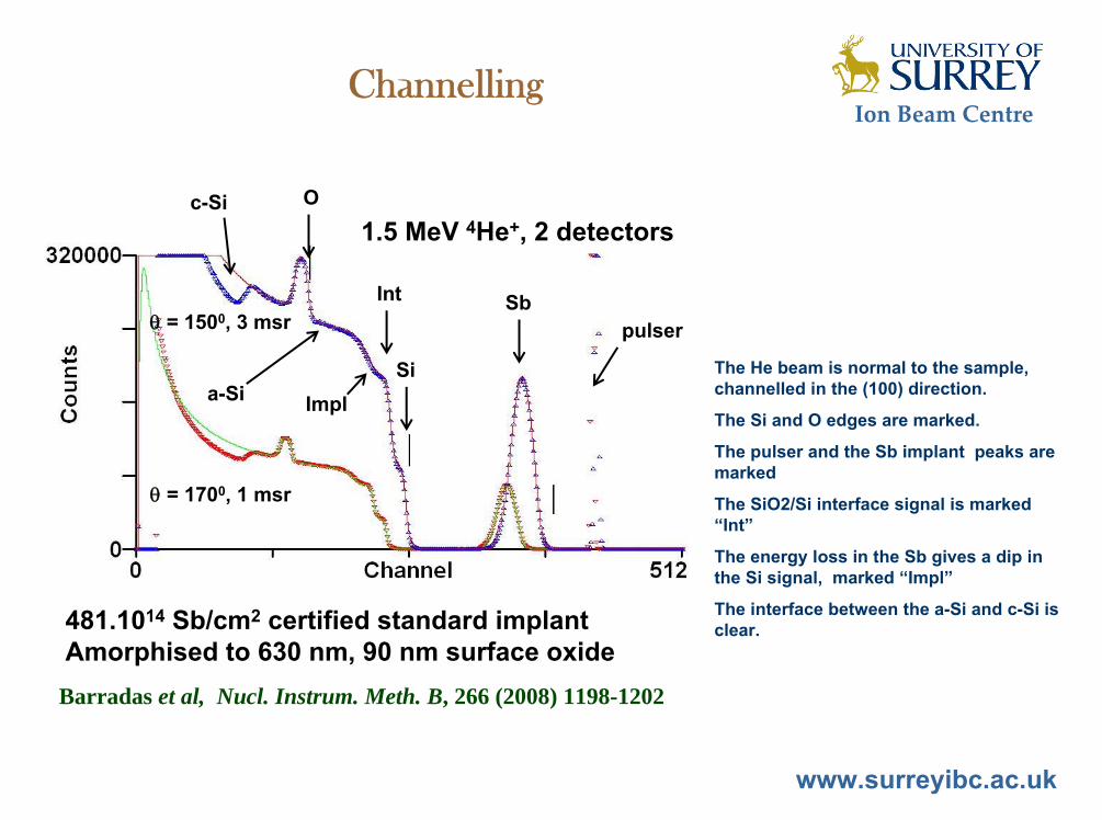
Channelling
Barradas et al, Nucl. Instrum. Meth. B, 266 (2008) 1198-1202
481.1014 Sb/cm2 certified standard implantAmorphised to 630 nm, 90 nm surface oxide
= 1500, 3 msr
= 1700, 1 msr
pulserSb
Si
O
Int
Impla-Si
c-Si1.5 MeV 4He+, 2 detectors
The He beam is normal to the sample, channelled in the (100) direction.
The Si and O edges are marked.
The pulser and the Sb implant peaks are marked
The SiO2/Si interface signal is marked “Int”
The energy loss in the Sb gives a dip in the Si signal, marked “Impl”
The interface between the a-Si and c-Si is clear.

Ion Beam Centre
www.surreyibc.ac.uk
Contents
• Introduction – Materials Analysis– Surrey Ion Beam Centre
• Materials Modification by Ion Implantation• Thin Film Depth Profiling by Ion Beam Analysis
– Ion Beam Analysis Overview• Atomic Excitation – PIXE• Nuclear Excitation – RBS, EBS, ERD, NRA• Scanning microbeam, channelling, external beam
– PIXE + Backscattering – self-consistent analysis– Accurate analysis!
• The 28Si()28Si reaction• Summary

Ion Beam Centre
www.surreyibc.ac.uk
Towards truly simultaneous PIXE and RBS analysis of layered objects in cultural heritage
C. Pascual-Izarra (Madrid), N. P. Barradas, M. A. Reis (Lisbon), C. Jeynes (Surrey), M. Menu, B. Lavdrine, J. J. Ezrati, S. Röhrs (Louvre)
Nucl. Instrum. Methods B261, 426-429 (2007)
NiepceNiepce’’s first Heliography:s first Heliography:Paysage Paysage àà SaintSaint--Loup de Varennes (1827)Loup de Varennes (1827)
Corrosion products demonstrated Corrosion products demonstrated by PIXE/RBS/EBS to be tin oxide in by PIXE/RBS/EBS to be tin oxide in a tin/lead matrixa tin/lead matrix
3 MeV H EBS
3 MeV H PIXE
3 MeV He RBSFitted depth
profile

Ion Beam Centre
www.surreyibc.ac.uk
Characterization of paint layers by simultaneous self-consistent fitting of RBS/PIXE spectra using simulated annealingL. Beck (Louvre), C. Jeynes (Surrey), N.P.Barradas (Lisbon)Nucl. Instrum. Methods B266, 1871-1874 (2008)
AGLAE : AccAGLAE : Accéélléérateur Grand rateur Grand Louvre dLouvre d’’Analyse Analyse ÉÉlléémentairementaire
““La BohLa Bohéémiennemienne”” , Frans Hals 1630, Frans Hals 1630
Ochre (haematite) Ochre (haematite) pigment located pigment located and quantified by and quantified by PIXE/RBS/EBSPIXE/RBS/EBS
3 MeV H EBS
Fitted depth profile
Ion Beam Centre
www.surreyibc.ac.uk
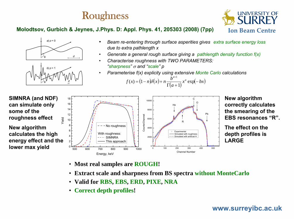
RoughnessMolodtsov, Gurbich & Jeynes, J.Phys. D: Appl. Phys. 41, 205303 (2008) (7pp)
• Beam re-entering through surface asperities gives extra surface energy loss due to extra pathlength x
• Generate a general rough surface giving a pathlength density function f(x) • Characterise roughness with TWO PARAMETERS:
“sharpness” and “scale” p• Parameterise f(x) explicity using extensive Monte Carlo calculations
exp1
1)(1
bxxabnxnxf a
a
SIMNRA (and NDF) can simulate only some of the roughness effect
New algorithm calculates the high energy effect and the lower max yield
New algorithm correctly calculates the smearing of the EBS resonances “R”.
The effect on the depth profiles is LARGE
• Most real samples are ROUGH!• Extract scale and sharpness from BS spectra without MonteCarlo• Valid for RBS, EBS, ERD, PIXE, NRA• Correct depth profiles!

Ion Beam Centre
www.surreyibc.ac.uk
RBS/EBS/PIXE measurement of single-walled carbon nanotube modification by nitric acid purification treatment
J.C.G. Jeynes, C. Jeynes, K.J. Kirkby, M. Rümmeli, S.R.P. Silva Nucl. Instrum. Methods B266, 1569-1573 (2008)
O quantified by PIXE/RBS/EBS in the O quantified by PIXE/RBS/EBS in the presence of mixed heavy metal presence of mixed heavy metal catalystcatalystcontent of CNT with heavy roughnesscontent of CNT with heavy roughness
Catalyst contains Pt, Rh, Re, Fe, Cu, Cl, Catalyst contains Pt, Rh, Re, Fe, Cu, Cl, Na. Roughness effects marked Na. Roughness effects marked ““RR””
SEM
RBS
SWCNT on mylar Fitted depth profileRBS/EBS & PIXE data & fits
2582keV H2582keV H

Ion Beam Centre
www.surreyibc.ac.uk
Characterisation of thin film chalcogenide PV materials using MeV ion beam analysis
• CIAS semiconductor on Mo electrode• Precursor material (not selenided yet)• Al invisible in backscattering• Strong layering: PIXE uninterpretable without
profile independently available• Differential PIXE to profile Al• Essential to fit roughness (non-uniform
thickness) to reproduce RBS spectra• Good fit essential for reliable profile
Chris Jeynes, G.Zoppi, I.Forbes, M.J.Bailey, N.PengIEEE Proceedings of Supergen Conference, Nanjing, April 2009, DOI:10.1109/SUPERGEN.2009.5348162
1.5MeV He RBS150O and 170O
Mo
CuInAl
0O beam in, 45O X-ray out
45O beam in, 0O X-ray out
He-PIXE simulated by equivalent-velocity H-PIXE
Ion Beam Centre
www.surreyibc.ac.uk
Elastic (non-Rutherford) BackScattering
EBSEBSFrom Table 1 in Jeynes, Webb & Lohstroh, From Table 1 in Jeynes, Webb & Lohstroh, Reviews of Reviews of Accelerator Science and TechnologyAccelerator Science and Technology, 4 (2011) 41, 4 (2011) 41––8282
NB:For 2 MeV protons cross-sections are EBS right up to Fe!
Notes:“>>Data” (red): data not available for these nuclei, but boundary known to be (much) lower than point shown.“Optical Model”: semi-classical quantum mechanics calculation
Energy at which proton scattering cross-section deviates from Rutherford by 4% (NB: gaps in data)
0
500
1000
1500
2000
2500
3000
0 10 20 30 40 50 60 70
Atomic mass (amu)
Ener
gy (k
eV)
Data
>>Data
Optical Model

Ion Beam Centre
www.surreyibc.ac.uk
Information in BS spectra with few countsInformation in BS spectra with few counts
N.P.Barradas, C. Jeynes, M. Jenkin, P.K. MarriottThin Solid Films 343-344 (1999) 31-34
• PIXE cross-sections >> RBS• 100 pA common for PIXE • Poor statistics in particle spectra!
This Bayesian analysis demonstrates that even spectra with very small collected charge have lots of information!
Don’t throw away good data!Always do BS with PIXE!Always do PIXE with BS!
1.5 MeV He RBS

Ion Beam Centre
www.surreyibc.ac.uk
Contents
• Introduction – Materials Analysis– Surrey Ion Beam Centre
• Materials Modification by Ion Implantation• Thin Film Depth Profiling by Ion Beam Analysis
– Ion Beam Analysis Overview• Atomic Excitation – PIXE• Nuclear Excitation – RBS, EBS, ERD, NRA• Scanning microbeam, channelling, external beam• Accurate analysis!
– PIXE + Backscattering – self-consistent analysis– Accurate analysis!
• The 28Si()28Si reaction• Summary

Ion Beam Centre
www.surreyibc.ac.uk
Accurate analysis
dx.doi.org/10.1021/ac300904c
C. Jeynes, N.P.Barradas, E. Szilágyi, Analytical Chemistry, 84(14) (2012) 6061-6069
First critical 1% accuracy RBS report, 2012

Ion Beam Centre
www.surreyibc.ac.uk
Contents
• Introduction – Materials Analysis• The 28Si()28Si reaction
– Why?– Previous work – R-matrix modelling– Beam Energy– Benchmarking – Results
• Summary

Ion Beam Centre
www.surreyibc.ac.uk
Why Silicon ()?
• Silicon – Important semiconductor material– 2nd most abundant element in the earth’s crust
• Si is the matrix for many samples of interest – without Si cross-sections we cannot use EBS!– For example, the strong 12C()12C resonance at 4262 keV– For example, the strong 14N()14N resonance at 4475 keV– For example, thick samples need high energy
• Si is important in the stellar nucleosynthesis process– 28Si is 8th and 32S is 10th most abundant element in Universe– Elastic scattering cross-sections central to the nuclear model– Astrophysics interest

Ion Beam Centre
www.surreyibc.ac.uk
• K.-M. Källman, Zeitschrift fűr Physik A, 356, 287 (1996); 3.6 – 5.8 MeV, 19 angles 173°-82°, thick film target; R-matrix analysis (reported in Nuclear Data Sheets 2011)
• A. Coban, F.Z. Khiari, M.S. Abdelmonem, A. Aksoy, A.A. Naqvi, Nuclear Physics A 678, 3 (2000); 130° to 170° with 10° step; 3.1 – 7.7 MeV
• J.J. Lawrie, A.A. Cowley, D.M. Whittal, S.J. Mills, W.R.McMurray, Zeitschrift fűr Physik A, 325, 175 (1986); 168°, 126°, 89°, 70°; 4.9 - 11.7 MeV
• M.K. Leung, Ph.D. dissertation, Univ. of Kentucky (1972); 165°, 2.5 - 6 MeV• H.-S. Cheng, H. Shen, F. Yang, J.-Y. Tang, Nuclear Instruments and Methods
in Physics Research B 85, 47 (1994); 170°, 2 – 9 MeV• R. Somatri, J.F. Chailan, A.Chevarier, N.Chevarier et al., Nucl. Instrum.
Methods in Physics Research B 113, 284 (1996); 172°, 3.8 – 4.6 MeV
Previous Work: 28Si()28Si measurements

Ion Beam Centre
www.surreyibc.ac.uk
Previous Work

Ion Beam Centre
www.surreyibc.ac.uk
Previous Work: spectral simulation code
N.P.Barradas & C.Jeynes, Nucl. Instrum. Methods B, 266 (2008) 1875-1879DataFurnace (NDFv.9) code to calculate sharp resonances correctly
4.2 m Ni
2070 keV H EBS
buried 1 m mylar
4.2 m Ni
1734 keV C(p,p) resonance

Ion Beam Centre
www.surreyibc.ac.uk
EBSEBS• Kinematics the same as for RBS • Scattering cross-sections not Coulomb• Can be fitted by solving Schrödinger’s equation • See SigmaCalc at www-nds.iaea.org/ibandl
Gurbich, NIM B268 (2010) 1703• Greatly enhanced cross-sections
often available for light elements• Cross-sections vary strongly with scattering angle
1752keV H+
= 1720
Mg1483keV
Mg1630keV
C O Mg
Benchmark experiment for evaluated Mg cross-sectionsGurbich & Jeynes, Nucl.Instrum.Methods B265 (2007) 447-452
Mg substrate+ 68.1015 C/cm2
+ 800.1015 MgO/cm2
Natural Mg(p,p) cross-sections
0
1
2
3
4
5
500 1000 1500 2000 2500
Proton Energy (keV)
Rel
ativ
e to
Rut
herfo
rd
172deg148deg
Natural Mg(p,p) cross-sections
0
5
10
15
20
1460 1470 1480 1490 1500Proton Energy (keV)
Rel
ativ
e to
Rut
herfo
rd
172deg148deg
SigmaCalc scattering cross-sections for natural Mg (the isotopes behave differently) at two different scattering angles
Sharp resonance at 1483keV, 2 angles (FWHM 400eV)
For sharp resonances one must correctly calculate energy straggling in depth (this effect can be LARGE)
Barradas et al, NIM B247 (2006) 381
Previous Work: spectral simulation code

Ion Beam Centre
www.surreyibc.ac.uk
Previous Work: evaluations A.F.Gurbich, Nucl.Instrum.Methods B, 268 (2010) 1703-1710

Ion Beam Centre
www.surreyibc.ac.uk
Previous Work: evaluations A.F.Gurbich, Nucl.Instrum.Methods B, 268 (2010) 1703-1710

Ion Beam Centre
www.surreyibc.ac.uk
56Fe(p,p)56Fe
Previous Work: evaluations A.F.Gurbich, Nucl.Instrum.Methods B, 268 (2010) 1703-1710

Ion Beam Centre
www.surreyibc.ac.uk
R-matrix modelling
• R-matrix with formalism as before (Gurbich, 2008)
• Phase shifts use Saxon–Woods potential wells (not hard-sphere) to take into account direct potential scattering
• Data invites qualitative as well as quantitative fitting (JJ)
400 500 6000
50
100
150
200
250
300
J=0+
J=1-Cou
nts/
Cha
nnel
Channel Number
E=3.948 MeV =172.8o
See: A.F.Gurbich, Evaluation of non-Rutherford proton elastic scatteringcross section for nitrogen, Nucl.Instrum.Methods B, 266 (2008) 1193–1197

Ion Beam Centre
www.surreyibc.ac.uk47
• Schrödinger’s equation solved for all open channels in nuclear scattering problem (see Gurbich 2010)
• Free parameters determined using nuclear data (including measured scattering cross-sections)
• For critical applications (whereUncertainty Budgets are constructed) RBS and EBS must be distinguished!
• Dramatically enhanced EBS sensitivities at resonances.
• Resonances can be used analytically where evaluated data exist.
• This resonance is at 3038±1 keV (see current SigmaCalc)
IBANDL (IBA Nuclear Data Library) :- www-nds.iaea.org/ibandl
SigmaCalc (evaluated cross-section on-line calculator) :- www-nds.iaea.org/sigmacalc
A.F.Gurbich, "The interaction of charged particles with nuclei", Chapter 3 in 2010 Handbook of Modern IBA (Y.Q.Wang & M.Nastasi, eds, 2nd Ed., Pittsburgh: MRS)
Finding the beam energy: using EBS resonances as calibration points

Ion Beam Centre
www.surreyibc.ac.uk48
Finding the beam energy
• Use Si\SiO2\Ni\Au sample• Channel to reduce background on O signal (not essential)• Gain (hence PHD) determined directly by sample
Au
Nia-Si
c-Si
O
Spectra acquired at
3040 keV
3100 keV

Ion Beam Centre
www.surreyibc.ac.uk49
Finding the beam energy
• Use Si\SiO2\Ni\Au sample• Channel to reduce background on O signal (not essential)• Gain (hence PHD) determined directly by sample• Multiple spectra at multiple energies• Spectral shape determines energy (fitting in NDF)• 16O()16O resonance at 3038±1 keV
3040 keV3050 keV3070 keV3090 keV3100 keV
3100
keV
3090
keV
3070
keV
3050
keV
3040
keV
O signal30
40 k
eV30
50 k
eV
3070
keV
3090
keV
3100
keV
Au signal
3040 keV3050 keV3070 keV3090 keV3100 keV

Ion Beam Centre
www.surreyibc.ac.uk50
Finding the beam energy
• DetA/DetB = 1 + 0.05%. This is a fitting bias.• DetA/DetB uncertainty is 0.03%. This is a fitting uncertainty.• Calibration factor uncertainty is 0.07%. This can be interpreted as a
set-voltage uncertainty of 1 kV (or an energy uncertainty of 2 keV)Nominal Voltage
kV Det A(keV)
Det B(keV)
Average(keV)
1495 3019.90 3020.00 3019.95 0.999967 1.00331500 3030.80 3030.10 3030.45 1.000231 1.00351505 3040.10 3039.60 3039.85 1.000164 1.00331510 3055.40 3053.40 3054.40 1.000655 1.0048
1515 3065.30 3063.60 3064.45 1.000555 1.0048
1520 3076.30 3073.50 3074.90 1.000911 1.0049
1525 3085.80 3083.30 3084.55 1.000811 1.0048
1530 3095.10 3093.20 3094.15 1.000614 1.0046
1535 3102.80 3102.20 3102.50 1.000193 1.0041
1.000456 1.0042
0.031% 0.065%St deviation
FittedRatio
Det A / Det BCalibration
factor
AverageNB: fixed gain & PHD!

Ion Beam Centre
www.surreyibc.ac.uk
Contents
• Introduction – Materials Analysis• The 28Si()28Si reaction
– Why?– Previous work – R-matrix modelling– Beam Energy– Benchmarking– Results
• Summary

Ion Beam Centre
www.surreyibc.ac.uk
• Set of data :-• 2 detectors• ~ 150° & 170°• 78 energies• 3717 – 6167 keV
• Energy calibration:• 3038 keV 16O()• 4262 keV 12C()
• ± 4 keV systematic• ± 2 keV random
4479 keV
4565 keV
4595 keV
4642 keV 5113 keV
4950 keV
4903 keV
4796 keV
Ion Beam Centre
www.surreyibc.ac.uk
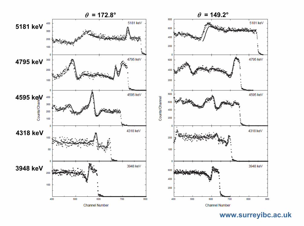
= 172.8° = 149.2°
5181 keV
4318 keV
4795 keV
4595 keV
3948 keV

Ion Beam Centre
www.surreyibc.ac.uk
Results
• NPA1990: P.M.Endt, Nuclear Physics A, 521 (1990) 1-830• NDS2011: C.Ouellet & B.Singh, Nuclear Data Sheets 112 (2011) 2199-2355. These
values in NDS2011 are based on K.-M.Källman, Z. Phys. A, 356 (1996) 287-291
Ex (keV) J (keV) NPA 1990
NDS 2011 Surrey NPA
1990NDS 2011 Surrey NPA
1990 NDS 2011 Surrey
10332 10369 10340 1- (0+) 1- 6.1 5.8 3.610457 10500 10500 0+ (0+) 0+ 1.7 1.7 1.710550 10570 10570 0 (0+) 0+ 8 1.2 1.210701 10658 10623 1- (1-) 3- 21 2.3 1.310769 10745 10718 2- (0+) 0+ 5.1 8.9 8.9
--- 10816 10781 (3-, 5-) 3- 4.7 3.310826 10868 10824 1- (2+) 0+ 22 7.7 4.710916 10956 10921 1- (0+) 0+ 1.6 2.9 1.9
--- 11107 11054 (2+) 0+ 67.4 0.611140 11130 11166 1+ (0+) 5- 2.6 1.8 6.7
--- 11254 11236 (3-) 2+ 1.1 2.1--- 11410 11383 (3-) 5- 1.9 0.6

Ion Beam Centre
www.surreyibc.ac.uk
Summary
• Accurate EBS cross-sections are:– essential for IBA (PIXE + backscattering)– valuable also for astrophysics!
• Evaluated cross-sections:– Thin targets are best for making measurements– Thick targets are best for benchmarking– R-matrix fitting, Saxon-Woods absorption (not hard sphere)
• Set of EBS spectra:– Very accurate detector calibration valid for whole set– Very accurate 2 MV HVEE Tandetron calibration (4 keV systematic
from known EBS resonances; 2 keV random from setting errors)– Correlation of all reactions reduces uncertainty– Ex, J and values from spectral data– Values contradict NDS

Ion Beam Centre
www.surreyibc.ac.uk56
Thanks for listening!
Ion Beam Analysis : amazingly powerful!


![nitric oxide [15N]arginine-to-[15N]citrulline - pnas.org · period, in healthy subjects receiving an adequate arginine intake. Thisinvestigation establishes anexperimentalbasisfor](https://img.dokumen.tips/doc/110x75/5d402ba788c99377448bcf7f/nitric-oxide-15narginine-to-15ncitrulline-pnasorg-period-in-healthy.jpg)