Embed Size (px)
Citation preview

EVALUATION OF INVITRO AND INVIVO ANTI DIABETIC ACTIVITY OF
ETHANOLIC EXTRACT OF Portulaca Quadrifida L. ON STREPTOZOTOCIN
INDUCED DIABETES IN RATS
A dissertation submitted to
THE TAMILNADU DR.M.G.R MEDICAL UNIVERSITY
CHENNAI - 600 032.
In the partial fulfillment of the requirements
for the award of the degree of
MASTER OF PHARMACY
IN
PHARMACOLOGY
Submitted by
Reg No: 261226054
INSTITUTE OF PHARMACOLOGY
MADRAS MEDICAL COLLEGE
CHENNAI – 600 003.
APRIL – 2013-14
CERTIFICATE
This is to certify that the dissertation entitled “EVALUATION OF INVITRO AND INVIVO ANTI-
DIABETIC ACTIVITY OF WHOLE PLANT OF Portulaca quadrifida Linn. ON
STREPTOZOTOCIN INDUCED DIABETES IN RATS” submitted by Registration No.261226054
in partial fulfillment of the requirements for the award of Degree of Master of Pharmacy in Pharmacology
by the Tamilnadu Dr. M. G. R. Medical University, Chennai is a bonafide work done by her during the
academic year 2013-2014.
The Dean,
Madras Medical College,
Chennai-600 003.

CERTIFICATE
This is to certify that the dissertation entitled“EVALUATION OF INVITRO AND INVIVO ANTI-
DIABETIC ACTIVITY OF WHOLEPLANT OF Portulaca quadrifida Linn. ON
STREPTOZOTOCIN INDUCED DIABETES IN RATS”submitted by Registration No.261226054 in
partial fulfillment of the requirements for the award of Degree of Master of Pharmacy in Pharmacology
by the Tamilnadu Dr. M. G. R. Medical University, Chennai is a bonafide work done by her during the
academic year 2013-2014.
The Director and HOD,
Institute of Pharmacology,
Madras Medical College,
Chennai - 600 003.
CERTIFICATE
This is to certify that the dissertation entitled “EVALUATION OF INVITRO AND INVIVO ANTI-
DIABETIC ACTIVITY OF WHOLE PLANT OF Portulaca quadrifida Linn. ON
STREPTOZOTOCIN INDUCED DIABETES IN RATS” submitted by Registration No.261226054 in
partial fulfillment of the requirements for the award of Degree of Master of Pharmacy in Pharmacology by
the Tamilnadu Dr. M. G. R. Medical University, Chennai is a bonafide work done by her during the
academic year 2013-2014.
Mrs. M. Sakthi abirami, M.pharm.,
Tutor in Pharmacy,
Institute of Pharmacology,
Madras Medical College,
Chennai - 600 003.

Though words are seldom sufficient to express my gratitude and feelings. It somehow
gives an opportunity to thank those who helped me during the tenure of my study.
I express my honourable thanks to The Dean, Dr. R. Vimala, M.D., Madras Medical
College, for permitting me to undertake the project during the period of my academic study.
I express my heartfelt gratitude and humble thanks to Dr. R. Nandini M.D., The Director
and Professor, Institute of Pharmacology, Madras Medical College for her immense support and
encouragement throughout the project.
It is a great privilege to honour and convey my gratitude to Mrs. M. Sakthi Abirami
M.Pharm., Tutor, Institute of Pharmacology, Madras Medical College, for her guidance, editing
suggestions, clearing the path towards completion of my project.
My Sincere thanks to Dr. B. Kalaiselvi M.D., Dr. B. Vasanthi M.D., Additional
professors, Institute of Pharmacology, Madras Medical College, for their support throughout the
project work.
My Sincere thanks to Dr. K. M. Sudha M.D., Associate professor, Institute of
Pharmacology, Madras Medical College, for her support throughout the project work.
I would like to thank Dr. A.C. Yegneshwaran M.D., tutor in Clinical Pharmacology,
Institute of Pharmacology, Madras Medical College, for his support throughout the project work.
I would like to thank Mrs. R. Indumathy M.Pharm., and Mrs. G. Sasikala M.Sc.,
M.Phil., Tutors, Institute of Pharmacology, Madras Medical College for their help rendered
during the study.
ACKNOWLEDGEMENT

I express my thanks to Dr. Vijayarani, M.D., Dr. V. Chenthamarai M.D., Dr. K.M
Malathi M.D., and Dr. V. Deepa M.D., Assistant Professors in Institute of Pharmacology,
Madras Medical College, for their support throughout the project work.
I would like to extend my thanks to Dr. Joseph Dixon B.V.Sc., Special Veterinary
Officer In charge for his support during the study. I also thanks to Mr. Kandhasamy and Mr.
Jothilingam for their help rendered during the study.
I am Very thankful to Mr. M. Pasupathiraja, Mr. R.V.Siva subramani, Mr. C.
Vijaykumar, Mr. J. Sivaraman, Ms. K. Abirami, Mrs. M. Devibabu, Ms. N. Ramya and
Ms. L. Abhayadav for their support during the study.
I am thankful to my friends and my juniors for their help and support during the study.
My thanks to Lab technicians, Institute of Pharmacology, Madras Medical College, for
their support during the study.

ABBREVIATIONS
DM Diabetes Mellitus
WHO World Health Organisation
PQ Portulaca quadrifida
GOD-POD Glucose Oxidase-Peroxidase
HMP Hexose Mono Phosphate
G6P Glucose-6-phosphate
LDH Lactate dehydrogenase
G6PD Glucose-6-phosphate dehydrogenase
NADP Nicotinamide adenine dinucleotide phosphate
GLP-1 Glucagon-likepeptide-1
CPCSEA Committee for the Purpose of Control and Supervision on
Experiment on Animals
GI Gastro intestinal
SU Sulfonyl Urea
PPAR Peroxisome Proliferator Activated receptor
DPP-4 Dipeptidyl Peptidase inhibitors
STZ Streptozotocin
PPHG Post-prandial hyperglycemia
ALT Alanine Aminotransferase

AST Aspartate Aminotransferase
FDA Food and Drug Administration
Rtd Retired
CSIR Council for Scientific and Industrial Research
EDTA Ethylene diamine tetra acetic acid
ATP Adenine Tri Phosphate
NAD Nicotinamide Adenine Dinucleotide
NADH Reduced Nicotinamide Adenine Dinucletide
IAEC Institutional Animal Ethical Committee
OECD Organisation for Economic Co-operation and Development
RBC Red Blood Corpuscles
SGOT Serum Glutamate Oxaloacetic Transaminase
SAP Serum Alkaline Phosphate
SGPT Serum Glutamic Pyruvic Transaminase
HDL High Density Lipoprotein
H2O2 Hydrogn Peroxide
ALP Alkaline Phosphatase
ACP Acid Phosphatase
HK Hexokinase
ANOVA Analysis of Variance
SD Standard Deviation

ROS Reactive Oxygen Species
GD Glucose Diffusion
p.o Oral route
b.w Body Weight
rpm Revolutions per minute
mins Minutes
Hrs Hours

S.NO
TITLE
PAGE NO.
1
INTRODUCTION
1
2
AIM AND OBJECTIVE
4
3
REVIEW OF LITERATURE
5
4
MATERIALS AND METHODS
28
5
RESULTS
53
6
DISCUSSION
75
7
CONCLUSION
81
8
REFERENCES
82
Introduction
Institute of Pharmacology, MMC Page 1
INTRODUCTION
India has an ancient heritage of traditional medicine. Traditional medicine is a very
important part of health care. Most of the population in the developing countries still relies
mainly on indigenous traditional medicines for satisfying their primary health care needs.
Materia medica of India provides much information on the folklore practices and traditional
aspects of therapeutically important natural product. Today we find in every country the
folklore living in the nook and corner are habituated to use their plant resources. They have
developed ethnic systems of medicines. Some of them have developed based on prolonged
experience (1).
Traditional use of herbal medicine is the basis and integral part of various cultures,
which was developed within an ethnic group before the developed and spread of modern
science. Herbal drugs constitute a major part in all the traditional systems of medicine. These
have made a great contribution in maintaining human health. A majority of the world’s
population still rely on herbal medicines to meet its health needs. The practice continues
today because of its biomedical benefits and its place in culture beliefs in many part of world.
India, china and several other nations have an ancient tradition of herbal remedies.
The written records in Ayurveda, the ancient system of medicine in India, contain more than
800 herbal remedies. The Charaka Samhita and Sushruta Samhita are two treasure troves
containing knowledge of plant based drugs and are even today, held in the highest esteem the
world over.
Inspite of all the advances in therapeutics, Diabetes still remain a major cause of
morbidity and mortality in the world. Diabetes Mellitus is the most common metabolic
disorder known to the ancient Indian physician some 3000 years ago, as can be seen, from the
medicinal texts such as Charaka Samhita and Sushruta Samhita. They have discussed the

Introduction
Institute of Pharmacology, MMC Page 2
honey urine in detail. The most ancient medicinal systems of India provide information that
madhumeha (diabetes) has long been treated with various herbs and herbomineral drugs. It is
observed that some are used as anti-diabetic medicine by folklore. Notable among them are
the use of fresh juice of Bael, Onion and Garlic.
However, the name Diabetes was given by the two Roman physician Celsius and
Aretaeus in 1st A.D in 1921.Banting and Best solved the problem of Diabetes to a great extent
by discovering Insulin as a therapeutic agent in Insulin Dependent Diabetes Mellitus. The
first oral hypoglycemic agents suitable for clinical use were the sulfonylureas developed by
Auguste Loubatieres in the year 1940.(2)
Conventionally, insulin dependent diabetes mellitus
is treated with exogenous insulin(3)
and non-insulin-dependent diabetes mellitus with
synthetic oral hypoglycemic agents like sulphonylureas and biguanides(4)
. However the
hormonrifails as curative agent for complications of diabetes and synthetic oral drugs produce
adverse health effects.(5)
Therefore, different medicinal systems are using the active plant
constituents, which discovered as natural hypoglycemic medicine came from the virtue of
traditional knowledge.(6)
Asia’s large population and rapid economic development have
made it an epicenter of the epidemic. Asian populations tend to develop diabetes at younger
ages and lower BMI levels. Several factors contribute to accelerated diabetic epidemic in
Asians, including the “normal-weight metabolically obese” phenotype, high prevalence of
smoking and heavy alcohol use; high intake of refined carbohydrates (e.g. White rice) and
dramatically decreased physical activity levels.
Estimates for the worldwide prevalence of diabetes have increased from around 60
million in 1980 to about 118 million in 1995 and are set to increase to 220 million by the year
2010(Amoes et al., 1997). According to the International Diabetes Federation (IDF), Diabetes
affects at least 285 million people worldwide, and that number will be expected to reach 438
million by the year 2030.
Introduction
Institute of Pharmacology, MMC Page 3
More than 400 different plants and plant extracts have been described as reputedly
beneficial for the diabetic patient. Most of these plants have been claimed to possess
hypoglycemic properties but most claims are anecdotal and few have received adequate
medical or scientific evaluation. Those that have been evaluated may be grouped into three
categories:
1. Plants from which a reputedly hypoglycemic compound or partially characterized
hypoglycemic fraction has been prepared.
2. Plants reported to exert a hypoglycemic effect, but the nature of the active principle is
unestablished.
3. Plants that reputedly exert a hypoglycemic effect, but the scientific evidence is
equivocal. These categories exclude the numerous traditional plants for which an
independent scientific or medical has not been published.
Portulaca quadrifida L. is an important medicinal herb and this plant is a small,
diffuse, annual and erect herb found throughout the tropical parts of India. It is said to be
useful in asthma, cough, urinary discharges, inflammations and ulcers. A poultice of the plant
is applied in abdominal complaints, erysipelas, and haemorrhoids. Portulaca quadrifida L.
has been reported to possess antifungal activity against Aspergillus fumigates and Candida
albicans. The effect of ethanolic extract of Portulaca quadrifida L. on central and peripheral
nervous system were studied by using spontaneous motor activity, in vivo muscle relaxant
activity (Grip strength) and anticonvulsant activity(11)
. However, till date there has been no
investigation supporting the anti-diabetic properties of this plant. Hence, this study has been
taken with an aim to evaluate the anti-diabetic potential of ethanolic extract of whole plant of
Portulaca quadrifida L. in in-vitro and in-vivo Streptozotocin model.

Aim and Objectives
Institute of Pharmacology, MMC Page 4
AIM AND OBJECTIVES
1. Phytochemical evaluation of the whole plant of ethanolic extract of Portulaca quadrifida L.
2. To determine the glucose diffusion property of the prepared extract of the Portulaca
quadrifida L. using dialysis tube and GOD-POD kit.
3. To evaluate the safety of the effective extract of Portulaca quadrifida L. by acute toxicity
study in Wistar albino rats.
4. To investigate the in-vivo anti diabetic effect of active extract of Portulaca quadrifida L.
in Streptozotocin induced diabetic Wistar albino rats.
Review of Literature
Institute of Pharmacology, MMC Page 5
REVIEW OF LITERATURE
DIABETES MELLITUS
The word “Diabetes” is derived from “Greek” word “Diabainein” which means “to
pass through”. It is characterized by an excess of glucose in blood and urine, hunger, thirst
and gradual loss of weight. Insulin is a hormone which regulates the carbohydrates and
triglyceride metabolism through its action at several sites and facilitates the entry of glucose
into the cell. Insulin also stimulates the synthesis of glucokinase and moderates the degree of
gluconeogenesis. In the diabetic patient, there is an aberration in the functioning of insulin.
WHO CLASSIFICATION OF DIABETES MELLITUS
1) TYPE 1 DIABETES MELLITUS(12)
The hallmark also known as Insulin Dependent Diabetes Mellitus (IDDM). It is not
associated with obesity, and may be associated with acidosis or ketosis. Administration of
insulin is essential in patients with type I diabetes. Type I diabetes is further subdivided into
immune and idiopathic types.
The immune form is the most common form of type I diabetes. Although most
patients are younger than 30 years of age at the time of diagnosis, the onset can occur at any
age. When it occurs at infancy (due to congenital disorder) or in childhood, it is called
Juvenile diabetes. Susceptibility appears to involve a multi factorial genetic linkage, but only
15-20% of patients have a positive family history.
Review of Literature
Institute of Pharmacology, MMC Page 6
Causes
i. Degeneration of β cells in the islets of Langerhans of pancreas.
ii. Destruction of β cells by viral infection.
iii. Congenital disorder of β cells.
iv. Destruction of β cells due to autoimmune disorder where there is development
of antibodies against β cells.
2) TYPE 2 DIABETES MELLITUS
Type 2 diabetes is characterized by tissue resistance to the action of insulin combined
with a relative deficiency in insulin secretion. It usually occurs after 40 years; hence it is
called as maturity onset diabetes mellitus. It is also known as adult-onset diabetes or Non-
Insulin Dependent Diabetes Mellitus (NIDDM).
The impaired insulin action affects fat metabolism, resulting in increased free fatty
acid flux and triglyceride levels, and reciprocally low high-density lipoprotein (HDL) levels.
Causes
In this type of diabetes, the structure and function of β cells and the blood level of
insulin are normal. The diabetes develops due to the absence or reduced number of insulin
receptors in the cells of the body.
The major causes for type 2 diabetes are:
Heredity disorders
Heredity disorders which may be associated with DM or impaired glucose tolerance
can be subdivided into syndromes such as maternally inherited diabetes, down, turner and

Review of Literature
Institute of Pharmacology, MMC Page 7
klinefelter syndromes, metabolic diseases such as cystic fibrosis and hemochromatosis.
Patients with a heredity disorder run a higher risk of developing DM.
Endocrine disorders
It is very common ‘in some of the disorders like Gigantism, Acromegaly, and
Cushing’s syndrome. The hyperglycemia in these conditions causes excess stimulation, in
turn causes burning out and degeneration of β cells. The β cells exhaustion leads to
permanent DM. This type of DM is called secondary diabetes.
3) GESTATIONAL DIABETES (GDM)(13)
It is defined as any abnormality in glucose levels noted for the first time during
pregnancy and resolves after delivery. Gestational diabetes mellitus develops during the
second or third trimester of pregnancy in about 2% of pregnancies and is treated by diet.
During pregnancy, the placenta and placental hormones create an insulin resistance that is
most pronounced in the last trimester. However, insulin injections may be required. Women
who have GDM are at higher risk for developing Type 2 diabetes mellitus within 5-10 years.
4) MATURITY ONSET DIABETES IN YOUNG (MODY)
MODY is defined as hyperglycemia diagnosed before the age of 25 years and
treatable for more than 5 years without insulin. Patients with MODY are found to have
mutation in the glucokinase (Hexokinase IV) gene on chromosome 7p, and the primary cause
of their NIDDM is due to reduction in glucose stimulated insulin secretion. This disease was
shown to be linked genetically to ADA (adenosine deaminase) locus on chromosome 20q.

Review of Literature
Institute of Pharmacology, MMC Page 8
5) GENETICS(14)
Susceptibility to both IDDM and NIDDM is determined to a substantial extent by
genetic factors. These probably interact with an environmental trigger to induce expression of
the disease state. The major contribution comes from a gene or genes within the HLA region,
on the short arm of chromosome 6, at least one of which lies close to or within the DQ sub
region.
There is a stronger inheritance pattern for type 2 diabetes. Those with first-degree
relatives with type 2 have a much higher risk of developing type 2 diabetes.
SIGNS AND SYMPTOMS OF DM(15)
These are mainly due to hyperglycemia with decreased utilization of glucose by cells;
as a result, there is an extracellular glucose excess and intracellular glucose deficiency, a
situation called starvation in the midst of plenty.
1. Hyperglycemia (raised blood glucose), it predisposes to infection like boils and
urinary tract infection.
2. Glycosuria (presence of glucose in the urine)
3. Polyuria (excessive urine production)
4. Dehydration
5. Polydipsia (increased thirst) is a result of the dehydration that results from the osmotic
diuresis.
6. Polyphagia (excessive eating)- low glucose utilization by glucostat cells of
ventromedial nucleus in hypothalamus(satiety center) results in no inhibition of lateral
nucleus in hypothalamus(feeding center) which eventually produces increased hunger.
7. Loss of weight.

Review of Literature
Institute of Pharmacology, MMC Page 9
8. Ketonuria.
9. Poor resistance to infections due to protein depletion.
10. Hyperlipedemia (abnormally high serum lipid levels). Insulin deficiency decreases
LDL receptor availability, which decreases serum cholesterol clearance. This
decreased clearance produces hypercholesterolemia, or high blood cholesterol.
11. Electrolyte depletion.
DIAGNOSIS OF DIABETES(16)
Diabetes can be confirmed by estimating the blood glucose level. This may be
performed using the Glucometer and Blood glucose test strips with small drop of blood
from finger.
Test for blood glucose levels are:
a) Fasting plasma glucose (FPG) test
It is also known as fasting blood sugar test. It measures blood glucose in a person who
has not eaten anything for at least 8 hours.
Plasma glucose result (mg/dL) Diagnosis
99 or below Normal
100 – 125 Pre-diabetes (impaired fasting glucose)
126 or above Diabetes

Review of Literature
Institute of Pharmacology, MMC Page 10
b) Postprandial blood glucose test
It is a test that determines the amount of glucose in the blood 2 hours after a meal.
Plasma glucose result (mg/dL) Diagnosis
<140 mg/dL Normal
140-200 mg/dL Pre-diabetes (impaired glucose tolerance)
>200 mg/dL Diabetes
c) Oral glucose tolerance test (OGTT)
This test is done to assess the glucose level of a person, 2 hours after the
administration of 75 gms of glucose dissolved in water with overnight fasting for atleast 8
hours.
2-hour plasma glucose result (mg/dL) Diagnosis
139 or below Normal
140-199 Pre-diabetes (impaired glucose tolerance)
200 or above Diabetes
d) Glycosylated haemoglobin test
A test that measures the amount of haemoglobin bound to glucose. It indicates the
amount of sugar in a person’s blood during the past 2-4 months. It is used to monitor
effectiveness of diabetes treatment.
Review of Literature
Institute of Pharmacology, MMC Page 11
Plasma glucose result (mg/dL) Diagnosis
4% - 6% Normal
More than 6% Diabetes
e) Random plasma glucose test
It is also known as casual plasma glucose test, which measures blood glucose level
randomly. Symptoms of diabetes + random plasma glucose concentration ≥ 200 mg/dL
(11.1mmol/l) is the diagnostic criteria for diabetes mellitus.
COMPLICATIONS OF DM(17)
(i)Diabetic ketoacidosis is a serious consequence of poorly controlled IDDM. It is
characterized by elevated blood glucose level, ketonemia, increased serum osmolarity and
elevated stress hormone levels (the counter regulatory hormones to insulin – cortisol, GH,
glucagon, epinephrine). These elevated hormones aggravate the metabolic disorder. The
patients have acidosis and decreased vascular volume.
(ii)Nonketotic Hyperosmolar Coma (NHC) can occur with either IDDM or NIDDM.
People with NHC have extremely high serum hyperosmolarity and glucose.
(iii)Insulin shock is produced due to the administration of excessive amounts of
insulin which can cause confusion, convulsions, loss of consciousness and even death.
LONG TERM SEQUELAE OF DM
Long-term problems associated with DM include Neuropathies, Nephropathies,
Microangiopathies, Macroangiopathies, and Retinopathies.
Review of Literature
Institute of Pharmacology, MMC Page 12
(i)Neuropathies: Peripheral nerve damage (neuropathy) can occur as a result of metabolic or
osmotic damage to neurons or Schwann cells. Diabetic patients can exhibit sensory loss,
paraesthesia and even pain as a result of the neurologic damage. Neuronal transmission is
slowed.
(ii)Nephropathies: Diabetes is a common cause of renal failure (nephropathy). The
glomerular capillary basement membrane thickens, which is thought to produce
glomerulosclerosis and subsequent renal insufficiency.
(iii)Microangiopathies: microscopic changes occur in the microcirculation with prominent
thickening of capillary basement membranes.
(iv)Macroangiopathies: Atherosclerosis develops in diabetic patients at an accelerated rate.
Diabetic patients are more likely to have coronary artery disease and myocardial infarction
than are non-diabetic individuals.
(v)Retinopathies: Retinal abnormalities develop in diabetic patients and are a major cause of
blindness in the United States. The retinal changes are characterized by aneurysms, increased
capillary permeability, small retinal hemorrhages and excessive micro vascular proliferation.
(vi)Nonretinal Visual Problems: As blood glucose and blood osmolarity rise, the volume of
the lens rise, distorting vision and the diabetic patients commonly have cataracts.
CHEMICAL AGENTS CAPABLE OF INDUCING DIABETES(18)
� Alloxan
� Streptozotocin

Review of Literature
Institute of Pharmacology, MMC Page 13
ALLOXAN INDUCED DIABETES
Alloxan, a cyclic urea analogue, was the first agent producing permanent diabetes in
animals. It is a highly reactive molecule readily reduced to diuleric acid, then auto-oxidized
back to alloxan resulting in the production of free radicals. These free radicals damage the
DNA of β-cells and cause cell death. Second mechanism proposed for alloxan is its ability to
react with protein SH groups, especially the membrane proteins like glucokinase on the β-
cells, finally resulting in cell necrosis.
Drawbacks
� High mortality in rats.
� Causes ketosis due to free fatty acid generation.
� Diabetes induced is reversible.
� Some species like guinea pigs are resistant to its diabetogenic action.
STREPTOZOTOCIN INDUCED DIABETES
STZ [2-deoxy-2-(3-methyl-3-nitrosourea) 1-D-glucopyranose] is a broad-spectrum
antibiotic, which is produced from Streptomyces achromogens. STZ causes β-cell damage by
process of methylation, free radical generation and Nitric oxide production.
Advantages
STZ has almost completely replaced alloxan for inducing diabetes because of:
� Greater selectivity towards β-cells
� Lower mortality rate and
� Longer or irreversible diabetes induction.
Review of Literature
Institute of Pharmacology, MMC Page 14
Disadvantage
Guinea pigs and rabbits are resistant to its diabetogenic action.
HORMONE-INDUCED DIABETES MELLITUS
Dexamethasone, a long acting glucocorticoid, is used to produce NIDDM at a dose of
2-5mg/kg i.p. twice daily over a number of days in rats.
INSULIN ANTIBODIES-INDUCED DIABETES
Giving bovine insulin along with CFA to guinea pigs produces anti-insulin antibodies.
Intravenous injection of 0.25-1.0 ml guinea pig anti-insulin serum to rats induces a dose
dependent increase in blood glucose levels up to 300%. This unique effect to guinea pig anti-
insulin serum is due to neutralization of endogenous insulin by the insulin antibodies. It
persists as long as the antibodies are capable of reacting with insulin remaining in the
circulation. Slow i.v. infusion or i.p. injection prolongs the effect for more than a few hours.
However, large doses and prolonged administration are accompanied by ketonemia,
ketonuria, glycosuria and acidosis and are fatal to animals. After lower doses, the diabetic
syndrome is reversible after a few hours.
VIRAL AGENTS-INDUCED DIABETES
Viruses are thought to be one of the etiologic agents for IDDM. Viruses may produce
diabetes mellitus by:
� Infecting and destroying of β-cells in pancreas,
� A less infecting or cytologic variant producing a comparable damage by eliciting
immune auto reactivity to the β-cells,
� Viruses producing systemic effect, not directly affecting the β-cells.
Review of Literature
Institute of Pharmacology, MMC Page 15
Various human viruses used for inducing diabetes include RNA picornoviruses,
Coxsackie-B4 (CB4), encephalomylocarditis (EMC-D and M variants), Mengo-2T, as well as
two other double stranded RNA viruses, reovirus and lymphocytic choriomeningitis virus
(LMCV, Armstrong variant).
SURGICALLY INDUCED DIABETES
Surgical removal of all or part of the pancreas can induce Diabetes Mellitus. In partial
pancreactectomy more than 90% of the organ must be removed to produce diabetes.
Depending on the amount of intact pancreatic cells, diabetes may range in duration from a
few days to several months. Total removal of the pancreas results in an insulin-dependent
form of diabetes, and insulin therapy is required to maintain experimental animals. The
portion of the pancreas usually left intact following a subtotal pancreatic resection is typically
the anterior lobe or a portion thereof.
Disadvantages
� Surgical removal of pancreas results in loss of α- and δ-cells in addition to β-cells.
This causes loss of counter-regulatory hormones, glucagon and somatostatin.
� There is a loss of the pancreatic enzymes necessary for proper digestion; therefore, the
diet must be supplemented with pancreatic enzymes.
MANAGEMENT OF DIABETES MELLITUS(19)
The goals of treatment for diabetes are to reduce and control blood glucose levels,
relieve the symptoms of the disease and prevent complications. Intensive treatment and
careful control of blood glucose levels can reduce the risk of complications of diabetes.(20)

Review of Literature
Institute of Pharmacology, MMC Page 16
I. NON PHARMACOLOGICAL INTERVENTIONS:
The major environmental factors that increase the risk of type 2 diabetes, presumably
in the setting of genetic risk are nutrition and sedentary lifestyle with consequent over weight
and obesity. Medical nutrition therapy (i.e., diet) and exercise are important aspects of non-
pharmacologic treatment for diabetes. Weight loss is a vital part of treatment for type 2
diabetes because it can help improve the sensitivity of cells to insulin and the uptake of
glucose by cells.
A goal of medical nutrition therapy is to attain and maintain blood glucose levels in
the normal range or as close to normal as possible. Patients with type 2 diabetes typically
have dyslipidemia and another goal for these patients is to improve the lipid profile.
II. PHARMACOLOGICAL INTERVENTIONS:
A) Injectable anti-diabetic agents(21)
1) INSULIN:
All patients with type 1 diabetes require insulin injections. Patients with type 2
disease who have multiple symptoms of hyperglycemia are pregnant or have ketosis also
should use insulin injections. Currently, insulin used for treatment is derived from beef and
pork pancreas as well as recombinant (human) DNA technology.
In the 1980s, the human insulin was produced by recombinant DNA technology in
Escherichia coli. The side effects of intensive treatment include serious hypoglycemia, local
reactions (swelling, erythema and stinging), allergy and edema. Newer insulin delivery
devices have been made to improve ease and accuracy of insulin administration. These are
insulin syringes, pen devices, inhaled insulin, insulin pumps, and implantable pumps (20)
.

Review of Literature
Institute of Pharmacology, MMC Page 17
Type
Zinc content
Action ( hours)
Onset peak Duration
Rapid:
a) Regular
soluble
b) Lispro
Intermediate:
a) NPH
b) Lente
Slow:
a) Ultralene
b) Protamine zinc
c) Glargine
0.01- 0.04
0.02
0.016
0.2-0.25
0.2-0.25
0.2-0.25
0.03
0.5-0.7
0.25
1-2
1-2
4-6
4-6
2-5
1.5-4
0.5-1.5
6-12
6-12
16-18
14-20
5-24
5-8
2-5
18-24
18-24
20-36
24-36
18-24
2) GLUCAGON LIKE PEPTIDE- 1 (GLP- 1) AGONIST:
The glucagon like peptide –1 (GLP-1) is an important incretin that is released from
the gut in response to oral glucose. It is difficult to use clinically because of rapid degradation
by the enzyme dipeptidyl peptidase-4 and it is injected subcutaneously twice daily one hour
before meals acts for 6-10 hours. Nausea is an important side effect.
3) AMYLIN AGONISTS:
This synthetic amylin (a polypeptide produced by pancreatic β-cells which reduces
glucagon secretion from α-cells and delays gastric emptying) analogue attenuates
postprandial hyperglycemia when injected subcutaneously just before meal and exerts a
centrally mediated anorectic action.

Review of Literature
Institute of Pharmacology, MMC Page 18
B) Oral hypoglycemic drugs:
Oral hypoglycemic drugs include sulfonyl ureas, biguanides, meglitinide
/phenylalanine analogues, thiazolidinediones, α-glucosidase inhibitors, dipeptidyl peptidase
inhibitors and incretin mimetics.
1) SULFONYL UREAS: [e.g. Glibenclamide, Glimepiride, Glipizide, etc.]
Sulfonyl ureas act primarily by increasing insulin secretion from pancreatic β-cells
and cause hypoglycemia. Side effects include hypoglycemia, weight gain, allergic rashes and
bone marrow damage.
2) BIGUANIDES: [e.g. Metformin]
Metformin reduces blood glucose concentrations by increasing glucose uptake in the
peripheral muscles and decreasing the amount of glucose produced and released in the liver
i.e., suppress hepatic gluconeogenesis. Side effects include lactic acidosis, vitamin B12
deficiency and gastrointestinal disturbances.
3) MEGLITINIDE / D-PHENYLALANINE ANALOGUES:
[e.g.Repaglinide, Nateglinide]
The glinides stimulates insulin secretion, although they bind to a different site within
the sulfonylurea receptor. They have a shorter circulating half-life than the sulfonylurea must
be administered more frequently.
4) THIAZOLIDINEDIONES:
Thiazolidinediones are insulin sensitizers that increase insulin sensitivity and action in
liver, muscle and fatty tissues to endogenous and exogenous insulin by binding and activating
Review of Literature
Institute of Pharmacology, MMC Page 19
the nuclear receptor peroxisome proliferator – activated receptor γ (PPAR-γ). Side effects
include weight gain, fluid retention and GI disturbances.
5) α-GLUCOSIDASE INHIBITORS :( e.g.Acarbose, Miglitol, Voglibose)
These drugs inhibit the action of intestinal disaccharidases enzymes that break down
carbohydrates. These oral anti-diabetic agents delay glucose absorption. Side effects include
flatulence, abdominal bloating and diarrhea.
6) DIPEPTIDYL PEPTIDASE INHIBITORS :( e.g.Sitagliptin)
DPP-4 inhibitors increase insulin secretion and decrease glucagon level. It does not
affect weight and does not induce hypoglycemia. Side effects include Gastro intestinal
disturbances, headache, peripheral edema, and severe pain in upper stomach spreading to
back.
7) INCRETIN MIMETICS :( e.g.Liraglutide)
These drugs increase insulin secretion, suppress glucagon secretion and slows gastric
emptying. Side effects include Gastro intestinal disturbances, headache, dizziness, asthenia,
hypoglycemia, increase sweating and injection site reaction.
ALTERNATIVE MEDICINES FOR DIABETES(22)
Insulin therapy is the only satisfactory approach in diabetes mellitus, even though it
has several drawbacks like insulin resistance, anorexia, brain atrophy and fatty liver in
chronic treatment. There are several oral hypoglycemic agents used therapeutically but
certain adverse effects and weak effectiveness of them has led to the search for more
effective agents. Therefore, herbal drugs are gradually gaining popularity in the treatment of

Review of Literature
Institute of Pharmacology, MMC Page 20
diabetes mellitus. The major qualities of herbal medicine seem to be their supposed efficacy,
low incidence of serious adverse effects and low cost.
MOST STUDIED AND COMMONLY USED ANTI DIABETIC
MEDICINAL PLANTS
Kumari et al., 1995,(23)
Allium cepa (Onion) is an essential plant cultivated throughout India, belongs to
the family Liliaceae. Various parts such as seedling, callus, bulb etc., are known to possess
anti-diabetic activity. Investigations reveal the presence of sulfur containing amino acid,
which when administered orally to alloxan-induced diabetic rats (200 mg/kg for 45 days)
significantly controlled blood glucose and lipids in serum and tissues and normalized the
activity of liver hexokinase, glucose-6-phosphatase and HMG-CoA reductase. The effect
was in accordance with that of glibenclamide and insulin.
Zacharias et al., 1980.(24)
Allium sativum (Garlic) belongs to the family of Alliaceae. Aqueous extract of
garlic increased hepatic glycogen, and free amino acid content when given orally to sucrose
fed rabbits (10 ml/kg/day). Garlic is known to decrease fasting blood sugar, triglyceride level
in serum liver and aorta and protein levels when compared with sucrose controls.
Faiyaz Ahmed and Asna Urooj, et al., 2008(25)
.
The present investigation evaluated the antihyperglycemic activity of the bark
powder and aqueous extract of Ficus glomerata (Moraceae) in streptozotocin induced
diabetic rats. Oral administration of bark powder (FGB) and aqueous extract (FGAE) at 500
mg/kg caused 21% and 52% reduction in fasting blood glucose, respectively and also

Review of Literature
Institute of Pharmacology, MMC Page 21
decreased glycosuria significantly. Histology of pancreas suggested normalization of islets of
Langerhans and β-cells with respect to their number and cellular architecture. The results
suggest that, the bark of Ficus glomerata has significant anti-hyperglycemic activity in
experimental animals and has potential to be used as an adjunct in the management of
diabetes mellitus.
M.R.M.Rafiullah and A.W.Siddiqui et al., 2006.(26)
The effect of aqueous extract of Syzygium cumini.Linn, Gymnema sylvestre (Retz)
Schult and Portulaca olearacea.Linn were investigated in fasting normal and streptozotocin
(STZ) induced diabetic rats. The effects of extract on oral glucose tolerance in normal fasting
rats were also studied. The aqueous extracts of S.cumini (200 mg/kg) and G. sylvestre (200
mg/kg) decrease the blood glucose in normal rats significantly at 2 and 4 hour of extracts
administration (p < 0.05, p < 0.01). The S. cumini and G.sylvestre extracts decrease the
increase of glucose levels significantly (p < 0.05) at 90 and 180 minutes after the glucose
load in glucose tolerance test. In STZ induced diabetic animals, the aqueous extracts of
S.cumini and G.sylvestre decrease the blood glucose significantly (p < 0.05) at 4 hour. The
aqueous extract of P.olearacea did not show any hypoglycemic activity.
Rajasekaran et al., 2005.(27)
Aloe barbabensis, commonly called as Aloe vera is a medicinal herb; belongs to the
family Liliaceae; Leaf gel, Leaf pulp and dried sap are known to possess anti-diabetic and
anti-oxidant activity oral administration of ethanoloic extract at a concentration of 300 mg/kg
body weight for 21 days, proved more effective in controlling oxidative stress found in
diabetes.

Review of Literature
Institute of Pharmacology, MMC Page 22
Gomes et al., 1995, Anderson and Polansky, 2002.(30)
Camellia sinensis belongs to the family Theaceae, commonly known as Tea. The
blood glucose level lowering activity has been extensively investigated. Antihyperglycemic
activity of hot water extract of green tea in STZ induced diabetic rats were studied by Gomes
et al.(1995), these findings have been supported by Anderson and Polansky(2002). Tea
polyphenols possess antioxidant capacity; have also been reported to inhibit α-amylase.
AVAILABLE STUDIES ON Portulaca quadrifida Linn.
Syed kamil M et al., 2010.(31)
The effect of ethonalic extract of Portulaca quadrifida Linn.were studied in mice
using various models. The effect of ethonalic extract of Portulaca quadrifida Linn.on central
and peripheral nerves system were studied by using spontaneous motor activity, anti-
nociceptive activity, in vivo muscle relaxants activity (grip strength) and anti-convulsant
activity. The extract (400 and 800 mg/kg i.p) showed a significant reduction in spontaneous
motor activity, antinociceptive activity and also showed reduction in time to recover from the
electrically induced convulsion. The effect of extract on grip strength was found non-
significant. Results from the present study indicate that Portulaca quadrifida Linn. has
significant effect on central nerves system but not on peripheral nerves system.
Patil AG, Patil DA et al., 2012(32)
The herbal products today symbolize safety in contrast to synthetic drugs that are
regarded unsafe to the human and environment. Portulaca quadrifida Linn. a traditional
medicinal plant, valued for its benefits in the management of urinary and inflammatory
disorders. The decoction of plant acts as anthelmintic and used in the treatment of stomach
complaints and gonorrhea. The powder microscopy showed the presence of tannins,
mucilage, steroidal compounds and carbohydrates.
Syed kamil mulla and Paramjyothi swami, 2012(33)
Portulaca quadrifida Linn. (Portulacaceae) is traditionally used for the treatment
against various ailments in tropical and sub-tropical parts of India without any scientific
knowledge. Polyphenols, present in different fruits and vegetables, have retained attention in
Review of Literature
Institute of Pharmacology, MMC Page 23
recent years. In the present study the effects of ethanol and polyphenol extracts of Portulaca
quadrifida Linn. on the proliferation of human colon cancer HT-29 and a normal L-6 cell
lines were investigated by MTT assay and Trypan blue dye exclusion assay followed by
DNA fragmentation assay. The results confirm that the ethanolic and polyphenolic extracts
exhibited fragmentation of DNA in HT-29 cell lines and are found less effective against
normal L-6 cell lines indicating the cancer specific effect of Portulaca quadrifida Linn.

Review of Literature
Institute of Pharmacology, MMC Page 24
PLANT PROFILE(29)
Name : Portulaca quadrifida L.
Synonym : Portulaca anceps A.Rich
Portulaca geniculate Royle
TAXONOMY
Kingdom : Plantae
Phylum : Magnoliophyta
Class : Magnoliatae
Order : Caryophyllales
Genus : Portulaca
Family : Portulacaceae
Vernacular names : English - chickenweed
Tamil - Siru pasalai, Tharai pasalai
Sanskrit - Paciri, Paviri
Telugu - Goddu paveli
Malayalam - Neelakeera
Others - Lunki buti, Pulikeerai
Habitat : Arenosol, subtropical, tropical, prostrate herb, upto 20cm.
Distribution : Pan tropical, Common in tropical India and Africa, U.S.
Virgin Islands and Asia.
Review of Literature
Institute of Pharmacology, MMC Page 25
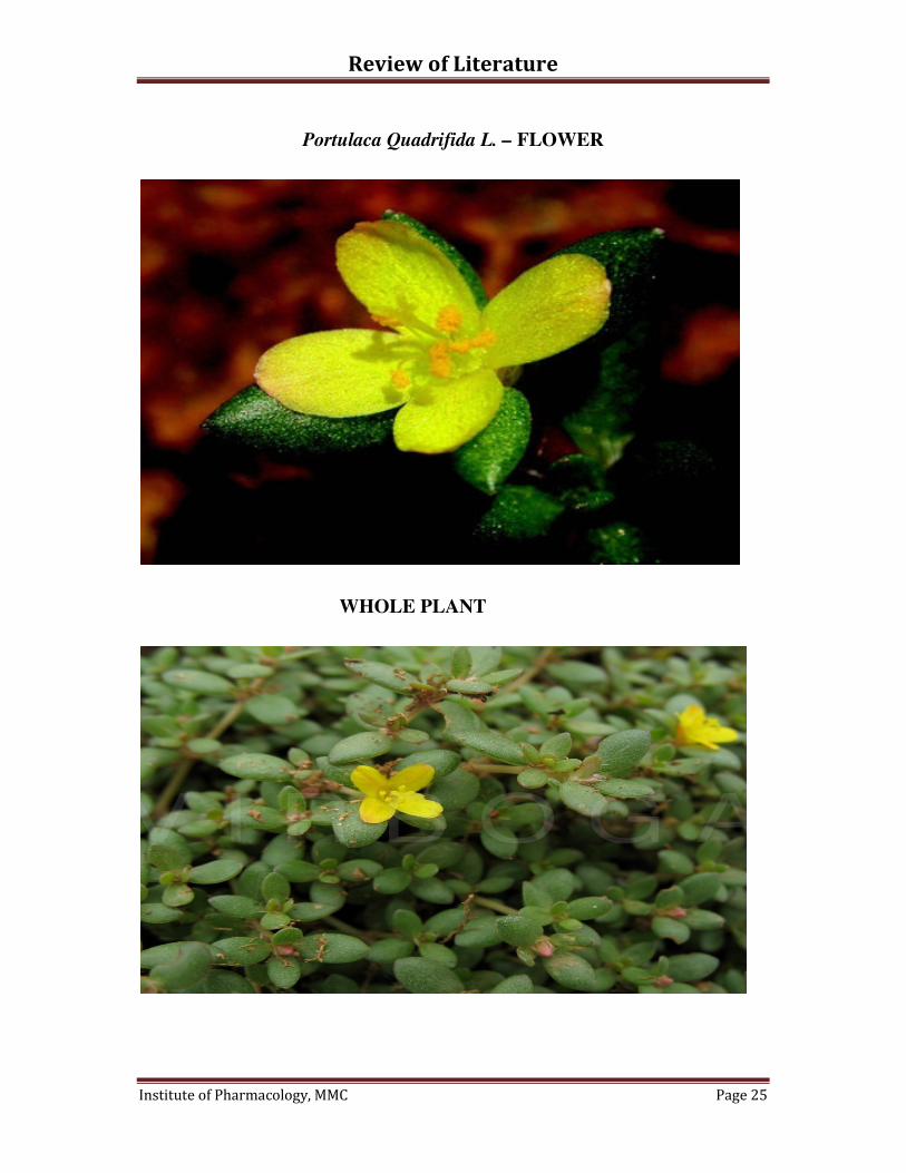
Portulaca Quadrifida L. – FLOWER
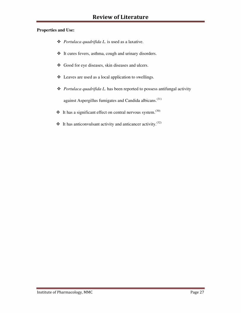
WHOLE PLANT

Review of Literature
Institute of Pharmacology, MMC Page 26
Description
The plant:
A small diffused succulent, annual herb/forb. Stem is rooting at the nodes. The
diameter of stem was found to be 0.1 cm.
Leaves:
� Leaves are opposite, fleshy, ovate, acute apex, entire margin; stipulate
with a ring of silvery hairs.
� The length of leaf is about 0.9 cm, and breadth is about 0.3 cm.
Flowers:
� Flowers are small, Dicot, terminal, and solitary, surrounded by a 4-leaved
involucre and copious white hair.
� Petals are four and yellow in colour.
Capsule:
Capsule dehisces horizontally, and contains minutely tubercled seeds.
Chemical composition:
Alkaloids, glycosides, mucilage,
Tannins, proteins, flavonoids,
Saponins, carbohydrates, triterpenoids
Parts used:
Whole plant.
Review of Literature
Institute of Pharmacology, MMC Page 27
Properties and Use:
� Portulaca quadrifida L. is used as a laxative.
� It cures fevers, asthma, cough and urinary disorders.
� Good for eye diseases, skin diseases and ulcers.
� Leaves are used as a local application to swellings.
� Portulaca quadrifida L. has been reported to possess antifungal activity
against Aspergillus fumigates and Candida albicans.(31)
� It has a significant effect on central nervous system.(30)
� It has anticonvulsant activity and anticancer activity.(32)

Materials and Methods
Institute of Pharmacology, MMC Page 28
STUDY DESIGN
Portulaca quadrifida L.
Authentication of plant material
Collection and processing of plant
Extraction with Ethanol
Phytochemical studies Pharmacological studies
Invivo study Invitro studies
1. Glucose diffusion assay
2. Glucose adsorption assay
• Blood glucose and
body weight changes.
• Haematological &
Biochemical
parameters.
• Tissue homogenate-
Estimation of
Lysosomal enzymes,
Carbohydrate
metabolizing enzymes.
• Histopathological study
Acute toxicity study
(OECD Guidelines 423)

Materials and Methods
Institute of Pharmacology, MMC Page 29
MATERIALS AND METHODS
PLANT COLLECTION AND IDENTIFICATION
Dried entire plant of Portulaca quadrifida L. was collected from the forest
around Komaneri, Tuticorin district, Tamilnadu (INDIA), in the month of July. It was
authenticated by Prof. V. Chelladurai, Research Officer- Botany (Scientist - C) (Rtd), Central
Council for Research in Ayurveda & Siddha, Govt. of India.
PREPARATION OF PLANT EXTRACT
The powdered plant material (50 g) was extracted by hot continuous soxhlet
extraction method. The plant material was extracted with ethanol (99.9% v/v) (500 ml) for 2
days in a percolator.
It is a process of continuous extraction method in which the solvent can be
circulated through the extractor for several times. The vapours from the solvent are taken to
the condenser and the condensed liquid is returned to the extract for continuous extraction.
The apparatus consist of body of extractor (thimble) attached with side siphon tube, lower
end attached with distillation flask and the mouth of extractor is fixed to the condenser by the
standard joints.
Procedure:
� Weighed about 50g of dried powdered plant and transferred into thimble for packing.
� While packing, the content was wetted with ethanol (99.9% v/v) and then poured until
the siphon tube was filled.
� A piece of porcelain was added into the round bottom flask to avoid bumping effect.
Materials and Methods
Institute of Pharmacology, MMC Page 31
solution to second portion and the gelatin salt reagent to third portion. Precipitation with the
latter reagent or with both the gelatin salt reagent was indicative of the presence of tannins.
Precipitation of salt solution indicated a false-positive test. Positive tests were further
confirmed by the addition of a few drops of dilute ferric chloride (1% FeCl3) to test extracts,
which gave black or green colouration.
The extract was mixed with lead acetate solution and observed for white precipitate.
4. Test for saponins:
• Foam test: A small amount of extract was extracted with petroleum ether. To the
insoluble residue left after extraction, a few ml of water was added and shaken
vigorously for 15 minutes and was observed for the formation of honeycomb froth
that persisted for at least 30 minutes.
5. Test for terpenoids:
• Noller’s test: The extract was warmed with tin and thionyl chloride. Pink colouration
indicates the presence of terpenoids.
6. Test for glycosides:
• Borntrager’s test: A small amount of extract was hydrolyzed with hydrochloric acid
for few hours on a water bath and the hydrolysate was extracted with benzene. The
benzene layer was treated with dilute solution and was observed for formation of
reddish pink colour.
• Legal test: the extract was dissolved in pyridine and was made alkaline with few
drops of 10% sodium hydroxide and then freshly prepared sodium nitroprusside was
added and observed for the formation of blue colour.
Materials and Methods
Institute of Pharmacology, MMC Page 32
7. Test for phytosterols:
• Libermann – Burchard test: 1 mg of the extract was dissolved in few drops of
chloroform, 3 ml of acetic anhydride and 3 ml of glacial acetic acid. Warmed, cooled
under tap water and drops of concentrated sulphuric acid was added along the side of
the test tube, formation of bluish green colour indicates the presence of steroids.
DRUGS AND CHEMICAL
Sodium Chloride, D- glucose, Ethanol, Sodium citrate tribasic-dehydrate,
Citric acid monohydrate, Streptozotocin, Glibenclamide.
II) IN VITRO ANTIDIABETIC EVALUATION
PHYSICAL METHOD
Ability of the plant materials to retard the movement of glucose from the
intestine into the blood was evaluated by physical methods in vitro, because the absorbed
glucose from the intestine will cause the rise in postprandial hyperglycemia (PPHG) which
complicates the diabetic condition associated with risk factors. The following are the
convenient models for assessing the materials which affect the absorption of glucose in vitro.
A. GLUCOSE DIFFUSION ASSAY(33)
:
Plant extracts were mixed with glucose and placed in the sealed dialysis membrane
and kept, and in the orbit shaker bath at 37º C, at 150rpm, and the movement of glucose
across the membrane into the external solution was measured at periodic intervals using
commercial GOD-POD kit.

Materials and Methods
Institute of Pharmacology, MMC Page 33
REQUIREMENTS:
o Dialysis membrane
o 0.15 M Sodium chloride solution
o D- (+) – Glucose - (25Mm in sodium chloride solution)
o Orbit shaker
o GOD – POD kit
PROCEDURE:
Dialysis membrane containing 2 ml of 25mM glucose solution was mixed with 1ml of
different concentrations of plant extracts (100, 250,500,1000 and 1500µg/ml) and was placed
in the centrifuge tube containing 45ml 0.15M NaCl and then kept in orbit shaker bath at
37ºC,at 150rpm. The movement of glucose into the external solution was monitored at 0, 1,
2, 3, 4, 5 hours using GOD-POD kit. Glucose concentration in the external solution was
expected as mg/dl/hr.
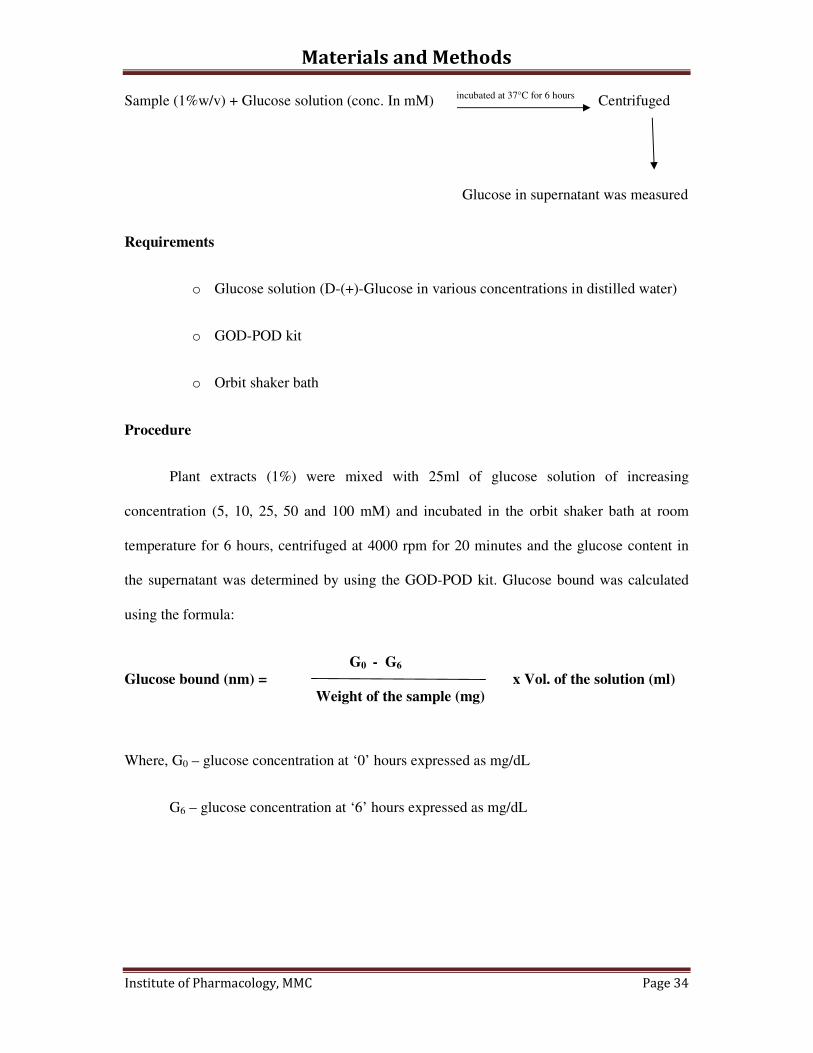
B.GLUCOSE ADSORPTION ASSAY(34)
Principle
Glucose binding ability of the plant materials was determined by the method of Ou
et al., 2001. Plant extract samples were incubated for 6 hours with various concentrations of
glucose and the free glucose concentration at the end of the incubation period was determined
using GOD-POD kit.
Materials and Methods
Institute of Pharmacology, MMC Page 34
Sample (1%w/v) + Glucose solution (conc. In mM) incubated at 37°C for 6 hours
Centrifuged
Glucose in supernatant was measured
Requirements
o Glucose solution (D-(+)-Glucose in various concentrations in distilled water)
o GOD-POD kit
o Orbit shaker bath
Procedure
Plant extracts (1%) were mixed with 25ml of glucose solution of increasing
concentration (5, 10, 25, 50 and 100 mM) and incubated in the orbit shaker bath at room
temperature for 6 hours, centrifuged at 4000 rpm for 20 minutes and the glucose content in
the supernatant was determined by using the GOD-POD kit. Glucose bound was calculated
using the formula:
G0 - G6
Glucose bound (nm) = x Vol. of the solution (ml)
Weight of the sample (mg)
Where, G0 – glucose concentration at ‘0’ hours expressed as mg/dL
G6 – glucose concentration at ‘6’ hours expressed as mg/dL

Materials and Methods
Institute of Pharmacology, MMC Page 35
III) IN VIVO ANTI DIABETIC STUDIES
Experimental Animals:
The present study was conducted after obtaining approval from the Institutional Animal
Ethics Committee and this protocol met the requirements of national guidelines of CPCSEA
(PROPOSAL NO: IAEC 13/243/CPCSEA). The Wistar albino rats (150-200 gm) used for
this study were procured from Animal house, Madras Medical College, Chennai, India.
Quarantine and Acclimatization:
Quarantine is the separation of newly received animals from those already in the facility
until the health and possibly the microbial status of the newly received animals have been
determined. The newly procured Wistar albino rats were quarantined for the period of one
week to minimize the chance of introduction of pathogens into established animals and
allowed to develop the psychological, physiological and nutritional stabilization before their
use.
Housing:
The animals were housed in well ventilated animal house which was maintained at a
constant temperature and relative humidity of 55 to 60%. The animals were housed in
spacious polypropylene cages and paddy husk was utilized as bedding material.
Diet and water:
The animals were maintained on standard pellet diet and purified water. The animals
were provided with food and water ad libitum except during fasting. The bed material was
changed twice a week.
Materials and Methods
Institute of Pharmacology, MMC Page 36
Animal identification:
All animal cages used in the study had a proper identification i.e., labels. Each animal
in the cage was marked either on head or body or tail with picric acid for their appropriate
identification
A) TOXICITY STUDIES:
Acute toxicity was designed as per the OECD guidelines (423).(37)
ACUTE TOXICITY STUDY:
Principles and purpose:
The main purpose of acute toxicity is to evaluate the degree of toxicity in a
quantitative and qualitative manner with the purpose of comparing it with other drug
substances (e.g. other drug candidates for the same indication).
Experimental Animals:
Three healthy adult Wistar albino rats weighing between 150-250g were selected for
the study. For all the three animals food, but not water was withheld overnight prior to
dosing.
Selection of dose levels and administration of doses:
Being a traditional herbal medicine, the mortality was unlikely at the highest starting
dose level (2000mg/kg body weight). Hence a limit test at one dose level of 2000mg/kg body
weight was conducted in all the three animals.
Materials and Methods
Institute of Pharmacology, MMC Page 38
B) INVIVO ANTIDIABETIC EVALUATION:
The antidiabetic activity of Portulaca quadrifida Linn was evaluated in diabetic
Wistar rats. Diabetes was induced by intra-peritoneal injection of Streptozotocin 50mg/kg
body weight. The antidiabetic effect of plant extract was compared with standard drug
Glibenclamide.
(i) Induction of diabetes in rats:
After a week of acclimatization, the rats were subjected to overnight fasting. Diabetes
was induced by intraperitoneal injection of Streptozotocin, freshly dissolved in citrate buffer
pH 4.5. The animals were allowed to drink water 5% glucose solution overnight to overcome
the drug induced hypoglycemia due to massive release of insulin from β-cells. After 3 days,
blood glucose levels were measured and the animals with a blood concentration of more than
250 mg/dl were considered as diabetic and taken for the experiment. Administration of the
plant extract was started on the 4th
day after streptozotocin injection and this was considered
as the 1st day of treatment, which was continued for 21 days.
(ii)Experimental design:
The fasting glucose and body weight of all animals were recorded at the beginning of
the study. The blood glucose was checked by one – touch glucometer throughout the study,
in the experiments, 24 rats were divided into 4 groups of six rats each.
GROUP 1: Normal control rats, received distilled water.
GROUP 2: STZ induced diabetic rats received distilled water and served as diabetic
control for 21 days.
GROUP 3: STZ induced diabetic rats received standard drug Glibenclamide (5mg/kg BW
p.o) for 21 days.
Materials and Methods
Institute of Pharmacology, MMC Page 39
GROUP 4: STZ induced diabetic rats received the plant extract (200mg/kg BW p.o) for
21 days.
GROUP 5: STZ induced diabetic rats received the plant extract (400mg/kg BW p.o) for
21 days
For all rats, body weight was measured before the induction of diabetes (-4th
day) and
on 7, 14 and 21st day of the treatment. Blood glucose level was measured on 1, 7, 14 and 21
st
days throughout the study period by tail tip cutting method. At the end of the experiment on
21st day, sufficient blood was collected by retro-orbital bleeding from all the animals under
anaesthesia for estimation of haematological and biochemical parameters.
(iii) Biochemical parameters
The blood samples were centrifuged at 3000rpm for 5 minutes using REMI (412
LAG) cooling centrifuge. The serum was kept at -80°C until analyzed. Levels of serum
glutamate oxaloacetic transaminase (SGOT), serum glutamate pyruvic transaminase (SGPT),
serum alkaline phosphatase (ALP), serum creatinine, urea, total cholesterol, triglycerides
(TGL), HDL and total protein were determined with an automatic analytical instrument
(Hitachi 911, Japan).
A.ESTIMATION OF TOTAL PROTEIN (Biuret Method)(38)
The serum content of the soluble proteins, those circulating in extracellular and
intracellular fluids, has been used as a marker to aid in clinical diagnosis. Serum total protein
including albumin is mainly involved in the maintenance of normal water distribution
between tissues and the blood and responsible for maintaining the osmotic pressure of plasma
and is used to transport many substances including macromolecules.
Principle
In the biuret reaction, a chelate is formed between the Cu2+
ion and the peptide bonds
of the proteins in alkaline solutions to form a violet coloured complex whose absorbance is

Materials and Methods
Institute of Pharmacology, MMC Page 40
measured photometrically. The intensity of the colour produced is proportional to the
concentration of the protein in the sample.
Cu2+
+ Serum protein Copper-protein complex
Reagents
� Biuret reagent
� 1ml of protein standard
� Serum sample
Procedure
1ml of biuret reagent which is stored under 2-8°C and 10µl of serum sample/standard
are mixed well and then it is incubated for 5 mins. The intensity of the colour is measured at
540 nm.
Calculation
Absorbance of test
Total protein in gm/dL = X 5.5
Absorbance of standard
B.ESTIMATION OF LIPID PROFILE(39)
Principle
The cholesterol esters and cholesterol present in the sample are acted upon by
Cholesterol Esterase to release Cholesterol and Fatty acids. The Cholesterol is oxidized by
Cholsterol Oxidase to yield 4-Cholesten 3-one and Hydrogen peroxide as by product.
Hydrogen peroxide together with 4-aminoantipyrin and phenolic compound in the presence
of peroxidase gives the coloured complex. The intensity of the colour is proportional to the
total cholesterol in the sample and is measured at 550 nm or with Green filter.

Materials and Methods
Institute of Pharmacology, MMC Page 41
Cholesterol ester + H2O Cholesterol Esterase
cholesterol + Fatty acids
Cholesterol +O2 cholesterol oxidase
4-Cholesten-3-one-H2O2
2H2O2 + Phenol + 4-aminoantipyrine peroxidase
Red quinone + 4H2o
Requirements
� Enzyme reagent
� HDL ppt reagent
Procedure
1ml of enzyme reagent and 10µl of test/standard were mixed well and incubated at
37°C for 5mins. The absorbance of test and standard were measured at 505 nm or using
Green filter.
Calculation
Absorbance of test
Cholesterol conc. mg/dl = x Conc. of Standard (200)
Absorbance of standard
HDL Cholesterol(40)
:
Procedure
Step 1:
200µl of serum and 300 µl of HDL ppt reagent were mixed well and allowed to for
10mins. Then the mixture was centrifuged at 3000rpm for 10mins and the supernatant was
separated.
Step 2:
1ml of enzyme reagent and 100 µl of supernatant from step A were mixed together
and incubated for 5mins at 37°C. The absorbance is read at 505 nm.

Materials and Methods
Institute of Pharmacology, MMC Page 42
Calculation
Absorbance of test
HDL Cholesterol conc. mg/dl = x Conc. of Standard (50)
Absorbance of standard
TRIGLYCERIDE(41)
In human nutrition, triglyceride is the most prevalent glycerol esters encountered.
They constitute 95% of tissue storage fat are the predominant form of glycerol ester found in
plasma. The investigation of triglyceride is part of the overall evaluation of lipids disorders.
Principle
Triglyceride + H2O LPL
Glycerol + free fatty acid
Glycerol + ATP GK
Glycerol-3-phosphate + ADP
Glycerol-3-phosphate + O2 GPO
DAP + H2O2
H2O2 + 4AAP + 3, 5-DHBS Peroxidase
Quinoeimine Dye + 2H2O2
Requirements
� Pipes buffer pH 7.0
� 4-AAP 0.4mmol/l
� Magnesium 2mmol/l
� ATP 2mmol/l
� GK
� POD
� LPL
� GPO
� Surfactants

Materials and Methods
Institute of Pharmacology, MMC Page 43
Procedure
1ml of reagent mixed with 10 µl of sample and incubated for 5min at 37°C.Then the
absorbance is measured at 505 nm.
Calculation
Triglyceride mg/dl = Ax/As x Concentration of Standard
Where, Ax – absorbance of sample, As- absorbance of standard
C. ESTIMATION OF UREA(41)
Urea is the end product of the protein metabolism. It is synthesized in liver from the
ammonia produced by the catabolism of ammonia acids. It is transported by the blood to the
kidneys from where it is excreted. Increased levels are bound in renal diseases, urinary
obstructions, shock, congestive heart failure and burns. Decreased levels are found in liver
failure and pregnancy.
Principle:
Urea is an acidic medium condenses with Diacetyl monoxime at 100oc to form a red
coloured complex. Intensity of the colour formed is directly proportional to the amount of
urea present in the sample.
Urea + H2O Urease
2NH3 + CO2
Urea + Diacetyl monoxime ------------------� Red Coloured Complex
Contents: 25 ml 75 ml
L1: Urea Reagent 75 ml 150 ml
L2: Acid Reagent 75 ml 150 ml
L3: DAM Reagent 75 ml 150 ml

Materials and Methods
Institute of Pharmacology, MMC Page 44
S: Urea Standard (40 mg/dl) 5 ml 5 ml
Procedure
Addition sequence Blank (B) ml Standard (S) ml Test (T) ml
Urea reagent (L1)
Acid reagent (L2)
DAM reagent
Distilled water
Urea Standard (S)
Sample
1.0
1.0
1.0
0.01
-
-
1.0
1.0
1.0
-
0.01
-
1.0
1.0
1.0
-
-
0.01
Pipette into clean dry test tubes labeled as Blank (B), Standard (S) and Test (T).
Mix well and keep the test tubes in boiling water (100oc) for 10min. Cool under running tap
water and measure the absorbance of the standard (Abs. S), and Test sample (Abs. T) against
the blank.
Calculation
Abs. T
Urea in mg/ dl = ----------------------- X 40
Abs. S

Materials and Methods
Institute of Pharmacology, MMC Page 45
D. ESTIMATION OF CREATININE(41)
Creatinine is a waste product formed from creatine phosphate, a high energy storage
compound. It is removed from plasma by glomerular filtration and then excreted in urine.
Creatinine is a useful indicator of renal function.
Elevated levels of creatinine are associated with abnormal renal function as it relates
to glomerular filtration.
Serum creatinine levels are used in combination with Urea/BUN to differentiate
between pre-renal and renal causes of azotemia (condition of increased BUN level)
Principle
Creatinine reacts with picric acid in alkaline environment to form an orange-red
colour complex. Developing of this orange-red colour may be followed photometrically at
500-520 nm.
Creatinine + Picric acid NaOH
orange-red formation
Requirements
� Picric acid 11mmol/l
� NaOH 0.3mmol/l
� Creatinine standard
Procedure
1ml of reagent and 100µl of sample/standard are mixed and incubated for 60seconds
at 37°C. Then the absorbance (A1) measured at 505 nm. After exactly 60second absorbance
(A2) was measured.
Calculation
Creatinine mg/dl = A2 – A1(sample)/ A2 – A1(standard) x concentration of standard

Materials and Methods
Institute of Pharmacology, MMC Page 46
E.ESTIMATION OF ASPARTATE AMINOTRANSFERASE (AST/ SGOT)(42)
Aspartate transaminase (AST) also referred to serum glutamate oxaloacetate
transferase (SGOT) is an enzyme involved in amino acid metabolism. AST is widely
distributed in liver, RBCs, heart, pancreas and kidney. Low level of SGOT in blood is
observed in severe liver disease, myocardial infarction, heart failure, kidney disease and lung
disease.
Principle
α-Ketoglutarate + L-Aspartate SGOT
L-Glutamate + Oxaloacetate
Oxaloacetate + NADH+ H+
MDH L-Malate + NAD
+
The rate of NADH consumption is measured photometrically and is directly
proportional to the SGOT concentration in the sample.
Reagents
� L-Aspartate >200mmol/l
� Malate dehydrogenase > 200mmol/l
� α-Ketoglutarate >35mmol/l
� NADH >1.05mmol/l
Procedure
800µl of L-Aspartate & Malate dehydrogenase and 200µl of α-Ketoglutarate are
mixed together and incubated at 37°C for 2 minutes and 100µl of sample is added. The
change in absorbance is measured at 340 nm.
Calculation
AST = ∆Abs/ min x Factor (1746)
Materials and Methods
Institute of Pharmacology, MMC Page 47
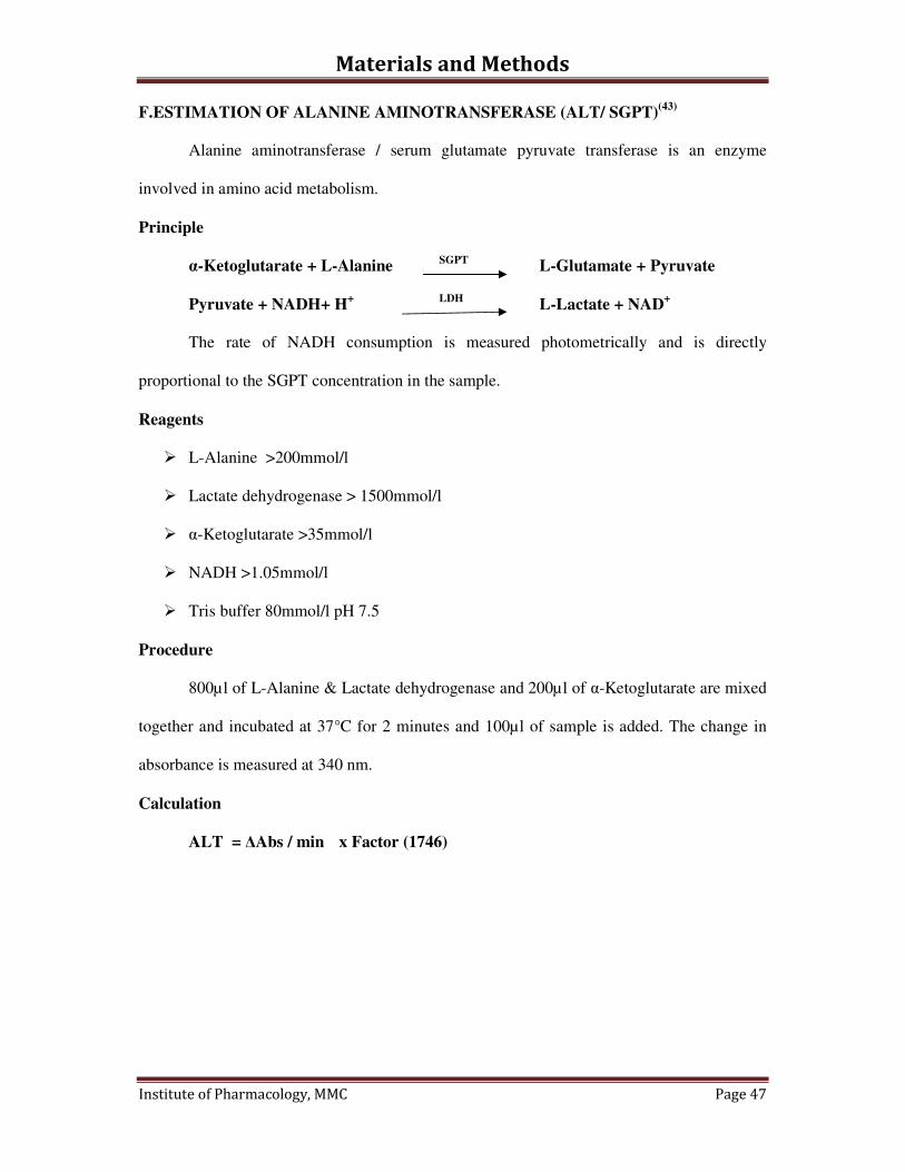
F.ESTIMATION OF ALANINE AMINOTRANSFERASE (ALT/ SGPT)(43)
Alanine aminotransferase / serum glutamate pyruvate transferase is an enzyme
involved in amino acid metabolism.
Principle
α-Ketoglutarate + L-Alanine SGPT
L-Glutamate + Pyruvate
Pyruvate + NADH+ H+
LDH L-Lactate + NAD
+
The rate of NADH consumption is measured photometrically and is directly
proportional to the SGPT concentration in the sample.
Reagents
� L-Alanine >200mmol/l
� Lactate dehydrogenase > 1500mmol/l
� α-Ketoglutarate >35mmol/l
� NADH >1.05mmol/l
� Tris buffer 80mmol/l pH 7.5
Procedure
800µl of L-Alanine & Lactate dehydrogenase and 200µl of α-Ketoglutarate are mixed
together and incubated at 37°C for 2 minutes and 100µl of sample is added. The change in
absorbance is measured at 340 nm.
Calculation
ALT = ∆Abs / min x Factor (1746)

Materials and Methods
Institute of Pharmacology, MMC Page 48
(iv) Estimation in tissue homogenates
A) PREPARATION OF TISSUE HOMOGENATE
At the end of 21st day, all the experimental animals were sacrificed and liver, pancreas
and kidney were removed, free from adhering tissues and washed with ice-cold normal saline
solution (0.9%). About 0.3g tissue was homogenized in 3ml of 0.01 M Tris-HCl with the help
of Teflon-homogenizer. The homogenate was centrifuged at 10,000 rpm for 20mins at 4°C.
The supernatant obtained was used for estimation of lysosomal enzymes and some
carbohydrate metabolizing enzymes
B) CARBOHYDRATE METABOLIZING ENZYMES ESTIMATION
1) HEXOKINASE ESTIMATION(44)
Principle
Hexokinase (HK) activity was determined by the method of Brandstrup et al., (1957).
The assay is based upon the reduction of NAD+ through a coupled reaction with glucose-6-
phosphate dehydrogenase and is determined spectrophotometrically by measuring the
increase in absorbance at 340nm.
D-Glucose + ATP hexokinase
Glucose-6-phosphate + ADP
Glucose-6-phosphate 2P
Gluconate-6-phosphate + NADH+H
+
One unit of activity reduces one micromole of NAD+/min at 30°C and pH
8.0 under
the specified conditions.
Reagents
� 0.05M Tris-Hcl buffer, pH8.0 with 13.3mM MgCl2
� 0.67M Glucose in above Tris-MgCl2 buffer
� 16.5mM Adenosine 5’Triphosphate in above Tris-MgCl2 buffer
� 6.8mM NAD in above Tris-MgCl2 buffer

Materials and Methods
Institute of Pharmacology, MMC Page 49
� Glucose -6-phosphate dehydrogenase (G6PD)
Procedure
The assay mixture containing 2.28 ml of Tris-MgCl2 buffer, 0.5 ml of 0.67M Glucose,
0.1 ml of 16.5mM ATP, 6.8mM NAD and 0.01 ml of G6PD was mixed well. To the above
mixture 0.1ml of tissue homogenate was added and the increase in absorbance was measured
at 340nm for 5minutes.
2) ESTIMATION OF GLUCOSE-6-PHOSPHATE DEHYDROGENASE (G6PD)(45)
Principle
G6PD activity was estimated by the method of Anderson & Nordlie et al., (1968).
This G6PD is rather unique in that it possesses dual coenzyme specificity. When assayed
under conditions that are optimal for the particular coenzyme, the ratio of observed catalytic
activity is NAD/NADP = 1.8. The reaction velocity is determined by measuring the increase
in absorbance at 340nm resulting from the reduction of NAD or NADP.
Reagents
� 0.055M Tris-HCl buffer pH 7.8 containing 0.0033 M Magnesium Chloride
� 0.06M Nicotinamide Adenine Dinucleotide (NAD)
� 0.1M Glucose-6-phosphate
� G6PD enzyme
Procedure
The assay mixture containing 2.7 ml of 0.055M Tris-HCl buffer, pH7.8 with 0.0033M
MgCl2, 0.1ml of 0.06M NAD and 0.1 ml of Glucose-6-phosphate. 0.1 ml of tissue
homogenate was added to the above assay mixture and the change in absorbance was
measured at 340nm for 5mins

Materials and Methods
Institute of Pharmacology, MMC Page 50
3) LACTATE DEHYDROGENASE (LDH) ASSAY(46)
Principle
The assay was performed according to King’s et al., (1965) method, when the enzyme
is supplied with Pyruvate and NADH+, the LDH catalyzed reaction starts to produce lactate.
At certain time point the reaction is terminated by the addition of 2,4-
Dinitrophenylhydrazine, which reacts with lactate at acidic pH. After alkalization (addition
of NaOH) the resulting hydrazine derivate gives a yellowish-orange colour suitable for
quantification by means of spectrophotometry at the wavelength of 440 nm.
Requirements
� 0.1M phosphate buffer (pH 7.5)
� NADH(6.6mM)
� Sodium pyruvate (30mM)
� Dinitro phenyl hydrazine
Procedure
0.1ml of tissue homogenate was added with 2.7 ml buffer, 0.1ml NADH, and 0.1ml
sodium pyruvate. The mixture was heated for 15 mins at 37°C. Then 0.5ml of dinitro phenyl
hydrazine was added and incubated at room temperature for 15 mins. The reaction was
stopped by addition of 5ml 0.1N NaOH. The developed colour was measured at 440nm.
C.LYSOSOMAL ENZYMES ESTIMATION IN TISSUE PREPARATIONS
1) ASSAY OF ALKALINE PHOSPHATASE (ALP)(47)
Principle
When the enzyme incubated with p-Nitro phenyl phosphate and Tris buffer (pH 9.6),
in alkaline condition inorganic phosphate and p-Nitro phenol are formed by the catalytic

Materials and Methods
Institute of Pharmacology, MMC Page 51
action of alkaline phosphatase. Amount of p-nitro phenol liberated by the enzyme is
measured at 420 nm.
2-amino-methyl-1-propanol + p-Nitro phenyl phosphate + H2O
4-Nitro phenol + 2-amino-2-methyl-1-propanol phosphate
Requirements
� P-nitro phenyl phosphate (10 mM)
� Tris-HCl pH 9.6 (80mM)
� NaOH (0.1N)
Procedure
1ml of p-nitro phenyl phosphate and 1.5 ml of buffer were added with 100µl of
homogenate. The mixture was incubated at 37°C for 30mins. Then the reaction was stopped
by addition of 0.1 N NaOH. The absorbance of liberated p-nitro phenol was measured at
420nm.
Calculation
ALP U/I = ∆A/ 2764
2) ASSAY OF ACID PHOSPHATASE (ACP)(48)
(α-Naphthylphosphate Kinetic method)
Principle
In acidic condition, the incubation of α-Naphthylphosphate with ACP will liberate α-
Naphthol and inorganic phosphate due to catalytic action of ACP. The α–Naphthol is coupled
with Fast Red to form a diazo dye complex. The rate of formation of this complex is
measured as an increase in absorbance which is proportional to the ACP activity in the
sample.
α –Naphtholphosphate + H2O ACP
α –Naphthol + Phosphate

Materials and Methods
Institute of Pharmacology, MMC Page 52
α –Naphthol + Fast Red TR Salt Diazo dye complex
Requirements
� α –Naphthol phosphate (4.5mM)
� acetate buffer (pH5.0)
� NaOH (0.2N)
Procedure
1ml of α –Naphthol and 1.9 ml of buffer were added with 100µl of homogenate. The
mixture was incubated at 37°C for 30mins. The absorbance of liberated α–Naphthol was
measured at 420 nm.
Calculation
ACP activity in U/L = ∆A/min x 750
HISTOPATHOLOGICAL STUDIES:
At the end of 21st day, all the animals were sacrificed to collect the pancreas and liver.
The organs were rinsed in ice cold 0.9% saline and were fixed in 10% formalin embedded in
paraffin and cut into 5 µm thick sections in a microtome. Sections were mounted on glass
slides using standard techniques. After staining with Hematoxylin-Eosin, the sections were
examined under 100X magnification and photographed under a light microscope equipped
for photography (Olympus CK 40).
STATISTICAL ANALYSIS:
All the parameters were analysed by ANAVO followed by Dunnet’s test. The results
were expressed as Mean ± SD. The statistical analysis was performed by using Graph and
Prism, version 6.0 software.
Results
Institute of Pharmacology, MMC Page 53
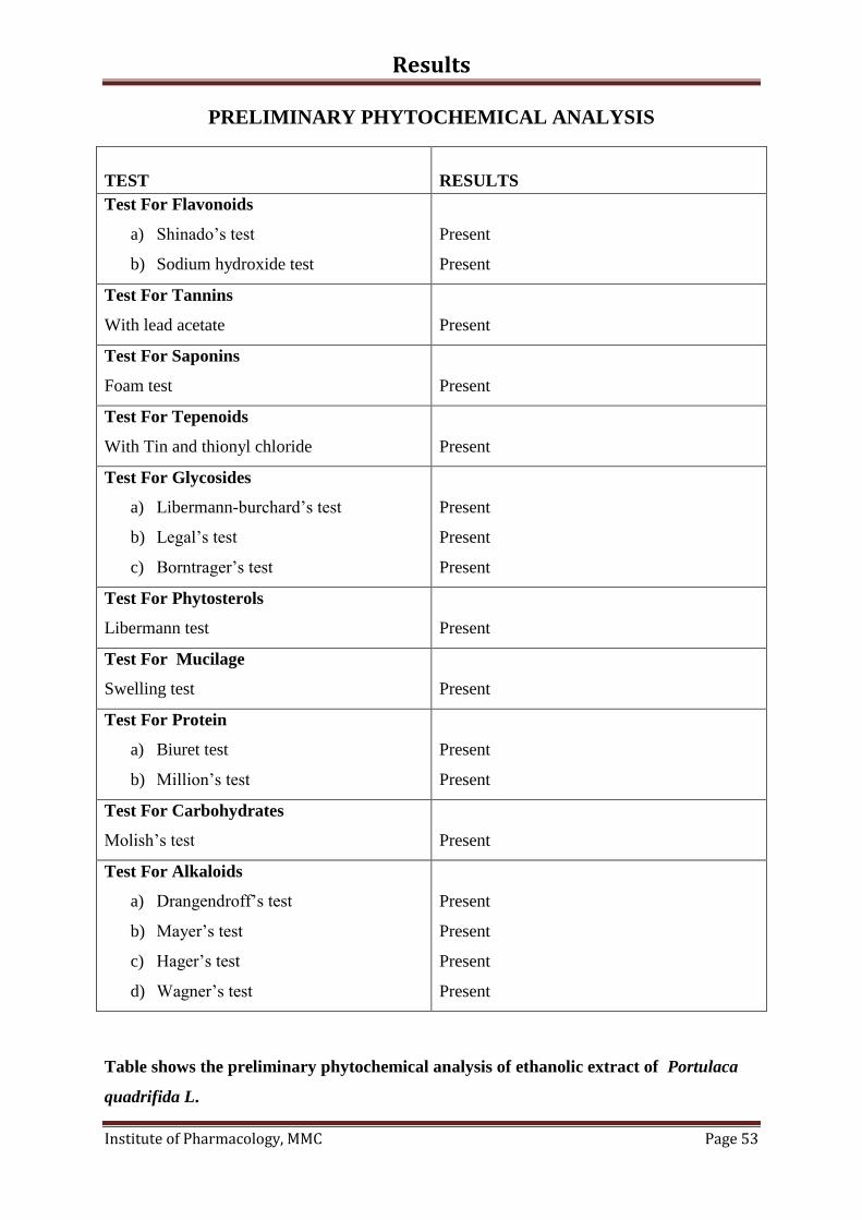
PRELIMINARY PHYTOCHEMICAL ANALYSIS
TEST
RESULTS
Test For Flavonoids
a) Shinado’s test
b) Sodium hydroxide test
Present
Present
Test For Tannins
With lead acetate
Present
Test For Saponins
Foam test
Present
Test For Tepenoids
With Tin and thionyl chloride
Present
Test For Glycosides
a) Libermann-burchard’s test
b) Legal’s test
c) Borntrager’s test
Present
Present
Present
Test For Phytosterols
Libermann test
Present
Test For Mucilage
Swelling test
Present
Test For Protein
a) Biuret test
b) Million’s test
Present
Present
Test For Carbohydrates
Molish’s test
Present
Test For Alkaloids
a) Drangendroff’s test
b) Mayer’s test
c) Hager’s test
d) Wagner’s test
Present
Present
Present
Present
Table shows the preliminary phytochemical analysis of ethanolic extract of Portulaca
quadrifida L.

Results
Institute of Pharmacology, MMC Page 54
TOXICITY STUDIES
ACUTE TOXICITY STUDY:
The crude ethanolic extract of Portulaca quadrifida L. was subjected to acute toxicity study.
S.NO.
PARAMETERS
RESULT
1. Toxic signs Absent
2. Pre-terminal deaths Nil
3. Body weight No specific change
4. Cage side observation Normal
5. Motor activity Normal
6. Tremors Absent
7. Convulsions Absent
8. Straub reaction Absent
9. Pilo erection Absent
10. Righting reflex Present
11. Lacrimation and salivation Normal
12. Unusual vocalization Absent
13. Sedation Absent
14. Body temperature Normal
15. Analgesia Absent
16. Ptosis Absent
17. Diarrhea Absent
18. Skin colour Normal
19. Respiration Normal
20. Scratching Absent
21. Grooming Absent
22. Aggressiveness and restlessness Absent

Results
Institute of Pharmacology, MMC Page 55
Animals were observed for behavioral signs of toxicity like motor activity, tremor,
ect., and no significant toxic signs were observed during 14 days. The results of the acute
toxicological studies revealed that the administration of ethanolic extract of Portulaca
quadrifida L. by oral route upto 2000mg/kg body weight did not produce any mortality and it
was tolerated.

Results
Institute of Pharmacology, MMC Page 56
Table 2 GLUCOSE ADSORPTION ASSAY
Table 2 shows the glucose adsorption capacity of PQ extract at 1% concentration, values
expressed as mean glucose bound (nm) with SD.
Glucose solution in a concentration of 50mM showed maximum glucose adsorption of 53 nm,
at 100mM concentration the value was reduced to 27 nm.
Figure 1 GLUCOSE ADSORPTION ASSAY
Figure 1 shows the graphical representation of mean glucose adsorption
0.0
10.0
20.0
30.0
40.0
50.0
60.0
5mM 10mM 25mM 50mM 100mM
Glu
cose
bo
un
d(n
m)
Concentration of Glucose(1% of Pq)
S.No. Concentration of the Glucose
Solution (mM)
Glucose bound
(nm)
1
5
8.6±0.88
2
10
23.5±1.31
3
25
39.5±0.71
4
50
53.73±0.87
5
100
26.77±o.74

Results
Institute of Pharmacology, MMC Page 57
Table 3 GLUCOSE DIFFUSION ASSAY
Concentration
(µg/ml)
Glucose concentration (mg/ml)
0 hour 1st hour 2
nd hour 3
rd hour 4
th hour 5
th hour
100 2.48±0.08
33.69±1.44 44.44±1.69 43.54±0.49 47.37±1.13 66.83±2.23
250 2.79±0.02
32.02±1.75 41.9±2.52 45.07±1.26 45.96±2.07 54.76±0.81
500 2.41±0.01
27.93±1.34 38.2±1.03 42.1±1.12 443.2±2.81 50.63±1.52
1000 2.54±0.04
25.63±0.93 30.7±0.70 34.9±0.91 39.37±0.61 44.83±2.21
1500 2.43±0.17
23.63±0.82 25.63±0.52 27.1±1.39 34.1±4.06 33.37±1.44
Control
(0 µg/ml)
3.16±0.34
35.37±1.15 47.3±0.4 53.57±0.49 58.87±1.59 73.57±2.16
Table 3 shows the mean glucose intensity in the external solution of various
concentrations of PQ at different time intervals. Glucose values are expressed as Mean
± SD
PQ extract decreased the concentration of glucose into the external solution by
retarding its diffusion through membrane. After 5 hrs the control showed 73.57 mg/dl of
glucose in the external solution whereas the PQ extract at 1500µg/ml showed only 33.3 mg/dl
of glucose. PQ extract possesses a dose dependent effect in glucose diffusion and it showed
highest activity at concentration of 1500µg/ml.
Figure 2 is the graphical representation of Table
0
10
20
30
40
50
60
70
80
100 250 500 1000 1500 0
Glu
cose
Con
c.(m
g/d
l)
Concentration (µg/ml)
Glucose diffusion assay
0 hour
1 hour
2 hours
3 hours
4 hours
5 hours

Results
Institute of Pharmacology, MMC Page 58
INVIVO ANTIDIABETIC ACTIVITY
Table 4: Effect of ethanolic extract of Portulaca quadrifida L. on Body Weight in
Streptozotocin induced diabetic rats.
Data were expressed as mean ±SD, n=6
Table shows the mean body weight during the study period of all groups. The mean body
weight showed a decrease in GP2, GP3, GP2 & GP5 at 21st day on comparing with 1
st day.
GP2 (diabetic rats) showed a significant reduction in body weight P<0.05.when compared
within the groups.
GROUP
TREATMENT
AVERAGE BODY WEIGHT IN DAYS(gm)
1st Day 7
th Day 14
th Day 21
st Day
1 Normal control
147.9±2.9
149.63±2.3
154.5±2.4
162.85±2.9
2
Diabetic control
STZ (150mg/kg)
150.65±1.0
144.21±2.9
138.05±2.8
131.3±2.0
3
Glibenclamide
(5mg/kg)
152.06 147.9±2.3 144.18±1.2 139.05±1.07
4 Portulaca
quadrifida
(200mg/kg)
151.7±1.49 144.5±1.32 141.75±1.9 136.5±3.15
5
Portulaca
quadrifida
(400mg/kg)
150.42±2.14 145.95±1.9 143.33±1.8 140.8±0.9
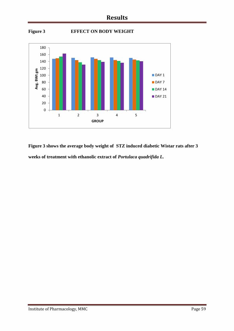
Results
Institute of Pharmacology, MMC Page 59
Figure 3 EFFECT ON BODY WEIGHT
Figure 3 shows the average body weight of STZ induced diabetic Wistar rats after 3
weeks of treatment with ethanolic extract of Portulaca quadrifida L.
0
20
40
60
80
100
120
140
160
180
1 2 3 4 5
Avg
. B
Wt
gm
GROUP
DAY 1
DAY 7
DAY 14
DAY 21

Results
Institute of Pharmacology, MMC Page 60
Table 5: Effect of ethanolic extract of Portulaca quadrifida L on Whole blood Glucose
(WBG) in Streptozotocin induced diabetic rats.
GROUP
TREATMENT
Whole Blood Glucose (mg/dL)
1st Day 7
th Day 14
th Day 21
st Day
1 Normal control
79.3±3.02
75.07±0.67
80.05±0.80
92.32±0.44
2
Diabetic control
STZ (150mg/kg)
395.5±1.46
411.05±1.52
419.02±1.41
423.3±1.3
3
Glibenclamide
(5mg/kg)
346.62±2.04 275.97±1.42 214.58±0.69 188.1±1.19
4 Portulaca
quadrifida
(200mg/kg)
355.22±7.53 281.81±1.01 218.48±1.44 190.93±1.06
5
Portulaca
quadrifida
(400mg/kg)
411.4±4.97 383.72±1.15 252.03±1.52 185.23±9.58
Data were expressed as mean ±SD, n=6
Within group analysis:
The mean blood glucose showed a significant reduction in both GP3, GP4& GP5 on
comparing 21st day with 1
st day

Results
Institute of Pharmacology, MMC Page 61
Between group analysis:
When compared with GP2, the mean body glucose level was lower in GP3, GP4 &
GP3 at day 21. On comparing GP1 with GP2, GP3, GP4 & GP5, a statistical significant
(p<0.05) difference was observed in GP2. On comparing GP2 with GP3, GP4 & GP5, a
statistically significant (p<0.05) difference was seen in both the groups;
Figure 4 MEAN BLOOD GLUCOSE
Figure 4 is the graphical representation of mean blood glucose level.
0
50
100
150
200
250
300
350
400
450
GROUP 1 GROUP 2 GROUP 3 GROUP 4 GROUP 5
Blo
od
Glu
cose
leve
l (m
g/d
l)
DAY 1
DAY 7
DAY 14
DAY 21

Results
Institute of Pharmacology, MMC Page 62
Table 6 LIPID PROFILE
Group
Treatment Cholesterol Triglycerides HDL
1
Normal control 109.2± 0.65 106.7± 0.63 16.6± 0.9
2
Diabetic control
STZ(150mg/kg)
155.9± 3.5 252.2± 0.9 25.12± 0.49
3
Glibenclamide
(5mg/kg)
130.63± 1.06 164.7± 0.6 16.6± 0.7
4
Pq (200mg/kg) 139.7± 0.6 172.9± 2.14 18.3± 0.57
5
Pq (400mg/kg) 133.4± 1.9 158.43±0.49 21.06± 1.8
The value are expressed as mean± SD
The mean cholesterol, triglycerides levels were higher in GP2, than the groups with GP3,
GP4 & GP5.
GP2 Vs GP3, GP4 & GP5- A significant (p< 0.05) decrease in Triglycerides Cholesterol
level was observed
Figure 5 LIPID PROFILE
Figure 5 is the graphical representation of Lipid profile level.
0
50
100
150
200
250
300
1 2 3 4 5
mg/d
l
Group
Cholesterol
Triglycerides
HDL

Results
Institute of Pharmacology, MMC Page 63
Table 7 ASSAY OF ALP (U/ml)
The values are expressed as mean ± SD, n=6
In GP2 the mean ALP was higher than GP1, GP3 & GP4.
GP1 Vs GP2, GP3, GP4 &GP5- A significant (p <0.05) increase in ALP level was observed
in GP2, GP3, GP4 & GP5.
Figure 6 ALKALINE PHOSPHATE
Figure 6 is the graphical representation of ALP levels
0.0
1.0
2.0
3.0
4.0
5.0
6.0
7.0
8.0
GP1 GP2 GP3 GP4 GP5
ALP
(un
its/
ml)
Group
Group
Treatment ALP (U/ml)
1
Normal control 0.9±0.47
2
Diabetic control
(STZ 5mg/kg)
7.1±0.21
3
Glibenclamide
(5mg/kg)
1.7±0.21
4
Pq (200mg/kg) 1.4±0.25
5
Pq (400mg/ml) 1.3±0.23

Results
Institute of Pharmacology, MMC Page 64
Table 8 ASSAY OF ACP (U/ml)
Group
Treatment ACP (U/ml)
1
Normal control 0.6±0.2
2
Diabetic control
(STZ 5mg/kg)
1.8±0.08
3
Glibenclamide
(5mg/kg)
0.7±0.12
4
Pq (200mg/kg) 0.6±0.15
5
Pq (400mg/ml) 0.56±0.20
The values are expressed as mean ± SD, n=6
In GP2 the mean ACP level was higher than GP1, GP3, GP4 & GP5.
GP1 Vs GP2, GP3, GP4 & GP5 - A significant (p<0.05) decrease in ACP level was seen in
GP3, GP4 & GP5.
Figure 7 ACID PHOSPHATASE
Figure 7 is the graphical representation of ACP level
0
0.2
0.4
0.6
0.8
1
1.2
1.4
1.6
1.8
2
GP1 GP2 GP3 GP4 GP5
AC
P(u
nit
s/m
l)
Group

Results
Institute of Pharmacology, MMC Page 65
Table 9 ESTIMATION OF HEXOKINASE (U/ml) ACTIVITY
Group
Treatment Hexokinase(U/ml)
1
Normal control 2.5±0.08
2
Diabetic control
(STZ 5mg/kg)
1.2±0.22
3
Glibenclamide
(5mg/kg)
2.3±0.09
4
Pq (200mg/kg) 2.4±0.12
5
Pq (400mg/ml) 2.56±0.17
The values are expressed as mean ± SD, n=6
In GP2 the mean hexokinase level was lower than GP1, GP3, GP4 & GP5.
GP1 Vs GP2, GP3, GP4 & GP5- A significant (p<0.05) decrease in the HK level was
observed in GP2
Figure 8 HEXOKINASE
Figure 8 is the graphical representation of Hexokinase activity.
0
0.5
1
1.5
2
2.5
3
GP1 GP2 GP3 GP4 GP5
He
xoki
nas
e(U
/ml)
Group

Results
Institute of Pharmacology, MMC Page 66
Table 10 ESTIMATION OF G6PD (U/ml) ACTIVITY
Group
Treatment G6PD(U/ml)
1
Normal control 10.63±0.67
2
Diabetic control
(STZ 5mg/kg)
2.4±0.22
3
Glibenclamide
(5mg/kg)
3.56±0.2
4
Pq (200mg/kg) 3.36±0.6
5
Pq (400mg/ml) 3.43±0.25
The values are expressed as mean ± SD, n=6
In GP2 the mean G6PD level was lower than GP1, GP3, GP4 & GP5.
GP1 Vs GP2, GP3, GP4 & GP5- A significant (p<0.05) decrease in the G6PD level was
observed in GP2
Figure 9 G6PD ESTIMATION
Figure 9 the graphical representation of G6PD levels
0
2
4
6
8
10
12
GP1 GP2 GP3 GP4 GP5
G6
PD
( u
nit
s/m
l)
Group
GP1
GP2
GP3
GP4
GP5

Results
Institute of Pharmacology, MMC Page 67
Table 11 ESTIMATION OF LDH (U/ml) ACTIVITY
Group
Treatment G6PD(U/ml)
1
Normal control 13.63±0.7
2
Diabetic control
(STZ 5mg/kg)
25.76±1.3
3
Glibenclamide
(5mg/kg)
16.7±1.02
4
Pq (200mg/kg) 18.9±0.4
5
Pq (400mg/ml) 11.8±0.73
The values are expressed as mean ± SD, n=6
In GP2 the mean G6PD level was higher than GP1, GP3, GP4 & GP5.
A significant increase was observed in LDH level when compared to other groups.
GP1 Vs GP2, GP3, GP4 & GP5 - A significant (p<0.05) rise in the G6PD level was observed
in GP2
Figure 10 LDH ESTIMATION
Figure 10 is the graphical representation of LDH level
0
5
10
15
20
25
30
GP1 GP2 GP3 GP4 GP5
LDH
(un
its/
ml)
group

Results
Institute of Pharmacology, MMC Page 68
Table 12 ESTIMATION OF SGOT
Group
Treatment SGOT(U/ml)
1
Normal control 144.05±2.0
2
Diabetic control
(STZ 5mg/kg)
205.1±3.15
3
Glibenclamide
(5mg/kg)
172.9±1.84
4
Pq (200mg/kg) 163.23±2.4
5
Pq (400mg/ml) 159.38±2.52
The values are expressed as mean ± SD, n=6
In GP2 the mean SGOT level was higher than GP1, GP3, GP4 & GP5.
A significant increase was observed in SGOT level when compared to other groups.
GP1 Vs GP2, GP3, GP4 & GP5 - A significant (p<0.05) rise in the SGOT level was observed
in GP2
Figure 11 SGOT
Figure 11 is the graphical representation of SGOT level
0
50
100
150
200
250
GP1 GP2 GP3 GP4 GP5
SGO
T
Group
GP1
GP2
GP3
GP4
GP5

Results
Institute of Pharmacology, MMC Page 69
Table 13 ESTIMATION OF SGPT
Group
Treatment SGPT(U/ml)
1
Normal control 103.68±2.0
2
Diabetic control
(STZ 5mg/kg)
157.91±1.23
3
Glibenclamide
(5mg/kg)
116.75±0.9
4
Pq (200mg/kg) 135.97±2.7
5
Pq (400mg/ml) 122.27±0.89
The values are expressed as mean ± SD, n=6
In GP2 the mean SGPT level was higher than GP1, GP3, GP4 & GP5.
A significant increase was observed in SGPT level when compared to other groups.
GP1 Vs GP2, GP3, GP4 & GP5 - A significant (p<0.05) rise in the SGOT level was observed
in GP2
Figure 12 SGPT
Figure 12 is the graphical representation of SGPT
0
20
40
60
80
100
120
140
160
180
GP1 GP2 GP3 GP4 GP5
SGP
T
Group

Results
Institute of Pharmacology, MMC Page 70
Table 14 TOTAL PROTEIN
Group
Treatment Total protein
1
Normal control 5.96± 0.55
2
Diabetic control
STZ(150mg/kg)
4.31± 0.62
3
Glibenclamide (5mg/kg) 6.0±0.44
4
Pq (200mg/kg) 6.05± 0.62
5
Pq (400mg/kg) 5.98± 0.62
The values are expressed as mean± SD
On comparing GP1 with GP2, GP3, GP4 & GP5, there was a significant (p<0.05)
decrease in total protein.
Figure 13
Figure 13 is a graphical representation of Total protein.
0
1
2
3
4
5
6
7
1 2 3 4 5
Tota
l pro
tein
(g/
dl)
Group
Total protein

Results
Institute of Pharmacology, MMC Page 71
Table 15 ESTIMATION OF UREA (mg/dl)
The values are expressed as mean± SD
The mean blood urea level was higher in GP2 than other groups. A significant increase was
observed in blood urea in GP2.
Figure 14
Figure 14 is a graphical representation of blood urea
0
10
20
30
40
50
60
1 2 3 4 5
Blo
od
Ure
a (m
g/d
l)
Group
Urea
Group
Treatment Urea
1
Normal control 40.17± 1.74
2
Diabetic control
STZ(150mg/kg)
48.08± 0.82
3
Glibenclamide (5mg/kg) 36.35±1.58
4
Pq (200mg/kg) 41.8± 2.59
5
Pq (400mg/kg) 35.11±0.66

Results
Institute of Pharmacology, MMC Page 72
Table 16 ESTIMATION OF CREATININE
The values are expressed as mean± SD
The mean blood urea level was higher in GP2 than other groups. A significant increase was
observed in blood urea in GP2.
On comparing GP4 Vs GP5, there was a significant difference in creatinine (p< 0.05).
Figure 15
Figure 15 is the graphical representation of Creatinine.
0
0.5
1
1.5
2
1 2 3 4 5
Cre
atin
ine
(m
g/m
l)
Group
Creatinine
Group
Treatment Creatinine
1
Normal control 0.77± 0.17
2
Diabetic control
STZ(150mg/kg)
1.58± 0.15
3
Glibenclamide (5mg/kg) 0.86± 0.12
4
Pq (200mg/kg) 1.1± 0.21
5
Pq (400mg/kg) 0.8± 0.16
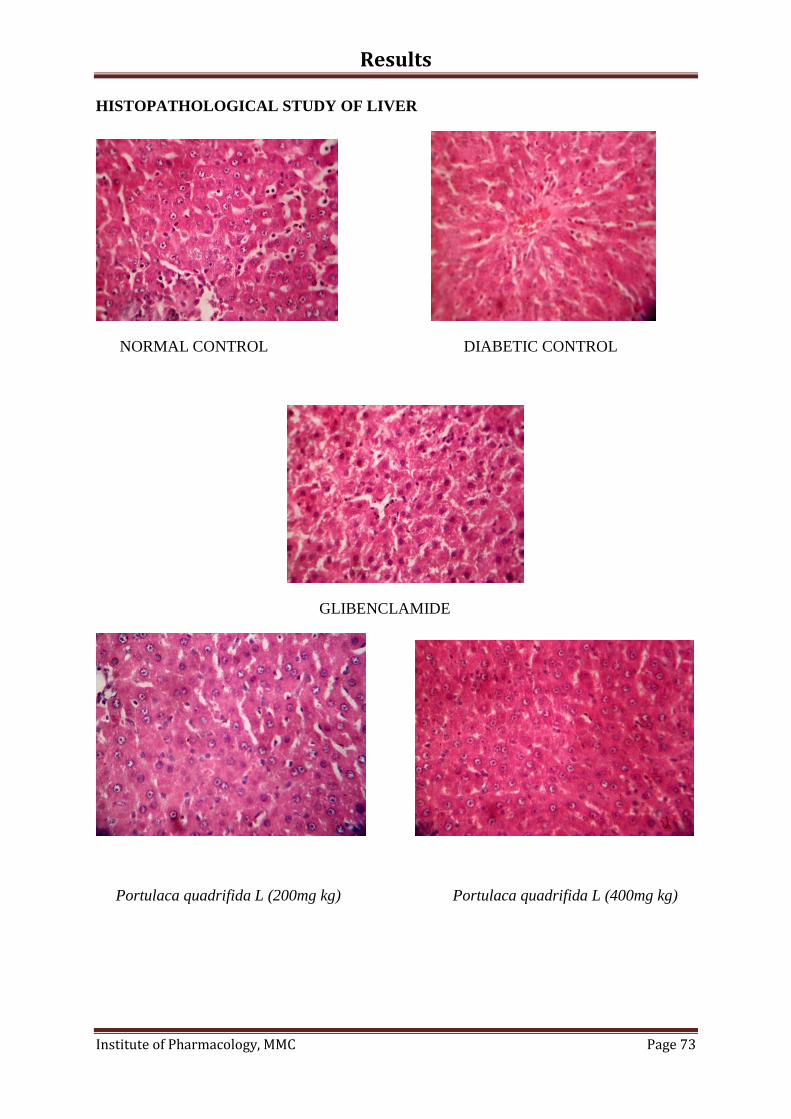
Results
Institute of Pharmacology, MMC Page 73
HISTOPATHOLOGICAL STUDY OF LIVER
NORMAL CONTROL DIABETIC CONTROL
GLIBENCLAMIDE
Portulaca quadrifida L (200mg kg) Portulaca quadrifida L (400mg kg)

Results
Institute of Pharmacology, MMC Page 74
HISTOPATHOLOGICAL STUDY OF PANCREAS
NORMAL CONTROL DIABETIC CONTROL
GLIBENCLAMIDE Portulaca quadrifida L (200mg kg)
Portulaca quadrifida L (400mg kg)

Discussion
Institute of Pharmacology, MMC Page 75
Discussion
Diabetes mellitus is a metabolic disorder, characterized by chronic hyperglycemia
with disturbance of carbohydrate, fat and protein metabolism resulting from defects in insulin
secretion, insulin action or both. Chronic hyperglycemia during diabetes leads to accelerated
generation of reactive oxygen species (ROS) through oxidative phosphorylation during
anaerobic glycolysis, glucose autoxidation, glucosamine pathway and a decline in the
antioxidant defense. The ROS formed during hyperglycemia causes damage to the membrane
lipids and proteins by lipid peroxidation and subsequent development of diabetic
complications. Recently, there has been considerable interests in finding natural
hypoglycemic agents from plant materials to replace synthetic ones to combat diabetic
complications are going on relentlessly.(49)
The goals of treatment for diabetes are to reduce and control blood glucose levels,
relieve the symptoms of disease, and prevent complications. The use of herbal medicine by
the suffers of chronic diseases is encouraged by the concern about the adverse effects of
chemical drugs and treatment using medicines of natural origin appears to offer gentler
means of managing such diseases(50)
.
Over 150 plant extracts and some of the active principle including flavonoids are
known to be used for the treatment of diabetes. In our study the whole plant extract of
chemical constituents such as Carbohydrates, Flavonoids, Terpenes, Glycosides, Tannins,
Saponins, Mucilage, Proteins and Sterols were used.
Acute toxicity study revealed that the ethanolic extract of Potulaca quadrifida L. was
relatively nontoxic upto 2000mg/kg/bw p.o indirectly pronouncing safety profile of the
extract.

Discussion
Institute of Pharmacology, MMC Page 76
Portulaca quadrifida L. is a tropical herb/forb which has been traditionally used in the
management of DM. this present study has been undertaken to evaluate its antidiabetic
property by preliminary phytochemical analysis, in vitro, Toxicological and in vivo studies in
Wistar albino rats.
The phytochemical analysis of ethanolic extract of Portulaca quadrifida L. showed
the presence of Alkaloids, Flavonoids, Tannins, Protein, Carbohydrates, Mucilage,
Phytosterols, Saponins, Glycosides and Terpenoids.
Glucose adsorption capacity of Portulaca quadrifida L. extract showed maximum
glucose bound at 50mM of glucose solution (53nm). Published research suggests that there is
a direct relationship between a plant’s ability to inhibit glucose absorption and viscosity of
the plant’s constituents such as soluble polysaccharides, supported by Gallagher et al.,(2003)
and Wood et al., (1994)(51)
The invitro antidiabetic evaluation was assessed by glucose diffusion assay.
Portulaca quadrifida L. ethanolic extract showed maximum decrease in glucose diffusion
when compared to control values. Glucose diffusion is useful invitro index to predict the
effect of plant fibers on the delay in glucose absorption in GI tract. In addition to glucose
adsorption, the retardation in glucose diffusion might be attributed to the physical obstacle
presented by fiber particles towards glucose molecules and entrapment of glucose within the
network formed by fibers. Based on this invitro study result, ethanolic extract of Portulaca
quadrifida L. was selected for invivo study.
Vehicle control animals were found to be almost stable in their body weight but
diabetic induced rats showed significant reduction in body weight. The administration of
Portulaca quadrifida L. whole plant extracts (200,400 mg/kg) and Glibenclamide (5mg/kg)
to the diabetic rats restored the changes in the body weight. On comparing 200mg Vs 400mg,

Discussion
Institute of Pharmacology, MMC Page 77
there was a significant difference in body weight (P<0.05). The dose dependent antidiabetic
property of the ethanolic extract of Portulaca quadrifida L. exhibited improvement in body
weight.
Plants may act on blood glucose through different mechanisms, some of them may
have insulin-like substances.(52)
Some may stimulate β-cells to produce more insulin and
others may increase β-cells in the pancreas by activating regeneration of pancreatic cells. The
fiber of the plants may also interfere with carbohydrate absorption; thereby affecting blood
glucose.
In our study, after the administration of extract (200,400mg/kg), the fall in fasting
blood glucose was evident at the end. This fall in fasting blood glucose level progressively
increases till the end of 3rd
week.
When the reduction glucose level with 200mg/kg & 400mg/kg were compared, there
was a statically significant reduction in blood glucose levels (P<0.05) with 400mg of the
extract.
The integrity of the lysosomal membrane is crucial to the maintenance of normal
levels of lysosomal glycohydrolases in tissue and body fluids. A compromise of lysosomal
membrane integrity may lead to an undesirable elevation of lysosomal enzymes in both intra-
and extra-cellular space, which could pave the way for cellular and tissue disorders. Eg.
Diabetes, arthritis, allergic response etc.(53)
. In the present study the diabetic control group,
there was an increase in alkaline phosphate and acid phosphate. This may due to the damage
of the lysosomal membrane in the diabetic animals. But in the Portulaca quadrifida L. extract
treated group, there was a significant reduction of these enzyme level which reveal the
protective nature of the plant source.

Discussion
Institute of Pharmacology, MMC Page 78
The hexokinase involved in the phosphorylation step of glucose in glycolysis. Hepatic
hexokinase is the most sensitive indicator of the glycolytic pathway in diabetes and its
increased level can increase the utilization of blood glucose for glycogen storage in liver.
Glucose-6-phosphate dehydrogenase activity was decreased in DM, resulting in diminished
functioning of HMP pathway and there by the production of reducing equivalent such as
NADH and NADPH.(54)
In the present study, the hexokinase and Glucose-6-phosphate
dehydrogenase activity were reduced in diabetic control group. The Portulaca quadrifida L.
extract treated group, there was an increase in the activity of both hexokinase and Glucose-6-
phosphate dehydrogenase. This shows that the Portulaca quadrifida L. improves the activity
of carbohydrate metabolizing enzymes (HK & G6PD).
Lactate dehydrogenase (LDH) is a cytosolic enzyme that catalyzes the conversion of
pyruvate to lactate in anaerobic glycolysis, which is subsequently converted to glucose in
glucogenic flux. Increased activity of lactate dehydrogenase interferes with normal glucose
metabolism and insulin secretion secretary defects in diabetes.(55)
In the present study, the
LDH activity was significantly elevated in diabetic control group. An increase in LDH
activity is observed in diabetes due to impairment in glucose stimulated insulin secretion. In
the Portulaca quadrifida L. extract treated group, the LDH activity was suppressed
significantly. The suppression of LDH activity shows the beneficial effects of plant source.
The levels of total serum cholesterol, triglycerides and HDL were raised in diabetic
rats which were lowered significantly with the treatment of Portulaca quadrifida L. On
comparing groups 3,4,5 with group 2 there was a significant reduction in total cholesterol,
triglycerides and HDL level (P<0.05). This shows that ethanolic extract of Portulaca
quadrifida L. is effective in reducing the total cholesterol, triglycerides, HDL levels (P<0.05)
in streptozotocin induced diabetic rats and its hypolipidemic effect could represent a

Discussion
Institute of Pharmacology, MMC Page 79
protective mechanism against the development of atherosclerosis which is usually associated
with diabetes.
Ghosh and Suryawanshi, 2001 observed elevation in transaminase activity (SGOT
and SGPT) in liver and kidney in diabetic rats.(56)
Increased gluconeogenesis and ketogenesis
observed in diabetic may be due to the high level in the activities of these transaminases.
Similarly in our study also there was an increase in SGOT and SGPT in group 2 animals
(Diabetic control). The restoration of SGOT and SGPT to their respective normal levels was
seen after the treatment with both Glibenclamide and ethanolic extract of Portulaca
quadrifida L. This supports the antidiabetogenic effect of Portulaca quadrifida L. Moreover,
SGOT and SGPT levels also act as indicators of liver function and restoration of these
parameters to normal levels indicate normal functioning of liver.
On comparing groups 3, 4 and 5 with group 2, there was a significant difference
(P<0.05) in total protein. In diabetic control rats, there was a decrease in serum protein.
Portulaca quadrifida L. extract prevented the reduction of serum protein in groups 4& 5.
Group 5 showed a statistically significant difference when compared to group 4 (P<0.05).
Blood urea and creatinine was elevated in group 2 (Diabetic control). When
comparing the groups 3, 4 & 5 with group 2, there was a significant difference (P<0.05) in
blood urea and creatinine. This shows that in the groups which received Portulaca quadrifida
L. extract, there was no significant increase in urea and creatinine.
The renewal of β cells in diabetes has been studied in different animal models. The
total β-cells mass reflects the balance between the renewal and loss of these cells. It has been
suggested that regeneration of islet β-cells following destruction by streptozotocin, may be
the primary cause of the recovery of streptozotocin injected guinea pigs from the effect of the
drug(57)
.
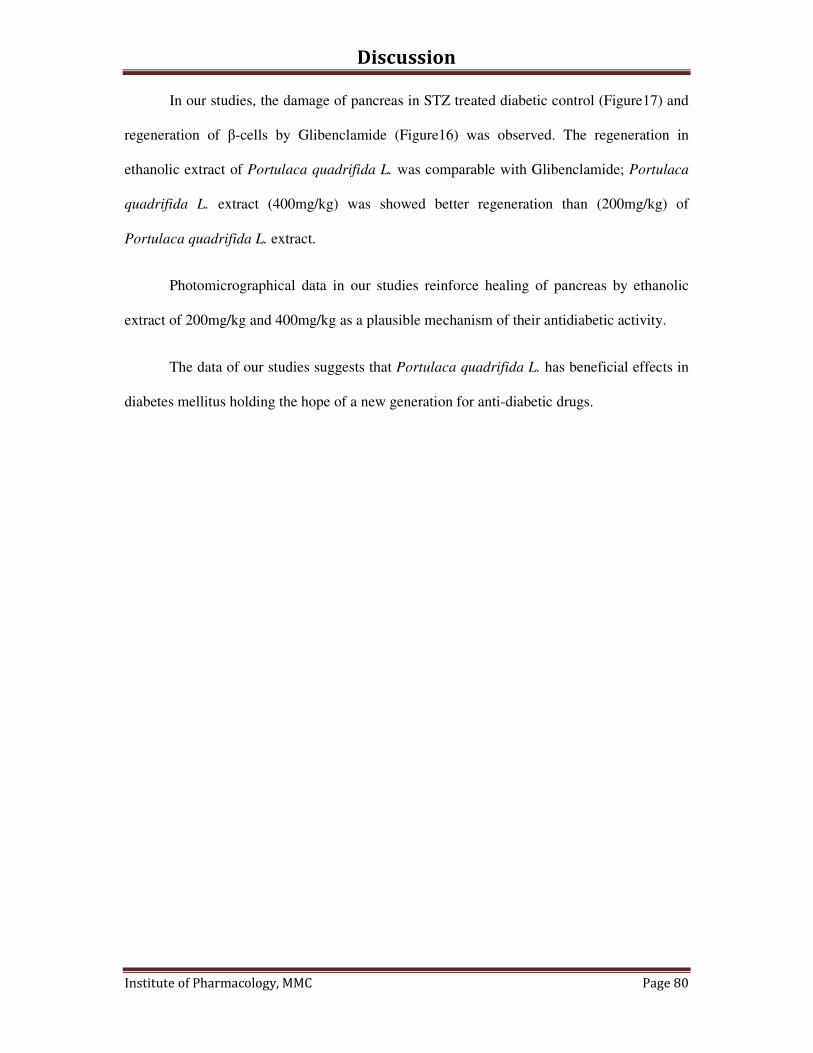
Discussion
Institute of Pharmacology, MMC Page 80
In our studies, the damage of pancreas in STZ treated diabetic control (Figure17) and
regeneration of β-cells by Glibenclamide (Figure16) was observed. The regeneration in
ethanolic extract of Portulaca quadrifida L. was comparable with Glibenclamide; Portulaca
quadrifida L. extract (400mg/kg) was showed better regeneration than (200mg/kg) of
Portulaca quadrifida L. extract.
Photomicrographical data in our studies reinforce healing of pancreas by ethanolic
extract of 200mg/kg and 400mg/kg as a plausible mechanism of their antidiabetic activity.
The data of our studies suggests that Portulaca quadrifida L. has beneficial effects in
diabetes mellitus holding the hope of a new generation for anti-diabetic drugs.

Conclusion
Institute of Pharmacology, MMC Page 81
CONCLUSION
� From the present study we conclude that the preliminary phytochemical analysis of
Portulaca quadrifida L. indicated the presence of alkaloids, flavonoids, proteins,
saponins, carbohydrates, terpenoids, mucilage, glycosides, tannins and phytosterols.
� Invitro Glucose adsorption assay of Portulaca quadrifida L. extract was evaluated
using GOD-POD kit.
� Invitro Glucose diffusion properties of Portulaca quadrifida L. extract was evaluated
using dialysis membrane and GOD-POD kit.
� Ethanolic extract of Portulaca quadrifida L. was safe in dose upto 2000mg/kg by
acute toxicity study and there was no toxic effects produced.
� The ethanolic extract of Portulaca quadrifida L. exhibited significant antidiabetic
activity in Streptozotocin induced diabetic rats.
� In vivo studies of Portulaca quadrifida L. ethanolic extract revealed the glucose
lowering effect& improved activity of carbohydrate metabolizing enzymes. Thus it
was concluded that the ethanolic extract of Portulaca quadrifida L. has anti-diabetic
activity.

References
Institute of Pharmacology, MMC Page 82
REFERENCES
1. Pulok.K.Mukerjee, Quality control of Herbal Drugs, An approach to evaluation of
Botanicals, 1st edition, Business Horizons, New Delhi, 2001, p.17-20
2. Robert. B. Tattesall., The history of Diabetes Mellitus, Text book of Diabetes, 3rd
edition, Vol. John. C. Pickup, Gareth Williams, Blackwell, USA, 2003, p. 1.1-1.17.
3. Felig. G., Bergman, M., Felig, C., The Endocrine Pancreas: Diabetes Mellitus, 3rd
ed.
McGraw-Hill, New York, 1995, p.1107-1250.
4. Rosac. C., “The Pathophysiological Basis of Efficacy and Clinical Experience with
the new Oral Anti-diabetic Agents”. J.J Diab. Compli., 2002; p.123-132.
5. Raheja. B.S., “Oral Hypoglycemic Agent in the Management of Maturity- Onset
Diabetes”. Journal J.J Group Hospitals, 2005; p.24-28.
6. Mukerjee. P.K., Maiti, K., Mukerjee, K. and Houghton, P.J., “Leads from Indian
medicinal Plants with Hypoglycemic Potentials”. Journal of Ethnopharmacology,
2006; p. 1-28.
7. Frank B.Hu. Department of Nutrition and Epidemiology, Harvard Medical School,
Havard school of Health., Boston, March 2011.
8. Goodman & Gillman’s The Pharmacological Basis of Therapeutics, McGraw Hill,
10th
edition, Pg. 1687.
9. International Diabetes Federation, IDF, Diabetes Atlas, Epidemiology and Morbidity,
March 2011.
10. Polyxeni Alexion and Vassilis J. Demopoulos, Medicinal Plants used for the
treatment of Diabetes and its long-term complications, Plants in Traditional and
Modern Medicine: Chemistry and Activity, 2010,p. 69-175.

References
Institute of Pharmacology, MMC Page 83
11. Patil AG., Joshi VS., Koli SP., Patil DA, “Pharmacognistical and Phytochemical
Analysis of Portulaca quarifida L.” , Research Journal of Pharmaceutical, Biological
and Chemical Sciences, Jan-March, 2012, vol.3, Pg. 90-100.
12. Harsh Mohan, Textbook of Pathology: Systemic Pathology, 5th
edition, Jaypee
Brother’s Medical Publishers, New delhi, 2005, Pg.842-53.
13. Kasper DL, Braunwald E, Fauci AS, Hauser SL, Longo DL and Janeson JL,
Harrison’s Principle of Internal Medicine, Vol.1 & 2.
14. Barret TG, “Mitochondrial Diabetes, DIDMOAD and other inherited diabetic
syndromes”, Journals of Clinical Endicrinology and Metabolism, 15(3): 325-43.
15. Endocrine Physiology, 2nd
edition, Susan Porterfield, Jaypee Publishers, p. 99-100.
16. Diagnosis of Diabetes, NIH Publications, October 2008, No.09-4642.
17. Textbook of Physiology, Vol.2, Prof. A. K. Jain, Arichal Publishing Company,
Pg.728-731.
18. SK. Gupta, Drug Screening Methods, 2nd
edition, Jaypee Brothers, New Delhi, 2009,
Pg.590-594.
19. Hardman., Limbird., Gilman, Goodman &Gilman’s The Pharmacological Basis of
Therapeutics, 10th
edition, Pg.1692.
20. ADA, Standards of Medical care for patients with Diabetes mellitus, Position
statement, Diabetes care, 2003, S5-S20.
21. Tripathi KD, Essentials of Medicinal Pharmacology, 6th
edition, Jaypee Brothers,
New Delhi, 2007,Pg.266-273
22. Arun Babu Vuyyuru, Govindarao M, Ravi Chandra Sekhara Reddy D., Harish B.,
Vishwanath J and Amarnath Reddy G, Antidiabetic activity of hydroalcolic extract of
Ananas comosus L. leaves in Streptozotocin induced diabetic rats. International
Journal of Pharmacy, Issue 2, P.142-147.

References
Institute of Pharmacology, MMC Page 84
23. Kumari, K., Mathew, B.C. and Augusti, K.T., Antidiabetic and Hypolipidemic effects
of S-methyl cysteine sulfoxide isolated from Allium epa Linn., Indian Journal of
Biochem.Biophys, 1995; 32 (1): 49-54.
24. Zacharias, N.T., Sebastian, K.L., Philip, B. and Augusti, K.T.(1980), Hypoglycemic
and Hypolipidemic effects of garlic in sucrose fed rabbits. Indian Journal of
Physiology and Pharmacology, 24: 151-154.
25. Faiyaz Ahmed, Asna Urooj, Antihyperglycemic activity of Ficus glomerata stem bark
in Streptozotocin induced diabetic rats. Global Journal of Pharmacology, 2008,p.41-
48
26. M.R.M.Rafiullah and A.W.Siddiqui, M R Mir, M. Ali, K.K.Pillai , Surender singh.
Antidiabetic activity of some Medicinal Plants. J pharmaceutical biology.44; 2: 95-
99.
27. Rajasekaran S,Sivagnanam, K., and Subramaniam, S. (2005) Modulatory effects of
Aloe vera leaf gel extract on oxitative stress in rats treated with Streptozotocin.
Journal of Pharmacy and Pharmacology, 57(2): 241-246.
28. Gomes A, Vedasiromoni JR, Das M, Sharma RM, Ganguly DK.(1995)
Antihyperglycemic effect of black tea (Camellia sinensis) in rats. Journal of
Ethnopharmacology, 45: 223-226.
29. C.P.Khare, Indian Medicinal Plants, Springer, New Delhi,2007, Pg.515-516.
30. Syed Kamil M, Liyakha T Ahmed MD, Paramjyothi S, Neuropharmacological Effects
of ethanolic extract of Portulaca quadrifida L in mice, International Journal of
Pharm Tech Research, Vol.2 No.2, April-June, p. 1386-1390.
31. Patil AG, Joshi VS, Koli SP, Patil DA, Pharmacognistical and Phytochemical
Analysis of Portulaca quadrifida L, Research Journal of Pharmaceutical, Biological
and Chemical Sciences, Vol.3, Jan-March 2012, p.90.
References
Institute of Pharmacology, MMC Page 85
32. Syed Kamil Mulla, Paramjyothi Swamy, Anticancer activity of ethanol and
polyphenol extracts of Portulaca quadrifida L on Human Colon Cancer cell lines,
International Journal of Pharma and Bio Sciences, July 2012, 3(3): p.488-498
33. A.M. Gallagher, P.R. Flatt, G. Duffy, Y.H.A. Abdel- Wahab, The effects of
Traditional Antidiabetic plants on in vitro Glucose Diffusion, Nutrition Research
2003; 23:413-424.
34. Faiyaz Ahmed NS, Siddaraju, Asna Uooj. In vitro hypoglycemic effects of Gymnema
sylvestre, Tinospora cordifolia, Eugenia jambolana and Aegle marmelos. Journal of
Natural Pharmaceuticals, 2011; 2(2): 52-55.
35. Kokate C K, Purohit A P, Gokhale S B. Pharmacognosy. 35th
edition. Pune: Nirali
Prakashan; 2006. Appendix I, Pathways to screen phytochemical nature of natural
drugs; p. 593-594.
36. Dr.K R Khandewal, Practical Pharmacognosy, 19th
edition, Nirali Prakashan, OECD
Guideline for testing of Chemicals 423[online]; 2001 December; [Cited on 2010
Feb11]. Available from URL. http://iccvam.niehs.nih.gov/SuppDocs/OECD/OECD
GL425.pdf.
37. Young DS. Effects of Drugs on Clinical Laboratory Tests, 5th
edition, AACC Press,
2000.
38. Alan H Gowenlock, Vanley’s Practical Clinical Biochemistry, 6th
edition, 1988:p.664.
39. Dace. J. V. , Practical Hematology, 6th
edition, 1985:p.28.
40. Burtis, C.A., Ashwood, E.R., Tietz Textbook of Clinical Chemistry, 2nd
edition,
Philadelphia, W.B. Saunders Company, 1994: p.1016-1018.
41. National Committee for Clinical Laboratory Standards. User evaluation Precision
Performance of Clinical Chemistry Devices. NCCLS; 1984, NCCLS publications.
http://www.thermo.com/eThermo/CMA/PDF/s/Product/productPDF_50918.pdf.

References
Institute of Pharmacology, MMC Page 86
42. Chernecky CC, Berger VJ, eds. Laboratory Tests and Diagonostic Proceduces, 4th
edition. Philadelphia: Saunders, 2004.
43. Brandstrup N, Kirk JE, Bruni C. Determination of Hexokinase in tissues. Journal of
Gerontol, 1957; 12: 166-71.
44. Anderson W, Nordlie R. Glucose Dehydrogenase activity of Yeast Glucose-6-
Phosphate Dehydrogenase. I. Selective Stimulation by Bicarbonate, Phosphate and
Sulphate. Journal of Biochemistry, 1968; 7: 1479.
45. King J. A routine method for the estimation of Lactate dehydrogenase activity.
Journal of Medical Lab. Technol, 1959; 16:265-272.
46. King J, the Hydrolases-Acid and Alkaline Phosphatases, in: D. Van (Ed.), Practical
Clinical Enzymology, Nostrand Company Limited, London, 1965; pp.191-208.
47. Hillman, GC., Klin Biochemistry,1971,9:273-274.
48. Seller D,et al., Journal of Clinical Chemistry and Clinical Biochemistry, 1983,21:519.
49. Selvaraj Sivakumar, Sorimuthu Pillai Subramanian. Pancreatic tissue protective
nature of D-pinitol studied in STZ mediated oxidative stress in experimental diabetic
rats. Eur. J. Pharmacol, 2009; 622: 65-70.
50. WHO. Traditional Medicine Strategy 2002-2005. WHO publications 2002;p. 1-6.
51. Wood PJ, Braaten JT, Scott FW, Riedel KD, Wolyntz MS, Collins MW. Effect of
dose and modification of viscous properties of oat gum on plasma glucose and insulin
following an oral load. Br J Nutr, 1994; 72: 731-43.
52. A.N.Nagappa. et.al. Antidiabetic activityof Terminalia catappa Linn fruits. J
Ethnopharmacol, 88 (2003), P.45-50.
53. Stoka, V., Turk, V., and Turk, B. Lysosomal cysteine cathepsins: signaling pathways
in apoptosis. Biol.Chem 2007; 388: 555-560.

References
Institute of Pharmacology, MMC Page 87
54. Leelavinothan Pari, Selvaraju Suman. Efficacy of Naingin on hapatic enzymes of
carbohydrate metabolism in STZ-Nicotinamide induced Type 2 diabetes rats. Int.J
Pharm. Biol.Archi 2010; 1(2): 280-286.
55. Gopalan Sriram Prasath, Sorimuthu Pillai Subramanian. Modulatory effects of Fiestin,
a bioflavoniod, on hyperglycemia by attenuating the key enzymes of carbohydrate
metabolism in hepatic and renal tissues in STZ-induced diabetic rats.
Eur.J.Pharmacol, 2011; 668:492-496.
56. Bhupesh chander semwal et.al., Antidiabetic activity of stem bark of ,Antidiabetic
activity of stem bark of Berberis asistata in alloxan induced diabetic rats. Internal J
Pharmocol, 2008. Vol.1.
57. Gorray. et al., Response of pancreatic β cells to alloxan and STZ in the guinea pig
pancreas, p.130-138