Embed Size (px)
Citation preview

From the Department of Molecular Medicine and Surgery
Karolinska Institutet, Stockholm, Sweden
EVALUATION OF CARBON DIOXIDE INSUFFLATION INTO THE
OPEN SURGICAL WOUND; INFLUENCE ON WOUND
TEMPERATURE, CORE TEMPERATURE, AND
POSTOPERATIVE OUTCOME
Joana Frey, MD
STOCKHOLM 2016
brought to you by COREView metadata, citation and similar papers at core.ac.uk
provided by Publications from Karolinska Institutet

Joana Frey
All previously published papers were reproduced with permission from the publisher.
Published by Karolinska Institute.
Printed by E-print AB 2016
© Joana Frey, 2016
ISBN 978-91-7676-201-1

EVALUATION OF CARBON DIOXIDE INSUFFLATION INTO THE OPEN SURGICAL
WOUND; INFLUENCE ON WOUND TEMPERATURE, CORE TEMPERATURE, AND
POSTOPERATIVE OUTCOME
THESIS FOR DOCTORAL DEGREE (Ph.D.)
By
Joana Frey
Principal Supervisor:
Professor Jan van der Linden
Karolinska Institutet
Department of Molecular Medicine
and Surgery
Division of Cardiothoracic anesthesia and
Intensive care
Co-supervisor:
Docent Peter Svenarud
Karolinska Institutet
Department of Molecular Medicine
and Surgery
Division of Cardiothoracic surgery
Opponent:
Professor Henrik Ahn
Hälsouniversitetet Linköping
Department of Medicine and Care
Division of Cardiothoracic surgery
Examination Board:
Professor Lars Wiklund
Uppsala Universitet
Department of Surgical science
Division of Anesthesiology and Intensive Care
Docent Louis Riddez
Karolinska Institutet
Department of Molecular Medicine
and Surgery
Division of Acute Surgery and Trauma
Docent Hans Hjelmqvist
Karolinska Institutet
Department of Clinical science, intervention and
technic
Division of Anesthesiology and Intensive Care

Joana Frey
2
The concept of keeping the wounds moist, warm, clean and protected is not new. There is
documentation indicating that the ancient Mesopotamians dressed their wounds with fine
linen soaked in oil. The Greeks applied animal fat and wrapped the wounds with greasy
sheepskin and the Roman applied ashes, oil and herbs. So the old expression from
Hippocrates, one of the most outstanding figures in the history of medicine, still holds true
today, 2500 years later.
The Healing Hand: Man and Wound in the Ancient World by Guido Majno - 1991
To my mother who still believes that I am going to get
the Nobel Prize, one day!

3
ABSTRACT
Introduction: When the internal organ tissues are abruptly exposed to the relative cold and
dry ambient air during open surgery, body heat is lost through radiation, evaporation, and
convection. Also general and neuraxial anesthesia contributes to a decrease in core
temperature, mainly due to a shift of the threshold for thermoregulatory defense mechanism
toward lower temperatures. It is well known that perioperative hypothermia is
disadvantageous for the patient, since it increases the risk of surgical wound infections, blood
loss, morbid cardiac events and postoperative shivering. Guidelines to keep the patient warm
during surgery are today common practice, but despite routine preventive measures, mild
intraoperative hypothermia is still common and contributes to postoperative morbidity and
mortality. The aim of this thesis was to investigate if local insufflation of CO2 could increase
both the open surgical wound temperature and core temperature, and affect postoperative
outcome.
Methods: I. In 10 patients undergoing cardiac surgery, the sternotomy wound was insufflated
with dry, room-tempered CO2 via a gas diffuser for two minutes. A heat-sensitive camera
measured the wound temperature before, during and after insufflation. II. 80 patients
undergoing open colon surgery were randomized to either standard warming measures (n=39)
or additional local wound insufflation (n=41) of warmed (30°C) humidified (93% rH) CO2
via a gas diffuser. A heat-sensitive camera measured the wound temperature and an ear
thermometer measured the core temperature. III. 83 patients undergoing open colon surgery
were randomized to either standard warming measures (n=39) or additional local wound
insufflation (n=40) of warmed (37°C) humidified (100% rH) CO2 via a gas diffuser. A heat-
sensitive camera measured the wound temperature and an ear thermometer measured the core
temperature. IV. This is a post hoc retrospective study of study II and III, where patients were
randomized to warmed humidified CO2 (n=80) or not (n=78).
Results: I. Exposure to dry CO2 increased the median temperature of the whole wound by
0.5°C (p=0.01). The temperature of the area distant to the diffuser increased by 1.2°C
(p<0.01) whereas in the area close to the diffuser it decreased by 1.8 °C (p<0.01). II. The
median wound area and wound edge temperatures were 1.2ºC (p<0.001) and 1.0ºC (p=0.002)
higher in the CO2 group, respectively, than in the control group. The mean core temperature
after intubation was the same (35.9ºC) in both groups, but at end of surgery the two groups
differed with a mean of 36.2 ± 0.5 ºC in the CO2 group and 35.8±0.5ºC in the control group
(p=0.003). III. The mean wound area temperature during surgery was 31.3ºC in the CO2
group compared with 29.6ºC in the control group (p<0.001). Also, the mean wound edge

Joana Frey
4
temperature during surgery was 30.3ºC compared with 28.5ºC in the control group (p<0.001).
Mean core temperature before start of surgery was similar with 36.7 ± 0.5ºC in the CO2 group
versus 36.6 ± 0.5ºC in the control group. At end of surgery the two groups differed
significantly with 36.9 ± 0.5ºC in the CO2 group versus 36.3 ± 0.5ºC in the control group
(p<0.001). IV. A multivariate analysis adjusted for age (p=0.001) and cancer (p=0.165)
showed that the larger the temperature difference between final core temperature and wound
edge temperature, the lower the overall survival rate (p=0.050). A lower end-of-operation
wound edge temperature was negatively associated with mortality (OR=0.80, 95%CI=0.68-
0.95, P=0.011), whereas age (10-year increase, OR=1.78, 95% CI=1.37-2.33, P<0.001) and
cancer (OR=8.1, 95% CI=1.95-33.7, P=0.004) were positively associated with mortality.
Conclusions: The major finding of this thesis is that insufflation of dry room-tempered CO2
with a gas diffuser increases the average surface temperature in an open wound cavity.
Insufflation of warmed humidified CO2 in an open surgical wound cavity results in significant
increases of the surgical wound temperature as well as the core temperature. Insufflation of
warmed fully humidified CO2 in an open surgical wound cavity increases surgical wound and
core temperatures, and helps to maintain normothermia. A small end-of-operation
temperature difference between final core and final wound edge temperature was positively
associated with patient survival in open colon surgery, and a lower end-of-operation wound
edge temperature was negatively associated with mortality.

5
SAMMANFATTNING
Introduktion: När de inre organens vävnader plötsligt exponeras för den relativt kalla och
torra omgivande luften under öppen kirurgi så förloras kroppsvärme genom strålning,
avdunstning och konvektion. Även generell och regional anestesi bidrar till en sänkning av
kroppstemperaturen, mestadels beroende på en nedsatt tröskel för termoregulation och
därmed ett minskat försvar mot kyla. Det är välkänt att perioperativ hypotermi är till nackdel
för patienten eftersom det leder till ökad risk för postoperativa sårinfektioner, ökad blödning,
kardiovaskulär morbiditet och mortalitet samt postoperativ shivering. Guidelines för att hålla
patienten varm är idag rutin på operationsavdelningar men trots detta är mild intraoperativ
hypotermi vanligt och bidrar till postoperativ morbiditet och mortalitet. Syftet med denna
avhandling var att undersöka om lokal tillförsel av koldioxid kunde öka både sårtemperaturen
och kroppstemperaturen under öppen kirurgi, samt om det påverkade det postoperativa
förloppet.
Metoder: I. Hos 10 patienter som genomgick öppen hjärtkirurgi lät vi tillföra torr,
rumstempererad CO2 till det kirurgiska såret via en gas-diffusor i 2 minuter. En värmekänslig
kamera mätte sårtemperaturen före, under och efter tillförseln. II. 80 patienter som
genomgick öppen kolonkirurgi blev randomiserade till antingen rutinmässiga
värmningsmetoder (n=39) eller dessutom lokal tillförsel (n=41) av värmd (30°C) och
befuktad (93 % rH) CO2 via en gas-diffusor. En värmekänslig kamera mätte sårtemperaturen
och en öron termometer mätte kroppstemperaturen. III. 83 patienter som genomgick öppen
kolonkirurgi blev randomiserade till antingen rutinmässiga värmningsmetoder (n=39) eller
dessutom lokal tillförsel (n=40) av värmd (37°C) och befuktad (100 % rH) CO2 via en gas-
diffusor. En värmekänslig kamera mätte sårtemperaturen och en öron termometer mätte
kroppstemperaturen. IV. Detta är en post hoc, retrospektiv studie av studie II och III där
patienter blev randomiserade till värmd och befuktad CO2 (n=80) eller inte (n=78).
Resultat: I. Exponering för CO2 ökade mediantemperaturen i hela såret med 0.5°C (p=0,01).
Temperaturen i området distalt om diffusorn ökade med 1.2°C (p<0,01), medan i området
nära diffusorn minskade temperaturen med 1.8°C (p<0,01). II. Mediantemperaturerna i
sårytan och sårkanterna var 1.2ºC (p<0,001) respektive 1.0ºC (p=0,002) högre i CO2-gruppen
jämfört med kontrollgruppen. Medelkroppstemperaturen efter intubation var samma (35.9ºC)
i båda grupper, men vid slutet av kirurgin skilde sig de båda grupperna åt med en
medeltemperatur på 36.2 ± 0.5 ºC i CO2-gruppen och 35.8 ± 0.5ºC i kontrollgruppen
(p=0,003). III. Medeltemperaturen i sårytan under kirurgi var 31.3ºC i CO2-gruppen jämfört
med 29.6ºC i kontrollgruppen (p<0,001). Dessutom var medeltemperaturen i sårkanterna
under kirurgi 30.3ºC jämfört med 28.5ºC i kontrollgruppen (p<0,001).
Medelkroppstemperaturen före start av kirurgi var nästan densamma med 36.7 ± 0.5ºC i CO2-
gruppen jämfört med 36. ± 0.5ºC i kontrollgruppen. Vid slutet av kirurgin skilde sig de två
grupperna signifikant åt med 36.9 ± 0.5ºC i CO2-gruppen jämfört med 36.3 ± 0.5ºC i
kontrollgruppen (p<0,001). IV. En multivariat analys justerad för ålder (p=0,001) och cancer
(p=0,165) visade att ju större temperaturdifferens mellan slutlig kroppstemperatur och slutlig
sårkantstemperatur desto lägre total överlevnad (p=0,050). En lägre sårkantstemperatur vid

Joana Frey
6
slutet av kirurgi var negativt associerad med mortalitet (OR=0.80, 95 % CI=0.68-0.95,
P=0,011), medan ålder (10-års ökning, OR=1.78, 95 % CI=1.37–2.33, P<0,001) och cancer
(OR=8,1, 95 % CI=1,95–33,7, P=0,004) var positivt associerade med mortalitet.
Slutsats: De huvudsakliga fynden i denna avhandling är att tillförsel av torr, rumstempererad
CO2 med en gas-diffusor ökar den genomsnittliga areatemperaturen i en öppen kirurgisk
sårhåla. Tillförsel av värmd och befuktad CO2 i en öppen kirurgisk sårhåla resulterar i en
signifikant ökning av sårtemperaturen liksom av kroppstemperaturen. Tillförsel av värmd och
100 % befuktad CO2 i en öppen kirurgisk sårhåla ökar sår- och kroppstemperaturen och
bidrar till att bibehålla normal kroppstemperatur. En liten differens mellan slutlig
kroppstemperatur och slutlig sårkantstemperatur var positivt associerat med bättre överlevnad
efter öppen kolonkirurgi. En lägre sårkantstemperatur vid slutet av kirurgi var negativt
associerad med mortalitet.

7
LIST OF SCIENTIFIC PAPERS
This thesis is based on the following papers that are referred to by their Roman numerals as
follows:
I. CO2 insufflation influences the temperature of the open surgical wound
Frey J, Svegby H, Svenarud P, van der Linden J
Wound Rep Reg 2010; 18: 378-382
II. Intraoperative local insufflation of warmed humidified CO2 increases
open wound and core temperatures: A randomized clinical trial
Frey J, Janson M, Svanfeldt M, Svenarud P, van der Linden J.
World J Surg 2012; 36:2567–2575
III. Local insufflation of warm humidified CO2 increases open wound and
core temperatures during open colon surgery: A randomized clinical
trial.
Frey J, Janson M, Svanfeldt M, Svenarud P, van der Linden J
Anesth Analg 2012; 115:1204–1211
IV. Relation of intra-operative temperature to postoperative mortality in
open colon surgery - an analysis of two randomized controlled trials
Frey J, Holm M, Janson M, Egenvall M, van der Linden J.
Int J Colorectal Dis 2015; published online Dec 13, 2015

Joana Frey
8

9
TABLE OF CONTENTS
INTRODUCTION ................................................................................................................ 11
Perioperative core hypothermia .................................................................................... 11
Consequences of open surgery...................................................................................... 12
Guidelines ...................................................................................................................... 12
NICE guidelines (National Institute for Health and Care Excellence) .............. 12
Need for improvement .................................................................................................. 13
Warming the open surgical wound ..................................................................... 13
Humidifying the open surgical wound ................................................................ 13
Qualities of local CO2 gas ............................................................................................. 14
AIMS OF THE THESIS....................................................................................................... 15
PATIENTS AND METHODS ............................................................................................. 17
Study I ............................................................................................................................ 17
Study II and III .............................................................................................................. 18
Study IV ......................................................................................................................... 21
Ethics ............................................................................................................................. 23
Statistical analysis ......................................................................................................... 23
RESULTS ............................................................................................................................. 25
Study I ............................................................................................................................ 25
Study II .......................................................................................................................... 26
Study III ......................................................................................................................... 30
Study IV ......................................................................................................................... 35
GENERAL DISCUSSION ................................................................................................... 41
Negative consequences of perioperative core hypothermia ......................................... 41
The significance of the open wound temperature ........................................................ 42
Insufflation of dry CO2 ........................................................................................ 42
Insufflation of warmed humidified CO2 ............................................................. 44
More effective heating and humidifying system ................................................ 45
Local wound temperatures during open colorectal surgery ............................... 47
Core temperature during open colorectal surgery .............................................. 48
Relation of intraoperative temperature to postoperative mortality in open
colon surgery ........................................................................................... 49
Surgical site infections .................................................................................................. 50
Limitations ..................................................................................................................... 51
Future studies ................................................................................................................. 52
CONCLUSIONS .................................................................................................................. 55
ACKNOWLEDGEMENTS ................................................................................................. 57
REFERENCES ..................................................................................................................... 59

Joana Frey
10
LIST OF ABBREVIATIONS
ºC
CO2
kPa
NHS
Degree Celsius
Carbon dioxide
Kilopascal
National Health Service
NICE
PtO2
rH
SSI
National Institute for Health and Care Excellence
Tissue oxygen tension
Relative Humidity
Surgical site infections

INTRODUCTION
11
INTRODUCTION
The human body temperature is strictly regulated to range from 36.5ºC to 37.5ºC. This
temperature threshold is set by complicated and not fully understood postsynaptic potentials
in the hypothalamic neurons. Very roughly the body can be divided in two compartments; a
core thermal compartment that consists of the well-perfused tissues in the trunk and head, and
a peripheral that consists of the arms and legs. Within the core compartment the temperature
remains relatively uniform and changes very little between the different tissues and over time
while in the periphery the temperature is non-homogeneous, has a much greater variability
over time, and is 2-4ºC below core temperature under normal conditions. This creates a core-
to-peripheral temperature gradient that is lowered by a warm environment or vasodilatation
and increased by a cold environment or vasoconstriction. The only natural internal source of
heat production is metabolism and it is mainly the brain and major organs in the trunk that are
metabolically active. All produced metabolic heat must eventually by dispersed to the
environment to keep thermal steady state. A waste majority, 95%, of this heat is lost through
the skin surface while the rest, 5%, is lost through respiration[1].
The definition of normal core temperature in this context has to be clarified. The
‘expected normal core temperature range of adult patients…’ is ‘…between 36.5ºC and
37.5ºC’ according to the National Institute for Health and Clinical Excellence guideline [2].
Moreover, the comprehensive review in Anesthesiology by D. Sessler states that ‘Normal
core temperatures in humans typically range from 36.5ºC and 37.5ºC; values less than 36ºC
or greater than 38ºC usually indicate loss of control or a thermal environment so extreme that
it overcomes thermoregulatory defenses’[3].
PERIOPERATIVE CORE HYPOTHERMIA
Under neuraxial and general anesthesia there is always a drop in temperature of 1-3ºC
typically, depending on type and dose of anesthesia, type and duration of surgery, and
ambient temperature. This development of perioperative hypothermia follows a characteristic
three-phase pattern with the highest temperature drop of 1-1.5 ºC during the first hour, a
slower, linear drop during the following 2-3 hours and finally a plateau phase during which
core temperature remains constant. The first phase depends on a redistribution of heat from
the core to the periphery caused by vasodilatation mainly due to a shift of the centrally
mediated threshold for thermoregulatory defense mechanism toward lower temperatures, but
also as a direct effect of the anesthetic drugs. The next linear phase is simply a result of heat
loss exceeding heat production, since metabolic rate is reduced by 15-40% during general
anesthesia[3]. Heat loss through the skin surface is mediated by radiation, convection,

Joana Frey
12
conduction, and evaporation. Radiation usually contributes the most to heat loss but does not
depend at all on the temperature of the surrounding air. Convection is the second most
important source of heat loss during surgery and anesthesia and is a direct transfer of heat
from one surface to another adjacent surface, depending on the temperature gradient between
them. Conduction and evaporation contributes normally very little to heat loss through intact
skin but from a surgical incision the loss can be substantial, which presumably explains why
major operations results in more pronounced core hypothermia than small procedures [4]. It
is during this linear phase that efforts to reduce heat loss, like for example passive insulation
or active warming, are most effective.
CONSEQUENCES OF OPEN SURGERY
Open surgery abruptly exposes the internal organ tissues to a new hostile environment of
relatively dry and cold ambient air in the operating room. With the protective barrier of the
skin broken, the impact of radiation, convection, and evaporation from the open surgical
wound can be assumed to cause local desiccation, a decrease of open wound temperature, and
possibly also lower core temperature.
GUIDELINES
Growing awareness of the negative consequences of perioperative hypothermia has led to the
development of guidelines and to increased use of countermeasures to avoid hypothermia.
Widely applied warming strategies include passive insulating measures or, even more
effective, active transfer of heat to the body with resistive-heating or forced-air warming
blankets, as well as fluid warming systems.
NICE guidelines (National Institute for Health and Care Excellence)
NICE clinical guidelines[2] are recommendations about the treatment of care of people with
specific diseases and conditions in the NHS (National Health Service) in England and Wales,
and have a strong impact all over Europe. This guidance represents the view of the Institute,
which was arrived at after careful consideration of the evidence available. However, the
guidance does not override the individual responsibility of health care professionals to make
decisions appropriate to the circumstances of the individual patients. NICE defines
hypothermia as a patient core temperature of below 36.0°C. The key priorities for
implementation in this guideline provide strong direction for healthcare professionals in
helping to prevent perioperative hypothermia in adults undergoing surgery. The guidelines
state that during the intraoperative phase:

INTRODUCTION
13
The patient's temperature should be measured and documented before induction of
anesthesia and then every 30 minutes until the end of surgery.
Induction of anesthesia should not begin unless the patient's temperature is 36.0°C or
above (unless there is a need to expedite surgery because of clinical urgency, for
example bleeding or critical limb ischemia).
Intravenous fluids (500 ml or more) and blood products should be warmed to 37°C
using a fluid warming device.
Patients who are at higher risk of inadvertent perioperative hypothermia and who are
having anesthesia for less than 30 minutes should be warmed intraoperatively from
induction of anesthesia using a forced air warming device.
All patients who are having anesthesia for longer than 30 minutes should be warmed
intraoperatively from induction of anesthesia using a forced air warming device.
NEED FOR IMPROVEMENT
Warming the open surgical wound
Despite all efforts made to keep the patient warm, about half of general surgery patients
undergoing abdominal operations become hypothermic during surgery, and a significant
proportion are still hypothermic on arrival in the recovery room [5-7]. Considering the well-
known clinical complications of hypothermia it seems wise to keep not only the patient but
also the surgical wound at normal temperatures during surgery. Nowadays, forced air-
warming can be regarded as standard of care for skin-surface warming, but obviously this
method cannot be effective when skin-surface covering is limited for surgical reasons. Even
if the rest of the patient is well protected, there will still be a substantial evaporative heat loss
from the unprotected open surgical wound [3].
Humidifying the open surgical wound
Desiccation is one of the causes of inadvertent loss of peritoneal mesothelium [8-10].
Traditionally, desiccation is reduced by use of irrigating lavage and by placing wet packs into
the abdominal cavity. However, criticism is growing against the use of intra-peritoneal
lavage, as it may increase the risk of post-operative complications by disrupting the
peritoneal mesothelium. Furthermore, it is not effective in reducing the risk of surgical site
infection [11, 12]. Also, rubbing the peritoneum with a wet pack can probably cause
mesothelial damage [13, 14]. Insufflation with humidified CO2 will differ fundamentally

Joana Frey
14
from this standard practice of care. Warmed fully humidified CO2 will humidify not only the
wound edges but the complete surface of the wound cavity, without any direct contact with
foreign material on the tissue surface, thus preventing any possible contact injury.
QUALITIES OF LOCAL CO2 GAS
Several both experimental and clinical studies have shown that an artificial CO2-atmosphere
within the open surgical wound cavity protects against heat loss. In order to create an
atmosphere of almost 100% CO2 the gas has to be delivered via a specially designed gas
diffuser from within a wound cavity at a low velocity while at a flow rate high enough to
create a local environment with a high concentration of CO2[15]. First, CO2 is heavier than
air and settles in the wound cavity like a protective layer. This minimizes both water
diffusion and the convective air currents caused by the operating room ventilation. Second,
CO2 is a greenhouse gas that reflects radiant heat from the wound[15]. Third, the addition of
warm water vapor to the insufflated CO2 has experimentally been shown to increase its heat
transfer, prevent evaporation and desiccation, and result in an increased local wound
temperature[16]. Fourth, CO2 has a bacteriostatic feature compared with air, where the
bacterial growth rate is exponential. This specific effect of CO2 is dependent on a high
concentration and explains why CO2 gas is used as a preservative in the food industry[17].
Fifth, CO2 insufflation of the wound via a gas diffuser results in a continuous laminar
overflow of CO2 from the wound opening that repels and transports bacteria-carrying
particles away from the surgical area. Experimentally the method reduces direct airborne
contamination by approximately 80%[18]. This may be of importance since the first few
hours following bacterial contamination constitute a decisive period during which infection is
established[19]. The effects of hypothermia induced hypoperfusion are thus especially
important during the intraoperative period. Techniques aimed at improving resistance to
surgical wound infections are therefore most likely to succeed if implemented
intraoperatively[19].

AIMS OF THE THESIS
15
AIMS OF THE THESIS
The specific aims of this thesis were to investigate if:
Dry, room-tempered insufflation with CO2 gas in an open surgical wound could affect
the surgical wound temperature.
Local insufflation of warmed (30°C) humidified CO2 (93% rH) could increase both
the open surgical wound and core temperature.
Local insufflation of warmed (37°C) fully humidified CO2 (100% rH) could increase
both the open surgical wound and core temperature, and maintain normothermia.
Increased wound and core temperatures during major open colorectal surgery induced
by local insufflation of warmed humidified CO2 could affect long term overall
mortality.

Joana Frey
16

PATIENTS AND METHODS
17
PATIENTS AND METHODS
STUDY I
Study I was conducted in 10 adult patients undergoing cardiac valve procedures, because
they routinely obtain CO2 insufflation to prevent air-embolism, at the Department of
Cardiothoracic Surgery & Anesthesiology at the Solna site of Karolinska University Hospital
in Sweden during 2007. Using our earlier published data[15] we calculated that only 6
patients were needed in each group to find a statistical difference (alpha-error 5% level,
statistical power 80% level). Moreover, by using each patient as his own control we avoided
the influence of inter-individual differences. Still, since we used a less controlled
environment in this study, we expanded the calculated needed number to 10.
Medical CO2 gas was delivered at a flow rate of 5 L/minute from a pressurized gas
cylinder, controlled with back-pressure–compensated flow meters (AGA gas AB, Stockholm,
Sweden) via a single use gas diffuser device (CarbonAid™, Cardia Innovation AB,
Stockholm, Sweden). It consists of a PVC tube with an inner diameter of 1/4 inch (6.35 mm),
a gas filter, and a distal 2.5 mm tube with a diffuser made of polyurethane plastic foam at its
end. The polyurethane foam together with the cylindrical shape diverts the gas jet into
multiple directions via the many small paths inside the foam. The gas is thus uniformly
distributed and the large diffuser surface greatly reduces the velocity of the outflow[20].
The delivered CO2 was dry (0% rH) and had a constant temperature equal to that of the
air in the operating room, approximately 20°C. After the pericardium had been opened and
before cannulation, the diffuser was positioned at the caudal end of the wound cavity, close to
the diaphragm approximately 5 cm below the wound edge. During the first two minutes, the
CO2 flow was not turned on, followed by two minutes with a CO2 flow of 5 L/minute. Then,
the CO2 flow was kept turned off for two minutes. We anticipated that the effect lasts as long
as the open wound is insufflated with CO2 to create a local CO2 atmosphere. Once
insufflation is stopped, the effect will be reversed by the counteracting effect of diffusion,
which will disperse CO2 from the wound cavity and fill the wound with ambient air[21].

Joana Frey
18
Figure 1. The thermal image of the open cardiothoracic wound area was divided into three equal thirds. Area I consists of the caudal area excluding the diffuser and its tube. Area II is the middle area, and Area III is the cranial third
The temperature in the wound cavity was measured with a heat-sensitive infrared
camera (ThermaCAM™ B2, FLIR Systems AB, Danderyd, Sweden). Images were taken
every two minute during 6 minutes. This resulted in three images per patient. First, the wound
area was divided into three equal thirds. Then, the following five areas were created: Area I
consisted of the caudal third but excluded the diffuser. Area II represented the middle third,
and Area III the cranial third. Area IV was created by adding Areas II and III and Area V was
created by adding Areas I, II, and III After delineating the respective areas, the program
automatically calculated the average temperature for each area with its corresponding
standard deviation (Figure 1).
STUDY II AND III
Study II and III were conducted at the Huddinge site of Karolinska University
Hospital between March 2007 and May 2008 (Study II) and between November 2008 and
February 2010 (Study III). Only adults above 18 years who were included to undergo
elective colon surgery were eligible for inclusion in the study. Exclusion criteria were acute
surgery and preoperative core temperature of ≥37.5ºC. In Study II 80 adult patients were
randomized to a CO2 group (n=39) or a control group (n=41) and in Study III 83 adult
patients were randomized to a CO2 group (n=42) or a control group (n=41). Randomization
was achieved with sealed envelopes, each of which was opened as the patient was being

PATIENTS AND METHODS
19
transported to the operation room. Nurses from an independent hospital department had
generated the random allocation sequence and they were kept uninformed of all other parts of
the study. When arriving to the pre-anesthetic room, all patients were covered with heated
blankets. The operating room temperature was kept at 20-22°C. Standard warming
procedures included maximal heating with forced-air heating blankets over the upper part of
the body and whenever possible over the lower part of the body, insulation of the limbs and
the head, and preheated fluids that were additionally warmed to 39-40°C via a fluid warmer
when given to the patient. A thoracic epidural catheter was placed in all patients, except two
in the humidified CO2 group (Study II), and three in the humidified CO2 group and five in
the control group (Study III), respectively. A 3 ml test dose of marcain 5 mg/ml +
epinephrine 5 ug/ml (AstraZeneca AB, Södertälje, Sweden) was immediately given, after
which an infusion of ‘‘Breiviks mix’’ (bupivacain 1 mg/ml + fentanyl 2 ug/ml + epinephrine
2 ug/ml, Apoteket AB, Stockholm, Sweden) was started at a rate of 12–15 ml/h. All patients
were given standardized premedication, anesthesia, and analgesia. Anesthesia was induced
with fentanyl (B. Braun Medical AB, Danderyd, Sweden) 1–2 ug/kg, propofol (B. Braun
Medical AB, Danderyd, Sweden) 1–2 mg/kg, and atracurium (Glaxo-SmithKline AB, Solna,
Sweden) 0.5 mg/kg or rocuronium (Schering-Plough AB, Stockholm, Sweden) 0.6 mg/kg,
and continued with inhalation of Sevoflurane (Baxter Medical AB, Kista, Sweden). The lungs
were mechanically ventilated using a circle circuit with absorption of CO2 at 1 L/ min fresh
gas flow. Mechanical ventilation was adjusted to keep end-tidal CO2 at 4-5 kPa.
Phenylephrine or norepinephrine was given i.v. at the discretion of the individual anesthetist
to keep a mean blood pressure above 70 mmHg. Atracurium or rocuronium was given
intermittently during the operation to keep a train of four value of 0, and reversed with
neostigmin 50 ug/kg + glykopyrron 10 ug/kg (Meda AB, Solna, Sweden). Intraoperatively,
all patients received fluids intravenous according to a standardized protocol: glucose 25
mg/ml (Glukos Buffrad, Baxter, Kista, Sweden) 1 ml/kg per h, hydroxyethyl starch 60 mg/ml
(Voluven, Fresenius Kabi AB, Uppsala, Sweden), 2 ml/kg per h, and Ringer’s acetate 2 ml/kg
per h (Baxter Medical AB, Kista, Sweden). Additional Voluven or Ringer’s acetate was given
if the attending anesthetist diagnosed hypovolemia. Transfusions were given at the discretion
of the attending anesthetist. Six respectively seven senior surgeons participated in Study II
and III, and they all used a circular abdominal wall retractor (GO400/GO450 General
Retractor System, Omni-Tract Surgical, St. Paul, MN). The competence of the bowel
anastomosis was, whenever applicable, tested by filling the abdominal cavity with 200-500
ml of preheated (37ºC) saline (0.9 %), and followed by insufflation of air into the rectum. The
fluid was thereafter removed with a suction device.
Patients randomized to the CO2 group of Study II and III, respectively, received
insufflation of warmed humidified CO2 into the open wound cavity. The gas diffuser was
positioned inside the open abdominal wound cavity (in the right cranial quadrant) at a depth
of approximately four cm from the skin as soon as the surgeon had fixated the wound edges
with the circular abdominal wall retractor. Two additional abdominal retracting pads, each

Joana Frey
20
consisting of a flat metal rod enclosed in polyurethane foam measuring 4.5 x 4.5 x 5cm
(Disarp, Unomedical A/S, Birkerød, Denmark), were then positioned around the abdominal
wound edge to increase the depth of the surgical wound cavity.
Figure 2. To the left the noncommercial humidification system. To the right the more effective commercial humidification system, and the gas diffuser.
In Study II dry medical CO2 was delivered at a flow rate of 10 L/min at a pressure of
4.5 bar from a pressurized gas cylinder (AGA gas AB, Stockholm, Sweden) via a 1/4 in.
(6.35 mm) PVC tube to a plastic bottle containing sterile water (Hudson Oxygen humidifier
without jet adaptor, Meteko Instrument AB, Esbo, Finland), which humidified the passing
CO2. The bottle was submerged in a heated water bath (Water thermostat-bath WB-4MS,
BioSan, Riga, Latvia), which was kept at 40ºC. The humidified and warmed CO2 then passed
through a 2.5 meter long PVC tube with polyurethane foam at its end, the gas diffuser
(CarbonAid™ Cardia Innovation AB, Stockholm, Sweden) (Figure 2). The CO2 delivered to
the patient had a temperature of 30ºC and an rH of 93%. This corresponds to 28.2 mg of
water per liter CO2.
In Study III the same flow rate was used but a more effective heating system delivered
humidified CO2 at 37°C and 100% rH to the surgical wound (HumiGard™, Fisher & Paykel
HealthCare Ltd, Auckland, New Zealand). This corresponds to 43.9 mg of water per liter
CO2, which is 55% more than delivered with the noncommercial device used in Study II.
The system used in Study III consists of a bacterial filter, and a humidification chamber
filled with 180 ml sterile water, positioned on a humidifier controller that includes an

PATIENTS AND METHODS
21
integrated temperature and flow sensor. The outlet of the humidification chamber is
connected to the CarbonVita™ gas diffuser (Cardia Innovation AB, Stockholm, Sweden) that
maintains temperature and humidity of the gas to its outlet (Figure 2). The arrangement is
very simple and does not need any attention after start except when after approximately 8
hours an alarm will indicate that water has to be refilled. Insufflation of warmed humidified
CO2 was thereafter started and continued until the abdominal retractor and the rector pads
were removed just before surgical closure of the wound.
The temperature in the wound cavity was measured with a heat sensitive infrared
camera (Figure 3) that was mounted on a rigid pod one meter above the wound to ensure that
all images from each patient showed the wound surface area from the same position (Figure
4). Images were taken every 10th minute with start just before the surgical incision until the
wound was closed. Core temperature was measured at the tympanic membrane every 30th
minute with a thermometer (CORE-CHECK Tympanic Thermometer System, Cardinal
Health, Dublin, OH) from the time that the patient was anesthetized until end of surgery. The
used tympanic thermometer has an accuracy ± 0.1°C. All temperatures were measured by a
specially trained nurse to avoid and minimize inter- and intra-examiner variability,
respectively.
In Study II the temperatures are presented for every 30: th min for the first 3 h only, as
thereafter fewer than 50 % of the patients were available for comparison. The median group
temperatures were calculated by using the median temperature for each patient during the
operation i.e., the area under the curve. Thus, the problems of analyzing repetitive
measurement as well as differences in operative time among the patients were avoided. In
Study III the wound and core temperatures are presented for every 10th and 30th minute,
respectively, during the first 4 hours, after that only around 40% of the patients remained.
Also in this study, the mean group temperatures were calculated by using the mean
temperature for each patient during the operation, i.e., the area under the curve.
STUDY IV
This study is a post hoc retrospective single-center study of Study II (n=72) and III (n=70)
and 16 additional patients, who had all been randomized to a warmed humidified CO2 group
(n= 80) or a control group (n=78). All patients underwent elective major open colon surgery
between March 2007 and November 2013 at Karolinska University Hospital. In addition to
the exclusion criteria described for Study II and III, patients who underwent colostomy
surgery were excluded, since the focus was on major colon surgery. Postoperative morbidity
and mortality were obtained in May, 2015 from the hospital’s medical records that are linked

Joana Frey
22
to the national Swedish database on mortality, the Total Population Registry. In Study II a
noncommercial system that delivered humidified CO2 at 93% rH and 30°C was used, whereas
in Study III and in the 16 additional patients a commercial system delivered 100% rH and
37°C to the surgical wound (HumiGard™, Fisher & Paykel HealthCare Ltd, Auckland, New
Zealand). All but 10 patients (6%) received a thoracic epidural blockade in addition to
general anesthesia.
Heat sensitive infrared camera
A heat sensitive infrared camera was used in Study I-III (ThermaCAM™ B2, FLIR
Figure 3. ThermaCAMTM B2 Figure 4. The ThermaCAMTM B2’s position during the measurements. The camera is mounted on a rigid pod
Systems AB, Danderyd, Sweden) to take images from the open surgical wound whereby it
measures the emitted infrared radiation from the open wound surface. The fact that radiation
is a function of object surface temperature makes it possible for the camera to calculate and
display the temperature of the observed area. Single shot images (160 × 120 pixels) can be
stored, whereby the device stores each pixel of the image together with its corresponding
temperature. Thus, when taking a thermographic image, the temperatures of all the pixels in
the image area are registered. The images were later analyzed with a software program
(ThermaCAM™ Researcher Basic 2.8 SR-1, FLIR Systems AB), which allows delineation of
the wound cavity. After delineating the respective area, the program automatically calculated
the average temperature for each area with its corresponding standard deviation. According to
the manufacturer, the thermal sensitivity of the detector is 0.1°C at 30°C; the accuracy is ±

PATIENTS AND METHODS
23
2°C. By standardizing the time the camera is switched on, by using internal calibration, an
accuracy of 0.1°C for sequential images can be reached (ThermaCAM™ B2-operator´s Guide.
FLIR Systems, August, 2004).
ETHICS
The local Ethical Committee approved all studies. Informed consent was obtained from
involved patients when judged appropriate by the local Ethical committee. All procedures
performed were in accordance with the ethical standards of the institutional and/or national
research committee and with the 1964 Helsinki declaration and its later amendments or
comparable ethical standards.
STATISTICAL ANALYSIS
Results were analyzed using SPSS for Windows version 15.0. Data are presented as means ±
SD when normally distributed or otherwise as medians and 25th/75th percentiles. The
Student t-test was used when variables were normally distributed. The χ2-test or the Fisher
exact test was used for nominal data, and the Mann-Whitney U or the Wilcoxon’s tests were
used when variables were not normally distributed. Statistical significance was accepted for p
values ≤ 0.05. All tests were two-sided.
Study IV
Survival in the CO2 group and the treatment group, as well as final core temperature ≥
36.0ºC, respectively, were analyzed using Kaplan-Meier curves. To identify variables
associated with mortality, univariate Cox regression analysis was performed. The relationship
between core and wound edge temperature differences at the end of surgery, and mortality,
was analyzed using the Cox proportional hazards model adjusted for age and cancer. The p-
values for the differences in patient characteristics were obtained by χ2 or t-tests. SPSS
software (version 22, SPSS Inc, Chicago, Ill) was used for statistical analyses. All tests were
two-sided. Statistical significance was accepted for p values ≤ 0.05.

Joana Frey
24

RESULTS
25
RESULTS
STUDY I
Table 1 describes the median temperature at each measurement. Insufflation with CO2 after
pericardiotomy but before cannulation significantly increased the median temperature of Area
IV (Area II+ Area III) with 1.2ºC and of Area V (I+II+III) with 0.5ºC, whereas the
temperature of Area I, close to the gas diffuser, decreased significantly with 1.8ºC.
Table 1. Median temperatures of the wound cavity area during the three time points of the study; 1=before; 2=during, 3=after insufflation of the wound cavity with CO2
Wound Area Time Median
Temperature C
25th / 75th percentile
Range P
Area V (I+II+III = total wound)
1 32.5 31.9/32.7 31.4-33.7 0.01#
2 33.0 32.4/33.5 31.3-34.2 0.01*
3 32.6 32.6/33.0 30.9-34.7 0.67&
Area IV (II+III = middle and cranial third)
1 32.5 32.1/32.9 31.4-33.8 <0.01#
2 33.7 31.2/34.0 31.4-34.7 <0.01*
3 32.7 32.3/33.4 31.6-33.8 0.09&
Area I (caudal third excluding temperature of diffuser and tube)
1 32.2 31.3/32.7 30.5-33.8 <0.01#
2 30.4 29.7/31.5 29.0-32.5 0.01*
3 31.6 30.6/32.5 29.3-33.6 0.03& #=Time 1 vs. 2; *=Time 2 vs. 3; &=Time 1 vs. 3
The three graphs of Figure 5 illustrate the changes in median temperature, with corresponding
25th/75th percentiles, of the areas V, IV, and I before, during, and after insufflation of CO2.
The wound temperature in Area IV and V almost completely regained their start temperatures
after cessation of insufflation with CO2, whereas area I remained significantly cooler, 1.2ºC,
than its start temperature.
Figure 5. Median temperatures with corresponding 25th/75th percentiles of the cardiothoracic open wound Area V (Area I + II + III = total wound area excluding the diffuser and its tube), Area IV (Area II + III = middle and cranial third of the wound area), and Area I (Area I = caudal third of the wound area, excluding the diffuser and its tube)

Joana Frey
26
Figure 6 depicts the thermal images during insufflation of the wound cavity with CO2. These
images easily allow the identification of the room tempered sternal retractor and the diffuser
with its tube. As one can see, the wound edges and the wound area are warmer after 2
minutes of insufflation with dry CO2. Besides, the temperature in close proximity to the
diffuser was the lowest in the image, approximately 20ºC.
Figure 6: Infra-red pictures from an open cardiothoracic wound before (left) and 2 min after insufflation with dry CO2 (right).
STUDY II
Initially eighty patients were randomized 41 in the CO2 group, and 39 in the control group.
Six patients were excluded before statistical analysis, three from the CO2 group and three
from the control group, leaving 74 to be analyzed. There were no significant differences
between the two groups in age, gender, length, weight, or BMI (Table 2). Neither were there
any significant differences between the groups regarding patients’ diagnoses or operation
codes.
Intraoperative variables are listed in Table 3. Forced air heating blankets covered the
upper part of the body in all patients and the lower part of the body in 61-63% in both groups.
The abdominal cavity was rinsed with a small amount of saline almost equally often in both
groups. The mean operating time was similar in both groups, with 219 ± 104 minutes and 205
± 85 minutes for the CO2 group and the control group, respectively (p=0.550). Hospital stay
and volume of intraoperative bleeding did not differ significantly between the groups.
However, patients in the CO2 group were extubated 8 minutes earlier compared to the control
group (p=0.035).

RESULTS
27
Table 2. Group characteristics (mean ± SD, or number of patients and percentage)
CO2 group
(n=38)
Control group
(n=36)
P
Age (years) 61 ± 14 66 ± 19 0.222‡
Sex
Male
Female
21 (55%)
17 (45%)
23 (64%)
13 (36%)
0.486†
Length (m) 1.72 ± 0.1 1.68 ± 0.1 0.132‡
Weight (kg) 76 ± 14 73.6 ± 22 0.554‡
BMI (kg/m2) 25.8 ± 4 25.7 ± 6 0.978‡
Diagnosis
Cancer
Diverticulitis
Inflammatory bowel disease
Other benign
25 (66%)
4 (11%)
8 (21%)
2 (5%)
24 (67%)
2 (6%)
7 (19%)
4 (11%)
0.936§
0.675†
0.863§
0.424†
Operation codes
Ileocolic resection
Total colectomy
Subtotal colectomy
Right hemicolectomy
Transverse colectomy
Left hemicolectomy
Sigmoid colectomy
High anterior resection
Proctocolectomy
Rectal resection
Rectal amputation
Surgical treatment of intra-abdominal
adherences
Partial small bowel resection
Colostomy
Sigmoidostomy
Ileostomy
Colostomy closure
Kock´s pouch
Small pelvic reservoir
Cholecystectomy
Nephrectomy
Partial liver resection
Salpingo-oophorectomy
Hysterectomy
Closure of colostomy hernia
5 (13%)
2 (5%)
0 (0%)
12 (32%)
0 (0%)
1 (3%)
6 (16%)
3 (8%)
2 (5%)
3 (8%)
2 (5%)
2 (5%)
4 (11%)
3 (8%)
2 (5%)
1 (3%)
2 (5%)
1 (3%)
2 (5%)
2 (5%)
1 (3%)
3 (8%)
1 (3%)
0 (0%)
1 (3%)
1 (3%)
1 (3%)
3 (8%)
1 (3%)
7 (19%)
1 (3%)
2 (6%)
4 (11%)
3 (8%)
0 (0%)
4 (11%)
3 (8%)
4 (11%)
2 (6%)
3 (8%)
3 (8%)
4 (11%)
0 (0%)
0 (0%)
3 (8%)
0 (0%)
0 (0%)
1 (3%)
1 (3%)
1 (3%)
1 (3%)
1 (3%)
0.200†
0.670†
0.486†
0.232§
0.486†
0.610†
0.737†
1.000†
0.494†
0.707†
0.670†
0.424†
0.675†
1.000†
0.670†
0.194†
0.494†
0.486†
0.670†
0.494†
0.486†
0.615†
1.000†
0.486†
1.000†
1.000†
* Mann-Whitney U-test, †Fisher exact test, ‡ t test, § χ2 test. Please note that a patient undergoing a procedure may receive
several operation codes.

Joana Frey
28
The median wound area and the median wound edge temperatures were both significantly
higher in the CO2 group than in the control group (Table 4). The median core, wound area,
and wound edge temperatures for the two groups during the first three hours of surgery are
shown in Figure 7. Also, the mean core temperature after intubation was the same (35.9ºC) in
both groups, but at end of surgery the two groups differed significantly with 36.2ºC in the
CO2 group and 35.8ºC in the control group (p=0.003). In accordance with this finding, the
median core temperature was 0.5ºC higher in the CO2 group during the operation.
Table 3. Intraoperative variables and hospital stay (mean ± SD, median with 25th/75th percentile, or n and percentage)
CO2 group
(n=38)
Control group
(n=36)
P
Forced-air heating blankets covering
Upper part of the body
Lower part of the body
38 (100%)
24 (63%)
36 (100%)
22 (61%)
1.000†
0.856†
Abdominal cavity rinsing (n)
17 (45%) 17 (47%) 0.831†
Operating time (min)
219 ± 104 205 ± 85 0.550‡
Intraoperative Bleeding (ml)
325
(137.5/525)
300
(100/575)
0.992*
End of surgery to extubation (min)
18 ± 8 26 ± 21 0.035‡
Hospital stay (days)
9.5 (7/15) 9 (7/14) 0.895*
*Mann-Whitney U-test, †Fisher exact test, ‡ t test, §χ2 test
Table 4. Intraoperative temperatures (mean ± SD, or median with 25th/75th percentile)
CO2 group
(n=38)
Control group
(n=36)
P
Core temperature °C
After intubation
At start of surgery
At end of surgery
35.9 ± 0.5
35.6 ± 0.5
36.2 ± 0.5
35.9 ± 0.4
35.5 ± 0.6
35.8 ± 0.5
0.877*
0.252*
0.003*
Core temperature during surgery °C
(median AUC)
36.0
(35.6/36.2)
35.5
(35.4/36.0)
0.028†
Wound area temperature during surgery °C
(median AUC)
31.1
(29.9/31.7)
29.9
(29.3/30.4)
<0.001†
Wound edge temperature during surgery °C
(median AUC)
29.5
(28.4/30.5)
28.5
(27.7/29.3)
0.002†
*t- test, † Mann-Whitney U-test, AUC=area under the curve.

RESULTS
29
Figure 7. The mean wound area (a), and wound edge (b) and core (c) temperatures for the two groups
during the first three hours of surgery
a)
b)
c)

Joana Frey
30
STUDY III
Eighty three patients were initially randomized to either CO2 or control. Four patients were
excluded before statistical analysis, two from the warmed humidified CO2 group and two
from the control group, leaving 79 patients available for analysis. The two groups did not
differ significantly in their clinical variables including age, gender, length, weight, and BMI.
There were no significant differences between the groups regarding patients’ diagnoses or
operation codes (Table 5). Forced air heating blankets covered the upper part of the body in
all patients and the lower part of the body in 85% of the patients. Median (25th/75th
percentile) operating time did not differ significantly between the groups, with 181.5
(147.5/288) minutes in the warmed humidified CO2 group versus 217 (149/288) minutes in
the control group (p=0.83). Hospital stay, volume of intraoperative bleeding, and transfusion
of red blood cells were also similar in both groups. There was a clear tendency towards more
rinsing of the abdominal cavity in the control group (75%) versus the warmed humidified
CO2 group (56%, p=0.058). Patients in the warmed humidified CO2 group had a small but
non-significant tendency (p=0.249) to be extubated earlier compared with the control group,
11 and 21 minutes in median, respectively, after end of surgery (Table 6).

RESULTS
31
Table 5. Group characteristics (mean ± SD, median with 25th/75th percentile, or number of patients and
percentage)
Warmed humidified CO2
group
(n=40)
Control
group
(n=39)
P
Age (years) 63.5 (56/73) 63.5 (53/72) 0.965*
Sex
Male
Female
21 (53%)
19 (47%)
23 (58%)
16 (42%)
0.562§
Height (m) 1.71 ± 0.1 1.71 ± 0.1 0.844‡
Weight (kg) 72.5 ± 13 75.7 ± 15 0.299‡
BMI (kg/m2) 25.0 ± 4.6 25.9 ± 4.7 0.378‡
Earlier abdominal surgery 22 (55%) 22 (56%) 0.900§
Diagnosis
Cancer
Diverticulitis
Inflammatory bowel disease
Other benign
22 (55%)
4 (10%)
10 (25%)
4 (10%)
23 (59%)
2 (5%)
7 (18%)
7 (18%)
0.721§
0.675†
0.446§
0.348†
Operation codes
Colon surgery
Ileocolic resection
Total colectomy
Right hemicolectomy
Left hemicolectomy
Sigmoid colectomy
High anterior resection
Colostomy
Rectal amputation
Rectopexy
Surgical treatment of intra-
abdominal adherences
Partial small bowel resection
New ileostomy
Closure of ileostomy
Urinary bladder resection
Ileal pouch-anal anastomosis
Cholecystectomy
Nephrectomy
Partial liver resection
Salpingo-oophorectomy
Appendectomy
Intestinal malrotation (LADDS)
Incisional hernia
33 (83%)
4 (10%)
3 (8%)
8 (20%)
3 (8%)
8 (20%)
5 (13%)
3 (8%)
3 (8%)
0 (0%)
3 (8%)
1 (3%)
2 (4%)
7 (18%)
1 (3%)
1 (3%)
1 (3%)
1 (3%)
3 (8%)
1 (3%)
0 (0%)
0 (0%)
5 (13%)
34 (87%)
2 (5%)
0 (0%)
8 (21%)
4 (10%)
6 (15%)
5 (13%)
5 (13%)
6 (15%)
1 (3%)
2 (5%)
1 (3%)
1 (3%)
4 (10%)
0 (0%)
1 (3%)
0 (0%)
0 (0%)
0 (0%)
1 (3%)
1 (3%)
2 (5%)
1 (3%)
0.747†
0.675†
0.241†
0.955§
0.712†
0.591§
0.966§
0.481†
0.311†
0.494†
1.000†
1.000†
1.000†
0.518†
1.000†
1.000†
1.000†
1.000†
0.241†
1.000†
0.494†
0.241†
0.201†
*Mann-Whitney U-test, †Fisher exact test, ‡t-test, §χ2 test. Please note that a patient under-going a procedure may receive
several operation codes. The variables height, weight, and BMI were normally distributed according to the Lilliefors' one-
sample test (all P > 0.067)
Joana Frey
32
Table 6. Intraoperative variables and hospital stay (mean ± SD, median with 25th/75th percentile, or n
and percentage)
Warmed
humidified CO2
group
(n=40)
Control group
(n=39)
P
Forced-air heating blankets covering
Upper part of the body
Lower part of the body
40 (100%)
37 (93%)
39 (100%)
33 (85%)
1.000†
0.311†
Abdominal cavity rinsing with in average
500 ml saline at 37 °C (n)
22 (56%) 30 (75%) 0.058†
Operating time (min)
181.5
(147.5/288)
217
(149/288)
0.312
Intraoperative Bleeding (ml)
300
(100/500)
250
(100/800)
0.424*
Transfusion of RBC units (n)
0.5 ± 1.4 1.3 ± 4 0.319*
End of surgery to extubation (min)
11 (8/15)
21 (15/26)
0.249*
Hospital stay (days)
7 (5/9)
7 (5/12)
0.818*
*Mann-Whitney U-test, †χ2-test, ‡t-test, RBC=Red blood cells. None of the variables were normally distributed according to the
Lilliefors' one-sample test.
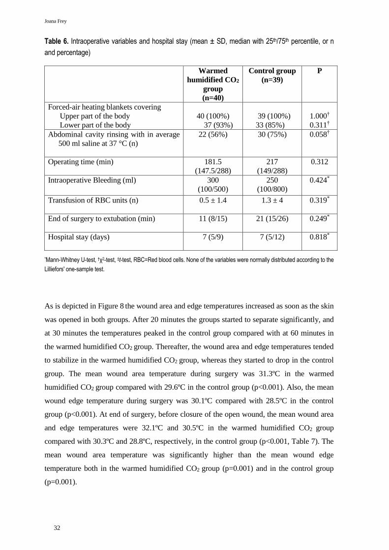
As is depicted in Figure 8 the wound area and edge temperatures increased as soon as the skin
was opened in both groups. After 20 minutes the groups started to separate significantly, and
at 30 minutes the temperatures peaked in the control group compared with at 60 minutes in
the warmed humidified CO2 group. Thereafter, the wound area and edge temperatures tended
to stabilize in the warmed humidified CO2 group, whereas they started to drop in the control
group. The mean wound area temperature during surgery was 31.3ºC in the warmed
humidified CO2 group compared with 29.6ºC in the control group (p<0.001). Also, the mean
wound edge temperature during surgery was 30.1ºC compared with 28.5ºC in the control
group (p<0.001). At end of surgery, before closure of the open wound, the mean wound area
and edge temperatures were 32.1ºC and 30.5ºC in the warmed humidified CO2 group
compared with 30.3ºC and 28.8ºC, respectively, in the control group (p<0.001, Table 7). The
mean wound area temperature was significantly higher than the mean wound edge
temperature both in the warmed humidified CO2 group (p=0.001) and in the control group
(p=0.001).

RESULTS
33
Figure 8. Mean ± SEM wound area (a) wound edge (b), and core temperatures (c) during the first 4 hours of the operation. Also, included are fifth-degree polynomials, all with R2 close to 0.9.

Joana Frey
34
Table 7. Perioperative temperatures (mean ± SD).
Warmed
humidified
CO2 group
(n=40)
Control
group
(n=39)
P 95%
confidence
interval
Wound area temperature during
surgery °C
(mean AUC)
31.3 ± 1.2
29.6 ± 1.3
<0.001 1.2 – 2.3
Wound edge temperature during
surgery °C
(mean AUC)
30.3 ± 1.1
28.5 ± 1.1
<0.001 1.3 – 2.3
Wound area temperature
before closure of open wound °C
32.1 ± 1.6 30.3 ± 1.8 <0.001 1.0 - 2.5
Wound edge temperature
before closure of open wound °C
30.5 ± 1.7 28.8 ± 1.7 <0.001 0.9 - 2.4
Core temperature °C
Before start of surgery
At end of surgery
36.7 ± 0.5
36.9 ± 0.5
36.6 ± 0.5
36.3 ± 0.5
0.179
<0.001
0.4 - (-0.1)
0.38 - 0.82
Core temperature at end of surgery
<36.5°C
<36.0°C
8 (20%)
0 (0%)
24 (62%)
7 (18%)
0.001
0.005
Core temperature during surgery °C
(mean AUC)
36.5 ± 0.5
36.1 ± 0.5
0.001 0.2 – 0.7
t- Test, AUC=area under the curve.
The mean core temperature before start of surgery was similar with 36.7ºC in the warmed
humidified CO2 group and 36.6ºC in the control group. However, at end of surgery the two
groups differed significantly with 36.9ºC in the warmed humidified CO2 group and 36.3ºC in
the control group (p<0.001). At end of surgery only 8 patients out of 40 in the warmed
humidified CO2 group had a core temperature< 36.5ºC (20%), while in the control group this
was the case in 24 out of 39 (62%) patients (p=0.001). With a cut-off at <36.0ºC none of the
patients in the warmed humidified CO2 group compared with 7 patients (18%) in the control
group were hypothermic at end of surgery (p=0.005). In accordance with this finding, the
mean core temperature during surgery, calculated as area under the curve, was 36.5ºC in the
warmed humidified CO2 group compared with 36.1ºC in the control group (p=0.001). There
was a significant positive correlation between duration of surgery and core temperature at end
of surgery in the warmed humidified CO2 group (r=0.46, p=0.02), whereas these factors did
not significantly correlate in the control group (r=0.03, p=0.84).

RESULTS
35
STUDY IV
Median follow-up was 70.9 months and no patients were lost to follow up. Preoperative
patient characteristics did not differ significantly between the treatment groups as described
in Table 8. Peri- and postoperative end points are presented in Table 9. All temperatures at
end of surgery as well as the temperature differences between core and wound were
significantly higher in the CO2 group. Mean operating time was 218 minutes in both groups
and all remaining end points tended to be in favor of the CO2 group.
*Data are presented as mean ± SD for quantitative variables, and as No. (%) for qualitative variables. BMI: body mass index
Figure 9 depicts the relationship between study groups and survival in all subjects. We could
not show that patients receiving insufflation of warmed humidified CO2 had a better overall
survival compared with control patients (p=0.508). But as shown in Figure 10, patient with a
core temperature ≥36.0ºC at end of surgery exhibited a better overall survival compared with
to those who did not (OR 0.5, CI95% 0.26-096, p=0.035).
The effectiveness of the commercially available system used in Study III was
compared with the noncommercial system used in Study II, analyzing the differences in
temperatures between the CO2 groups in both studies. Mean and final core, wound area and
wound edge temperatures were all higher in the CO2 group of Study III compared with the
CO2 group of Study II (Table 10), whereas other pre- and perioperative data did not differ
significantly.
Table 8. Demographic characteristics of the study cohort including comparisons between patients with and without humidified warmed CO2
Clinical parameters
Humid warmed
CO2
(n=80)
Controls
(n=78)
P-value
Age, years 62.9 ± 14.0 63.4 ± 17.7 0.833
Male gender 46 (57.5%) 45 (57.7%) 0.980
BMI, kg/m2 25.5 ± 4.5 25.3 ± 4.4 0.787
Colon/rectal cancer 59 (73.8%) 58 (74.4%) 0.930
Primary surgery 72 (90.0%) 64 (82.1%) 0.149

Joana Frey
36
Table 9. End points
*Data are presented as No. (%), mean ± standard deviation, or as median [range].
End point
Humid warmed
CO2
(n=80)
Controls
(n=78)
P-value
Operation duration (min) 218.0 ± 97.2 218.0 ± 94.2 1.0
Anesthesia time (min) 297.3 ± 108.9 302.5 ± 104.9 0.775
Intra operative bleeding 473.7 ± 613.1 468.3 ± 537.3 0.954
Mean core temperature 36.2 ± 0.6 35.9 ± 0.5 0.005
Mean core temperature ≥36.0°C 51 (64.6%) 32 (42.7%) 0.006
Mean wound edge temperature 29.8 ± 1.2 28.5 ± 1.1 <0.001
Mean wound area temperature 31.0 ± 1.2 29.7 ± 1.1 <0.001
Final core temperature 36.5 ± 0.6 36.1 ± 0.6 <0.001
Final core temperature ≥36.0°C 66 (82.5%) 49 (65.3%) 0.015
Final wound edge temperature 29.7 ± 1.9 28.5 ± 1.7 <0.001
Final wound area temperature 31.2 ± 2.0 30.1 ± 1.9 0.001
Mean core – mean wound edge temp 6.4 ± 1.1 7.4 ± 1.1 <0.001
Mean core – mean wound area temp 5.2 ± 1.1 6.2 ± 1.2 <0.001
Final core – last wound edge temp 6.8 ± 1.8 7.7 ± 1.7 0.006
Final core – last wound area temp 5.3 ± 1.9 6.0 ± 1.9 0.023
Wound rupture 1 (1.3%) 3 (3.8%) 0.364
Re-operation 7 (8.8%) 7 (9.0%) 0.960
Surgical site infection <30 days 13 (16.3%) 13 (16.7%) 0.944
Mortality 19 (23.8%) 22 (28.2%) 0.519
Readmission <30 days 12 (15.0%) 13 (16.7%) 0.774
PRBC transfused (units) 0 [0-13] 0 [0-9] 0.738
Plasma transfusion 0 [0-12] 0 [0-5] 0.600
Platelet transfusion 0 [0-4] 0 [0-2] 0.992

RESULTS
37
Overall univariate mortality predictions for all patients during elective major open
colon cancer surgery are shown in Table 11. As expected, age and cancer showed a strong
impact on mortality (p=<0.001 and p=0.004, respectively). Moreover, a final core
temperature ≥36.0ºC (p=0.035) and a higher final wound edge temperature (p=0.011) were
associated with lower mortality, as well as a smaller difference between final core and final
wound edge temperature (p=0.017) improved survival. A multivariate analysis adjusted for
age (p= 0.001) and cancer (p=0.165) showed that the temperature difference between final
core and final wound edge temperature was associated with a better overall survival
(p=0.050).
Table 10. Heating efficiency of the commercially available system used in study III compared with the noncommercial system by analyzing the differences in temperatures between the CO2 groups in both studies.
End point
Noncommercial
(n=37)
HumiGard™
(n=43)
P-value
Mean core temperature 35.9 ± 0.4 36.5 ± 0.5 <0.001
Mean core temperature ≥36.0°C 17 (45.9%) 34 (81.0%) 0.001
Mean wound edge temperature 29.5 ± 1.2 30.2 ± 1.0 0.011
Mean wound area temperature 30.7 ± 1.2 31.3 ± 1.1 0.032
Final core temperature 36.2 ± 0.5 36.9 ± 0.6 <0.001
Final core temperature ≥36.0°C 26 (70.3%) 40 (93.0%) 0.008
Final wound edge temperature 29.3 ± 2.1 30.3 ± 1.6 0.024
Final wound area temperature 30.6 ± 2.2 31.9 ± 1.5 0.008

Joana Frey
38
Figure 9. Cumulative survival in the CO2 and control group in all subjects after major open colon surgery (log rank p=0.508). Small vertical lines represent end of follow up.
Figure 10. Cumulative survival in patients with a core temperature ≥36.0 °C and <36.0 °C at end of surgery in all subjects after major open colon surgery (log rank p=0.035). Small vertical lines represent end of follow up.

RESULTS
39
Table 11. Cox analysis for the prediction of mortality
Variable Univariate analysis Multivariate analysis
OR (95% CI) P-value HR
(95% CI)
P-value
Mean core – mean
wound edge temp
1.24 (0.96 – 1.59) 0.097
Mean core – mean
wound area temp
1.15 (0.90 – 1.48) 0.256
Final core – Final
wound edge temp
1.24 (1.04 – 1.47) 0.017 1.20
(1.00-1.44)
0.050
Final core – Final
wound area temp
1.13 (0.97 – 1.32) 0.125
Age (10-year increase) 1.78 (1.37 – 2.33) <0.001 1.05
(1.02-1.08)
0.001
Cancer 8.1 (1.95 – 33.7) 0.004 2.92
(0.64-13.3)
0.165
Final core temperature
≥36°C
0.50 (0.26 – 0.96) 0.035
Mean core temperature 0.95 (0.54 – 1.69) 0.869
Mean core temperature
≥36°C
0.93 (0.50 – 1.75) 0.821
Mean wound area
temperature
0.87 (0.68 – 1.10) 0.242
Mean wound edge
temperature
0.81 (0.63 – 1.03) 0.089
Final core temperature 0.86 (0.51 – 1.43) 0.551
Humidified warmed
CO2
0.80 (0.43 – 1.50) 0.490
Final wound area
temperature
0.88 (0.76 – 1.02) 0.095
Final wound edge
temperature
0.80 (0.68 – 0.95) 0.011

Joana Frey
40

GENERAL DISCUSSION
41
GENERAL DISCUSSION
A major finding of this thesis was that insufflation of dry room-tempered CO2 with a gas
diffuser increases the average surface temperature in an open surgical wound cavity.
Furthermore, when the CO2 gas was warmed and humidified both the open wound
temperature as well as the core temperature significantly increased. The effect was even
stronger when the insufflated CO2 gas was better warmed and fully humidified. This
improved the maintenance of normothermia throughout the surgical procedure.
Normothermia at end of surgery and a small end-of-operation temperature difference between
final core and wound edge temperature were significantly associated with better patient
survival in open colon surgery.
NEGATIVE CONSEQUENCES OF PERIOPERATIVE CORE HYPOTHERMIA
Perioperative core hypothermia is known to trigger thermoregulatory vasoconstriction, which
decreases subcutaneous oxygen tension [22, 23]. Also, hypothermia shifts the oxygen
saturation curve to the left, whereby tissue oxygen tension will be reduced (the Bohr Effect).
Several clinical studies have found that the incidence of postoperative wound infections
correlates with intraoperative subcutaneous oxygen tension [24, 25]. Subcutaneous tissue is
particularly vulnerable to vasoconstriction, since there is little regulation of blood flow,
except in response to locally applied heat [23, 26]. Inadvertent perioperative hypothermia,
defined in our studies as <36.0 ºC core temperature, is well known to be associated with a
multitude of poor outcomes. Large prospective, randomized clinical trials have shown that
even mild hypothermia between 34-36 ºC causes numerous adverse outcomes in a variety of
patient populations. These include increased risk for surgical wound infections[27-29],
increased blood loss through impaired platelet function[30], prolonged duration of anesthetic
drugs[3], delayed wound healing[27], delayed post anesthetic recovery[31], prolonged
hospitalization[27], and postoperative shivering and thermal discomfort[3]. In a randomized
study, patients undergoing colon surgery with an average 1.9 ºC fall in core temperature had a
tripled risk of surgical wound infection and prolonged hospitalization compared with patients
who were actively kept normothermic[27]. In high-risk heart patients a slight fall in core
temperature by only 1.3ºC implied a three-fold higher risk for adverse myocardial outcomes
[32]. Hypothermia also causes hypertension in elderly patients and individuals at high risk for
cardiac complications [33]. A retrospective study from 2014 found that unintentional
perioperative hypothermia during elective surgery was associated with a 4-fold increase in
mortality and a doubled complication rate, in which sepsis and stroke increased the most [34].

Joana Frey
42
THE SIGNIFICANCE OF THE OPEN WOUND TEMPERATURE
The significance of the temperature of the open wound and the possibility to warm it during
surgery are almost unexplored in humans and very little is known about what happens when
the internal tissues are abruptly exposed to the relatively cold and dry ambient air in the
operating room. It is not known what the optimal wound temperature is during open surgery,
however, normally the abdominal viscera is particle free, body tempered, and has tissue
surfaces moist with peritoneal fluid. Thus, one may presume that during open surgery a local
temperature increase towards normal and humidification is advantageous.
Insufflation of dry CO2
In Study I we demonstrated that it is possible to rapidly increase the surface temperature in a
thoracic surgical wound by insufflating the wound cavity with dry, room-tempered CO2 via a
gas diffuser for as short time as two minutes. The patients underwent open cardiac surgery,
since it implicates a large, deep wound cavity. Besides, in our cardiothoracic department we
routinely fill the open surgical wound cavity with CO2 for the prevention of arterial air
embolism in patients undergoing open heart surgery [15, 20, 21, 35]. The wound cavity was
insufflated with CO2 for 2 minutes, recognizing that at a flow rate of 5 L/minute about 1
minute is needed to fill up the wound cavity with CO2 (>99% CO2)[21]. The flow rate of 5
L/minute is the lower limit that counteracts the influence of diffusion during steady state in a
cardiothoracic wound model[20]. We limited the insufflation period to 2 minutes, as we
presumed that this short period would ensure an effect of CO2 as well as a constant core
temperature. The results confirmed that the total wound surface area temperature returned to
start temperature 2 minutes after cessation of CO2 insufflation. We did not humidify CO2
since dry gas insufflation is currently used for de-airing in cardiac procedures.
The rapid increase in surface temperature can be explained by the green-house
properties of CO2, which provides an effective thermal insulation of the open wound as
evaporation and convection are reduced[15]. It could also be that CO2 induces a local
vasodilatation within the wound. This might lead to a redistribution of body heat from deeper
compartments into the surface of surgical wound.
The surface temperature increased significantly in the whole wound (Area V), and in
the two cranial thirds proximal to the gas diffuser (Area IV). However, in the third area
closest to the gas diffuser (Area I) we unexpectedly found a cooling effect, which according
to the images was coolest in the proximity of the diffuser. This localized cooling effect was
most probably caused by convection around the diffuser from the delivered laminar flow of

GENERAL DISCUSSION
43
room-tempered dry CO2, although the outflow velocity was as low as 0.1 m/sec[20].
Moreover, already before insufflation the diaphragmal third of the wound area (Area I) was
cooler than the other two thirds, indicating a lower tissue perfusion in this area. Its lower
perfusion rate may have amplified the cooling effect of convection and this would explain
why this area did not recover its original temperature 2 minutes after cessation of CO2
insufflation. A possible way to overcome the problem of convection would be to lower the
CO2-flow. This would decrease the cooling effect, but at the same time, due to the impact of
diffusion, increase the air content in the wound cavity. Thus, a reduced flow may interfere
with the warming effect. Another solution would be to use polyurethane foam in the diffuser
with more but smaller paths, which would lower the outflow velocity even more. However,
this would increase the resistance of the foam and thus would build up a pressure gradient
proximal to the diffuser. A larger diffuser would also reduce the local cooling effect by
decreasing the flow velocity without interfering with the flow. The drawback of this
alternative is that a larger diffuser may interfere with surgery in the limited space of the
surgical field. Insufflation of CO2 might alternatively be enhanced by using two or more
diffusers. Again, this may interfere with surgery. Finally, the best solution in our view would
be to use warmed humidified CO2 that has a much higher heat capacity. This would promote
heat transfer from the heated gas to the tissue as well as avoiding the cooling effect of
evaporation. In addition, water vapor is known to be a greenhouse gas.
Study I may be criticized for lacking an important control with the exposure of the
wound to some other gas that would be hypothesized not to produce a temperature raising
effect. We used the wound temperature after the first two minute period as a control with the
exposure of the wound to ambient air (mainly nitrogen and oxygen gas). Again, we analyzed
the impact of exposing the wound to ambient air on wound temperature after the third two
minute period (which followed the period of insufflation with CO2). Thus, we used the
patient’s wound temperature during exposure to ambient air as his own control.
It may be argued that one should include a measure of core temperature and
protected skin temperature to determine how the temperature changes related to changes
occurring in body temperatures unrelated to experimental manipulation of the wound. We
decided not to measure that in Study I because the two minute period of insufflation with
CO2 is far too short to affect core temperature and protected skin temperature. During the two
minutes of insufflation we detected a significant rise in average wound temperature that we

Joana Frey
44
believe cannot be explained by anything else than CO2 itself, since the patient was not
warmed by any other arrangements.
One may also ask if the drop in surface temperature in wound area I could be a
reflection of the endothermic effect of gas expansion as it came out of the diffuser device and
if so, could one use a heater on the diffuser to reduce this effect? The endothermic effect of
gas expansion is restricted to the site of the pressure reducing port (down to 4.5-5.5 kPa),
which is positioned at the CO2 gas cylinder. Indeed, this metal port is cooled down to subzero
(°C) temperatures during insufflation of CO2 over hours. However, the dry CO2 leaving the
flow meter, which is connected distally to the port, will very quickly adopt room temperature
when it travels along the 2.5 m PVC tube to and trough the gas diffuser.
Insufflation of warmed humidified CO2
The technique of Study II and III with insufflation of warmed humidified CO2 opens a new
way to protect the tissues in open surgical wounds that are exposed to dry cold ambient air
and airborne contamination. The local temperature of the open surgical wound and especially
that of the open abdominal wound has until now hardly been studied in humans, although any
change in local and indirectly in core temperature will affect local tissue perfusion and vice-
versa. We do not yet know what the optimal local wound temperature is during surgery, but
we do know that even a mild drop in core temperature especially in abdominal surgery affects
patients’ outcome including morbidity, mortality, and costs.
We used a continuous high inflow (10 L/min) of CO2 to counteract the strong force of
diffusion of surrounding air into the open wound cavity and to compensate for any turbulence
caused by the surgeon’s hands in the wound. A local atmosphere of CO2 in the open wound
has been shown to have an insulating effect, both experimentally [15, 16] and clinically [39].
Humidification of CO2 with 93% rH at 30°C (Study II) and even better with 100% rH at
37°C (Study III) prevents evaporation from the open wound and the cooling effect that
otherwise would have occurred[15, 16]. Warm humidified CO2 has a much higher heat
capacity than warm dry CO2 and promotes heat transfer to tissues. In fact the heating capacity
of the fully humidified gas increases logarithmically with temperature. In addition, water
vapor itself is also a greenhouse gas. Furthermore, both a higher temperature and a higher
CO2 content will shift the oxyhemoglobin curve to the right, which will result in hemoglobin
proteins releasing their load of oxygen. This improves local oxygen extraction at the tissue
level (the Bohr Effect, Figure 11).

GENERAL DISCUSSION
45
Figure 11. The oxygen-hemoglobin dissociation curve.
Since general anesthesia causes vasodilatation, heat is more easily redistributed from
the core out to the wound surface. The lower the surface temperature the greater is the core-
to-peripheral temperature gradient and thus the rate of heat redistribution and heat loss. Vice
versa, if the wound surface temperature is warmed, the heat will redistribute to deeper
departments within the body and core temperature increases and thus the possible risk of
wound infection may be attenuated. A warm humidified CO2 atmosphere provides an
effective thermal insulation of the open surgical wound and the resulting increase in wound
and core temperature may help to reduce the negative consequences of mild perioperative
hypothermia.
More effective heating and humidifying system
In Study II our simple heating system did not deliver fully humidified CO2 at body
temperature (93% rH and 30°C) to the open surgical wound. This called for technical
improvements of the heating system. Thus, in Study III the same flow rate was used but a
more effective heating system delivered humidified CO2 at 100% rH and 37 °C to the surgical
wound. This corresponds to 43.9 mg of water per liter CO2, which is 55% more than
delivered with the noncommercial device used in Study II.

Joana Frey
46
Why a clinical study in patients undergoing open colorectal surgery?
Over the last decades open abdominal surgery has lost ground to laparoscopic surgery. The
latter technique has several advantages including that it prevents exposure of the internal
tissues to ambient dry and cold air and airborne contamination. Instead, dry CO2 is usually
insufflated as a jet through a thin port into the peritoneal cavity, and CO2’s high solubility
explains why CO2 will cause much less harm than air if unintentionally introduced into the
venous or arterial system. Many animal and clinical studies have been undertaken to
investigate if the insufflated dry CO2 jet causes local desiccation of the peritoneum and
promotes local inflammation and whether or not this can be prevented by humidification.
Based on the results of these studies many centers now use commercially available
techniques to apply warmed and humidified CO2 during laparoscopic surgery.
The positive effects of laparoscopically insufflated CO2 have been evaluated both in
animal and human studies. In rats CO2 pneumoperitoneum has a protective effect against
induced bacterial peritonitis. Furthermore, two recent meta-analyses demonstrated that
insufflation of warmed humidified CO2 compared with dry gas during laparoscopic surgery
reduced the incidence of intraoperative hypothermia, postoperative pain, and opiate use [36,
37]. However, one must bear in mind the many basic differences between open and
laparoscopic surgery, including the negative effects of an increased intraperitoneal pressure in
the latter.
We chose to investigate the effects of warmed and humidified CO2 during open
abdominal surgery in patients because of many reasons. First, this type of surgery implicates
a large, deep wound cavity, which exposes a very large wound surface to ambient air
promoting evaporation, heat loss and the risk of intraoperative hypothermia. Second, the
clinical application of warmed humidified gas is now common in laparoscopic surgery. Third,
the technique of CO2-insufflation via a diffuser is clinically used in cardiothoracic surgery.
Forth, perioperative core hypothermia worsens clinical outcome i.e. increases the risk of
surgical wound infection[16-18], morbid cardiac events[23], as well as prolongs hospital stay
and increases costs. Fifth, despite growing awareness of the negative consequences of
perioperative hypothermia more than one-third of general surgery patients undergoing
abdominal operations will be hypothermic on arrival in the postoperative care unit [30, 32].
Sixth, since the rate of surgical site infection is 5-10 times higher in open colon surgery
compared with cardiothoracic surgery, the potential impact of the technique is much larger in
open colon surgery. Seventh, the technique opens a new way to protect exposed tissues in

GENERAL DISCUSSION
47
open surgical wounds, where such a vital parameter as local temperature has hardly been
studied in humans.
Local wound temperatures during open colorectal surgery
Both in Study II and III the wound area and edge temperatures increased during the first
hour of surgery in the groups with warmed humidified CO2, after which these temperatures
stabilized. Also in the control groups these temperatures increased at start, but peaked already
after 30 minutes of surgery, although at lower values, after which there was a tendency to a
steady decline (Figure 7 and 8).
In both Study II and III, as shown in Figure 7 and 8, the actual wound temperatures
were surprisingly low. In Study III there was a peak wound area and edge temperature of
30.3ºC and 29.0ºC in the control group and 31.7ºC and 30.7º in the warmed humidified CO2
group, respectively. It is not known what the optimal wound temperature is during open
surgery, nor is it known if the 1.8ºC increase in wound area at end of surgery in the warmed
humidified CO2 group is of clinical importance. However, as the normal temperature of the
abdominal viscera is close to the core temperature of 37ºC in a normothermic healthy
adult,[38] one may presume that a temperature increase towards normal is advantageous. The
initial wound temperature increase and the following stabilization in the treatment group both
in Study II and III is most likely explained by the heating effects of warmed humidified
CO2. In the control group both in Study II and III where the open surgical wound is
unprotected, there was in contrast a continuous drop in temperature most probably due to a
loss of heat through evaporation throughout surgery. This heat loss seems to be the main
reason why the core temperature of the control group was significantly lower than in the
treatment group.
Further aspects of the local wound temperature measurements are also of interest. In
both groups of Study II there were a significant difference between the wound area and edge
temperatures with the area temperatures being ~ 1.5°C warmer than the edges (slightly
smaller difference than in Study III). The reason for this is probably multifactorial. Normal
skin temperature is lower than that of deeper tissues and the edges are also exposed to a
greater tension by the circular abdominal wall retractor, which must restrict the local
perfusion. When comparing Study II and III, the wound edge temperature in the CO2 group
was approx. 0.8°C higher in Study III. This was expected with the increased temperature and
humidity of the insufflated CO2. In Study III, the tendency to more frequent rinsing with
saline of the abdominal cavity in the control group versus the warmed humidified CO2 group

Joana Frey
48
at end of surgery cannot explain the drop in wound temperatures that were seen in the control
group. On the contrary, this would rather have increased wound temperatures, as the saline
was preheated to 37°C.
Core temperature during open colorectal surgery
Both in Study II and III there was a significant difference in core temperature between the
two groups despite our efforts with conventional methods to avoid perioperative hypothermia
in all patients. In Study II the warmed humidified CO2 group had a median core temperature
during surgery of 36.0°C, while the control group had 35.5°C. Both groups started at the
same core temperature, 35.9°C, but at the end of surgery there was an increase with 0.3°C in
the warmed humidified CO2 group compared with a decrease of 0.1°C in the control group.
In Study III the mean core temperature during surgery was 36.5°C and 36.1°C in the
warmed humidified CO2 group and in the control group, respectively. Also, both groups
started at approximately the same core temperature, ~36.6°C. Thereafter, it increased with
0.2°C in the warmed humidified CO2 group, but decreased with 0.3°C in the control group to
end of surgery. Depending on where one wants to define the lower cut-off point for
normothermia, one gets different percentages of patients that were hypothermic at end of
surgery in each group, although the groups still differed significantly. When using the
traditional definition of hypothermia, i.e. a core temperature of <36.0°C, in Study II 55%
(20/36) of the patients in the control group were hypothermic at end of surgery compared
with 24% (9/38) in the warmed humidified CO2 group (p<0.01). In Study III 18% (7/39) in
the control and none of the patients in the warmed humidified CO2 group were hypothermic
at end of surgery. With a core temperature cut off at <36.5ºC, 62% (24/39) of the patients in
the control group were hypothermic, while this was the case in 20% (8/40) in the warmed
humidified CO2 group at end of surgery in Study III. It may still be argued that a difference
in core temperature between the groups of about 0.5°C in both studies is rather modest, but
some patients in the control group in Study II even dropped down to 34°C in core
temperature. To what extent the attained 0.5°C difference in core temperature can be proven
to make a difference in objective outcome data remains to be settled in a much larger study.
However, in Study IV, the patients with a core temperature ≥36.0°C compared to the ones
with a core temperature of < 36°C at end of surgery had a better overall survival (p=0.035).
Besides, in Study II patients in the treatment group were extubated significantly earlier. This
may possibly be explained by the higher core temperature and thereby a faster elimination of
anesthetic drugs. However, patients in the treatment group of Study III were not extubated
significantly earlier, indicating that other factors than warming could have been of

GENERAL DISCUSSION
49
importance, like when the anesthetic staff shifted in the afternoon, or that the anesthetic staff
did not follow strict criteria i.e. normothermia for extubation. Moreover, any awake patient
will feel distressed and be prone to shivering when subjected to mild core hypothermia
(<36.0°C). Because of the recognized drawbacks of perioperative hypothermia, centers
usually have to follow international guidelines [40, 41] and some centers do not even get
reimbursement for patients if perioperative hypothermia occurs.
Relation of intraoperative temperature to postoperative mortality in open colon surgery
The retrospective Study IV was an almost 6-year follow up of all patients included in Study
II and III. We looked at the potential ability of increased wound temperature to improve long
term survival after major open colon surgery. This can be attributed to at least three different
mechanisms.
First, perioperative hypothermia has been confirmed to increase cardiac demand and,
consequently, to increase the risk of cardiac morbidity [[42]]. Those patients who survived a
postoperative cardiac event continued to have a substantial risk of cardiac death, with a
hazard ratio of 18 (95% CI, 6-57) in the first 6 month after discharge. In patients with cardiac
risk factors who undergo non-cardiac surgery, perioperative maintenance of normothermia
has been associated with a reduced rate of morbid cardiac events and ventricular tachycardia
[39]. These data are consistent with our findings that patients with a core temperature
≥36.0ºC at end of surgery demonstrated a significantly better overall survival compared with
those with core temperature <36.0ºC at end of surgery. Also, the treatment group with
insufflation of warmed humidified CO2 was inclined to have a better long time survival,
although this did not reach significance, possibly due to a Type II error.
Second, insufflation of warmed humidified CO2 in the open surgical wound increased
core and wound temperatures and decreased the difference between core and wound
temperatures. These changes may indicate an improved perfusion and a better oxygenation of
the open surgical wound, where wound edge temperature is a more sensitive indicator of
wound tissue perfusion than wound area, since the latter temperature is influenced by all
exposed internal tissues. A recently published rat model showed that insufflation of warmed
humidified CO2 into the abdominal cavity during open abdominal surgery caused a rapid
raise in wound tissue oxygen tension[29]. The humidification and warming to physiological
temperature of the insufflated CO2 decreases desiccation from the open wound and increases
overall wound temperature thereby improving general wound perfusion and oxygenation.

Joana Frey
50
Third, averting desiccation of the exposed peritoneal mesothelium by insufflation of
warmed humidified CO2 has been shown to reduce intra-peritoneal tumor dissemination in
animal models, a finding which is consistent with maintaining the physiological integrity of
the mesothelium as an intact barrier to tumor infiltration[3, 41]. The peritoneum was the only
site of metastasis in > 50 % of patients with metastatic disease[23] and such metastasis
remains fatal[43]. This could have influenced the long term survival in Study IV.
The strength of Study IV is that the long term effects of intraoperative wound area and
wound edge temperatures have to our knowledge not been studied before. Importantly,
patients were warmed with standard warming measures according to the NICE guidelines[2].
In addition, 94% of the patients received epidural analgesia together with general anesthesia
and this combination has been shown to increase long-term survival after colon surgery in a
retrospective study[43], which could be due to a reduction in neuroendocrine response and
attenuated immunosuppression. Furthermore, none of our patients were lost to follow up.
SURGICAL SITE INFECTIONS
Wound healing and wound infection rates correlate with the perfusion and thus the
oxygenation of the wound tissues and edges [24, 25]. Lowering of local wound and edge
temperatures as well as a lowering core temperature will directly and indirectly cause
vasoconstriction, hypoperfusion, local hypoxia, and hence increase the risk of surgical site
infection[44]. A recent experimental study in rats has shown that insufflation of warmed,
humidified CO2 following open abdominal surgery increases intraoperative PtO2 and
decreases loss of peritoneal mesothelium[45]. In the former study insufflation of warmed,
humidified CO2 into the abdominal cavity during open surgery caused an immediate and
clinically significant increase in local PtO2 compared with exposure to ambient air. The
increase in PtO2 was an additive result of the delivery of CO2 and avoidance of evaporative
cooling via the delivery of gas humidified at body temperature. It is likely that an increase in
local PtO2 and protection against loss of peritoneal mesothelium will interact to prevent
postoperative surgical site infections. The role of high PtO2 in avoiding wound infections is
well established[44], however, it is likely that maintenance of an intact mesothelial layer may
further decrease the risk of infection, as the mesothelial layers provides both a physical
barrier and also plays an active role in immune response[46].
Assuming a power of 80%, a type I error of 0.05, and that the method would half a
surgical site infection rate from 5% to 2.5% or from 10% to 5%, 1806 and 864 patients,

GENERAL DISCUSSION
51
respectively, would have to be recruited to a large multicenter trial involving major colon
surgery. Since the number of included patients in Study II and III, each represent less than
10% of the necessary number of patients needed we did not think it made sense to study SSI
rate. In addition, the rate of postoperative SSI was not studied prospectively and not all
patients underwent major colon surgery. Since we in none of our studies investigated the SSI
rate prospectively, any retrospective review of data would have to be afflicted with
uncertainty, especially when the standard definitions of SSI cannot be followed. However,
when looking at a non-disputable variable such as wound ruptures that needed reoperation,
we found in Study IV that this occurred twice in the control group only.
LIMITATIONS
In Study II and III the operating team was not blinded to type of treatment because
insufflation of warmed humidified CO2 into the surgical wound was immediately sensed by
the surgical team and both seen and heard by all personnel in the operating room. However,
none of the surgeons experienced the device getting in their way during surgery, given the
small size of the gas diffuser.
Patients in Study II and III did not receive preoperative active warming and where
only covered with preheated blankets when arriving at the pre-anesthetic room according to
the clinical routine.
Inhalation of high concentrations of CO2 may increase the breathing rate or even cause
fainting-fits. However, due to the high density of CO2, surplus CO2 will drain away quickly
from the surgical wound to the floor. The gas finally escapes via the extraction system as do
anesthetic gases. Besides, when CO2 is insufflated at a flowrate of 10L/min via the gas
diffuser, the CO2 concentration at 10 cm above the wound opening is less than 1%.
Furthermore, CO2 has been used for about 50 years now and there have not been any reports
of such side-effects. When considering a method that is based on a continuous outflow of
greenhouse gas, such as CO2, we need to understand to what extent this will contribute to the
worldwide CO2 discharge. Continuous wound ventilation during a 3-hour operation will use
about 3 kg CO2. This corresponds to the discharge from a regular car during 15 minutes
‘drive. It should also be noted that medical CO2 is a recycled by-product from beer, spirits
and fertilizer production. If not recovered, it would be vented to the atmosphere. Thus, the
CO2 used does not contribute to extra CO2 being released into the earth’s atmosphere.

Joana Frey
52
Potential limitations of Study IV are that this was a retrospective post hoc study of
postoperative morbidity and mortality, although patients had been randomized before
surgery. Also, two different heating systems were used in Study IV. Moreover, Study IV
was relatively small with a concurrent probability of Type II errors.
FUTURE STUDIES
As with any technology the new method will most likely be improved over time. Originally,
we showed that insufflation of cold and dry CO2 through a gas diffuser into the cardiothoracic
wound cavity during open heart surgery could prevent arterial air embolism[35]. Since we
became concerned that the use of dry CO2 might cause desiccation and cooling of exposed
tissue surfaces within the open wound cavity we have improved the technique by warming
and humidifying the delivered gas. By doing so we have experimentally shown that this
improvement could prevent evaporation, cooling, and desiccation of exposed surfaces.
Also, CO2 itself is a bacteriostatic drug. For this thesis we have mainly studied patients
undergoing open colon surgery. The method can, however, be used in more or less any open
surgical procedure, provided that a cavity is created around the open wound surface e.g. by
placing a brim around the wound edges.
There is need for larger, randomized studies investigating the influence of
intraoperative insufflation of warm humidified CO2 on postoperative complications like
surgical site infections, adhesion, and cancer recurrence. A recently published study [47]
showed that a low maximal body temperature (<37.4°C) after rectal surgery was an
independent predictor of recurrence and survival in rectal cancer patients. The authors
suggested that these patients might gain a survival benefit from warming strategies during the
perioperative treatment.
It would also be of interest to look at biopsies from the peritoneum and determine if
intraoperative insufflation of warm humidified CO2 gas can prevent local desiccation and
damage of the mesothelium. However, as these changes are most likely to appear 12 hours
postoperative, this would be difficult to study in humans.
One possible further alternative would be to warm the humidified CO2 gas to > 37°C to
achieve more effective heating. However, this temperature could also have negative
consequences on local tissue and thus has first to be tested in comprehensive animal studies.

GENERAL DISCUSSION
53
Another interesting thought would be to use the CO2 gas as a carrier for delivery of
local antibiotics or alcohol to the open wound. The latter possibility is bacteriostatic in an
experimental model and the alcohol vapor becomes non-explosive when combined with the
fire extinguisher CO2 [48].


CONCLUSIONS
55
CONCLUSIONS
This thesis concludes that
Shortterm insufflation of dry room-tempered CO2 gas in an open wound increases the
surface temperature significantly during open cardiac surgery.
Insufflation of warmed (30°C) humidified (93% rH) CO2 in an open surgical wound
cavity increases both the surgical wound temperature as well as the core temperature
significantly during open abdominal procedures.
Insufflation of warmed (37°C) humidified (100% rH) CO2 increases both wound and
core temperature during open abdominal procedures and helps to maintain
normothermia.
Normothermia at end of surgery and a small end-of-operation temperature difference
between final core and wound edge temperature were significantly associated with
better patient survival in open colon surgery.

Joana Frey
56

ACKNOWLEDGEMENTS
57
ACKNOWLEDGEMENTS
I am sincerely grateful to all involved in the completion of this work, and particularly to:
Professor Jan van der Linden, my supervisor and friend. Without you I would never have
become a PhD, ever! Thank you for excellent guidance in the world of science, always with
great curiosity and enthusiasm. Your support has been immense both at work, in research and
lots of private ups and downs. Thank you for always having time for my needs and for all the
nice moments during hard work, travels and lunchtimes.
Peter Svenarud, my co-supervisor and colleague for always supporting me when needed.
Martin Janson and Monika Egenvall (former Svanfeldt), surgeons at Karolinska University
Hospital, Huddinge, for your help with patient information and inclusion, and patience during
surgery.
Kristina Kilsand, research nurse, for your professionalism and patience, for all your help in
compiling study data and creating a nice and inspiring atmosphere around us.
Henrik Svegby for all the help with study I
Manne Holm who helped me with all statistics in study IV
Jean Marshall, Dean Reynecke and Michael Blackhurst for general support and for
helping me with the difficutle Englich langauge ;)
Marie van der Linden, for patience while Jan is occupied somewhere else than at home with
some research project (sometimes mine, especially during the last months).
Åke Hellström, former chief of the cardiothoracic anesthesiologists and intensivists. I know
that you have always believed in me and you are the one that really made me grow as a
cardiac anesthesiologist. Your door has always been open for discussions, both professional
and private, and I certainly miss that now when you are no longer here.
Lena Nilsson, colleague and interim chief of the anesthesiologists and intensivists, for
always trying to satisfy my need for research time and free time. You have always managed
to make my schedule wishes come true.
Ulf Lockowandt, Head of the Dept. of Cardiothoracic Surgery & Anesthesiology for your
kind support.
Gabriella Lindvall, dear colleague, for being such a generous humanist (the world need
more people like you), and always having time for support and a chat.
All other colleagues at the Dept. of Cardiothoracic Surgery and Anesthesiology, Karolinska
University Hospital, Solna. Thank you for always believing in me and cheering me up in my
dark moments.

Joana Frey
58
Late professor emeritus Willem van der Linden, for excellent help with the English
language.
All the patients who participated in the study I-IV.
Francisca Herodes, mentor and oldest friend. I can always count on you no matter what.
My other dear, dear friends, who bring me a dimension of trust, joy and happiness to life
that cannot be exchanged by other sensation.
Friedel, my deeply beloved mother, who always believed in me, supported me and still is my
biggest fan. Without you and your endless love I would never be where I am today, and I am
so grateful to be your daughter.
Alexandra and Andreas, my wonderful children for reminding me of all other things than
work that are important in life. Since the minute you were born, I love you, always.
Georgios, my life companion for many years. Without you nothing is possible, together with
you everything is possible. I look forward to all the loving and exciting years with you after
the completion of both our theses.
The work with this thesis was supported by grants from the regional agreement on medical
training and research (ALF) between Stockholm County Council and Karolinska Institutet,
the Swedish Heart and Lung Foundation, and Karolinska Institutet.

REFERENCES
59
REFERENCES
1. Sessler, D.I., Perioperative heat balance. Anesthesiology, 2000. 92(2): p. 578-96.
2. NICE (2008). Perioperative hypothermia (inadvertent): the management of inadvertent
peri-operative hypothermia in adults. NICE Clinical Guideline. 65 London: National
Institute for Health and Clinical Excellence. http://www.nice.org.uk/GC065 (accessed 02
⁄ 18 ⁄ 2011)
3. Sessler, D.I., Temperature monitoring and perioperative thermoregulation.
Anesthesiology, 2008. 109(2): p. 318-38.
4. Torossian, A., Thermal management during anaesthesia and thermoregulation
standards for the prevention of inadvertent perioperative hypothermia. Best Pract Res
Clin Anaesthesiol, 2008. 22(4): p. 659-68.
5. Sun, Z., et al., Intraoperative core temperature patterns, transfusion requirement, and
hospital duration in patients warmed with forced air. Anesthesiology, 2015. 122(2): p.
276-85.
6. Forbes, S.S., et al., Evidence-Based Guidelines for Prevention of Perioperative
Hypothermia. Journal of the American College of Surgeons, 2009. 209(4).
7. Hedrick, T.L., et al., Efficacy of protocol implementation on incidence of wound
infection in colorectal operations. J Am Coll Surg, 2007. 205(3): p. 432-8.
8. Binda, M.M., et al., Conditioning of the abdominal cavity reduces tumor implantation in
a laparoscopic mouse model. Surg Today, 2014. 44(7): p. 1328-35.
9. Binda, M.M., et al., Effect of desiccation and temperature during laparoscopy on
adhesion formation in mice. Fertil Steril, 2006. 86(1): p. 166-75.
10. Carpinteri, S., et al., Peritoneal Tumorigenesis and Inflammation are Ameliorated by
Humidified-Warm Carbon Dioxide Insufflation in the Mouse. Ann Surg Oncol, 2015.
11. Hellebrekers, B.W. and T. Kooistra, Pathogenesis of postoperative adhesion formation.
Br J Surg, 2011. 98(11): p. 1503-16.
12. Mutsaers, S.E., et al., The origin of regenerating mesothelium: A historical perspective.
International Journal of Artificial Organs, 2007. 30(6): p. 484-494.
13. van den Tol, M.P., et al., Reduction of intraperitoneal adhesion formation by use of non-
abrasive gauze. Br J Surg, 1997. 84(10): p. 1410-5.

Joana Frey
60
14. von Ruhland, C.J., et al., Can artifact mimic the pathology of the peritoneal
mesothelium? Perit Dial Int, 2003. 23(5): p. 428-33.
15. Persson, M., H. Elmqvist, and J. van der Linden, Topical humidified carbon dioxide to
keep the open surgical wound warm: the greenhouse effect revisited. Anesthesiology,
2004. 101(4): p. 945-9.
16. Persson, M. and J. van der Linden, Can wound desiccation be averted during cardiac
surgery? An experimental study. Anesth Analg, 2005. 100(2): p. 315-20.
17. Persson, M., et al., Carbon dioxide inhibits the growth rate of Staphylococcus aureus at
body temperature. Surg Endosc, 2005. 19(1): p. 91-4.
18. Persson, M. and J. van der Linden, Wound ventilation with carbon dioxide: a simple
method to prevent direct airborne contamination during cardiac surgery? J Hosp Infect,
2004. 56(2): p. 131-6.
19. Gottrup, F., Prevention of surgical-wound infections. N Engl J Med, 2000. 342(3): p.
202-4.
20. Persson, M. and J. van der Linden, De-airing of a cardiothoracic wound cavity model
with carbon dioxide: theory and comparison of a gas diffuser with conventional tubes. J
Cardiothorac Vasc Anesth, 2003. 17(3): p. 329-35.
21. Svenarud, P., M. Persson, and J. van der Linden, Intermittent or continuous carbon
dioxide insufflation for de-airing of the cardiothoracic wound cavity? An experimental
study with a new gas-diffuser. Anesth Analg, 2003. 96(2): p. 321-7.
22. Greif, R., et al., Supplemental perioperative oxygen to reduce the incidence of surgical-
wound infection. Outcomes Research Group. N Engl J Med, 2000. 342(3): p. 161-7.
23. Sheffield, C.W., et al., Centrally and locally mediated thermoregulatory responses alter
subcutaneous oxygen tension. Wound Rep Reg, 1996. 4: p. 339-45.
24. Greif, R. and D.I. Sessler, Supplemental oxygen and risk of surgical site infection. Jama,
2004. 291(16): p. 1957; author reply 1958-9.
25. Hopf, H.W., et al., Wound tissue oxygen tension predicts the risk of wound infection in
surgical patients. Archives of Surgery, 1997. 132(9): p. 997-1005.
26. Rabkin, J.M. and T.K. Hunt, Local heat increases blood flow and oxygen tension in
wounds. Arch Surg, 1987. 122(2): p. 221-5.

REFERENCES
61
27. Kurz, A., D.I. Sessler, and R. Lenhardt, Perioperative normothermia to reduce the
incidence of surgical-wound infection and shorten hospitalization. New England Journal
of Medicine, 1996. 334(19): p. 1209-1215.
28. Melling, A.C., et al., Effects of preoperative warming on the incidence of wound
infection after clean surgery: A randomised controlled trial. Lancet, 2001. 358(9285): p.
876-880.
29. Sessler, D.I., Complications and treatment of mild hypothermia. Anesthesiology, 2001.
95(2): p. 531-43.
30. Rajagopalan, S., et al., The effects of mild perioperative hypothermia on blood loss and
transfusion requirement. Anesthesiology, 2008. 108(1): p. 71-7.
31. Lenhardt, R., et al., Mild intraoperative hypothermia prolongs postanesthetic recovery.
Anesthesiology, 1997. 87(6): p. 1318-23.
32. Frank, S.M., et al., Perioperative maintenance of normothermia reduces the incidence of
morbid cardiac events. A randomized clinical trial. Jama, 1997. 277(14): p. 1127-34.
33. Frank, S.M., et al., The catecholamine, cortisol, and hemodynamic responses to mild
perioperative hypothermia. A randomized clinical trial. Anesthesiology, 1995. 82(1): p.
83-93.
34. Billeter, A.T., et al., Unintentional perioperative hypothermia is associated with severe
complications and high mortality in elective operations. Surgery, 2014. 156(5): p. 1245-
52.
35. Svenarud, P., M. Persson, and J. van der Linden, Effect of CO2 insufflation on the
number and behavior of air microemboli in open-heart surgery: a randomized clinical
trial. Circulation, 2004. 109(9): p. 1127-32.
36. Sajid, M.S., et al., Effect of heated and humidified carbon dioxide on patients after
laparoscopic procedures: a meta-analysis. Surg Laparosc Endosc Percutan Tech, 2008.
18(6): p. 539-46.
37. Sammour, T., A. Kahokehr, and A.G. Hill, Meta-analysis of the effect of warm
humidified insufflation on pain after laparoscopy. Br J Surg, 2008. 95(8): p. 950-956.
38. Pearson, J., et al., Pulmonary artery and intestinal temperatures during heat stress and
cooling. Med Sci Sports Exerc, 2012. 44(5): p. 857-62.

Joana Frey
62
39. Frey, J.M., et al., CO2 insufflation influences the temperature of the open surgical
wound. Wound Repair Regen, 2010. 18(4): p. 378-82.
40. Radauceanu, D.S., D. Dragnea, and J. Craig, NICE guidelines for inadvertent peri-
operative hypothermia. Anaesthesia, 2009. 64(12): p. 1381-2.
41. Sessler, D.I., New surgical thermal management guidelines. Lancet, 2009. 374(9695): p.
1049-50.
42. Devereaux, P.J., et al., Perioperative cardiac events in patients undergoing noncardiac
surgery: a review of the magnitude of the problem, the pathophysiology of the events and
methods to estimate and communicate risk. CMAJ, 2005. 173(6): p. 627-34.
43. Vogelaar, F.J., et al., Epidural analgesia associated with better survival in colon cancer.
Int J Colorectal Dis, 2015. 30(8): p. 1103-7.
44. Hopf, H.W. and M.D. Rollins, Wounds: an overview of the role of oxygen. Antioxid
Redox Signal, 2007. 9(8): p. 1183-92.
45. Marshall, J.K., et al., Desiccation of the peritoneum by exposure to operating room
ventilation during open abdominal surgery causes loss of peritoneal mesothelium that is
prevented by insufflation of humidified–warm CO2 in rats. (In manuscript).
46. Mutsaers, S.E. and S. Wilkosz, Structure and function of mesothelial cells. Cancer
treatment and research, 2007. 134: p. 1-19.
47. Huichuan Y., Yanxin L., et al., The predicting value of postoperative body
temperatureon long-term survival in patients with rectal cancer. Tumor Biol, 2015. 36:p.
8055–8063.
48. Persson M, Flock JI, van der Linden J. Antiseptic wound ventilation with a gas diffuser:
a new intraoperative method to prevent surgical wound infection? J Hosp Infect.
2003;54(4):294-9.