Embed Size (px)
Citation preview

www.eurasianjvetsci.org
RESEARCH ARTICLE The effects of different extracts of Glycine max (L.)
Merr. on primordial, primary and secondary follicles
Yeşim Ceylantekin¹*, Esra Küpeli Akkol², Vural Özdemir³
¹Department of Nursing, ³Department of Anatomy, Afyon Kocatepe University, 03106, Afyonkarahisar, ²Department of Pharmacognosy, Gazi University, Ankara, Turkey
Received: 29.10.2016, Accepted: 15.12.2016 *[email protected]
Soyanın (Glycine max L.) farklı ekstraktlarının pimordial, primer ve sekonder folliküllere etkisi
Eurasian Journalof Veterinary Sciences
Eurasian J Vet Sci, 2017, 33, 1, 1-7
1
Öz
Amaç: Soya (Glycine max L.)’ın dişi üreme sistemi üzerine etkileri araştırmaktır.
Gereç ve Yöntem: Soya bitkisi n-hekzan, etil asetat ve etanol ekstrak edilerek ratlara oral yolla 1 ay süre ile 100 ve 200 mg/kg dozlarında (SID) verildi. Ratların diöstrus durumu vajinal smear yöntemi ile menstrual siklus gözlenerek belir-lendi. Deneme sürecinin sonunda hayvanlar ötenazi edildi. Üreme sistemi organları stereolojik yöntemlerle ratlardan alınarak analiz edildi. Ovaryumdaki foliküller stereolojik ola-rak optik disektör metodu ile sayıldı. Ovaryum ve uterusun ağırlığı da değerlendirildi.
Bulgular: 200 mg/kg dozunda soya etanol ekstresi verilen ratların genital sisteminde bazı değişiklikler meydana geldi. Test gruplarında vajinal açıklık arttı, önemli bir ağırlık artışı gözlenmezken uterin hiperemi oluştuğu belirlendi. Relatif ovaryum ağırlığı, uterus ağırlığı arttı ve epitelyal hücrele-rin irileştiği gözlendi. Primordial, primer folikül sayılarında tüm gruplarda önemli bir fark gözlenmedi. Sekonder folikül çapı azalırken sekonder foliküllerin sayısı 200 mg/kg etanol ekstresi verilen grupta, kontrol grubuna kıyasla arttığı belir-lendi. İzoflavon içeriği aktif ekstrakt üzerinde yüksek basınç kromatografi (HPLC) tekniği kullanılarak belirlendi. Etanol ekstraktında daidzein ve genistein miktarlarısırasıyla 3.851 µg/g ve 3.127 µg/g olarak belirlendi.
Öneri: Soya bitkisi fitoöstrojenlerin dişi genital sistemi üze-rine doza bağlı olarak etkileri olabileceği ifade edilebilir.
Anahtar kelimeler: Fitoöstrojen, Glycine max L., reprodük-tif sistem, stereoloji
Abstract
Aim: The effects of plant soya (Glycine max L.) on the female genital system have been investigated.
Materials and Methods: n-hexane, ethyl acetate, ethanol extracts of soya plant were administered to the rats by oral route at 100 and 200 mg/kg doses (SID) for 1 month. By mo-nitoring the menstrual cycle with vaginal smear method, di-estrus stage of the rats were determined. At the end of expe-rimental period the animals were euthanized. Genital system organs taken of rats were analyzed by stereological methods. Follicules were counted with optical dissector method in the ovaryum stereologically, the weights of the ovaries, uterus were also evaluated.
Results: Some changes occured in genital system of the rats which were given at 200 mg/kg dose of soy ethanol extract. In test groups vaginal aperture increased, uterine appeared likehyperemia while significant weight increase did not oc-cur. The weight of relative ovarium, uterus weight increased, coarsening of epithelial cells was observed. No significant difference was observed in all groups regarding the numbers of primordial, primary follicles. Diameter of secondaryfol-licule reduced while the number of secondary follicule inc-reased in 200 mg/kg ethanol extract administered group, when compared to control group. The amount of isoflavone content was determined by using high pressure liquid chro-matography technique on the active extract. The amount of daidzein, genistein were found to be 3.851 µg/gand 3.127 µg/g, respectively in the ethanol extract.
Conclusion: It could be stated that phytoestrogens in soya plant dose-dependently affect the female genital system.
Keywords: Phytoestrogen, Glycine max L., reproductive system, stereology
Eurasian J Vet Sci, 2017, 33, 1, 1-7DOI: 10.15312/EurasianJVetSci.2016.128

Eurasian J Vet Sci, 2017, 33, 1, 1-7
Introduction
Glycine max (L.) Merr. (soy, soybean, soya bean), belongs to the Fabaceae family, is cultivated to produce oil and flour es-pecially in Asian countries (Modaresi et al 2011). Soy is rich in protein content, approximately 40%, including mainly es-sential amino acids (El Din et al 2011, Modaresi et al 2011). This plant is very important due to its phytoestrogen con-tent (Modaresi et al 2011). Phytoestrogens are plant-derived nonsteroidal secondary metabolites have the ability to ca-use estrogenic or/and antiestrogenic activities due to their structural similarity with estradiol (17-β-estradiol) (Jeffor-son 2010, Modaresi et al 2011).
Many studies have indicated positive effects of isoflavons on hormone-dependent disorders, cardiovascular diseases, cancer and osteoporosis. Their negative effects on menstru-al cycle were also reported in several researches (Setchell et al 1998, Lee et al 2004). Isoflavons accumulate mainly in uterus, ovary and vagina rather than the other peripheral or-gans. Soya consumption of women is related with the morp-hology and function of the ovary (Brasil et al 2009, Modaresi et al 2011).
Abnormal estrus cycle and uterotrophic effects have been reported due to the consumption of soya during the neona-tal and mature period (Brasil et al 2009). Gallo et al (1999) reported that the average period of the menstrual cycle inc-reased and vaginal opening occurred earlier in the animals receiving soy supplemented feed. In high-dose females, ef-fects on the uterine including increases in weight, oedema, endothelial hyperplasia and vaginal inflammation were ob-served (Gallo et al 1999). Contraceptive effects of soya were also demonstrated (Jefferson 2010).
Although many researches were conducted about the effetcts of soya on the female genital system, to the best of our know-ledge, no data has been found regarding the effects of soya on the primordial, primary and secondary follicles (Goncharova et al 1997, Delclos et al 2001). According to the considerati-ons form previously published data regarding the effects of soya on the female genital system, it has been hypothesized that soya may effect primordial, primary and secondary fol-licles progression and these changes in tne ovary may be de-termined by stereologic methods (Awoniyi et al 1998, Dins-dale and Ward 2010).
The present study was designed to investigate the potential effects of the different extracts of Glycine maxonprimordial, primary and secondaryfollicles by using stereologic met-hods. Due to the lack of information about the effects of soy on female genital system, as well as in terms of its metho-dology in which optical disector method is used, the present study will be highly important for the further researches.
2
...........
Material and Methods
Material
The plant material was provided from Çukurova Agricultural Research Institute, Turkey, in May 2014.
Extraction of the material for activity assessment
Plant materialwas shade dried and powdered. The extracts were prepared according to previously reported method (Carrao-Panizzi et al 2002). Briefly, n-hexane extract: Dri-ed plant material (100 g) was extracted with n-Hexane at room temperature three times (x1000 mL). The combined n-hexanephases were evaporated to dryness in vacuo to give crude n-hexane extract (Yield: 17.2%). Ethyl acetate extract: Remaining plant materialswere extracted with ethyl acetateat room temperature three times (x1000 mL). The combined ethyl acetate (EtOAc) phases were lyophilized to give the crude ethyl acetate extract (Yield: 21.8%). Ethanol extract (EtOH): Remaining plant materialswere extracted with 0.1% acetic acid in 70% ethanol at room temperature four times(x1000 mL). The combined ethanol phases were lyophilizedto give the crude ethanol extract (Yield: 48.9%). In thin layer chromatography (TLC) analysis, n-hexane and ethyl acetateextracts were found to be devoid of isoflavon contents, while ethanol extract was found to be rich in isoflavons. Therefo-re, HPLC analysis was performed on ethanol extract.For this purpose, powdered material was macerated with 0.1% ace-tic acid in 70% ethanolfor two days. After filtration the ext-ract was evaporated to dryness under reduced pressure andat 40oC. Isoflavon stock solutions were prepared in 1 mg/mL concentration.
Chemicals
Standard daidzein (EC 207–635–4, 25 mg, St. Louis, USA) and genistein (EC Number 207-174-9, 25 mg, St. Louis, USA) were purchased from Sigma Aldrich Chemicals. All the sol-vents used were of analytical grade (Merck).
HPLC conditions
The analysis was performed with a LC system consisting of an Varian Modular Analytical HPLC Systems quaternary pump with degasser and photodiode array detector. Samples were injected with an HP Agilent 1100 autosamplers with a thermostatted column compartment on an ACE–5 C18 column (5 μm, 250 mm; 4.6 mm) at 40°C. The system was controlled and data analyses were performed with Agilent ChemStation. All the calculations for quantitative analysis were performed with external standardization by measure-ment of peak areas. A mobile phase consisting of formic acid (0.2% v/v-Solvent A):Acetonitrile (Solvent B) was chosen to achieve maximum separation and sensitivity (Kledjus et al
Ceylantekin et alSoybean and follicules

Eurasian J Vet Sci, 2017, 33, 1, 1-7
3
2004). Standard stock solutions contained 808 μg/mL daid-zein and 1000 μg/mL genistein in HPLC grade methanol. To establish the linear detection range for each standard, indivi-dual standard stock solutions were prepared in mobile phase in 10 mL measuring flasks.
Aliquots of these solutions were diluted and analyzed to de-termine method linearity. Calibration ranges were 0.1263–202 μg/mL for daidzein and 0.07813–125 μg/mL for ge-nistein. Triplicates of 10 μL injections were made for each standard solution. The limit of detection (LOD) was establis-hed at a signal-to-noise ratio (S/N) of 3. The limit of quantifi-cation (LOQ) was established at S/N 10.
Pharmacological procedures
Animals
Eight-week-old female, intact, 64 Sprague Dawley rats we-ighing 180-220 g were purchased from the animal breeding laboratories of Afyon Kocatepe University (Afyonkarahisar, Turkey). The animals were housed in polysulfone cages at 21-24oC, at 40-45% humidity, and light-controlled (12 h light/12 h dark) conditions. A minimum of eight animals was used in each group. All animals were maintained in accor-dance with the directions of Guide for the Care and Use ofLaboratory Animals, and the experimens were approved bythe Experimental Animal Ethics Committee of Afyon Kocate-pe University (AKÜHADYEK-83-12).
Preparation of test samples and dose estimation for bioassays
Since no ethnomedical data was available on the amount for treatment in traditional medicine, all extracts in the activity testing were administered in 100 and 200 mg/kg/day doses after suspending in 0.5% carboxymethyl cellulose in distil-led H2O for 1 month. The control group animals received the same experimental handling as those of the test groups ex-cept that the drug treatment was replaced with appropriate
volumes of the dosing vehicle. On the 30th day of the experi-ment, diestrus animals were sacrificed under general anest-hesia and the organs were removed. Other animals were followed 12 hour-intervals for diestrus and then euthanized (Myers et al 2004).
Determination of estrus cycle by vaginal lavage method
By using a plastic pipette filled with 10 µL of 0.9% NaCl, va-ginal secretion was collected. Afterwards, vaginal fluid was placed on glass slides and the material was observed under a light microscope (Olympus CX21FS1), with 10 and 40 x ob-jective lenses. The phases of the estrous cycle were determi-ned according to the proportion among leukocytes, epithelial cells, cornified cells (Marcondes et al 2002).
Dissection of the rats
All rats in diestrus cycle were weighed and anesthetized by intramuscular injection of 50 mg/kg Ketamin HCl and 10 mg/kg Ksilazin HCl (Tapısız and Özat 2009, Erdem et al 2014). Animals were sacrificed by cervical dislocation and bilateral uterus and ovaries were removed and weighed.
Histological analysis
All ovaries were fixed in the neutral buffered formalin solu-tion during 24 hours and embedded in paraffin after histolo-gical processing.
Evaluation of the uterus
All uterinal tissues were sliced into 1 mm³ pieces and fixed in the 1/15 M phospat buffered glutaraldehyde solution (2.5%, 7.4 pH) at 40oC.
The tissue pieces were washed four times with phospat buf-fer and fixed in the 1/15 M phospat buffered glutaraldehyde solution (1%, 7.4 pH) at 40oC. After histological processing
Figure 1. Ethanol group (100 mg/kg) view of primary follicle. Figure 2. Ethanol group (200 mg/kg) view of secondary, primary follicle.
Ceylantekin et alSoybean and follicules
Eurasian J Vet Sci, 2017, 33, 1, 1-7
4
the tissues were dried during two hours at the room tempe-rature and two hours at the incubator adjusted to 400oC. All uterinal tissues embedded into gelatine. All uteri and ovaries were sliced by microtome (Leica RM 2155) and slices were stained by Giemsa. The slices were observed withOlympus BH2 microscope under X40 magnification. The diameter of the follicules, shape of the thecal cells and corpus luteum were evaluated by Image Pro-Plus 5.1 cell analysis software.
Estimation of the primordial, primary, and seconder follicule numbers
The follicule types were determined according to the size of the follicules, and shape and size of the granular cells surro-unded the oocyte (Myers et al 2004).The counting was per-formed with follicules as counting units. The number of the follicules was estimated using the computer software loaded Shtereom I 12, Olympus BH2 light microscope with motori-zed stage (Lang MS 316) (for the step lengths on the X, Y axis) and 14 MP MShot camera (China), under x40 lens objective.
The thickness of the tissue was measured and the move-ments in the Z axis were controlled using a microcator (Hei-denhain, Germany).
All follicule counts were performed by one of the stereolo-gical methods; optical disector (Figure 1). For counting ne-arly 100-200 follicules per ovary were maden according to disector principle (Gundersen et al 1999). Area of the coun-ting frame was 4225 μm2 and the step lengths for the X and Y axis was 110 μm. The height of the disector was 11.2 μm (Figure 2).
Statistical analysis
Statistical analyses were performed by one-way analysis of variance (ANOVA) and Tukey test (SPSS 10.00, Chicago, IL, USA). Values are presented as means ± SE. P<0.05 level was accepted statitically signifcant.
Results
Ingredients of the extracts
According to TLC analyses, isoflavons were not detected in the n-hexane and EtOAc extracts, while they were rich in the ethanol extract. Therefore, HPLC analyses were conducted on the ethanol extract. In this method, the retention times of daidzein and genistein were detected as 22.3 and 25.8 mi-nutes. The concentrations of daidzein and genistein in the sample were 3.851 µg/g and 3.127 µg/g, respectively.
Live weight, relative ovary and uterus weight
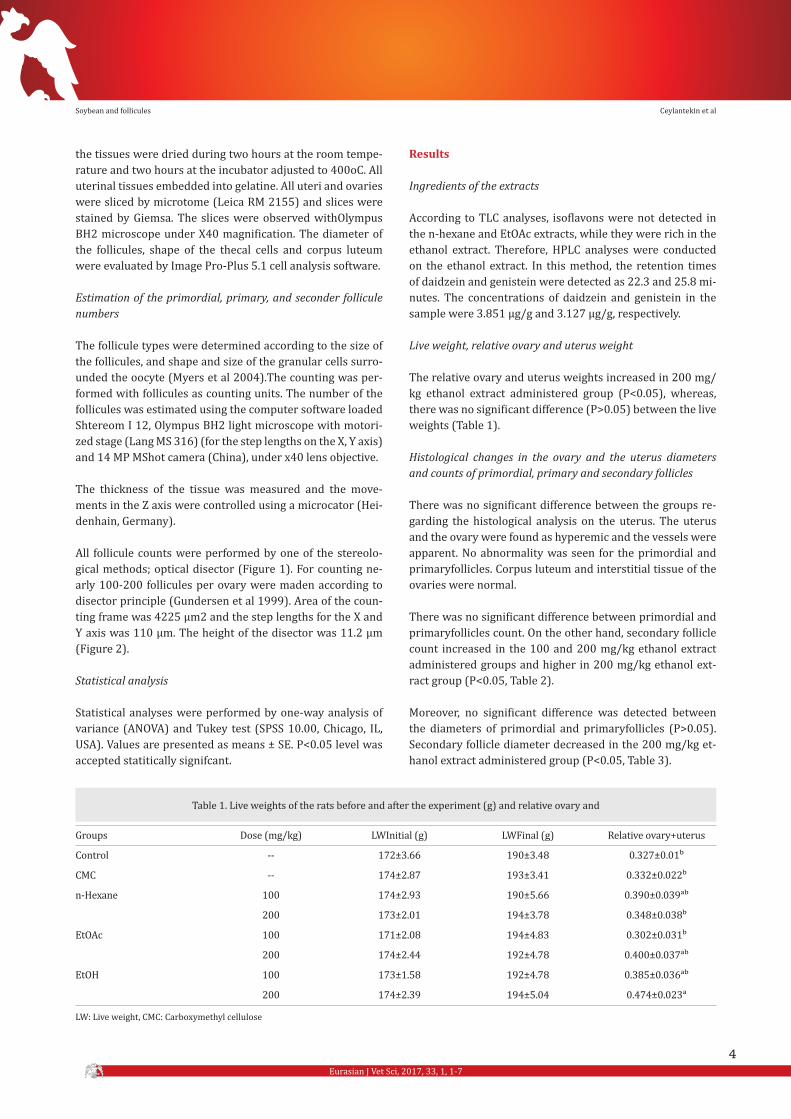
The relative ovary and uterus weights increased in 200 mg/kg ethanol extract administered group (P<0.05), whereas, there was no significant difference (P>0.05) between the live weights (Table 1).
Histological changes in the ovary and the uterus diameters and counts of primordial, primary and secondary follicles
There was no significant difference between the groups re-garding the histological analysis on the uterus. The uterus and the ovary were found as hyperemic and the vessels were apparent. No abnormality was seen for the primordial and primaryfollicles. Corpus luteum and interstitial tissue of the ovaries were normal.
There was no significant difference between primordial and primaryfollicles count. On the other hand, secondary follicle count increased in the 100 and 200 mg/kg ethanol extract administered groups and higher in 200 mg/kg ethanol ext-ract group (P<0.05, Table 2).
Moreover, no significant difference was detected between the diameters of primordial and primaryfollicles (P>0.05). Secondary follicle diameter decreased in the 200 mg/kg et-hanol extract administered group (P<0.05, Table 3).
LW: Live weight, CMC: Carboxymethyl cellulose
Groups
Control
CMC
n-Hexane
EtOAc
EtOH
Table 1. Live weights of the rats before and after the experiment (g) and relative ovary and
Dose (mg/kg)
--
--
100
200
100
200
100
200
LWInitial (g)
172±3.66
174±2.87
174±2.93
173±2.01
171±2.08
174±2.44
173±1.58
174±2.39
LWFinal (g)
190±3.48
193±3.41
190±5.66
194±3.78
194±4.83
192±4.78
192±4.78
194±5.04
Relative ovary+uterus
0.327±0.01b
0.332±0.022b
0.390±0.039ab
0.348±0.038b
0.302±0.031b
0.400±0.037ab
0.385±0.036ab
0.474±0.023a
Ceylantekin et alSoybean and follicules

Eurasian J Vet Sci, 2017, 33, 1, 1-7
5
Discussion
Glycine max is rich in phytoestrogens, however the types and amounts of phytoestrogens change according to the extrac-tion method and the solvent used forthe extaction (Anthony et al 1996, Delclos et al 2001, Jung et al 2004, Medigovic et al 2012). Luthria and Natarajan (2009) used seven different solvent mixture and various extraction methods for the ext-raction of isoflavons from soy. The method in which dimethyl sulfoxide: ethanol: water (5:70:25, v/v/v) mixture was used, was found to be the most efficient method (Luthria and Na-tarajan 2009). In the present study, different extracts were prepared from the powdered plant material. TLC analysis showed the presence of isoflavons in the ethanol extract, therefore, HPLC analysis was conducted on this extract for quantification.
As it is known, phytoestrogens bind estrogen receptors and exert estrogen-like activity resulting in secrteion and prolife-ration in the uterus and cause increase in the uterus weight (Erlandsson et al 2005, Dinsdale and Ward 2010). Similarly, uterus weights of the animals, which were fed phytoestrogen rich diet, were detected to be increased. Genistein found in the soy caused proliferation of the epithelium cells and enlar-
ged uterus when administered to the rats at high doses (Er-landsson et al 2005, Dinsdale and Ward 2010). The results of the present study, in which relative ovary and uterus weights incresed in 200 mg/kg ethanol extract administered animals (P<0.05, Table 1), supported the previous findings (Whitten et al 1992, Erlandsson et al 2005, Dinsdale and Ward 2010, Jefferson et al 2012). On the other hand, no statistical signi-ficant difference was detected in 100 mg/kg ethanol extract administered animals, which could be related with the low concentretion of the isoflavons (Table 2).
In the present study, soy extracts did not cause any differn-ce in the uterus histology, however, hiperemia was detected in 200 mg/kg ethanol extract administered group (P>0.05). A previous study revealed that isoflavons affect endometri-um histology. Soy isoflavons did not cause changes in uterus morphology at normal doses, however, caused abnormal ovarian follicles and abnormal cell maturation in vagina at higher doses (Delclos et al 2001). Awoniyiet al (1998) sho-wed that genistein caused supression of birth weight and decrease in the body weight in female rats.
Due to the fact that a follicle starts from primordial phase and goes through the preantral, antral, gaaf and preovulatory
Groups
Control
CMC
n-Hexane
EtOAc
EtOH
Table 2. Primordial, primary and secondary follicle count.
Dose (mg/kg)
--
--
100
200
100
200
100
200
PMFC
13954±1447
14917±806
13589±1233
13322±1533
15179±1068
13125±1278
14123±2142
15320±2023
PFC
3219±349.7
3444±216.3
3908±441.0
3105±302.0
3789±672.0
3570±563.9
4161±559.5
4789±442.4
SFC
3.33±0.55c
4.00±0.37c
5.00±0.36c
4.66±0.55c
3.50±0.50c
5.66±0.91c
11.66±0.76b
17.00±1.09a
PMFS: Primordial follicle count; PFS: Primary follicle count; SFS: Secondary follicle count; CMC: Carboxymethyl cellulose.
Groups
Control
CMC
n-Hexane
EtOAc
EtOH
Table 3. Primordial, primary and secondary follicle diameters
Dose (mg/kg)
--
--
100
200
100
200
100
200
PMFD
16.42±1.52
15.54±2.06
18.40±1.53
19.30±2.97
16.90±2.357
16.02±1.78
19.72±3.14
18.88±3.07
PFD
38.61±4.26c
44.22±5.37c
58.64±3.61ba
60.42±6.08a
70.56±5.57a
57.39±3.77ba
59.20±3.96ba
57.60±4.57ba
SFD
154.27±10.32ab
166.04±7.32a
155.84±15.45ab
164.16±16.01a
154.46±13.59ab
166.40±10.40a
124.84±5.023bc
94.55±10.40c
PMFD: Primordial follicle diameters; PFD: Primary follicle diameters; SFD: Secondary follicle diameters; CMC: Carboxymethyl cellulose.
Ceylantekin et alSoybean and follicules

Eurasian J Vet Sci, 2017, 33, 1, 1-7
6
follicle phases, we focused on folliculogenesis in the present study. Primordial follicle originates from germ cell and primi-tive cell pool begins to expand. Primodial follicles occur the third day of the birth and develops to antral follicle in three weeks. The mature secondary follicles appear in the 7th day. Apoptosis of the minimal ovarian cell starts in the 18th day. Follicles develop until ovulation or atresia. In adults at furt-her ages, follicle resource diminishes because primordial pool runs out (Tingen et al 2009). In a previous study, pri-mordial and primary follicle count decreased in organic and transgenic soy administered female rats (Brasil et al 2009). The effects of the estrogen on the follicle development were assessed and estrogen was found to control the dimensions of the follicles (Britt et al 2004). According to a study byMe-digovicet al (2012), genistein decreased primordial, primary, secondary follicle count but increased atretic follicle count, revealing its estrogen antagonist effect. On the other hand, estrogen agonist effect of genistein was also demonstrated by its capacity to enhance the number of the follicles which develop from preantral phase to antral phase (Medigovic et al 2012). In another study, the toxicity of genistein was in-vestigated and it was reported that there was no significant difference in the primordial follicle number (Lamartiniere et al 1998). In the present study, no significant difference was shown in the primordial follicle number (P>0.05, Table 2), similarly. Therefore, it was suggested that soy did not cau-se primordial follicle to change primary follicle and did not change the speeding process and mother follicle pool. In the ethanol extract administered group, primordial, primary and secondary follicles increased when compared to control and other test groups, but the difference was not statistically sig-nificant for the primordial and primary follicles. Similarly in literature reports, decrease in primary and secondary follicles, and reduciton in the diameter of secondary follicle were detected in the present study. As the size of the follicle incresed, the number of the follicle progressively decreased. In the ethanol extract administered group, increse of secon-dary follicle count indicated the inhibition of the follicle de-velopment and reduction the tertiary folliclegrowth. It was suggested that, due to the apoptotic effect of the estrogen, secondary follicle diameter decresed in 200 mg/kg ethanol extract administered group owing to high genistein and da-idzein concentration. High levels of these compounds in the ethanol extract could be also associated with the increase in primordial follicle count. On the other hand, primordial follic-les were reported to be the smallest and the most abundant ones among the other follicles. Similar to a previous report by Britt et al (2004), primary follicle count decreased when compared to the primordial follicle count (Britt et al 2004).
Conclusion
In conclusion, the ethanol extract of Glycine max was found to change the follicle development in a dose-dependent man-ner. Future works are necessary to clarify the releationship
between these effects and hormonal mechanisms as well as apoptosis.
Acknowledgement
This study was supported by Scientific Research Project Committee of Afyon Kocatepe University, Afyonkarahisar, Turkey (12.SAĞ.BİL.05).This study was presented orally at the Veterinary Congress in Fırat University (7-10 September 2015) and published ın congress abstracts book
References
Anthony MS, Clarkson TB, Hughes CL, Morgan TM, Burke GL, 1996. Soybean isoflavones improve cardiovascular risk factors without affecting the reproductive system of peri-pubertal rhesus monkeys. J Nutr, 1, 43-50.
Awoniyi CA, Roberts D, Veeramachaneni DN, Hurst BS, Tuc-ker KE, Schlaff WD, 1998. Reproductive sequelae in female rats after in utero and neonatal exposure to the phytoest-rogen genistein. Fertil Steril, 3, 440-447.
Brasil FB, Soares LL, Faria TS, Boaventura GT, Sampaio FJ, Ra-mos CF, 2009. The impact of dietary organic and transgenic soy on the reproductive system of female adult rat. Anat Rec, 4, 587-594.
Britt KL, Saunders PK, McPherson SJ, Misso ML, Simpson ER, Findlay JK, 2004. Estrogen actions on follicle formation and early follicle development. Biol Reprod, 5, 1712-1723.
Carrao-Panizzi M, Pedroso S, Kikuchi A, 2002. Extraction time for soybean isoflavone determination. Braz ArchBiol Techn, 4, 515-518.
Delclos KB, Bucci TJ, Lomax LG, Latendresse JR, Warbritton A, Weis CC, Newbold R, 2001. Effects of dietary geniste-in exposure during development on male and female CD (Sprague-Dawley) rats. Reprod Toxicol, 6, 647-663.
Dinsdale EC, Ward WE, 2010. Early exposure to soy isoflavo-nes and effects on reproductive health: A review of human and animal studies. Nutrients, 11, 1156-1187.
El Din S, Balta H, Elazim A, El Fattah AN,2011. Effect of soy-bean on fertility of male and female albino rats. J Am Sci, 6, 872-883.
Erdem MK, Yurdakan G, Yilmaz-Sipahi E, 2014. The effects of ketamine, midazolam and ketamine/xylazine on acute lung injury induced by alpha-naphthylthiourea in rats. Adv Clin Exp Med, 3, 343-351.
Erlandsson MC, Islander U, Moverare S, Ohlsson C, Carlsten H, 2005. Estrogenic agonism and antagonism of the soy isoflavone genistein in uterus, bone and lymphopoiesis in mice. APMIS, 5, 317-323.
Gallo D, Cantelmo F, Distefano M, Ferlini C, Zannoni GF, Riva A, Morazzoni P, Bombardelli E, Mancuso S, Scambia G, 1999. Reproductive effects of dietary soy in female Wistar rats. Food Chem Toxicol, 5, 493-502.
Goncharova EN, Timoshin SS, Radivoz MI, Komolykh OM, 1997. Effect of soybean products on the reproductive
Ceylantekin et alSoybean and follicules

Eurasian J Vet Sci, 2017, 33, 1, 1-7
7
system of sexually mature female albino rats. Vopr Pitan, 1, 17-20.
Gundersen HJ, Jensen EB, Kieu K, Nielsen J, 1999. The effici-ency of systematic sampling in stereology- reconsidered. J Microsc, 3, 199-211.
Jefferson WN, 2010. Adult ovarian function can be affected by high levels of soy. J Nutr, 12, 2322-2325.
Jefferson WN, Patisaul HB, Williams CJ, 2012. Reproductive consequences of developmental phytoestrogen exposure. Reproduction, 3, 247-260.
Jung EY, Lee BJ, Yun YW, Kang JK, Baek IJ,Yon J, Lee Y, Sohn H, Lee JY, Kim K, Yu W, Do C, Kim YC Nam S, 2004. Effects of exposure to genistein and estradiol on reproductive development in immature male mice weaned from dams adapted to a soy-based commercial diet. J Vet Med Sci, 11, 1347-1354.
Kledjus B, Mikelova R, Adam V, Zehnalek J, Vacek JR, Kizek R, 2004. Liquid chromatographic-mass spectrometric de-termination of genistin and daidzin in soybean food samp-les after accelerated solvent extraction with modified con-tent of extraction cell. Anal Chim Acta, 517, 1-11.
Lamartiniere CA, Zhang JX, Cotroneo MS, 1998. Genistein studies in rats: Potential for breast cancer prevention and reproductive and developmental toxicity. Am J Clin Nutr, 6, 1400-1405.
Lee B, Jung E, Won Yun Y, Kang J, Baek I, Yon J, Lee Y, Sohn H, Lee J, Kim K, Nam S, 2004. Effects of exposure to genistein during pubertal development on the reproductıve system of male mice. J Reprod Develop, 4, 399-409.
Luthria DL, Natarajan SS, 2009. Influence of sample prepa-
ration on the assay of isoflavones. Planta Med, 7, 704-710.Marcondes FK, Bianchi FJ, Tanno AP, 2002. Determination of
the estrous cycle phases of rats: Some helpful considerati-ons. Braz J Biol, 4, 609-614.
Medigovic I, Ristic N, Trifunovic S, Manojlovic-Stojanoski M, Milosevic V, Zikic D, Nestorovic N, 2012. Genistein affects ovarian folliculogenesis: A stereological study. Microsc Res Tech, 12, 1691-1699.
Modaresi M, Messripour M, Khorami H, 2011. Effect of soybe-an on male reproductive physiology in mice. International Conference on Life Science and Technology, 3, 15-18.
Myers M, Britt KL, Wreford NG, Ebling FJ, Kerr JB, 2004. Met-hods for quantifying follicular numbers within the mouse ovary. Reproduction, 5, 569-580.
Setchell K, Nechemias L, Cai J, Heubi J, 1998. Isoflavone con-tent of infant formulas and the metabolic fate of these phytoestrogens in early life. AJCN, 6, 1453-1461.
Tapısız Ö, Özat M, Aytan H, Mülazımoğlu B, Köse K, Güngör T, Bilge Ü, Mollamahmutoğlu L, 2009. Histerektominin stero-idojenik hormonlar olan estraiol, progesteron ve testeste-ron düzeyleri üzerine etkisi: Deneysel dişi rat modeli. Gazi Tıp Der, 4, 163-168.
Tingen CM, Bristol-Gould SK, Kiesewetter SE, Wellington JT, Shea L, Woodruff TK, 2009. Prepubertal primordial follicle loss in mice is not due to classical apoptotic pathways. Biol Reprod, 1, 16-25.
Whitten PL, Russell E, Naftolin F, 1992. Effects of a normal, human-concentration, phytoestrogen diet on rat uterine growth. Steroids, 3, 98-106.
Ceylantekin et alSoybean and follicules