Embed Size (px)
Citation preview

Research ArticleEuphorbia bicolor (Euphorbiaceae) Latex Extract ReducesInflammatory Cytokines and Oxidative Stress in a Rat Model ofOrofacial Pain
Paramita Basu, Rebecca S. Hornung, Dayna L. Averitt , and Camelia Maier
Department of Biology, Texas Woman’s University, Denton, 76204 TX, USA
Correspondence should be addressed to Camelia Maier; [email protected]
Received 17 May 2019; Accepted 10 August 2019; Published 12 September 2019
Academic Editor: Manuela Curcio
Copyright © 2019 Paramita Basu et al. This is an open access article distributed under the Creative Commons Attribution License,which permits unrestricted use, distribution, and reproduction in any medium, provided the original work is properly cited.
Recent studies have reported that the transient receptor potential V1 ion channel (TRPV1), a pain generator on sensory neurons, isactivated and potentiated by NADPH oxidase-generated reactive oxygen species (ROS). ROS are increased by advanced oxidationprotein products (AOPPs), which activate NADPH oxidase by upregulating Nox4 expression. Our previous studies reported thatEuphorbia bicolor (Euphorbiaceae) latex extract induced peripheral analgesia, partly via TRPV1, in hindpaw-inflamed male andfemale rats. The present study reports that E. bicolor latex extract also can evoke analgesia via reduction of oxidative stressbiomarkers and proinflammatory cytokines/chemokines in a rat model of orofacial pain. Male and female rats were injectedwith complete Freund’s adjuvant (CFA) into the left vibrissal pad to induce orofacial inflammation, and mechanical allodyniawas measured by the von Frey method. Twenty-four hours later, rats received one injection of E. bicolor latex extract or vehicleinto the inflamed vibrissal pad. Mechanical sensitivity was reassessed at 1, 6, 24, and/or 72 hours. Trigeminal ganglia and trunkblood were collected at each time point. In the trigeminal ganglia, ROS were quantified using 2′,7′-dichlorodihydrofluoresceindiacetate dye, Nox4 protein was quantified by Western blots, and cytokines/chemokines were quantified using a cytokine array.AOPPs were quantified in trunk blood using a spectrophotometric assay. E. bicolor latex extract significantly reduced orofacialmechanical allodynia in male and female rats at 24 and 72 hours, respectively. ROS, Nox4, and proinflammatorycytokines/chemokines were significantly reduced in the trigeminal ganglia, and plasma AOPP was significantly reduced in thetrunk blood of extract-treated compared to vehicle-treated rats. In vitro assays indicate that E. bicolor latex extract possessedantioxidant activities by scavenging free radicals. Together our data indicate that the phytochemicals in E. bicolor latex mayserve as novel therapeutics for treating oxidative stress-induced pain conditions.
1. Introduction
Pain is a major submodality of the somatosensory systemthat serves as a warning to alert the organism to actual injuryor the threat of injury. However, pain also can develop in theabsence of injury or continue following the resolution ofinjury leading to a transition from acute to chronic pain.Acute and chronic pain manifest as the development andmaintenance of hyperalgesia and/or allodynia. The Interna-tional Association for the Study of Pain defines hyperalgesiaas an increased sensitivity to noxious stimuli, while allodyniais defined as an increased sensitivity to nonnoxious stimuli.The transition mechanism from acute to chronic pain is
not entirely understood, and once chronic pain has devel-oped, it is hard to treat without the long-term use ofaddictive opioid-based narcotics. The identification of non-opioid pharmaceutical targets is needed to improve chronicpain management.
A potential target for chronic pain management may bemanaging the noxious effects of oxidative stress on peripheralsensory neurons. Patients with spinal cord injury and dia-betic neuropathy [1, 2] suffer from hyperalgesia and allody-nia arising, in part, from oxidative stress due to eitherexcessive formation of reactive oxygen species (ROS) or adecrease in antioxidant capacity [3]. This is supported bypreclinical studies reporting that superoxide (reactive
HindawiOxidative Medicine and Cellular LongevityVolume 2019, Article ID 8594375, 19 pageshttps://doi.org/10.1155/2019/8594375

oxygen) and peroxynitrite (nitrogen species) led to the devel-opment of hyperalgesia [4–6]. Furthermore, ROS are keymediators in the development of peripheral and central sen-sitization in various pain etiologies, including neuropathic,inflammatory, opioid-induced, and orofacial pain [7]. Oneof the most common orofacial pain disorders is temporo-mandibular joint disorder (TMD) pain. It has been reportedrecently that oxidative stress contributes to TMD pain. Oxi-dative stress biomarkers are significantly elevated in TMDpatients [8] correlating with a corresponding reduction intotal antioxidant capacity [9]. These data indicate that painmedications that include antioxidant and free radical scav-enging activity may be beneficial for reducing TMD pain.
Recent studies have reported that ROS may cause painthrough activation of the transient receptor potential V1ion channel (TRPV1), a pain generator in peripheral sensoryneurons. It was reported that ROS, such as nitric oxide, canactivate TRPV1 [10, 11] to increase entry of calcium ions inthe cytosol, thus enhancing the channel’s sensitivity to acidand heat and contributing to pain signalling [10]. Also,TRPV1 can be activated and potentiated by NADPH oxidasegenerated ROS [12, 13]. The source of in vivo ROS is attrib-uted to advanced oxidation protein products (AOPPs),dityrosine-containing cross-linking protein products formedprimarily because of oxidative stress [14]. AOPPs are knownto activate NADPH oxidase by increasing the expression ofits regulatory subunits, Nox1, Nox2, and Nox4 [13, 15].Nox4 mRNA is present in sensory neurons, and Nox4-derived ROS contribute to pain signalling after peripheralnerve injury [16]. Indeed, AOPPs activate TRPV1 viaNADPH oxidase 4-dependent ROS production leading tothe development of hyperalgesia [13].
The development of hyperalgesia and allodynia alsoinvolves sensitization of sensory neurons by inflammatorycytokines and chemokines. Tissue injury induces the releaseof bradykinin, which leads to the release of tumor necrosisfactor alpha (TNFα), interleukin 6 (IL-6), and interleukin 1beta (IL-1β) [17]. Cytokines directly sensitize sensory neu-rons by increasing neuronal responses to thermal, mechani-cal, and chemical stimuli [18–21]. This can occur viaTRPV1 as TNFα enhances capsaicin responses in sensoryneurons through neuronal production of prostaglandins[18]. Also, TNFα, IL-1β, and IL-6 increase neuronal excit-ability via TRPV1 [22, 23]. Furthermore, the TNFα receptorsare coexpressed with TRPV1 in sensory neurons and IL-1βand IL-6 increase TRPV1 expression in sensory neurons viathe extracellular signal-regulated kinase pathway [24, 25].
Many Euphorbia species are known to possess phyto-chemicals with antioxidant and anti-inflammatory proper-ties [26–33]. We previously reported that Euphorbia bicolor(Euphorbiaceae) latex extract evokes long-lasting peripheralanalgesia that does not involve opioid receptors but occursin part through TRPV1, in a rat inflammatory pain model[34]. Given that other Euphorbia species display antioxidantand anti-inflammatory properties and E. bicolor is able toinduce robust analgesia, we hypothesized that E. bicolor latexextract evokes analgesia via downregulation of oxidativestress biomarkers and proinflammatory cytokines in a ratmodel of orofacial pain.
2. Materials and Methods
2.1. Plant Collection and Latex Extract Preparation. E. bicolor(Euphorbiaceae) plants were collected from prairies in Den-ton County, Texas, USA, and a voucher specimen was placedin the Texas Woman’s University Herbarium. Fresh latexwas collected from the stem, leaf, and inflorescence bractsand extracted in 80% methanol (1 : 40 w/v) at room temper-ature for two days. The extract was centrifuged at 3500 rpmfor 20 minutes, and the supernatant was filtered throughWhatman #54 filter paper and stored at -20°C for future use.
2.2. Phytochemical Analyses
2.2.1. Total Phenolic Content. Total phenolic content of thelatex extract was determined by the Folin–Ciocalteu method[35]. 400 μL of latex extract was mixed with 1.6mL of 7.5%sodium carbonate and 2mL of Folin–Ciocalteu reagent(diluted 10 times in deionized water). The reaction mixtureswere incubated at room temperature for 1 hour. The absor-bances were measured at 765nm. A gallic acid standardcurve was used to estimate the total phenolic content in thelatex extract, and the results were expressed as mg gallic acidequivalents (GAE)/g of latex fresh weight (FW).
2.2.2. Total Flavonoid Content. Total flavonoid content wasdetermined by the method of Ordonez et al. [36]. 500 μL oflatex extract was mixed with 500μL of 2% aluminium chlo-ride prepared in ethanol and incubated at room temperaturefor 1 hour. The absorbances were measured at 430nm. Aquercetin standard curve was used to estimate the total flavo-noid content in the latex extract, and the results wereexpressed as mg quercetin equivalents (QE)/g of latex FW.
2.2.3. Total Proanthocyanidin Content. Total proanthocyani-din content was determined by the method of Aiyegoro andOkoh [37]. 500 μL of latex extract, 3mL of 4% (v/v) vanil-lin-methanol, and 1.5mL of hydrochloric acid were mixedand vortexed thoroughly and allowed to stand at room tem-perature for 15 minutes. The absorbances were measured at500 nm. The total proanthocyanidin content of the latexextract was estimated using a gallic acid standard curve andexpressed as mg GAE/g of latex FW.
2.2.4. Total Terpenoid Content. Total terpenoid content wasestimated by the modified method of Ghorai et al. [38]. Thelatex extract was centrifuged at 4000 g for 15 minutes at roomtemperature. 200 μL of supernatant was mixed with 1.5mL ofchloroform. The standard was prepared by adding 200 μL of2000μg/mL of linalool in methanol to 1.5mL of chloroform,and serial dilutions were prepared in the range of 100mg/200μL-1mg/200μL. The sample and standard mixtures werevortexed thoroughly and allowed to stand for three minutes,after which 100 μL of sulfuric acid (H2SO4) was added. Thesample mixture was incubated at room temperature for 1.5-2 hours in the dark, and the standard was incubated for 5minutes at room temperature in the dark. A reddish-brownprecipitate formed at the end of the incubation. The superna-tants were decanted from both sample and standard mix-tures, and the precipitates were dissolved in 1.5mL of 95%
2 Oxidative Medicine and Cellular Longevity

(v/v) methanol and vortexed thoroughly until dissolvedcompletely. The absorbances were measured at 538nmusing 95% (v/v) methanol as a blank. The total terpenoidconcentration of the latex extract was estimated as linaloolequivalents using a linalool standard curve.
2.3. In Vitro Antioxidant and Radical Scavenging Activities ofE. bicolor Latex Extract. A series of in vitro assays wereemployed to determine the in vitro antioxidant and radicalscavenging activities of E. bicolor latex extract. All assays wereperformed according to the original methods specified foreach assay and modified by Basu and Maier [39].
2.3.1. Ferric Reducing Power. Ferric reducing power of thelatex extract was performed by the method of Oyaizu [40].Increasing concentrations (20 μg/mL–100μg/mL) of 1mLlatex extract were mixed with 2.5mL of 0.2M phosphatebuffer (pH 6.6) and 2.5mL of 1% w/v K3Fe(CN)6. Thereaction mixtures were incubated at 50°C for 20min, afterwhich 2.5mL of 10% w/v trichloroacetic acid (TCA) wasadded and the mixtures were centrifuged at 3000 rpm for10min. 2.5mL of supernatants was mixed with 2.5mLdeionized water and 0.5mL of 0.1% w/v FeCl3. The resultswere presented as absorbances measured at 700nm.Butylated hydroxylated toluene (BHT) was used as a pos-itive control.
2.3.2. 2,2′-Azino-bis-(3-ethylbenzothiazoline-6-sulphonic Acid)(ABTS) Scavenging Activity. 2,2′-Azino-bis-(3-ethylbenzo-thiazoline-6-sulphonic acid) (ABTS) scavenging activity wasdetermined by the method of Re et al. [41]. ABTS workingsolution was prepared by mixing equal amounts of 7mMABTS and 2.4mM potassium persulfate in the dark at roomtemperature for 12h. Increasing concentrations (20 μg/mL-100μg/mL) of 1mL latex extract were mixed with 1mL ofABTS solution. The reaction mixtures were incubated at roomtemperature for 7 minutes, and absorbances were measured at734nm. The results were presented as percent inhibition ofABTS radical calculated according to equation (1). Ascorbicacid was used as a standard.
% of free radical scavenging activity =Abscontrol −Abssample
Abscontrol× 100
1
2.3.3. 2,2-Diphenyl-1-picrylhydrazyl (DPPH) ScavengingActivity. Free radical scavenging activity was determined bymeasuring the 2,2-diphenyl-1-picrylhydrazyl (DPPH) radicalscavenging activity was determined using a modifiedmethod of Gülçin et al. [42]. Increasing concentrations (20μg/mL–100μg/mL) of 0.05mL latex extract were mixedwith 2.95mL of 0.1mM DPPH in methanol, thoroughlyvortexed, and incubated in the dark for 30 minutes. Theabsorbances were recorded at 517nm, and results werepresented as percent inhibition of DPPH radical calculatedaccording to equation (1). Ascorbic acid was used as astandard.
2.3.4. Hydrogen Peroxide Scavenging Activity. Hydrogen per-oxide scavenging activity was determined by the method ofRuch et al. [43]. Increasing concentrations (20 μg/mL-100μg/mL) of 4mL latex extract were mixed with 600μL of4mM H2O2 solution prepared in 0.1M phosphate buffer(pH 7.4). The reaction mixtures were incubated for 4minutes, and the absorbances were recorded at 230nmagainst blank solution containing latex extract withoutH2O2. The results were presented as percent inhibition ofH2O2 radical calculated according to equation (1). Butylatedhydroxylated toluene (BHT) was used as a standard.
2.3.5. Nitric Oxide Scavenging Activity. Nitric oxide (NO)scavenging activity was determined by the modified methodof Balakrishnan et al. [44]. Increasing concentrations(20 μg/mL-100μg/mL) of 1mL latex extract were mixed with2mL of sodium nitroprusside prepared in phosphate-buffered saline (PBS). The reaction mixtures were incubatedat 25°C for 150min, after which 0.5mL of Griess reagentwas added to the incubating mixture. The absorbances weremeasured at 540 nm, and results were presented as percentinhibition of NO radical calculated according to equation(1). Quercetin was used as a standard.
2.4. IC50 and Pearson’s Correlation. IC50 values, i.e., the con-centration required to scavenge 50% of free radicals, were cal-culated by linear regression analysis. Pearson’s correlationswere performed between the IC50 values of radical scaveng-ing activities and total phenolic, flavonoid, proanthocyani-din, and terpenoid contents of the latex extract.
2.5. Animals. A total of 70 male and 70 female (250-350 g)adult Sprague-Dawley rats (Charles River Laboratories,Wilmington, MA, USA) were separated by sex and housedin cages in a 12 : 12 hour light : dark cycle with ad libitumaccess to food and water. Rats were maintained in the animalfacility for a minimum of five days before testing. All studieswere conducted under the approval of the Texas Woman’sUniversity Institutional Animal Care & Use Committee andunder the strict guidelines of Committee for Research andEthical Issues of the International Association for the Studyof Pain and Animal Welfare Act, implementing Animal Wel-fare Regulations, and the principles of the Guide for the Careand Use of Laboratory Animals. The experimenters wereblind to the treatment groups during behaviour testing, andall rats were acclimated to the testing room and apparatustwenty-four hours prior to testing.
2.6. Behaviour Testing. Sensitivity to a nonnoxiousmechanical (touch) stimulus was measured as the forceto withdraw from contact with a blunt von Frey filament(North Coast Medical Inc., Gilroy, CA), as previouslycharacterized [45]. For this test, a filament of 2.0 g fornoninflamed tissue and 0.16 g for inflamed tissues was firstapplied to the vibrissal pad. When no response wasobserved, 30 seconds later, the next thicker filament wasapplied and the process was repeated until a withdrawresponse was observed. When a withdraw response wasobserved, the next thinnest filament was applied 30 sec-onds later, and the process was repeated until no withdraw
3Oxidative Medicine and Cellular Longevity

response was observed. The filament size that produced atleast three withdrawals was recorded as the thresholdgrams of pressure required to elicit a head withdrawalresponse as a measure of mechanical allodynia.
For the experiment, baseline mechanical sensitivity wasrecorded followed by injections of 50 μL complete Freund’sadjuvant (CFA; 1 : 1 in 0.9% sterile saline; Sigma-Aldrich,St. Louis, MO, USA) into the left vibrissal pad to induceinflammation. Mechanical allodynia was then confirmed24 hours following CFA injections. Rats received one injec-tion of either E. bicolor latex extract (300 μg/mg in 0.9%saline and <5% methanol; n = 10 males and n = 10 females)or vehicle (0.9% saline and <5% methanol; n = 10 malesand n = 10 females) into the inflamed vibrissal pad.Mechanical allodynia was then reassessed at 1, 6, and 24hours in males and 1, 6, and 72 hours in females. The latexextract concentration and time points were selected basedon our previous report that the onset of extract-inducedanalgesia in male and female rats with hindpaw inflamma-tion occurs at 6 and 72 hours, respectively [34].
2.7. Tissue Collection and Analysis. A total of 60 male and60 female rats (n = 10 per time point, per sex, and pertreatment) were injected with CFA into the left vibrissalpad and saline into the right vibrissal pad. At 1, 6, and24 hours in males and 1, 6, and 72 hours in females, ratswere rapidly decapitated under brief gas anesthesia (iso-flurane; 3%). A separate control group of rats (n = 10males and n = 10 females) received only one saline injec-tion into the left vibrissal pad. 24 hours post saline injec-tion, male and female rats were rapidly decapitated underbrief gas anesthesia (isoflurane; 3%). Trunk blood was col-lected in BD Vacutainer® spray-coated K2EDTA collectiontubes on ice (Pulmolab, CA, US). Immediately after collec-tion, the cells were removed from plasma by centrifugationat 1000–2000 x g for 10 minutes. The separated plasmawas then transferred into clean polypropylene tubes andstored at −80°C. The trigeminal ganglia (TGs) were bilat-erally extracted from the same rats and either immediatelyused or stored at −80°C.
2.7.1. Quantification of Advanced Oxidation ProteinProduct (AOPP). The level of AOPP was quantified inthe plasma of male and female rats treated with either E.bicolor latex extract or vehicle (n = 10 per sex and pertreatment) by the modified method of Witko-Sarsat et al.[14]. For this method, 2mL of plasma was diluted inPBS at a 1 : 5 ratio; 10 μL of 1.16M potassium iodide(KI) was added, followed by 20 μL of acetic acid after2min. The absorbance of the reaction mixture was mea-sured at 340 nm against a blank containing 2mL of PBS,10 μL of KI, and 20μL of acetic acid. The AOPP levelwas determined as μmol·L−1 chloramine-T equivalents byusing a chloramine-T linear curve ranging from 0 to 100μM.
2.7.2. Quantification of Reactive Oxygen Species (ROS).The level of ROS was detected in the TGs isolated frommale and female rats treated with either E. bicolor latexextract or vehicle (n = 4 per sex and per treatment) by
the method of Chung et al. [46], using a cell-permeant2′,7′-dichlorodihydrofluorescein diacetate (H2DCFDA), areduced form of fluorescein used as ROS indicator. Thenonfluorescent H2DCFDA is converted into the highlyfluorescent 2′,7′-dichlorofluorescein when the acetategroups of H2DCFDA are cleaved by intracellular oxida-tion. H2DCFDA detects levels of hydrogen peroxide, per-oxyl radicals, and peroxynitrite in dissociated cells andintact tissues [47]. Freshly extracted TGs were quicklywashed, minced, and incubated in 96-well plates with200 μL PBS for 30 minutes at 37°C. After 30 minutes,the background fluorescence was detected (Biotek’s SynergyHT fluorimeter) at 485nm excitation and 535nm emission.After recording the background reading, H2DCFDA wasadded into each well at a final concentration of 10 μM. Theplates were incubated for another 30 minutes at 37°C, afterwhich the fluorescence was remeasured. The level of ROSwas detected as the intensity of fluorescence after subtract-ing the background fluorescence. TGs from the saline-treated vibrissal pad were used as internal controls anddata normalization.
2.7.3. Quantification of Nox4 Protein Expression by WesternImmunoblotting. TGs isolated from male and female ratstreated with either E. bicolor latex extract or vehicle (n = 4per sex and per treatment) were homogenized in radioimmu-noprecipitation (RIPA) lysis and extraction buffer containingHalt protease inhibitor cocktail (Thermo Scientific; part no.78430) to prevent proteolysis. Tissue homogenization wasperformed at 6.5m/s, 3 times for 10 seconds each, in aFastPrep®-24 homogenizer (MP Biomedicals; Santa Ana,CA). Homogenates were centrifuged at 13000 rpm for 15minutes at 4°C. The protein concentration was determinedby using Pierce BCA™ protein assay kit. Protein lysate(30 μg) was resolved by sodium dodecyl sulfate polyacryl-amide gel electrophoresis (SDS-PAGE), followed by transferonto a polyvinylidene difluoride (PVDF) membrane (Bio-Rad; Hercules, CA) using Bio-Rad’s Western blot unit.Membranes were blocked with 5% bovine serum albumin(BSA) in TBS-Tween 20 and probed with monoclonalanti-Nox4 (1 : 500; 67 kDa; Abcam) overnight, followed byincubation with secondary antibody (1 : 8000, Alexa Fluor)at room temperature for one hour. To confirm the selec-tivity of the antibody, blots were preincubated with block-ing peptide (ab155071, Abcam) for Nox4. Immunoreactivebands were detected with a Licor Odyssey imaging system.Beta actin was used as an internal control to normalizeprotein expression, and the percentage of β-actin was cal-culated based on at least four independent experiments.
2.7.4. Cytokine and Chemokine Proteome Profiling. A Prote-ome Profiler Rat Cytokine Array Kit, column A (R&D Sys-tems Inc., Minneapolis, MN), containing 4 membranescoated with 29 cytokines/chemokines (Table 1) was probedwith rat protein samples extracted from TGs, and the relativecytokine levels were compared. Briefly, 200 μg of proteinextracted from vehicle- or extract-treated TGs from male orfemale rats (n = 4 males and n = 4 females per treatment)was mixed with a cocktail of biotinylated detection
4 Oxidative Medicine and Cellular Longevity
antibodies. The nitrocellulose membranes were blockedaccording to the manufacturer’s protocol. The protein andantibody mixtures were incubated with the membrane con-taining immobilized antibodies for 29 rat cytokines. Boundprotein was detected with streptavidin conjugated to horse-radish peroxidase (HRP). Membranes were washed anddeveloped with chemiluminescent detection reagents. Thecytokine/chemokine spots were detected with a Bio-RadChemiDoc™ MP imaging system.
2.8. Statistical Analyses. All data were analyzed usingGraphPad Prism software version 7 (GraphPad, SanDiego, CA, USA). Behavioural data were presented asmeans ± SEM grams and analyzed by repeated measurestwo-way analysis of variance (ANOVA) followed by Bon-ferroni post hoc analysis. In vitro antioxidant and radicalscavenging activities were analyzed by ordinary two-wayANOVA followed by Bonferroni post hoc analysis withstatistical significance tested at p ≤ 0 05. AOPP and ROSlevels in saline- and CFA-treated animals were analyzedby the Student t-test with a statistical significance testedat p ≤ 0 05. Western blot bands were quantified by densi-tometry using the Licor Odyssey imaging system ImageStudio 2.0, and cytokine/chemokine levels were quantifiedby densitometry using ImageJ version 1.8.0 (NationalInstitutes of Health, Bethesda, MD, USA). Densitometrydata were analyzed by two-way ANOVA followed by Bon-ferroni post hoc analysis with statistical significance testedat p ≤ 0 05.
3. Results
3.1. E. bicolor Latex Extract Displays In Vitro Antioxidantand Free Radical Scavenging Activities. E. bicolor latexextract significantly contained more total phenolics (TPC),proanthocyanidins (TPrC), and terpenoids (TTC) than totalflavonoids (TFC) (F 3, 8 = 24 34; p ≤ 0 05) (Figure 1(a)).The in vitro antioxidant and radical scavenging activities ofthe E. bicolor latex extract were evaluated through a series ofin vitro assays. E. bicolor latex extract significantly andconcentration-dependently increased the ferric reducingactivity compared to the standard BHT (F 1, 20 = 902 7;p ≤ 0 05) (Figure 1(b)). The extract inhibited approximately80% of the ABTS radical at 20-100 μg/mL, which was asignificantly greater inhibition as compared to that ofthe ascorbic acid standard (F 1, 20 = 6729; p ≤ 0 05)(Figure 1(c)). Additionally, the latex extract reducedapproximately 8% of DPPH radical at 20-100μg/mL(Figure 1(d)), whereas H2O2 radical was reduced byapproximately 30% at 20-100μg/mL (Figure 1(e)). Theascorbic acid standard induced significantly higherscavenging activities than the latex extract on DPPH(F 1, 20 = 100787; p > 0 05) (Figure 1(d)), while theextract induced H2O2 scavenging activities comparable tothe BHT standard (F 1, 20 = 0 34; p > 0 05) (Figure 1(e)).The latex extract reduced nitric oxide (NO) radical in aconcentration-dependent manner (Figure 1(f)) and wassignificantly higher compared to the quercetin standard(F 1, 20 = 2983; p > 0 05).
Table 1: Rat cytokine/chemokine array coordinates (adapted from the manufacturer’s guidelines for the Proteome Profiler Rat CytokineArray Kit, column A, R&D Systems).
A B C D
1 Ref spot — — Ref spot
2 Ref spot — — Ref spot
3 CINC-1 IL-1α or IL-1F1 IL-13 RANTES or CCL5
4 CINC-1 IL-1α or IL-1F1 IL-13 RANTES or CCL5
5 CINC-2α/β IL-1β or IL-1F2 IL-17 Thymus chemokine or CXCL7
6 CINC-2α/β IL-1β or IL-1F2 IL-17 Thymus chemokine or CXCL7
7 CINC-3 IL-1ra or IL-1F3 IP-10 or CXCL10 TIMP-1
8 CINC-3 IL-1ra or IL-1F3 IP-10 or CXCL10 TIMP-1
9 CNTF IL-2 LIX TNF-α or TNFSF1A
10 CNTF IL-2 LIX TNF-α or TNFSF1A
11 Fractalkine or CX3CL1 IL-3 L-Selectin or CD62L/LECAM-1 VEGF or VEGF-A/vasculotropin
12 Fractalkine or CX3CL1 IL-3 L-Selectin or CD62L/LECAM-1 VEGF or VEGF-A/vasculotropin
13 GM-CSF IL-4 MIG or CXCL9 —
14 GM-CSF IL-4 MIG or CXCL9 —
15 sICAM-1/CD54 IL-6 MIP-1α or CCL3 —
16 sICAM-1/CD54 IL-6 MIP-1α or CCL3 —
17 IFN-γ IL-10 MIP-3α or CCL20 Negative control or control (-)
18 IFN-γ IL-10 MIP-1α or CCL20 Negative control or control (-)
19 Ref spot — — —
20 Ref spot — — —
Alternate nomenclatures are in bold.
5Oxidative Medicine and Cellular Longevity

Total polyphenolic contents were estimated and corre-lated with the IC50 values of the radical scavenging activitiesof the E. bicolor latex extract (Figure 1(g) and Table 2).Latex extract significantly (F 3, 8 = 10 71; p ≤ 0 05)quenched 50% of ABTS, DPPH, H2O2, and NO radicals(Figure 1(g)). The IC50 values of the latex extractrequired to quench 50% of free radicals are ranked as fol-lows: NO (1 6 ± 0 04 μg/mL), ABTS (18 2 ± 13 μg/mL),
H2O2 50 ± 6 6 μg/mL < DPPH 68 4 ± 3 8 μg/mL . ABTSradical scavenging activity showed high correlations withflavonoid (R2 = 0 866) and proanthocyanidin (R2 = 0 861)contents of the latex extract. The DPPH (R2 = 0 929)and NO (R2 = 0 975) radical scavenging activities of theextract showed high correlations with phenolics (R2 = 0 929)and terpenoids (R2 = 0 979). H2O2 radical scavenging activityshowed low to negative correlations with polyphenolics,
TPC
(mg
GA
E/g)
TFC
(mg
QE/
g)
TPrC
(mg
GA
E/g)
TTC
(mg
linal
ool/g
)0
5
10
15
20
25
Poly
phen
olic
cont
ent
⁎
⁎
⁎
(a)
20 �휇
g/m
l
40 �휇
g/m
l
60 �휇
g/m
l
80 �휇
g/m
l
100 �휇
g/m
l0.0
0.1
0.2
0.3
0.4
0.5
Abso
rban
ce at
700
nm
ExtractBHT
⁎ ⁎⁎
⁎⁎
(b)
20 �휇
g/m
l
40 �휇
g/m
l
60 �휇
g/m
l
80 �휇
g/m
l
100 �휇
g/m
l
ExtractAscorbic acid
0
20
40
60
80
100
% in
hibi
tion
ABT
S ra
dica
l
⁎ ⁎ ⁎ ⁎ ⁎
(c)
20 �휇
g/m
l
40 �휇
g/m
l
60 �휇
g/m
l
80 �휇
g/m
l
100 �휇
g/m
l
ExtractAscorbic acid
0
10
20
30
40
50
% in
hibi
tion
DPP
H ra
dica
l
⁎
⁎
⁎⁎
⁎
(d)
20 �휇
g/m
l
40 �휇
g/m
l
60 �휇
g/m
l
80 �휇
g/m
l
100 �휇
g/m
l
ExtractBHT
0
10
20
30
40
% in
hibi
tion
H2O
2 rad
ical
(e)
20 �휇
g/m
l
40 �휇
g/m
l
60 �휇
g/m
l
80 �휇
g/m
l
100 �휇
g/m
l
ExtractQuercetin
0
20
40
60
80
100
% in
hibi
tion
NO
radi
cal
⁎ ⁎ ⁎ ⁎⁎
(f)
ABT
S
DPP
H
H2O
2
NO
0
20
40
60
80
100
IC50
val
ues (�휇
g/m
l)
⁎
⁎
(g)
Figure 1: Total polyphenolic contents, in vitro antioxidant, in vitro free radical scavenging activities, and IC50 values of E. bicolor latex extract.Total phenolic (TPC), flavonoid (TFC), proanthocyanidin (TPrC), and terpenoid (TTC) contents of latex extract (a). Asterisks indicatesignificance compared to TFC at p ≤ 0 05 by one-way ANOVA with Tukey’s analysis. Reducing power (b), percent inhibition of the2,2′-azino-bis-(3-ethylbenzothiazoline-6-sulphonic acid) (ABTS) radical (c), percent inhibition of the 2,2-diphenyl-1-picrylhydrazyl(DPPH) radical (d), percent inhibition of the hydrogen peroxide (H2O2) radical (e), and percent inhibition of the nitric oxide (NO)radical (f). For each assay, extract (black bars) was compared to its corresponding standard (grey bars) by two-way ANOVA with Bonferronipost hoc analysis at p ≤ 0 05. ∗Significant difference compared to the positive control. IC50 values of radical scavenging activities of latexextract (g). Asterisks indicate significance compared to IC50 of DPPH scavenging activity at p ≤ 0 05 by one-way ANOVAwith Tukey’s analysis.
6 Oxidative Medicine and Cellular Longevity

indicating that other phytochemicals are involved. NO radi-cal scavenging activity showed positive and high correlationswith phenolics (R2 = 0 975) and terpenoids (R2 = 0 999)(Table 2).
3.2. E. bicolor Latex Extract Reduced Orofacial MechanicalAllodynia. E. bicolor latex extract significantly reducedorofacial mechanical sensitivity in male rats (F 1, 18 = 38 6;p ≤ 0 05) at 24 hours (Figure 2(a)) and in female rats(F 1, 18 = 15 71; p ≤ 0 05) at 72 hours (Figure 2(b)) ascompared to vehicle-treated rats and in accordance withour previous timeline of analgesia in hindpaw-inflamed rats[34]. In male rats, extract treatment significantly increasedthe withdrawal threshold from 0.6 g to 4.4 g, and in femalerats, extract treatment significantly increased the withdrawalthreshold from 0.74 g to 4.7 g. No significant differenceswere observed between the vehicle- and extract-treatedgroups at 1 and 6 hours in male and at 1, 6, and 24 hoursin female rats (p > 0 05).
3.3. E. bicolor Latex Extract Reduced AOPP, ROS, and Nox4Levels. CFA inflammation significantly increased plasmaAOPP levels in both male (t = 14 66, df = 18; p ≤ 0 05)(Figure 3(a)) and female (t = 135 5, df = 18; p ≤ 0 05)(Figure 3(b)) rats as compared to vehicle-treated controls.Female rats had significantly greater CFA-evoked AOPPlevels when compared to males (F 1, 36 = 24 18; p ≤ 0 05).E. bicolor extract treatment significantly decreased the AOPPlevels in males at 6 and 24 hours (F 1, 54 = 212 8; p ≤ 0 05)(Figure 3(c)) and at 1 and 72 hours posttreatment in femalerats (F 1, 54 = 395 5; p ≤ 0 05) (Figure 3(d)). CFA inflam-mation also significantly increased ROS levels in the trigem-inal ganglia of both male (t = 11 75, df = 6) (Figure 4(a)) andfemale (t = 4 06, df = 6) (Figure 4(b)) rats compared tovehicle-treated controls, and CFA-evoked ROS were compa-rable between the sexes (F 1, 12 = 0 02; p > 0 05). Extracttreatment significantly decreased ROS levels in males at 6and 24 hours (F 1, 18 = 63 43; p ≤ 0 05) (Figure 4(c)) andat 72 hours posttreatment in female rats (F 1, 18 = 12 36;p ≤ 0 05) (Figure 4(d)). Further, CFA inflammation signifi-cantly increased Nox4 protein expression in the trigeminalganglia of both male (t = 15 99, df = 6) (Figure 5(a)) andfemale (t = 4 62, df = 6) (Figure 5(b)) rats compared tovehicle-treated controls. No significant difference was
observed in CFA-evoked Nox4 protein expression betweenmale and female rats (F 1, 12 = 1 96; p > 0 05). Extracttreatment significantly decreased Nox4 expression at 24hours posttreatment in male rats (F 1, 18 = 36 24; p ≤ 0 05)(Figure 5(e)) and at 72 hours posttreatment in females(F 1, 18 = 23 03; p ≤ 0 05) (Figure 5(f)).
3.4. E. bicolor Latex Extract Modulated Cytokines/Chemokines.In male rats, E. bicolor latex extract significantly downregu-lated several proinflammatory cytokines (notably IL-1α, IL-1β, IL-2, IL-3, and IL-17), chemotactic cytokines (MIP-1αand MIP-3α), and proinflammatory chemokines (CINC-1,MIG) and upregulated an anti-inflammatory chemokine(TIMP) in the trigeminal ganglia 1 hour post E. bicolorlatex extract treatment compared to vehicle controls(F 1, 44 = 125 2; p ≤ 0 05) (Figure 6(a)). At 6 hours oftreatment, proinflammatory cytokines (sICAM-1, IL-1α)and proinflammatory chemokines (CINC-1, VEGF) weresignificantly downregulated compared to vehicle controls(F 1, 20 = 256 7; p ≤ 0 05) (Figure 6(b)). At 24 hours postE. bicolor latex extract treatment, the proinflammatory cyto-kine IL-1α and proinflammatory chemokines LIX andRANTES were significantly downregulated compared tovehicle controls (F 1, 20 = 12 64; p ≤ 0 05) (Figure 6(c)).
In female rats, E. bicolor latex extract significantly down-regulated several proinflammatory cytokines (notably IL-1β,IL-2, and IL-3) and proinflammatory chemokines (MIG) inthe trigeminal ganglia 1 hour post E. bicolor latex extracttreatment compared to vehicle controls (F 1, 38 = 189 5;p ≤ 0 05) (Figure 7(a)). No significant differences betweenvehicle- and extract-treated groups were observed at 6hours (F 1, 20 = 0 85; p > 0 05) (Figure 7(b)) or 72 hours.At 72 hours, a reduction in the proinflammatory cytokine IL-1β was observed and an increase in the anti-inflammatorychemokine TIMP was observed.
4. Discussion
Painand inflammatoryconditionsare treatedmainlywithopi-oids and nonsteroidal anti-inflammatory drugs (NSAIDs),which are responsible for adverse reactions, such as gastro-intestinal disturbances, renal damage, respiratory depres-sion, and possible dependence [48–50]. These negativeside effects have led to the search for alternative therapeu-tics, such as phytomedicines [51–54]. As extracts fromdifferent species of Euphorbia possess antioxidant andanti-inflammatory activities [55–57], we have examinedthe previously untested E. bicolor as a potential phytome-dicine. Here, we report for the first time that E. bicolorlatex extract (1) possesses in vitro antioxidant and freeradical scavenging activities; (2) reduces orofacial mechan-ical allodynia in both male and female rats; (3) downregu-lates the oxidative stress biomarkers AOPP, ROS, andNox4; and (4) alters the expression levels of pro- andanti-inflammatory cytokines/chemokines.
E. bicolor latex extract displayed in vitro free radical scav-enging activities by inhibiting ABTS, DPPH, H2O2, and NO.The ferric reducing power of the latex extract was signifi-cantly more potent than that of the BHT standard. The
Table 2: Correlation between IC50 of in vitro radical scavengingactivities and phenolic, flavonoid, proanthocyanidin, andterpenoid contents of E. bicolor latex extract.
Correlation (R)TPC TFC TPrC TTC
ABTS radical 0.500 0.866 0.861 0.640
DPPH radical 0.929∗ -0.370 0.361 0.979∗
H2O2 radical -1.00 -0.002 0.011 -0.985∗
NO radical 0.975∗ 0.223 -0.214 0.999∗
TPC: total phenolic content; TFC: total flavonoid content; TPrC: totalproanthocyanidin content; TTC: total terpenoid content. The coefficientvalues with asterisks indicate significant difference at p ≤ 0 05.
7Oxidative Medicine and Cellular Longevity

DPPH radical scavenging activity of the latex extract wasfound to be significantly lower than ABTS scavenging activ-ity. This could be explained by the fact that the latex extract
contains more hydrophobic than hydrophilic phytochemi-cals with antioxidant properties, since the ABTS assay ismore suitable for hydrophilic antioxidants while the DPPH
Base
line
Post-
CFA
1 ho
ur
6 ho
urs
24 h
ours
012345678
Time post-injection
Forc
e to
with
draw
(g)
VehicleExtract
⁎
(a)
Base
line
Post-
CFA
1 ho
ur
6 ho
urs
72 h
ours
24 h
ours
Time post-injection
VehicleExtract
012345678
Forc
e to
with
draw
(g)
⁎
(b)
Figure 2: E. bicolor latex extract reduces orofacial mechanical sensitivity in both male and female rats. Orofacial mechanical allodynia wasreduced in males (a) at 24 hours and in females (b) at 72 hours post E. bicolor latex extract injection (closed bars) as compared to vehicletreatment (open bars). ∗p ≤ 0 05 compared to vehicle by repeated measures two-way ANOVA with Bonferroni post hoc analysis.
0
200
400
600
Plas
ma A
OPP
leve
l
⁎
SalineCFA
(a)
0
200
400
600
Plas
ma A
OPP
leve
l
⁎
SalineCFA
(b)
1 hr 6 hr0
200
400
600
Plas
ma A
OPP
leve
l
VehicleExtract
Saline
⁎⁎
24 hr
(c)
1 hr 6 hr 72 hr0
200
400
600
Plas
ma A
OPP
leve
l
VehicleExtract
Saline
⁎
⁎
(d)
Figure 3: E. bicolor latex extract reduces plasma AOPP levels in both male and female rats. CFA inflammation (closed bars) increased AOPPlevels in male (a) and female (b) rats as compared to saline treatment (diagonal bars). ∗p ≤ 0 05 compared to vehicle by the Student t-testanalysis. Plasma AOPP levels decreased in males (c) at 6 and 24 hours and in females (d) at 1 and 72 hours post E. bicolor latex extractinjection (closed bars) as compared to vehicle treatment (open bars). Dotted lines indicate the plasma AOPP level in noninflamedrats. ∗p ≤ 0 05 compared to vehicle by two-way ANOVA with Bonferroni post hoc analysis.
8 Oxidative Medicine and Cellular Longevity

assay is more applicable for hydrophobic antioxidants [58].In addition, total phenolic and terpenoid contents of the latexextract resulted in stronger DPPH and NO scavenging activ-ities, whereas total flavonoid content contributed to ABTSscavenging activity. Low and negative correlation valuesindicate that other groups of phytochemicals present inthe latex extract contributed to the ABTS and H2O2 radicalscavenging activities. Therefore, the differences in the corre-lations between IC50 values and polyphenolic contentsexplain why DPPH, H2O2, and NO radical scavengingactivities of the extract are not as potent as their corre-sponding standards.
The orofacial region is innervated by the trigeminalnerves and is one of the most densely innervated regions ofthe body making trigeminal system-associated pain, such asmigraine, headache, temporomandibular joint disorder, andtrigeminal neuralgia, difficult to manage. In the presentstudy, E. bicolor latex extract significantly reduced orofacialmechanical sensitivity in both male and female rats at 24hours and 72 hours, respectively, which correspond to thesame onset time of analgesia observed in our previous studieson hindpaw-inflamed male and female rats [34]. In support,a recent systematic review reported that extracts from variousplants are quite effective at alleviating orofacial pain in pre-clinical experimental models [59]. For example, plantextracts from Sida cordifolia [60], Hyptis pectinata [61, 62],
Hyptis fruticosa [63], Ocimum basilicum [64], Acmella olera-cea [65], and Syzygium cumini [66] reduce orofacial pain.This suggests that plant phytochemicals may be an effectivealternative or adjuvant therapy to opioids and NSAIDs formanaging orofacial pain.
Elevated AOPP levels are linked to many diseases, such ascancer [67], chronic irritable bowel syndrome [68], coronaryartery disease [69], and diabetes [70, 71], and are thought tocontribute to pain arising from these diseases. In the presentstudy, CFA-evoked inflammation significantly increasedplasma AOPP levels, corroborating a previous study report-ing that CFA induced a 1.6-fold increase in AOPP levels inrats [13]. Interestingly, we also found that CFA evokedhigher AOPP levels in female compared to male rats, indicat-ing that females may have a higher level of oxidative stress inresponse to CFA. Saline treatment induced a decrease in thelevel of AOPP in female rats. This could be attributed to theantioxidant properties of estrogens. Several studies reportedthe neuroprotective effect of estradiol through the involve-ment of antioxidant signalling [72–75]. Estradiol also upreg-ulated the expression of antioxidant enzyme manganesesuperoxide dismutase (MnSOD) via activation of the MAPkinase signalling pathway, which further confirms the anti-oxidant properties of estrogens [76]. Therefore, it can bespeculated that the endogenous estradiol reduced the levelof plasma AOPP in saline-treated female rats.
0
2
4
6
ROS
fold
chan
ge(n
orm
aliz
ed n
aive
TG
)⁎
SalineCFA
(a)
0
2
4
6
ROS
fold
chan
ge(n
orm
aliz
ed n
aive
TG
)
⁎
SalineCFA
(b)
1 hr 6 hr0
2
4
6
ROS
fold
chan
ge(n
orm
aliz
ed n
aive
TG
)
VehicleExtract
Saline
⁎⁎
24 hr
(c)
1 hr 6 hr 72 hr
VehicleExtract
0
2
4
6
ROS
fold
chan
ge(n
orm
aliz
ed n
aive
TG
)
Saline
⁎
(d)
Figure 4: E. bicolor latex extract reduces ROS levels in both male and female rats. CFA inflammation (closed bar) increased ROS levels in thetrigeminal ganglia of male (a) and female (b) rats as compared to saline treatment (diagonal bar). ∗p ≤ 0 05 compared to vehicle by the Studentt-test analysis. ROS levels decreased in the trigeminal ganglia of males (c) at 6 and 24 hours and females (d) at 72 hours post E. bicolor latexextract injection (closed bars) as compared to vehicle treatment (open bars). Dotted lines indicate the ROS level in noninflamed rats. ∗p ≤ 0 05compared to vehicle by two-way ANOVA with Bonferroni post hoc analysis.
9Oxidative Medicine and Cellular Longevity

Treatment with E. bicolor latex extract reduced plasmaAOPP levels in male rats at 6 and 24 hours and in female ratsat 72 hours. Together, these data indicate that the antioxidantactivity of E. bicolor latex extract correlates to the onset ofanalgesia but varies by tissue type and sex. Composite meth-anol extract of Aegle marmelos, Azadirachta indica,Murrayakoenigii, Ocimum sanctum leaves, and Syzygium cuminifruits [77] and ethyl acetate extract of Anthyllis henonianaflowers [78] also reduce AOPP levels in alloxan-induced dia-betic rats. The aforementioned studies reported the effects ofextracts on the AOPP level in male rats only, whereas thepresent study reports the effects of E. bicolor latex extracton the AOPP levels in both male and female rats. To the bestof our knowledge, this is the first study that reports the sexdifferences in AOPP level.
Generated ROS as a result of nerve injury or inflamma-tion in peripheral tissues are known to induce pain behav-iours [79–81]. In our study, ROS levels were significantlyand comparably increased in the trigeminal ganglia ofCFA-inflamed male and female rats. These results are inaccordance with those of a study reporting that CFA inflam-mation in rat masseter muscle significantly upregulated ROSlevels in the trigeminal ganglia as compared to naïve malerats [46]. Niu et al. [82] also reported the significant upregu-lation of ROS in CFA-induced mechanical muscular hyperal-gesia in male rats. The present study reports that E. bicolorlatex extract significantly reduced CFA-evoked ROS levelsin the trigeminal ganglia of both male and female rats andalong the same time course as that for the AOPP level reduc-tion. Other Euphorbia species also display antioxidant
0
100
200
300
400
500
% co
ntro
l �훽-a
ctin
⁎
SalineCFA
(a)
0
100
200
300
400
500
% co
ntro
l �훽-a
ctin
⁎
SalineCFA
(b)
CFA
1 h 1 hControl 1 h 1 h 6 h 6 h 6 h 6 h 24 h67 kDa
24 h 24 h 24 h
Vehicle − + − − − + − − − + − − −
Extract − − − + − − − + − − − + −
NOX4
CFA CFA CFA CFA CFA
SAL SAL SAL SAL SAL SAL SAL�훽-Actin 42 kDa
(c)
−
CFA
1 h 1 h
67 kDa1 h 1 h 6 h 6 h 6 h 6 h 72 h72 h 72 h
Vehicle − + − − − + − − − + − − −
Extract − − − + − − − + − − − + −
NOX4
CFA CFA CFA CFA CFA
SAL SAL SAL SAL SAL SAL SAL
72 hControl
�훽-Actin 42 kDa
(d)
1 h 6 h 24 h0
100
200
300
400
500
% co
ntro
l �훽-a
ctin
VehicleExtract
⁎
(e)
1 h 6 h 72 h
VehicleExtract
0
100
200
300
400
500
% co
ntro
l �훽-a
ctin
⁎
(f)
Figure 5: E. bicolor latex extract reduces Nox4 protein expression in both male and female rats. CFA inflammation (gray bars) increasedNox4 protein expression in the trigeminal ganglia of male (a) and female (b) rats as compared to saline treatment (diagonalbars). ∗p ≤ 0 05 compared to vehicle by the Student t-test analysis. E. bicolor latex extract induced significant reduction in Nox4protein expression in male rats ((c) representative immunoblot) at 24 hours posttreatment ((e) quantification of immunoblots) and infemale rats ((d) representative immunoblot) at 72 hours posttreatment ((f) quantification of immunoblots). Note that the 1-hourCFA-treated band is 25 hours following CFA and 1 hour following vehicle treatment and the control band is 25 hours followingsaline. ∗p ≤ 0 05 compared to vehicle by two-way ANOVA with Bonferroni post hoc analysis (c, d).
10 Oxidative Medicine and Cellular Longevity

ABCD
1 hVehicle
ABCD
1 hExtract
Ref
35
7
9
11
Ref
1517
CIN
C-1
CNTF
Frac
talk
ine
sICA
M-1
IL-1�훼
IL-1�훽
IL-1
ra
IL-2
IL-3
IL-1
0
IL-1
3
IL-1
7
IP-1
0
LIX
L-Se
lect
in
MIG
MIP
-1�훼
MIP
-3�훼
RAN
TES
Thym
us ch
emok
ine
TIM
P-1
VEG
F
0
50
100
150
Mea
n pi
xel d
ensit
y
VehicleExtract
⁎ ⁎ ⁎ ⁎ ⁎ ⁎ ⁎ ⁎ ⁎ ⁎
⁎ ⁎
⁎⁎ ⁎
⁎
⁎
⁎
(a)
ABCD
6 hVehicle
ABCD
6 hExtract
Ref
35
79
11
Ref
15
CIN
C-1
Frac
talk
ine
sICA
M-1
IL-1�훼
LIX
L-Se
lect
in
RAN
TES
Thym
us ch
emok
ine
TIM
P-1
VEG
F
0
20
40
60
80
100
Mea
n pi
xel d
ensit
y
⁎
⁎
⁎
⁎⁎
⁎
⁎
VehicleExtract
(b)
Figure 6: Continued.
11Oxidative Medicine and Cellular Longevity

activity by reducing ROS levels. For example, Ricinus com-munis reduces ROS generation in macrophage cells [83]and E. supina reduces ROS generation in fibrosarcoma cells[84]. The production of ROS is a physiological function ofthe nicotinamide adenine dinucleotide phosphate (NADPH)oxidase, a membrane-bound enzyme complex [85]. Inrodents, four different catalytic subunits of NOX genes(NOX1-NOX4) are expressed in a tissue-specific manner[86]. Nox4 has been known to contribute to pain signallingafter peripheral nerve injury [16]. In the present study,CFA inflammation significantly and comparably upregulatedNox4 protein expression in both males and females and E.bicolor latex extract treatment decreased Nox4 expressionin both male and female rats at 24 hours and 72 hours,respectively. The time point for decreased Nox4 proteinexpression corresponded to that of analgesia onset in bothmale and female rats, indicating that the extract-inducedanalgesia may be working at least in part by downregulatingNox4 protein. Since it has been reported that ROS can acti-vate TRPV1 [10, 11] and that E. bicolor evokes analgesia inpart via TRPV1 [34], it is possible that E. bicolor latex extracttreatment reduces AOPP, Nox4, and ROS levels leading toreduced TRPV1 activity contributing to analgesia. Thus, E.
bicolor latex phytochemicals can reduce oxidative stresseffects on TRPV1, thus reducing pain.
The present study focused on the effects of E. bicolor latexextract on AOPP-mediated activation of TRPV1 via Nox4-dependent production of ROS. Other studies [11, 87, 88]have also reported the involvement of Nox1-derived ROSin dorsal root ganglion neurons and Schwann cells under dif-ferent pain conditions. Marone et al. reported that stimula-tion of proalgesic transient receptor potential ankyrin 1(TRPA1) channel led to the activation of NOX1/2 in thesoma of trigeminal ganglion neurons [89]. The TRPA1 acti-vation and ensuing oxidative stress may sensitize the menin-geal nociceptors and the second order trigeminal neurons toshow periorbital allodynia, which might be relevant to glyc-eryl trinitrate-evoked migraine-like headaches in humans.Savini et al. found that capsaicin regulates Ecto-NOX1(plasma membrane external NADH oxidase) expressionthrough TRPV1 and ROS in human platelets [90]. Therefore,future studies will explore the potential reduction of Nox1-mediated ROS production by E. bicolor latex extract in differ-ent pain conditions.
ROS are also known to modulate proinflammatory cyto-kines [91–93]. In the current study, several proinflammatory
ABCD
24 hVehicle
ABCD
24 hExtract
Ref
3579
11
Ref17
15
CIN
C-1
Frac
talk
ine
sICA
M-1
IL-1�훼
LIX
L-Se
lect
in
RAN
TES
Thym
us ch
emok
ine
TIM
P-1
VEG
F
0
50
100
150
Mea
n pi
xel d
ensit
y
⁎
⁎⁎
⁎
VehicleExtract
(c)
Figure 6: E. bicolor latex extract modulated cytokines/chemokines in the trigeminal ganglia of male rats. Representative cytokine/chemokinemembranes and the respective mean pixel density of detected cytokine or chemokine from the array from extract- (closed bars) and vehicle-treated (open bars) male rats at 1 hour (a), 6 hours (b), and 24 hours posttreatment (c). Columns A–D illustrate cytokine or chemokineexpression levels by row (see Table 1). ∗p ≤ 0 05 compared to vehicle by two-way ANOVA with Bonferroni post hoc analysis.
12 Oxidative Medicine and Cellular Longevity

ABCD
1 hVehicle
ABCD
1 hExtract
Ref
3579
11
Ref17
15
13
CIN
C-1
CNTF
Frac
talk
ine
sICA
M-1
IL-1�훽
IL-1
ra
IL-2
IL-3
IL-1
0
IL-1
7
IP-1
0
LIX
L-Se
lect
in
MIG
MIP
-3�훼
RAN
TES
Thym
us ch
emok
ine
TIM
P-1
VEG
F
0
20
40
60
80
100
Mea
n pi
xel d
ensit
y
VehicleExtract
⁎ ⁎ ⁎ ⁎ ⁎ ⁎
⁎
⁎
(a)
0
20
40
60
80
100
Mea
n pi
xel d
ensit
y
VehicleExtract
CIN
C-1
Frac
talk
ine
sICA
M-1
IL-1�훼
LIX
L-Se
lect
in
RAN
TES
Thym
us ch
emok
ine
TIM
P-1
VEG
F
6 hVehicle
6 hExtract
ABCD ABCD
3
79
11
Ref
15
5
Ref
(b)
Figure 7: Continued.
13Oxidative Medicine and Cellular Longevity

cytokines/chemokines were detected in CFA-treated trigem-inal ganglia, similar to reports from a rat model of temporo-mandibular joint disorder pain [94]. The anti-inflammatoryactivity of the extract was displayed by not only inhibitingproinflammatory cytokines/chemokines but also increasingthe expressions of anti-inflammatory cytokines/chemokines.In the present study, significantly upregulated cytokines/che-mokines were found, such as ICAM-1, CINC-3, IL-3, thymuschemokine, GM-CSF, VEGF, fractalkine, MIP-3α, TNF-α,CINC-2α/β, CNTF, IL-17, IL-1β, IL-13, TIMP-1, LIX, L-selectin, IFN-γ, and RANTES. Durham et al. showed thatseveral cytokines/chemokines, including LIX, L-selectin,TIMP-1, and VEGF, were significantly upregulated in malerats at 2 hours post-TNFα injection [95]. Similar results wereobtained in the present study at 1 hour post extract injection.Inflammation and pronociceptive cytokines, such as TNFα,modulate the expression of other proinflammatory cytokineswithin the trigeminal ganglion, thus promoting sensitizationof neurons [95]. In the present study, 1 hour post extracttreatment in males significantly reduced several proinflam-matory cytokines, including IL-1β and IL-17, which areknown to directly activate nociceptors [96, 97]. While thisreduction did not yet translate into pain inhibition, further
reduction of the proinflammatory cytokines, such as IL-1α,at 6 and 24 hours postinjection correlated to a reduction inpain behaviour in male rats.
Chemokines, such as monokine induced by interferon-γ(MIG), macrophage inflammatory protein 1α (MIP-1α),and MIP-3α, play an important role in rheumatoid arthritispathology [98–102]. In the present study, all the above-mentioned chemokines were significantly downregulatedin male rats. Other cytokines implicated in rheumatoidarthritis pathology, such as serum levels of soluble intercel-lular adhesion molecule-1 (sICAM-1) and vascular endo-thelial growth factor (VEGF), were significantly reducedin male rats at 6 hours post extract treatment. Also, a che-mokine named RANTES (or CCL5) that is involved in Tcell migration and immunity during infection was signifi-cantly reduced in male rats at 24 hours [103–105]. No sig-nificant differences in cytokine/chemokine levels wereobserved between vehicle- and extract-treated groups at 6hours. E. bicolor latex extract significantly increased tissueinhibitors of metalloproteinase (TIMP) level in the trigem-inal ganglia of female rats at 72 hours. Others havereported that intrathecal injection of TIMPs significantlyreduces neuropathic pain [106] and inhibits joint damage
72 hVehicle
72 hExtract
ABCDABCD
3579
11
1715
Ref
Ref
Mea
n pi
xel d
ensit
y
VehicleExtract
CIN
C-1
Frac
talk
ine
sICA
M-1
IL-1�훼
IL-1�훽
LIX
L-Se
lect
in
RAN
TES
Thym
us ch
emok
ine
TIM
P-1
VEG
F
0
50
100
150
(c)
Figure 7: E. bicolor latex extract modulated cytokines/chemokines in the trigeminal ganglia of female rats. Representativecytokine/chemokine membranes and the respective mean pixel density of detected cytokine or chemokine in the array from extract-(closed bars) and vehicle-treated (open bars) female rats 1 hour (a), 6 hours (b), and 72 hours posttreatment (c). Columns A–D illustratecytokine or chemokine expression levels by row (see Table 1). ∗p ≤ 0 05 compared to vehicle by two-way ANOVA with Bonferroni posthoc analysis.
14 Oxidative Medicine and Cellular Longevity
by osteoarthritis in rats [107]. Together, these data indicatethat E. bicolor latex phytochemicals may be effective againstneuropathic pain and osteoarthritis.
The present study also reports a sexually dimorphic mod-ulation of cytokines/chemokines by E. bicolor latex treat-ment. In female rats, fewer cytokines/chemokines, such asCNTF, lL-1β, IL-1ra, IL-2, IL-3, IL-10, L-selectin, and MIG,were significantly altered at 1 hour posttreatment. WhilelL-1β was no longer detectable in males after 1 hour, itwas present and lowered by E. bicolor treatment in femalesat 72 hours. During acute inflammatory responses, resi-dent leukocytes present in the resting tissues modulatethe levels of circulating cytokines and recruit blood leuko-cytes. Scotland et al. reported that the numbers of leuko-cytes present in the naïve peritoneal and pleural cavitieswere higher in female than in male rodents, indicating thatincreased leukocyte population in female rats may contrib-ute to the recognition and elimination of infectious stimuliwithout recruiting neutrophils or producing excessive cyto-kines [108]. The results on sex differences in cytokine/-chemokine levels reported in the current study supportpreviously reported differences in tissue and immune cellphenotype between males and females.
5. Conclusions
ROS are key players in the development of peripheral andcentral sensitization in various pain etiologies, includingneuropathic, inflammatory, opioid-induced, and orofacial
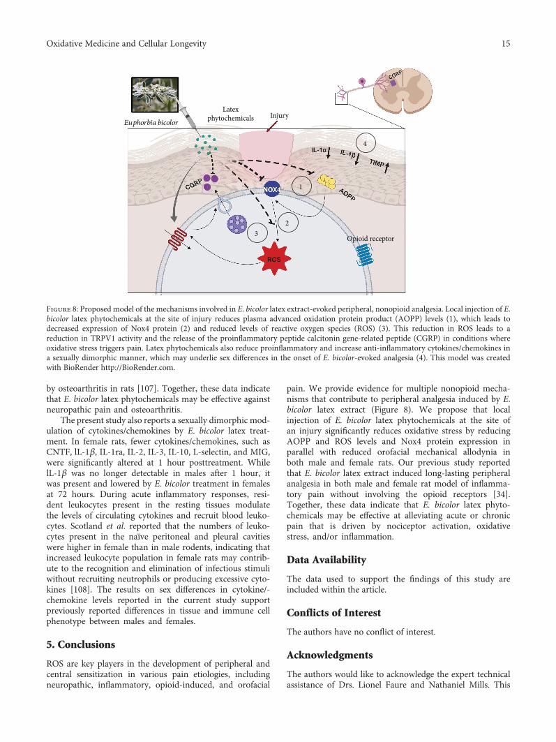
pain. We provide evidence for multiple nonopioid mecha-nisms that contribute to peripheral analgesia induced by E.bicolor latex extract (Figure 8). We propose that localinjection of E. bicolor latex phytochemicals at the site ofan injury significantly reduces oxidative stress by reducingAOPP and ROS levels and Nox4 protein expression inparallel with reduced orofacial mechanical allodynia inboth male and female rats. Our previous study reportedthat E. bicolor latex extract induced long-lasting peripheralanalgesia in both male and female rat model of inflamma-tory pain without involving the opioid receptors [34].Together, these data indicate that E. bicolor latex phyto-chemicals may be effective at alleviating acute or chronicpain that is driven by nociceptor activation, oxidativestress, and/or inflammation.
Data Availability
The data used to support the findings of this study areincluded within the article.
Conflicts of Interest
The authors have no conflict of interest.
Acknowledgments
The authors would like to acknowledge the expert technicalassistance of Drs. Lionel Faure and Nathaniel Mills. This
4
InjuryLatex
phytochemicals
Opioid receptor
Euphorbia bicolor
1
23
Figure 8: Proposed model of the mechanisms involved in E. bicolor latex extract-evoked peripheral, nonopioid analgesia. Local injection of E.bicolor latex phytochemicals at the site of injury reduces plasma advanced oxidation protein product (AOPP) levels (1), which leads todecreased expression of Nox4 protein (2) and reduced levels of reactive oxygen species (ROS) (3). This reduction in ROS leads to areduction in TRPV1 activity and the release of the proinflammatory peptide calcitonin gene-related peptide (CGRP) in conditions whereoxidative stress triggers pain. Latex phytochemicals also reduce proinflammatory and increase anti-inflammatory cytokines/chemokines ina sexually dimorphic manner, which may underlie sex differences in the onset of E. bicolor-evoked analgesia (4). This model was createdwith BioRender http://BioRender.com.
15Oxidative Medicine and Cellular Longevity
research was supported by TWU Research EnhancementGrants awarded to CM and DLA, a TWU Center for StudentResearch Small Grant awarded to PB, and a TWU QualityEnhancement Program Learn by Doing Grant awarded toPB.
References
[1] R. B. Griggs, R. R. Donahue, B. G. Adkins, K. L. Anderson,O. Thibault, and B. K. Taylor, “Pioglitazone inhibits thedevelopment of hyperalgesia and sensitization of spinalnociresponsive neurons in type 2 diabetes,” The Journal ofPain, vol. 17, no. 3, pp. 359–373, 2016.
[2] J. Y. Moon, S. R. Choi, D. H. Roh et al., “Spinal sigma-1receptor activation increases the production of D-serinein astrocytes which contributes to the development ofmechanical allodynia in a mouse model of neuropathicpain,” Pharmacological Research, vol. 100, pp. 353–364,2015.
[3] J. Yowtak, K. Y. Lee, H. Y. Kim et al., “Reactive oxygen speciescontribute to neuropathic pain by reducing spinal GABArelease,” Pain, vol. 152, no. 4, pp. 844–852, 2011.
[4] D. Salvemini and W. L. Neumann, “Peroxynitrite: a strategiclinchpin of opioid analgesic tolerance,” Trends in Pharmaco-logical Sciences, vol. 30, no. 4, pp. 194–202, 2009.
[5] D. Salvemini and W. Neumann, “Targeting peroxynitritedriven nitroxidative stress with synzymes: a novel therapeuticapproach in chronic pain management,” Life Sciences, vol. 86,no. 15-16, pp. 604–614, 2010.
[6] T. Doyle, A. Finley, Z. Chen, and D. Salvemini, “Role for per-oxynitrite in sphingosine-1-phosphate-induced hyperalgesiain rats,” Pain, vol. 152, no. 3, pp. 643–648, 2011.
[7] D. Salvemini, J. W. Little, T. Doyle, and W. L. Neumann,“Roles of reactive oxygen and nitrogen species in pain,”Free Radical Biology & Medicine, vol. 51, no. 5, pp. 951–966, 2011.
[8] D. R. de Sotillo, A. M. Velly, M. Hadley, and J. R. Fricton,“Evidence of oxidative stress in temporomandibular disor-ders: a pilot study,” Journal of Oral Rehabilitation, vol. 38,no. 10, pp. 722–728, 2011.
[9] C. de Almeida and J. M. Amenabar, “Changes in the salivaryoxidative status in individuals with temporomandibular dis-orders and pain,” Journal of Oral Biology and CraniofacialResearch, vol. 6, Supplement 1, pp. S1–S4, 2016.
[10] T. Yoshida, R. Inoue, T. Morii et al., “Nitric oxide activatesTRP channels by cysteine S-nitrosylation,” Nature ChemicalBiology, vol. 2, no. 11, pp. 596–607, 2006.
[11] M. Ibi, K. Matsuno, D. Shiba et al., “Reactive oxygen speciesderived from NOX1/NADPH oxidase enhance inflammatorypain,” The Journal of Neuroscience, vol. 28, no. 38, pp. 9486–9494, 2008.
[12] M. Naziroglu, B. Cig, and C. Ozgul, “Neuroprotectioninduced by N-acetylcysteine against cytosolic glutathionedepletion-induced Ca2+ influx in dorsal root ganglion neu-rons of mice: role of TRPV1 channels,” Neuroscience,vol. 242, pp. 151–160, 2013.
[13] R. Ding, H. Jiang, B. Sun et al., “Advanced oxidation proteinproducts sensitized the transient receptor potential vanilloid1 via NADPH oxidase 1 and 4 to cause mechanical hyperalge-sia,” Redox Biology, vol. 10, pp. 1–11, 2016.
[14] V. Witko-Sarsat, M. Friedlander, C. Capeillère-Blandin et al.,“Advanced oxidation protein products as a novel marker ofoxidative stress in uremia,” Kidney International, vol. 49,no. 5, pp. 1304–1313, 1996.
[15] X. F. Wei, Q. G. Zhou, F. F. Hou, B. Y. Liu, and M. Liang,“Advanced oxidation protein products induce mesangial cellperturbation through PKC-dependent activation of NADPHoxidase,” American Journal of Physiology-Renal Physiology,vol. 296, no. 2, pp. F427–F437, 2009.
[16] Y. B. Im, M. K. Jee, J. I. Choi, H. T. Cho, O. H. Kwon, andS. K. Kang, “Molecular targeting of NOX4 for neuropathicpain after traumatic injury of the spinal cord,” Cell Death &Disease, vol. 3, no. 11, article e426, 2012.
[17] S. Poole, F. Q. Cunha, S. Selkirk, B. B. Lorenzetti, and S. H.Ferreira, “Cytokine-mediated inflammatory hyperalgesialimited by interleukin-10,” British Journal of Pharmacology,vol. 115, no. 4, pp. 684–688, 1995.
[18] G. D. Nicol, J. C. Lopshire, and C. M. Pafford, “Tumor necro-sis factor enhances the capsaicin sensitivity of rat sensoryneurons,” The Journal of Neuroscience, vol. 17, no. 3,pp. 975–982, 1997.
[19] L. S. Sorkin, W.-H. Xiao, R. Wagner, and R. R. Myers,“Tumour necrosis factor-α induces ectopic activity in noci-ceptive primary afferent fibres,” Neuroscience, vol. 81, no. 1,pp. 255–262, 1997.
[20] H. Junger and L. S. Sorkin, “Nociceptive and inflammatoryeffects of subcutaneous TNFα,” Pain, vol. 85, no. 1,pp. 145–151, 2000.
[21] O. Obreja, P. K. Rathee, K. S. Lips, C. Distler, and M. Kress,“IL-1β potentiates heat-activated currents in rat sensory neu-rons: involvement of IL-1RI, tyrosine kinase, and proteinkinase C,” The FASEB Journal, vol. 16, no. 12, pp. 1497–1503, 2002.
[22] M. Schafers and L. Sorkin, “Effect of cytokines on neuronalexcitability,” Neuroscience Letters, vol. 437, no. 3, pp. 188–193, 2008.
[23] R. J. Miller, H. Jung, S. K. Bhangoo, and F. A. White, “Cyto-kine and chemokine regulation of sensory neuron function,”Handbook of Experimental Pharmacology, vol. 194, pp. 417–449, 2009.
[24] S. Hensellek, P. Brell, H. G. Schaible, R. Bräuer, andG. Segond von Banchet, “The cytokine TNFalpha increasesthe proportion of DRG neurones expressing the TRPV1receptor via the TNFR1 receptor and ERK activation,”Molec-ular and Cellular Neurosciences, vol. 36, no. 3, pp. 381–391,2007.
[25] A. A. Khan, A. Diogenes, N. A. Jeske, M. A. Henry,A. Akopian, and K. M. Hargreaves, “Tumor necrosis factorα enhances the sensitivity of rat trigeminal neurons to capsa-icin,” Neuroscience, vol. 155, no. 2, pp. 503–509, 2008.
[26] H. Van Sam, P. Baas, and P. J. A. Kessler, “Traditional medic-inal plants in Ben En National Park, Vietnam,” Blumea - Bio-diversity, Evolution and Biogeography of Plants, vol. 53, no. 3,pp. 569–601, 2008.
[27] Q. W. Shi, X. H. Su, and H. Kiyota, “Chemical and phar-macological research of the plants in genus Euphorbia,”Chemical Reviews, vol. 108, no. 10, pp. 4295–4327, 2008.
[28] M. J. Iadarola and G. L. Gonnella, “Resiniferatoxin for paintreatment: an interventional approach to personalized painmedicine,” The Open Pain Journal, vol. 6, no. 1, pp. 95–107,2013.
16 Oxidative Medicine and Cellular Longevity

[29] A. Upadhyay, P. Chattopadhyay, D. Goyary, P. M. Mazum-der, and V. Veer, “Anti-inflammatory Effect of Euphorbiahirta Leaf Extract in Rats and Modulation of Inflammation-Associated Prostaglandins (PGE-2) and Nitric Oxide (NO)Expression in RAW264.7 Macrophage,” Journal of Pharma-ceutical Sciences and Pharmacology, vol. 1, no. 1, pp. 68–73,2014.
[30] A. Vasas and J. Hohmann, “Euphorbia diterpenes: isolation,structure, biological activity, and synthesis (2008-2012),”Chemical Reviews, vol. 114, no. 17, pp. 8579–8612, 2014.
[31] M. Majid, M. R. Khan, N. A. Shah et al., “Studies on phyto-chemical, antioxidant, anti-inflammatory and analgesic activ-ities of Euphorbia dracunculoides,” BMC Complementaryand Alternative Medicine, vol. 15, no. 1, article 349, 2015.
[32] P. Palit, D. Mukherjee, P. Mahanta et al., “Attenuation ofnociceptive pain and inflammatory disorders by total steroidand terpenoid fraction of Euphorbia tirucalli Linn root inexperimental in vitro and in vivo model,” Inflammopharma-cology, vol. 26, no. 1, pp. 235–250, 2018.
[33] J. Sdayria, I. Rjeibi, A. Feriani et al., “Chemical Compositionand Antioxidant, Analgesic, and Anti-Inflammatory Effectsof Methanolic Extract of Euphorbia retusa in Mice,” PainResearch &Management, vol. 2018, article 4838413, 11 pages,2018.
[34] P. Basu, S. A. Tongkhuya, T. L. Harris et al., “Euphorbiabicolor (Euphorbiaceae) latex phytochemicals induce long-lasting non-opioid peripheral analgesia in a rat model ofinflammatory pain,” Frontiers in Pharmacology, vol. 10,p. 958, 2019.
[35] V. L. Singleton, R. Orthofer, and R. M. Lamuela-Raventos,“Analysis of total phenols and other oxidation substratesand antioxidants by means of Folin-Ciocalteu reagent,” Oxi-dants and Antioxidants, Part A, vol. 299, pp. 152–178, 1999.
[36] A. Ordonez, J. Gomez, M. Vattuone, and M. Lsla, “Antioxi-dant activities of Sechium edule (Jacq.) Swartz extracts,” FoodChemistry, vol. 97, no. 3, pp. 452–458, 2006.
[37] O. A. Aiyegoro and A. I. Okoh, “Preliminary phytochemicalscreening and in vitro antioxidant activities of the aqueousextract of Helichrysum longifolium DC,” BMC Complemen-tary and Alternative Medicine, vol. 10, no. 1, article 21, 2010.
[38] N. Ghorai, N. Ghorai, S. Chakraborty, S. Gucchait, S. K. Saha,and S. Biswas, “Estimation of total terpenoids concentrationin plant tissues using a monoterpene, linalool as standardreagent,” Protocol Exchange, vol. 5, no. 10, p. 1038, 2012.
[39] P. Basu and C. Maier, “In vitro antioxidant activities andpolyphenol contents of seven commercially available fruits,”Pharmacognosy Research, vol. 8, no. 4, pp. 258–264, 2016.
[40] M. Oyaizu, “Studies on products of browning reaction - anti-oxidative activities of products of browning reaction preparedfrom glucosamine,” The Japanese Journal of Nutrition andDietetics, vol. 44, no. 6, pp. 307–315, 1986.
[41] R. Re, N. Pellegrini, A. Proteggente, A. Pannala, M. Yang, andC. Rice-Evans, “Antioxidant activity applying an improvedABTS radical cation decolorization assay,” Free RadicalBiology & Medicine, vol. 26, no. 9-10, pp. 1231–1237,1999.
[42] I. Gulcin, E. Kirecci, E. Akkemik, F. Topal, and O. Hisar,“Antioxidant, antibacterial, and anticandidal activities of anaquatic plant: duckweed (Lemna minor L., Lemnaceae),”Turkish Journal of Biology, vol. 34, no. 2, pp. 175–188,2010.
[43] R. J. Ruch, S.-j. Cheng, and J. E. Klaunig, “Prevention of cyto-toxicity and inhibition of intercellular communication byantioxidant catechins isolated from Chinese green tea,” Car-cinogenesis, vol. 10, no. 6, pp. 1003–1008, 1989.
[44] N. Balakrishnan, A. B. Panda, N. R. Raj, A. Shrivastava, andR. Prathani, “The evaluation of nitric oxide scavenging activ-ity of Acalypha Indica Linn root,” Asian Journal of Researchin Chemistry, vol. 2, pp. 148–150, 2009.
[45] K. Ren and R. Dubner, “Inflammatory models of pain andhyperalgesia,” ILAR Journal, vol. 40, no. 3, pp. 111–118, 1999.
[46] M. K. Chung, J. Asgar, J. Lee, M. S. Shim, C. Dumler, and J. Y.Ro, “The role of TRPM2 in hydrogen peroxide-inducedexpression of inflammatory cytokine and chemokine in rattrigeminal ganglia,” Neuroscience, vol. 297, pp. 160–169,2015.
[47] A. Behndig, “Corneal endothelial integrity in aging mice lack-ing superoxide dismutase-1 and/or superoxide dismutase-3,”Molecular Vision, vol. 14, pp. 2025–2030, 2008.
[48] T. Grosser, E. Smyth, and G. A. FitzGerald, “Anti-inflamma-tory, antipyretic, and analgesic agents; pharmacotherapy ofgout,” Goodman and Gilman's the pharmacological basis oftherapeutics, vol. 12, pp. 959–1004, 2011.
[49] A. Farshchi, G. Ghiasi, P. Malek Khatabi, H. Farzaee, andA. Niayesh, “Antinociceptive effect of promethazine in mice,”Iranian Journal of Basic Medical Sciences, vol. 12, no. 3-4,pp. 140–145, 2009.
[50] A. M. Vittalrao, T. Shanbhag, M. Kumari, K. L. Bairy, andS. Shenoy, “Evaluation of antiinflammatory and analgesicactivities of alcoholic extract of Kaempferia galanga in rats,”Indian Journal of Physiology and Pharmacology, vol. 55,no. 1, pp. 13–24, 2011.
[51] J. D. Phillipson, “Phytochemistry and medicinal plants,” Phy-tochemistry, vol. 56, no. 3, pp. 237–243, 2001.
[52] N. Kumara, “Identification of strategies to improve researchon medicinal plants used in Sri Lanka,” in In WHO Sympo-sium, pp. 12–14, University of Ruhuna, Galle, Sri Lanka,2001.
[53] M. G. Dharmasiri, J. R. A. C. Jayakody, G. Galhena, S. S. P.Liyanage, and W. D. Ratnasooriya, “Anti-inflammatory andanalgesic activities of mature fresh leaves of Vitex negundo,”Journal of Ethnopharmacology, vol. 87, no. 2-3, pp. 199–206, 2003.
[54] R. W. Li, S. P. Myers, D. N. Leach, G. D. Lin, and G. Leach, “Across-cultural study: anti-inflammatory activity of Australianand Chinese plants,” Journal of Ethnopharmacology, vol. 85,no. 1, pp. 25–32, 2003.
[55] A. A. Basma, Z. Zakaria, L. Y. Latha, and S. Sasidharan,“Antioxidant activity and phytochemical screening of themethanol extracts of Euphorbia hirta L,” Asian PacificJournal of Tropical Medicine, vol. 4, no. 5, pp. 386–390,2011.
[56] N. Sharma, K. Samarakoon, R. Gyawali et al., “Evaluation ofthe antioxidant, anti-inflammatory, and anticancer activitiesof Euphorbia hirta ethanolic extract,” Molecules, vol. 19,no. 9, pp. 14567–14581, 2014.
[57] L. M. Ndam, A. M. Mih, A. S. Tening, A. G. N. Fongod, N. A.Temenu, and Y. Fujii, “Phytochemical analysis, antimicrobialand antioxidant activities of Euphorbia golondrina L.C.Wheeler (Euphorbiaceae Juss.): an unexploredmedicinal herbreported from Cameroon,” Springerplus, vol. 5, no. 1, article264, 2016.
17Oxidative Medicine and Cellular Longevity
[58] A. Floegel, D. O. Kim, S. J. Chung, S. I. Koo, and O. K. Chun,“Comparison of ABTS/DPPH assays to measure antioxidantcapacity in popular antioxidant-rich US foods,” Journal ofFood Composition and Analysis, vol. 24, no. 7, pp. 1043–1048, 2011.
[59] P. S. Siqueira-Lima, J. C. Silva, J. S. S. Quintans et al., “Naturalproducts assessed in animal models for orofacial pain - a sys-tematic review,” Revista Brasileira de Farmacognosia, vol. 27,no. 1, pp. 124–134, 2017.
[60] L. R. Bonjardim, A. M. Silva, M. G. B. Oliveira et al., “Sidacordifolia leaf extract reduces the orofacial nociceptiveresponse in mice,” Phytotherapy Research, vol. 25, no. 8,pp. 1236–1241, 2011.
[61] M. S. Paixão, M. S. Melo, M. G. B. Oliveira et al., “Hyptis pec-tinata: redox protection and orofacial antinociception,” Phy-totherapy Research, vol. 27, no. 9, pp. 1328–1333, 2013.
[62] M. S. Paixão, M. S. Melo, N. P. Damascena et al., “Hyptis pec-tinata gel prevents alveolar bone resorption in experimentalperiodontitis in rats,” Revista Brasileira de Farmacognosia,vol. 25, no. 1, pp. 35–41, 2015.
[63] A. C. B. de Lima, M. S. Paixão, M. Melo et al., “Orofacialantinociceptive effect and antioxidant properties of thehydroethanol extract of Hyptis fruticosa salmz ex Benth,”Journal of Ethnopharmacology, vol. 146, no. 1, pp. 192–197, 2013.
[64] A. M. Venâncio, M. Marchioro, C. S. Estavam et al., “Oci-mum basilicum leaf essential oil and (-)-linalool reduce oro-facial nociception in rodents: a behavioral andelectrophysiological approach,” Revista Brasileira de Farma-cognosia, vol. 21, no. 6, pp. 1043–1051, 2011.
[65] E. C. O. Nomura, M. R. A. Rodrigues, C. F. da Silva et al.,“Antinociceptive effects of ethanolic extract from the flowersof Acmella oleracea (L.) R.K. Jansen in mice,” Journal of Eth-nopharmacology, vol. 150, no. 2, pp. 583–589, 2013.
[66] J. S. S. Quintans, R. G. Brito, P. G. V. Aquino et al., “Antino-ciceptive activity of Syzygium cumini leaves ethanol extracton orofacial nociception protocols in rodents,” Pharmaceuti-cal Biology, vol. 52, no. 6, pp. 762–766, 2014.
[67] F. Kosova, B. Çetin, M. Akıncı et al., “Advanced oxidationprotein products, ferrous oxidation in xylenol orange, andmalondialdehyde levels in thyroid cancer,” Annals of SurgicalOncology, vol. 14, no. 9, pp. 2616–2620, 2007.
[68] F. Xie, S. Sun, A. Xu et al., “Advanced oxidation protein prod-ucts induce intestine epithelial cell death through a redox-dependent, c-jun N-terminal kinase and poly (ADP-ribose)polymerase-1-mediated pathway,” Cell Death & Disease,vol. 5, no. 1, article e1006, 2014.
[69] M. Skvarilova, A. Bulava, D. Stejskal, S. Adamovska, andJ. Bartek, “Increased level of advanced oxidation products(AOPP) as a marker of oxidative stress in patients with acutecoronary syndrome,” Biomedical Papers, vol. 149, no. 1,pp. 83–87, 2005.
[70] M. Kalousová, T. Zima, V. Tesař, and J. Lachmanová,“Advanced glycation end products and advanced oxidationprotein products in hemodialyzed patients,” Blood Purifica-tion, vol. 20, no. 6, pp. 531–536, 2002.
[71] P. Martín-Gallán, A. Carrascosa, M. Gussinyé, andC. Domínguez, “Biomarkers of diabetes-associated oxidativestress and antioxidant status in young diabetic patients withor without subclinical complications,” Free Radical Biology& Medicine, vol. 34, no. 12, pp. 1563–1574, 2003.
[72] K. Botsakis, S. Theodoritsi, K. Grintzalis et al., “17β-Estra-diol/N-acetylcysteine interaction enhances the neuroprotec-tive effect on dopaminergic neurons in the weaver model ofdopamine deficiency,” Neuroscience, vol. 320, pp. 221–229,2016.
[73] D. Shen, X. Tian, B. Zhang, and R. Song, “Mechanistic evalu-ation of neuroprotective effect of estradiol on rotenone and 6-OHDA induced Parkinson’s disease,” PharmacologicalReports, vol. 69, no. 6, pp. 1178–1185, 2017.
[74] S. Chaychi, A. Polosa, S. Chemtob, and P. Lachapelle, “Eval-uating the neuroprotective effect of 17β-estradiol in rodentmodels of oxidative retinopathies,”Documenta Ophthalmolo-gica, vol. 137, no. 3, pp. 151–168, 2018.
[75] H. Xiao, M. Deng, B. Yang, Z. Hu, and J. Tang, “Pretreatmentwith 17β-estradiol attenuates cerebral ischemia-inducedblood-brain barrier disruption in aged rats: involvement ofantioxidant signaling,” Neuroendocrinology, vol. 106, no. 1,pp. 20–29, 2018.
[76] J. Viña, C. Borrás, J. Gambini, J. Sastre, and F. V. Pallardó,“Why females live longer than males? Importance of theupregulation of longevity-associated genes by oestrogeniccompounds,” FEBS Letters, vol. 579, no. 12, pp. 2541–2545,2005.
[77] B. K. Tiwari, D. Kumar, A. B. Abidi, and S. I. Rizvi, “Efficacyof composite extract from leaves and fruits of medicinalplants used in traditional diabetic therapy against oxidativestress in alloxan-induced diabetic rats,” ISRN Pharmacology,vol. 2014, Article ID 608590, 7 pages, 2014.
[78] A. B. Younes, M. B. Salem, H. El Abed, and R. Jarraya, “Phy-tochemical screening and antidiabetic, antihyperlipidemic,and antioxidant properties of Anthyllis henoniana (Coss.)flowers extracts in an alloxan-induced rats model of diabe-tes,” Evidence-based Complementary and Alternative Medi-cine, vol. 2018, Article ID 8516302, 14 pages, 2018.
[79] X. Gao, H. K. Kim, J. Mo Chung, and K. Chung, “Reactiveoxygen species (ROS) are involved in enhancement ofNMDA-receptor phosphorylation in animal models of pain,”Pain, vol. 131, no. 3, pp. 262–271, 2007.
[80] J. Wang, V. Cochran, S. Abdi, J. M. Chung, K. Chung, andH. K. Kim, “Phenyl N-t-butylnitrone, a reactive oxygen spe-cies scavenger, reduces zymosan-induced visceral pain inrats,” Neuroscience Letters, vol. 439, no. 2, pp. 216–219, 2008.
[81] J. E. Linley, L. Ooi, L. Pettinger et al., “Reactive oxygen speciesare second messengers of neurokinin signaling in peripheralsensory neurons,” Proceedings of the National Academy ofSciences of the United States of America, vol. 109, no. 24,pp. E1578–E1586, 2012.
[82] K. Y. Niu, Y. Zhang, and J. Y. Ro, “Effects of gonadal hor-mones on the peripheral cannabinoid receptor 1 (CB1R) sys-tem under a myositis condition in rats,” Pain, vol. 153, no. 11,pp. 2283–2291, 2012.
[83] V. Nemudzivhadi and P. Masoko, “In Vitro Assessment ofCytotoxicity, Antioxidant, and Anti-Inflammatory Activitiesof Ricinus communis (Euphorbiaceae) Leaf Extracts,” Evi-dence-based Complementary and Alternative Medicine,vol. 2014, Article ID 625961, 8 pages, 2014.
[84] H. M. Choi and S. Y. Lim, “Effect of Euphorbia supina extracton antioxidant activity,” The Open Nutraceuticals Journal,vol. 7, no. 1, pp. 28–32, 2014.
[85] M. Skonieczna, T. Hejmo, A. Poterala-Hejmo, A. Cieslar-Pobuda, and R. J. Buldak, “NADPH oxidases: insights intoselected functions and mechanisms of action in cancer and
18 Oxidative Medicine and Cellular Longevity

stem cells,” Oxidative Medicine and Cellular Longevity,vol. 2017, Article ID 9420539, 15 pages, 2017.
[86] H. Suzuki, N. Hatano, Y. Muraki et al., “The NADPH oxidaseinhibitor diphenyleneiodonium activates the human TRPA1nociceptor,” American Journal of Physiology. Cell Physiology,vol. 307, no. 4, pp. C384–C394, 2014.
[87] X. Cao, X. Dai, L. M. Parker, and D. L. Kreulen, “Differentialregulation of NADPH oxidase in sympathetic and sensoryganglia in deoxycorticosterone acetate salt hypertension,”Hypertension, vol. 50, no. 4, pp. 663–671, 2007.
[88] F. De Logu, R. Nassini, S. Materazzi et al., “Schwann cellTRPA1 mediates neuroinflammation that sustainsmacrophage-dependent neuropathic pain in mice,” NatureCommunications, vol. 8, no. 1, article 1887, 2017.
[89] I. M. Marone, F. de Logu, R. Nassini et al., “TRPA1/NOX inthe soma of trigeminal ganglion neurons mediatesmigraine-related pain of glyceryl trinitrate in mice,” Brain,vol. 141, no. 8, pp. 2312–2328, 2018.
[90] I. Savini, R. Arnone, A. Rossi, M. V. Catani, D. del Principe,and L. Avigliano, “Redox modulation of Ecto-NOX1 inhuman platelets,” Molecular Membrane Biology, vol. 27,no. 4-6, pp. 160–169, 2010.
[91] V. Lakshminarayanan, D. W. A. Beno, R. H. Costa, and K. A.Roebuck, “Differential Regulation of Interleukin-8 and Inter-cellular Adhesion Molecule-1 by H2O2 and Tumor NecrosisFactor-α in Endothelial and Epithelial Cells,” The Journal ofBiological Chemistry, vol. 272, no. 52, pp. 32910–32918, 1997.
[92] J. Chen, M. Kinter, S. Shank, C. Cotton, T. J. Kelley, and A. G.Ziady, “Dysfunction of Nrf-2 in CF epithelia leads to excessintracellular H2O2 and inflammatory cytokine production,”PLoS One, vol. 3, no. 10, article e3367, 2008.
[93] F. Ma, L. Zhang, and K. N. Westlund, “Reactive oxygen spe-cies mediate TNFR1 increase after TRPV1 activation inmouse DRG neurons,” Molecular Pain, vol. 5, pp. 1744–8069, 2009.
[94] J. Hawkins and P. Durham, “Prolonged jaw opening pro-motes nociception and enhanced cytokine expression,”Journal of Oral & Facial Pain and Headache, vol. 30,no. 1, pp. 34–41, 2016.
[95] Z. L. Durham, J. L. Hawkins, and P. L. Durham, “Tumornecrosis factor-alpha stimulates cytokine expression andtransient sensitization of trigeminal nociceptive neurons,”Archives of Oral Biology, vol. 75, pp. 100–106, 2017.
[96] A. M. Binshtok, H. Wang, K. Zimmermann et al., “Nocicep-tors are interleukin-1beta sensors,” The Journal of Neurosci-ence, vol. 28, no. 52, pp. 14062–14073, 2008.
[97] F. Richter, G. Natura, M. Ebbinghaus et al., “Interleukin-17sensitizes joint nociceptors to mechanical stimuli and con-tributes to arthritic pain through neuronal interleukin-17receptors in rodents,” Arthritis and Rheumatism, vol. 64,no. 12, pp. 4125–4134, 2012.
[98] A. E. Koch, S. L. Kunkel, L. A. Harlow et al., “Macrophageinflammatory protein-1 alpha. A novel chemotactic cytokinefor macrophages in rheumatoid arthritis,” The Journal ofClinical Investigation, vol. 93, no. 3, pp. 921–928, 1994.
[99] Y. Hatano, T. Kasama, H. Iwabuchi et al., “Macrophageinflammatory protein 1 alpha expression by synovial fluidneutrophils in rheumatoid arthritis,” Annals of the RheumaticDiseases, vol. 58, no. 5, pp. 297–302, 1999.
[100] T. Matsui, T. Akahoshi, R. Namai et al., “Selective recruit-ment of CCR6‐expressing cells by increased production of
MIP‐3α in rheumatoid arthritis,” Clinical and ExperimentalImmunology, vol. 125, no. 1, pp. 155–161, 2001.
[101] R. García-Vicuña, M. V. Gómez-Gaviro, M. J. Domínguez-Luis et al., “CC and CXC chemokine receptors mediatemigration, proliferation, and matrix metalloproteinase pro-duction by fibroblast-like synoviocytes from rheumatoidarthritis patients,” Arthritis and Rheumatism, vol. 50,no. 12, pp. 3866–3877, 2004.
[102] T. Tsubaki, S. Takegawa, H. Hanamoto et al., “Accumulationof plasma cells expressing CXCR3 in the synovial subliningregions of early rheumatoid arthritis in association with pro-duction of Mig/CXCL9 by synovial fibroblasts,” Clinical andExperimental Immunology, vol. 141, no. 2, pp. 363–371, 2005.
[103] P. Rathanaswami, M. Hachicha, M. Sadick, T. J. Schall, andS. R. McColl, “Expression of the cytokine RANTES in humanrheumatoid synovial fibroblasts. Differential regulation ofRANTES and interleukin-8 genes by inflammatory cyto-kines,” The Journal of Biological Chemistry, vol. 268, no. 8,pp. 5834–5839, 1993.
[104] E. Robinson, E. C. Keystone, T. J. Schall, N. Gillett, and E. N.Fish, “Chemokine expression in rheumatoid arthritis (RA):evidence of RANTES and macrophage inflammatory protein(MIP)‐1β production by synovial T cells,” Clinical and Exper-imental Immunology, vol. 101, no. 3, pp. 398–407, 1995.
[105] P. A. Klimiuk, M. Fiedorczyk, S. Sierakowski, andJ. Chwiecko, “Soluble cell adhesion molecules (sICAM‐1,sVCAM‐1, and sE‐selectin) in patients with early rheumatoidarthritis,” Scandinavian Journal of Rheumatology, vol. 36,no. 5, pp. 345–350, 2007.
[106] Y. Kawasaki, Z. Z. Xu, X.Wang et al., “Distinct roles of matrixmetalloproteases in the early- and late-phase development ofneuropathic pain,” Nature Medicine, vol. 14, no. 3, pp. 331–336, 2008.
[107] M. J. Janusz, A. M. Bendele, K. K. Brown, Y. O. Taiwo,L. Hsieh, and S. A. Heitmeyer, “Induction of osteoarthritisin the rat by surgical tear of the meniscus: inhibition of jointdamage by a matrix metalloproteinase inhibitor,” Osteoar-thritis and Cartilage, vol. 10, no. 10, pp. 785–791, 2002.
[108] R. S. Scotland, M. J. Stables, S. Madalli, P. Watson, and D. W.Gilroy, “Sex differences in resident immune cell phenotypeunderlie more efficient acute inflammatory responses infemale mice,” Blood, vol. 118, no. 22, pp. 5918–5927, 2011.
19Oxidative Medicine and Cellular Longevity

Stem Cells International
Hindawiwww.hindawi.com Volume 2018
Hindawiwww.hindawi.com Volume 2018
MEDIATORSINFLAMMATION
of
EndocrinologyInternational Journal of
Hindawiwww.hindawi.com Volume 2018
Hindawiwww.hindawi.com Volume 2018
Disease Markers
Hindawiwww.hindawi.com Volume 2018
BioMed Research International
OncologyJournal of
Hindawiwww.hindawi.com Volume 2013
Hindawiwww.hindawi.com Volume 2018
Oxidative Medicine and Cellular Longevity
Hindawiwww.hindawi.com Volume 2018
PPAR Research
Hindawi Publishing Corporation http://www.hindawi.com Volume 2013Hindawiwww.hindawi.com
The Scientific World Journal
Volume 2018
Immunology ResearchHindawiwww.hindawi.com Volume 2018
Journal of
ObesityJournal of
Hindawiwww.hindawi.com Volume 2018
Hindawiwww.hindawi.com Volume 2018
Computational and Mathematical Methods in Medicine
Hindawiwww.hindawi.com Volume 2018
Behavioural Neurology
OphthalmologyJournal of
Hindawiwww.hindawi.com Volume 2018
Diabetes ResearchJournal of
Hindawiwww.hindawi.com Volume 2018
Hindawiwww.hindawi.com Volume 2018
Research and TreatmentAIDS
Hindawiwww.hindawi.com Volume 2018
Gastroenterology Research and Practice
Hindawiwww.hindawi.com Volume 2018
Parkinson’s Disease
Evidence-Based Complementary andAlternative Medicine
Volume 2018Hindawiwww.hindawi.com
Submit your manuscripts atwww.hindawi.com

![(Euphorbia esula L.) - ARS Home : USDA ARS. NPJ_11...The survey included most of the 2003 Deep Fire Burn. Survey data were used to determine leafy spurge ( Euphorbia esula L. [Euphorbiaceae])](https://img.dokumen.tips/doc/110x75/5b5ae9767f8b9a885b8cf212/euphorbia-esula-l-ars-home-usda-ars-npj11the-survey-included-most-of.jpg)