Embed Size (px)
Citation preview

MOLECULAR AND CELLULAR BIOLOGY, Jan. 2003, p. 687–698 Vol. 23, No. 20270-7306/03/$08.00�0 DOI: 10.1128/MCB.23.2.687–698.2003Copyright © 2003, American Society for Microbiology. All Rights Reserved.
Eukaryotic Initiation Factors 4G and 4A Mediate ConformationalChanges Downstream of the Initiation Codon of the
Encephalomyocarditis Virus InternalRibosomal Entry Site
Victoria G. Kolupaeva,1 Ivan B. Lomakin,1 Tatyana V. Pestova,1,2
and Christopher U. T. Hellen1*Department of Microbiology and Immunology, State University of New York Downstate Medical Center,
Brooklyn, New York 11203,1 and A. N. Belozersky Institute of Physico-Chemical Biology,Moscow State University, 119899 Moscow, Russia2
Received 10 July 2002/Returned for modification 9 September 2002/Accepted 11 October 2002
Initiation of translation of encephalomyocarditis virus mRNA is mediated by an internal ribosome entry site(IRES) comprising structural domains H, I, J-K, and L immediately upstream of the initiation codon AUG atnucleotide 834 (AUG834). Assembly of 48S ribosomal complexes on the IRES requires eukaryotic initiationfactor 2 (eIF2), eIF3, eIF4A, and the central domain of eIF4G to which eIF4A binds. Footprinting experimentsconfirmed that eIF4G binds a three-way helical junction in the J-K domain and showed that it interactsextensively with RNA duplexes in the J-K and L domains. Deletion of apical hairpins in the J and K domainssynergistically impaired the binding of eIF4G and IRES function. Directed hydroxyl radical probing, done byusing Fe(II) tethered to surface residues in eIF4G’s central domain, indicated that it is oriented with its Nterminus towards the base of domain J and its C terminus towards the apex. eIF4G recruits eIF4A to a definedlocation on the IRES, and the eIF4G/eIF4A complex caused localized ATP-independent conformationalchanges in the eIF4G-binding region of the IRES. This complex also induced more extensive conformationalrearrangements at the 3� border of the ribosome binding site that required ATP and active eIF4A. We proposethat these conformational changes prepare the region flanking AUG834 for productive binding of the ribosome.
Translation of a subset of cellular and viral mRNAs is ini-tiated by end-independent binding of ribosomes to an internalribosome entry site (IRES) in the 5� untranslated region.IRESs were first identified in different picornavirus RNA ge-nomes, and these IRESs are now classified into two majorgroups, one of which includes those of the encephalomyocar-ditis virus (EMCV), Theiler’s murine encephalitis virus(TMEV), and foot-and-mouth disease virus (FMDV) (17).The EMCV IRES is �450 nucleotides (nt) long and comprisesH, I, J-K, and L structural domains upstream of the initiationcodon AUG at nucleotide 834 (AUG834). Ribosomal initiationcomplexes attach directly to AUG834, and initiation does notinvolve scanning (19, 35). Recent analysis has begun to resolvemolecular details of the mechanism of initiation on EMCV-like IRESs. Viral and cellular IRES-containing mRNAs can betranslated under conditions when the cap-mediated mode ofend-dependent initiation that is used by most mRNAs is in-hibited (12), and studies of initiation on viral IRESs are there-fore yielding insights into mechanisms that enable selectivetranslation of mRNAs in the cell.
The stage that differentiates IRES-mediated initiation fromthe conventional cap-mediated mode of initiation is the mech-anism by which the 43S ribosomal preinitiation complex en-
gages mRNA to form a 48S complex at the initiation codon(16, 36). The 43S complex consists of a 40S ribosomal subunit,initiator tRNA, and eukaryotic initiation factors (eIFs), includ-ing eIF2 and eIF3. In the process of cap-mediated initiation,eIF4F is thought to create a binding site for this complexadjacent to the 5�-terminal m7G cap by inducing local confor-mational changes in bound mRNA (42) and by enhancing thebinding of the eIF3 component of 43S complexes to mRNA(5). The 43S complex binds to the mRNA through a network ofprotein-protein and protein-RNA interactions and then scansto the initiation codon. Many of these interactions involveeIF4F, which comprises the cap-binding protein eIF4E, theRNA helicase eIF4A, and either the eIF4GI or eIF4GII iso-form. The amino-terminal one-third of eIF4G (amino acidresidues 1 to 675) binds eIF4E and the poly(A) binding protein(10, 24). The central one-third of eIF4G, which binds to eIF3and contains one of the two eIF4A-binding sites, consists offive pairs of �-helices that are known as HEAT repeats (29).
Biochemical reconstitution of 48S complex formation frompurified components on the EMCV IRES has shown that thisprocess is ATP dependent and that it requires the same initi-ation factors as cap-dependent initiation, except for eIF1,eIF1A, and intact eIF4F (34, 35, 38). Similar results have beendescribed for FMDV and TMEV IRESs (40). 48S complexesassembled in this way are competent to complete all remainingsteps in initiation (T. V. Pestova, unpublished data). Initiationon the EMCV IRES has no requirement for eIF4E, and eIF4Fcan be replaced by eIF4A and a central domain of eIF4G,namely, eIF4GI, located from amino acid residues 737 to 989
* Corresponding author. Mailing address: Department of Microbi-ology and Immunology, State University of New York DownstateMedical Center, 450 Clarkson Ave., Brooklyn, NY 11203. Phone: (718)270-1034. Fax: (718) 270-2656. E-mail: [email protected].
687
Dow
nloa
ded
from
http
s://j
ourn
als.
asm
.org
/jour
nal/m
cb o
n 26
Nov
embe
r 20
21 b
y 27
.133
.9.9
4.

(eIF4GI737-989) (26). The requirement for eIF4A and eIF4G isconsistent with the profound inhibition of EMCV and FMDVtranslation by dominant-negative mutant forms of eIF4A, suchas the R362Q substitution mutant, which sequester the eIF4A/eIF4G complex in an inactive form (33, 47). Initiation on theEMCV IRES does not involve scanning, and the basis for therequirement for ATP and for active eIF4A for initiation on theIRES is not known. eIF4A binds to the central �-helical do-mains of eIF4GI737-989 and eIF4GII745-1003 and greatly en-hances their affinity for the IRES in an ATP-independentmanner (26, 29).
In addition to requiring canonical initiation factors, assem-bly of 48S complexes on some picornavirus IRESs is enhancedby IRES trans-acting factors that bind to defined sites on themand induce conformational changes in these IRESs that facil-itate binding of the eIF4G/eIF4A complex (3, 20, 35, 37, 40).IRESs are therefore not static scaffolds but undergo confor-mational transitions as they bind components of the translationapparatus during the initiation process. Initiation on the IRESof the EMCV strain used in the experiments described heredoes not require IRES trans-acting factors (35). However, theEMCV IRES becomes dependent on the pyrimidine tract-binding protein as a result of mutations either in the domain ofthe IRES to which eIF4G binds or in the coding region adja-cent to the initiation codon (20).
EMCV-like IRESs have conserved structures that arethought to orient binding sites for components of the transla-tion apparatus to promote recruitment of the 43S complex andconsequent assembly of the 48S complex (17, 37). eIF4G bindsspecifically to the J-K domain of these IRESs, and this inter-action is necessary for initiation (22, 26, 27, 29, 38, 40, 46). Thebinding site on the EMCV IRES has been localized by foot-printing (22); residues are protected in the A-rich loop at thejunction of the J and K domains and in the flanking J2, J3, andK1 helices (Fig. 1). Deletion of nucleotides in the A-rich loopseverely reduces both binding of eIF4G/eIF4A and IRES-me-diated initiation (35). Although the J-K domain is essential, itis by itself not sufficient for EMCV IRES function, and exper-iments to date account only for the function of the central coreof this domain. The extensive sequence conservation inEMCV-like IRESs extends over much of the J-K domain (18),including regions such as the terminal loops of the J and Kdomains and the J3 helix that have been found to be importantfor initiation (13, 32, 48). The adjacent domain I also plays anessential but as yet undefined role in EMCV IRES function,and the flanking domain H, domain L, and sequences down-stream of domain L all also play accessory roles (7, 9, 18, 20).The molecular basis for the importance of these regions of theIRES has not yet been determined.
The interaction of the eIF4G/eIF4A complex with theEMCV IRES is therefore of central importance in this initia-tion mechanism, but important details of its function remainunresolved. We conducted a series of experiments to deter-mine the orientation of the central HEAT-repeat domain ofeIF4G on the J-K domain by using directed hydroxyl radicalcleavage to characterize the nucleotide determinants in theIRES required for binding the eIF4G/eIF4A complex and toinvestigate the consequences on the conformation of the IRESand downstream coding sequences of binding to this complexin the presence and absence of ATP. We report that eIF4G
makes much more extensive interactions with the J-K and Ldomains when it binds to the IRES than was previously rec-ognized. The integrity of the terminal hairpins of J and Kdomains is necessary for stable binding of eIF4G and eIF4F tothe EMCV IRES and for initiation on it. The bound eIF4G/eIF4A complex induced conformational changes in the codingregion adjacent to the IRES that are ATP dependent and thatrequire eIF4A to be active. We propose that these conforma-tional changes in the IRES mediated by eIF4G/eIF4A preparethe region of the IRES flanking the initiation codon so that the40S subunit can bind to it productively, leading to initiation oftranslation.
MATERIALS AND METHODS
Enzymes and reagents. DNA restriction endonucleases and modifying en-zymes were from New England BioLabs (Beverly, Mass.). Rabbit reticulocytelysate (RRL) for in vitro translation and avian myeloblastosis virus reversetranscriptase (AMV-RT) were purchased from Promega Corp. (Madison, Wis.).RRL for purification of ribosomes and initiation factors was from Green Hect-ares (Oregon, Wis.). Native rabbit tRNA was from Novagen (Madison, Wis.).Unlabeled nucleoside triphosphates, RNAguard RNase inhibitor, and RNase V1were from Amersham Biosciences (Piscataway, N.J.). Dimethyl sulfate (DMS)was from Aldrich (Milwaukee, Wis.). Radiochemicals [35S]methionine (44 TBq/mmol), 35S-dATP (37 TBq/mmol), and [32P]dATP (220 TBq/mmol) were fromICN Radiochemicals (Irvine, Calif.). Escherichia coli methionyl-tRNA synthetasefrom E. coli strain MRE 600, ribosomal 40S subunits, and native and recombi-nant initiation factors were purified as described previously (26, 29, 34, 35, 37,38).
Plasmids. EMCV pTE1 (9), pTE3-GUS (2), and pJK (26) transcription vec-tors and vectors for expression of recombinant wild-type eIF4A (35), R362Qmutant eIF4A (37), eIF4GI737-1116, eIF4GI737-1009, and eIF4GI737-1600 (26) andeIF4GII745-1003 (29) have been described previously. The eIF4GI plasmids havebeen renamed to take into account a recent revision to the sequence of thelargest eIF4GI isoform, extending its amino terminus by 40 amino acid residues(4). Deletion mutants of the EMCV IRES were generated in pTE3-GUS andpJK plasmids by using a two-step PCR. Substitution mutations in eIF4GI737-1116
were made exactly as described previously (26). All mutations were confirmed bysequencing the complete IRES or the eIF4GI737-1116 coding sequence, as ap-propriate.
In vitro translation. mRNA comprising the EMCV IRES linked to the �-glu-curonidase open reading frame was transcribed by using T7 RNA polymeraseand was translated in RRL in accordance with the manufacturer’s instructions inthe presence of [35S]methionine. Translation products were resolved by electro-phoresis with 12% polyacrylamide gel. Gels were dried and exposed to X-rayfilm. The efficiency of translation was quantified by using a Molecular DynamicsPhosphorImager.
Assembly and analysis of ribosomal complexes. Ribonucleoprotein and ribo-somal 48S complexes were assembled on EMCV RNAs and analyzed by primerextension by using the primers 5�-GTCAATAACTCCTCTGG-3� (complemen-tary to EMCV nt 957 to 974) and 5�-GGGGGATGTGCTGCAACC-3� (com-plementary to nt 368 to 351 in the polylinker of pTZ18R), as appropriate, andAMV-RT in the presence of [32P]dATP, as described previously (26, 35, 38).
Footprinting analysis of initiation factor-IRES complexes. Free or initiationfactor-bound RNAs in binding buffer were probed with DMS or RNase V1, andchemically modified or enzymatically cleaved RNAs were analyzed by reversetranscription, as described previously (21, 22, 40). ATP was present in all probingreactions that contained eIF4A or eIF4F. Hydroxyl radical footprinting was doneessentially as described previously (15).
Preparation of Fe(II)-BABE-derivatized eIF4G. Cysteine-containing mutantsof eIF4GI737-1116 (0.2 to 0.5 �g/�l in 95 �l of buffer containing 80 mM K�-HEPES [pH 7.6], 300 mM KCl, and 10% glycerol) were conjugated with 5 �l of20 mM Fe(II)–bromoacetyl-amidobenzyl-EDTA (BABE) (Dojindo MolecularTechnologies, Inc., Gaithersburg, Md.) by incubation at 37°C for 30 min essen-tially as described previously (6). Unincorporated Fe-BABE was separated fromderivatized eIF4G by loading reaction mixtures onto pretreated MicroconYM-30 microconcentrators and washing four times with the incubation buffer.Fe-eIF4G samples were typically recovered in 50 to 80 �l of buffer. The acces-sibility of each introduced cysteine residue for derivatization and the efficiency ofthe Fe(II)-BABE tethering reaction were assessed by using the reaction with the
688 KOLUPAEVA ET AL. MOL. CELL. BIOL.
Dow
nloa
ded
from
http
s://j
ourn
als.
asm
.org
/jour
nal/m
cb o
n 26
Nov
embe
r 20
21 b
y 27
.133
.9.9
4.

thiol-specific fluorescent coumarin derivative DCIA (Molecular Probes, Eugene,Oreg.) as described previously (6).
Directed hydroxyl radical probing. Ribonucleoprotein complexes were assem-bled by incubating 0.2 �g of EMCV RNA (nt 280 to 974) in 40 �l of buffercontaining 80 mM K�-HEPES (pH 7.6), 300 mM KCl, 2 mM MgCl2, and 10%glycerol with 0.8 to 1 �g of Fe-eIF4G (and, where indicated, 1 �g of eIF4A) at37°C for 5 min and then placing them on ice. Fenton chemistry was initiated bythe addition of 2 �l of a freshly prepared mixture of 0.1 M ascorbic acid and 0.5%H2O2 to each sample to generate hydroxyl radicals in the vicinity of the tetheredFe(II). Reactions were quenched after incubation for 10 min in ice by theaddition of 20 �l of 20 mM thiourea. EMCV RNA was isolated immediately by
phenol extraction followed by ethanol precipitation, and sites of hydroxyl radicalcleavage were located by primer extension analysis with the primer 5�-CGGTATTGTAGAGCAGAGC-3� (complementary to EMCV nt 854 to 836) andAMV-RT in the presence of [32P]dATP.
RESULTS
In vitro translation of mRNAs containing mutations in theEMCV IRES J-K domain. To address the basis for the impor-tance of the terminal J5 and K2 hairpins for IRES activity,
FIG. 1. Interactions of eIF4A, eIF4B, and eIF4G with the EMCV IRES assessed by chemical and enzymatic footprinting. Sites at whichchemical modification by DMS and CMCT and enzymatic cleavage by RNase V1 of the IRES were altered by eIF4G737-1600 alone and in thepresence of eIFA/eIF4B are indicated by the symbols shown in the key at bottom right. Footprinting data for eIF4G alone are from previousanalyses (22) except for those for protection of A704 (see Fig. 4A). These data are mapped onto a secondary structure model of nt 449 to 904 ofthe EMCV IRES and the adjacent coding region. Helical segments of the J-K domain in this model are designated J1 to J5 and K1 and K2 (13).The initiation codon AUG834 is boxed. A proposed RNA structure (14) in the region encoding the leader (L) protein is included. Only the lowerpart of domain I is shown; the structure shown is that proposed previously (39). Nucleotides deleted in the �J5, �K2, and �K2� mutants areindicated by gray lines. The positions of the toe prints due to RT arrest in the J-K domain by bound eIF4G and downstream of the initiation codonby bound eIF4G/eIF4A are indicated by black and dashed arrows, respectively. The positions of toe prints �15 to �17 nt downstream of theinitiation codon caused by RT arrest by bound 48S complexes are indicated by gray arrows.
VOL. 23, 2003 INITIATION FACTOR-MEDIATED CHANGE IN IRES CONFORMATION 689
Dow
nloa
ded
from
http
s://j
ourn
als.
asm
.org
/jour
nal/m
cb o
n 26
Nov
embe
r 20
21 b
y 27
.133
.9.9
4.
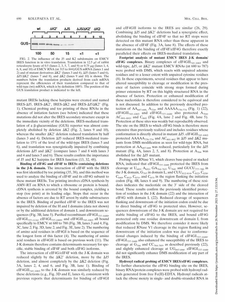
mutant IRESs lacking these hairpins were created and namedIRES-�J5, IRES-�K2�, IRES-�K2 and IRES-�J5�K2� (Fig.1). Chemical probing and toe printing of these RNAs in theabsence of initiation factors (see below) indicated that thesemutations did not alter the IRES secondary structure except inthe immediate vicinity of the deletions. IRES-mediated trans-lation of a �-glucuronidase (GUS) reporter was almost com-pletely abolished by deletion �K2 (Fig. 2, lanes 9 and 10),whereas the smaller �K2� deletion reduced translation by half(lanes 3 and 4). Deletion �J5 reduced IRES-mediated trans-lation to 15% of the level of the wild-type IRES (lanes 5 and6), and translation was synergistically impaired by combiningdeletions �J5 and �K2� (compare lanes 7 and 8 with lanes 3and 4 and lanes 5 and 6). These results confirm the importanceof J5 and K2 hairpins for IRES function (13, 32, 48).
Binding of eIF4G and eIF4F to IRESs containing deletionsin the J-K domain. The interaction of eIF4F with the IRESwas first identified by toe printing (35, 38), and this method wasused to analyze the binding of eIF4F and its eIF4G subunit tothese mutant IRESs. Toe printing involves cDNA synthesis byAMV-RT on RNA to which a ribosome or protein is bound.cDNA synthesis is arrested by the bound complex, yielding astop (toe print) at its leading edge. Stops that occur in theabsence of factors are due to RT arrest by stable substructuresin the IRES. Binding of purified eIF4F to the IRES was notimpaired by deletion of the H and I domains (data not shown)or by the additional deletion of domain L and downstream se-quences (Fig. 3B, lane 5). Purified recombinant eIF4GI737-1600,eIF4GI737-1116, eIF4GI737-1009, and eIF4GII745-1003 all boundspecifically to EMCV nt 680 to 788 (Fig. 3B, lanes 3 and 4; Fig.3C, lane 2; Fig. 3D, lane 2; and Fig. 3E, lane 2). The numberingof amino acid residues in eIF4GI is based on the sequence ofthe longest form of this factor (4). The numbering of aminoacid residues in eIF4GII is based on previous work (11). TheJ-K domain therefore contains determinants necessary for spe-cific, stable binding of eIF4F and both eIF4G isoforms.
The interaction of eIF4G/eIF4F with the J-K domain wasreduced slightly by the �K2� deletion, more by the �J5deletion, and almost completely by the �K2 deletion (Fig.3A, lanes 2, 4, and 6, and Fig. 3B, lane 1). Binding ofeIF4GII745-1003 to the J-K domain was similarly reduced bythese deletions (e.g., Fig. 3D and E, lanes 4), consistent withprevious reports that determinants for binding of eIF4GI
and eIF4GII isoforms to the IRES are similar (26, 29).Combining �J5 and �K2� deletions had a synergistic effect,abolishing the binding of eIF4F so that no RT stops weredetected on this mutant RNA other than those apparent inthe absence of eIF4F (Fig. 3A, lane 8). The effects of thesemutations on the binding of eIF4F/eIF4G therefore exactlyparalleled their effects on IRES-mediated translation.
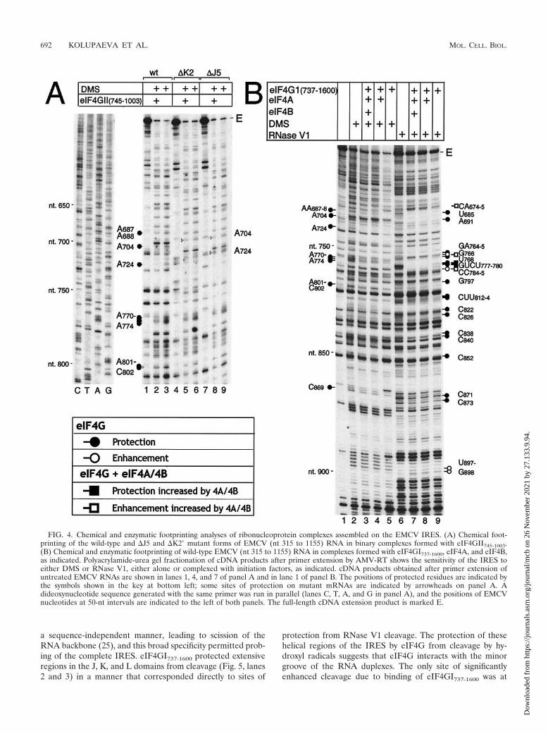
Footprint analysis of mutant EMCV IRES J-K domain/eIF4G complexes. Binary complexes of eIF4GII745-1003 andwild-type, �J5, or �K2� mutant EMCV RNAs (nt 680 to 787)were probed with DMS, which reacts with unpaired adenineresidues and to a lesser extent with unpaired cytosine residues(8). In these experiments, several residues that appear to havealtered susceptibility to cleavage or modification in the pres-ence of factors coincide with strong stops formed duringprimer extension by RT on this highly structured RNA in theabsence of factors. Protection or enhanced modification ofthese nucleotides is therefore considered to be equivocal andis not discussed. In addition to the previously described pro-tection of AA687-688, A724, and AAAAA770-774 (Fig. 1) (22),eIF4GII745-1003 and eIF4GI737-1600 also protected A704,AC801-802, and C869 (Fig. 4A, lane 2 and Fig. 4B, lane 5).Protection at these sites was weakly but reproducibly observed.The site on the IRES to which eIF4G binds is therefore moreextensive than previously realized and includes residues whoseconformation is directly altered in mutant �J5. eIF4GII745-1003
protected AAAAA770-774 and AC801-802 in �J5 and �K2 mu-tants from DMS modification as seen for wild-type RNA, butprotection at AA687-688 was reduced, particularly for the �J5mutant (Fig. 4A, lanes 2, 5, and 8). Protection at A704 wasabolished for the �J5 mutant.
Probing with RNase V1, which cleaves base-paired or stackedRNA, indicated that eIF4GI737-1600 protected the IRES fromcleavage at U685, A691, GA764-765, U768, and GUCU777-780 inthe J-K domain, G797 in domain L, and CUU812-814, C822, C838,C840, C852, C871, and C873 in the region flanking the initiationcodon (Fig. 4B, lanes 6 and 9). The numbering of these resi-dues indicates the nucleotide on the 3� side of the cleavedbond. These results confirm the previously identified protec-tion of residues in the J-K domain (22) and the interaction ofeIF4G with domain L (22). Reduced cleavage of sequencesflanking and downstream of the initiation codon could be dueto direct binding of eIF4G to protected sites. However, se-quences downstream of the J-K domain are not required forstable binding of eIF4G to the IRES, and bound eIF4Gprotected only one residue downstream of domain L frommodification by DMS. We therefore consider it more likelythat reduced RNase V1 cleavage in the region flanking anddownstream of the initiation codon was due to conforma-tional changes induced by the binding of eIF4GI737-1600.eIF4GI737-1600 also enhanced the susceptibility of the IRES tocleavage at G766 and CC784-785, as described previously (22),and slightly enhanced cleavage at UG897-898. eIF4GI737-1600
did not significantly enhance DMS modification of any part ofthe IRES.
Hydroxyl radical probing of EMCV IRES/eIF4G complexes.To further characterize the eIF4G binding site on the IRES,binary RNA/protein complexes were probed with hydroxyl rad-icals generated from free Fe(II)-EDTA. Hydroxyl radicals at-tack the ribose moiety in single- and double-stranded RNA in
FIG. 2. The influence of the J5 and K2 subdomains on EMCVIRES function in in vitro translation. Translation in 12.5 �l of rabbitreticulocyte lysate of 0.5 (lanes 2, 3, 5, 7, and 9) or 0.75 �g (lanes 1, 4,6, 8, and 10) of EMCV RNA (nt 315 to 844)/GUS mRNA (lanes 1 and2) and of mutant derivatives �K2� (lanes 3 and 4), �J5 (lanes 5 and 6),�J5/�K2� (lanes 7 and 8), and �K2 (lanes 9 and 10) is shown. Thenumbers below the translation products derived from each mRNArepresent the efficiencies of their translation compared to that ofwild-type (wt) mRNA, which is by definition 100%. The position of theGUS translation product is indicated to the left.
690 KOLUPAEVA ET AL. MOL. CELL. BIOL.
Dow
nloa
ded
from
http
s://j
ourn
als.
asm
.org
/jour
nal/m
cb o
n 26
Nov
embe
r 20
21 b
y 27
.133
.9.9
4.

FIG. 3. Influence of the J5 and K2 subdomains on the interaction of eIF4G and eIF4F with the J-K domain of the EMCV IRES. Shown is toeprint analysis of ribonucleoprotein complex formation by binding of eIF4F (A and B), eIF4GI fragments (B and C), and eIF4GII fragments (Dand E) to wild-type and mutant EMCV (nt 315 to 844) RNAs (A) and wild-type and mutant EMCV (nt 680 to 788) RNAs (B to E), as indicated.The position of the stop site due to binding of eIF4G/eIF4F is indicated at C786, and the positions of full-length cDNAs and cDNAs with internaldeletions are marked as E and E�, respectively. Reference lanes C, T, A, and G depict the EMCV cDNA sequence except in panel B, which showsthe sequence of the �K2 mutant.
VOL. 23, 2003 INITIATION FACTOR-MEDIATED CHANGE IN IRES CONFORMATION 691
Dow
nloa
ded
from
http
s://j
ourn
als.
asm
.org
/jour
nal/m
cb o
n 26
Nov
embe
r 20
21 b
y 27
.133
.9.9
4.
a sequence-independent manner, leading to scission of theRNA backbone (25), and this broad specificity permitted prob-ing of the complete IRES. eIF4GI737-1600 protected extensiveregions in the J, K, and L domains from cleavage (Fig. 5, lanes2 and 3) in a manner that corresponded directly to sites of
protection from RNase V1 cleavage. The protection of thesehelical regions of the IRES by eIF4G from cleavage by hy-droxyl radicals suggests that eIF4G interacts with the minorgroove of the RNA duplexes. The only site of significantlyenhanced cleavage due to binding of eIF4GI737-1600 was at
FIG. 4. Chemical and enzymatic footprinting analyses of ribonucleoprotein complexes assembled on the EMCV IRES. (A) Chemical foot-printing of the wild-type and �J5 and �K2� mutant forms of EMCV (nt 315 to 1155) RNA in binary complexes formed with eIF4GII745-1003.(B) Chemical and enzymatic footprinting of wild-type EMCV (nt 315 to 1155) RNA in complexes formed with eIF4GI737-1600, eIF4A, and eIF4B,as indicated. Polyacrylamide-urea gel fractionation of cDNA products after primer extension by AMV-RT shows the sensitivity of the IRES toeither DMS or RNase V1, either alone or complexed with initiation factors, as indicated. cDNA products obtained after primer extension ofuntreated EMCV RNAs are shown in lanes 1, 4, and 7 of panel A and in lane 1 of panel B. The positions of protected residues are indicated bythe symbols shown in the key at bottom left; some sites of protection on mutant mRNAs are indicated by arrowheads on panel A. Adideoxynucleotide sequence generated with the same primer was run in parallel (lanes C, T, A, and G in panel A), and the positions of EMCVnucleotides at 50-nt intervals are indicated to the left of both panels. The full-length cDNA extension product is marked E.
692 KOLUPAEVA ET AL. MOL. CELL. BIOL.
Dow
nloa
ded
from
http
s://j
ourn
als.
asm
.org
/jour
nal/m
cb o
n 26
Nov
embe
r 20
21 b
y 27
.133
.9.9
4.

AA678-679 at the junction of the I and J-K domains. Theseresults are summarized in Fig. 1.
Together, these findings confirm and extend previous obser-vations concerning the interaction of eIF4G with the IRES(22) and indicate that the binding site is more extensive thanpreviously recognized. These findings are consistent with theeffects, described above, of deletions in apical regions of theJ-K domain on binding of eIF4G and consequently on IRESfunction.
Mapping the orientation of eIF4G on the IRES by directedhydroxyl radical probing. The core domain of eIF4G that isnecessary and sufficient to support initiation on the IRES con-sists of five stacked pairs of �-helical repeats, numbered 1a-1b,2a-2b, etc. (29) (Fig. 6D). We used directed hydroxyl radicalprobing to orient this core domain of eIF4GI on the IRES. Inthis approach, based on methods used to map the binding sitesfor factors and ribosomal proteins on prokaryotic rRNA (6),Fe(II) tethered to a specific site on the surface of eIF4G isused to generate hydroxyl radicals to cleave the IRES.
eIF4GI737-1116 contains six cysteine residues: C820, C822,C848, C920, C935, and C937. Site-directed mutagenesis was usedto create mutants of this eIF4GI domain lacking C920, C935,and C937 (mutant 1), lacking C820, C822, C920, C935, and C937
(mutant 2) or lacking all cysteine residues (mutant 3). Allcysteine residues were replaced by alanine residues except forsubstitution of C848 by threonine. The cysteineless mutant 3was used as the basis for the introduction of single cysteineresidues at five different positions on the surface of eIF4GI(based on the known structure of the homologous eIF4GII[29]). In addition to A753C, M776C, T830C, and S989C sub-stitution mutants, a D929DC insertion mutant was also ob-tained. The activity of these mutant polypeptides in binding tothe EMCV IRES and in supporting 48S complex formation onthe IRES in in vitro assembly reactions was comparable to thatof wild-type eIF4GI737-1116 (data not shown).
Fe(II) was then tethered to the protein via BABE. The ac-tivity of each Fe(II)-conjugated mutant polypeptide was com-parable to the activity of unmodified wild-type eIF4GI737-1116
in binding to the IRES and in supporting 48S complex forma-tion on the IRES in vitro (Fig. 6A and data not shown). Toidentify the nucleotides in the IRES that are in close proximityto bound eIF4GI, Fe(II)-conjugated polypeptides were boundto the IRES in the presence and absence of eIF4A (whichenhances eIF4G’s interaction with the IRES [26]). Hydroxylradicals were then generated to cleave the IRES in the vicinityof the tethered Fe(II). Sites of cleavage mapped by primerextension (Fig. 6B) are indicated on the secondary structure ofthe J-K domain (Fig. 6C). Three of the eight mutants repro-ducibly caused specific and characteristic cleavages in theIRES that were all restricted to the J-K domain. The T830Cmutation is located in the long, crystallographically disorderedloop between helices 2b and 3a. The eIF4GI737-1116 T830Cmutant induced weak cleavage at AA699-700 and AUC724-6 onopposing sides of the J3/J4 helical junction, and at UAU713-715;cleavage at AA699-700 and particularly at AUC724-726 wasstrongly enhanced by eIF4A (Fig. 6B, lanes 2 and 7). TheD929DC insertion is located immediately after helix 4a, and itinduced cleavage at UAU713-715 that was strongly enhanced byeIF4A (Fig. 6B, lanes 3 and 8). The eIF4GI737-1116 (C920A,C935A, C937A) substitution mutant retains C820 and C822 res-idues in helix 2b and C848 in helix 3a. The cleavage that thismutant induced at GCGU775-778 was enhanced by eIF4A(Fig. 6B, lanes 5 and 10). Mutant 3 retains only a singlecysteine residue (C848) and did not induce cleavage at this site(data not shown). These results suggest a model for theeIF4GI737-1116/IRES complex (Fig. 6C and D) in which the Nterminus of this domain of eIF4G is orientated towards helixJ1 of the J-K domain and the C terminus of this domain isoriented towards helix J5.
FIG. 5. Hydroxyl radical probing of ribonucleoprotein complexescomprising EMCV RNA (nt 280 to 1155) and eIF4GI737-1600. Poly-acrylamide-urea gel fractionation of cDNA products after primer ex-tension by AMV-RT shows the sensitivity of the IRES to cleavage byhydroxyl radicals, either alone or with initiation factors, as indicated.cDNAs obtained after primer extension of untreated EMCV RNAsare shown in lane 1. Residues or subdomains with altered sensitivity tocleavage in the presence of initiation factors are indicated to the rightof the panel. A site of increased sensitivity to cleavage is indicated byan arrow. A dideoxynucleotide sequence generated with the sameprimer was run in parallel (lanes C, T, A, and G), and the positions ofEMCV nucleotides at 50-nt intervals are indicated to the left.
VOL. 23, 2003 INITIATION FACTOR-MEDIATED CHANGE IN IRES CONFORMATION 693
Dow
nloa
ded
from
http
s://j
ourn
als.
asm
.org
/jour
nal/m
cb o
n 26
Nov
embe
r 20
21 b
y 27
.133
.9.9
4.
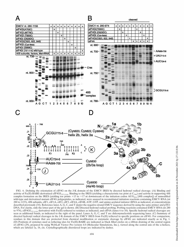
FIG. 6. Defining the orientation of eIF4G on the J-K domain of the EMCV IRES by directed hydroxyl radical cleavage. (A) Binding andactivity of Fe(II)-BABE-derivatized eIF4GI737-1116. Binding to the IRES (yielding a characteristic toe print at C786) and activity in supporting 48Scomplex formation on the IRES (yielding toe prints �15 to �17 nt downstream of the initiation codon AUG834 [48S complex]) of unmodifiedwild-type and derivatized mutant eIF4G polypeptides, as indicated, were assayed in reconstituted initiation reactions containing EMCV RNA (nt280 to 1155), 40S subunits, eIF1, eIF1A, eIF2, eIF3, eIF4A, eIF4B, ATP, GTP, and amino-acylated initiator tRNA as indicated, at concentrationsdescribed previously (34). Reference lanes A, G, C, and T depict the negative-strand EMCV sequence derived by using the same primer and pTE1DNA. For clarity, only the lower part of the gel is shown. (B) Directed hydroxyl radical probing. Probing reactions contained EMCV RNA (nt 280to 974), eIF4GI737-1116 derivatives with Fe(II) tethered to residues as indicated, and eIF4A (lanes 6 to 10). Specific hydroxyl radical cleavages areseen as additional bands, as indicated to the right of the panel. Lanes A, G, C, and T are dideoxynucleotide sequencing lanes. (C) Summary ofdirected hydroxyl radical cleavages in the J-K domain of the EMCV IRES from Fe(II) tethered to specific positions on eIF4G. For comparison,residues in this domain that are protected from chemical modification or enzymatic cleavage by eIF4G are indicated exactly as in Fig. 1.(D) Positions of cysteines used as tethering sites for Fe(II)-BABE are indicated as black filled circles on a ribbon drawing of the central regionof eIF4G (29), prepared by using WebLab Viewer Pro version 4.0 (Molecular Simulations, Inc.), viewed along the central axis of the �-helices,which are labeled 1a, 1b, etc. Crystallographically disordered loops are indicated by dashes.
694
Dow
nloa
ded
from
http
s://j
ourn
als.
asm
.org
/jour
nal/m
cb o
n 26
Nov
embe
r 20
21 b
y 27
.133
.9.9
4.
Footprinting complexes of eIF4G, eIF4A, and eIF4B on theEMCV IRES. eIF4A, eIF4B, and eIF4G interact synergisticallyto form a ribonucleoprotein complex on the IRES of EMCVand related viruses (26, 38, 40). We used RNA footprinting tocompare the interactions of this complex and of eIF4G alonewith the EMCV IRES. eIF4A enhanced eIF4GI737-1600’s pro-tection of the IRES from RNase V1 cleavage at GUCU777-780,reduced protection at CUU812-814, and enhanced cleavage atCA674-675 at the base of domain I, at G766 and CC784-785 in theJ-K domain, and weakly at UG897-898 downstream of the ini-tiation codon AUG834 (Fig. 4B, lanes 7 to 9). Some of thesechanges, for example at CA674-675 and G766, were slightly en-hanced by eIF4B. These changes were also observed when theR362Q trans-dominant mutant form of eIF4A replaced thewild-type factor (data not shown). eIF4A and eIF4B did notsignificantly alter the pattern of DMS modification of RNA inthe eIF4GI737-1600/IRES complex (Fig. 4B, lanes 3 to 5). Theseresults are summarized in Fig. 1.
ATP-dependent conformational changes in EMCV RNAdownstream of the initiation codon induced by eIF4F. TheATP dependence of initiation on the IRES and the require-ment for active eIF4A (33, 35) suggest that ribosomal loadingonto the initiation codon AUG834 may involve restructuring ofthe IRES by the eIF4G/eIF4A complex. Our probing analyses(22) (Fig. 4B) indicated that sequences immediately down-stream of the J-K domain are not extensively unwound by eIFs4A, 4B, and 4G but that some conformational changes areinduced in structures flanking and downstream of the initiationcodon.
ATP-dependent conformational changes downstream of theinitiation codon AUG834 induced by eIF4G/eIF4A were alsoidentified by toe printing done by using EMCV RNA (nt 378 to1155, comprising the entire IRES and 322 nt of the adjacentcoding region), eIF4A, and eIF4GI737-1116 (which containsonly one of the two eIF4A-binding sites in eIF4G [Fig. 7]).Binding of eIF4GI737-1116 in the presence of eIF4A enhancedstops at CGC865-867 in a manner that was further enhanced bybut did not depend on the presence of ATP or on ATP hydro-lysis (lanes 3 and 6). Additional new toe prints appeared atC841, A858 and (more weakly) G862, and G898 only in thepresence of eIF4F, wild-type eIF4A, and ATP (lanes 7 and 12)or eIF4GI737-1116, wild-type eIF4A, and ATP (lane 5). All butone of these stops occur in a proposed irregular stem-loopstructure (14) encompassing nt 854 to 902 (Fig. 1). These stopswere not detected in reactions lacking eIF4A (lanes 2 and 3).They were only weakly apparent in the absence of ATP (lanes4 and 11) or on inclusion of AMP-PNP (a nonhydrolyzableATP analog) in place of ATP (lane 6). However, this resultmust be treated cautiously because these reactions also con-tained dATP (an essential constituent of primer extension re-actions), which can partially substitute for ATP in eIF4A-dependent initiation on the EMCV IRES [38]). Significantly,these additional stops were not detected in reactions in whichR362Q trans-dominant mutant eIF4A replaced wild-type eIF4Ain the presence or absence of ATP (lanes 8 and 9). This eIF4Amutant is unable to utilize ATP or dATP to mediate confor-mational changes in RNA. Inclusion of R362Q mutant eIF4Ain toe printing reactions with eIF4GI737-1116 enhanced the toeprints at C786 and CGC865-867 to a similar extent as whenwild-type eIF4A was present (compare lanes 2, 4, and 8 and
lanes 5 and 9). These results indicate that mutant eIF4A wasincorporated into the eIF4G/eIF4A complex on the J-K do-main but that the resulting complex was unable to induce ATP-dependent conformational changes in the IRES/factor com-plex downstream of the initiation codon.
DISCUSSION
We have extended our previous characterization of the bind-ing site for eIF4G on the EMCV IRES (22) by using footprint-ing and mutagenesis and have determined the orientation ofthe essential core domain of eIF4G on this IRES by usingdirected hydroxyl radical cleavage. eIF4G recruits eIF4A tothe IRES, and we found that the bound eIF4G/eIF4A complexinduces limited local conformational changes in the IRES in anATP-independent manner as well as more extensive ATP-dependent changes immediately downstream of the ribosomebinding site. eIF4B, which also binds to the IRES, slightlyenhanced binding of the eIF4G/eIF4A complex. These obser-vations extend our understanding of the mechanism of IRES-mediated translation initiation by suggesting how binding ofinitiation factors to the IRES can actively prepare it for ribo-somal attachment.
eIF4G recognizes a large and structurally complex bindingsite on the EMCV IRES that encompasses sequences in the J,K, and L domains. This conclusion is based on the results ofhydroxyl radical and chemical and enzymatic probing (Fig. 4A,4B, and 5A). We have previously reported that interaction ofeIF4G, either alone or as part of eIF4F, with the IRES andconsequent initiation of translation require the oligo(A) loopat the junction of the J and K domains (35). We have nowfound that binding eIF4G and consequent initiation also de-pend on the apical J5 and K2 hairpins, which act synergisticallyin these processes (Fig. 2 and 3). Taken together with obser-vations that eIF4G/eIF4F binds specifically to the J-K domainsof human parechovirus type 1 (our unpublished data), FMDV,and TMEV IRESs (27, 28, 40, 46), these findings suggest thatthe sequence conservation in the central region of the J-Kdomain and the extensive but covariant sequence differences inthese peripheral helices of the J-K domain (17) are indicativeof a need to maintain its structural integrity to enable eIF4G tobind. This requirement would account for the deleterious ef-fects of mutations in these apical helices on EMCV IRESfunction noted here and elsewhere (13, 14, 32, 48).
The central domain of eIF4G that binds specifically to theJ-K domain consists of five �-helical HEAT repeats: eIF4G isthe first HEAT-repeat polypeptide that is known to bind spe-cifically to nucleic acid (29), and the results reported heretherefore define aspects of a new mode of RNA-protein inter-action. The HEAT-repeat domain of eIF4G binds directly to asmall number of bases in the J-K domain, most significantly inthe A-rich bulge at the junction of the J2, J3, and K1 helices(Fig. 1), but hydroxyl radical probing indicated that eIF4Gbound more extensively to RNA duplexes in the J, K, and Ldomains (Fig. 1 and 5A). Hydroxyl radicals cleave the sugar-phosphate backbone of RNA at ribose C1� and C4� positions(25), so the observed protection indicates that eIF4G binds inthe minor groove of these RNA duplexes. Other polypeptidesthat bind double-stranded RNA, such as the double-strandedRNA (dsRNA)-binding domain of proteins such as Staufen,
VOL. 23, 2003 INITIATION FACTOR-MEDIATED CHANGE IN IRES CONFORMATION 695
Dow
nloa
ded
from
http
s://j
ourn
als.
asm
.org
/jour
nal/m
cb o
n 26
Nov
embe
r 20
21 b
y 27
.133
.9.9
4.

Xlrbpa, and the dsRNA-dependent protein kinase PKR (1, 41,45), all do so through its wide minor groove. The functional2�-hydroxyl and phosphate groups in the RNA minor groovedo not allow sequence-specific recognition, and these proteinsshow no sequence specificity in their interactions with RNA invitro. A similar ability of eIF4G to bind dsRNA in a sequence-independent manner could bear on its activity as a cofactor
that enhances the ATP-dependent RNA helicase activity ofeIF4A (43, 44), for example by reducing dissociation of eIF4Afrom duplex RNA during cycles of step-wise unwinding ofRNA.
eIF4B also binds directly to EMCV-like IRESs (28, 31, 38),and footprinting data (Fig. 4B) indicate that it weakly enhancesbinding of the eIF4G/eIF4A complex to the EMCV IRES.
FIG. 7. ATP-dependent conformational changes induced eIF4G/4A in EMCV RNA downstream of the initiation codon. Primer extensionanalysis was done on EMCV RNA (nt 378 to 1155) in the presence of ATP or AMP-PNP and translation initiation components, as indicated. Thereaction mixture in lane 7 that yielded a 48S complex on the EMCV IRES contained eIF1, eIF1A, eIF2, eIF3, 40S subunits, GTP, and amino-acylated initiator tRNA as well as other translation initiation components as indicated, at concentrations described previously (34). The cDNAproducts marked C786, C841, A858, G862, CGC865-867, and G898 terminated at these nucleotides. The cDNA products marked �15-�17 terminatedat stop sites 15, 16, and 17 nucleotides from the initiation codon AUG834. Reference lanes A, G, C, and T depict the negative strand EMCVsequence derived by using the same primer and pTE1 DNA. For clarity, only the lower part of the gel downstream of EMCV nt 774 is shown.
696 KOLUPAEVA ET AL. MOL. CELL. BIOL.
Dow
nloa
ded
from
http
s://j
ourn
als.
asm
.org
/jour
nal/m
cb o
n 26
Nov
embe
r 20
21 b
y 27
.133
.9.9
4.

This conclusion is consistent with previous results obtained byusing EMCV and FMDV IRESs in UV cross-linking and toeprinting assays (38, 40). However, this enhancement is weak, soit may be a less important contributing factor in eIF4B’s activ-ity in enhancing ribosomal binding to this group of IRESs (35)than the interaction of eIF4B with the eIF3 component of the43S complex (30).
eIF4GI and eIF4GII both bind specifically to the J-K do-main and recruit eIF4A, which by itself does not bind specif-ically or stably to the IRES. eIF4A interacts strongly with theIRES in the resulting heterotrimeric complex and also signif-icantly enhances eIF4G’s binding to the IRES (22, 26, 38). TheeIF4G/eIF4A complex induced limited local conformationalchanges in the immediate vicinity of its binding site on theIRES (Fig. 4B), suggesting that binding may involve a degreeof induced fit. It also induced structural rearrangements in theIRES that reduced RNase V1 cleavage at several sites flankingthe initiation codon, in the region between domain L and aproposed irregular stem-loop structure (14) encompassing nt854 to 902 (Fig. 1). These changes are probably due to areduction in secondary structure in this region. Toe printing of48S complexes indicated that the leading edge of a bound 40Ssubunit is at G849, in the immediate vicinity of the inducedconformational changes (Fig. 7, lane 7). We therefore suggestthat the eIF4G/eIF4A complex prepares a binding site for theribosome on the IRES by inducing concerted conformationalchanges that include a reduction in the secondary structure ofsequences centered on the initiation codon so that this regionbecomes accessible to an incoming 43S ribosomal complex.
The eIF4G/eIF4A complex also enhanced existing weakstops and induced new toe prints in an ATP-dependent man-ner downstream of the initiation codon, primarily at and in thevicinity of nt 858. These new toe prints indicate that the sta-bility of this region had increased, which could be due toinduced and possibly long-range conformational rearrange-ments involving sequences in this region and upstream se-quences that had become unpaired by the helicase activity ofthe eIF4A/eIF4G complex. The possibility that sequences inthe vicinity of nt 858 switch between two alternative confor-mations that involve similar degrees of base pairing couldaccount for the minor changes in sensitivity of this region tochemical and enzymatic probes. Alternatively, the increasedstability of this region could be due to binding of the eIF4Aand/or the eIF4GI737-1116 fragment (which contains a bindingsite for one molecule of eIF4A). Such interactions could in-volve additional molecules of one or both of these factors, orcould involve a single eIF4G/eIF4A complex anchored on theJ-K domain that makes additional interactions with down-stream sequences, which would be possible if the active centerof this complex that is involved in its helicase activity is distinctfrom the RNA-binding surface that interacts with the J-Kdomain.
We are currently unable to distinguish between these twopossible explanations for the enhanced stability of sequences inthe vicinity of nt 858, which, we note, are not mutually exclu-sive. Either possibility would imply that the region between�nt 850 to 900 plays a specific role in IRES function; there isevidence for this and, more specifically, for both genetic andfunctional interactions between this domain and the J-K do-main (14, 20). A substitution in this domain suppressed the
defect caused by a substitution in the A-rich loop between theJ and K domains (14), and the translation defect caused by a1-nt insertion in this loop became apparent only if sequencesincluding the domain from �nt 850 to 900 were deleted (20).The position of this stabilized domain at the 3� border of thesequence covered by a 40S subunit bound to the initiationcodon AUG834 could constrain the site of ribosomal attach-ment and thus play a role in enhancing the accuracy of initia-tion codon selection.
In summary, we suggest that the eIF4G/eIF4A complex in-duces concerted conformational changes in the IRESs thatprepare a site on the IRES to which the ribosome can bindefficiently and accurately. A requirement for these conforma-tional changes for initiation on the IRES can account for thedependence of EMCV translation initiation on ATP (35) andon an enzymatically active eIF4G/eIF4A complex (33, 47). Thebasis for these requirements has until now been obscure. Thus,in addition to enhancing the affinity of eIF4G for the EMCVIRES (26) and for eIF3, a major component of the 43S com-plex (23), eIF4A also induces local changes in IRES confor-mation that prepare the region flanking and downstream of theinitiation codon AUG834 so that the 43S complexes can bind toit productively, leading to initiation of translation.
ACKNOWLEDGMENTS
This material is based upon work supported by the National ScienceFoundation under grant no. 0110834.
V.G.K. and I.B.L. contributed equally to this work.
REFERENCES
1. Bevilacqua, P. C., and T. R. Cech. 1996. Minor-groove recognition of double-stranded RNA by the double-stranded RNA-binding domain from the RNA-activated protein kinase PKR. Biochemistry 35:9983–9994.
2. Borovjagin, A. V., M. V. Ezrokhi, V. M. Rostapshov, T. Y. Ugarova, T. F.Bystrova, and I. N. Shatsky. 1991. RNA-protein interactions within theinternal translation initiation region of encephalomyocarditis virus RNA.Nucleic Acids Res. 19:4999–5005.
3. Borovjagin, A., T. Pestova, and I. Shatsky. 1994. Pyrimidine tract bindingprotein strongly stimulates in vitro encephalomyocarditis virus RNA trans-lation at the level of preinitiation complex formation. FEBS Lett. 351:299–302.
4. Bradley, C. A., J. C. Padovan, T. L. Thompson, C. A. Benoit, B. T. Chait, andR. E. Rhoads. 2002. Mass spectrometric analysis of the N terminus of trans-lational initiation factor eIF4G-1 reveals novel isoforms. J. Biol. Chem.277:12559–12571.
5. Carberry, S. E., and D. J. Goss. 1991. Interaction of wheat germ proteinsynthesis initiation factors eIF-3, eIF-(iso)4F, and eIF4F with mRNA ana-logues. Biochemistry 30:6977–6982.
6. Culver, G. M., and H. F. Noller. 2000. Directed hydroxyl radical probing ofRNA from iron(II) tethered to proteins in ribonucleoprotein complexes.Methods Enzymol. 318:461–475.
7. Duke, G. M., M. A. Hoffman, and A. C. Palmenberg. 1992. Sequence andstructural elements that contribute to efficient encephalomyocarditis viralRNA translation. J. Virol. 66:1602–1609.
8. Ehresmann, C., F. Baudin, M. Mougel, P. Romby, J. P. Ebel, and B. Ehres-mann. 1987. Probing the structure of RNA in solution. Nucleic Acids Res.15:9109–9128.
9. Evstafieva, A. G., T. Y. Ugarova, B. K. Chernov, and I. N. Shatsky. 1991. Acomplex RNA sequence determines the internal initiation of encephalomyo-carditis virus RNA translation. Nucleic Acids Res. 19:665–671.
10. Gingras, A. C., B. Raught, and N. Sonenberg. 1999. eIF4 initiation factors:effectors of mRNA recruitment to ribosomes and regulators of translation.Annu. Rev. Biochem. 68:913–963.
11. Gradi, A., H. Imataka, Y. V. Svitkin, E. Rom, B. Raught, S. Morino, and N.Sonenberg. 1998. A novel functional human eukaryotic translation initiationfactor 4G. Mol. Cell. Biol. 18:334–342.
12. Hellen, C. U. T., and P. Sarnow. 2001. Internal ribosome entry sites ineukaryotic mRNA molecules. Genes Dev. 15:1593–1612.
13. Hoffman, M. A., and A. C. Palmenberg. 1995. Mutational analysis of the J-Kstem-loop region of the encephalomyocarditis virus IRES. J. Virol. 69:4399–4406.
VOL. 23, 2003 INITIATION FACTOR-MEDIATED CHANGE IN IRES CONFORMATION 697
Dow
nloa
ded
from
http
s://j
ourn
als.
asm
.org
/jour
nal/m
cb o
n 26
Nov
embe
r 20
21 b
y 27
.133
.9.9
4.
14. Hoffman, M. A., and A. C. Palmenberg. 1996. Revertant analysis of J-Kmutations in the encephalomyocarditis virus internal ribosomal entry sitedetects an altered leader protein. J. Virol. 70:6425–6430.
15. Huttenhofer, A., and H. F. Noller. 1994. Footprinting mRNA-ribosome com-plexes with chemical probes. EMBO J. 13:3892–3901.
16. Jackson, R. J. 2000. A comparative view of initiation site selection mecha-nisms, p.127–183 In N. Sonenberg, M. B. Mathews and J. W. B. Hershey(ed.), Translational control of gene expression. Cold Spring Harbor Labo-ratory Press, Cold Spring Harbor, N.Y.
17. Jackson, R. J., and A. Kaminski. 1995. Internal initiation of translation ineukaryotes: the picornavirus paradigm and beyond. RNA 1:985–1000.
18. Jang, S. K., and E. Wimmer. 1990. Cap-independent translation of encepha-lomyocarditis virus RNA: structural elements of the internal ribosomal entrysite and involvement of a cellular 57-kD RNA-binding protein. Genes Dev.4:1560–1572.
19. Kaminski, A., M. T. Howell, and R. J. Jackson. 1990. Initiation of encepha-lomyocarditis virus RNA translation: the authentic initiation site is not se-lected by a scanning mechanism. EMBO J. 9:3753–3759.
20. Kaminski, A., and R. J. Jackson. 1998. The polypyrimidine tract bindingprotein (PTB) requirement for internal initiation of translation of cardiovi-rus RNAs is conditional rather than absolute. RNA 4:626–638.
21. Kolupaeva, V. G., C. U. T. Hellen, and I. N. Shatsky. 1996. Structural analysisof the interaction of the pyrimidine tract-binding protein with the internalribosomal entry site of encephalomyocarditis virus and foot-and-mouth dis-ease virus RNAs. RNA 2:1199–1212.
22. Kolupaeva, V. G., T. V. Pestova, C. U. T. Hellen, and I. N. Shatsky. 1998.Translation eukaryotic initiation factor 4G recognizes a specific structuralelement within the internal ribosome entry site of encephalomyocarditisvirus RNA. J. Biol. Chem. 273:18599–18604.
23. Korneeva, N. L., B. J. Lamphear, F. L. Hennigan, and R. E. Rhoads. 2000.Mutually cooperative binding of eukaryotic translation initiation factor (eIF)3 and eIF4A to human eIF4G-1. J. Biol. Chem. 275:41369–41376.
24. Lamphear, B. J., R. Kirchweger, T. Skern, and R. E. Rhoads. 1995. Mappingof functional domains in eukaryotic protein synthesis initiation factor 4G(eIF4G) with picornaviral proteases. Implications for cap-dependent andcap-independent translational initiation. J. Biol. Chem. 270:21975–21983.
25. Latham, J. A., and T. R. Cech. 1989. Defining the inside and outside of acatalytic RNA molecule. Science 245:276–282.
26. Lomakin, I. B., C. U. T. Hellen, and T. V. Pestova. 2000. Physical associationof eukaryotic initiation factor 4G (eIF4G) with eIF4A strongly enhancesbinding of eIF4G to the internal ribosomal entry site of encephalomyocar-ditis virus and is required for internal initiation of translation. Mol. Cell.Biol. 20:6019–6029.
27. Lopez de Quinto, S., and E. Martinez-Salas. 2000. Interaction of the eIF4Ginitiation factor with the aphthovirus IRES is essential for internal transla-tion initiation in vivo. RNA 6:1380–1392.
28. Lopez de Quinto, S., E. Lafuente, and E. Martinez-Salas. 2001. IRES inter-action with translation initiation factors: functional characterization of novelRNA contacts with eIF3, eIF4B and eIF4GII. RNA 7:1213–1226.
29. Marcotrigiano, J., I. B. Lomakin, N. Sonenberg, T. V. Pestova, C. U. T.Hellen, and S. K. Burley. 2001. A conserved HEAT domain within eIF4Gdirects assembly of the translation initiation machinery. Mol. Cell 7:193–203.
30. Methot, N., M. S. Song, and N. Sonenberg. 1996. A region rich in asparticacid, arginine, tyrosine, and glycine (DRYG) mediates eukaryotic initiationfactor 4B (eIF4B) self-association and interaction with eIF3. Mol. Cell. Biol.16:5328–5334.
31. Meyer, K., A. Petersen, M. Niepmann, and E. Beck. 1995. Interaction ofeukaryotic initiation factor eIF-4B with a picornavirus internal translationinitiation site. J. Virol. 69:2819–2824.
32. Oudshoorn, P., A. Thomas, G. Scheper, and H. O. Voorma. 1990. An initi-ation signal in the 5� untranslated leader sequence of encephalomyocarditisvirus RNA. Biochim. Biophys. Acta 1050:124–128.
33. Pause, A., N. Methot, Y. Svitkin, W. C. Merrick, and N. Sonenberg. 1994.Dominant negative mutants of mammalian translation initiation factoreIF-4A define a critical role for eIF-4F in cap-dependent and cap-indepen-dent initiation of translation. EMBO J. 13:1205–1215.
34. Pestova, T. V., S. I. Borukhov, and C. U. T. Hellen. 1998. Eukaryotic ribo-somes require eIFs 1 and 1A to locate initiation codons. Nature 394:854–859.
35. Pestova, T. V., C. U. T. Hellen, and I. N. Shatsky. 1996. Canonical eukaryoticinitiation factors determine initiation of translation by internal ribosomalentry. Mol. Cell. Biol. 16:6859–6869.
36. Pestova, T. V., V. G. Kolupaeva, I. B. Lomakin, E. V. Pilipenko, I. N. Shatsky,V. I. Agol, and C. U. T. Hellen. 2001. Molecular events in initiation oftranslation in eukaryotes. Proc. Natl. Acad. Sci. USA 98:7029–7036.
37. Pestova, T. V., I. N. Shatsky, S. P. Fletcher, R. J. Jackson, and C. U. T.Hellen. 1998. A prokaryotic-like mode of cytoplasmic eukaryotic ribosomebinding to the initiation codon during internal initiation of translation ofhepatitis C virus and classical swine fever virus RNAs. Genes Dev. 12:67–83.
38. Pestova, T. V., I. N. Shatsky, and C. U. T. Hellen. 1996. Functional dissectionof eukaryotic initiation factor 4F: the 4A subunit and the central domain ofthe 4G subunit are sufficient to mediate internal entry of 43S preinitiationcomplexes. Mol. Cell. Biol. 16:6870–6878.
39. Pilipenko, E. V., V. M. Blinov, B. K. Chernov, T. M. Dmitrieva, and V. I.Agol. 1989. Conservation of the secondary structure elements of the 5�-untranslated region of cardio- and aphthovirus RNAs. Nucleic Acids Res.17:5701–5711.
40. Pilipenko, E. V., T. V. Pestova, V. G. Kolupaeva, E. V. Khitrina, A. N.Poperechnaya, V. I. Agol, and C. U. T. Hellen. 2000. A cell cycle-dependentprotein serves as a template-specific translation initiation factor. Genes Dev.14:2028–2045.
41. Ramos, A., S. Grunert, J. Adams, D. R. Micklem, M. R. Proctor, S. Freund,M. Bycroft, D. St. Johnston, and G. Varani. 2000. RNA recognition by aStaufen double-stranded RNA-binding domain. EMBO J. 19:997–1009.
42. Ray, B. K., T. G. Lawson, J. C. Kramer, M. H. Cladaras, J. A. Grifo, R. D.Abramson, W. C. Merrick, and R. E. Thach. 1985. ATP-dependent unwind-ing of messenger RNA structure by eukaryotic initiation factors. J. Biol.Chem. 260:7651–7658.
43. Rogers, G. W., N. R. Richter, W. F. Lima, and W. C. Merrick. 2001. Mod-ulation of the helicase activity of eIF4A by eIF4B, eIF4H, and eIF4F. J. Biol.Chem. 276:30914–30922.
44. Rozen, F., I. Edery, K. Meerovitch, T. E. Dever, W. C. Merrick, and N.Sonenberg. 1990. Bidirectional RNA helicase activity of eucaryotic transla-tion initiation factors 4A and 4F. Mol. Cell. Biol. 10:1134–1144.
45. Ryter, J. M., and S. C. Schultz. 1998. Molecular basis of double-strandedRNA-protein interactions: structure of a dsRNA-binding domain complexedwith dsRNA. EMBO J. 17:7505–7513.
46. Saleh, L., R. C. Rust, R. Fullkrug, E. Beck, G. Bassili, K. Ochs, and M.Niepmann. 2001. Functional interaction of translation initiation factoreIF4G with the foot-and-mouth disease virus internal ribosome entry site.J. Gen. Virol. 82:757–763.
47. Svitkin, Y. V., A. Pause, A. Haghighat, S. Pyronnet, G. Witherell, G. J.Belsham, and N. Sonenberg. 2001. The requirement for eukaryotic initiationfactor 4A (elF4A) in translation is in direct proportion to the degree ofmRNA 5� secondary structure. RNA 7:382–394.
48. Witherell, G. W., C. S. Schultz-Witherell, and E. Wimmer. 1995. Cis-actingelements of the encephalomyocarditis virus internal ribosomal entry site.Virology 214:660–663.
698 KOLUPAEVA ET AL. MOL. CELL. BIOL.
Dow
nloa
ded
from
http
s://j
ourn
als.
asm
.org
/jour
nal/m
cb o
n 26
Nov
embe
r 20
21 b
y 27
.133
.9.9
4.