Embed Size (px)
Citation preview
Review
Pathophysiology of Takotsubo Syndrome
Francesco Pelliccia, MD, PhD1, Juan Carlos Kaski, MD2,
Filippo Crea, MD3, and Paolo G. Camici, MD4
From the Department of Cardiovascular Sciences, Sapienza University, Rome, Italy
(1); Molecular and Clinical Sciences Research Institute, St George’s, University of
London, London, UK (2); Institute of Cardiology, Catholic University, Rome, Italy (3)
and Vita-Salute University and San Raffaele Hospital, Milan, Italy (4)
Word count: 5596 words (w/o ref.)
References: 100
Numbers of figures and tables: 8
Unstructured abstract: 301 words
Short title: Pathophysiology of Takotsubo
Correspondence to:
Paolo G. Camici MD FACC FAHA FESC FRCP
Vita-Salute University, Via Olgettina, 58,
20132 Milan, Italy
Tel: +39 02 2643 6206; Fax: +39 02 2643 6218,
E-mail: [email protected]
1

Review
ABSTRACT
Originally described by Japanese authors in the 1990s, Takotsubo Syndrome (TTS)
generally presents as an acute myocardial infarction characterized by severe left
ventricular (LV) dysfunction. TTS, however, differs from an acute coronary syndrome
because patients have generally a normal coronary angiogram and LV dysfunction,
which extends beyond the territory subtended by a single coronary artery and
recovers within days or weeks. The prognosis was initially thought to be benign, but
subsequent studies have demonstrated that both acute and long-term mortality are
higher than previously recognized. Indeed, mortality reported during the acute phase
in hospitalized patients is ~4-5%, a figure comparable to that of ST elevation
myocardial infarction in the era of primary percutaneous coronary interventions.
Despite extensive research, the etiology and pathogenesis of TTS remain
incompletely understood. The aim of the present review is to discuss the
pathophysiology of TTS with particular emphasis on the role of the central and
autonomic nervous systems.
Different emotional or psychological stressors have been identified to precede the
onset of TTS. The anatomical structures that mediate the stress response are found
both in the central and autonomic nervous systems. Acute stressors induce brain
activation, increasing bioavailability of cortisol and catecholamine. Both circulating
epinephrine and norepinephrine, released from adrenal medullary chromaffin cells,
and norepinephrine released locally from sympathetic nerve terminals are
significantly increased in the acute phase of TTS. This catecholamine surge leads,
through multiple mechanisms - i.e. direct catecholamine toxicity, adrenoceptor-
mediated damage, epicardial and microvascular coronary vasoconstriction and/or
spasm, and increased cardiac workload - to myocardial damage whose functional
2

Review
counterpart is the transient apical LV ballooning. The relative preponderance among
post-menopausal women suggests that estrogen deprivation may play a facilitating
role probably mediated by endothelial dysfunction. Despite the substantial
improvement in our understanding of the pathophysiology of TTS, a number of
knowledge gaps still remain.
Key Words: cardiomyopathy; catecholamine; ischemic heart disease; Takotsubo
syndrome.
3
Review
Originally described by Sato et al.1 in the 1990’s, Takotsubo Syndrome (TTS)
presents as an acute coronary syndrome (ACS) characterized by severe left
ventricular (LV) dysfunction that typically recovers spontaneously within days or
weeks. Patients may present with abrupt onset chest pain and/or dyspnea. Several
stressors have been identified to precede the onset of TTS in a substantial proportion
of patients.2 Emotional or psychological stress due to the unexpected death of a
relative or a friend, suppressed terror, the occurrence of natural disasters or
strenuous physical stress usually precede its onset.3 About one in 5 patients,
however, does not report any form of stress preceding the onset of the condition.
Recently, it has been shown that TTS can also occur following a ‘positive’ life event,
hence the recently proposed denomination of ‘happy heart syndrome’.4
Despite extensive research, the etiology and pathogenesis of TTS remain
incompletely understood. The aim of the present review is to discuss the
pathophysiology of TTS with particular emphasis on the role of the central and
autonomic nervous systems.
Clinical presentation
Symptoms, clinical signs, echocardiographic and electrocardiographic findings in
TTS patients are suggestive of an acute coronary syndrome.5 The most common
symptoms at presentation are chest pain and dyspnea. TTS can also present as
syncope and pulmonary edema. Cardiac arrest, cardiogenic shock, and serious
ventricular arrhythmias occur more rarely in TTS patients. Symptoms such as
generalized weakness, unexplained cough, and fever have also been reported.6
ECG patterns
4
Review
Abnormalities on the ECG are common at the time of presentation. The most
frequent finding on the admission ECG is ST-segment elevation, which most often
occurs in the precordial leads.7 The magnitude of ST-segment elevation and the
number of leads with this pattern is usually less in patients with TTS than in cases of
ST-elevation myocardial infarction (STEMI).8 Interestingly, reciprocal ST segment
changes and abnormal Q waves are often absent in TTS. Moreover, ST segment
depression is less common in TTS, as compared to coronary artery disease-related
ACS.9 Some TTS patients may present with diffuse T-wave inversion particularly in
the anterior and lateral leads of the ECG. A prolongation in the QT interval -
corrected for heart rate - has been reported in a substantial proportion of TTS
patients. These ECG changes are often transient and their presence or absence
depends on when the ECG is recorded after symptom onset. However, it is
challenging to distinguish TTS from an ACS on the basis of the ECG alone and
therefore access to emergency coronary angiography should not be delayed.5
Biomarkers
Typically, TTS patients manifest modest increases in creatine kinase-MB and cardiac
troponin concentrations as compared to STEMI patients. Of interest, in TTS there is a
disparity between the degree of biomarker elevation and extent of myocardial
dysfunction observed at left ventriculography. In a minority of TTS patients, however,
the elevation of biomarkers of necrosis can be substantial, probably reflecting more
severe myocardial damage.5 Significantly elevated serum brain natriuretic peptide or
N-terminal pro-brain natriuretic peptide can also be detected during the acute phase
of TTS.10 The production and release of these peptides appears to be mainly related
to ventricular stretching.11 Because in most cases TTS is characterized by LV
5

Review
distension and relatively mild tissue necrosis, a greater increase in plasma natriuretic
peptides compared with biomarkers indicative of necrosis can be detected.10
Coronary and LV angiography
Diagnostic coronary angiography shows normal coronary arteries or non-obstructive
coronary artery disease in the vast majority of patients.5 Yet, about 15% of patients
with TTS have obstructive coronary atherosclerosis.6 In these patients, the diagnosis
of TTS is suggested by the fact that the area of dysfunction detected on LV
angiography extends beyond the territory subtended by a single coronary artery and
by the reversibility of LV dysfunction. Hence, the mere presence of obstructive
coronary atherosclerosis does not allow for excluding the diagnosis of TTS.12
Different types of LV dysfunction
Different patterns of LV dysfunction have been reported in TTS, including the
classical apical variant, a mid-ventricular variant, a basal or inverted variant and
regional variants.13 About 80% of patients exhibit the apical variant.14 As the heart is
densely innervated by sympathetic nerves that follow a regional distribution, it has
been hypothesized that the typical apical pattern of LV dysfunction results from this
anatomical substrate,3 as well as from the regional distribution of sympathetic
adrenoceptors. 15
Clinical Outcome
The prognosis of TTS was initially thought to be benign.16 Subsequent series,
however, have demonstrated that both acute and long-term
mortality are higher than
previously recognized.17 Indeed, mortality reported during the acute phase in
6
Review
hospitalized patients is ~4-5%, a figure comparable to that of STEMI in the era of
primary percutaneous coronary interventions.18 Of interest, despite the recovery of
LV function and absence of significant coronary disease in most cases, mortality after
hospital discharge is worse than that in an aged-matched healthy population.19 A
recent meta-analysis of clinical correlates of acute mortality in TTS has reported that
the average in-hospital mortality is 4.5%.20 Japanese investigators have recently
pointed out that TTS is associated with an elevated in-hospital mortality due to co-
existing chronic comorbidities and acute medical illnesses.21 In one of the largest
published series (n=1750), Templin et al. reported a 30-day mortality of 5.9% and a
long-term death rate of 5.6% per patient per year.6 Major adverse events, including
cardiogenic shock, cardiac arrest and mortality, are more frequent in women than in
men with TTS. There is uncertainty as to the real recurrence rate of TTS due to the
paucity of data on the risk of a further episode after the index event. Available
evidence points to figures ranging from 0 to 22% depending upon the size of the
population investigated and the duration of follow up.5
TTS ‘Phenocopies’
Generally, TTS is preceded by intense emotional triggers, although in up to one third
of patients no trigger can be identified. A TTS-like syndrome can be observed in
several medical conditions including sepsis, neurological disorders (e.g.
subarachnoid hemorrhage, seizures, stroke/transient ischemic attack, cerebral
tumors, head trauma)22 and pheochromocytoma.23 Furthermore, a TTS-like
syndrome can be triggered by drugs (e.g. dopamine, dobutamine, epinephrine, or
norepinephrine in the setting of cardiovascular stress tests, anesthesia, etc.).24 In our
7

Review
view, all these conditions should be differentiated from the classic ‘phenotype’ of TTS
and could be labeled as TTS ‘phenocopies’.
Epidemiology
About 90% of patients with TTS are post-menopausal females with a similar
prevalence across ethnic groups.6 In recent years, the increasing number of patients
referred to coronary angiography with suspected ACS has allowed better
appreciation of the true incidence of TTS. At present, it is estimated that
approximately 2% of all patients undergoing emergency coronary angiography for a
suspected ACS are TTS12, and it has been calculated that the incidence of TTS is
approximately 100 new cases per million population per annum.25 Indeed, the
improved clinical characterization of the condition has led to a paradigm shift: from
what was initially the exclusion of STEMI, to the recognition that TTS has a number
of distinctive diagnostic features.
Pathophysiology
Sympathetic activation in TTS and its mechanisms
The environmental events experienced by the majority of these patients and
perceived as threatening become profoundly stressful if one is not able to cope with
them.26 Stress is a physiological response that mediates the action of a stressor on
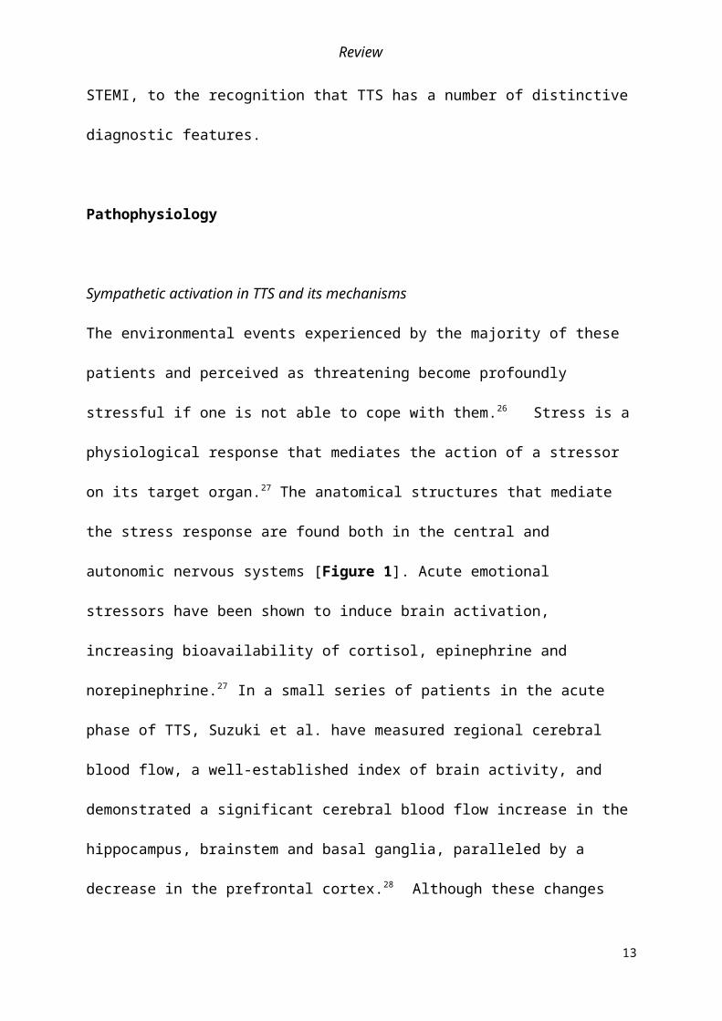
its target organ.27 The anatomical structures that mediate the stress response are
found both in the central and autonomic nervous systems [Figure 1]. Acute
emotional stressors have been shown to induce brain activation, increasing
bioavailability of cortisol, epinephrine and norepinephrine.27 In a small series of
8

Review
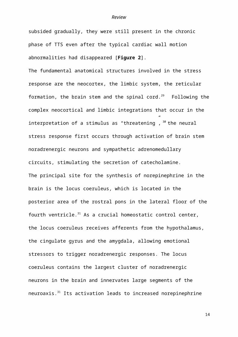
patients in the acute phase of TTS, Suzuki et al. have measured regional cerebral
blood flow, a well-established index of brain activity, and demonstrated a significant
cerebral blood flow increase in the hippocampus, brainstem and basal ganglia,
paralleled by a decrease in the prefrontal cortex.28 Although these changes subsided
gradually, they were still present in the chronic phase of TTS even after the typical
cardiac wall motion abnormalities had disappeared [Figure 2].
The fundamental anatomical structures involved in the stress response are the
neocortex, the limbic system, the reticular formation, the brain stem and the spinal
cord.29 Following the complex neocortical and limbic integrations that occur in the
interpretation of a stimulus as “threatening”, 30 the neural stress response first occurs
through activation of brain stem noradrenergic neurons and sympathetic
adrenomedullary circuits, stimulating the secretion of catecholamine.
The principal site for the synthesis of norepinephrine in the brain is the locus
coeruleus, which is located in the posterior area of the rostral pons in the lateral floor
of the fourth ventricle.31 As a crucial homeostatic control center, the locus coeruleus
receives afferents from the hypothalamus, the cingulate gyrus and the amygdala,
allowing emotional stressors to trigger noradrenergic responses. The locus coeruleus
contains the largest cluster of noradrenergic neurons in the brain and innervates
large segments of the neuroaxis.31 Its activation leads to increased norepinephrine
secretion, which in turn stimulates the hypothalamic-pituitary-adrenal axis.29
Adrenal medullary chromaffin cells synthesize, store, and release predominantly
epinephrine and norepinephrine which constitute the hormonal output of the
neuroendocrine stress-response axis.32 Activation of the latter is crucial to maintain
high levels of stress arousal for prolonged periods. The hypothalamic-pituitary-
adrenal axis is a complex set of direct influences and feedback interactions among
9
Review
three endocrine glands: the hypothalamus, the pituitary gland, and the adrenal gland
[Figure 1].
Apart from the locus coeruleus, the neural impulses also descend into the posterior
hypothalamus, i.e. the pathway of sympathetic activation. From here, sympathetic
neural pathways descend through the cranial and sacral spinal cord regions and
trigger the release of norepinephrine.33 There are sympathetic preganglionic neurons
that lay in the lateral grey column from T1 to L2/3 which synapse with their
postganglionic neurons. Sympathetic cardiac innervation originates mainly in the
right and left stellate ganglia. These fibers travel along the epicardial vascular
structures of the heart into the underlying myocardium and end as sympathetic nerve
terminals reaching the heart muscle and coronary circulation. The sympathetic nerve
endings release norepinephrine directly into the synaptic cleft activating and
postsynaptic adrenoceptors.34 Thus, all the epinephrine in the body and a significant
amount of circulating norepinephrine derive from the adrenal medulla, and the total
amount of catecholamine presented to cardiac adrenergic receptors at any given
time is composed of circulating norepinephrine and epinephrine coupled with
norepinephrine released locally from sympathetic nerve terminals.35 In normal
humans, under resting conditions, only about 2-8% of the circulating norepinephrine
is released by the adrenal medulla and the rest is released by sympathetic nerve
endings.26
Increased circulating and myocardial catecholamine levels
Akashi and colleagues were the first to report elevated serum catecholamine levels in
patients with TTS.36 Wittstein et al. subsequently showed that in the acute phase,
TTS patients have increased concentrations of plasma catecholamines (i.e.
10
Review
epinephrine, norepinephrine, and dopamine) and stress-related circulating
neuropeptides that are several times higher than those in patients with STEMI.
These levels remain markedly elevated even a week after the onset of symptoms
[Figure 3].37
A recent study in a murine model has demonstrated that the infusion of high
concentrations of epinephrine can produce the characteristic reversible apical LV
ballooning coupled with basal hyper-contractility observed in patients with TTS.38
Indeed in the acute phase of TTS, along with an increased concentration of
circulating catecholamine, 37 there is also evidence of increased catecholamine at the
myocardial level. Kume et al. have demonstrated increased norepinephrine spillover
in the coronary sinus in a small series of TTS patients, suggesting increased local
myocardial release of catecholamine.39 Increase in local catecholamine levels has
been demonstrated also in the so-called neurogenic stunned myocardium which
appears to be mediated by neuronally transmitted norepinephrine.40 The clinical
presentation of this condition, which is found in patients with aneurysm-related
subarachnoid hemorrhage, resembles closely that of TTS and is characterized by a
fully reversible form of acute LV dysfunction.41 Accordingly, experimental work has
shown that elevated activity of the sympathetic nervous system in the acute phase of
subarachnoid hemorrhage induces myocardial damage and contributes to the
development of cardiac dysfunction.42
The local release of catecholamine from cardiac nerve endings results in an elevated
norepinephrine concentration in the synaptic cleft due to increased exocytosis of
norepinephrine from presynaptic vesicles, paralleled by a decrease in terminal nerve
axon norepinephrine re-uptake through the specific uptake-1 transporter [Figure 4].43
This process has been originally demonstrated by means of iodine-123 meta-iodo-
11
Review
benzyl-guanidine, i.e. a gamma-emitting norepinephrine analogue that is used to
image myocardial sympathetic nerve terminals with single photon emission computed
tomography. Akashi et al. examined 8 patients with TTS within 3 days of admission
and at 3 month follow up after normalization of LV dysfunction.44 The acute scan
showed a pattern of cardiac sympathetic hyperactivity with improvement at follow
up.44 The evidence of reduced iodine-123 meta-iodo-benzyl-guanidine retention in
dysfunctional segments is consistent with a regional disturbance of sympathetic
neuronal activity that can persist for months.45 Recently, Christensen et al. have
demonstrated myocardial sympathetic hyperactivity in the subacute phase of TTS
paralleling plasma epinephrine levels, that were also elevated compared with follow-
up concentrations.46
Role of endothelial dysfunction and estrogen deficiency
New information has shed additional light on the pathogenesis of TTS supporting the
concept that the condition differs markedly from cardiomyopathies, as currently
defined. Specifically, recent data show that endothelial dysfunction is common in
patients with TTS, which could explain the propensity to epicardial and/or
microvascular coronary artery spasm which are two likely pathogenetic mechanisms
for TTS.47 Indeed, endothelial dysfunction, a pathological state of the endothelium
characterized by an imbalance between vasoconstricting and vasodilating factors,
may represent an important link between stress and myocardial dysfunction in TTS.47
Therefore, transient myocardial ischemia followed by stunning might be the cause
underlying the typical, reversible LV dysfunction.
Endothelial dysfunction can also explain why TTS is more common in post-
menopausal women, as they have been shown to have both age-related and
12
Review
estrogen deficiency-related coronary vasomotor abnormalities.48-50 Under physiologic
circumstances estrogen beneficially affects the coronary microcirculation via
endothelium-dependent and -independent mechanisms, improving coronary blood
flow.50 During menopause, both increased sympathetic drive and endothelial
dysfunction are a consequence of reduced estrogen levels.49 In an elegant
experimental study in animals, Ueyema et al. have shown that stress-induced LV
apical ballooning can be prevented by pretreatment with - and β-adrenoceptor
blockers and estrogen.51 Estrogen supplementation attenuated the stressinduced
hypothalamosympatho-adrenal outflow from the central nervous system to the target
organs. In addition, estrogen treatment upregulated the cardiac levels of
cardioprotective substances, such as atrial natriuretic peptide and heat shock protein
70. These data suggest that estrogen deficiency following menopause might facilitate
the occurrence of TTS - particularly that linked to emotional stress - either by indirect
action on the nervous system or by direct action on the heart. Moreover, impairment
of endothelial function is associated with the presence of traditional risk factors and
has been described in the setting of various systemic inflammatory disorders with
high cardiovascular morbidity and mortality.49 Recently, data in large cohorts have
shown that TTS patients have a non-negligible prevalence of cardiovascular risk
factors, i.e. hypertension, hypercholesterolemia, and smoking.2,6,18 In addition, there
is now evidence that most cases of TTS occur in patients with various co-morbidities,
including neurologic, psychiatric, pulmonary, kidney, liver and connective tissue
disease,2 that are associated with endothelial dysfunction and might therefore
constitute a previously unrecognized predisposing factor for TTS.52
13
Review
Based on these multiple observations, the possibility exists that endothelial
dysfunction might constitute a crucial link between a sympathetic surge and
myocardial ischemia in TTS.
Mechanisms of LV dysfunction induced by sympathetic hyperactivity
Although there is agreement that TTS is characterized by increased circulating and
cardiac catecholamine levels, how this translates into the typical LV dysfunction
remains incompletely understood. 53 Multiple mechanisms have been postulated to
explain the cardiotoxicity of catecholamines.54 The surge in stimulation of
adrenoceptors enhances heart rate and cardiac contractility with a secondary
imbalance in the ratio of oxygen supply to oxygen demand thus creating areas of
myocellular hypoxia.55 Myocyte hypoxia can be further aggravated by metabolic
changes, 56 such as excessive deposition of lipid droplets in cardiomyocytes. These
changes might result in an uncoupling of oxidative phosphorylation in mitochondria
which inhibits the coupling between the electron transport and phosphorylation
reactions that in turn will interfere with ATP synthesis.57 Changes in membrane
permeability might also lead to electrolyte changes. These include hypokalemia,
hypocalcemia and hypomagnesemia with resultant elevations in parathyroid
hormone, and hypozincemia with hyposelenemia, which compromise antioxidant
defenses. Altered cationic homeostasis might affect several cellular processes and
contribute to myocardial toxicity.58 Norepinephrine and epinephrine are also potential
sources of free radicals. These oxygen-derived free radicals may interfere with
calcium and sodium transporters, which may result in additional myocyte
dysfunction.3
14
Review
Direct catecholamine toxicity
Some authors favor the hypothesis of direct catecholamine-induced myocardial
toxicity in TTS. For instance, myocardial necrosis can occur in patients with acute
neurovascular events, and this is caused by direct toxicity of endogenous
catecholamine released into the heart via nerve terminals.59 Catecholamine released
directly into the myocardium via sympathetic nerves has been suggested to have a
greater “toxic” effect than that reaching the heart via the bloodstream.60 Indeed,
norepinephrine spillover from the cardiac sympathetic nerve terminals can decrease
myocyte viability through cyclic adenosine monophosphate-mediated calcium
overload resulting, histologically, in contraction band necrosis which is one of the
pathological hallmarks of TTS,61 along with increased production of extracellular
matrix leading to a rapid increase in fibrosis and mild neutrophil infiltration. Nef et al.
studied serial myocardial biopsies in 8 patients with TTS during the phase of severe
LV dysfunction and found histological signs of catecholamine toxicity, i.e. focal
mononuclear inflammatory cells, areas of fibrotic response, and characteristic
contraction bands. They noted that TTS can be accompanied by “severe
morphological alterations potentially resulting from catecholamine excess followed by
microcirculatory dysfunction and direct cardiotoxicity. However, the affected
myocardium represents a high potential of structural reconstitution which correlates
with the rapid functional recovery function is accompanied by an equally fast
reconstitution of myocardial integrity”. 62
Contraction band necrosis is a unique form of myocyte injury characterized by
hypercontracted sarcomeres, dense eosinophilic transverse bands, and an interstitial
mononuclear inflammatory response and is distinct from the polymorphonuclear
inflammation seen in infarction.61 Contraction band necrosis has been found in
15

Review
patients with pheochromocytoma, 23 and those with subarachnoid hemorrhage40 both
characterized by a catecholamine excess. It has also been observed post-mortem in
people who died under terrifying circumstances such as fatal asthma and violent
assault, suggesting that catecholamine excess represents an important link between
emotional stress and cardiac injury.63 The lack of persistent, significant morphological
changes in most cases of TTS is further demonstrated by data accrued so far with
cardiac magnetic resonance (CMR). Different studies have pointed out that the acute
phase of the disease is characterized only by remarkable myocardial edema with no
evidence of significant late gadolinium enhancement [Figure 5].64 These findings are
of major importance, as they exclude the possibility that TTS is mainly the
consequence of a catecholamine-mediated myocarditis, which commonly occurs in
pheochromocytoma.23 Although TTS and pheochromocytoma are both
characterized by increased catecholamine concentrations, this causes a distinct
entity in pheochromocytoma only, leading to degenerative changes in muscle fibers,
foci of necrosis, acute inflammation, chronic interstitial inflammatory exudation, and
reparative fibrosis.65 At CMR, these abnormalities may be observed noninvasively
as myocardial necrosis (late enhancement), edema, and focal and diffuse fibrosis
that may lead to short- or long-term LV dysfunction.23 A different pattern is observed
in TTS patients. Testa and Feola performed serial CMR scans in patients with TTS
and found no evidence of delayed enhancement either in the acute phase or at 3
months follow up, suggesting that the damage in dysfunctional myocardium was
transient and did not include significant tissue fibrosis.66
In summary, it may be hypothesized that direct catecholamine toxicity plays a role
both in TTS and pheochromocytoma, but with important quantitative differences. The
myocardial damage in pheochromocytoma is probably more extensive because of
16

Review
the persistent exposition of patients to elevated catecholamine levels. In TTS, the
elevation is transient and generally results in less evident damage as demonstrated
by the relatively mild elevation of necrosis biomarkers, 5,6 and absence of late
enhancement at CMR in most cases.66 The latter in particular might be a
consequence of the limited spatial resolution of CMR, i.e. 0.6 to 1 cm3, which may not
detect smaller or patchy areas of damage that are present in TTS.67
Microvascular spasm
A further “vascular” pathogenetic mechanism that could be involved in TTS is acute,
transient myocardial ischemia. Since the first description of TTS, coronary
vasospasm has been suggested as a plausible causative factor. In their original
report, Dote et al. hypothesized that TTS was caused by multivessel coronary
vasospasm, as 4 of 5 patients in their series had “spontaneous” or induced coronary
vasospasm at coronary angiography.68 Sato et al. reported epicardial coronary artery
spasm in 8 of 35 patients (23%) and diffuse coronary vasoconstriction in 19 (54%).1
Similarly, Tsuchihashi et al. reported epicardial coronary spasm in 10 of 48 TTS
patients (21%).69 Although the causative role of coronary spasm has been questioned
by many authors, in a prospective study Angelini et al. confirmed the development of
coronary spasm in TTS patients who underwent acetylcholine testing.70 Indeed,
severe, sub-occlusive epicardial coronary artery spasm occurred in these patients,
which was associated with echocardiographic evidence of transient LV dysfunction,
as classically observed in TTS. Another epicardial coronary abnormality that might
cause TTS is spontaneous coronary artery dissection, which is a form of TTS
triggered by an ischemic insult leading to post-ischemic myocardial stunning.71
In addition to abnormalities of the epicardial arteries, coronary microvascular
dysfunction could play a pathogenetic role in TTS [Figure 6].72
17

Review
Abnormal coronary microvascular responses have been documented in TTS using
invasive and non-invasive diagnostic tools.73 Reduced TIMI (Thrombolysis in
Myocardial Infarction) frame count in most patients undergoing emergency coronary
angiography with spontaneous improvement of coronary flow reserve at 1-month
follow-up has been reported by some authors,16 albeit this has not been a universal
finding.74,75 Interestingly, using myocardial contrast echocardiography, Galiuto et al.
demonstrated reversible coronary microvascular dysfunction in TTS patients.76 A
clear perfusion defect was observed in the LV segments showing reduced
contractility. In contrast to what is commonly observed in patients with ST elevation
myocardial infarction, the perfusion defect in TTS patients transiently improved
following the infusion of intracoronary adenosine and recovered permanently at 1-
month of follow-up [Figure 7]. The close relationship between the improvement of
myocardial perfusion and LV dysfunction observed in this study suggests a
pathogenetic role for the coronary microvascular dysfunction in this condition.76
Several single photon emission computed tomography perfusion studies have shown
a decrease in tracer uptake during the acute phase of TTS and a return to normal at
follow-up, suggesting a role for coronary microvascular dysfunction as a trigger of
myocardial ischemia in this condition.77-79
Mechanisms of myocardial protection
The severe wall motion abnormalities seen in TTS are transient in the vast majority of
patients which strongly suggests that protective mechanisms are likely to operate to
preserve myocardial integrity. Overactive adrenoceptor signaling, in the presence of
supra-physiological catecholamine concentrations, might be the trigger of LV
dysfunction.80 It is well established that catecholamine signaling through β-
adrenoceptors mediates endogenous regulation of chronotropic, inotropic, and
18

Review
lusitropic cardiac functions. There is general consensus that this “brain-cardiac”
process occurs via the β-adrenoceptor–mediated cyclic-AMP–dependent protein
kinase pathway.81 Regional differences in adrenoceptor density might explain the
pattern of LV dysfunction often seen in TTS. Experimental data have shown that β2-
adrenoceptors are more frequently expressed in apical than in basal segments of the
LV while a reverse distribution is present for norepinephrine β1-adrenoceptors and
sympathetic nerve terminals of the neuro-cardiac axis, which are much more
expressed at the base than at the apex of the LV.15
With this background, it might be considered that both epinephrine and
norepinephrine elicit positive inotropic responses through Gs-coupling protein, but
they function differently when activating the β2-adrenoceptors. Indeed, supra-
physiological levels of epinephrine trigger β2-adrenoceptor to switch from Gs to Gi
coupling.82 The switch to Gi, which causes a negative inotropic response thus
contributing to the apical ballooning, may be a mechanism to protect myocytes from
the cardiotoxic activation of β1- and β2-adrenoceptor Gs pathways, thus limiting the
degree of acute myocardial injury in response to the catecholamine storm. This
mechanism has been elegantly shown by Paur et al. who demonstrated that high-
dose epinephrine can induce direct myocyte cardio-depression and cardio-protection
in a Gi-dependent manner.39 In a rat model, these authors showed that high-dose of
intravenous epinephrine given quickly as a bolus, to mimic the catecholamine surge
following acute stress, produced the characteristic reversible apical depression of
myocardial contraction coupled with basal hypercontractility, whereas an equivalent
bolus of norepinephrine did not.39 This implies that the mechanism is epinephrine-
specific and confirms the observation that dysfunction is not typically observed in the
region with the highest density of norepinephrine-releasing sympathetic nerve
19

Review
terminals.83
Besides the inhibition of Gs protein, a major signaling pathway regulated by β2-
adrenoceptors in TTS seems to be the phosphatidylinositol-3-kinase (PI3K) and
protein kinase B (AKT) signaling cascade. 57 Gene expression profiling, using the
microarray technique, has demonstrated that genes coding for the PI3K/AKT
signaling pathway proteins are differentially expressed in TTS. Indeed, Nef et al.
analyzed biopsies from 16 patients and found that increased catecholamine levels in
TTS activate the PI3K/AKT signaling pathway in the acute phase of the disease, as
shown by an upregulation of PI3K, an increase in AKT phosphorylation and a down-
regulation of the PI3K antagonist PTEN.84 AKT is critical for postnatal cardiac growth
and coronary angiogenesis. Also, its downstream targets, especially mechanistic
target of rapamycin (mTOR) and glycogen synthase kinase 3 (GSK3), have been
shown to play crucial roles in cell survival.85 Noteworthy, mechanisms of myocardial
protection seem to act differently in different patients as a consequence of genetic
variability. Over the past decade, several studies analyzing polymorphisms
potentially involved in the pathogenesis of TTS have demonstrated differences in the
various subtypes of adrenoceptors,86 and estrogen receptors.87 Genetic
predisposition to TTS might explain why some patients may develop the disease
even with no preceding stressor and are at risk of recurrence.88 Finally, since
myocardial ischemia seems to play a key role in the pathophysiology of TTS - see
the next section of this review - it could also be hypothesized that mechanisms
triggered by transient ischemia could confer some additional myocardial protection.
Putting it all together
20

Review
The most recent evidence supports the concept that in the acute phase of TTS there
is an increased concentration of catecholamine that might induce direct myocardial
injury and coronary spasm, mostly at the microvascular level, together with an
increased cardiac workload that contribute to an acute situation of ‘supply-demand
mismatch” followed by post-ischemic stunning. The functional counterpart at the LV
level would be the typical apical ballooning that persists due to the presence of
stunned myocardium, but which is followed by complete functional recovery over
relatively short periods of time in most cases [Figure 8].
Physiologically, small coronary arteries and arterioles are the principal determinants
of coronary vascular resistance. These vessels receive autonomic innervation and
their diameter is modified by activation of these nerves. In normal subjects the overall
response to sympathetic activation is vasodilatation mainly through activation of
coronary 2-adrenoceptors. Conversely, increased cardiac sympathetic activity can
induce coronary microvascular constriction in the context of endothelial dysfunction
instead of the vasodilatation observed normally because -adrenergic
vasoconstriction becomes unrestrained and powerful enough to reduce coronary
blood flow thus contributing to myocardial ischemia.89-91 Both 1- and 2-
adrenoceptors mediate coronary vasoconstriction, with 1-adrenoceptors
predominant in larger vessels and 2--adrenoceptors more abundant in the
microcirculation.92,93 In the context of endothelial dysfunction, both 1- and 2-
adrenoceptors and microvascular constriction are augmented and can induce
myocardial ischemia.94
Cardiac sympathetic hyperactivity in the acute phase of TTS is accompanied by
metabolic abnormalities appearing as a flow/metabolism mismatch.95,96 Specifically,
cardiac positron emission tomography with [18F]2-fluoro-deoxy-glucose has
21

Review
demonstrated reduced glucose metabolism in the context of normal myocardial
perfusion. Similar findings have been observed using free fatty acid analogs. This
pattern is known as ‘inverse metabolic perfusion mismatch’ and represents a
transient metabolic abnormality despite preserved myocardial blood flow which is
typically observed in stunned myocardium.97-99 Using positron emission tomography
in the acute phase, Feola et al. have demonstrated impairment of tissue metabolism
in the dysfunctional myocardium, mainly at the apex and progressively less in the
mid-ventricular myocardium, which normalized at 3 month follow up. In the same
study, hyperemic myocardial blood flow and coronary flow reserve were shown to be
reduced in dysfunctional myocardium, and these abnormalities recovered at follow-
up.98
There are some apparent discrepancies between the flow data obtained with
echocardiography and single photon emission computed tomography on one hand
and positron emission tomography on the other. Two main reasons likely explain
these differences. The first is the time in the course of the disease when the studies
were performed and the second is related to differences inherent to these
techniques. In fact, positron emission tomography is the only technique that can
provide absolute myocardial blood flow in mL/min per gram of tissue. By contrast,
echocardiography and single photon emission computed tomography only provide
relative regional differences in tracer concentration that, also for differences within
the normal range, will appear as regional defects: e.g., if one myocardial region has
an absolute resting flow of 0.7 mL/min per gram and another a flow of 0.9 mL/min per
gram (both these values are within the normal baseline flow range), single photon
emission computed tomography and echocardiography might show a defect in the
22

Review
former relative to the latter whereas positron emission tomography will show that
both flows are within the normal range.
The possibility exists that ischemic stunning confers protection against subsequent
episodes of ischemia and preserves energy metabolism by down-regulating
contractile function and metabolism, thus facilitating recovery of LV systolic
function.100 It is likely that different pathogenetic mechanisms operate in different
patients presenting with TTS and thus further mechanistic research is required to
appropriately unveil the different etiologies responsible for this intriguing and complex
condition.
Conclusions and perspectives
In the past few years, several studies have clarified mechanisms responsible for TTS
showing that a catecholamine surge results in direct and indirect myocardial damage.
The frequent spontaneous resolution of LV dysfunction appears to be related to the
activation of survival pathways like those observed in post-ischemic stunning. The
high prevalence in post-menopausal women suggests that estrogen deprivation may
play a facilitating role, probably mediated by endothelial dysfunction.
In spite of the substantial increment in our knowledge of TTS, a number of
knowledge gaps still remain, including: 1) causes of the sympathetic surge in patients
who have TTS in the absence of psychological or physical stress, of neurological
disorders, or pheochromocytoma; 2) mechanisms making patients, at a certain point
in time of their life, susceptible to develop TTS in the presence of a catecholamine
surge; 3) reasons of the different distributions of wall motion abnormalities in different
patients; 4) causes of the poor outcome observed in a sizeable proportion of these
patients; 5) causes of the recurrence of TTS occasionally observed during medium-
23

Review
term follow-up.6 These important issues need to be carefully addressed in future
studies together with work to identify targeted therapies.
24

Review
REFERENCES
1. Sato TH, Uchida T, Dote K, Ishihara M. Tako-tsubo-like left ventricular
dysfunction due to multivessel coronary spasm. In: Kodama K, Haze K, Hori M,
eds. Clinical aspect of myocardial injury: fom ischemia to heart failure. Tokyo,
Japan: Kagakuhyoronsha Publishing Co;1990; 56–64.
2. Pelliccia F, Parodi G, Greco C, Antoniucci D, Brenner R, Bossone E, Cacciotti
L, Capucci A, Citro R, Delmas C, Guerra F, Ionescu CN, Lairez O, Larrauri-
Reyes M, Lee PH, Mansencal N, Marazzi G, Mihos CG, Morel O, Nef HM,
Nunez Gil IJ, Passaseo I, Pineda AM, Rosano G, Santana O, Schneck F, Song
BG, Song JK, Teh AW, Ungprasert P, Valbusa A, Wahl A, Yoshida T, Gaudio C,
Kaski JC. Comorbidities frequency in Takotsubo syndrome: an international
collaborative systematic review including 1109 patients. Am J Med. 2015;
128:654.e11-19.
3. Y-Hassan S. Acute cardiac sympathetic disruption in the pathogenesis of the
takotsubo syndrome: a systematic review of the literature to date. Cardiovasc
Revasc Med. 2014;159:35-42.
4. Ghadri JR, Sarcon A, Diekmann J, Bataiosu DR, Cammann VL, Jurisic S, Napp
LC, Jaguszewski M, Scherff F, Brugger P, Jäncke L, Seifert B, Bax JJ,
Ruschitzka F, Lüscher TF, Templin C; InterTAK Co-investigators. Happy
heart syndrome role of positive emotional stress in takotsubo syndrome.
Eur Heart J. 2016;37:2823-2829.
5. Akashi YJ, Nef HM, Lyon AR. Epidemiology and pathophysiology of Takotsubo
syndrome. Nat Rev Cardiol. 2015;12:387-397.
6. Templin C, Ghadri JR, Diekmann J, Napp LC, Bataiosu DR, Jaguszewski M,
Cammann VL, Sarcon A, Geyer V, Neumann CA, Seifert B, Hellermann J,
25

Review
Schwyzer M, Eisenhardt K, Jenewein J, Franke J, Katus HA, Burgdorf C,
Schunkert H, Moeller C, Thiele H, Bauersachs J, Tschöpe C, Schultheiss HP,
Laney CA, Rajan L, Michels G, Pfister R, Ukena C, Böhm M, Erbel R, Cuneo A,
Kuck KH, Jacobshagen C, Hasenfuss G, Karakas M, Koenig W, Rottbauer W,
Said SM, Braun-Dullaeus RC, Cuculi F, Banning A, Fischer TA, Vasankari T,
Airaksinen KE, Fijalkowski M, Rynkiewicz A, Pawlak M, Opolski G,
Dworakowski R, MacCarthy P, Kaiser C, Osswald S, Galiuto L, Crea F, Dichtl
W, Franz WM, Empen K, Felix SB, Delmas C, Lairez O, Erne P, Bax JJ, Ford I,
Ruschitzka F, Prasad A, Lüscher TF. Clinical Features and Outcomes of
Takotsubo (Stress) Cardiomyopathy. N Engl J Med. 2015;373:929-938.
7. Kurisu S, Inoue I, Kawagoe T, Ishihara M, Shimatani Y, Nakamura S, Yoshida
M, Mitsuba N, Hata T, Sato H. Time course of electrocardiographic changes in
patients with tako-tsubo syndrome: comparison with acute myocardial infarction
with minimal enzymatic release. Circ J. 2004; 68: 77-81.
8. Frangieh AH, Obeid S, Ghadri JR, Imori Y, D'Ascenzo F, Kovac M, Ruschitzka
F, Lüscher TF, Duru F, Templin C; InterTAK Collaborators. ECG Criteria to
Differentiate Between Takotsubo (Stress) Cardiomyopathy and Myocardial
Infarction. J Am Heart Assoc. 2016;5. pii: e003418
9. Kosuge M, Ebina T, Hibi K, Morita S, Okuda J, Iwahashi N, Tsukahara K,
Nakachi T, Kiyokuni M, Ishikawa T, Umemura S, Kimura K. Simple and
accurate electrocardiographic criteria to differentiate takotsubo cardiomyopathy
from anterior acute myocardial infarction. J Am Coll Cardiol. 2010; 55, 2514-
2516.
10. Fröhlich GM, Schoch B, Schmid F, Keller P, Sudano I, Lüscher TF, Noll G,
Ruschitzka F, Enseleit F. Takotsubo cardiomyopathy has a unique cardiac
26

Review
biomarker profile: NT-proBNP/myoglobin and NT-proBNP/troponin T ratios for
the differential diagnosis of acute coronary syndromes and stress induced
cardiomyopathy. Int J Cardiol. 2012;154:328-332.
11. Omland T, Persson A, Ng L, O'Brien R, Karlsson T, Herlitz J, Hartford M,
Caidahl K. N-terminal pro-B-type natriuretic peptide and long-term mortality in
acute coronary syndromes. Circulation. 2002; 106: 2913-2918.
12. Prasad A, Lerman A, Rihal CS. Apical ballooning syndrome (Tako-Tsubo or
stress cardiomyopathy): a mimic of acute myocardial infarction. Am Heart J.
2008; 155:408-417.
13. Kurowski, V, Kaiser A, von Hof K, Killermann DP, Mayer B, Hartmann F,
Schunkert H, Radke PW. Apical and midventricular transient left ventricular
dysfunction syndrome (tako-tsubo cardiomyopathy): frequency, mechanisms,
and prognosis. Chest. 2007; 132: 809-816.
14. Ono R, Falcao LM. Takotsubo cardiomyopathy systematic review:
Pathophysiologic process, clinical presentation and diagnostic approach to
Takotsubo cardiomyopathy. Int J Cardiol. 2016;209:196-205.
15. Ancona F, Bertoldi LF, Ruggieri F, Cerri M, Magnoni M, Beretta L, Cianflone
D, Camici PG. Takotsubo cardiomyopathy and neurogenic stunned
myocardium: similar albeit different. Eur Heart J. 2016;37:2830-2832.
16. Elesber A, Lerman A, Bybee KA, Murphy JG, Barsness G, Singh M, Rihal CS,
Prasad A. Myocardial perfusion in apical ballooning syndrome: correlate of
myocardial injury. Am Heart J. 2006;152:469.e9-469.e13.
17. Sharkey SW, Windenburg DC, Lesser JR, Maron MS, Hauser RG, Lesser
JN, Haas TS, Hodges JS, Maron BJ. Natural history and expansive clinical
27

Review
profile of stress (tako-tsubo) cardiomyopathy. J Am Coll Cardiol. 2010;55:333-
341.
18. Tornvall P, Collste O, Ehrenborg E, JärnbertPetterson H. A case-control study
of risk markers and mortality in Takotsubo stress cardiomyopathy. J Am Coll
Cardiol. 2016;67:1931–1936.
19. Song BG, Yang HS, Hwang HK, Kang GH, Park YH, Chun WJ, Oh JH. The
impact of stressor patterns on clinical features in patients with Takotsubo
cardiomyopathy: Experiences of two tertiary cardiovascular centers. Clin
Cardiol. 2012;35:E6-E13.
20. Singh K, Carson K, Shah R, Sawhney G, Singh B5 Parsaik A, Gilutz H, Usmani
Z, Horowitz J. Meta-analysis of clinical correlates of acute mortality in takotsubo
cardiomyopathy. Am J Cardiol. 2014; 113: 1420-1428.
21. Isogai T, Yasunaga H, Matsui H, Tanaka H, Ueda T, Horiguchi H, Fushimi K.
Out-of-hospital versus in-hospital Takotsubo cardiomyopathy: analysis of 3719
patients in the Diagnosis Procedure Combination database in Japan. Int J
Cardiol. 2014; 176: 413-417.
22. Finsterer J, Wahbi K. CNS disease triggering Takotsubo stress cardiomyopathy.
Int J Cardiol. 2014;177:322-329.
23. Ferreira VM, Marcelino M, Piechnik SK, Marini C, Karamitsos TD, Ntusi
NA, Francis JM, Robson MD, Arnold JR, Mihai R, Thomas JD, Herincs
M, Hassan-Smith ZK, Greiser A, Arlt W, Korbonits M, Karavitaki N, Grossman
AB, Wass JA, Neubauer S. Pheochromocytoma is characterized
by catecholamine-mediated myocarditis, focal and diffuse myocardial fibrosis,
and myocardial dysfunction. J Am Coll Cardiol. 2016;67:2364-2374.
28

Review
24. Amariles P, Cifuentes L. Drugs as possible triggers of Takotsubo
Cardiomyopathy: A comprehensive literature search - Update 2015. Curr Clin
Pharmacol. 2016;11:95-109.
25. Deshmukh, A. et al. Prevalence of Takotsubo cardiomyopathy in the United
States. Am Heart J. 2012;164: 66-71
26. Janig W. The Integrative Action of the Autonomic Nervous System. 2006;
Cambridge University Press. The Edinburgh Building, Cambridge, UK
27. Steptoe A, Kivimäki M. Stress and cardiovascular disease. Nat Rev
Cardiol. 2012;9:360–370.
28. Suzuki H, Matsumoto Y, Kaneta T, Sugimura K, Takahashi J, Fukumoto
Y, Takahashi S, Shimokawa H. Evidence for brain activation in patients with
takotsubo cardiomyopathy. Circ J. 2013;78:256-258.
29. Crossman AR, Neary D. Neuroanatomy. 2nd. London: Churchill Livingston;
2000.
30. LeDoux JE. Emotion circuits in the brain. Annu Rev Neurosci. 2000;23:155–
184.
31. Sved AF, Cano G, Passerin AM, Rabin BS. The locus coeruleus, Barrington’s
nucleus, and neural circuits of stress. Physiol Behav. 2002;77:737-742.
32. Mazeh H, Paldor I, Chen H. The endocrine system: Pituitary and adrenal
glands. ACS Surgery: Principles and Practice, 2012; 1–13.
33. Francis GS. Modulation of peripheral sympathetic nerve transmission. J Am Coll
Cardiol. 1988;12:250–254.
34. Lymperopoulos A, Rengo G, Koch WJ. Adrenal adrenoceptors in heart failure:
fine-tuning cardiac stimulation. Trends Mol Med. 2007;13:503–511
29

Review
35. Florea VG, Cohn JN. The autonomic nervous system and heart failure. Circ
Res. 2014;114:1815-1826.
36. Akashi YJ, Nakazawa K, Sakakibara M, Miyake F, Musha H, Sasaka K. 123I-
MIBG myocardial scintigraphy in patients with "takotsubo" cardiomyopathy. J
Nucl Med. 2004; 45:1121-112.
37. Wittstein IS, Thiemann DR, Lima JA, Baughman KL, Schulman SP, Gerstenblith
G, Wu KC, Rade JJ, Bivalacqua TJ, Champion HC. Neurohumoral features of
myocardial stunning due to sudden emotional stress. N Engl J Med.
2005;352:539-548.
38. Paur H, Wright PT, Sikkel MB, Tranter MH, Mansfield C, O'Gara P, Stuckey
DJ, Nikolaev VO, Diakonov I, Pannell L, Gong H, Sun H, Peters NS, Petrou
M,Zheng Z, Gorelik J, Lyon AR, Harding SE. High levels of circulating
epinephrine trigger apical cardiodepression. Circulation. 2012;126:697–706.
39. Kume T, Akasaka T, Kawamoto T, Yoshitani H, Watanabe N, Neishi Y, Wada
N, Yoshida K. Assessment of coronary microcirculation in patients with
takotsubo-like left ventricular dysfunction. Circ J. 2005;69:934-939.
40. Moussouttas M, Mearns E, Walters A, DeCaro M. Plasma catecholamine
profile of subarachnoid hemorrhage patients with neurogenic cardiomyopathy.
Cerebrovasc Dis Extra. 2015;5:57-67.
41. Ohtsuka T, Hamada M, Kodama K, Sasaki O, Suzuki M, Hara Y, Shigematsu Y,
Hiwada K. Images in cardiovascular medicine. Neurogenic stunned
myocardium. Circulation. 2000;101:2022-2024.
42. Masuda T, Sato K, Yamamoto S, Matsuyama N, Shimohama T, Matsunaga A,
Obuchi S, Shiba Y, Shimizu S, Izumi T. Sympathetic nervous activity and
30

Review
myocardial damage immediately after subarachnoid hemorrhage in a unique
animal model. Stroke. 2002; 33:1671-1676.
43. Lymperopoulos A, Rengo G, Koch WJ. Adrenergic nervous system in heart
failure: pathophysiology and therapy. Circ Res. 2013;113:739–753.
44. Akashi YJ, Nakazawa K, Sakakibara M, Miyake F, Musha H, Sasaka K. 123I-
MIBG myocardial scintigraphy in patients with "takotsubo" cardiomyopathy. J
Nucl Med. 2004;45:1121-1127.
45. Cimarelli S, Sauer F, Morel O, Ohlmann P, Constantinesco A, Imperiale A.
Transient left ventricular dysfunction syndrome: patho-physiological bases
through nuclear medicine imaging. Int J Cardiol. 2010;144:212-218.
46. Christensen TE, Bang LE, Holmvang L, Skovgaard DC, Oturai DB, Søholm
H, Thomsen JH, Andersson HB, Ghotbi AA, Ihlemann N, Kjaer A,Hasbak P.
123I-MIBG scintigraphy in the subacute state of Takotsubo Cardiomyopathy.
JACC Cardiovasc Imaging. 2016; 9: 982-990.
47. Naegele M, Flammer AJ, Enseleit F, Roas S, Frank M, Hirt A, Kaiser P,
Cantatore S, Templin C, Fröhlich G, Romanens M, Lüscher TF, Ruschitzka F,
Noll G, Sudano I. Endothelial function and sympathetic nervous system activity
in patients with Takotsubo syndrome. Int J Cardiol. 2016 Dec 1;224:226-230.
48. Camici PG, Crea F. Microvascular angina. A women’s affair? Circ Cardiovasc
Imaging. 2015. doi: 10.1161/CIRCIMAGING.115.003252.
49. Vitale C, Mendelsohn ME, Rosano GM. Gender differences in the
cardiovascular effect of sex hormones. Nat Rev Cardiol. 2009;6:532-542.
50. Kaski JC. Cardiac syndrome X in women: the role of oestrogen deficiency.
Heart. 2006 May;92 Suppl 3: iii5-9.
31

Review
51. Ueyama T, Ishikura F, Matsuda A, Asanuma T, Ueda K, Ichinose M, Kasamatsu
K, Hano T, Akasaka T, Tsuruo Y, Morimoto K, Beppu S. Chronic estrogen
supplementation following ovariectomy improves the emotional stress-induced
cardiovascular responses by indirect action on the nervous system and by
direct action on the heart. Circ J. 2007;71:565-573.
52. Pelliccia F, Greco C, Vitale C, Rosano G, Gaudio C, Kaski JC. Takotsubo
syndrome (stress cardiomyopathy): an intriguing clinical condition in search of
its identity. Am J Med. 2014;127:699-704.
53. Templin C, Napp LC, Ghadri JR. Takotsubo Syndrome: underdiagnosed,
underestimated, but understood? J Am Coll Cardiol. 2016;67:1937-1940.
54. Liaudet L, Calderari B, Pacher P. Pathophysiological mechanisms of
catecholamine and cocaine mediated cardiotoxicity. Heart Fail Rev.
2014;1:815–824.
55. Zhang X, Szeto C, Gao E, Tang M, Jin J, Fu Q, Makarewich C, Ai X, Li Y, Tang
A, Wang J, Gao H, Wang F, Ge XJ, Kunapuli SP, Zhou L, Zeng C, Xiang KY,
Chen X. Cardiotoxic and cardioprotective features of chronic -adrenergic
signaling. Circ Res. 2008;112:498–509.
56. Okonko DO, Shah AM. Heart failure: mitochondrial dysfunction and oxidative
stress in CHF. Nat Rev Cardiol. 2015; 12: 6–8.
57. Behonick GS, Novak MJ, Nealley EW, Baskin SI. Toxicology update: the
cardiotoxicity of the oxidative stress metabolites of catecholamines
(aminochromes). J Appl Toxicol. 2001;21:S15–S22.
58. Borkowski BJ, Cheema Y, Shahbaz AU, Weber KT. Cation dyshomeostasis and
cardiomyocyte necrosis: the Fleckenstein hypothesis revisited. Eur Heart J.
2011;32:1846–1853.
32

Review
59. Cheung RT, Hachinski V. The insula and cerebrogenic sudden death. Arch
Neurol. 2000;57:1685-1688.
60. Raab W, Stark E, Macmillan WH, Gigee WR. Sympathogenetic origin and
antiadrenergic prevention of stress-induced myocardial lesions. Am J Cardiol.
1961;8:203-211.
61. Basso B, Thiene G. The pathophysiology of myocardial reperfusion: a
pathologist's perspective. Heart. 2006; 92:1559–1562.
62. Nef HM, Möllmann H, Kostin S, Troidl C, Voss S, Weber M, Dill T, Rolf A,
Brandt R, Hamm CW, Elsässer A. Tako-Tsubo cardiomyopathy: intraindividual
structural analysis in the acute phase and after functional recovery. Eur Heart J.
2007;28:2456-2464.
63. Lacy CR, Contrada RJ, Robbins ML, Tannenbaum AK, Moreyra AE, Chelton
S, Kostis JB. Coronary vasoconstriction induced by mental stress (simulated
public speaking). Am J Cardiol. 1995;75:503-505.
64. Eitel I, Grothoff M, Sareban M, Schuler G, Thiele H, Gutberlet M. Inflammation
in takotsubo cardiomyopathy: insights from cardiovascular magnetic resonance
imaging. Eur Radiol. 2010; 20: 422-431.
65. Lenders JWM, Eisenhofer G, Mannelli M, Pacak K. Phaeochromocytoma.
Lancet. 2005;366:665–675.
66. Testa M, Feola M. Usefulness of myocardial positron emission
tomography/nuclear imaging in Takotsubo cardiomyopathy. World J Radiol.
2014; 6: 502-506.
67. Saeed M, Van TA, Krug R, Hetts SW, Wilson MW. Cardiac MR imaging: current
status and future direction. Cardiovasc Diagn Ther. 2015;5:290-310.
33

Review
68. Dote K, Sato H, Tateishi H, Uchida T, Ishihara M. Myocardial stunning due to
simultaneous multivessel coronary spasms: a review of 5 cases. J Cardiol.
1991;21:203-214.
69. Tsuchihashi K, Ueshima K, Uchida T, Oh-mura N, Kimura K, Owa
M, Yoshiyama M, Miyazaki S, Haze K, Ogawa H, Honda T, Hase M, Kai
R, Morii I. Transient left ventricular apical ballooning without coronary artery
stenosis: a novel heart syndrome mimicking acute myocardial infarction. Angina
Pectoris-Myocardial Infarction Investigations in Japan. J Am Coll Cardiol.
2001;38:11-18.
70. Angelini P. Transient left ventricular apical ballooning: a unifying
pathophysiologic theory at the edge of Prinzmetal angina. Catheter Cardiovasc
Interv. 2008;71:342-352.
71. Y-Hassan S. Post-ischemic myocardial stunning was the starting point of
takotsubo syndrome: restitution is justified after falling down. Int J Cardiol. 2015;
198: 174–175.
72. Crea F, Camici PG, Bairey Merz CN. Coronary microvascular dysfunction:
an update. Eur Heart J. 2014;35:1101-1111.
73. Vitale C, Rosano GM, Kaski JC. Role of coronary microvascular dysfunction in
Takotsubo Cardiomyopathy. Circ J. 2016;80:299-305.
74. Khalid N, Ikram S. Coronary flow assessment in Takotsubo cardiomyopathy
with TIMI frame count. Int J Cardiol. 2015;197:208.
75. Ito K, Sugihara H, Kawasaki T, Yuba T, Doue T, Tanabe T, Adachi Y, Katoh S,
Azuma A, Nakagawa M. Assessment of ampulla (Takotsubo) cardiomyopathy
with coronary angiography, two-dimensional echocardiography and 99mTc-
34

Review
tetrofosmin myocardial single photon emission computed tomography. Ann
Nucl Med. 2001;15:351-355.
76. Galiuto L, De Caterina AR, Porfidia A, Paraggio L, Barchetta S, Locorotondo G,
Rebuzzi AG, Crea F. Reversible coronary microvascular dysfunction: a common
pathogenetic mechanism in Apical Ballooning or Tako-Tsubo Syndrome. Eur
Heart J. 2010; 31:1319–1327.
77. Hadase M, Kawasaki T, Asada S, Kamitani T, Kawasaki S, Sugihara H.
Reverse redistribution of Tc-99m tetrofosmin in a patient with "takotsubo"
cardiomyopathy. Clin Nucl Med. 2003;28:757-759.
78. Nishikawa S, Ito K, Adachi Y, Katoh S, Azuma A, Matsubara H. Ampulla
('takotsubo') cardiomyopathy of both ventricles: evaluation of microcirculation
disturbance using 99mTc-tetrofosmin myocardial single photon emission
computed tomography and doppler guide wire. Circ J. 2004;68:1076-1080.
79. Ito K, Sugihara H, Kinoshita N, Azuma A, Matsubara H. Assessment of
Takotsubo cardiomyopathy (transient left ventricular apical ballooning) using
99mTc-tetrofosmin, 123I-BMIPP, 123I-MIBG and 99mTc-PYP myocardial
SPECT. Ann Nucl Med. 2005;19:435–445.
80. Chen W, Dilsizian V. Cardiac sympathetic disturbance in Takotsubo
Cardiomyopathy: Primary etiology or a compensatory response to heart
failure? JACC Cardiovasc Imaging. 2016; 9:991-993.
81. Murchison CF, Schutsky K, Jin SH, Thomas SA. Norepinephrine and β1-
adrenergic signaling facilitate activation of hippocampal CA1 pyramidal neurons
during contextual memory retrieval. Neuroscience. 2011;181:109–116.
82. Heubach JF, Ravens U, Kaumann AJ. Epinephrine activates both Gs and Gi
pathways, but norepinephrine activates only the Gs pathway through human
35

Review
beta2-adrenoceptors overexpressed in mouse heart. Mol Pharmacol.
2004;65:1313–1322.
83. Bybee KA, Prasad A Stress-related cardiomyopathy syndromes. Circulation.
2008; 118: 397–409.
84. Nef HM, Möllmann H, Hilpert P, Troidl C, Voss S, Rolf A, Behrens CB, Weber
M, Hamm CW, Elsässer A. Activated cell survival cascade protects
cardiomyocytes from cell death in Tako-Tsubo cardiomyopathy. Eur J Heart
Fail. 2009;11:758-764.
85. Zhang X, Szeto C, Gao E, Tang M, Jin J, Fu Q, Makarewich C, Ai X, Li Y, Tang
A, Wang J, Gao H, Wang F, Ge XJ, Kunapuli SP, Zhou L, Zeng C, Xiang KY,
Chen X. Cardiotoxic and cardioprotective features of chronic β-adrenergic
signaling. Circ Res. 2013;112:498-509.
86. Triposkiadis F, Karayannis G, Giamouzis G, Skoularigis J, Louridas G, Butler J.
The sympathetic nervous system in heart failure physiology, pathophysiology,
and clinical implications. J Am Coll Cardiol. 2009;54:1747-1762.
87. Goodloe AH, Evans JM, Middha S, ePrasad A, Olson TM. Characterizing
genetic variation of adrenergic signalling pathways in Takotsubo (stress)
cardiomyopathy exomes. Eur J Heart Fail. 2014;16:942–949.
88. Limongelli G, Masarone D, Maddaloni V, Rubino M, Fratta F, Cirillo A, Ludovica
SB, Pacileo R, Fusco A, Coppola GR, Pisacane F, Bossone E, Calabrò P,
Calabrò R, Russo MG, Pacileo G.. Genetics of Takotsubo Syndrome. Heart Fail
Clin. 2016;12:499-506.
89. Heusch G, Baumgart D, Camici P, Chilian W, Gregorini L, Hess O, Indolfi
C, Rimoldi O. alpha-adrenergic coronary vasoconstriction and myocardial
ischemia in humans. Circulation. 2000;101:689-694.
36

Review
90. Seitelberger R, Guth BD, Heusch G, Lee JD, Katayama K, Ross J Jr.
Intracoronary α2-adrenergic receptor blockade attenuates ischemia in conscious
dogs during exercise. Circ Res. 1988;62:436–442.
91. Jones CHJ, Kuo L, Davis MJ, Chilian WM. α-Adrenergic responses of isolated
canine coronary microvessels. Basic Res Cardiol. 1995;90:61–69.
92. Chilian WM. Functional distribution of α1- and α2-adrenergic receptors in the
coronary microcirculation. Circulation. 1991;84:2108–2122.
93. Heusch G. α-Adrenergic mechanisms in myocardial ischemia.
Circulation. 1990; 81:1–13.
94. Baumgart D, Haude M, Görge G, Liu F, Ge J, Grosse-Eggebrecht C, Erbel R,
Heusch G. Augmented alpha-adrenergic constriction of atherosclerotic human
coronary arteries. Circulation. 1999;99:2090-2097.
95. Obunai K, Misra D, Van Tosh A, Bergmann SR. Metabolic evidence of
myocardial stunning in takotsubo cardiomyopathy: a positron emission
tomography study. J Nucl Cardiol. 2005;12:742-744.
96. Bybee KA, Prasad A, Barsness GW, Lerman A, Jaffe AS, Murphy JG, Wright
RS, Rihal CS. Acute impairment of regional myocardial glucose uptake in the
apical ballooning (takotsubo) syndrome. J Nucl Cardiol. 2006;13:244-250.
97. Yoshida T, Hibino T, Kako N, Murai S, Oguri M, Kato K, Yajima K, Ohte
N, Yokoi K, Kimura G. A pathophysiologic study of tako-tsubo cardiomyopathy
with F-18 fluorodeoxyglucose positron emission tomography. Eur Heart J. 2007;
28: 2598-2604.
98. Feola M, Chauvie S, Rosso GL, Biggi A, Ribichini F, Bobbio M. Reversible
impairment of coronary flow reserve in takotsubo cardiomyopathy: a myocardial
PET study. J Nucl Cardiol. 2008;15:811-817.
37
Review
99. Rendl G, Rettenbacher L, Keinrath P, Altenberger J, Schuler J, Heigert M,
Pichler M, Pirich C. Different pattern of regional metabolic abnormalities in
Takotsubo cardiomyopathy as evidenced by F-18 FDG PET-CT. Wien Klin
Wochenschr. 2010;122:184-185.
100. Lyon AR, Bossone E, Schneider B, Sechtem U, Citro R, Underwood SR,
Sheppard MN, Figtree GA, Parodi G, Akashi YJ, Ruschitzka F, Filippatos G,
Mebazaa A, Omerovic E. Current state of knowledge on Takotsubo syndrome:
a Position Statement from the Taskforce on Takotsubo Syndrome of the Heart
Failure Association of the European Society of Cardiology. Eur J Heart Fail.
2016;18:8-27.
38

Review
Funding Sources
Ministero della Salute, BANDO 2011-2012 progetti di Ricerca, Project Code: NET-
2011-02347173, P.I. Camici Paolo, IRCCS Ospedale San Raffaele.
Conflict of Interest Disclosure
Francesco Pelliccia: None
Juan Carlos Kaski: None
Filippo Crea: None
Paolo G Camici: Consultant for Servier; speaking engagements with Menarini
39
Review
Legends to the figures
Figure 1. Central and autonomic nervous system interplay
Right: somatic nervous system (motor system and sensory systems) and
environment. Left: autonomic nervous system, neuroendocrine system and body
organs. In the middle, spinal cord, brain stem, hypothalamus’ limbic system and
neocortex. The afferent feedback from the body is neuronal, hormonal and humoral
(physicochemical; e.g., glucose concentration, osmolality) and of other types (e.g.,
body temperature). Solid line arrows, neuronal; dashed line arrow, hormonal; dotted,
neuroendocrine system, hormonal and humoral feedback. Limbic system is
anatomically descriptive and a collective term denoting brain structures common to
all mammals that include hippocampus, dentate gyrus with archicortex, cingulate
gyrus, septal nuclei and amygdala. These forebrain structures are functionally
heterogeneous and not a unitary system (as the term ‘‘limbic system’’ may imply).
They are involved in the generation of emotional and motivational aspects of
behavior). Note the reciprocal communication between hypothalamus, limbic system
and neocortex (symbolized by the shaded arrows) indicating that the centers of the
cerebral hemispheres have powerful influence on all autonomic regulations.
[Reprinted with permission from Janig W. The Integrative Action of the Autonomic
Nervous System. 2006; Cambridge University Press. The Edinburgh Building,
Cambridge, UK. Ref. 26].
Figure 2. Brain activation in patients with TTS
LV angiograms (top) and single photon computed emission tomography images of
the brain in 3 patients with TTS. In patient 1, LV angiograms showed typical apical
40

Review
ballooning in the acute phase (day of onset; A), which disappeared in the chronic
phase (29 days after onset; B). Normalization of wall motion abnormality was
confirmed by echocardiography in patients 2 and 3. In all patients, brain activation
was noted in the acute phase (1, 1 and 4 days after onset, respectively; C, E, G),
which subsided in the chronic phase (28, 28 and 39 days after onset, respectively; D,
F, H). The yellow, red, green and blue arrowheads indicate the hippocampus,
brainstem, basal ganglia and prefrontal cortex, respectively.
[Reprinted with permission from Suzuki H. Evidence for brain activation in patients
with takotsubo cardiomyopathy. Circ J. 2014; 78: 256-258. Ref. 28].
Figure 3. Plasma catecholamine levels in patients with TTS and patients with
myocardial infarction
In the acute phase, TTS patients have increased concentrations of plasma
catecholamine (i.e. epinephrine, norepinephrine, and dopamine) and stress-related
circulating neuropeptides that are several times higher than those in patients with
myocardial infarction and remain markedly elevated even a week after the onset of
symptoms
[Modified from Wittstein IS, et al. Neurohumoral features of myocardial stunning due
to sudden emotional stress. N Engl J Med. 2005;352: 539-548. Ref. 37].
Figure 4. Autonomic Nervous System activation and cardiovascular system
Autonomic Nervous System (ANS) activation is mediated by release of
norepinephrine and epinephrine and occurs via the following mechanisms: (1)
norepinephrine is released by cardiac sympathetic nerve terminals (resulting in
tachycardia and an increased force of contraction); (2) epinephrine is released into
41

Review
the circulation by the adrenal medulla, modulating both myocardium and peripheral
vessels; and (3) local release of norepinephrine and epinephrine by various
peripheral ANS’s that can synthesize and release these catecholamines in an
autocrine/paracrine manner and are located in blood vessels and in cardiac
myocytes themselves.
[Reprinted with permission from Lymperopoulos A et al. Adrenergic nervous system
in heart failure. Pathophysiology and therapy. Circ Res. 2013; 113:739-753. Ref 43]
Figure 5. Cardiovascular magnetic resonance identification of myocardial
edema in TTS
T2-weighted images (short-axis view, top row) demonstrating normal signal intensity
of the basal myocardium (left panel), but global edema of the mid and apical
myocardium (middle and right panels). Computer-aided signal intensity analysis
(bottom row) of the T2- weighted images with color-coded display of relative signal
intensity normalized to skeletal muscle (blue indicates a signal intensity ratio of
myocardium to skeletal muscle of 1.9 or higher, indicating edema; green/yellow
indicates a normal signal intensity ratio of 1.9) confirm the presence of global mid
and apical edema.
[Reprinted with permission from Eitel I et al. Clinical characteristics and
cardiovascular magnetic resonance findingsin stress (Takotsubo) cardiomyopathy.
JAMA. 2011; 306:277-286. Ref 64]
Figure 6. Mechanism of myocardial ischemia
In addition to the ‘classic mechanisms’ (i.e. atherosclerotic disease and epicardial
coronary vasospasm) that lead to myocardial ischemia, coronary microvascular
42
Review
dysfunction has recently emerged as a ‘third’ potential mechanism of myocardial
ischaemia. As in the case of the other two mechanisms, coronary microvascular
dysfunction (alone or in combination with the other two) can lead to transient
myocardial ischemia as in patients with coronary artery disease (CAD) or
cardiomyopathy (CMP) or to severe acute ischemia as observed in TTS.
(CFR= coronary flow reserve)
[Reprinted with permission from Crea F, Camici PG, Bairey Merz CN. Coronary
microvascular dysfunction: an update. Eur Heart J. 2014; 35:1101-1111. Ref 72].
Figure 7. Myocardial contrast echocardiography in TTS
Under baseline condition, a clear perfusion defect is present within LV apical
myocardium (panel A) while during adenosine a significant decrease in the extent of
the perfusion defect is evident (panel B). Contrast score index (panels C and D;
arbitrary units) and contrast defect length (panels E and F) at baseline (Bsl), at peak
of 90 s adenosine infusion (Adn) and at 1-month follow-up (FUP) in patients with TTS
(ABS) and patients with ST-elevation myocardial infarction (STEMI). *P<0.001 vs.
baseline.
[Reprinted with permission from Galiuto L et al. Reversible coronary microvascular
dysfunction: a common pathogenetic mechanism in apical ballooning or Takotsubo
syndrome. Eur Heart J 2010;31:1319–1327. Ref 76]
Figure 8. Key pathogenetic aspects in TTS
The interplay among triggers, pathogenetic factors, mechanisms of cardiac injury and
clinical consequences.
43
Review
(ANS= autonomic nervous system; CNS= central nervous system; LV=left
ventricular; MVO2= myocardial oxygen consumption).
44


![openaccess.sgul.ac.ukopenaccess.sgul.ac.uk/108572/1/benchmarking_final_20150604[1].docx · Web viewBenchmarking the Hypertensive Disorders of Pregnancy. Charlene Thornton1, Jane Tooher2,](https://img.dokumen.tips/doc/110x75/5d0235f488c993ac088bee79/1docx-web-viewbenchmarking-the-hypertensive-disorders-of-pregnancy-charlene.jpg)