Embed Size (px)
Citation preview

Accepted Manuscript
Title: Estrogen receptor-� mediates human multidrugresistance associated protein 3 induction by17�-ethynylestradiol. Role of activator protein-1
Author: Marı́a Laura Ruiz Juan Pablo Rigalli Agostina AriasSilvina Stella Maris Villanueva Claudia Banchio Mary VoreAldo Domingo Mottino Viviana Alicia Catania
PII: S0006-2952(13)00351-1DOI: http://dx.doi.org/doi:10.1016/j.bcp.2013.05.025Reference: BCP 11653
To appear in: BCP
Received date: 17-4-2013Revised date: 20-5-2013Accepted date: 21-5-2013
Please cite this article as: Ruiz ML, Rigalli JP, Arias A, Villanueva SSM, Banchio C, VoreM, Mottino AD, Catania VA, Estrogen receptor-� mediates human multidrug resistanceassociated protein 3 induction by 17�-ethynylestradiol. Role of activator protein-1,Biochemical Pharmacology (2013), http://dx.doi.org/10.1016/j.bcp.2013.05.025
This is a PDF file of an unedited manuscript that has been accepted for publication.As a service to our customers we are providing this early version of the manuscript.The manuscript will undergo copyediting, typesetting, and review of the resulting proofbefore it is published in its final form. Please note that during the production processerrors may be discovered which could affect the content, and all legal disclaimers thatapply to the journal pertain.
Page 1 of 37
Accep
ted
Man
uscr
ipt
1
Estrogen receptor-α mediates human multidrug resistance
associated protein 3 induction by 17α-ethynylestradiol. Role of
activator protein-1.
María Laura Ruiz1, Juan Pablo Rigalli1, Agostina Arias1, Silvina Stella Maris
Villanueva1, Claudia Banchio2, Mary Vore3, Aldo Domingo Mottino1, Viviana
Alicia Catania1
1 Instituto de Fisiología Experimental (CONICET) - Facultad de Ciencias
Bioquímicas y Farmacéuticas (UNR). Suipacha 570 (2000) Rosario.
ARGENTINA. [email protected], [email protected],
[email protected], [email protected],
[email protected], [email protected].
2 Instituto de Biología Molecular y Celular de Rosario (CONICET) - Facultad de
Ciencias Bioquímicas y Farmacéuticas (UNR). Rosario. ARGENTINA.
3 Graduate Center for Toxicology, University of Kentucky, KY, USA.
Corresponding author:
Viviana A. Catania, Ph. D.
Instituto de Fisiología Experimental (CONICET) - Facultad de Ciencias
Bioquímicas y Farmacéuticas (UNR).
Suipacha 570. (2000) Rosario. Argentina.
TE: 54 341 4305799/ FAX: 54 341 4399473.

Page 3 of 37
Accep
ted
Man
uscr
ipt
3
ABSTRACT
Previously, we have demonstrated that 17α-ethynylestradiol (EE) induces rat
multidrug-resistance associated protein 3 (Mrp3, Abcc3) expression
transcriptionally through estrogen receptor-α (ER-α) activation. We explored the
effect of EE on MRP3 expression of human origin. HepG2 cells were
transfected with ER-α and incubated with EE (1-10-50 µM) for 48 h. MRP3
protein and mRNA levels were measured by Western blotting and Real time
PCR, respectively. EE up-regulated MRP3 protein and mRNA at 50 µM only in
ER-α(+)-HepG2 cells. The in silico analysis of mrp3 promoter region
demonstrated absence of estrogen response elements, but showed several Ap-
1 binding sites. We further evaluated the potential involvement of the
transcription factors c-JUN and c-FOS (members of Ap-1) in MRP3 up-
regulation. ER-α(+) HepG2 cells were incubated with EE and c-FOS and c-JUN
levels measured by Western blotting in nuclear extracts. EE up-regulated only
c-JUN. Experiments of overexpression and knock-down of c-JUN by siRNA
further demonstrated that this transcription factor is indeed implicated in MRP3
upregulation by EE. Co-immunoprecipitation assay demonstrated that EE
induces c-JUN/ER-α interaction, and chromatin immunoprecipitation assay
showed that this complex is recruited to the AP-1 binding consensus element
present at the position (-1300/-1078bp) of human mrp3 promoter. We conclude
that EE induces MRP3 expression through ER-α, with recruitment of ER-α in
complex with c-JUN to the human mrp3 promoter.
Key words: drug transporter, AP-1, estrogen, multidrug resistance, nuclear
receptor, c-JUN.
Page 4 of 37
Accep
ted
Man
uscr
ipt
4
1. INTRODUCTION.
Multidrug resistance-associated protein 3 (MRP3/ABCC3) is a member
of the superfamily of adenosine tri-phosphate (ATP)-binding cassette (ABC)
transporters. It is expressed in several epithelial cells including hepatocytes (1).
Due to its basolateral localization it transports into blood a wide range of
endogenous and exogenous compounds such as sulphated bile salts, bilirubin
glucuronides, specific anti-cancer drugs such as methotrexate, etoposide,
teniposide, etc (2-6). Under physiological conditions its expression is low, but it
is highly inducible by drugs such as acetaminophen (7) and phenobarbital (8),
or under several cholestatic situations such as later stages of primary biliary
cirrhosis and extrahepatic cholestasis of different etiology (9). Induction of
MRP3 activity results in re-directioning the secretion of common MRP
substrates from the apical to the basolateral pole.
It is known that estrogens are involved in the pathogenesis of both oral
contraceptive-induced cholestasis and cholestasis of pregnancy (10, 11). In
previous work we demonstrated that 17α-ethynylestradiol (EE), a synthetic
estrogen widely used in contraceptive formulations and in estrogen replacement
therapies, up-regulates hepatic Mrp3 in rats (12, 13). We later demonstrated
that induction of Mrp3 by EE in rat liver is independent of cholestasis and
occurs via activation of ER (14), though the underlying mechanism down-
stream ER remains unknown. The canonical model for ER-mediated regulation
of gene expression involves the direct binding of dimeric ER to DNA sequences
known as estrogen response elements (ERE), which are specific, inverted

Page 5 of 37
Accep
ted
Man
uscr
ipt
5
palindromic sequences. In addition, ER can indirectly associate with promoters
through protein-protein interactions with other DNA-binding transcription factors
such as specificity protein 1 (Sp1) or activator protein-1 (AP-1), a complex
composed of c-JUN protein homodimers or c-JUN/c-FOS heterodimers. In
either case, interaction of ERs with estrogens leads to transcriptional activation
of the associated genes via recruitment of co-activators and components of the
basal transcriptional machinery (15). The liver expresses predominantly ER-α
(16) and its expression is under multihormonal regulation.
Until present there are no reports about the effect of estrogens on human
MRP3 expression. As interactions of nuclear receptors with selective ligands
may vary between species and considering the differences between rat and
human mrp3 promoters (17, 18), we explored whether EE is also able to induce
MRP3 in HepG2 cells, a human hepatoma cell line, and whether ER-α is
involved. Additionally, we explored in these cells the intervention of potential
candidates that can mediate the response downstream ER-α, such as the
transcription factor AP-1.

Page 6 of 37
Accep
ted
Man
uscr
ipt
6
2. MATERIALS AND METHODS.
2.1. Chemicals.
EE, leupeptin, phenylmethylsulfonyl fluoride, pepstatin A, were obtained
from Sigma Aldrich Chemical Company (St. Louis, MO). All other chemicals
were of analytical grade purity, and used as supplied.
2.2. Cell culture and treatment with EE.
HepG2 cells were grown in monolayer culture and maintained in F-12
(Invitrogen, Carlsbad, CA, USA) and phenol-red DMEM (1:1) supplemented
with 10% FBS (PAA, Pasching, Austria) (v/v) in a humidified atmosphere of 5%
CO2 and 37ºC. Three days prior to harvesting, 3.5 x 10⁵ cells were seeded into
six-well plates and 24 h later, the medium was changed to phenol red-free
DMEM/F-12 supplemented with 10% charcoal-dextran treated FBS (Hyclone
Laboratories, Logan, UT), and treated with EE at 1, 10 or 50 µM for 48 h.
2.3. Transfection and treatment with EE.
The expression vector encoding human ER-α (pCMV5-hERα) and the
estrogen-responsive reporter plasmid 4XERE-TK-Luc, which contains four
copies of ERE in front of a thymidine kinase promoter reporter construct, were
a gift from Dr. Benita Katzenellenbogen (University of Illinois, Urbana-
Champaign, IL). The expression vectors encoding c-JUN and the empty vector

Page 7 of 37
Accep
ted
Man
uscr
ipt
7
were a gift from Dr. Beatriz Caputto (Universidad Nacional de Córdoba,
Córdoba, Argentina). Plasmid DNA was purified by using EndoFree Plasmid
Maxi kit from Qiagen Sciences (Maryland, USA). HepG2 cells were grown as
described above. A day before transfection, 3.5 x 10⁵ cells were subcultured
into 6 well-plates in phenol red-free DMEM/F12 supplemented with 10%
charcoal stripped FBS and glutamine. Transfections were performed by using
Lipofectamine2000 (Invitrogen, Carlsbad, CA) following manufacturer’s
instructions. Cells were transfected with 5 µg of pCMV5-hERα or pCMV5
(empty vector) as a negative control. In transcription factor over-expression
experiments, cells were transfected with 4 µg of pCMV5-hERα and 1 µg of c-
JUN plasmids or with their respective empty vectors. To monitor the estrogen-
induced transcriptional activation, cells were subcultured into a 10 cm plate,
and 24 h later, transfected with 5 µg of the estrogen-responsive reporter
plasmid 4X ERE-TK-Luc, and 5 µg of pCMV5-hERα or 5 µg of empty vector
pCMV5 as a negative control. Eighteen h after transfection, the media were
removed and the cells washed twice with PBS and re-plated in a 96-well plate.
The cells were treated with vehicle (dimethyl sulfoxide) or estrogens (EE at 1,
10 or 50 µM or 17β-estradiol at 1, 10 or 100 nM). The final concentration of
dimethyl sulfoxide was adjusted to 0.08% (v/v). After 48 h, cells were washed
with PBS and luciferase activity measured using Luciferase Assay System
following the manufacturer’s protocol (Promega, Madison, WI). For Western
blot or real time PCR studies, the media were removed 18 h after transfection,
the cells washed twice with PBS, and replaced with fresh media containing EE
(1, 10 or 50 µM). After 48 h of incubation, cells were washed with PBS and
harvested for preparation of whole-cell extract or total RNA extraction.

Page 8 of 37
Accep
ted
Man
uscr
ipt
8
2.4. Western blot studies.
HepG2 cells were harvested and lysed in RIPA buffer (Pierce, Rockford,
IL), 0.1 µM phenylmethylsulfonyl fluoride and 35 nM leupeptin. Nuclear extracts
were prepared for detection of c-JUN and c-FOS. Briefly, HepG2 cells were
harvested in sucrose 0.3 M, 0.1 µM phenylmethylsulfonyl fluoride and 35 nM
leupeptin (Sigma Aldrich Chemical Company, St. Louis, MO), and samples
sonicated and centrifuged at 500 g for 10 min. The pellets were resuspended in
RIPA buffer plus protease and phosphatase inhibitors and protein concentration
measured using bovine serum albumin as standard (19). Equal amounts of cell
protein extract were resolved on an 8% (to detect MRP3) or 12% (to detect c-
FOS and c-JUN) SDS-PAGE and transferred to nitrocellulose membranes. The
blots were then exposed to primary antibodies: anti-MRP3 (M3II-21, Sigma, St.
Louis, MO), anti-ER-α, anti c-JUN, anti c-FOS, anti-histone or anti-β-Actin
(Santa Cruz Biotechnology Inc., Santa Cruz, CA). Immunoreactive bands were
detected using a chemiluminescence kit (ECL + Plus, Amersham Pharmacia
Biotech, Inc., Piscataway, NJ) and quantified using the Gel-Pro Analyzer
(Media Cybernetics, Silver Spring, MD) software.
2.5. Quantitative Real Time PCR.
Total RNA was isolated from HepG2 cells using Trizol (Invitrogen,
Carlsbad, CA) following the manufacturer’s protocol. cDNA was produced by
using the SuperScript Preamplification System for first-strand cDNA synthesis

Page 9 of 37
Accep
ted
Man
uscr
ipt
9
according to the manufacturer’s instructions (Invitrogen, Carlsbad, CA). Real-
time quantitative PCR was performed on cDNA samples using the MX3000P
system (Agilent Technologies, Santa Clara CA, USA) with Platinum Taq DNA
Polymerase (Invitrogen, Carlsbad, CA). The amount of template was quantified
with SYBR green (Invitrogen) (20). For MRP3 mRNA amplification the forward
primer sequence was 5´ gtccgcagaatggacttgat 3´ while the reverse primer
sequence was 5´ tcaccacttggggatcattt 3´. Results for MRP3 were normalized to
18S rRNA as housekeeping gene using the following primers: forward 5´
cgccgctagaggtgaaattc 3´ and reverse 5´ ttggcaaatgctttcgctc 3´. All the primers
were used at a final concentration of 1 µM. The thermocycling regime was 95
°C for 2 min followed by 40 cycles of 95 °C for 15 s, 55 °C for 30 s and 72 °C
for 30 s. The amplified product size was 120 and 62 bp for mrp3 and 18S,
respectively. PCR product sizes were confirmed by gel electrophoresis.
Relative levels of MRP3 mRNA normalized to 18S rRNA were calculated based
on the 2-ΔΔCt method. Specificity of the reaction was verified with a dissociation
curve between 55°C and 95°C with continuous fluorescence measure (20).
2.6. RNA interference experiment.
HepG2 cells (5x10⁴ cells/well) were seeded in 24 well plates, incubated
at 37°C and subjected to transfection 24 h later. Human c-JUN was transiently
knocked down with c-JUN siRNA (Santa Cruz Biotechnology, sc-29223),
targeting the human transcription factor mRNA. Control siRNA-A (Santa Cruz
Biotechnology, sc-37007), a non-targeting siRNA, was used as a negative
control. Both groups of cells were transfected also with ER-α plasmid.

Page 10 of 37
Accep
ted
Man
uscr
ipt
10
Transfections were performed using Dharmafect4 Transfection Reaction
(Dharmacon, Lafayette, CO, USA) according to the manufacturer’s instructions.
At 24 h post-transfection, the cells were exposed to EE (50 µM) for additional
48 h, and then harvested and subject to Western blot analyses.
2.7. Co-immunoprecipitation assay.
2.5x10⁶ HepG2 cells were plated into a 100-mm plate, transfected with
ER-α, and treated as described in section 2.2. They were harvested 48 h after
EE treatment. Cell lysates and immunoprecipitation were performed using
Protein A agarose (Roche Applied Science, Germany). The antibodies used for
immunoprecipitation were anti ER-α, c-JUN or normal rabbit IgG (H-184, SC-
45X, SC-2027, respectively, Santa Cruz Biotechnology, Inc.). Western blotting
of immunoprecipitated ER-α and c-JUN was performed using anti ER-α or anti
c-JUN (2Q418, SC-1694, respectively, Santa Cruz Biotechnology, Inc.).
2.8. Chromatin immunoprecipitation (ChIP) assay.
2.5x10⁶ HepG2 cells were plated into a 100-mm plate and transfected
with ER-α as described above. ER-α transfected HepG2 cells were treated with
EE (50 µM, 48 h) or vehicle and cross-linked with 1% formaldehyde (37°C, 10
min). ChIP assay was performed following manufacturer protocol (Upstate
Biotech, NY) using rabbit antibodies to c-JUN or normal rabbit IgG as a
negative control (SC-45X, SC-2027, respectively, Santa Cruz Biotechnology,
Inc.). An aliquot of lysates (20 µl) was taken out as input control. DNA was

Page 11 of 37
Accep
ted
Man
uscr
ipt
11
purified by phenol/chloroform extraction and ethanol precipitation. For PCR
analysis, we used primers that amplify 222 and 183 bp fragments of the human
mrp3 promoter region from -1300 to -1078 and -501 to -318 bp, relative to the
transcription start site, which include the two putative AP-1 binding sites ChIP-
1: 5´gagtccgccgtccacaccca3´ and ChIP-2: 5´cttctgtcccgttgtcgcccta3´ for AP-1
(A) and ChIP-3: 5´tagacattttacccttcccgaa3´ and ChIP-4:
5´cacatagggtcaagggggaac3´ for AP-1 (B) (Fig 4). The thermocycling regime
was 35 cycles at 95°C for 30 sec, 55°C for 30 sec, and 72°C for 30 sec.
2.9. Statistical Analysis.
Results are expressed as mean ± S.D. Statistical analysis was
performed using Student t test or one-way ANOVA followed by Newman Keuls
test. Values of p< 0.05 were considered to be statistically significant.
Page 12 of 37
Accep
ted
Man
uscr
ipt
12
3. RESULTS.
3.1. ER-α is necessary for EE to up-regulate MRP3 in HepG2 cells.
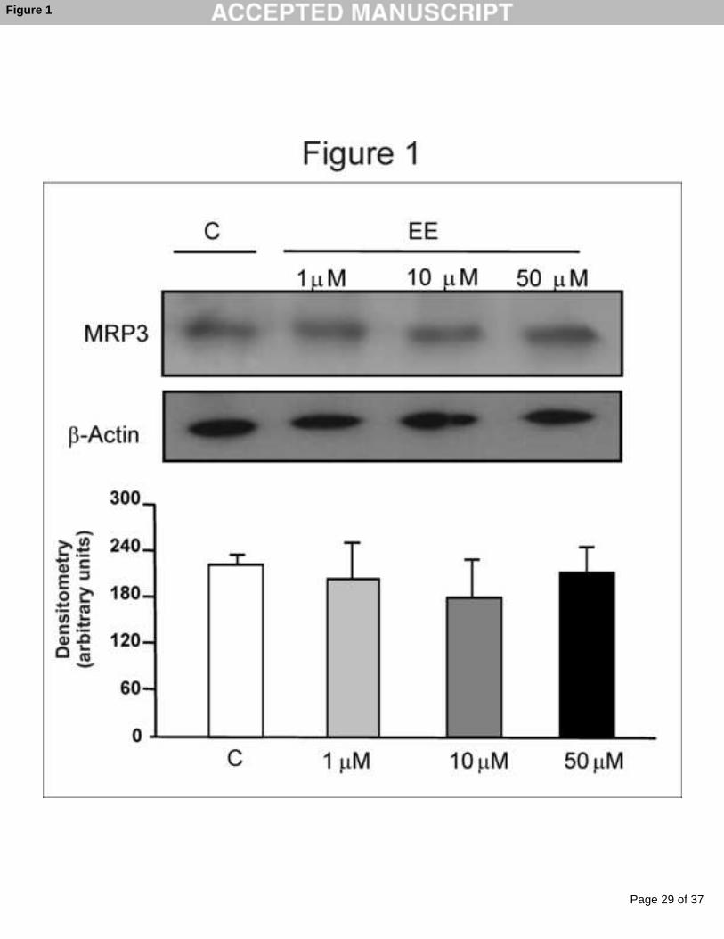
HepG2 are well differentiated cells, retain many hepatocyte-specific
features, and represent a tool for understanding the regulation of MRP3 in a
human liver cell line (21). We found no changes in MRP3 protein expression at
any concentrations of EE analyzed (1, 10 or 50 µM) (Fig 1). HepG2 cells
express ER at low levels that are insufficient for ligands to induce
transactivation of an ERE-containing synthetic target gene under transient
transfection conditions (22, 23). We therefore transfected HepG2 cells with a
plasmid encoding human ER-α, and confirmed ER-α expression by Western
blotting (data not shown). The activity of ER-α was monitored by introducing an
estrogen-responsive indicator in which four copies of the ERE were linked to a
thymidine kinase promoter of a luciferase expression plasmid (4XERE-TK-Luc).
As shown in Fig 2, EE (upper panel) and 17β-estradiol (positive control, lower
panel) treatments activated 4XERE-TK-Luc in a dose dependent manner in
HepG2 cells transfected with ER-α but not in those transfected with the empty
vector. These data demonstrate that ER-α was active and able to transduce the
estrogen signal in HepG2 cells. Western blot studies indicate that MRP3
expression significantly increased (270%) in cells transfected with ER-α and
incubated with 50 µM EE (Fig 3A), whereas it remained unchanged in the
empty vector-transfected HepG2 cells at all concentrations of EE tested (Fig
3B). Real time PCR studies showed an increase in the content of MRP3 mRNA
in ER-α transfected HepG2 cells incubated with 50 µM EE (180%) with respect

Page 13 of 37
Accep
ted
Man
uscr
ipt
13
to control cells, whereas no changes were observed in empty vector-
transfected cells (Fig 3C).
3.2. Changes in the expression of AP-1 are responsible for MRP3 up-
regulation by EE.
To understand how ER-α might mediate MRP3 induction by EE, we
performed an in silico analysis using the TFSearch database
(http://www.cbrc.jp/research/db/TFSEARCH.html). Although we could not
identify any ERE in the mrp3 promoter sequence, the analysis located two
binding sites for AP-1 (Fig 4). This led us to hypothesize an indirect action of
the ER-α involving these transcription factors. To test this, we evaluated the
effect of EE on expression of c-JUN and c-FOS. We found that c-JUN but not c-
FOS protein level was increased in ER-α expressing HepG2 cells after
treatment with 50 µM EE (Fig 5A-B). To explore if c-JUN up-regulation may be
a mediator of MRP3 induction, we analyzed the effect of c-JUN over-expression
on MRP3 levels. HepG2 cells were co-transfected with c-JUN together with ER-
α cDNA or their empty vectors as controls. As shown in Fig 5C, over-
expression of c-JUN in ER-α expressing cells led to up-regulation of MRP3,
even in the absence of incubation with EE, and to a similar extent to that
produced by EE treatment in cells transfected with ER-α only. This same Fig
also shows that EE treatment of cells overexpressing c-JUN did not further
increase MRP3 expression when compared with these same cells incubated
with EE vehicle. ER-α non-expressing HepG2 cells did not show any increase
in MRP3 protein levels in response to transfection with c-JUN (Fig 5D). To

Page 14 of 37
Accep
ted
Man
uscr
ipt
14
further confirm that c-JUN modulation is unambiguously involved in MRP3 up-
regulation, we knocked down c-JUN and tested the effect of EE on MRP3
expression. Figure 6A demonstrates a decrease of 50% in c-JUN protein level
in ER-α-transfected HepG2 cells after treatment with small interference RNA
(siRNA), either in the presence or absence of EE in the incubation medium. As
shown in Fig 6B, knock-down of c-JUN prevented from any MRP3 induction by
EE. In contrast, treatment of control siRNA scrambled-transfected HepG2 cells
with EE resulted in up-regulation of MRP3 by 230% (Fig 6B). This induction is
similar in extent to that shown above for cells not transfected with siRNA or its
control (Fig 3A).
3.3. c-JUN interacts with ER and binds to the mrp3 promoter after EE
stimulus.
The mrp3 proximal promoter region lacks ERE elements but contains
AP-1 binding consensus elements (Fig 4). Our results strongly suggest that ER-
αpresence and c-JUN up-regulation are needed for EE to induce MRP3
expression. Because both actions are needed at the same time, we further
investigated whether EE treatment promotes ER-α/AP-1 interaction. We
performed co-immunoprecipitation studies in ER-α-transfected HepG2 cells.
We observed that a greater fraction of ER-α co-precipitated with c-JUN in the
EE treated cells (Fig 7A). This ER-α/c-JUN interaction was confirmed by
reverse co-immunoprecipitation with ER-α. In this case, the amount of c-JUN
associated with ER-α was greater in EE-treated cells than in control cells (Fig
7B).

Page 15 of 37
Accep
ted
Man
uscr
ipt
15
To address whether c-JUN is recruited to the promoter region of mrp3
after EE treatment, we performed ChIP assays using two pairs of primers that
include the two putative AP-1 binding sites identified in the mrp3 promoter
region (Fig 4). The data show that specific anti c-JUN antibody, but not the non-
immune IgG control, successfully co-immunoprecipitated c-JUN with significant
quantities of the mrp3 promoter (-1300/-1078 bp). Interestingly, in cells treated
with EE, the amount of mrp3 promoter immunoprecipitated with c-JUN
increased significantly (Fig 7C), indicating that EE promotes the binding of AP-1
to the (-1300/-1078 bp) region of the endogenous mrp3 promoter. No
amplification product was detected with the primers ChIP 3 and ChIP 4 (-501/ -
318 bp) (data not shown).
4. DISCUSSION.
ER activated by ligand can regulate transcription through either a
classical or a non-classical mechanisms (15). The in silico analysis of the mrp3
promoter region did not show any ERE sequence but showed several potential
binding sites for AP-1. Based on this information, we proposed that EE-
induction of mrp3 transcription is mediated by ER-α acting not on ERE
sequences but on Sp1 or AP-1 elements (non-classical pathway). Regulation of
AP-1 activity can be achieved through changes in transcription of genes
encoding AP-1 subunits, independent of changes in its phosphorylation status
(24, 25). We found that c-JUN protein expression was increased in nuclear
extracts from HepG2 cells transfected with ER-α and incubated with EE, and

Page 16 of 37
Accep
ted
Man
uscr
ipt
16
that over-expression of c-JUN led to induction of MRP3 expression to the same
extent as found under EE treatment conditions. Experiments in c-JUN knock-
down cells confirmed that this transcription factor is indeed involved in MRP3
up-regulation by EE. Whether AP-1 non-classical pathway is the only
mechanism mediating MRP3 induction by EE is uncertain, however, the
experiments showing that MRP3 induction by c-JUN over-expression was not
further exacerbated by EE treatment (Fig 5C), strongly pointed to c-JUN
induction as a key player.
Several other studies indirectly implicate AP-1 as a key mediator in
increasing Mrp3/MRP3 expression. AP-1 transcriptional activity is increased
300% in HepG2 cells after exposure to taurolithocholic acid (26), and could
explain findings of Teng et al. (27) demonstrating that taurolithocholic acid
increased MRP3 mRNA in human hepatoma HuH7 cell line. Bile duct ligation in
mice increases AP-1 binding activity in liver (28), which could be linked to
studies demonstrating that Mrp3 is up-regulated in liver of bile-duct ligated rats
(29). Liver of mice treated with lipopolysaccharide showed an increase of AP-1
binding activity detected by gel shift assays (30), whereas independent studies
described an up-regulation of Mrp3 after similar treatment in rats (31, 32). It was
reported that binding of c-Jun to electrophile responsive elements (EpRE)
sequences increased subsequent to 4-hydroxy-2-nonenal exposure in HepG2
cells (33), and at the same time produces a marked up-regulation of MRP3 in
human bronchial epithelial and Keap1 wild-type non-small-cell lung carcinoma
(NSCLC) cells (34). Appropriate experiments are needed to establish whether
AP-1 activation and Mrp3 up-regulation are causally or casually related under

Page 17 of 37
Accep
ted
Man
uscr
ipt
17
conditions different from that of EE treatment. MRP3/Mrp3 up-regulation was
demonstrated to be an adaptive response to deal with liver toxicity of harmful
endogenous metabolites, xenobiotics and drugs, either under cholestatic or
non-cholestatic conditions (7, 35). We proposed AP-1 activation to be a
significant participant in this adaptive response.
The mechanisms to explain the increased expression of c-JUN by EE,
and the subsequent interaction with the promoter region of mrp3, remain poorly
understood. It was demonstrated that human c-Jun promoter region has
several transcription factor binding sites including AP-1 sites (36), indicating
that c-Jun could be positively auto-regulated. Oxidative metabolites of EE, i.e.,
the 2-OH and 4-OH catechols, produce reactive oxygen species in rat liver (37),
which in turn up-regulate AP-1 (38). It is thus possible that these same species
contribute to AP-1 induction in our experimental conditions. Further studies are
necessary to elucidate the mechanism of AP-1 up-regulation by EE. Our
findings demonstrating that the over-expression of AP-1 requires the presence
of ER-α to induce MRP3 suggest that they might need to interact with each
other. This hypothesis was confirmed by co-immunoprecipitation assays, which
further demonstrated that EE likely exacerbated this interaction. Moreover,
ChIP analysis shows that from the two putative mrp3 promoters containing AP-
1 binding sites, only the distal one (-1300/-1078 bp) binds the complex c-JUN/
ER-α.
In conclusion, the present data demonstrate for the first time that EE
induce MRP3 in HepG2 cells via ER-α through the non-classical pathway

Page 18 of 37
Accep
ted
Man
uscr
ipt
18
mediated by AP-1. A graphical scheme summarizes the proposed mechanism
(Fig 8).
ACKNOWLEDGEMENTS:
This work was supported by grants from Agencia Nacional de Promoción
Científica y Tecnológica [PICT 2011-0360 and PICT 2010-1072], Consejo Nacional
de Investigaciones Científicas y Técnicas [PIP 112-2008-01-00029/00691],
Universidad Nacional de Rosario [BIO 214], and Fundación Alberto J. Roemmers,
Argentina, and by the National Institute of Child Health and Human Development
[Grant HD58299].
AUTHOR CONTRIBUTIONS.
Participated in research design: Ruiz, Banchio, Mottino, Catania.
Conducted experiments: Ruiz, Rigalli, Arias, Villanueva.
Contributed analytic tools: Banchio, Vore.
Performed data analysis: Ruiz, Rigalli, Mottino, Catania.
Wrote or contributed to the writing of the manuscript: Ruiz, Rigalli, Vore,
Mottino, Catania.

Page 19 of 37
Accep
ted
Man
uscr
ipt
19
REFERENCES
1. König J, Rost D, Cui Y, Keppler D. Characterization of the human
multidrug resistance protein isoform MRP3 localized to the basolateral
hepatocyte membrane. Hepatology 1999;29:1156-1163.
2. Akita H, Suzuki H, Hirohashi T, Takikawa H, Sugiyama Y. Transport
activity of human MRP3 expressed in Sf9 cells: comparative studies with rat
MRP3. Pharm Res 2002;19:34-41.
3. Hirohashi T, Suzuki H, Sugiyama Y. Characterization of the transport
properties of cloned rat multidrug resistance-associated protein 3 (MRP3). J
Biol Chem 1999;274:15181-15185.
4. Hirohashi T, Suzuki H, Takikawa H, Sugiyama Y. ATP-dependent
transport of bile salts by rat multidrug resistance-associated protein 3 (Mrp3). J
Biol Chem 2000;275:2905-2910.
5. König J, Nies AT, Cui Y, Leier I, Keppler D. Conjugate export pumps of
the multidrug resistance protein (MRP) family: localization, substrate specificity,
and MRP2-mediated drug resistance. Biochim Biophys Acta 1999;1461:377-
394.
6. Kool M, van der Linden M, de Haas M, Scheffer GL, de Vree JM, Smith
AJ, Jansen G, et al. MRP3, an organic anion transporter able to transport anti-
cancer drugs. Proc Natl Acad Sci U S A 1999;96:6914-6919.
7. Ghanem CI, Ruiz ML, Villanueva SS, Luquita MG, Catania VA, Jones B,
Bengochea LA, et al. Shift from biliary to urinary elimination of acetaminophen-
glucuronide in acetaminophen-pretreated rats. J Pharmacol Exp Ther
2005;315:987-995.

Page 20 of 37
Accep
ted
Man
uscr
ipt
20
8. Ogawa K, Suzuki H, Hirohashi T, Ishikawa T, Meier PJ, Hirose K,
Akizawa T, et al. Characterization of inducible nature of MRP3 in rat liver. Am J
Physiol Gastrointest Liver Physiol 2000;278:G438-446.
9. Chai J, He Y, Cai SY, Jiang Z, Wang H, Li Q, Chen L, et al. Elevated
hepatic multidrug resistance-associated protein 3/ATP-binding cassette
subfamily C 3 expression in human obstructive cholestasis is mediated through
tumor necrosis factor alpha and c-Jun NH2-terminal kinase/stress-activated
protein kinase-signaling pathway. Hepatology 2012;55:1485-1494.
10. Reyes H, Simon FR. Intrahepatic cholestasis of pregnancy: an estrogen-
related disease. Semin Liver Dis 1993;13:289-301.
11. Vore M. Estrogen cholestasis. Membranes, metabolites, or receptors?
Gastroenterology 1987;93:643-649.
12. Ruiz ML, Villanueva SS, Luquita MG, Ikushiro S, Mottino AD, Catania
VA. Beneficial effect of spironolactone administration on ethynylestradiol-
induced cholestasis in the rat: involvement of up-regulation of multidrug
resistance-associated protein 2. Drug Metab Dispos 2007;35:2060-2066.
13. Ruiz ML, Villanueva SS, Luquita MG, Vore M, Mottino AD, Catania VA.
Ethynylestradiol increases expression and activity of rat liver MRP3. Drug
Metab Dispos 2006;34:1030-1034.
14. Ruiz ML, Rigalli JP, Arias A, Villanueva S, Banchio C, Vore M, Mottino
AD, et al. Induction of hepatic multidrug resistance-associated protein 3 by
ethynylestradiol is independent of cholestasis and mediated by estrogen
receptor. Drug Metab Dispos 2013;41:275-280.
15. Marino M, Galluzzo P, Ascenzi P. Estrogen signaling multiple pathways
to impact gene transcription. Curr Genomics 2006;7:497-508.

Page 21 of 37
Accep
ted
Man
uscr
ipt
21
16. Alvaro D, Alpini G, Onori P, Perego L, Svegliata Baroni G, Franchitto A,
Baiocchi L, et al. Estrogens stimulate proliferation of intrahepatic biliary
epithelium in rats. Gastroenterology 2000;119:1681-1691.
17. Bohan A, Chen WS, Denson LA, Held MA, Boyer JL. Tumor necrosis
factor alpha-dependent up-regulation of Lrh-1 and Mrp3(Abcc3) reduces liver
injury in obstructive cholestasis. J Biol Chem 2003;278:36688-36698.
18. Tzeng SJ, Chang WC, Huang JD. Transcriptional regulation of the rat
Mrp3 gene promoter by the specificity protein (Sp) family members and
CCAAT/enhancer binding proteins. J Biomed Sci 2005;12:741-761.
19. Lowry OH, Rosebrough NJ, Farr AL, Randall RJ. Protein measurement
with the Folin phenol reagent. J Biol Chem 1951;193:265-275.
20. Rigalli JP, Ruiz ML, Perdomo VG, Villanueva SS, Mottino AD, Catania
VA. Pregnane X receptor mediates the induction of P-glycoprotein by
spironolactone in HepG2 cells. Toxicology 2011;285:18-24.
21. Chen W, Cai SY, Xu S, Denson LA, Soroka CJ, Boyer JL. Nuclear
receptors RXRalpha:RARalpha are repressors for human MRP3 expression.
Am J Physiol Gastrointest Liver Physiol 2007;292:G1221-1227.
22. Hanet N, Lancon A, Delmas D, Jannin B, Chagnon MC, Cherkaoui-Malki
M, Latruffe N, et al. Effects of endocrine disruptors on genes associated with
17beta-estradiol metabolism and excretion. Steroids 2008;73:1242-1251.
23. Marino M, Distefano E, Trentalance A, Smith CL. Estradiol-induced IP(3)
mediates the estrogen receptor activity expressed in human cells. Mol Cell
Endocrinol 2001;182:19-26.
24. Karin M, Liu Z, Zandi E. AP-1 function and regulation. Curr Opin Cell Biol
1997;9:240-246.

Page 22 of 37
Accep
ted
Man
uscr
ipt
22
25. Wagner EF. AP-1-Introductory remarks. Oncogene 2001;20:2334-2335.
26. Pusl T, Vennegeerts T, Wimmer R, Denk GU, Beuers U, Rust C.
Tauroursodeoxycholic acid reduces bile acid-induced apoptosis by modulation
of AP-1. Biochem Biophys Res Commun 2008;367:208-212.
27. Teng S, Jekerle V, Piquette-Miller M. Induction of ABCC3 (MRP3) by
pregnane X receptor activators. Drug Metab Dispos 2003;31:1296-1299.
28. Bird MA, Lange PA, Schrum LW, Grisham JW, Rippe RA, Behrns KE.
Cholestasis induces murine hepatocyte apoptosis and DNA synthesis with
preservation of the immediate-early gene response. Surgery 2002;131:556-563.
29. Donner MG, Keppler D. Up-regulation of basolateral multidrug resistance
protein 3 (Mrp3) in cholestatic rat liver. Hepatology 2001;34:351-359.
30. Trauner M, Arrese M, Lee H, Boyer JL, Karpen SJ. Endotoxin
downregulates rat hepatic ntcp gene expression via decreased activity of critical
transcription factors. J Clin Invest 1998;101:2092-2100.
31. Cherrington NJ, Slitt AL, Li N, Klaassen CD. Lipopolysaccharide-
mediated regulation of hepatic transporter mRNA levels in rats. Drug Metab
Dispos 2004;32:734-741.
32. Donner MG, Warskulat U, Saha N, Haussinger D. Enhanced expression
of basolateral multidrug resistance protein isoforms Mrp3 and Mrp5 in rat liver
by LPS. Biol Chem 2004;385:331-339.
33. Levy S, Jaiswal AK, Forman HJ. The role of c-Jun phosphorylation in
EpRE activation of phase II genes. Free Radic Biol Med 2009;47:1172-1179.
34. Mahaffey CM, Zhang H, Rinna A, Holland W, Mack PC, Forman HJ.
Multidrug-resistant protein-3 gene regulation by the transcription factor Nrf2 in
Page 23 of 37
Accep
ted
Man
uscr
ipt
23
human bronchial epithelial and non-small-cell lung carcinoma. Free Radic Biol
Med 2009;46:1650-1657.
35. Villanueva SS, Ruiz ML, Soroka CJ, Cai SY, Luquita MG, Torres AM,
Sanchez Pozzi EJ, et al. Hepatic and extrahepatic synthesis and disposition of
dinitrophenyl-S-glutathione in bile duct-ligated rats. Drug Metab Dispos
2006;34:1301-1309.
36. Angel P, Hattori K, Smeal T, Karin M. The jun proto-oncogene is
positively autoregulated by its product, Jun/AP-1. Cell 1988;55:875-885.
37. Yager JD. Endogenous estrogens as carcinogens through metabolic
activation. J Natl Cancer Inst Monogr 2000:67-73.
38. Dalton TP, Shertzer HG, Puga A. Regulation of gene expression by
reactive oxygen. Annu Rev Pharmacol Toxicol 1999;39:67-101.

Page 24 of 37
Accep
ted
Man
uscr
ipt
24
FIGURE LEGENDS
Figure 1: MRP3 protein expression in wild type HepG2 cells. Cells were
incubated with 17α-ethynylestradiol (EE 1, 10 or 50 µM, 48 h) or vehicle (C).
MRP3 was detected by Western blotting. Equal amounts of protein (15 µg) were
loaded in all lanes. Uniformity of protein loading and transfer from gel to
nitrocellulose membrane were controlled with Ponceau S and detection of β-
actin. Densitometric analyses represent means ± SD of 4 independent
experiments.
Figure 2: Activity of estrogen receptor-α (ER-α) in transfected HepG2 cells.
HepG2 cells were transfected with expression plasmid vectors for ER-α or
empty vector together with an estrogen reporter gene (4XERE-TK-Luc), and
treated with the indicated concentrations of 17α-ethynylestradiol (EE, upper
panel) or 17β-estradiol (E2, lower panel). Data represent mean ± SD of 4
independent experiments. a: Significantly different from control, p<0.05.
Figure 3: MRP3 protein expression in ER-α-transfected HepG2 cells.
A- MRP3 protein expression in HepG2 transfected with estrogen receptor-α
(ER-α) vector.
B- MRP3 protein expression in HepG2 transfected with empty vector. Cells
were incubated with 17α-ethynylestradiol (EE) or vehicle [control (C)] for 48 h.
Equal amounts of protein (15 µg) were loaded in all lanes. Uniformity of protein
loading and transfer from gel to nitrocellulose membrane were controlled with
Ponceau S and detection of β-actin. Densitometric analyses represent means ±

Page 25 of 37
Accep
ted
Man
uscr
ipt
25
SD of 4 independent experiments. a: significantly different from C, p<0.05. b:
significantly different from 17α-ethynylestradiol (EE) 1 µM, p<0.05.
C- MRP3 mRNA expression in HepG2 cells transfected with ER-α or empty
vector. Cells were incubated with EE (50 µM) or vehicle (C) for 48 h and mRNA
levels were detected by real time PCR. Data are presented as a percent of
control (100%) and were expressed as means ± SD of 4-6 independent
experiments. 18S rRNA was used as an internal control. a: significantly different
from C, p<0.05
Figure 4: MRP3 proximal promoter region analysis. Consensus elements for
AP-1 nuclear factors identified by TF-Search database are underlined. Numbers
represent the positions of nucleotides in relationship to the transcription
initiation site +1 indicated by arrows. ChIP 1, ChIP 2, ChIP 3 and ChIP 4
represents the primers utilized in ChIP assay.
Figure 5: Involvement of c-JUN in MRP3 induction.
A- c-JUN, and B- c-FOS, protein expression in nuclear extracts from HepG2
cells transfected with estrogen receptor-α (ER-α) and incubated with EE (50
µM) or vehicle (C).
C- HepG2 cells transfected with ER-α vector (ERα (+)), and D- HepG2 cells
transfected with empty vector (ERα (-)), were transfected with c-JUN (c-JUN
(+)) or empty vector (c-JUN (-)), and incubated with EE (50 µM) or vehicle (C).
MRP3 protein expression was then assessed by Western blotting in cell lysates.
Equal amounts of protein (5 µg for nuclear extracts and 15 µg for total lysates)
were loaded in all lanes. Uniformity of protein loading and transfer from gel to

Page 26 of 37
Accep
ted
Man
uscr
ipt
26
nitrocellulose membrane were controlled with Ponceau S and detection of
histone or β-actin. Densitometric analyses represent means ± SD of 4
independent experiments. a: significantly different from c-JUN(-) C, p<0.05.
Figure 6: MRP3 and c-JUN protein expression in c-JUN knock down HepG2
cells.
A- c-JUN protein expression in nuclear extracts from HepG2 cells transfected
with ER-α and c-JUN siRNA (c-JUN-) or siRNA scramble (c-JUN+).
B- MRP3 protein expression in cell lysates from HepG2 cells transfected with
ER-α and c-JUN siRNA (c-JUN-) or siRNA scramble (c-JUN+).
Cells were incubated with 17α-ethynylestradiol (EE, 50 µM for 48 h) or vehicle
(control (C)).
Equal amounts of protein (5 µg for nuclear extracts and 15 µg for total lysate)
were loaded in all lanes. Uniformity of protein loading and transfer from gel to
nitrocellulose membrane were controlled with Ponceau S and detection of
histone or β-actin. Densitometric analyses represent means ± SD of 4
independent experiments. a: significantly different from c-JUN (+) and c-JUN (-)
EE, p<0.05. b: significantly different from c-JUN (+) C, c-JUN (-) C and c-JUN (-
) EE, p<0.05.
Figure 7: Interaction of ER-α with c-JUN and recruitment of c-JUN to MRP3
promoter. ER-α-transfected HepG2 cells were treated with EE (50 µM, 48 h) or
vehicle (control (C)). Co-immunoprecipitation assays were performed by
treating cell lysates with either c-JUN antibody or normal rabbit IgG followed by
Western blot detection of ER-α (A) or by treating cell lysates with either ER-α

Page 27 of 37
Accep
ted
Man
uscr
ipt
27
antibody or normal rabbit IgG followed by Western blot detection of c-JUN (B).
The inputs represent 2% of total proteins in the cell lysates. (C) ChIP assay for
detection of c-JUN association to MRP3 promoter. An unrelated antibody, anti
IgG, was used as control. Similar results were obtained in a separated
experiment.
IP: immunoprecipitation.
Figure 8: ER-α and c-JUN mediation of human MRP3 up-regulation by 17-
α-ethynylestradiol (EE). The graphical scheme represents a sequence of
events starting with EE treatment of the HepG2 cells and leading ultimately to
induction of human MRP3 protein expression. As a first step of this sequence,
EE induces up-regulation of a major component of AP-1, c-JUN. Secondly, in
order to induce the expression of MRP3, c-JUN interacts with ER-α and the
resulting complex is recruited to a specific promoter region of the human mrp3
gene. On this regard, two putative regions were found to be potential targets for
the complex to bind to, but only one of them showed to be involved in the
current experimental conditions.

Page 28 of 37
Accep
ted
Man
uscr
ipt
28
FOOTNOTES.
These experiments were partially presented as a poster in the 47th
International Liver Congress 2012 in Barcelona, Spain. The presentation was
selected as TOP 10% by the EASL committee.
Abbreviations: MRP3, multidrug resistance-associated protein 3; EE, 17α-
ethynylestradiol; ER, estrogen receptor; AP-1, activating protein-1; DMEM,
Dulbecco´s Modified Eagle´s Medium; FBS, fetal bovine serum; ERE, estrogen
response elements.
Page 29 of 37
Accep
ted
Man
uscr
ipt
Figure 1

Page 30 of 37
Accep
ted
Man
uscr
ipt
Figure 2

Page 31 of 37
Accep
ted
Man
uscr
ipt
Figure 3

Page 32 of 37
Accep
ted
Man
uscr
ipt
Figure 4

Page 33 of 37
Accep
ted
Man
uscr
ipt
Figure 5

Page 34 of 37
Accep
ted
Man
uscr
ipt
Figure 6

Page 35 of 37
Accep
ted
Man
uscr
ipt
Figure 7

Page 36 of 37
Accep
ted
Man
uscr
ipt
Figure 8

Page 37 of 37
Accep
ted
Man
uscr
ipt
*Graphical Abstract (for review)