Embed Size (px)
Citation preview

1
rRNA分子を標的とした定量的RT-PCR 法によるヒト腸内細菌叢の解析法の開発
はじめに
ヒトの腸管内には多種多様な微生物群がバランスを保
ちながら生息しており、この複雑な微生物群集は総称し
て腸内細菌叢と呼ばれる。ヒト腸内細菌叢は Firmicutes、
Actinobacteria、Fusobacteria、Fibrobacteres、
Proteobacteria、Spirochaetes、 お よ び Bacteroidetes
の 7 つの門にまたがる 100 ~ 300 種類の菌種から構成さ
れている 1,2)。それらの生息レベルは菌種により糞便 1
g あたり 102 個から 1011 個に及ぶ範囲で非常に多様で
あるが、その構成は各個人に固有でありかつ安定してい
る。腸内細菌叢構成菌はさまざまな生理活性を有してお
り、その代表的なものとして食物を発酵し消化を助長す
ること、その過程で有機酸や腐敗物質を産生すること 3)、
外来物質(薬物)や内在物質(胆汁酸)を代謝すること 4)、
宿主の腸管の成熟を促すこと 5)、宿主の免疫系と相互作
用すること 6)、外来病原菌を排除すること 7)、などが知
られている。腸内細菌叢はこれらの機能を介して宿主の
健康状態と密接に関係しており、そのバランスが崩れ
rRNA 分子を標的とした定量的 RT-PCR 法によるヒト腸内細菌叢の解析法の開発
松 田 一 乗
Establishment of an Analytical System for the Human IntestinalMicrobiota, Based on Reverse Transcription–Quantitative PCR
Targeting rRNA Molecules
Kazunori Matsuda
Yakult Central Institute for Microbiological Research
1796 Yaho Kunitachi, Tokyo 186-8650 Japan
AbstractHuman intestinal tract harbors the complex microbial community (intestinal microbiota) that includes several hundreds of
species, which has been found to be closely associated with the human health. Recently, several molecular techniques such as
FISH, DNA sequencing, DNA fingerprinting, microarray, and quantitative PCR (qPCR) with rRNA gene-targeted oligonucleotide
probes or primers also have been developed for the intestinal microbiota analysis as a culture independent method. These new
technologies enabled accurate and convenient analysis of targeted predominant anaerobic bacterial species that are difficult to
be cultured. However, it has been demonstrated that sensitivity of above molecular techniques is not sufficient to quantify the
subdominant populations.
To resolve the problem, we have developed an analytical system based on reverse-transcription quantitative PCR (RT-
qPCR) using specific primers that target bacterial rRNA molecules. By targeting rRNA molecules, which are present at 1,000 to
10,000 copies per cell, RT-qPCR has 100 to 1,000 times the sensitivity of conventional methods. Thanks to its high sensitivity,
rRNA-targeted RT-qPCR covers a variety of intestinal bacterial populations including the subdominant bacteria such as lactic
acid bacteria and opportunistic commensal pathogens: Staphylococcus, Pseudomonas, and Clostridium perfringens. This RT-
qPCR method has the following several advantages; 1) High sensitivity (lower detection limit: 102 – 104 cells per g of feces), 2)
High precision and high reproducibility, 3) Rapidity. Therefore, it will enable the large-scale, systematic, quantitative analysis
of human intestinal microbiota, which should be effective for investigating several in situ aspects: the effects of probiotics or
prebiotics and the relationship between microbiota and the human health.
2
ヤクルト研究所研究報告集 第 31号, 01–14(2013)
ることにより腸の機能異常(下痢、便秘、過敏性腸症候
群)や腸疾患(感染性腸炎、潰瘍性大腸炎、クローン病、
大腸癌)が引き起こされることが示唆されている 8,9)。
また、近年では腸内細菌叢が肥満およびメタボリックシ
ンドローム発症に関係することを示す研究が相次いで発
表されている 10-12)。一方で、食事 13,14)や宿主の健康状
態(病気、年齢、ストレス)15-17)、医薬品の服用 18,19)な
どが腸内細菌叢の形成に重要な影響を与えることが知ら
れている。したがって、ヒトの健康増進において宿主と
腸内細菌叢との相互作用を明らかにすることは非常に重
要であり、そのためにはまずこの複雑多様な生態系の微
生物構成を正確に把握することが必要である。
ヒト腸内細菌叢の解析手法
腸内細菌叢の構成の解析は主に培養法により行われ
てきた 20-23)。本手法は、目的とする細菌が検体 1 g あ
たり 100 個程度の低い菌数レベルで存在する場合でも、
それを選択的に分離培養することができる。しかしな
がら、これらの解析作業には多大な労力と時間を必要
とするのみならず、結果が選択培地の性能による影響
を受けやすい、細菌の同定精度は技術者の経験に寄る
ところが大きいといった問題点があった。一方、1980
年代より分子・遺伝学的手法が細菌分類学に用いられ
るようになり、細菌のリボソームを構成する小型のサ
ブユニット(16S)に存在する RNA をコードする遺伝
子(16S rRNA 遺伝子)の塩基配列を指標とした系統分
類が幅広く用いられるようになった 24)。これを受けて、
rRNA 塩基配列を標的としたさまざまな分子生物学的手
法[FISH(fluorescence in situ hybridization)法 25-28)、
DNA シーケンス法 2,14,29-32)、フィンガープリント法 33-
39)、定量的PCR(qPCR)法 40-43)、等]が腸内細菌叢の解
析に幅広く用いられるようになり、これにより腸内細菌
叢に生息する難培養菌を含む多くの細菌を培養すること
なしに検出・同定・定量することが可能となった。しか
しながら、それらの手法の検出下限値は最も低いもので
も糞便 1 g あたり 105 個であり、これより低い菌数レ
ベルで存在する細菌を精度よく解析することができな
かった。そこで本研究では、ヒト腸内細菌を低い菌数
で存在する菌群も含めて網羅的に定量できる手法を確
立するため、細菌中に多コピー存在する rRNA 分子に
着目し、これを標的とした定量的 RT-PCR(RT-qPCR:
reverse transcription qPCR)法による腸内細菌叢解析
システムの構築を試みた。本稿では、RT-qPCR 法の開
発に関する研究成果を紹介した上で、本法の特徴と腸内
細菌叢の解析におけるその有効性を考察する。
RT-qPCR 法による高感度な腸内細菌叢解析法の構築
図 1 には rRNA 分子を標的とした RT-qPCR 法によ
る菌数定量の原理を示した。本法では、糞便より抽出
された総 RNA を鋳型として rRNA 遺伝子を標的とし
た菌群・菌種特異的プライマーを用いた RT-PCR を行
う。その増幅産物をリアルタイム PCR 装置により検出
し、糞便中の標的細菌の菌数をその標準菌株の RNA を
用いて作製する検量線により算出する。本法が標的と
する rRNA 分子は、リボソームの構成成分として細胞
内に数千から数万分子存在していることが知られてい
る 44,45)。したがって、本分子を標的とすることにより
従来の DNA を標的とする手法に比べて検出感度を飛躍
的に向上できる可能性がある。一方で、一般的に RNA
は DNA と比較して安定性が低く 46)、熱処理や栄養状態
の変化などの環境的要因、また増殖効率の違いなどで細
胞中の rRNA 量が変動することが報告されており 47-49)、
rRNA 分子を菌数測定の指標として使用するには、細
菌の増殖状態と rRNA 量の関係を明らかにする必要が
ある。そこで本研究では、rRNA 分子を標的とした RT-
qPCR 法を腸内細菌叢の解析に応用可能であることを確
認するため、最初に腸内の優勢菌群からサブドミナント
な菌群までを網羅するプライマーセットを作製し、本プ
ライマーを用いた RT-qPCR 法の検出感度および測定精
度を解析した。さらに、RT-qPCR 法により健常成人の
糞便を解析し、従来法による結果との比較を行った。
〈RT-qPCR 法による検出下限値の測定〉
腸内の優勢菌群からサブドミナントな菌群までを網
羅するため、16S または 23S rRNA 遺伝子配列をもと
に、既報 41,50-52)のものを含む 20 種類の菌群・菌属・
菌種特異的プライマーセットを作製した 53,54) (表 1)。
図 2 には Enterobacteriaceae および Enterococcus 特
異的プライマーの検出感度を解析した結果を示した。
Escherichia coli および Enterococcus faecalis の各純

3
rRNA分子を標的とした定量的RT-PCR 法によるヒト腸内細菌叢の解析法の開発
cDNA
PCR
rRNA RT-PCR
RNA
A
Threshold line
CT 105 104 103 102 101 100
PCR 0 10 30 20
B
RNA 100 102 104 106
CT
20
0
10
30
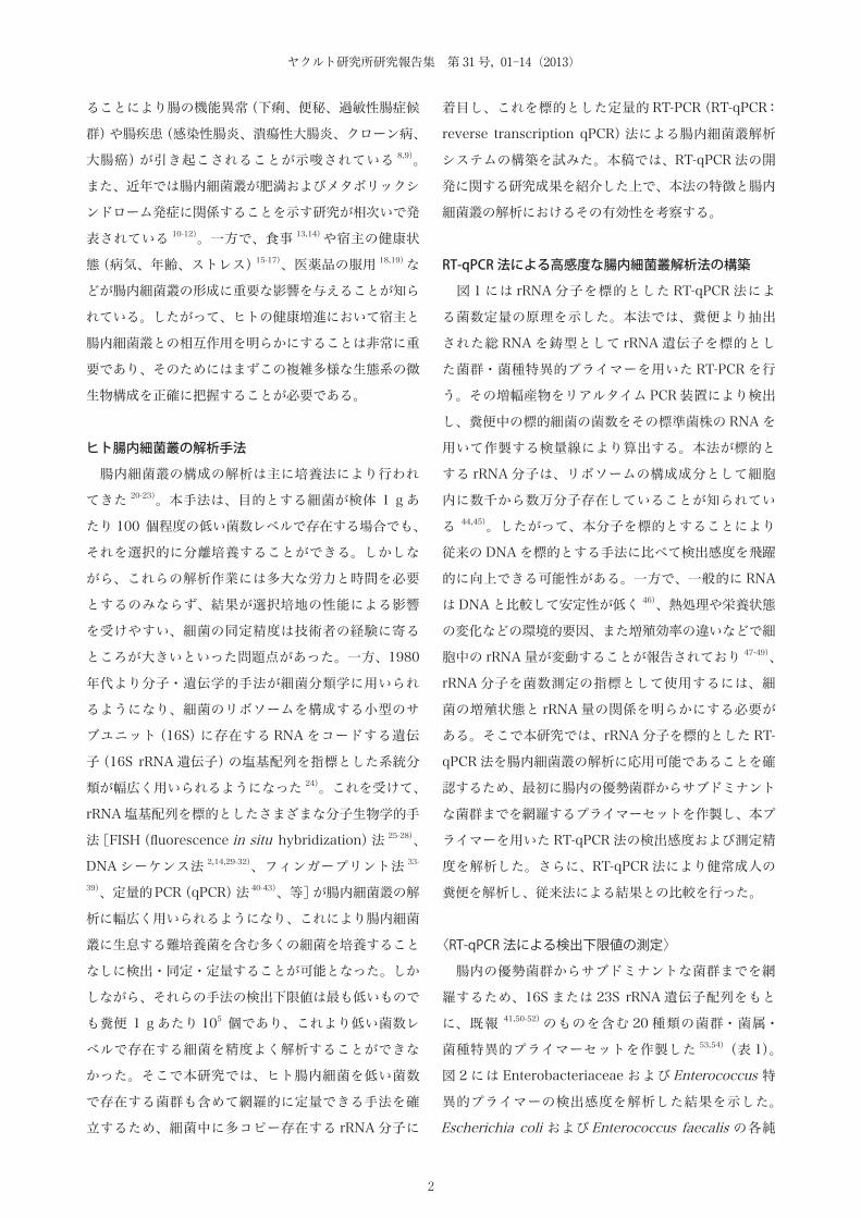
図 1. rRNA 分子を標的とした RT-qPCR 法による腸内細菌叢の解析原理(A)糞便より抽出された総 RNA を鋳型として rRNA 遺伝子を標的とした菌群・菌種特異的プライマーを用いた RT-PCR を行う。増幅された二本鎖 DNA は蛍光色素・SYBR® Green I によりラベルされ,その蛍光強度はリアルタイム PCR 装置により検出される。(B)糞便中の標的細菌の菌数をその標準菌株の RNA を用いて作製する検量線により算出する。最初に測定する細菌の標準菌株から総 RNA を抽出し,元の菌液の DAPI カウント法による菌数をもとに濃度調整した RNA を鋳型として RT-qPCR を行う。得られた増幅曲線上に Threshold ラインを設定し,それと増幅曲線との交点のサイクル数(CT 値)を求める。CT 値を縦軸に,供試した RNA 量(DAPI カウント菌数換算)を横軸にプロットし得られる近似曲線を検量線とする。
図 2. RT-qPCR 法および qPCR 法の検出感度の比較BHI ブロスおよび MRS ブロスで 37°C,18 時間培養した E. coli YIT 6044T(A)および E. faecalis YIT 2031T(B)の各菌液より総 RNA および DNA をそれぞれ抽出した。同時に菌液中の菌数を DAPI カウント法により測定し,10–3 個から 105 個に相当する総 RNA および DNA を鋳型として RT-qPCR(○)および qPCR(●)をそれぞれ行った。CT 値に対する近似曲線に直線性が認められる RNA および DNA の濃度範囲を求めた。
Log10 cells/reaction
CT v
alue
y = -3.36x + 24.42R2 = 0.999
y = -3.34x + 30.78R2 = 0.998
-4 -2 0 2 4 6
15 20 25 30 35
10
40
5
B
y = -3.23x + 29.13R2 = 0.998
y = -3.45x + 38.82R2 = 0.987
-2 0 2 4 6
15 20 25 30 35
10
40
5
A
4
ヤクルト研究所研究報告集 第 31号, 01–14(2013)
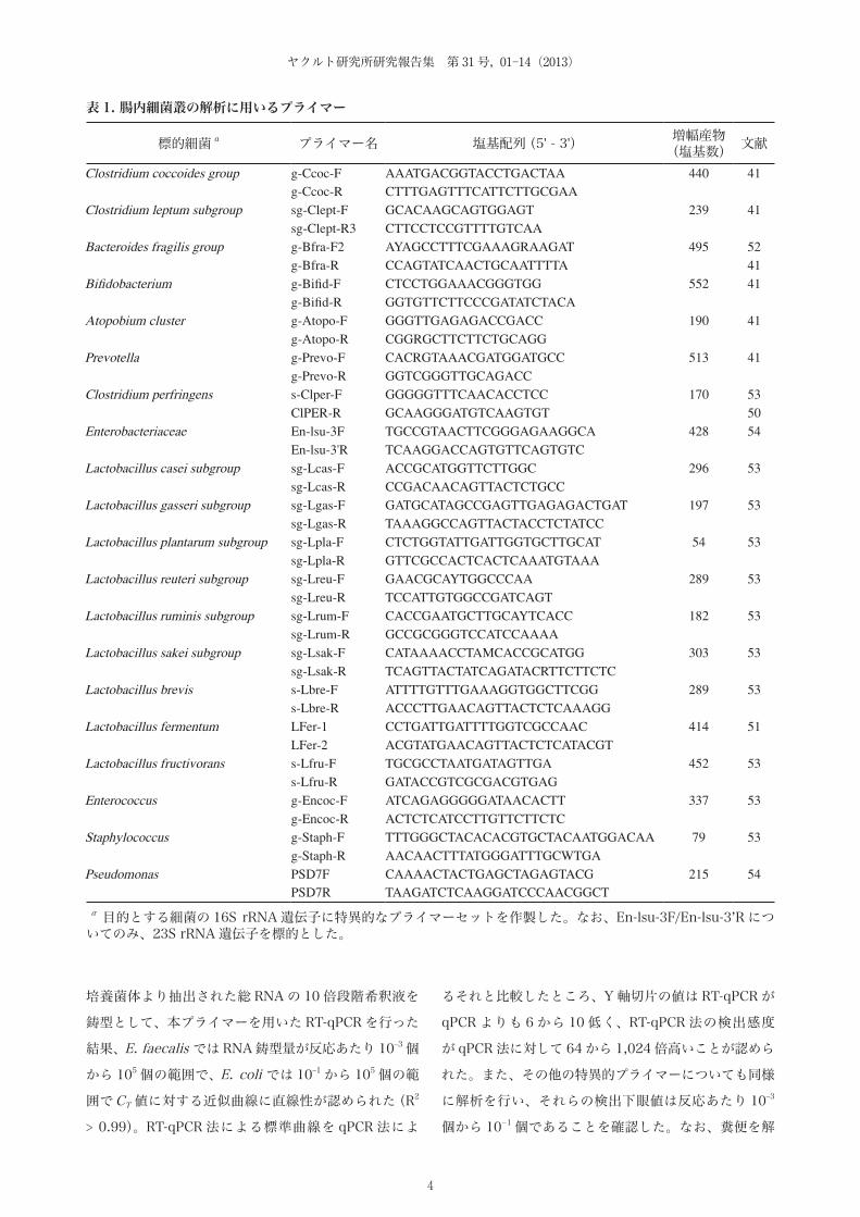
培養菌体より抽出された総 RNA の 10 倍段階希釈液を
鋳型として、本プライマーを用いた RT-qPCR を行った
結果、E. faecalis では RNA 鋳型量が反応あたり 10–3 個
から 105 個の範囲で、E. coli では 10–1 から 105 個の範
囲でCT 値に対する近似曲線に直線性が認められた(R2
> 0.99)。RT-qPCR 法による標準曲線を qPCR 法によ
るそれと比較したところ、Y 軸切片の値は RT-qPCR が
qPCR よりも 6 から 10 低く、RT-qPCR 法の検出感度
が qPCR 法に対して 64 から 1,024 倍高いことが認めら
れた。また、その他の特異的プライマーについても同様
に解析を行い、それらの検出下眼値は反応あたり 10–3
個から 10−1 個であることを確認した。なお、糞便を解
表 1. 腸内細菌叢の解析に用いるプライマー
標的細菌 a プライマー名 塩基配列(5' - 3') 増幅産物(塩基数) 文献
Clostridium coccoides group g-Ccoc-F AAATGACGGTACCTGACTAA 440 41 g-Ccoc-R CTTTGAGTTTCATTCTTGCGAA
Clostridium leptum subgroup sg-Clept-F GCACAAGCAGTGGAGT 239 41 sg-Clept-R3 CTTCCTCCGTTTTGTCAA
Bacteroides fragilis group g-Bfra-F2 AYAGCCTTTCGAAAGRAAGAT 495 52 g-Bfra-R CCAGTATCAACTGCAATTTTA 41
Bifidobacterium g-Bifid-F CTCCTGGAAACGGGTGG 552 41 g-Bifid-R GGTGTTCTTCCCGATATCTACA
Atopobium cluster g-Atopo-F GGGTTGAGAGACCGACC 190 41 g-Atopo-R CGGRGCTTCTTCTGCAGG
Prevotella g-Prevo-F CACRGTAAACGATGGATGCC 513 41 g-Prevo-R GGTCGGGTTGCAGACC
Clostridium perfringens s-Clper-F GGGGGTTTCAACACCTCC 170 53 ClPER-R GCAAGGGATGTCAAGTGT 50
Enterobacteriaceae En-lsu-3F TGCCGTAACTTCGGGAGAAGGCA 428 54 En-lsu-3'R TCAAGGACCAGTGTTCAGTGTC
Lactobacillus casei subgroup sg-Lcas-F ACCGCATGGTTCTTGGC 296 53 sg-Lcas-R CCGACAACAGTTACTCTGCC
Lactobacillus gasseri subgroup sg-Lgas-F GATGCATAGCCGAGTTGAGAGACTGAT 197 53 sg-Lgas-R TAAAGGCCAGTTACTACCTCTATCC
Lactobacillus plantarum subgroup sg-Lpla-F CTCTGGTATTGATTGGTGCTTGCAT 54 53 sg-Lpla-R GTTCGCCACTCACTCAAATGTAAA
Lactobacillus reuteri subgroup sg-Lreu-F GAACGCAYTGGCCCAA 289 53 sg-Lreu-R TCCATTGTGGCCGATCAGT
Lactobacillus ruminis subgroup sg-Lrum-F CACCGAATGCTTGCAYTCACC 182 53 sg-Lrum-R GCCGCGGGTCCATCCAAAA
Lactobacillus sakei subgroup sg-Lsak-F CATAAAACCTAMCACCGCATGG 303 53 sg-Lsak-R TCAGTTACTATCAGATACRTTCTTCTC
Lactobacillus brevis s-Lbre-F ATTTTGTTTGAAAGGTGGCTTCGG 289 53 s-Lbre-R ACCCTTGAACAGTTACTCTCAAAGG
Lactobacillus fermentum LFer-1 CCTGATTGATTTTGGTCGCCAAC 414 51 LFer-2 ACGTATGAACAGTTACTCTCATACGT
Lactobacillus fructivorans s-Lfru-F TGCGCCTAATGATAGTTGA 452 53 s-Lfru-R GATACCGTCGCGACGTGAG
Enterococcus g-Encoc-F ATCAGAGGGGGATAACACTT 337 53 g-Encoc-R ACTCTCATCCTTGTTCTTCTC
Staphylococcus g-Staph-F TTTGGGCTACACACGTGCTACAATGGACAA 79 53 g-Staph-R AACAACTTTATGGGATTTGCWTGA
Pseudomonas PSD7F CAAAACTACTGAGCTAGAGTACG 215 54 PSD7R TAAGATCTCAAGGATCCCAACGGCT
a 目的とする細菌の 16S rRNA 遺伝子に特異的なプライマーセットを作製した。なお、En-lsu-3F/En-lsu-3’R についてのみ、23S rRNA 遺伝子を標的とした。

5
rRNA分子を標的とした定量的RT-PCR 法によるヒト腸内細菌叢の解析法の開発
析する場合の検出下限値については糞便 1 g あたり 102
個から 104 個になると予測された。
〈培養状態の違いが RT-qPCR 菌数に及ぼす影響の解析〉
培養状態の違いが RT-qPCR 法による測定菌数に及ぼ
す影響を調べるため、様々な培養期の菌体を用いて、培
養法による測定菌数(CFU 数)と RT-qPCR 菌数との関
係を調べた(図 3)。E. coli およびE. faecalis を 60 時
間連続培養し、その間の菌数の変動を両測定法により調
べたところ、RT-qPCR 菌数は菌体の増殖時期に関わら
ず CFU 数とよく一致することが確認された。対数増殖
期の細菌では RT-qPCR 菌数が CFU 数を上回る傾向が
観察されたが、その差は最大でも 4 倍にとどまり両測
定法による菌数に有意な差はなかった。本結果は rRNA
分子が菌数測定の指標となり得ることを支持するもので
あり、本分子を標的とした RT-qPCR 法を菌数測定に適
用可能であると考えられた。
〈RT-qPCR 法によるヒト糞便細菌叢の解析〉
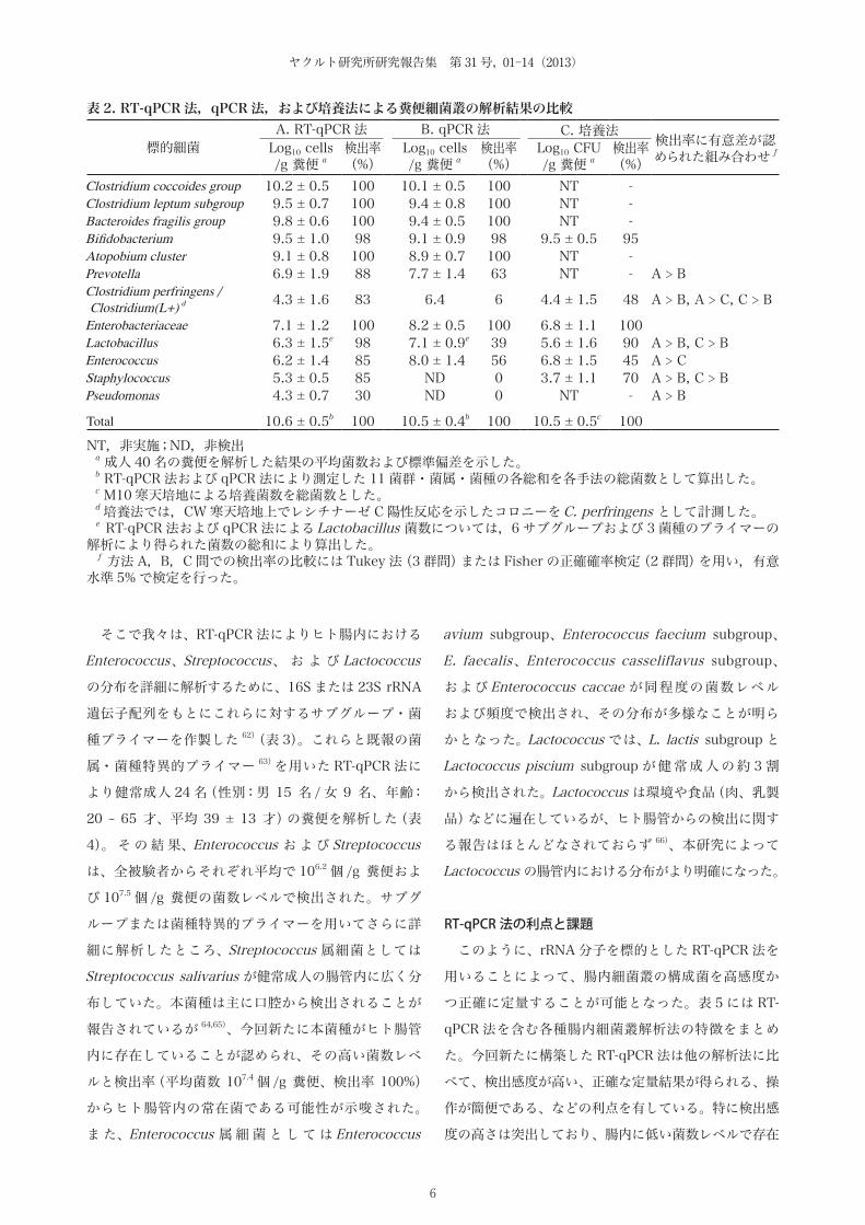
RT-qPCR 法により健常成人 40 名(男性 24 名、女性
16名;年齢 20 ~ 65歳[平均 41 ± 13 歳])の糞便を解析
し、測定結果を従来法と比較した(表 2)。ヒト腸内に優
勢に存在する嫌気性菌群であるClostridium coccoides
group、Clostridium leptum subgroup、Bacteroides
fragilis group、Bifidobacterium、 お よ び Atopobium
cluster に関しては、RT-qPCR 法による測定菌数およ
び検出率は qPCR 法による結果と同等であった。一方、
腸管内に低い菌数レベルで生息するC. perfringens、
Lactobacillus、Enterococcus、Staphylococcus、 お よ
びPseudomonas 等については、RT-qPCR 法による検出
率は qPCR 法あるいは培養法による結果よりも高かった
(P < 0.05)。上記結果より、RT-qPCR 法はヒト腸内の優
勢菌群および低い菌数レベルで存在する細菌を網羅的に
しかも正確に解析できる有効な手法であることが示唆さ
れた。
Enterococcus、Streptococcus、およびLactococcus の
菌属・サブグループ・菌種別測定系の構築と分布の解析
Enterococcus、Streptococcus、およびLactococcus
は、通性嫌気性、カタラーゼ陰性のグラム陽性球菌
で あ り、Streptococcus thermophilus、Lactococcus
lactis、E. faecalis と い っ た 菌 種 は 発 酵 食 品 に 広 く
用 い ら れ て い る 55-57)。 ま た、Enterococcus お よ び
Streptococcus は、ヒト腸内のサブドミナントな構成
菌としてヒト糞便からの検出が数多く報告されている58,59)。その一方で、Enterococcus およびStreptococcus
にはさまざまな感染症原因菌が含まれており、その中で
も高度薬剤耐性を獲得したバンコマイシン耐性腸球菌
(vancomycin resistant Enterococcus、VRE)の増加は
世界的な問題となっている 60,61)。
Incubation time (h)
Log 1
0 bac
teria
/
ml c
ultu
re
B
2
4
6
8
10
0 12 24 36 48 60
A
2
4
6
8
10
0 12 24 36 48 60
図 3. 培養状態の違いが RT-qPCR 法による測定菌数に及ぼす影響E. coli YIT 6044T(A)およびE. faecalis YIT 2031T(B)を 60 時間連続培養し,その間の菌数の変動を培養法(●)および RT-qPCR 法(○)により測定した。RT-qPCR 法の検量線の作製には,各菌株を 18 時間培養した菌液より抽出された総 RNA を使用した。
6
ヤクルト研究所研究報告集 第 31号, 01–14(2013)
そこで我々は、RT-qPCR 法によりヒト腸内における
Enterococcus、Streptococcus、 お よ び Lactococcus
の分布を詳細に解析するために、16S または 23S rRNA
遺伝子配列をもとにこれらに対するサブグループ・菌
種プライマーを作製した 62) (表 3)。これらと既報の菌
属・菌種特異的プライマー 63)を用いた RT-qPCR 法に
より健常成人 24 名(性別:男 15 名 / 女 9 名、年齢:
20 ~ 65 才、平均 39 ± 13 才)の糞便を解析した(表
4)。 そ の 結 果、Enterococcus お よ び Streptococcus
は、全被験者からそれぞれ平均で 106.2 個 /g 糞便およ
び 107.5 個 /g 糞便の菌数レベルで検出された。サブグ
ループまたは菌種特異的プライマーを用いてさらに詳
細に解析したところ、Streptococcus 属細菌としては
Streptococcus salivarius が健常成人の腸管内に広く分
布していた。本菌種は主に口腔から検出されることが
報告されているが 64,65)、今回新たに本菌種がヒト腸管
内に存在していることが認められ、その高い菌数レベ
ルと検出率(平均菌数 107.4 個 /g 糞便、検出率 100%)
からヒト腸管内の常在菌である可能性が示唆された。
ま た、Enterococcus 属 細 菌 と し て は Enterococcus
avium subgroup、Enterococcus faecium subgroup、
E. faecalis、Enterococcus casseliflavus subgroup、
および Enterococcus caccae が同程度の菌数レベル
および頻度で検出され、その分布が多様なことが明ら
かとなった。Lactococcus では、L. lactis subgroup と
Lactococcus piscium subgroup が 健 常 成 人 の 約 3 割
から検出された。Lactococcus は環境や食品(肉、乳製
品)などに遍在しているが、ヒト腸管からの検出に関す
る報告はほとんどなされておらず 66)、本研究によって
Lactococcus の腸管内における分布がより明確になった。
RT-qPCR 法の利点と課題
このように、rRNA 分子を標的とした RT-qPCR 法を
用いることによって、腸内細菌叢の構成菌を高感度か
つ正確に定量することが可能となった。表 5 には RT-
qPCR 法を含む各種腸内細菌叢解析法の特徴をまとめ
た。今回新たに構築した RT-qPCR 法は他の解析法に比
べて、検出感度が高い、正確な定量結果が得られる、操
作が簡便である、などの利点を有している。特に検出感
度の高さは突出しており、腸内に低い菌数レベルで存在
表 2. RT-qPCR 法,qPCR 法,および培養法による糞便細菌叢の解析結果の比較
標的細菌A. RT-qPCR 法 B. qPCR 法 C. 培養法 検出率に有意差が認
められた組み合わせ fLog10 cells/g 糞便 a
検出率 (%)
Log10 cells/g 糞便 a
検出率(%)
Log10 CFU/g 糞便 a
検出率(%)
Clostridium coccoides group 10.2 ± 0.5 100 10.1 ± 0.5 100 NT -Clostridium leptum subgroup 9.5 ± 0.7 100 9.4 ± 0.8 100 NT -Bacteroides fragilis group 9.8 ± 0.6 100 9.4 ± 0.5 100 NT -Bifidobacterium 9.5 ± 1.0 98 9.1 ± 0.9 98 9.5 ± 0.5 95 Atopobium cluster 9.1 ± 0.8 100 8.9 ± 0.7 100 NT -Prevotella 6.9 ± 1.9 88 7.7 ± 1.4 63 NT - A > BClostridium perfringens / Clostridium(L+) d 4.3 ± 1.6 83 6.4 6 4.4 ± 1.5 48 A > B, A > C, C > B
Enterobacteriaceae 7.1 ± 1.2 100 8.2 ± 0.5 100 6.8 ± 1.1 100 Lactobacillus 6.3 ± 1.5e 98 7.1 ± 0.9e 39 5.6 ± 1.6 90 A > B, C > BEnterococcus 6.2 ± 1.4 85 8.0 ± 1.4 56 6.8 ± 1.5 45 A > CStaphylococcus 5.3 ± 0.5 85 ND 0 3.7 ± 1.1 70 A > B, C > BPseudomonas 4.3 ± 0.7 30 ND 0 NT - A > B
Total 10.6 ± 0.5b 100 10.5 ± 0.4b 100 10.5 ± 0.5c 100 NT,非実施;ND,非検出 a 成人 40 名の糞便を解析した結果の平均菌数および標準偏差を示した。 b RT-qPCR 法および qPCR 法により測定した 11 菌群・菌属・菌種の各総和を各手法の総菌数として算出した。 c M10 寒天培地による培養菌数を総菌数とした。 d 培養法では,CW 寒天培地上でレシチナーゼ C 陽性反応を示したコロニーをC. perfringens として計測した。 e RT-qPCR 法および qPCR 法によるLactobacillus 菌数については,6 サブグループおよび 3 菌種のプライマーの解析により得られた菌数の総和により算出した。 f 方法 A,B,C 間での検出率の比較には Tukey 法(3 群間)または Fisher の正確確率検定(2 群間)を用い,有意水準 5% で検定を行った。

7
rRNA分子を標的とした定量的RT-PCR 法によるヒト腸内細菌叢の解析法の開発
する菌群を正確に定量するには RT-qPCR 法が最も有効
である。また、RT-qPCR 法による解析操作は RNA 抽
出および PCR のみと非常に簡便であり、それらを自動
化することが容易である。我々は既に糞便検体からの
RNA 抽出から RT-qPCR 解析までを全自動で行える機
器システムを構築している。これを用いることにより糞
便 96 検体から RNA を抽出し、それを 20 種類のプラ
イマーで解析する作業を約 5 日間で完了することがで
きる。したがって、RT-qPCR 法はプロバイオティクス
飲用による腸内細菌叢への効果を調べる大規模な介入試
験や、菌叢構成の変化が大きい乳児期の腸内細菌叢の形
成過程を調べる試験において非常に有効であると考えら
れる。
これに対して、本法の問題点としてまず懸念されるの
が標的分子の安定性であろう。一般的に RNA は DNA
よりも分解を受けやすいため、RT-qPCR 法による解析
では検体の保存状態が結果に及ぼす影響がより大きいと
考えられる。この問題点について、Life Technologies
社より市販されている RNA 安定化試薬:RNAlater に糞
便試料を懸濁しておくことで、室温でも 1 か月間糞便
中の rRNA 分子を安定な状態で保存できることを別の
試験で確認しており、RNA の安定性に関する問題は解
表 3. Enterococcus,Streptococcus,およびLactococcus の菌属・サブグループ・菌種プライマー
標的細菌 a プライマー名 塩基配列(5' - 3') 増幅産物(塩基数) 文献
Enterococcus genus g-Encoc-F ATCAGAGGGGGATAACACTT 336 53g-Encoc-R ACTCTCATCCTTGTTCTTCTC
Enterococcus faecalis s-Efs-F CCCGAGTGCTTGCACTCAATTGG 419 62s-Efs-R AGGGGACGTTCAGTTACTAACGT
Enterococcus caccae s-Ecacc-F CCGCATAATAGTCGACACC 318 62s-Ecacc-R GTCAAGGTAAGAGCAGTTACTCTCCTA
Enterococcus cecorum s-Ececo-F TTCCATTTACCGCATGGTAGATGGAT 311 62s-Ececo-R CCGTCAAGGGATGAACTTTCCAC
Enterococcus sulfureus subgroup sg-Esulf-F TTCTTTCTTATCGAACTTCGGTTCA 410 62sg-Esulf-R ACTCTCATCCTTGTTCTTCTC
Enterococcus casseliflavus subgroup sg-Ecass-F CACTATTTTCCGCATGGAAGAAAG 311 62sg-Ecass-R CCGTCAAGGGATGAACATTTTAC
Enterococcus avium subgroup sg-Eavi-F CAGCATCTTTTATAGGATGTTACTTTTCA 213 62sg-Eavi-R GGTCCTTCGACTATCTCACTGG
Enterococcus dispar s-Edis-F CCGCATAATATTAATGAACTCATGTTT 318 62s-Edis-R CCGTCAAGGGATGAACATTTTAC
Enterococcus faecium subgroup sg-Efm-F AGCTTGCTCCACCGGAAAAAGA 164 62sg-Efm-R ATCCATCAGCGACACCSKAA
Enterococcus faecium s-Efm-F GTCTGTCCAAGCAGTAAGTCTGAAGAG 65 62s-Efm-R CATCACAGCTTGTCCTTAAGAAAAG
Streptococcus genus g-Str-F AGCTTAGAAGCAGCTATTCATTC 309 63g-Str-R GGATACACCTTTCGGTCTCTC
Streptococcus agalactiae s-Sag-F GTTATTTAAAAGGAGCAATTGCTT 285 63s-Sag-R TTGGTAGATTTTCCACTCCTACCA
Streptococcus pyogenes s-Spy-F AAGAGAGACTAACGCATGTTAGTAATTT 443 63s-Spy-R AATGCCTTTAACTTCAGACTTAAAAA
Streptococcus pneumoniae / S. mitis s-Spn-F CAATGTGGACTCAAAGATTATAGAAGAATG 396 63s-Spn-R GTCATGATACTAAGGCGCCCTA
Streptococcus salivarius s-Ssal-F CAATGGATGACACATGTCATTTAT 682 62 subsp. salivarius / thermophilus s-Ssal-R GGCACTGAATCCCGGAAAGGATCCLactococcus lactis subgroup sg-Lclac-F TGTAGGGAGCTATAAGTTCTCTGTA 613 62
sg-Lclac-R GGCAACCTACTTYGGGTACTCCCLactococcus piscium subgroup sg-Lcpis-F GCTATCCAGCCCTAAGTGA 614 62
sg-Lcpis-R AAAGGTTAGCTCACCGGCTTTGGGTA
asg-Eavi-F/R, s-Efm-F/R, g-Str-F/R および s-Spn-F/R については目的とする細菌の 23S rRNA 遺伝子を、その他のプライマーについては 23S rRNA 遺伝子を標的とした。

8
ヤクルト研究所研究報告集 第 31号, 01–14(2013)
決されたと考えている 67)。また、RT-qPCR 法は qPCR
法や FISH 法と同様に、あらかじめ標的とする細菌の遺
伝子配列情報を必要とし、未知の細菌を検出するのには
適していない。さらに、RT-qPCR 法や qPCR 法で菌数
を測定するには検量線を作製するための標準菌株が必要
なため、菌株が分離されていない難培養菌の集団につ
いてはそれらの菌数を測定することは困難となる。RT-
qPCR 法により腸内細菌叢の構成菌を網羅的に解析する
には、DNA シーケンス解析等で得られた配列情報をも
とに系統的に幅広い菌種を網羅できる菌群プライマーを
設計する必要があり、この点は今後の課題と考えてい
る。
一方で、本手法は、その高い検出感度と迅速性から食
品衛生や臨床検査領域において非常に有効な細菌検出法
となり得る。Sakaguchi ら 63)は、癌化学療法施行後に
細菌感染症と診断された患児の血中細菌を RT-qPCR 法
および培養法により解析し、RT-qPCR 法による細菌検
出率は培養法よりも約 4 倍高かったことを報告してい
表 5. 腸内細菌叢解析手法の比較
培養法分子生物学的手法
RT-qPCR 法 qPCR 法 FISH 法 マイクロアレイ法 DNA シーケンス法(次世代高速シーケンス)
フィンガープリント法(DGGE,T-RFLP 等)
検出感度(CFU or cells/g) >102 >102 >105 >108 >107 >106 >108
定量性 高 高 高 高 高 高 半定量的
特異性 菌株 ~ 菌種 ~ 菌群 菌株 ~ 菌群 菌種 ~ 菌群 菌種 ~ 菌群 菌種 ~ 菌群 菌種 ~ 菌群
精度 低 高 高 中 中 高 低 ~ 中
簡便性 難 中 中 難 中 中 中 ~ 難
備考 培養できる菌株が限られる 予め標的菌の配列情報が必要 未知菌を含めて
解析することが可能
表 4. 健常成人におけるEnterococcus,Streptococcus,およびLactococcus のサブグループ・菌種分布の解析
標的細菌 菌数(Log10 cells/g feces)a 検出率(%)
Genus Enterococcus 6.2 ± 1.4 100 E. avium subgroup 5.4 ± 1.4 79 E. faecium subgroup 5.9 ± 1.6 46 E. faecalis 5.2 ± 1.4 46 E.casselifavus subgroup 4.4 ± 1.0 33 E. caccae 4.0 4 E. dispar ND 0 E. sulfureus subgroup ND 0 E. cecorum ND 0
Genus Streptococcus 7.5 ± 0.9 100 S. salivarius 7.4 ± 0.8 100 S. pneumoniae / S. mitis 5.7 ± 0.8 100 S. agalactiae 4.9 ± 0.6 29 S. pyogenes ND 0
L. lactis subgroup 5.0 ± 1.0 21 L. piscium subgroup 3.9 ± 1.2 13 a 成人 24 名の糞便を解析した結果の平均菌数および標準偏差を示した。

9
rRNA分子を標的とした定量的RT-PCR 法によるヒト腸内細菌叢の解析法の開発
る。今後、本法を当領域に応用する上で真菌、酵母を含
む病原微生物に対するプライマーの拡充、また病原性に
関与する遺伝子の検出システムの構築、さらには解析作
業の簡素化といったことが今後の課題となるだろう。
おわりに
本稿では、rRNA 分子を標的とした RT-qPCR 法につ
いて、その開発に関する研究成果を紹介するとととも
に、本手法が腸内細菌叢を高感度かつ迅速に解析する方
法として非常に有効であることを解説した。近年の分
子生物学的手法の発展、特に DNA シーケンサーの技術
革新を契機として腸内細菌叢の研究は新たな時代へと
突入しており、次世代型シーケンサーを用いた解析によ
り複雑な腸内細菌叢の全体像が明らかにされてきている8,10,11,68,69)。また、細菌叢の構成に加えて細菌叢の構成
ゲノム(マイクロバイオーム)を網羅的に解析すること
により、腸内細菌叢の形成に影響を及ぼす因子 70)や腸
内細菌叢と健康や腸疾患との関連性を考察する知見 71)
が数多く報告されてきている。これらの解析は腸内細菌
叢の構造および機能についてさまざまな側面から莫大な
情報を得ることができる一方で、解析対象は最優勢菌群
に限られており、また大量の検体の解析には適していな
い。したがって、今後はシーケンス解析で得られた情報
をもとに注目すべき菌群・菌属・菌種を選定し、それら
について RT-qPCR 法を用いて多くの被験者でより詳細
に解析するのが効果的であろう。これらの手法を用いた
今後の研究により、腸内細菌叢の形成に影響を及ぼす因
子や、腸内細菌叢と健康や疾患との関連性について、そ
の詳細が明らかになっていくことが期待される。また、
RT-qPCR 法は、その高い検出感度と迅速性から、食品
衛生や臨床検査領域において非常に有効な細菌検出法と
なり得る可能性も示唆されたことから、今後の応用研究
も重要な検討課題と考えられる。
要 約
ヒト腸管内には 100 種類以上の細菌が複雑な微生物
生態系(腸内細菌叢)を形成しており、宿主の健康と密
接に関係していることが明らかにされている。近年、
16S rRNA 遺伝子を標的としたさまざまな分子生物学
的手法[FISH 法、DNA シーケンス法、フィンガープリ
ント法、マイクロアレイ法、定量的 PCR(quantitative
PCR:qPCR)法、等]が腸内細菌叢の研究に幅広く用い
られるようになった。これらの方法を用いて、腸内に生
息する難培養菌を含む多くの細菌を培養することなしに
検出・同定・定量することが可能となったが、一方で
当該手法は検出限界が高いため腸内に低い菌数レベル
で存在する細菌の検出や定量には不向きであった。そ
こで本研究では、ヒト腸内細菌を低い菌数で存在する
菌群も含めて網羅的に定量できる手法として、細菌が
その細胞内に多コピーを有する rRNA 分子を標的とし
た定量的 RT-PCR(reverse transcription–quantitative
PCR:RT-qPCR)法による腸内細菌叢解析システムの
構築を試みた。RT-qPCR 法は従来の qPCR 法に比べて
100 倍から 1,000 倍程度検出感度が高く、そのためヒ
ト腸内の優勢菌群のみならず乳酸菌や日和見感染症原
因 菌(Staphylococcus、Pseudomonas、Clostridium
perfringens)などの低い菌数レベルで存在する細菌を
糞便 1 g あたり 102 ~ 104 個という高い感度で正確に定
量することができる。その他の RT-qPCR 法の利点とし
て、分類体系に基づいた正確な定量結果が得られる、操
作が簡便である、などが挙げられるが、これらの利点は
大規模な介入試験をもとにプロバイオティクス飲用によ
る腸内細菌叢への効果を調べたり、腸内細菌叢と健康と
の関連性を明らかにしていく上で非常に重要である。
謝 辞
本研究の遂行に当たり、株式会社ヤクルト本社中央研
究所 野本 康二 審議役、梅﨑 良則 特別研究員、髙橋 琢
也 主任研究員、辻 浩和 主任研究員、朝原 崇 主任研究
員には数多くのご指導、ご御鞭撻を頂きました。また、
松本 一政 主任研究員、高田 敏彦 主任研究員、松木 隆
広 主任研究員、角 有希子 主事補、倉川 尚 副指導研究
員、久保田 博之 研究員には数多くの貴重なご助言とご
協力を頂きました。ここに深く感謝の意を表します。
引用文献
1 Qin J., Li R., Raes J., Arumugam M., Burgdorf K.
S., Manichanh C., Nielsen T., Pons N., Levenez
F., Yamada T., Mende D. R., Li J., Xu J., Li S.,
Li D., Cao J., Wang B., Liang H., Zheng H., Xie
10
ヤクルト研究所研究報告集 第 31号, 01–14(2013)
Y., Tap J., Lepage P., Bertalan M., Batto J. M.,
Hansen T., Le Paslier D., Linneberg A., Nielsen
H. B., Pelletier E., Renault P., Sicheritz-Ponten T.,
Turner K., Zhu H., Yu C., Jian M., Zhou Y., Li Y.,
Zhang X., Qin N., Yang H., Wang J., Brunak S.,
Dore J., Guarner F., Kristiansen K., Pedersen O.,
Parkhill J., Weissenbach J., Bork P. and Ehrlich
S. D., A human gut microbial gene catalogue
established by metagenomic sequencing. Nature.
464 (7285), 59-65 (2010).
2 Eckburg P. B., Bik E. M., Bernstein C. N.,
Purdom E., Dethlefsen L., Sargent M., Gill S. R.,
Nelson K. E. and Relman D. A., Diversity of the
human intestinal microbial flora. Science. 308
(5728), 1635-8 (2005).
3 Sato T., Matsumoto K., Okumura T., Yokoi W.,
Naito E., Yoshida Y., Nomoto K., Ito M. and
Sawada H., Isolation of lactate-utilizing butyrate-
producing bacteria from human feces and in
vivo administration of Anaerostipes caccae
strain L2 and galacto-oligosaccharides in a rat
model. FEMS Microbiol Ecol . 66 (3), 528-36
(2008).
4 Takamine F. and Imamura T., Isolation and
characterization of bile acid 7-dehydroxylating
bacteria from human feces. Microbiol Immunol.
39 (1), 11-8 (1995).
5 Stappenbeck T. S., Hooper L. V. and Gordon
J. I., Developmental regulation of intestinal
angiogenesis by indigenous microbes via Paneth
cells. Proc Natl Acad Sci U S A. 99 (24), 15451-
5 (2002).
6 Atarashi K., Tanoue T., Shima T., Imaoka A.,
Kuwahara T., Momose Y., Cheng G., Yamasaki
S., Saito T., Ohba Y., Taniguchi T., Takeda K.,
Hori S., Ivanov, II, Umesaki Y., Itoh K. and
Honda K., Induction of colonic regulatory T
cells by indigenous Clostridium species. Science.
331 (6015), 337-41 (2011).
7 Lupp C., Robertson M. L., Wickham M. E.,
Sekirov I., Champion O. L., Gaynor E. C. and
Finlay B. B., Host-mediated inflammation
disrupts the intestinal microbiota and promotes
the overgrowth of Enterobacteriaceae. Cell Host
Microbe. 2 (2), 119-29 (2007).
8 Frank D. N., St Amand A. L., Feldman R. A.,
Boedeker E. C., Harpaz N. and Pace N. R.,
Molecular-phylogenetic characterization of
microbial community imbalances in human
inflammatory bowel diseases. Proc Natl Acad
Sci U S A. 104 (34), 13780-5 (2007).
9 Wu S., Rhee K. J., Albesiano E., Rabizadeh S.,
Wu X., Yen H. R., Huso D. L., Brancati F. L.,
Wick E., McAllister F., Housseau F., Pardoll D. M.
and Sears C. L., A human colonic commensal
promotes colon tumorigenesis via activation of
T helper type 17 T cell responses. Nat Med. 15
(9), 1016-22 (2009).
10 Ley R. E., Backhed F., Turnbaugh P., Lozupone
C. A., Knight R. D. and Gordon J. I., Obesity
alters gut microbial ecology. Proc Natl Acad Sci
U S A. 102 (31), 11070-5 (2005).
11 Ley R. E., Turnbaugh P. J., Klein S. and Gordon
J. I., Microbial ecology: human gut microbes
associated with obesity. Nature . 444 (7122),
1022-3 (2006).
12 Wen L., Ley R. E., Volchkov P. Y., Stranges P.
B., Avanesyan L., Stonebraker A. C., Hu C.,
Wong F. S., Szot G. L., Bluestone J. A., Gordon
J. I. and Chervonsky A. V., Innate immunity
and intestinal microbiota in the development of
Type 1 diabetes. Nature. 455 (7216), 1109-13
(2008).
13 Wu G. D., Chen J., Hoffmann C., Bittinger K.,
Chen Y. Y., Keilbaugh S. A., Bewtra M., Knights
D., Walters W. A., Knight R., Sinha R., Gilroy
E., Gupta K., Baldassano R., Nessel L., Li H.,
Bushman F. D. and Lewis J. D., Linking Long-
Term Dietary Patterns with Gut Microbial
Enterotypes. Science. 334 (6052), 105-8 (2011).

11
rRNA分子を標的とした定量的RT-PCR 法によるヒト腸内細菌叢の解析法の開発
14 De Filippo C., Cavalieri D., Di Paola M.,
Ramazzotti M., Poullet J. B., Massart S., Collini
S., Pieraccini G. and Lionetti P., Impact of
diet in shaping gut microbiota revealed by a
comparative study in children from Europe and
rural Africa. Proc Natl Acad Sci U S A. 107 (33),
14691-6 (2010).
15 Shimizu K., Ogura H., Goto M., Asahara
T., Nomoto K., Morotomi M., Yoshiya K.,
Matsushima A., Sumi Y., Kuwagata Y., Tanaka
H., Shimazu T. and Sugimoto H., Altered gut
flora and environment in patients with severe
SIRS. J Trauma. 60 (1), 126-33 (2006).
16 Hopkins M. J. and Macfarlane G. T., Changes
in predominant bacterial populations in human
faeces with age and with Clostridium difficile
infection. J Med Microbiol . 51 (5), 448-54
(2002).
17 Claesson M. J., Cusack S., O'Sullivan O., Greene-
Diniz R., de Weerd H., Flannery E., Marchesi J.
R., Falush D., Dinan T., Fitzgerald G., Stanton
C., van Sinderen D., O'Connor M., Harnedy N.,
O'Connor K., Henry C., O'Mahony D., Fitzgerald
A. P., Shanahan F., Twomey C., Hill C., Ross R. P.
and O'Toole P. W., Composition, variability, and
temporal stability of the intestinal microbiota
of the elderly. Proc Natl Acad Sci U S A. 108
Suppl 1, 4586-91 (2011).
18 Dethlefsen L. and Relman D. A., Incomplete
recovery and individualized responses of
the human distal gut microbiota to repeated
antibiotic perturbation. Proc Natl Acad Sci U S
A. 108 Suppl 1, 4554-61 (2011).
19 Kanno T., Matsuki T., Oka M., Utsunomiya H.,
Inada K., Magari H., Inoue I., Maekita T., Ueda
K., Enomoto S., Iguchi M., Yanaoka K., Tamai H.,
Akimoto S., Nomoto K., Tanaka R. and Ichinose
M., Gastric acid reduction leads to an alteration
in lower intestinal microflora. Biochem Biophys
Res Commun. 381 (4), 666-70 (2009).
20 Hungate R. E., The anaerobic mesophilic
cellulolytic bacteria. Bacteriol Rev. 14 (1), 1-49
(1950).
21 Mitsuoka T., Morishita Y., Terada A. and
Yamamoto S., A simple method ("plate-in-
bottle method") for the cultivation of fastidious
anaerobes. Jpn J Microbiol. 13 (4), 383-5 (1969).
22 Arank A., Syed S. A., Kenney E. B. and Freter
R., Isolation of anaerobic bacteria from human
gingiva and mouse cecum by means of a
simplified glove box procedure. Appl Microbiol.
17 (4), 568-76 (1969).
23 Moore W. E. and Holdeman L. V., Human
fecal flora: the normal flora of 20 Japanese-
Hawaiians. Appl Microbiol . 27 (5), 961-79
(1974).
24 Woese C. R., Bacterial evolution. Microbiol Rev.
51 (2), 221-71 (1987).
25 Franks A. H., Harmsen H. J., Raangs G.
C., Jansen G. J., Schut F. and Welling G.
W., Variations of bacterial populations in
human feces measured by fluorescent in situ
hybridization with group-specific 16S rRNA-
targeted oligonucleotide probes. Appl Environ
Microbiol. 64 (9), 3336-45 (1998).
26 Langendijk P. S., Schut F., Jansen G. J., Raangs
G. C., Kamphuis G. R., Wilkinson M. H. and
Welling G. W., Quantitative fluorescence in
situ hybridization of Bifidobacterium spp. with
genus-specific 16S rRNA-targeted probes and
its application in fecal samples. Appl Environ
Microbiol. 61 (8), 3069-75 (1995).
27 Takada T., Matsumoto K. and Nomoto K.,
Development of multi-color FISH method for
analysis of seven Bifidobacterium species in
human feces. J Microbiol Methods. 58 (3), 413-
21 (2004).
28 Harmsen H. J., Wildeboer-Veloo A. C., Grijpstra
J., Knol J., Degener J. E. and Welling G. W.,
Development of 16S rRNA-based probes for

12
ヤクルト研究所研究報告集 第 31号, 01–14(2013)
the Coriobacterium group and the Atopobium
cluster and their application for enumeration
of Coriobacteriaceae in human feces from
volunteers of different age groups. Appl Environ
Microbiol. 66 (10), 4523-7 (2000).
29 Wilson K. H. and Blitchington R. B., Human
colonic biota studied by ribosomal DNA
sequence analysis. Appl Environ Microbiol . 62
(7), 2273-8 (1996).
30 Hayashi H., Sakamoto M. and Benno Y.,
Phylogenetic analysis of the human gut
microbiota using 16S rDNA clone libraries
and strictly anaerobic culture-based methods.
Microbiol Immunol. 46 (8), 535-48 (2002).
31 Wang X., Heazlewood S. P., Krause D. O. and
Florin T. H., Molecular characterization of the
microbial species that colonize human ileal and
colonic mucosa by using 16S rDNA sequence
analysis. J Appl Microbiol. 95 (3), 508-20 (2003).
32 Shendure J. and Ji H., Next-generation DNA
sequencing. Nat Biotechnol . 26 (10), 1135-45
(2008).
33 Favier C. F., Vaughan E. E., De Vos W. M. and
Akkermans A. D., Molecular monitoring of
succession of bacterial communities in human
neonates. Appl Environ Microbiol . 68 (1), 219-
26 (2002).
34 Walter J., Hertel C., Tannock G. W., Lis C.
M., Munro K. and Hammes W. P., Detection
of Lactobacillus, Pediococcus, Leuconostoc,
and Weissella species in human feces by using
group-specific PCR primers and denaturing
gradient gel electrophoresis. Appl Environ
Microbiol. 67 (6), 2578-85 (2001).
35 Zoetendal E. G., Akkermans A. D. and De Vos
W. M., Temperature gradient gel electrophoresis
analysis of 16S rRNA from human fecal samples
reveals stable and host-specific communities of
active bacteria. Appl Environ Microbiol . 64 (10),
3854-9 (1998).
36 Liu W. T., Marsh T. L., Cheng H. and Forney
L. J., Characterization of microbial diversity by
determining terminal restriction fragment length
polymorphisms of genes encoding 16S rRNA.
Appl Environ Microbiol. 63 (11), 4516-22 (1997).
37 Matsumoto M., Sakamoto M., Hayashi H. and
Benno Y., Novel phylogenetic assignment
database for terminal-restriction fragment
length polymorphism analysis of human colonic
microbiota. J Microbiol Methods. 61 (3), 305-19
(2005).
38 Sakamoto M., Takeuchi Y., Umeda M., Ishikawa
I. and Benno Y., Application of terminal RFLP
analysis to characterize oral bacterial flora in
saliva of healthy subjects and patients with
periodontitis. J Med Microbiol. 52 (Pt 1), 79-89
(2003).
39 Suau A., Bonnet R., Sutren M., Godon J. J.,
Gibson G. R., Collins M. D. and Dore J., Direct
analysis of genes encoding 16S rRNA from
complex communities reveals many novel
molecular species within the human gut. Appl
Environ Microbiol. 65 (11), 4799-807 (1999).
40 Matsuki T., Watanabe K., Fujimoto J., Kado
Y., Takada T., Matsumoto K. and Tanaka R.,
Quantitative PCR with 16S rRNA-gene-targeted
species-specific primers for analysis of human
intestinal bifidobacteria. Appl Environ Microbiol.
70 (1), 167-73 (2004).
41 Matsuki T., Watanabe K., Fujimoto J., Takada T.
and Tanaka R., Use of 16S rRNA gene-targeted
group-specific primers for real-time PCR analysis
of predominant bacteria in human feces. Appl
Environ Microbiol. 70 (12), 7220-8 (2004).
42 Penders J., Vink C., Driessen C., London N.,
Thijs C. and Stobberingh E. E., Quantification
of Bifidobacterium spp., Escherichia coli and
Clostridium difficile in faecal samples of breast-
fed and formula-fed infants by real-time PCR.
FEMS Microbiol Lett. 243 (1), 141-7 (2005).

13
rRNA分子を標的とした定量的RT-PCR 法によるヒト腸内細菌叢の解析法の開発
43 Rinttila T., Kassinen A., Malinen E., Krogius L.
and Palva A., Development of an extensive set
of 16S rDNA-targeted primers for quantification
of pathogenic and indigenous bacteria in faecal
samples by real-time PCR. J Appl Microbiol. 97
(6), 1166-77 (2004).
44 Arfvidsson C. and Wahlund K. G., Time-
minimized determination of ribosome and tRNA
levels in bacterial cells using flow field-flow
fractionation. Anal Biochem. 313 (1), 76-85
(2003).
45 Hansen M. C., Nielsen A. K., Molin S., Hammer
K. and Kilstrup M., Changes in rRNA levels
during stress invalidates results from mRNA
blotting: fluorescence in situ rRNA hybridization
permits renormalization for estimation of
cellular mRNA levels. J Bacteriol . 183 (16),
4747-51 (2001).
46 Cenciarini C., Courtois S., Raoult D. and La
Scola B., Influence of long time storage in
mineral water on RNA stability of Pseudomonas
aeruginosa and Escherichia coli after heat
inactivation. PLoS One. 3 (10), e3443 (2008).
47 Asato Y., Control of ribosome synthesis
during the cell division cycles of E. coli and
Synechococcus. Curr Issues Mol Biol. 7 (1), 109-
17 (2005).
48 Condon C., Squires C. and Squires C. L.,
Control of rRNA transcription in Escherichia
coli. Microbiol Rev. 59 (4), 623-45 (1995).
49 Dennis P. P., Ehrenberg M. and Bremer H.,
Control of rRNA synthesis in Escherichia coli :
a systems biology approach. Microbiol Mol Biol
Rev. 68 (4), 639-68 (2004).
50 Miyamoto K. , Wen Q. and McClane B.
A., Multiplex PCR genotyping assay that
distinguishes between isolates of Clostridium
perfringens type A carrying a chromosomal
enterotoxin gene (cpe ) locus, a plasmid cpe
locus with an IS1470 -like sequence, or a
plasmid cpe locus with an IS1151 sequence. J
Clin Microbiol. 42 (4), 1552-8 (2004).
51 Watanabe K., Primers for Lactobacillus . Japan
patent application 1998-260041, publication
1999-151097. (September 1998).
52 Matsuki T., [Development of quantitative PCR
detection method with 16S rRNA gene-targeted
genus- and species-specific primers for the
analysis of human intestinal microflora and its
application]. Nippon Saikingaku Zasshi. 62 (2),
255-61 (2007).
53 Matsuda K., Tsuji H., Asahara T., Matsumoto
K., Takada T. and Nomoto K., Establishment
of an analytical system for the human fecal
microbiota, based on reverse transcription-
quantitative PCR targeting of multicopy rRNA
molecules. Appl Environ Microbiol . 75 (7),
1961-9 (2009).
54 Matsuda K., Tsuji H., Asahara T., Kado Y. and
Nomoto K., Sensitive quantitative detection of
commensal bacteria by rRNA-targeted reverse
transcription (RT)-PCR. Appl Environ Microbiol.
73 (1), 32-39 (2007).
55 Casalta E. and Montel M. C., Safety assessment
of dairy microorganisms: the Lactococcus genus.
Int J Food Microbiol. 126 (3), 271-3 (2008).
56 Furet J . P. , Quenee P. and Tai l l iez P. ,
Molecular quantification of lactic acid bacteria
in fermented milk products using real-time
quantitative PCR. Int J Food Microbiol. 97 (2),
197-207 (2004).
57 Ogier J. C. and Serror P., Safety assessment of
dairy microorganisms: the Enterococcus genus.
Int J Food Microbiol. 126 (3), 291-301 (2008).
58 Ruoff K. L., Whiley R. A. and D. Beighton.,
Streptococcus . p283-296. In P. R. Murray, E.
J. Baron, M.A.Pfaller, F.C. Tenover, and R. H.
Yolken (ed.), Manual of clinical microbiology,
7th ed. ASM press, Washington, D.C. (1998).
59 Gilmore M. S., Clewell D. B., Courvalin P.,

14
ヤクルト研究所研究報告集 第 31号, 01–14(2013)
Dunny G. M., Murray B. E. and Rice L. B.,
The Enterococci ; Pathogenesis, Molecular
Biology, and Antibiotic Resistance. ASM press,
Washington, D.C. (2002).
60 de Bruin M. A. and Riley L.W.,Does vancomycin
prescribing intervention affect vancomycin-
resistant enterococcus infection and colonization
in hospitals? A systematic review. BMC Infect
Dis. 7, 24 (2007).
61 Mundy L. M., Sahm D. F. and Gilmore M.,
Relationships between enterococcal virulence
and antimicrobial resistance. Clin Microbiol
Rev. 13 (4), 513-22 (2000).
62 Kubota H., Tsuji H., Matsuda K., Kurakawa T.,
Asahara T. and Nomoto K., Detection of human
intestinal catalase-negative, Gram-positive cocci
by rRNA-targeted reverse transcription-PCR.
Appl Environ Microbiol. 76 (16), 5440-51 (2010).
63 Sakaguchi S., Saito M., Tsuji H., Asahara T.,
Takata O., Fujimura J., Nagata S., Nomoto K.
and Shimizu T., Bacterial rRNA-targeted reverse
transcription-PCR used to identify pathogens
responsible for fever with neutropenia. J Clin
Microbiol. 48 (5), 1624-8 (2010).
64 Filkins L. M., Hampton T. H., Gifford A. H.,
Gross M. J., Hogan D. A., Sogin M. L., Morrison
H. G., Paster B. J. and O'Toole G. A., Prevalence
of streptococci and increased polymicrobial
diversity associated with cystic fibrosis patient
stability. J Bacteriol. 194 (17), 4709-17 (2012).
65 Paster B. J., Falkler Jr W. A., Jr., Enwonwu
C. O., Idigbe E. O., Savage K. O., Levanos V.
A., Tamer M. A., Ericson R. L., Lau C. N. and
Dewhirst F. E., Prevalent bacterial species and
novel phylotypes in advanced noma lesions. J
Clin Microbiol. 40 (6), 2187-91 (2002).
66 Woodmansey E. J., McMurdo M. E., Macfarlane
G. T. and Macfarlane S., Comparison of
compositions and metabolic activities of fecal
microbiotas in young adults and in antibiotic-
treated and non-antibiotic-treated elderly
subjects. Appl Environ Microbiol. 70 (10), 6113-
22 (2004).
67 Kurakawa T., Kubota H., Tsuji H., Matsuda
K., Asahara T., Takahashi T., Ramamurthy
T., Hamabata T., Takahashi E., Miyoshi S.,
Okamoto K., Mukhopadhyay A. K., Takeda Y.
and Nomoto K., Development of a sensitive
rRNA-targeted reverse transcription-quantitative
polymerase chain reaction for detection of
Vibrio cholerae/mimicus, V. parahaemolyticus/
alginolyticus and Campylobacter jejuni/coli.
Microbiol Immunol. 56 (1), 10-20.
68 Nakayama J., Kobayashi T., Tanaka S., Korenori
Y., Tateyama A., Sakamoto N., Kiyohara C.,
Shirakawa T. and Sonomoto K., Aberrant
structures of fecal bacterial community in
allergic infants profiled by 16S rRNA gene
pyrosequencing. FEMS Immunol Med Microbiol.
63 (3), 397-406 (2011).
69 Davis L. M., Martinez I., Walter J., Goin C. and
Hutkins R. W., Barcoded pyrosequencing reveals
that consumption of galactooligosaccharides
results in a highly specific bifidogenic response
in humans. PLoS One. 6 (9), e25200 (2011).
70 Islam K. B., Fukiya S., Hagio M., Fujii N.,
Ishizuka S., Ooka T., Ogura Y., Hayashi T.
and Yokota A., Bile Acid is a host factor
that regulates the composition of the cecal
microbiota in rats. Gastroenterology . 141 (5),
1773-81 (2011).
71 Manichanh C., Rigottier-Gois L., Bonnaud E.,
Gloux K., Pelletier E., Frangeul L., Nalin R.,
Jarrin C., Chardon P., Marteau P., Roca J. and
Dore J., Reduced diversity of faecal microbiota
in Crohn's disease revealed by a metagenomic
approach. Gut. 55 (2), 205-11 (2006).