Embed Size (px)
Citation preview
Hand Clin 24 (2008) 125–137
Essex-Lopresti InjuriesSeth D. Dodds, MD*, Peter C. Yeh, MD, Joseph F. Slade III, MD
Department of Orthopaedics and Rehabilitation, Yale University School of Medicine,
800 Howard Avenue, PO Box 208071, New Haven, CT 06520, USA
The Essex-Lopresti injury, or longitudinal ra-dioulnar dissociation, as originally described con-sists of (1) fracture of the radial head, (2) rupture of
the interosseous membrane (IOM), and (3) disrup-tion of the distal radioulnar joint (DRUJ) [1]. It isan injury that is typically difficult to diagnose and
even harder to treat. Today, many experts believethere should be another component to an Essex-Lopresti injury: disruption of the triangular
fibrocartilage complex (TFCC) [2,3]. Radioulnardissociation is usually a result of a high-energyfall onto anoutstretched hand, creating a longitudi-
nal compression force on the wrist, forearm, andultimately the elbow. Initially, attention bymedicalpersonnel is solely focused on the very painful andobvious radial head fracture on radiograph. In
many cases, during the acute stages of the injury,the wrist and forearm are minimally tender andwithout swelling. Often, the radial head fracture
provides sufficient elbow pain that patients are dis-tracted from any symptoms in the forearm. In thesesevere injuries, however, the forearm is rendered
unstable by the loss of continuity of the IOM.The radius migrates proximally, resulting in de-creased motion, weakness, and increased forearmand wrist pain over time. The key to Essex-
Lopresti injuries is early diagnosis because man-agement is guided by the discovery of this injury.
History
The recognition of this injury has only been
recent, having first been introduced into theliterature by Curr and Coe [4] in 1946 and thenagain in 1951 by Essex-Lopresti [1], for whom
this injury is named. His name is attached to this
* Corresponding author.
E-mail address: [email protected] (S.D. Dodds).
0749-0712/08/$ - see front matter � 2008 Elsevier Inc. All righ
doi:10.1016/j.hcl.2007.11.009
injury because he made many of the fundamentalobservations and conclusions regarding the clini-cal significance of longitudinal radioulnar dissoci-
ation through a series of case reports.Essex-Lopresti (1916–1951) was trained at the
London Hospital. He joined the Royal Army
Medical Corps serving as a surgical specialist inan airborne division. As a result of this experience,he was able to give a comprehensive report on the
injuries associated with 20,777 parachute jumpsmade by men in the Sixth British AirborneDivision, one of the first such reports. At the end
of World War II, he went back to the BirminghamAccident Hospital where he practiced as a surgeonand professor. It was at this point that his paper onradioulnar dislocation of the forearm was written.
Essex-Lopresti was a talented and energetic youngsurgeon, whose death at the age of 35 cut shorta promising career.
One of the observations in his paper was thatthe mechanism of injury involves a particularlyviolent longitudinal compression force with sub-
sequent injury to the radial head, IOM, andDRUJ.He also recognized that wrist and forearm symp-toms may not be present despite injury to wrist andforearm soft tissue structures. He further recom-
mended performing an open reduction and internalfixation of the fracture if possible. He suggestedthat in this clinical scenario, the radial head should
not be excised under any circumstance and that ifcomminution precludes fixation, a prosthetic radialhead should be used [1]. These conclusions, made
half a century ago, remain the principles that guidediagnosis and treatment today.
Anatomy
The IOM is a quadrangular sheath that
extends from the radius to the ulna, filling the
ts reserved.
hand.theclinics.com
126 DODDS et al
space between and linking the two bones of theforearm. It separates the anterior and the poste-rior compartments of the forearm. Proximally and
distally, the membrane is not continuous and isperforated by the posterior and anterior inteross-eous vessels. The membrane is arranged in a con-tinuous fashion in the anterior plane and it is
discontinuous in the posterior plane. It consists ofa membranous portion, a central band, accessorybands, and a proximal interosseous band [5].
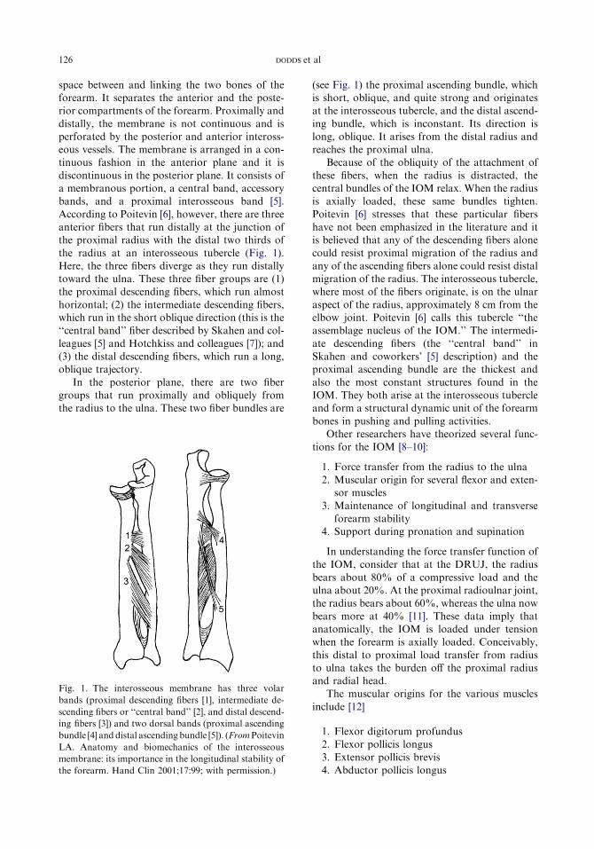
According to Poitevin [6], however, there are threeanterior fibers that run distally at the junction ofthe proximal radius with the distal two thirds of
the radius at an interosseous tubercle (Fig. 1).Here, the three fibers diverge as they run distallytoward the ulna. These three fiber groups are (1)the proximal descending fibers, which run almost
horizontal; (2) the intermediate descending fibers,which run in the short oblique direction (this is the‘‘central band’’ fiber described by Skahen and col-
leagues [5] and Hotchkiss and colleagues [7]); and(3) the distal descending fibers, which run a long,oblique trajectory.
In the posterior plane, there are two fibergroups that run proximally and obliquely fromthe radius to the ulna. These two fiber bundles are
Fig. 1. The interosseous membrane has three volar
bands (proximal descending fibers [1], intermediate de-
scending fibers or ‘‘central band’’ [2], and distal descend-
ing fibers [3]) and two dorsal bands (proximal ascending
bundle [4] anddistal ascendingbundle [5]). (FromPoitevin
LA. Anatomy and biomechanics of the interosseous
membrane: its importance in the longitudinal stability of
the forearm. Hand Clin 2001;17:99; with permission.)
(see Fig. 1) the proximal ascending bundle, whichis short, oblique, and quite strong and originatesat the interosseous tubercle, and the distal ascend-
ing bundle, which is inconstant. Its direction islong, oblique. It arises from the distal radius andreaches the proximal ulna.
Because of the obliquity of the attachment of
these fibers, when the radius is distracted, thecentral bundles of the IOM relax. When the radiusis axially loaded, these same bundles tighten.
Poitevin [6] stresses that these particular fibershave not been emphasized in the literature and itis believed that any of the descending fibers alone
could resist proximal migration of the radius andany of the ascending fibers alone could resist distalmigration of the radius. The interosseous tubercle,where most of the fibers originate, is on the ulnar
aspect of the radius, approximately 8 cm from theelbow joint. Poitevin [6] calls this tubercle ‘‘theassemblage nucleus of the IOM.’’ The intermedi-
ate descending fibers (the ‘‘central band’’ inSkahen and coworkers’ [5] description) and theproximal ascending bundle are the thickest and
also the most constant structures found in theIOM. They both arise at the interosseous tubercleand form a structural dynamic unit of the forearm
bones in pushing and pulling activities.Other researchers have theorized several func-
tions for the IOM [8–10]:
1. Force transfer from the radius to the ulna2. Muscular origin for several flexor and exten-
sor muscles
3. Maintenance of longitudinal and transverseforearm stability
4. Support during pronation and supination
In understanding the force transfer function ofthe IOM, consider that at the DRUJ, the radius
bears about 80% of a compressive load and theulna about 20%. At the proximal radioulnar joint,the radius bears about 60%, whereas the ulna now
bears more at 40% [11]. These data imply thatanatomically, the IOM is loaded under tensionwhen the forearm is axially loaded. Conceivably,
this distal to proximal load transfer from radiusto ulna takes the burden off the proximal radiusand radial head.
The muscular origins for the various muscles
include [12]
1. Flexor digitorum profundus2. Flexor pollicis longus3. Extensor pollicis brevis
4. Abductor pollicis longus
127ESSEX-LOPRESTI INJURIES
5. Extensor indicis6. Extensor pollicis longus
In the dissection of human cadaveric forearms,it was found that the central band (or intermediatedescending fibers) of the IOM was the most
dominant and consistent structure, whereas theproximal and distal ends of the IOM are thin andmembranous [5]. The central band originates onthe radius and inserts on the ulna and possesses
a distinct oblique radioulnar direction of 21degrees to the longitudinal axis of the ulna [5].The average length of the radial origin and ulnar
insertion of the central band have been found tobe about the same at 10.6 cm. On the dorsal sur-face of the radius, the origin is broad and fan-
shaped at the level of the abductor pollicis longusorigin. Distally, it fades to the sharp interosseousborder of the radius. On the palmar surface, the
origin is also broad at the level of the flexor polli-cis longus origin and gradually fades to the sharpinterosseous border distally.
The bony attachment of the ulnar insertion of
the IOM is quite different. The dorsal ulnarinsertion is broad along the entire dorsal flatsurface of the ulna; however, the palmar attach-
ment is limited to the sharp interosseous border.Because the central band fibers tend to fan out asthey pass from the radius to the ulna, the average
radial origin measures 3.4 cm, whereas the ulnarinsertion measures an average of 4.2 cm. Thecentral band origin can be found at an average of7.7 cm distal from the articular surface of the
radial head. The average insertion point is 13.7 cmdistal to the tip of the olecranon. The central bandis in general two to three times thicker than the
membranous portion [10].The accessory bands were separate and distinct
from the central band and not only were less
substantial but they also varied in number andorientation among cadaveric specimens. Theproximal interosseous band is found exclusively
on the dorsal surface of the proximal forearm.Fibers of this band are oriented proximal andulnar as they pass from the radius to the ulna.This structure shares a point of origin with the
central band, although its fibers are orientedperpendicular to the central, an average of 28degrees to the longitudinal axis of the ulna [5].
Histology
McGinley and colleagues [13] found that theIOM is primarily composed of collagen arranged
in fibrillar structures surrounded by elastin. Colla-gen was abundant in the proximal bundles anddecreased in the distal bundles. Histologic analysisof the IOM bundles obtained from cadaveric fore-
arms showed that collagen represented greaterthan 90% of the central band.
Biomechanics
There are several levels of restraint againstproximal migration of the radius. The primaryrestraint is that of the radius abutting the cap-
itellum. It has been shown that the TFCC andIOM are the secondary restraints [2]. Wallace andcolleagues [14] studied the structure and function
of the IOM in 11 cadaver preparations. Theymade a model of longitudinal radioulnar dissocia-tion by applying longitudinal tensile load untilrupture of the IOM. Seven of the specimens sus-
tained a midsubstance tear of the central bandof the IOM at a mean peak load of 1038 N [14].Hotchkiss and colleagues [7] found that the cen-
tral band portion of the IOM was responsiblefor 71% of the total longitudinal stiffness of theIOM after radial head excision.
Other authors have shown that varus-valgusmoments about the elbow, caused by transverseloads applied at the hand in intact specimens,
affect the magnitude of IOM load transfer [15,16].Interosseous ligament load transfer was found tobe consistently lower when the elbow experienceda valgus stress compared with a varus stress under
an axially applied load to the hand of 134 N [15].Several studies have examined the strain and
load distribution of the IOM in relation to fore-
arm rotation. Pfaeffle and colleagues [17] mea-sured three-dimensional force vectors acting inthe forearm when axially loaded to 136 N in intact
specimens. They showed that the IOM partici-pates not only in longitudinal load transfer, butalso in the maintenance of transverse stability of
the forearm. Forces in the IOM were significantlygreater in neutral rotation compared with supina-tion or pronation. Manson and colleagues [18]studied five intact cadaveric forearms and found
that the strain distribution of the IOM changedwith forearm rotation, again with the highestoverall strain in neutral rotation. In neutral and
pronation, higher strain was observed in the prox-imal region of the IOM. In supination, however,higher average strain was seen in the distal region
of the IOM.As Manson and colleagues [18] noted, these
findings may be helpful in answering questions
128 DODDS et al
of optimal placement of a reconstructive graft andthe ideal rotational position of the forearm duringgraft tensioning. For example, the results suggest
that to provide balanced constraint in differentpositions of forearm rotation, the ideal placementof a graft is in the proximal region of the IOM andtensioned in neutral rotation [18,19].
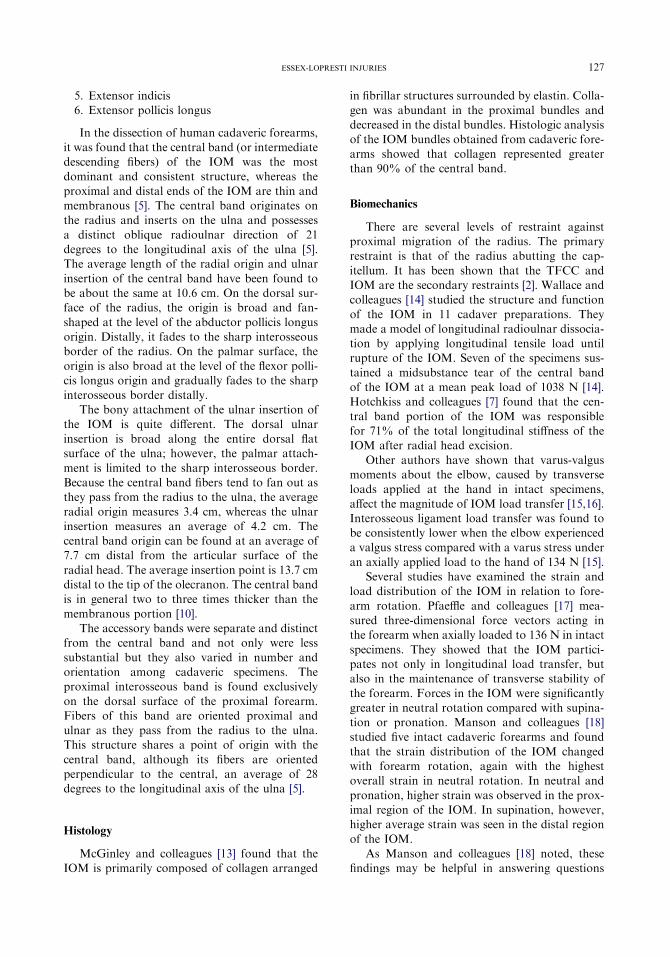
After radial head excision in cadavers, Rabi-nowitz and colleagues [20] and Skahen andcolleagues [5] demonstrated not only substantially
more strain in the IOM, but also increasing strainin the IOM as the forearm was rotated from supi-nation to pronation (Fig. 2). These findings con-
firm the IOM’s secondary role in maintainingdynamic radioulnar stability when changes toradial length occur.
Clinical presentation
An Essex-Lopresti injury is usually sustainedduring a high-energy axial load onto an out-stretched hand. As such, a longitudinal force istransmitted through the wrist to the radial head.
When increasingly sufficient force is exerted, thehead of the radius fractures or dislocates, the IOMruptures, and the DRUJ ruptures. Consequently,
the radius migrates proximally, leaving the patientwith complex instability of the forearm. Patientscomplain of elbow and wrist pain. Some may also
be acutely aware of pain throughout the forearm.
Fig. 2. This graph demonstrates the percentage strain
withstood by the central band with an intact radial
head compared with the increased strain after radial
head excision in cadaveric biomechanical testing.
(From Skahen JR III, Palmer AK, Werner FW, et al.
The interosseous membrane of the forearm: anatomy
and function. J Hand Surg [Am] 1997;22:985; with
permission.)
Physical examination often yields ulnar-sidedwrist tenderness with increasing pain as the fore-arm is rotated. The forearm itself may be tender
and ecchymotic, especially between the radius andulna in the interosseous space. Examination of theelbow may demonstrate lateral ecchymosis andtenderness. Attention must focus on the elbow,
forearm, and wrist to diagnose accurately thisdisruption of the forearm ‘‘ring.’’
The position of the forearm at the time of
impact greatly influences the kind of fracture thatresults. Through a cadaveric forearm axial load-ing model, McGinley and colleagues [21] found
that loading in supination produced both boneforearm fractures, loading in a neutral positionresulted in isolated radial head fractures, andloading in pronation resulted in Essex-Lopresti
injuries. In supination, there is the least amountof contact within the radiocapitellar joint andmaximum tension within the IOM. Following
sudden impact loading, forces are primarily trans-mitted from the radius through the IOM onto theulna without being transmitted to the radiocapi-
tellar joint. The large magnitude of the suddenload results in fractures of the radial and ulnarshafts without injury of the radial head.
In neutral position, there is moderate contactwithin the radiocapitellar joint and moderatetension in the IOM. As a result, the impact isprimarily absorbed by the radial head but also
reduced by transfer across the IOM, resulting inisolated marginal radial head fractures. In thepronated position, there is the most contact
between the radial head and capitellum and verylittle tension in the IOM. All of the force of theimpact is transferred onto the radial head, usually
comminuting the head. This results in a shift ormigration of the radius and subsequent tearing ofthe IOM [8,21–23]. With this knowledge in hand,the astute clinician knows when to suspect an
Essex-Lopresti lesion, especially if comminutionof the radial head is seen or if the clinical historysuggests that the forearm was loaded in pronation
at the time of injury.It should be noted that subtle proximal migra-
tion of the radius (approximately 2–3 mm) usually
occurs after fracture or excision of the radial head,but it is rarely symptomatic [10]. When both theprimary and secondary stabilizers of the forearm
are disrupted, however, proximal migration of theradial head usually averages greater than 7 mm,in which case patients become symptomatic bycomplaining of loss of wrist extension, forearm
rotation, ulnocarpal impaction symptoms, and
129ESSEX-LOPRESTI INJURIES
wrist pain. One study showed that patients withproximal radial translation greater than 1 cm usu-ally experienced pain and loss of motion, whereaspatients with translation less than 1 cm report
pain but retain motion [24]. With the relativeshortening of the radius compared with the ulna,the distal ulna loses its position in the sigmoid
notch and actually becomes dorsally positionedin relation to the carpus. Supination and wristextension are limited as the ulnar head comes
into contact with the dorsal carpus. In additionto the wrist symptoms, the proximally migratingradius abuts the capitellum, leading to elbow
pain and limitation of elbow motion.An Essex-Lopresti injury of the forearm often
is missed during assessment at the time of injury.A fracture of the radial head usually is apparent,
whereas injury to the IOM and TFCC is notsymptomatic in many patients at initial presenta-tion. This is where the clinician must obtain
a good history and have a high index of suspicionbased on the mechanism of injury. Whenevera high-energy longitudinal compression force
occurs (ie, fall from a height and motorcycleinjuries), the Essex-Lopresti injury must beentertained.
Diagnostic imaging
Radiographs of the elbow, forearm, and wristshould be obtained when clinical suspicion is highfor an Essex-Lopresti injury. It may also be helpful
to obtain contralateral wrist films to have a base-line for comparison of ulnar variance. Accordingto Epner and colleagues [25], the optimal view of
the DRUJ is obtained using a posteroanteriorradiograph with the shoulder at 90 degrees ofabduction, the elbow flexed at 90 degrees, and
the forearm in neutral rotation. According toYeh and colleagues [26], however, ulnar variancecan be assessed adequately with a routine poster-
oanterior radiograph of the wrist. Interestingly,Tomaino [27] took radiographs of the forearmwhile the patient was gripping to assess the changein ulnar variance and thereby indirectly evaluating
for alterations in the integrity of the IOM. Al-though the posteroanterior view of the wrist mayshow a markedly positive ulnar variance, the lat-
eral radiograph of the wrist is also helpful inassessing dorsal or volar subluxation of the ulna.
Biplanar radiographs of the forearm rule out
diaphyseal fractures. Elbow radiographs oftenshow a radial head or neck fracture with orwithout dislocation at the radiocapitellar joint,
usually with comminution. A CT scan (with three-dimensional reconstructions) of the elbow may behelpful in determining the degree of comminutionand articular involvement of the radial head.
Ultrasound, CT, and MRI are not typicallyused in diagnosing the soft tissue components ofradioulnar dissociation. Failla and colleagues [28]
used ultrasound to detect tears in the IOM in oneliving and two cadaveric forearms. In their study,the IOM is a very hyperechoic structure, with the
central third of the IOM seen as a thick, continu-ous white line. It is the only continuous andintensely hyperechoic structure that connects the
ulna to the radius. Any disruptions of the IOMshow a break in this continuity of the white linerepresenting the IOM. Wallace [29] described theultrasound technique as one using a high-
resolution dynamic machine with the transducerover the dorsal aspect of the forearm orientedtransversely. Jaakkola and colleagues [30] also
looked at the accuracy of ultrasound in detectingIOM disruptions and found a 96% accuracy ratewhen looking at cadaveric forearms. The sono-
graphic criterion used to determine IOM disrup-tion during the dynamic study was lack ofvisualization of a continuous hyperechoic band
passing between the radius and ulna througha region of at least 2 cm in length. Matsuokaand colleagues [31] took it one step further andshowed the accuracy of ultrasonography on living
patients with and without the injury. IOM disrup-tions were seen as hypoechoic regions in longitu-dinal and transverse views.
In a study by McGinley and colleagues [32],MRI determination of IOM injury demonstrateda positive predictive value of 100%, a negative
predictive value of 89%, a sensitivity of 87.5%,and a specificity of 100%. Further, they demon-strated that most lesions occurred along theIOM’s ulnar insertion, and in half of the injured
specimens there was concomitant dorsal obliquebundle disruption [32]. Sowa and colleagues [3]used MRI (specifically 1.6-T magnetic resonance
with a wrist coil) to diagnose a tear in the centralband of the IOM in one patient. Starch and Dabe-zies [33] concluded through cadaveric and live
patient investigation that the IOM can be evalu-ated best by axial T2-weighted fast spin echoimages with fat suppression.
Whereas the ultrasound and MRI studiesmentioned previously focused on the integrity ofthe central third of the IOM, Soubeyrand andcolleagues [34] found that a ‘‘muscular hernia
sign’’ could be elicited anywhere along the IOM
130 DODDS et al
by directing an anteroposterior load on the ante-rior aspect of the forearm at a specific level, andif the IOM was not intact at that level, anterior
musculature could be visualized by ultrasoundon the posterior aspect of the forearm. CT, a mo-dality more suited for evaluating bony anatomyand generally not useful in assessing IOM integ-
rity, can be used to assess DRUJ reduction.
Treatment
Although it may be easy to separate the
components of the injury into a fractured radialhead, torn IOM, and disrupted DRUJ, it isessential to consider the injury as a disruption of
the forearm ‘‘joint.’’ Treatment of the radial headfracture alone may lead to persistent problems atthe DRUJ. Healing and recovery depends on the
management of both the elbow and the wrist(Figs. 3–7).
Every effort should be made to save the native
radial head. If an open reduction and internalfixation cannot be achieved, then prosthetic re-placement should be considered. When the DRUJis dislocated in an Essex-Lopresti injury, some
surgeons have advocated pinning or temporarysyndesmotic screw placement between the radiusand ulna to allow healing of the IOM and distal
and proximal radioulnar ligaments. In theory,stabilization of the forearm using screws or wiresallows healing of the soft tissue structures to
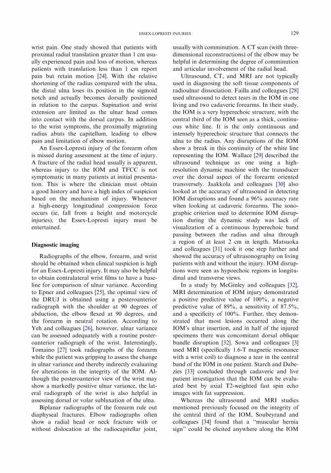
Fig. 3. (A) Anteroposterior radiograph of an Essex-Lopresti i
(B) On the lateral view, an arrow points out a malrotated and
elbow, forearm, and wrist pain.
occur. There is not clear evidence, however, thatwhen the IOM is immobilized by syndesmoticscrew fixation, it always heals [7].
Radial head excision alone leads to suboptimalresults. In one study, 15 of the 20 patientsreported severe pain at the DRUJ when the radialhead was excised [35]. Radial head allografts have
also not been very successful in restoring forearmstability [36]. Silicone prostheses have been associ-ated with gradual material failure and continued
proximal migration [37–40]. Currently, there area number of metallic radial head prostheses avail-able with different implant styles that cater to the
surgical approach and character of the radial headand neck fracture.
Central band repair and reconstruction hasbeen tried by many authors, all in limited numbers
with brief follow-up. Augmentation tissues haveincluded flexor carpi ulnaris tendon, palmarislongus tendon, tibia-fibula interosseous ligament
bone-ligament bone-autograft, and bone-ligament-bone allograft. Conversion to a one-bone forearm is the ultimate salvage alternative
in patients with chronic, symptomatic longitudi-nal forearm instability. It has been met with themost success in treating these patients [41]. It is
associated with loss of rotation, however, andrecent studies have called into question previousfavorable results [42].
Acceptable outcomes in the treatment of acute
radioulnar dissociation are incumbent on
njury demonstrating a comminuted radial head fracture.
displaced articular fragment. This patient presented with
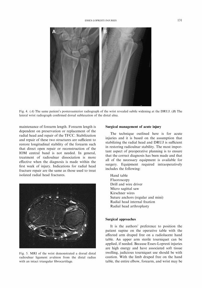
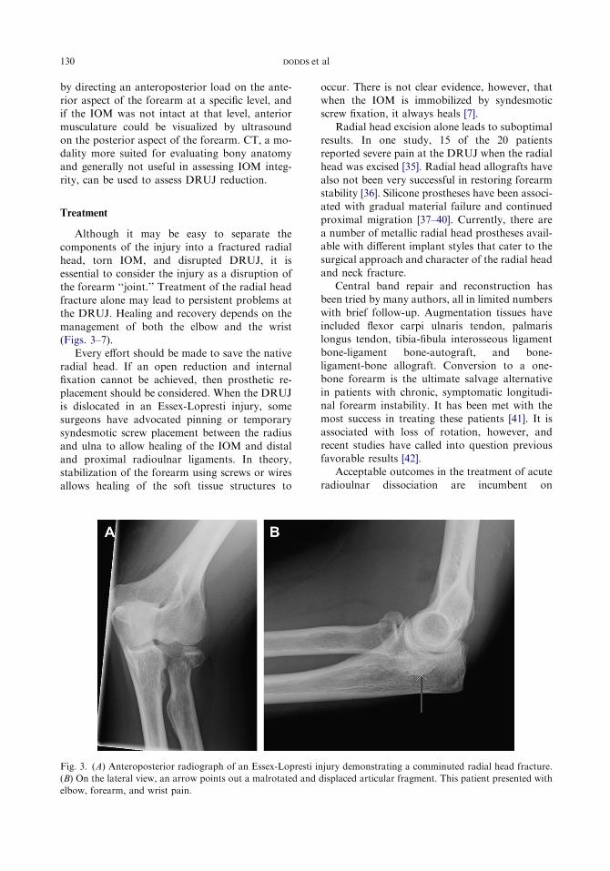
Fig. 4. (A) The same patient’s posteroanterior radiograph of the wrist revealed subtle widening at the DRUJ. (B) The
lateral wrist radiograph confirmed dorsal subluxation of the distal ulna.
131ESSEX-LOPRESTI INJURIES
maintenance of forearm length. Forearm length is
dependent on preservation or replacement of theradial head and repair of the TFCC. Stabilizationand repair of these two structures are sufficient to
restore longitudinal stability of the forearm suchthat direct open repair or reconstruction of theIOM central band is not needed. In general,treatment of radioulnar dissociation is more
effective when the diagnosis is made within thefirst week of injury. Indications for radial headfracture repair are the same as those used to treat
isolated radial head fractures.
Fig. 5. MRI of the wrist demonstrated a dorsal distal
radioulnar ligament avulsion from the distal radius
with an intact triangular fibrocartilage.
Surgical management of acute injury
The technique outlined here is for acuteinjuries and it is based on the assumption thatstabilizing the radial head and DRUJ is sufficient
in restoring radioulnar stability. The most impor-tant aspect of preoperative planning is to ensurethat the correct diagnosis has been made and thatall of the necessary equipment is available for
surgery. Equipment required intraoperativelyincludes the following:
Hand tableFluoroscopyDrill and wire driver
Micro sagittal sawKirschner wiresSuture anchors (regular and mini)
Radial head internal fixationRadial head arthroplasty
Surgical approaches
It is the authors’ preference to position thepatient supine on the operative table with theaffected arm draped free on a radiolucent hand
table. An upper arm sterile tourniquet can beapplied, if needed. Because Essex-Lopresti injuriesare high energy and have associated soft tissue
swelling, judicious tourniquet use should be withcaution. With the limb draped free on the handtable, the entire elbow, forearm, and wrist may be
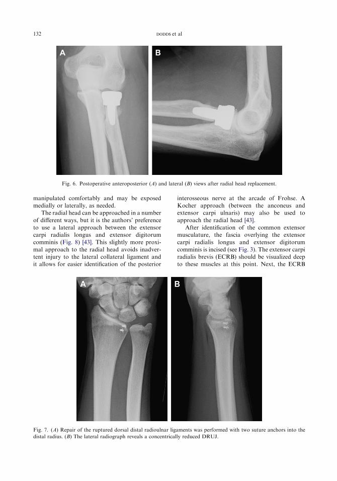
Fig. 6. Postoperative anteroposterior (A) and lateral (B) views after radial head replacement.
132 DODDS et al
manipulated comfortably and may be exposedmedially or laterally, as needed.
The radial head can be approached in a number
of different ways, but it is the authors’ preferenceto use a lateral approach between the extensorcarpi radialis longus and extensor digitorum
comminis (Fig. 8) [43]. This slightly more proxi-mal approach to the radial head avoids inadver-tent injury to the lateral collateral ligament andit allows for easier identification of the posterior
Fig. 7. (A) Repair of the ruptured dorsal distal radioulnar lig
distal radius. (B) The lateral radiograph reveals a concentrical
interosseous nerve at the arcade of Frohse. AKocher approach (between the anconeus andextensor carpi ulnaris) may also be used to
approach the radial head [43].After identification of the common extensor
musculature, the fascia overlying the extensor
carpi radialis longus and extensor digitorumcomminis is incised (see Fig. 3). The extensor carpiradialis brevis (ECRB) should be visualized deepto these muscles at this point. Next, the ECRB
aments was performed with two suture anchors into the
ly reduced DRUJ.
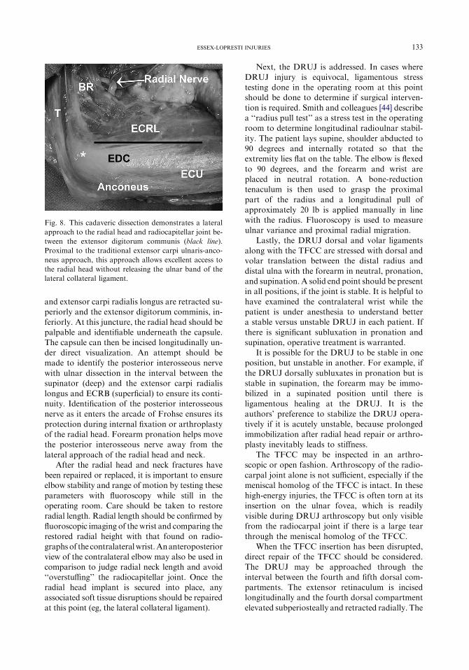
Fig. 8. This cadaveric dissection demonstrates a lateral
approach to the radial head and radiocapitellar joint be-
tween the extensor digitorum communis (black line).
Proximal to the traditional extensor carpi ulnaris-anco-
neus approach, this approach allows excellent access to
the radial head without releasing the ulnar band of the
lateral collateral ligament.
133ESSEX-LOPRESTI INJURIES
and extensor carpi radialis longus are retracted su-periorly and the extensor digitorum comminis, in-
feriorly. At this juncture, the radial head should bepalpable and identifiable underneath the capsule.The capsule can then be incised longitudinally un-der direct visualization. An attempt should be
made to identify the posterior interosseous nervewith ulnar dissection in the interval between thesupinator (deep) and the extensor carpi radialis
longus and ECRB (superficial) to ensure its conti-nuity. Identification of the posterior interosseousnerve as it enters the arcade of Frohse ensures its
protection during internal fixation or arthroplastyof the radial head. Forearm pronation helps movethe posterior interosseous nerve away from the
lateral approach of the radial head and neck.After the radial head and neck fractures have
been repaired or replaced, it is important to ensureelbow stability and range of motion by testing these
parameters with fluoroscopy while still in theoperating room. Care should be taken to restoreradial length. Radial length should be confirmed by
fluoroscopic imaging of the wrist and comparing therestored radial height with that found on radio-graphs of the contralateralwrist.Ananteroposterior
view of the contralateral elbow may also be used incomparison to judge radial neck length and avoid‘‘overstuffing’’ the radiocapitellar joint. Once theradial head implant is secured into place, any
associated soft tissue disruptions should be repairedat this point (eg, the lateral collateral ligament).
Next, the DRUJ is addressed. In cases whereDRUJ injury is equivocal, ligamentous stresstesting done in the operating room at this pointshould be done to determine if surgical interven-
tion is required. Smith and colleagues [44] describea ‘‘radius pull test’’ as a stress test in the operatingroom to determine longitudinal radioulnar stabil-
ity. The patient lays supine, shoulder abducted to90 degrees and internally rotated so that theextremity lies flat on the table. The elbow is flexed
to 90 degrees, and the forearm and wrist areplaced in neutral rotation. A bone-reductiontenaculum is then used to grasp the proximal
part of the radius and a longitudinal pull ofapproximately 20 lb is applied manually in linewith the radius. Fluoroscopy is used to measureulnar variance and proximal radial migration.
Lastly, the DRUJ dorsal and volar ligamentsalong with the TFCC are stressed with dorsal andvolar translation between the distal radius and
distal ulna with the forearm in neutral, pronation,and supination. A solid end point should be presentin all positions, if the joint is stable. It is helpful to
have examined the contralateral wrist while thepatient is under anesthesia to understand bettera stable versus unstable DRUJ in each patient. If
there is significant subluxation in pronation andsupination, operative treatment is warranted.
It is possible for the DRUJ to be stable in oneposition, but unstable in another. For example, if
the DRUJ dorsally subluxates in pronation but isstable in supination, the forearm may be immo-bilized in a supinated position until there is
ligamentous healing at the DRUJ. It is theauthors’ preference to stabilize the DRUJ opera-tively if it is acutely unstable, because prolonged
immobilization after radial head repair or arthro-plasty inevitably leads to stiffness.
The TFCC may be inspected in an arthro-scopic or open fashion. Arthroscopy of the radio-
carpal joint alone is not sufficient, especially if themeniscal homolog of the TFCC is intact. In thesehigh-energy injuries, the TFCC is often torn at its
insertion on the ulnar fovea, which is readilyvisible during DRUJ arthroscopy but only visiblefrom the radiocarpal joint if there is a large tear
through the meniscal homolog of the TFCC.When the TFCC insertion has been disrupted,
direct repair of the TFCC should be considered.
The DRUJ may be approached through theinterval between the fourth and fifth dorsal com-partments. The extensor retinaculum is incisedlongitudinally and the fourth dorsal compartment
elevated subperiosteally and retracted radially. The
134 DODDS et al
extensor digiti quinti minimi is retracted ulnarly.Careful examination of the dorsal joint may revealcapsular avulsion with or without disruption of the
dorsal distal radioulnar ligament. If inspection ofthe TFCC reveals injury of its insertion into theulnar fovea, this tissue is reattached with sutureanchors or through small bone tunnels.
There may be cases of continued DRUJinstability after repair of the TFCC insertionand supporting capsular structures. In these cases,
reconstruction of the dorsal and volar DRUJligaments may be undertaken with a palmarislongus autograft or allograft tendon. Fractures of
the ulnar styloid require reduction and repair withinternal fixation if the DRUJ is unstable. Thedistal forearm may be pinned with 0.062-inKirschner wires to protect the soft tissue repairs
or as an alternative to direct repair. Two Kirsch-ner wires are used and placed proximal to theDRUJ articular surfaces; these pins should be
prominent radially and ulnarly to facilitateremoval if they break.
Postoperative immobilization and rehabilitation
The patient should be initially immobilized ina well-padded and well-molded long posteriorsplint that extends to the distal palmar crease to
immobilize the wrist. Patient’s limb is mobilizeddepending on the patient’s injury and the stability
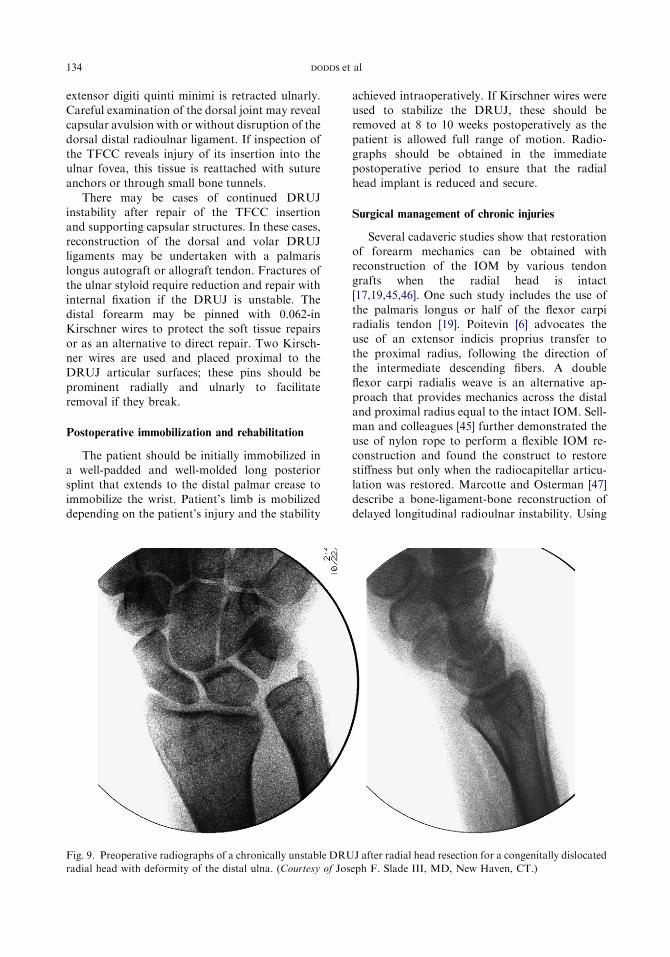
Fig. 9. Preoperative radiographs of a chronically unstable DRU
radial head with deformity of the distal ulna. (Courtesy of Jos
achieved intraoperatively. If Kirschner wires wereused to stabilize the DRUJ, these should beremoved at 8 to 10 weeks postoperatively as the
patient is allowed full range of motion. Radio-graphs should be obtained in the immediatepostoperative period to ensure that the radialhead implant is reduced and secure.
Surgical management of chronic injuries
Several cadaveric studies show that restorationof forearm mechanics can be obtained with
reconstruction of the IOM by various tendongrafts when the radial head is intact[17,19,45,46]. One such study includes the use of
the palmaris longus or half of the flexor carpiradialis tendon [19]. Poitevin [6] advocates theuse of an extensor indicis proprius transfer to
the proximal radius, following the direction ofthe intermediate descending fibers. A doubleflexor carpi radialis weave is an alternative ap-
proach that provides mechanics across the distaland proximal radius equal to the intact IOM. Sell-man and colleagues [45] further demonstrated theuse of nylon rope to perform a flexible IOM re-
construction and found the construct to restorestiffness but only when the radiocapitellar articu-lation was restored. Marcotte and Osterman [47]
describe a bone-ligament-bone reconstruction ofdelayed longitudinal radioulnar instability. Using
J after radial head resection for a congenitally dislocated
eph F. Slade III, MD, New Haven, CT.)
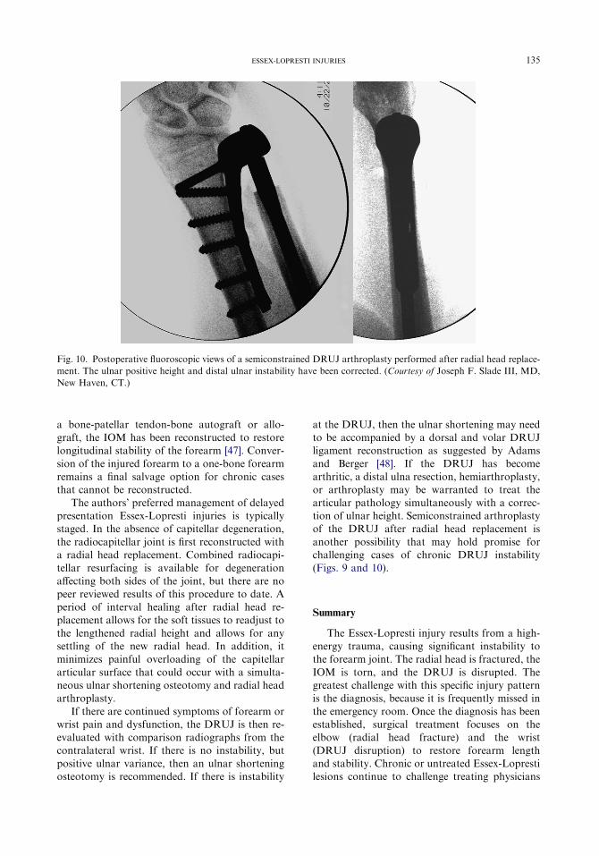
Fig. 10. Postoperative fluoroscopic views of a semiconstrained DRUJ arthroplasty performed after radial head replace-
ment. The ulnar positive height and distal ulnar instability have been corrected. (Courtesy of Joseph F. Slade III, MD,
New Haven, CT.)
135ESSEX-LOPRESTI INJURIES
a bone-patellar tendon-bone autograft or allo-graft, the IOM has been reconstructed to restorelongitudinal stability of the forearm [47]. Conver-
sion of the injured forearm to a one-bone forearmremains a final salvage option for chronic casesthat cannot be reconstructed.
The authors’ preferred management of delayedpresentation Essex-Lopresti injuries is typicallystaged. In the absence of capitellar degeneration,
the radiocapitellar joint is first reconstructed witha radial head replacement. Combined radiocapi-tellar resurfacing is available for degeneration
affecting both sides of the joint, but there are nopeer reviewed results of this procedure to date. Aperiod of interval healing after radial head re-placement allows for the soft tissues to readjust to
the lengthened radial height and allows for anysettling of the new radial head. In addition, itminimizes painful overloading of the capitellar
articular surface that could occur with a simulta-neous ulnar shortening osteotomy and radial headarthroplasty.
If there are continued symptoms of forearm orwrist pain and dysfunction, the DRUJ is then re-evaluated with comparison radiographs from thecontralateral wrist. If there is no instability, but
positive ulnar variance, then an ulnar shorteningosteotomy is recommended. If there is instability
at the DRUJ, then the ulnar shortening may needto be accompanied by a dorsal and volar DRUJligament reconstruction as suggested by Adams
and Berger [48]. If the DRUJ has becomearthritic, a distal ulna resection, hemiarthroplasty,or arthroplasty may be warranted to treat the
articular pathology simultaneously with a correc-tion of ulnar height. Semiconstrained arthroplastyof the DRUJ after radial head replacement is
another possibility that may hold promise forchallenging cases of chronic DRUJ instability(Figs. 9 and 10).
Summary
The Essex-Lopresti injury results from a high-energy trauma, causing significant instability tothe forearm joint. The radial head is fractured, the
IOM is torn, and the DRUJ is disrupted. Thegreatest challenge with this specific injury patternis the diagnosis, because it is frequently missed in
the emergency room. Once the diagnosis has beenestablished, surgical treatment focuses on theelbow (radial head fracture) and the wrist
(DRUJ disruption) to restore forearm lengthand stability. Chronic or untreated Essex-Loprestilesions continue to challenge treating physicians
136 DODDS et al
and often require salvage or reconstructive pro-cedures to minimize pain and return function.
References
[1] Essex-Lopresti P. Fractures of the radial head with
distal radio-ulnar dislocation: report of two cases.
J Bone Joint Surg [Br] - British Volume 1951;
33B(2):244–7.
[2] Rozental TD, Beredjiklian PK, BozentkaDJ. Longi-
tudinal radioulnar dissociation. J Am Acad Orthop
Surg 2003;11(1):68–73.
[3] Sowa DT, Hotchkiss RN, Weiland AJ. Symptom-
atic proximal translation of the radius following
radial head resection. Clin Orthop Relat Res 1995;
317:106–13.
[4] Curr JF, Coe WA. Dislocation of the inferior radio-
ulnar joint. Br J Surg 1946;34(133):74–7.
[5] Skahen JR III, Palmer AK, Werner FW, et al. The
interosseous membrane of the forearm: anatomy
and function. J Hand Surg [Am] 1997;22(6):981–5.
[6] Poitevin LA. Anatomy and biomechanics of the
interosseous membrane: its importance in the
longitudinal stability of the forearm. Hand Clin
2001;17(1):97–110, vii.
[7] Hotchkiss RN, An KN, Sowa DT, et al. An
anatomic and mechanical study of the interosseous
membrane of the forearm: pathomechanics of
proximal migration of the radius. J Hand Surg
[Am] 1989;14(2 Pt 1):256–61.
[8] Markolf KL, Lamey D, Yang S, et al. Radioulnar
load-sharing in the forearm: a study in cadavera.
J Bone Joint Surg [Am] 1998;80(6):879–88.
[9] McGinley JC, D’Addessi L, Sadeghipour K, et al.
Mechanics of the antebrachial interosseous
membrane: response to shearing forces. J Hand
Surg [Am] 2001;26(4):733–41.
[10] McGinley JC, Kozin SH. Interosseous membrane
anatomy and functional mechanics. Clin Orthop
Relat Res 2001;383:108–22.
[11] Halls AA, Travill A. Transmission of pressures
across the elbow joint. Anat Rec 1964;150:243–7.
[12] Hollister AM, GellmanH,Waters RL. The relation-
ship of the interosseous membrane to the axis of
rotation of the forearm. Clin Orthop Relat Res
1994;298:272–6.
[13] McGinley JC, Heller JE, Fertala A, et al. Biochemi-
cal composition and histologic structure of the
forearm interosseous membrane. J Hand Surg
[Am] 2003;28(3):503–10.
[14] Wallace AL, Walsh WR, van Rooijen M, et al. The
interosseous membrane in radio-ulnar dissociation.
J Bone Joint Surg [Br] 1997;79(3):422–7.
[15] MarkolfKL,DunbarAM,HannaniK.Mechanisms
of load transfer in the cadaver forearm: role of the
interosseous membrane. J Hand Surg [Am] 2000;
25(4):674–82.
[16] ShepardMF,Markolf KL, Dunbar AM. The effects
of partial and total interosseous membrane
transection on load sharing in the cadaver forearm.
J Orthop Res 2001;19(4):587–92.
[17] PfaeffleHJ, Stabile KJ, Li ZM, et al. Reconstruction
of the interosseous ligament restores normal forearm
compressive load transfer in cadavers. J Hand Surg
[Am] 2005;30(2):319–25.
[18] Manson TT, Pfaeffle HJ, Herdon JH, et al. Forearm
rotation alters interosseous ligament strain distribu-
tion. J Hand Surg [Am] 2000;25(6):1058–63.
[19] Skahen JR III, Palmer AK, Werner FW, et al.
Reconstruction of the interosseous membrane of
the forearm in cadavers. J Hand Surg [Am] 1997;
22(6):986–94.
[20] Rabinowitz RS, Light TR, Havey RM, et al. The
role of the interosseous membrane and triangular
fibrocartilage complex in forearm stability. J Hand
Surg [Am] 1994;19(3):385–93.
[21] McGinley JC, Hopgood BC, Gaughan JP, et al.
Forearm and elbow injury: the influence of
rotational position. J Bone Joint Surg [Am] 2003;
85-A(12):2403–9.
[22] af EkenstamFW, Palmer AK,GlissonRR. The load
on the radius and ulna in different positions of the
wrist and forearm: a cadaver study. Acta Orthop
Scand 1984;55(3):363–5.
[23] Morrey BF, An KN, Stormont TJ. Force transmis-
sion through the radial head. J Bone Joint Surg
[Am] 1988;70(2):250–6.
[24] Hotchkiss RN. Fractures of the radial head and
related instability and contracture of the forearm.
Instr Course Lect 1998;47:173–7.
[25] EpnerRA,BowersWH,GuilfordWB.Ulnar variance:
the effect ofwrist positioningand roentgenfilming tech-
nique. J Hand Surg [Am] 1982;7(3):298–305.
[26] Yeh GL, Beredjiklian PK, KatzMA, et al. Effects of
forearm rotation on the clinical evaluation of ulnar
variance. J Hand Surg [Am] 2001;26(6):1042–6.
[27] TomainoMM. The importance of the pronated grip
x-ray view in evaluating ulnar variance. J Hand Surg
[Am] 2000;25(2):352–7.
[28] Failla JM, Jacobson J, van Holsbeeck M.
Ultrasound diagnosis and surgical pathology of the
torn interosseous membrane in forearm fractures/
dislocations. J Hand Surg [Am] 1999;24(2):257–66.
[29] Wallace AL. Magnetic resonance imaging or ultra-
sound in assessment of the interosseous membrane
of the forearm. J Bone Joint Surg Am 2002;
84-A(3):496–7.
[30] Jaakkola JI, Riggans DH, Lourie GM, et al.
Ultrasonography for the evaluation of forearm
interosseous membrane disruption in a cadaver
model. J Hand Surg [Am] 2001;26(6):1053–7.
[31] Matsuoka J, Beppu M, Nakajima H, et al. Ultraso-
nography for the interosseous membrane of the
forearm. Hand Surg 2003;8(2):227–35.
[32] McGinley JC, Roach N, Hopgood BC, et al.
Forearm interosseous membrane trauma: MRI
diagnostic criteria and injury patterns. Skeletal
Radiol 2006;35(5):275–81.
137ESSEX-LOPRESTI INJURIES
[33] Starch DW, Dabezies EJ. Magnetic resonance
imaging of the interosseous membrane of the fore-
arm. J Bone Joint Surg [Am] 2001;83-A(2):235–8.
[34] Soubeyrand M, Lafont C, Oberlin C, et al. The
muscular hernia sign: an original ultrasonographic
sign to detect lesions of the forearm’s interosseous
membrane. Surg Radiol Anat 2006;28(4):372–8.
[35] Trousdale RT, Amadio PC, Cooney WP, et al.
Radio-ulnar dissociation: a review of twenty cases.
J Bone Joint Surg [Am] 1992;74(10):1486–97.
[36] Karlstad R, Morrey BF, Cooney WP. Failure of
fresh-frozen radial head allografts in the treatment
of Essex-Lopresti injury: a report of four cases.
J Bone Joint Surg [Am] 2005;87(8):1828–33.
[37] Levin PD. Fracture of the radial head with disloca-
tion of the distal radio-ulnar joint: case report.
Treatment by prosthetic replacement of the radial
head. J Bone Joint Surg [Am] 1973;55(4):837–40.
[38] Mayhall WS, Tiley FT, Paluska DJ. Fracture of
Silastic radial-head prosthesis: case report. J Bone
Joint Surg [Am] 1981;63(3):459–60.
[39] Vanderwilde RS, Morrey BF, Melberg MW, et al.
Inflammatory arthritis after failure of silicone
rubber replacement of the radial head. J Bone Joint
Surg [Br] 1994;76(1):78–81.
[40] Worsing RA Jr, Engber WD, Lange TA. Reactive
synovitis from particulate Silastic. J Bone Joint
Surg [Am] 1982;64(4):581–5.
[41] Lee SJ, Jazrawi LM, Ong BC, et al. Long-term
follow-up of the one-bone forearm procedure. Am
J Orthop 2000;29(12):969–72.
[42] Peterson CA II, Maki S, Wood MB. Clinical results
of the one-bone forearm. J Hand Surg [Am] 1995;
20(4):609–18.
[43] Patterson SD, Bain GI, Mehta JA. Surgical
approaches to the elbow. Clin Orthop Relat Res
2000;370:19–33.
[44] Smith AM, Urbanosky LR, Castle JA, et al. Radius
pull test: predictor of longitudinal forearm instabil-
ity. J Bone Joint Surg [Am] 2002;84-A(11):1970–6.
[45] Sellman DC, Seitz WH Jr, Postak PD, et al. Recon-
structive strategies for radioulnar dissociation:
a biomechanical study. J Orthop Trauma 1995;
9(6):516–22.
[46] Tomaino MM, Pfaeffle J, Stabile K, et al. Recon-
struction of the interosseous ligament of the forearm
reduces load on the radial head in cadavers. J Hand
Surg [Br] 2003;28(3):267–70.
[47] Marcotte AL, Osterman AL. Longitudinal radioul-
nar dissociation: identification and treatment of
acute and chronic injuries. Hand Clin 2007;23(2):
195–208, vi.
[48] AdamsBD, Berger RA. An anatomic reconstruction
of the distal radioulnar ligaments for posttraumatic
distal radioulnar joint instability. J Hand Surg [Am]
2002;27(2):243–51.


![Inestabilidad longitudinal del antebrazo. Fracturas …...sión de Essex-Lopresti a fracturas de escafoides o a luxa-ciones de codo. Kazuki et al. [23] asociaron una fractura de Essex-Lopresti](https://img.dokumen.tips/doc/110x75/5f4038c41a237b45ef005007/inestabilidad-longitudinal-del-antebrazo-fracturas-sin-de-essex-lopresti.jpg)