Embed Size (px)
Citation preview

1
Wound Management -
Essentials of Topical Therapy
Presented by
Sue Petros RN BSN CWOCN
Clinical Nurse Consultant
Objectives-Essentials of Topical
Therapy
Following this presentation the learner will be able to:
Utilize a framework of wound bed preparation to guide selection of dressings and skin care products
Describe main indications for common dressing categories used to fill and cover wounds
Select appropriate topical therapy for common wound care problems
General Principles of Wound
Management
Assess the wound
Identify wound etiology
Determine clinical outcomes
Identify and eliminate/minimize factors contributing to wound formation or interfering with healing
Utilize effective topical therapy
Evaluate patient progress and revise treatment plan as necessary

2
1. Assess the Wound
Location
Size
Base
Undermining, tunneling
Dead space
Drainage (exudate)
Odor
Wound edges
Surrounding skin
Pain
2. Identify Etiology (Cause) of
Wound
We‟re All in This Together!
Treatment plan and care-coordination
Nursing
Physician
• Primary
• Consulting
• PA‟S
Physical/occupational therapy
Nutritional therapy

3
Introduction to Skin
Skin Functions Protective barrier
Against aqueous,
chemical, mechanical,
biological and radiation
damage
Prevents desiccation
Loss of fluids and
electrolytes
Immune protection
Thermoregulation
Circulation and sweating
Metabolism
Synthesis of vitamin D
Communication and
Identification
Body image, auto self-
steam
Excretion
Sweat glands
Protective sensation
Pain, touch, temperature
and pressure
Layers of our Skin Epidermis
Outermost layer of skin
Provides protective barrier
Constantly renewed (28-42
days)
Dermis
Below epidermis
Vascular, nerve supply
Structural support-provided
by collagen & elastin
Layers of our Skin
Subcutaneous layer
Adipose (fatty) tissue
Insulates & cushions
underling structures
Shock absorber-
protects muscles and
organs
4
Effects on skinAging
Sun & Radiation
Dry Skin
IrritantsMedications
HemostaticPhase
InflammatoryPhase
Proliferative Phase
Remodeling Phase
Damage Hours Days Weeks Years
Type of Cells involved
Platelets
Platelets
Macrophages
Neutrophils
Macrophages
Lymphocytes
Fibroblasts
Keratinocytes
Endothelial cells
Myofibroblasts
Wound Healing Process
Injury Day 5
Occurs at injury
Day 3/4 Day 18/19
Day 21 1 ½ yrs
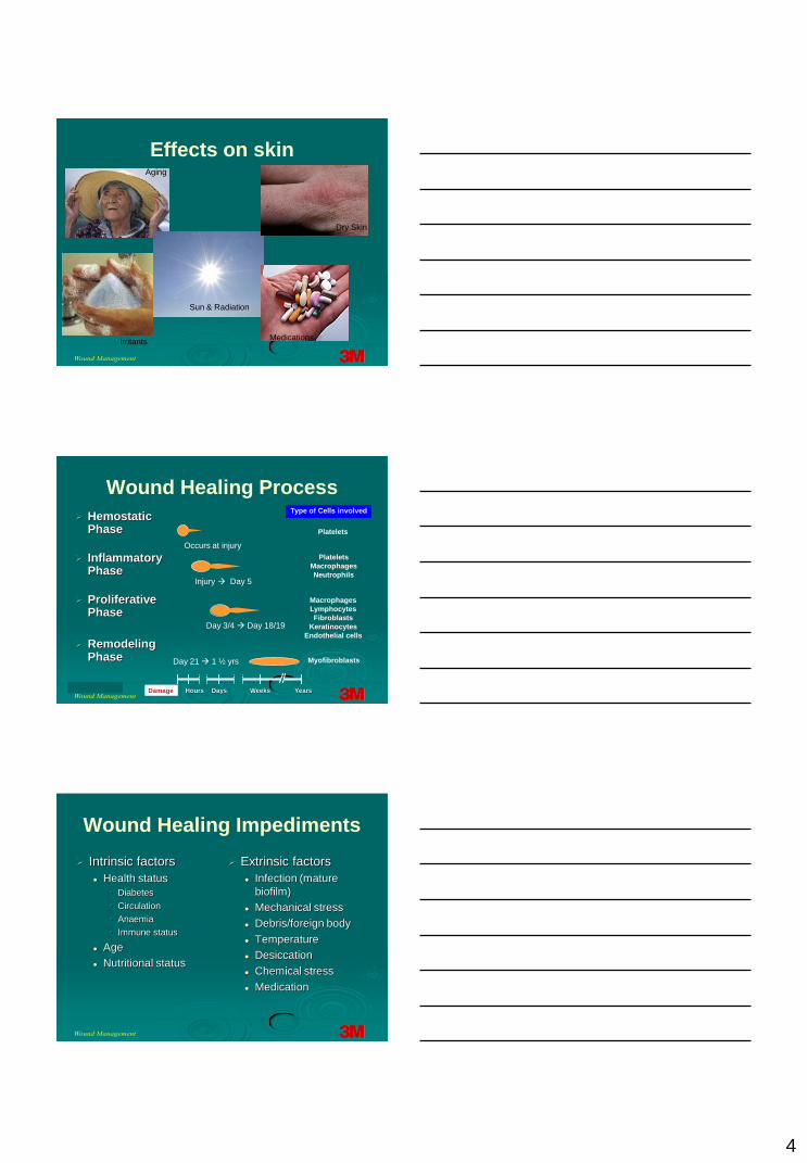
Wound Healing Impediments
Intrinsic factors
Health status
• Diabetes
• Circulation
• Anaemia
• Immune status
Age
Nutritional status
Extrinsic factors
Infection (mature
biofilm)
Mechanical stress
Debris/foreign body
Temperature
Desiccation
Chemical stress
Medication

5
Tissue Injury
Partial-Thickness:
Epidermis and superficial dermis
Painful
Regeneration
Full-Thickness
Total loss of skin layers, epidermis, dermis, subcutaneous layers, deeper tissue may be involved.
Repair
Connective tissue fills in defect
New tissue is never a strong as original tissue fills in defect
(Doughty, 2007)
Wound healing (con‟t)
Regeneration Repair
Identical tissue replaces
what was damaged
Dermis filled in with
collagen tissue, Replaced
by scar tissue. Deep dermis, fat,
muscle, bone not regenerated.
MDS 3.0 Tissue types
M0700 Most Severe Tissue Type for
Pressure Ulcer
Epithelial Tissue
Granulation Tissue
Slough
Necrotic Tissue (Eschar)

6
MDS 3.0
Granulation Tissue
Pink or red tissue with shiny, moist, granular appearance
Epithelial Tissue New skin growing in
superficial ulcer. It can be light pink and shiny, even in persons with darkly pigmented skin
Wound Base-Slough Nonviable tissue, loose or firm
Soft, tan, yellow, brown. Green
MDS definition – yellow or white tissue that adheres to the ulcer bed in strings or thick clumps, or is mucinous
Wound Base-Eschar
Necrotic tissue
MDS - Black or brown, or tan tissue that adheres firmly to the wound bed or ulcer edges, may be softer or harder
Loose or firm, hard, soft, or boggy
If wound is covered with eschar, wound size likely to INCREASE when necrotic tissue is debrided

7
Basic Principles of Wound Care for
Pressure Ulcers with Tissue Loss
(Stage II, III, IV, Unstageable)
Wound Bed Preparation
The “DIME” concept
• Debridement
• Infection/ Inflammation
• Moisture
• Edge
Effective Debridement Debridement removes:
Cellular debris that may be impairing healing
Microbes and toxins that may be prolonging the inflammatory phase
Chronic wounds require ongoing maintenance debridement rather than a single intervention
Selective and Non-selective methods
Selection of method
Urgency of the need for debridement
Skill level of the care provider
Availability of products and supplies
Debridement

8
Effective Topical Therapy
Eliminate non-viable
tissue and debris:
Debridement
• Surgical
• Sharp conservative
• Chemical
• Mechanical
• Autolytic
Mechanical Debridement
Irrigation
High pressure
Pulsatile
Whirlpool
Debridement cont.
Surgical
Sharp conservative
Chemical: tissue-specific enzymes
Mechanical
Autolytic: moisture retentive dressings
Biologic
9
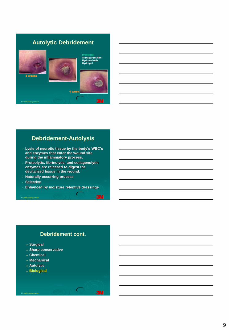
Autolytic Debridement
1 week
3 weeks
Dressings:Transparent filmHydrocolloidsHydrogel
Debridement-Autolysis
Lysis of necrotic tissue by the body‟s WBC‟s
and enzymes that enter the wound site
during the inflammatory process.
Proteolytic, fibrinolytic, and collagenolytic
enzymes are released to digest the
devitalized tissue in the wound.
Naturally occurring process
Selective
Enhanced by moisture retentive dressings
Debridement cont.
Surgical
Sharp conservative
Chemical
Mechanical
Autolytic
Biological

10
Debridement-Biologic
Maggots
Sterile
No reimbursement
Resistant organisms
Mail order
Stable Eschar
Do Not Debride
stable eschar!
Eschar on heels
Observe!
Debridement once
eschar separates
11
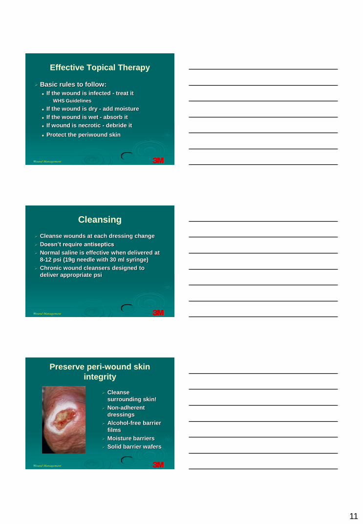
Effective Topical Therapy
Basic rules to follow:
If the wound is infected - treat it
• WHS Guidelines
If the wound is dry - add moisture
If the wound is wet - absorb it
If wound is necrotic - debride it
Protect the periwound skin
Cleansing
Cleanse wounds at each dressing change
Doesn‟t require antiseptics
Normal saline is effective when delivered at
8-12 psi (19g needle with 30 ml syringe)
Chronic wound cleansers designed to
deliver appropriate psi
Preserve peri-wound skin
integrity
Cleanse
surrounding skin!
Non-adherent
dressings
Alcohol-free barrier
films
Moisture barriers
Solid barrier wafers

12
Which Treatment Path Do You
Take?
Choose a Dressing That
protects the wound from the
surrounding environment
keeps the wound base moist
keeps the surrounding tissue dry
contains exudate without desiccating
the wound base
maintains the integrity of the primary
dressing**
13
What is “Moist” Wound Healing?
First described ~1940
Winter‟s publication-1962
Superficial wounds
Control group allowed to dry
Experimental group covered with thin
plastic film
Experimental group-2x faster re-
epithelialization
Advantages of Moisture
Retentive Dressings
Faster healing
Cytokines and growth factors remain
viable, cells
migrate unobstructed
Promotes granulation tissue formation
Supports autolysis
Reduces fibrosis: less scarring
Reduces pain
Objections to Moisture Retentive
Dressings
Objections
Concern regarding infection• Bacteria increase but not correlated with
higher incidence of infection
• Risk of infection may be reduced under dressings that form a barrier
Dressing cost: counteracted by longer wear time and improved outcome if used correctly
14
Key Consideration in Dressing
Selection
Management of
moisture
maintain a Moist
Environment
DRY - Add(+)
Moisten
WET - Subtract(--)
Absorb
Protect wound bed
Dressing Terminology
Primary
Direct contact with wound base
Secondary
Secures or protects primary dressing
Moisture retentive
„Modern dressings‟
Traditional
Tape & gauze
Dressing Categories-Gauze
Advantages
Readily available
Intuitive
Versatile
Disadvantages:
Microbial
Contamination
Labor intensive
Easy to misuse

15
Gauze cont.
Technique
Woven cotton or
appropriate synthetic
Correct size/shape
Pack lightly (fluff)
Moist versus wet
Change to prevent
strike-through
Composite Dressings
Combine features of
two or more types of
dressings
Primary or
secondary dressing,
e.g., non-adherent
pad and transparent
dressing
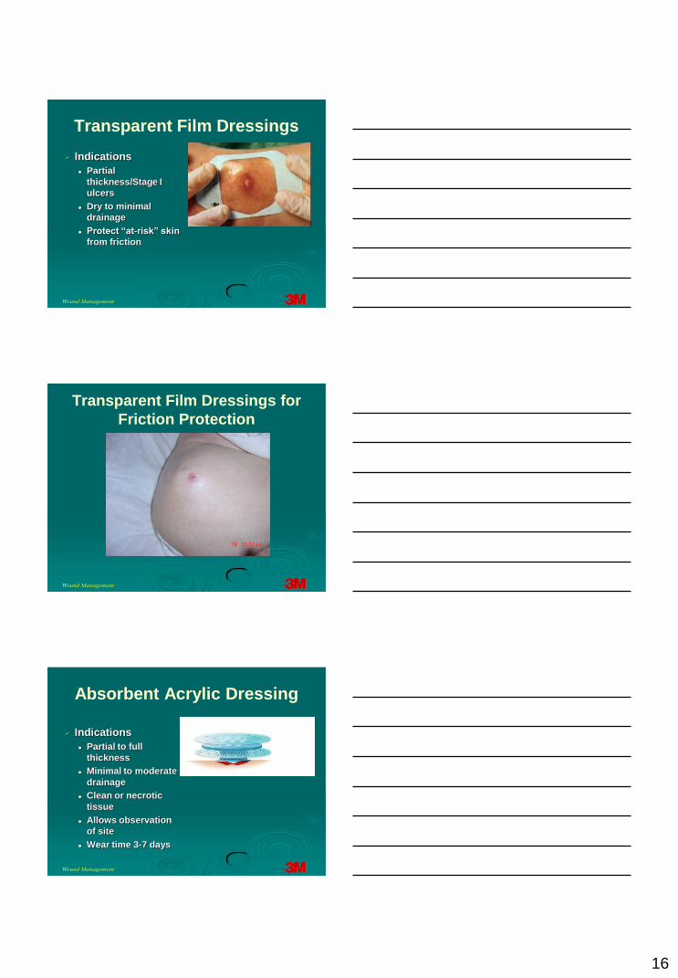
Transparent Film Dressings
Description:
Breathable,
polyurethane film
Barrier properties
Maintains moist
wound environment-
excellent at autolysis
Primary or
secondary dressing
16
Transparent Film Dressings
Indications
Partial
thickness/Stage I
ulcers
Dry to minimal
drainage
Protect “at-risk” skin
from friction
Transparent Film Dressings for
Friction Protection
Absorbent Acrylic Dressing
Indications
Partial to full
thickness
Minimal to moderate
drainage
Clean or necrotic
tissue
Allows observation
of site
Wear time 3-7 days

17
Absorbent Acrylic Dressing
Absorbent Acrylic Dressing
Hydrocolloid Dressings
Description Absorbent particles (e.g.
CMC) in adhesive matrix
Breathable or occlusive: determines ability to manage drainage
Indications Partial to full thickness
Minimal to moderate drainage
Clean or necrotic tissue
Wear time 3-7-days

18
Foam Dressings
Description
Polyurethane pads
Absorbent and
breathable
Primary or secondary
dressing
Foam Dressings
Indications
Partial and full thickness wounds
Min. to moderately-heavy draining wounds
e.g..
Variety of wounds-pressure ulcers, dehisced
surgical wounds, donor sites, venous ulcers,
trachs, partial thickness burns, etc.
Contact Layers
Contact layer
Various materials
Indications
Protect wound base
Prevent pain
19
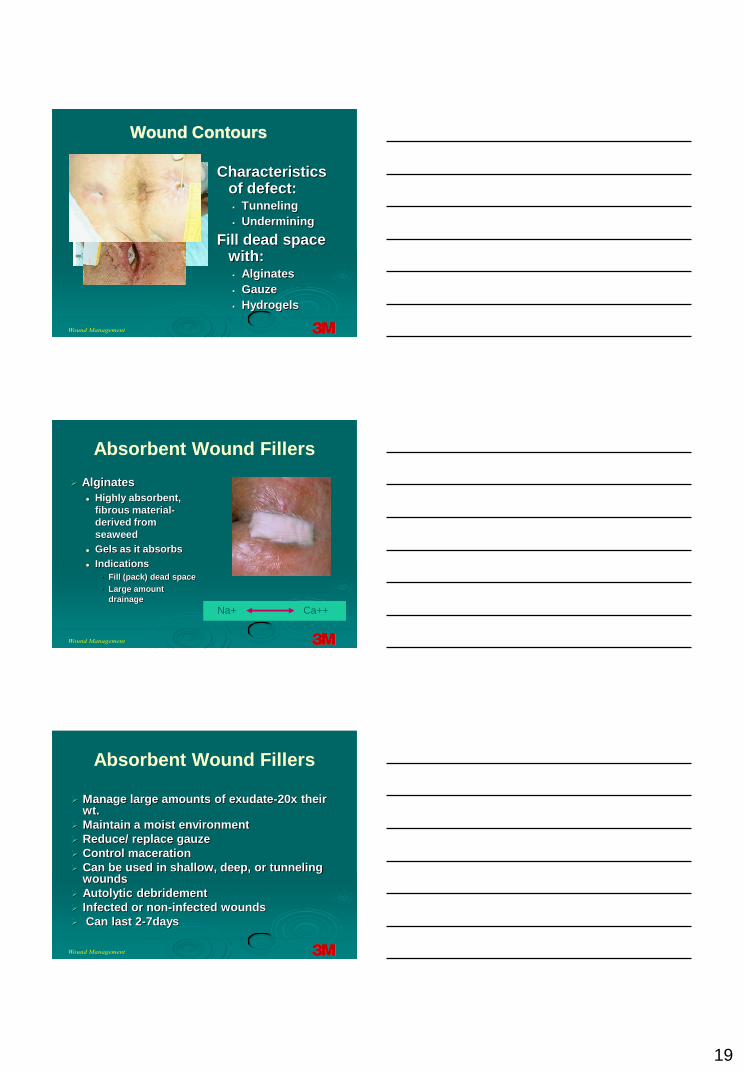
Characteristics of defect: Tunneling
Undermining
Fill dead space with: Alginates
Gauze
Hydrogels
Wound Contours
Absorbent Wound Fillers
Alginates
Highly absorbent,
fibrous material-
derived from
seaweed
Gels as it absorbs
Indications
• Fill (pack) dead space
• Large amount
drainage
Na+ Ca++
Manage large amounts of exudate-20x their wt.
Maintain a moist environment
Reduce/ replace gauze
Control maceration
Can be used in shallow, deep, or tunneling wounds
Autolytic debridement
Infected or non-infected wounds
Can last 2-7days
Absorbent Wound Fillers

20
Where Would You Use an
Alginate?
Wound Hydration
Hydrogel
Hydrogel Dressings
Description:
hydrating liquid or
liquid impregnated
gauze
Indications
Non-draining to
minimal drainage
Partial to full
thickness
Clean, necrotic or
infected wounds

21
Hydrogel Dressings cont.
Contain within
wound edges
Protect wound
edges/
surrounding skin
Change frequency
Keep wound base
moist
Every 24-48 hours
Evaluate Patient Progress- Be
Open to Change
Revise Treatment Plan as Necessary:
If the wound shows no improvement in
2 weeks…reevaluate for possible
change in treatment.
Why Does a Wound Fail to
Progress?
Local factors:
Infection
Inflammation
Ischemia
Other factors
↑ MMPs
↓ Growth factors
↓ Nitric oxide

22
Why Does a Wound Fail to
Progress?
Other factors
Necrotic tissue
Location
Size
Foreign objects
Infection
All chronic wounds are
colonized with bacteria
Infection = “105” col./ml.
(not always!)
Culture: if signs of
infection present• Biopsy-most accurate
• Swab-most common
• Never culture necrotic
tissue
Antimicrobials
Antiseptics
Examples-Povidone iodine, “Dakin’s, H202
Indication (traditional): Skin disinfection
Non-selective: Cytotoxic to viable cells

23
Antiseptic Solutions
Dakin‟s Solution
Effective against Staphylococcus and
Streptococcus
Toxic to fibroblasts
May injure periwound skin
Acetic Acid
Effective against pseudomonas
Toxic to fibroblasts
Modern Antimicrobials
Cadexomer iodine
Silver
Silver-Mechanism of Action

24
Silver Dressings
Antimicrobial barrier dressing
Controlled release of ionic silver
Non-cytotoxic
Effective for 3-7 days
Many substrates
Films, wound fillers, composites, contact layers, foams, hydrocolloids,etc.
*Dressings require moisture so silver can dissolve
Chronic/Stalled wounds
Wounds not responding to standard
treatment protocol in 4-6 weeks
Options
Collagen products w/wo silver
Matrix with metal ions
Growth Factors
Biosynthetic Dressings
Other Options for Non-healing
Wounds
PHI technology Metal ions, citric acid, polyethylene glycols
What are Growth factors? Proteins: Platelets, macrophages
Action• cell growth
• cell migration
• Regulatory effect
Necrotic tissue and infection must be managed!
Only one commercially available- Regranex®• PDGF
• Indications-limited

25
Considerations
Edema control
Nutrition
Topical care
Patient education
Case Study – Venous Ulcer
Is This a Stage II?
No! But why not?
Superficial moist lesions
with irregular borders
“Moisture lesions”
(DeFloor)
Causative factors
Moisture
• Incontinence
• Perspiration
• Friction
At risk-immobile, obese

26
Case Study-Skin Tear
Case Study – Neuropathic
UlcerConsiderations
Off-loading
Topical care
Patient education
Difficult Locations

27
Management of Patient-
intrinsic FactorsDiseases/condition
s:
Immuno-compromise
Diabetes
Reduced perfusion
• Arterial: LEAD Non-diabetic
Diabetes
• Venous
Malnutrition
Immobility
Cognitive deficits
Large wounds
Dressings
Negative Pressure
Wound Therapy
(NPWT)
• V.A.C.®Therapy
Pouching (rare)
Small wounds:
dressings
Management Options for
Difficult Sizes
28
Management of Foreign Objects: