Embed Size (px)
Citation preview

Epo Gene Regulation 233Tohoku J. Exp. Med., 2015, 235, 233-240
233
Received January 30, 2015; revised and accepted February 23, 2015. Published online March 17, 2015; doi: 10.1620/tjem.235.233.Correspondence: Norio Suzuki, Division of Interdisciplinary Medical Science, Center for Oxygen Medicine, United Centers for
Advanced Research and Translational Medicine, Tohoku University Graduate School of Medicine, 2-1 Seiryo-machi, Aoba-ku, Sendai, Miyagi 980-8575, Japan.
e-mail: [email protected]. Norio Suzuki is a recipient of the 2014 Gold Prize, Tohoku University School of Medicine.
Invited Review
Erythropoietin Gene Expression: Developmental-Stage Specificity, Cell-Type Specificity, and Hypoxia Inducibility
Norio Suzuki1
1Division of Interdisciplinary Medical Science, Center for Oxygen Medicine, United Centers for Advanced Research and Translational Medicine, Tohoku University Graduate School of Medicine, Sendai, Miyagi, Japan
Erythrocytes play an essential role in the delivery of oxygen from the lung to every organ; a decrease in erythrocytes (anemia) causes hypoxic stress and tissue damage. To maintain oxygen homeostasis in adult mammals, when the kidney senses hypoxia, it secretes an erythroid growth factor, erythropoietin (Epo), which stimulates erythropoiesis in the bone marrow. Recently, studies using genetically modified mice have shown that the in vivo expression profile of the Epo gene changes dramatically during development. The first Epo-producing cells emerge in the neural crest and neuroepithelium of mid-stage embryos and support primitive erythropoiesis in the yolk sac. Subsequently, Epo from the hepatocytes stimulates erythropoiesis in the fetal liver of later stage embryos in a paracrine manner. In fact, erythroid lineage cells comprise the largest cell population in the fetal liver, and hepatocytes are distributed among the erythroid cell clusters. Adult erythropoiesis in the bone marrow requires Epo that is secreted by renal Epo-producing cells (REP cells). REP cells are widely distributed in the renal cortex and outer medulla. Hypoxia-inducible Epo production both in hepatocytes and REP cells is controlled at the gene transcription level that is mainly mediated by the hypoxia-inducible transcription factor (HIF) pathway. These mouse studies further provide insights into the molecular mechanisms of the cell-type specific, hypoxia-inducible expression of the Epo gene, which involves multiple sets of cis- and trans-regulatory elements.
Keywords: anemia; erythropoiesis; genetically modified mouse; renal erythropoietin-producing cell; transcriptional regulationTohoku J. Exp. Med., 2015 March, 235 (3), 233-240. © 2015 Tohoku University Medical Press
IntroductionErythropoietin (Epo) is a 34-kDa glycoprotein essen-
tial for erythropoiesis. One Epo molecule binds to a homodimer of its specific receptors (EpoR) on the surface of immature erythroid cells, and transduces signals that repress apoptosis and promote cell proliferation and differ-entiation into mature erythrocytes (Wojchowski et al. 2010). Epo binding to the receptors causes conformation changes in the intracellular domains of an EpoR homodimer, bring-ing it into close proximity to initiate Epo-EpoR signal transduction (Suzuki et al. 2015). Because EpoR is highly expressed only in committed erythroid progenitors and erythroblastic cells, the principal function of Epo is funda-mentally restricted to erythropoiesis; it is not involved in the growth of other cell lineages (Suzuki et al. 2002; Paffett-Lugassy et al. 2007). Additionally, Epo signaling is important for the growth and terminal maturation of ery-
throid cells and not for the commitment of hematopoietic stem cells to the erythroid lineage (Wu et al. 1995).
The human Epo gene encodes the Epo precursor pro-tein, which consist of 193-amino acids, and the mature 166-amino-acid Epo is secreted from Epo-producing cells into the bloodstream after cleavage of the amino-terminal signal sequences. The Epo gene has been cloned from a variety of mammals as well as fish and frogs (Miyake et al. 1977; Jacobs et al. 1985; McDonald et al. 1986; Shoemaker and Mitsock 1986; Chou et al. 2004; Nogawa-Kosaka et al. 2010). However, even though anemic chicken plasma has historically exhibited high erythropoietic activity, avian Epo genes have not yet been identified (Yamamoto et al. 1985). The human Epo amino acid sequence exhibits an overall sequence homology of 80% to mouse Epo but less than 33% of the fish Epo protein (Chu et al. 2008). Interestingly, human Epo administration can stimulate not only mamma-lian erythropoiesis but also erythropoiesis in frogs, even
N. Suzuki234
with the amino acid sequence of Xenopus laevis Epo being only 38% identical to human Epo (Nogawa-Kosaka et al. 2010).
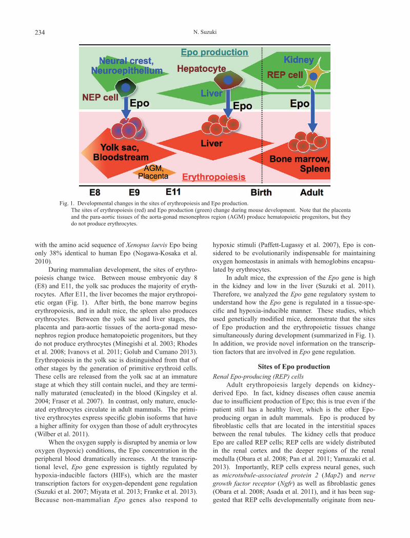
During mammalian development, the sites of erythro-poiesis change twice. Between mouse embryonic day 8 (E8) and E11, the yolk sac produces the majority of eryth-rocytes. After E11, the liver becomes the major erythropoi-etic organ (Fig. 1). After birth, the bone marrow begins erythropoiesis, and in adult mice, the spleen also produces erythrocytes. Between the yolk sac and liver stages, the placenta and para-aortic tissues of the aorta-gonad meso-nephros region produce hematopoietic progenitors, but they do not produce erythrocytes (Minegishi et al. 2003; Rhodes et al. 2008; Ivanovs et al. 2011; Golub and Cumano 2013). Erythropoiesis in the yolk sac is distinguished from that of other stages by the generation of primitive erythroid cells. These cells are released from the yolk sac at an immature stage at which they still contain nuclei, and they are termi-nally maturated (enucleated) in the blood (Kingsley et al. 2004; Fraser et al. 2007). In contrast, only mature, enucle-ated erythrocytes circulate in adult mammals. The primi-tive erythrocytes express specific globin isoforms that have a higher affinity for oxygen than those of adult erythrocytes (Wilber et al. 2011).
When the oxygen supply is disrupted by anemia or low oxygen (hypoxic) conditions, the Epo concentration in the peripheral blood dramatically increases. At the transcrip-tional level, Epo gene expression is tightly regulated by hypoxia-inducible factors (HIFs), which are the master transcription factors for oxygen-dependent gene regulation (Suzuki et al. 2007; Miyata et al. 2013; Franke et al. 2013). Because non-mammalian Epo genes also respond to
hypoxic stimuli (Paffett-Lugassy et al. 2007), Epo is con-sidered to be evolutionarily indispensable for maintaining oxygen homeostasis in animals with hemoglobins encapsu-lated by erythrocytes.
In adult mice, the expression of the Epo gene is high in the kidney and low in the liver (Suzuki et al. 2011). Therefore, we analyzed the Epo gene regulatory system to understand how the Epo gene is regulated in a tissue-spe-cific and hypoxia-inducible manner. These studies, which used genetically modified mice, demonstrate that the sites of Epo production and the erythropoietic tissues change simultaneously during development (summarized in Fig. 1). In addition, we provide novel information on the transcrip-tion factors that are involved in Epo gene regulation.
Sites of Epo productionRenal Epo-producing (REP) cells
Adult erythropoiesis largely depends on kidney-derived Epo. In fact, kidney diseases often cause anemia due to insufficient production of Epo; this is true even if the patient still has a healthy liver, which is the other Epo-producing organ in adult mammals. Epo is produced by fibroblastic cells that are located in the interstitial spaces between the renal tubules. The kidney cells that produce Epo are called REP cells; REP cells are widely distributed in the renal cortex and the deeper regions of the renal medulla (Obara et al. 2008; Pan et al. 2011; Yamazaki et al. 2013). Importantly, REP cells express neural genes, such as microtubule-associated protein 2 (Map2) and nerve growth factor receptor (Ngfr) as well as fibroblastic genes (Obara et al. 2008; Asada et al. 2011), and it has been sug-gested that REP cells developmentally originate from neu-
Fig. 1. Developmental changes in the sites of erythropoiesis and Epo production. The sites of erythropoiesis (red) and Epo production (green) change during mouse development. Note that the placenta
and the para-aortic tissues of the aorta-gonad mesonephros region (AGM) produce hematopoietic progenitors, but they do not produce erythrocytes.

Epo Gene Regulation 235
ral crest cells (Asada et al. 2011; Suzuki et al. 2013). This idea is supported by the observation that kidney progenitor cells produce renal tubules and glomeruli but not blood ves-sels or interstitial tissues, which contain REP cells (Matsumoto et al. 2012; Taguchi et al. 2014).
Most interstitial fibroblasts in the renal cortex have the potential to produce Epo, but even under chronic severe anemia, only 10% of these cells express the Epo gene (Yamazaki et al. 2013) (Fig. 2A). This observation sug-gests that all REP cells can be divided into 2 groups: OFF-REP cells that are at rest and ON-REP cells that produce Epo. The larger OFF-REP cell population may be an emer-gency reserve for Epo production. The total number of REP cells is constant, but the fraction of ON-REP cells increases in proportion to the severity of anemic or hypoxic stress (Obara et al. 2008; Yamazaki et al. 2013). Because the expression of hypoxia-inducible genes is higher in ON-REP cells than in OFF-REP cells, it has been suggested that ON-REP cells are more hypoxic than OFF-REP cells (Yamazaki et al. 2013) (Fig. 2B). Thus, hypoxia is the prin-cipal inducer of Epo gene expression in REP cells, and REP cells control oxygen homeostasis by regulating erythropoie-sis in adult mammals.
Because the blood delivers oxygen to REP cells after passing through the glomeruli, the basal oxygen supply to the REP cells seems to be lower than in other tissues. Additionally, the tubular cells around the REP cells may consume high amounts of oxygen to produce ATP for urine reabsorption. Thus, in the microenvironment of the REP cells, oxygen flux is very rapid due to low supply and high consumption. The oxygen state of the REP cells may help
sense decreases in oxygen uptake into the body, and it pro-vides insight into the reason why the kidneys produce Epo.
During the progression of kidney disease, fibrosis begins with the emergence of myofibroblasts in the intersti-tial tissue between damaged renal tubules. We have dem-onstrated that most myofibroblasts originate from the REP cells (Asada et al. 2011; Souma et al. 2013; Miyata et al. 2013). After their transformation to myofibroblasts, the REP cells begin producing smooth muscle actins and colla-gens, whereas the expression of neural and fibroblastic genes is repressed. Despite disease conditions that make the kidney much more hypoxic, the myofibroblastic-trans-formed REP cells lose their Epo-producing ability (Fig. 2B). Therefore, REP cells are responsible for both fibrosis and anemia in chronic kidney diseases.
HepatocytesAt the point during embryonic development when the
liver is an erythropoietic organ, hepatocytes are the major source of Epo (Fig. 1). In E15 embryos, erythroid lineage cells comprise the largest cell population in the fetal liver, and hepatocytes surround the erythroid cell clusters (Fig. 3A). Thus, Epo stimulates fetal liver erythropoiesis in a paracrine manner, whereas erythropoiesis in the yolk sac and bone marrow is promoted by endocrine Epo (Fig. 1).
The adult liver can still produce Epo in response to hypoxia, but the Epo production of each hepatocyte is very low compared with that of each REP cell. Therefore, the kidney is the major Epo-producing site in adults; hepatic Epo production is dispensable for normal adult erythropoie-sis (Suzuki et al. 2011). However, in embryos and neo-
Fig. 2. ON-REP, OFF-REP and MF-REP cells. (A) Kidney cryosection from a mouse suffering from chronic anemia. The green fluorescence is derived from the Epo-
GFP transgene and indicates the ON-REP cells with active Epo gene expression. The red fluorescence is derived from a tracer for cells that have expressed the Epo-Cre transgene in Rosa26R-tdTomato::Epo-Cre mice, and indicates total REP cells (Yamazaki et al. 2013). The nuclei are stained with DAPI (blue). The arrowheads indicate ON-REP cells that are positive for both green and red fluorescence. The OFF-REP cells express red fluorescence but not green fluores-cence. (B) Subtypes of REP cells. Most REP cells do not produce Epo under normal oxygen conditions (OFF-REP), whereas hypoxic microenvironments induce Epo production (ON-REP). Healthy REP cells are reversibly transformed to myofibroblastic REP cells (MF-REP) by kidney diseases (Souma et al. 2013).

N. Suzuki236
nates, hepatic Epo production is essential for erythropoiesis; Epo or EpoR knockout mice commonly show embryonic lethality due to severe anemia at E13, which occurs before the kidneys initiate Epo production (Wu et al. 1995).
We have generated a genetically modified mouse line (referred to as the ∆EpoHE mouse) in which the liver-spe-cific Epo gene enhancer (EpoHE) is deleted from the genome (Suzuki et al. 2011). In the late embryonic stages in ∆EpoHE mice, the expression of the Epo gene is reduced in the liver but normal in the kidney. The livers of ∆EpoHE mice are pale during the perinatal stages, whereas erythro-poiesis in their spleens and bone marrow is normal (Fig. 3B and C). This observation indicates that liver-derived Epo primarily stimulates fetal liver erythropoiesis. In contrast, kidney-derived Epo is required for adult erythropoiesis in the spleen and bone marrow (Fig. 1).
Neural Epo-producing (NEP) cellsIn Epo knockout mice, primitive erythropoiesis in the
yolk sac is partially defective (Wu et al. 1995; Suzuki et al. 2013). The abnormality in the differentiation of primitive erythroid cells is observed at approximately E9, before the fetal liver initiates Epo production (Makita et al. 2005; Obara et al. 2008; Suzuki et al. 2013). To determine the source of the Epo used for primitive erythropoiesis, we used the genetically modified mouse lines expressing green fluorescent protein (GFP) as a reporter under the control of the Epo gene regulatory system (the Epo-GFP mice). As expected, GFP efficiently labeled the Epo gene-expressing cells in the Epo-GFP mice, and the Epo-GFP+ cells in the neural crest and neuroepithelium were identified as neural Epo-producing cells (NEP cells in Fig. 4A and B) (Suzuki et al. 2013).
NEP cells produce functional Epo that induces the dif-ferentiation of yolk sac erythrocytes. As with neural crest cells, a subset of the NEP cells migrates from the dorsal areas of the neural tube toward the abdominal region during mouse development (Fig. 4A). However, because Epo expression in NEP cells is extinguished around E11, it is impossible to trace their fate using the Epo-GFP reporter. Because REP cells exhibit neural features (Obara et al. 2008; Asada et al. 2011), an attractive hypothesis is that NEP cells migrate into the kidney and differentiate into REP cells. In the embryonic brain, the NEP cells are pri-marily found in the rhombencephalon (Fig. 4B and C), and we have detected Epo-GFP+ cells expressing the endoge-nous Epo gene in the brain of hypoxic adult mice. This evidence suggests that NEP-derived cells in organs other than the kidney and liver may retain their ability to produce Epo under pathological conditions. Interestingly, in Zebrafish, one of the major Epo-producing sites is the brain (Paffett-Lugassy et al. 2007). Therefore, transient Epo pro-duction by NEP cells in mouse embryos may support the so-called “recapitulation theory”, which argues that ontog-eny recapitulates phylogeny (Sander 2002).
Regulatory factors in Epo gene expressionGATA factors
Because there are no kidney cell lines that inducibly express the Epo gene, human hepatoma cell lines (Hep3B
Fig. 3. Expression and function of Epo in the liver. (A) Hepatocytes that are positive for Epo-GFP transgene
expression (brown color staining) are surrounded by ery-throid lineage cells (round shape, nuclei are counter-stained blue by hematoxylin) in the fetal liver of Epo-GFP transgenic mice at E15. (B) The liver of a day 0 ∆EpoHE neonate is pale compared to that of a control lit-termate mouse. (C) No significant differences between the spleens of ∆EpoHE and control neonates were ob-served.

Epo Gene Regulation 237
and HepG2) that produce Epo in a hypoxia-inducible man-ner have been used to study Epo gene regulation (Goldberg et al. 1987; Pugh et al. 1991; Tarumoto et al. 2000). The genomic sequences flanking the Epo coding region are highly conserved among mammals, and these sequences have been analyzed to understand the regulatory mecha-nisms of Epo gene expression (Suzuki et al. 2007).
The upstream flanking region contains a basal pro-moter and a GATA factor-binding motif (Fig. 5). In hepa-toma cell lines, the promoter activity is enhanced when the GATA sequence is mutated (Tarumoto et al. 2000). Mouse
lines bearing mutant Epo-GFP transgenes in which the GATA motif has been mutated have demonstrated that the GATA motif is required for the suppression of ectopic and constitutive Epo gene expression and dispensable for the hypoxia-induced Epo gene expression in REP cells and hepatocytes (Obara et al. 2008). The ectopic expression of the transgene is observed exclusively in cells of the epithe-lial lineage, including those of the renal tubules, bile ducts and alveolar epithelia (Fig. 5). This tissue-specific suppres-sion system is a regulatory mechanism that is unique to the Epo gene. There may be an epithelial enhancer sequence near the Epo gene, and the GATA motif strongly inhibits this activation signal in epithelial cells.
All 6 GATA transcription factors except GATA1 are expressed in epithelial-lineage cells; these proteins may complementarily suppress ectopic Epo gene transcription by binding to the GATA motif (Obara et al. 2008). Thus, the upstream GATA motif and the GATA transcription fac-tors play essential roles in cell type-specific, hypoxia-inducible Epo production in vivo (Fig. 5). GATA factors in the epithelial cells are plausible drug targets to pharmaco-logically induce Epo production in Epo-deficiency anemia.
The prolyl-hydroxylase (PHD)-HIF systemThe downstream flanking sequence of the Epo gene
was first identified as a hypoxia-responsive enhancer (Fig. 5), and a HIF complex has been isolated as a factor that binds to this enhancer (Semenza and Wang 1992; Wang and Semenza 1993). To determine the in vivo roles of this enhancer sequence, we deleted it from the mouse genome. The resulting mice exhibited a reduction in Epo production in the liver, whereas Epo production in the kidneys was unaffected by the deletion (Suzuki et al. 2011). This obser-vation indicates that the downstream flanking sequence, EpoHE (Hepatic Enhancer), is a hepatocyte-specific, hypoxia-responsive enhancer of the Epo gene (Fig. 5). REP and NEP cells utilize a different cell-type specific enhancer(s) to regulate Epo gene expression, but the enhancer(s) has not been identified (Fig. 5). Interestingly, EpoHE is required for Epo production in the hepatocytes after E14, indicating that the Epo gene regulatory system changes during liver ontogeny (Suzuki et al. 2011).
The HIF complex consists of α and β (also known as ARNT) subunits and regulates a large set of hypoxia-induc-ible genes by binding to a specific sequence (the hypoxia-responsive element, or HRE) in the regulatory regions of its target genes (Wang et al. 1995). Mammalian cells have 3 HIF-α isoforms: HIF1α, HIF2α and HIF3α. Under normal conditions, HIF-α proteins are degraded via the hydroxyl-ation of specific proline residues, which is mediated by HIF-prolyl-hydroxylases (PHDs) that use oxygen as a sub-strate (Greer et al. 2012).
To elucidate the regulatory mechanisms of Epo pro-duction in vivo, we have analyzed mouse lines harboring conditional deletions of genes encoding HIF-prolyl-hydroxylase isoforms (PHD1, PHD2 and PHD3) (Takeda et
Fig. 4. NEP cells are located in the neural crest and neuroepi-thelium.
(A) NEP cells (green, arrowheads) migrate from the dor-sal regions to the ventral regions around the neural tube (NT) and the otic vesicle (e) at E11, and CD31-positive capillaries (red) are located around the path of the NEP cells. (B and C) Dorsal view of Epo-GFP transgene ex-pression (green in B) and neural crest marker (Rosa26R-LacZ::Wnt1-Cre) expression (blue in C) at E11.

N. Suzuki238
al. 2006). Inactivation of PHD2 in REP cells causes poly-cythemia, which is prevented by the loss of HIF2α. Surprisingly, under disease conditions, the loss of PHD2 in REP cells restores Epo production in myofibroblastic-trans-formed REP cells. These results demonstrate that a PHD2-HIF2α cascade regulates the Epo gene in REP cells and that this cascade is involved in the inactivation of Epo produc-tion in injured kidneys (Fig. 5). Indeed, gene expression analyses have demonstrated that, compared with other iso-forms, PHD2 and HIF2α are highly expressed in REP cells. In the liver, deleting any combination of two PHD isoforms induces polycythemia, whereas the liver-specific deletion of a single isoform causes no apparent phenotype. Poly-cythemia is prevented by the loss of either HIF2α or EpoHE. These results demonstrate that hepatic Epo expres-sion is regulated by a PHDs-HIF2α-EpoHE cascade and that the combined activity of all PHDs is more important than the specific function of each PHD isoform in this cas-cade. Thus, analyses of genetically modified mice have demonstrated the existence of distinct regulatory systems for hypoxia-induced Epo gene expression in the kidneys and liver.
PerspectivesStudies using genetically modified mouse lines have
provided important information on the tissue-specific and hypoxia-inducible Epo gene expression profile and the in vivo regulatory mechanisms of Epo production, which are mainly regulated in a hypoxia-dependent manner by the PHD-HIF system (Fig. 5). Further studies may elucidate
the developmental fate of newly identified NEP cells in the neural crest and the Epo-producing potential of NEP cell-derived cells in adult mice. The liver-specific enhancer of the Epo gene (EpoHE) has been identified in the proximal region downstream of the coding region, but little is known about the enhancer(s) for NEP and REP cells. Transgenic reporter analyses may identify the enhancer sequences in the mammalian genome in the near future.
Cells in the carotid body, which are derived from neu-ral crest and reside in the carotid bifurcation, play essential roles in the maintenance of oxygen homeostasis by regulat-ing respiration and blood pressure (Platero-Luengo et al. 2014). The carotid body senses hypoxic stresses in the carotid artery through channel molecule depolarization and acutely releases neurotransmitters toward the central ner-vous system. In addition to the regulation of respiration and blood pressure by carotid body cells, erythropoietic induction by REP cells is also important for the mainte-nance of oxygen homeostasis in adults through Epo produc-tion. The molecular basis of hypoxic responses in REP cells, which requires PHD-HIF system-mediated transcrip-tional induction, is completely different from the carotid body, which uses neurotransmitters. REP cell-mediated erythropoietic induction may contribute to chronic phases of hypoxic adaptation, compared with the acute response of the carotid body. Thus, elucidation of the mechanism of renal Epo production is important for understanding sys-temic responses to hypoxic stressors. However, because of the difficulty in culturing REP cells in vitro, little is known about the regulatory mechanisms of Epo gene expression in
Fig. 5. Schema of cell-type specific Epo gene regulation.

Epo Gene Regulation 239
these cells. The development of REP cell culture tech-niques will allow a deeper understanding of the mecha-nisms of hypoxia sensing, Epo gene regulation and kidney fibrosis.
AcknowledgmentsI am grateful to Drs. Masayuki Yamamoto and Toshio
Miyata for giving me the opportunity to carry out this study.
Conflict of InterestThe author declares no conflict of interest.
ReferencesAsada, N., Takase, M., Nakamura, J., Oguchi, A., Asada, M.,
Suzuki, N., Yamamura, K., Nagoshi, N., Shibata, S., Rao, T.N., Fehling, H.J., Fukatsu, A., Minegishi, N., Kita, T., Kimura, T., et al. (2011) Dysfunction of fibroblasts of extrarenal origin underlies renal fibrosis and renal anemia in mice. J. Clin. Invest., 121, 3981-3990.
Chou, C.F., Tohari, S., Brenner, S. & Venkatesh, B. (2004) Eryth-ropoietin gene from a teleost fish, Fugu rubripes. Blood, 104, 1498-1503.
Chu, C.Y., Cheng, C.H., Yang, C.H. & Huang, C.J. (2008) Eryth-ropoietins from teleosts. Cell. Mol. Life Sci., 65, 3545-3552.
Franke, K., Gassmann, M. & Wielockx, B. (2013) Erythrocytosis: the HIF pathway in control. Blood, 122, 1122-1128.
Fraser, S.T., Isern, J. & Baron, M.H. (2007) Maturation and enucleation of primitive erythroblasts during mouse embryo-genesis is accompanied by changes in cellsurface antigen expression. Blood, 109, 343-352.
Goldberg, M.A., Glass, G.A., Cunningham, J.M. & Bunn, H.F. (1987) The regulated expression of erythropoietin by two human hepatoma cell lines. Proc. Natl. Acad. Sci. USA, 84, 7972-7976.
Golub, R. & Cumano, A. (2013) Embryonic hematopoiesis. Blood Cells Mol. Dis., 51, 226-231.
Greer, S.N., Metcalf, J.L., Wang, Y. & Ohh, M. (2012) The updated biology of hypoxia-inducible factor. EMBO J., 31, 2448-2460.
Ivanovs, A., Rybtsov, S., Welch, L., Anderson, R.A., Turner, M.L. & Medvinsky, A. (2011) Highly potent human hematopoietic stem cells first emerge in the intraembryonic aorta-gonad-mesonephros region. J. Exp. Med., 208, 2417-2427.
Jacobs, K., Shoemaker, C., Rudersdorf, R., Neill, S.D., Kaufman, R.J., Mufson, A., Seehra, J., Jones, S.S., Hewick, R., Fritsch, E.F., Kawakita, M., Shimizu, T. & Miyake, T. (1985) Isola-tion and characterization of genomic and cDNA clones of human erythropoietin. Nature, 313, 806-810.
Kingsley, P.D., Malik, J., Fantauzzo, K.A. & Palis, J. (2004) Yolk sac-derived primitive erythroblasts enucleate during mamma-lian embryogenesis. Blood, 104, 19-25.
Makita, T., Duncan, S.A. & Sucov, H.M. (2005) Retinoic acid, hypoxia, and GATA factors cooperatively control the onset of fetal liver erythropoietin expression and erythropoietic differ-entiation. Dev. Biol., 280, 59-72.
Matsumoto, K., Yokoo, T., Matsunari, H., Iwai, S., Yokote, S., Teratani, T., Gheisari, Y., Tsuji, O., Okano, H., Utsunomiya, Y., Hosoya, T., Okano, H.J., Nagashima, H. & Kobayashi, E. (2012) Xenotransplanted embryonic kidney provides a niche for endogenous mesenchymal stem cell differentiation into erythropoietin-producing tissue. Stem Cells, 30, 1228-1235.
McDonald, J.D., Lin, F.K. & Goldwasser, E. (1986) Cloning, sequencing, and evolutionary analysis of the mouse erythro-poietin gene. Mol. Cell. Biol., 6, 842-848.
Minegishi, N., Suzuki, N., Yokomizo, T., Pan, X., Fujimoto, T., Takahashi, S., Hara, T., Miyajima, A., Nishikawa, S. &
Yamamoto, M. (2003) Expression and domain-specific func-tion of GATA-2 during differentiation of the hematopoietic precursor cells in midgestation mouse embryos. Blood, 102, 896-905.
Miyake, T., Kung, C.K. & Goldwasser, E. (1977) Purification of human erythropoietin. J. Biol. Chem., 252, 5558-5564.
Miyata, T., Suzuki, N. & van Ypersele de Strihou, C. (2013) Diabetic nephropathy: are there new and potentially promising therapies targeting oxygen biology? Kidney Int., 84, 693-702.
Nogawa-Kosaka, N., Hirose, T., Kosaka, N., Aizawa, Y., Nagasawa, K., Uehara, N., Miyazaki, H., Komatsu, N. & Kato, T. (2010) Structural and biological properties of eryth-ropoietin in Xenopus laevis. Exp. Hematol., 38, 363-372.
Obara, N., Suzuki, N., Kim, K., Nagasawa, T., Imagawa, S. & Yamamoto, M. (2008) Repression via the GATA box is essen-tial for tissue-specific erythropoietin gene expression. Blood, 111, 5223-5232.
Paffett-Lugassy, N., Hsia, N., Fraenkel, P.G., Paw, B., Leshinsky, I., Barut, B., Bahary, N., Caro, J., Handin, R. & Zon, L.I. (2007) Functional conservation of erythropoietin signaling in zebrafish. Blood, 110, 2718-2726.
Pan, X., Suzuki, N., Hirano, I., Yamazaki, S., Minegishi, N. & Yamamoto, M. (2011) Isolation and characterization of renal erythropoietin-producing cells from genetically produced anemia mice. PLoS One, 6, e25839.
Platero-Luengo, A., González-Granero, S., Durán, R., Díaz-Castro, B., Piruat, J.I., García-Verdugo, J.M., Pardal, R. & López-Barneo, J. (2014) An O2-sensitive glomus cell-stem cell synapse induces carotid body growth in chronic hypoxia. Cell, 156, 291-303.
Pugh, C.W., Tan, C.C., Jones, R.W. & Ratcliffe, P.J. (1991) Func-tional analysis of an oxygen-regulated transcriptional enhancer lying 3′ to the mouse erythropoietin gene. Proc. Natl. Acad. Sci. USA, 88, 10553-10557.
Rhodes, K.E., Gekas, C., Wang, Y., Lux, C.T., Francis, C.S., Chan, D.N., Conway, S., Orkin, S.H., Yoder, M.C. & Mikkola, H.K. (2008) The emergence of hematopoietic stem cells is initiated in the placental vasculature in the absence of circulation. Cell Stem Cell, 2, 252-263.
Sander, K. (2002) Ernst Haeckel’s ontogenetic recapitulation: irri-tation and incentive from 1866 to our time. Ann. Anat., 184, 523-533.
Semenza, G.L. & Wang, G.L. (1992) A nuclear factor induced by hypoxia via de novo protein synthesis binds to the human erythropoietin gene enhancer at a site required for transcrip-tional activation. Mol. Cell. Biol., 12, 5447-5454.
Shoemaker, C.B. & Mitsock, L.D. (1986) Murine erythropoietin gene: cloning, expression, and human gene homology. Mol. Cell. Biol., 6, 849-858.
Souma, T., Yamazaki, S., Moriguchi, T., Suzuki, N., Hirano, I., Pan, X., Minegishi, N., Abe, M., Kiyomoto, H., Ito, S. & Yamamoto, M. (2013) Plasticity of renal erythropoietin-producing cells governs fibrosis. J. Am. Soc. Nephrol., 24, 1599-1616.
Suzuki, N., Hirano, I., Pan, X., Minegishi, N. & Yamamoto, M. (2013) Erythropoietin production in neuroepithelial and neural crest cells during primitive erythropoiesis. Nat. Commun., 4, 2902.
Suzuki, N., Mukai, H.Y. & Yamamoto, M. (2015) In vivo regula-tion of erythropoiesis by chemically inducible dimerization of the erythropoietin receptor intracellular domain. PLoS One, [in press].
Suzuki, N., Obara, N., Pan, X., Watanabe, M., Jishage, K., Minegishi, N. & Yamamoto, M. (2011) Specific contribution of the erythropoietin gene 3′ enhancer to hepatic erythropoi-esis after late embryonic stages. Mol. Cell. Biol., 31, 3896-3905.
Suzuki, N., Obara, N. & Yamamoto, M. (2007) Use of gene-manipulated mice in the study of erythropoietin gene expres-
N. Suzuki240
sion. Methods Enzymol., 435, 157-177.Suzuki, N., Ohneda, O., Takahashi, S., Higuchi, M., Mukai, H.Y.,
Nakahata, T., Imagawa, S. & Yamamoto, M. (2002) Erythroid-specific expression of the erythropoietin receptor rescued its null mutant mice from lethality. Blood, 100, 2279-2288.
Taguchi, A., Kaku, Y., Ohmori, T., Sharmin, S., Ogawa, M., Sasaki, H. & Nishinakamura, R. (2014) Redefining the in vivo origin of metanephric nephron progenitors enables generation of complex kidney structures from pluripotent stem cells. Cell Stem Cell, 14, 53-67.
Takeda, K., Ho, V.C., Takeda, H., Duan, L.J., Nagy, A. & Fong, G.H. (2006) Placental but not heart defects are associated with elevated hypoxia-inducible factor alpha levels in mice lacking prolyl hydroxylase domain protein 2. Mol. Cell. Biol., 26, 8336-8346.
Tarumoto, T., Imagawa, S., Ohmine, K., Nagai, T., Higuchi, M., Imai, N., Suzuki, N., Yamamoto, M. & Ozawa, K. (2000) N(G)-monomethyl-L-arginine inhibits erythropoietin gene expression by stimulating GATA-2. Blood, 96, 1716-1722.
Wang, G.L., Jiang, B.H., Rue, E.A. & Semenza, G.L. (1995) Hypoxia-inducible factor 1 is a basic-helix-loop-helix-PAS heterodimer regulated by cellular O2 tension. Proc. Natl.
Acad. Sci. USA, 92, 5510-5514.Wang, G.L. & Semenza, G.L. (1993) General involvement of
hypoxia-inducible factor 1 in transcriptional response to hypoxia. Proc. Natl. Acad. Sci. USA, 90, 4304-4308.
Wilber, A., Nienhuis, A.W. & Persons, D.A. (2011) Transcriptional regulation of fetal to adult hemoglobin switching: new thera-peutic opportunities. Blood, 117, 3945-3953.
Wojchowski, D.M., Sathyanarayana, P. & Dev, A. (2010) Erythro-poietin receptor response circuits. Curr. Opin. Hematol., 17, 169-176.
Wu, H., Liu, X., Jaenisch, R. & Lodish, H.F. (1995) Generation of committed erythroid BFU-E and CFU-E progenitors does not require erythropoietin or the erythropoietin receptor. Cell, 83, 59-67.
Yamamoto, M., Yew, N.S., Federspiel, M., Dodgson, J.B., Hayashi, N. & Engel, J.D. (1985) Isolation of recombinant cDNAs encoding chicken erythroid delta-aminolevulinate synthase. Proc. Natl. Acad. Sci. USA, 82, 3702-3706.
Yamazaki, S., Souma, T., Hirano, I., Pan, X., Minegishi, N., Suzuki, N. & Yamamoto, M. (2013) A mouse model of adult-onset anaemia due to erythropoietin deficiency. Nat. Commun., 4, 1950.