Embed Size (px)
Citation preview

ORIGINAL PAPER
ERP Evidence of Atypical Face Processing in Young Childrenwith Autism
Sara J. Webb Æ Geraldine Dawson Æ Raphael Bernier ÆHeracles Panagiotides
Published online: 2 August 2006
� Springer Science+Business Media, Inc. 2006
Abstract Autism involves a basic impairment in social
cognition. This study investigated early stage face pro-
cessing in young children with autism by examining the
face-sensitive early negative event-related brain potential
component in 3–4 year old children with autism spectrum
disorder (ASD), typical development, and developmental
delay. Results indicated that children with ASD showed a
slower electrical brain response to faces and a larger
amplitude response to objects compared to children with
typical development and developmental delay. These
findings indicate that children with ASD have a disordered
pattern of brain responses to faces and objects at an early
age.
Keywords Autism Æ Event-related potentials Æ Faces ÆN170 Æ Children
Introduction
Recent evidence suggests that autism is a disorder char-
acterized by impairments in face recognition (Dawson
et al., 2002) and decreased attention to faces (Osterling &
Dawson, 1994, Osterling, Dawson, & Munson 2002).
Several investigators have theorized that a lack of social
motivation or social interest may reduce attention to faces
early in development (e.g., Carver & Dawson, 2002; Klin
et al., 1999). Further, aspects of face processing, such as
emotion perception and eye gaze detection, are critical to
the development of social relationships and theory of mind
(e.g., Baron-Cohen, 1995).
Beginning during the neonatal period, infants demon-
strate a preference for faces over other stimuli (Goren,
Sarty & Wu, 1975; Johnson, Dziurawiec, Ellis, & Morton,
1991; Valenza, Simion, Cassia, & Umilta, 1996) and prefer
to look at their mother’s face versus other female faces
(Bushnell, Sai & Mullin, 1989). By 4 months of age,
infants begin to show the inversion effect, better processing
of upright faces compared with inverted faces (Fagan,
1972); by 6 months of age, they show differential event-
related brain potentials to familiar versus unfamiliar faces
(de Haan & Nelson, 1997) as well as to familiar and
unfamiliar objects (de Haan & Nelson, 1999); and by
7 months infants are able to differentiate between a nega-
tive and neutral facial expression (Nelson & De Haan,
1996). While less is known about the development of face
processing during the toddler period, Carver et al. (2003)
found that ERP responses to familiar and novel faces
shifted in response between 18 and 54 months, with the
younger children demonstrating greater amplitude to the
familiar face and older children demonstrating greater
amplitude to the novel face. These results were interpreted
as age-related differences in the perceived salience of the
face of the primary caregiver versus and unfamiliar face.
Many of the early social impairments in autism, such as
eye contact, joint attention, responses to emotional dis-
plays, and face recognition, involve the ability to attend to
and process information from faces. Face processing
S. J. Webb (&)
Psychiatry and Behavioral Sciences, Box 357920 CHDD,
Seattle, WA 98195, USA
e-mail: [email protected]
G. Dawson Æ H. Panagiotides
Center on Human Development and Disability, University of
Washington, Box 357920, Seattle, WA 98195, USA
R. Bernier Æ G. Dawson
Department of Psychology, University of Washington, Box
357920, Seattle, WA 98195, USA
J Autism Dev Disord (2006) 36:881–890
DOI 10.1007/s10803-006-0126-x
123
abnormalities may be one of the earliest identifiable
markers of autism (Dawson, Webb, & McPartland, 2005)
as face processing and recognition are early developing
abilities. Autism typically is not diagnosed until about
3 years of age, although researchers are working to identify
children by 15–18 months (Filipek et al., 1999). Because
there exist few observations of infants with autism, very
little is known about the early development of face pro-
cessing abilities of young children with autism. In a ret-
rospective study using videotapes of first birthday parties,
the single best discriminator between infants who were
later diagnosed with an autism spectrum disorder versus
those with typical development was the failure to look at
others (Osterling & Dawson, 1994; also see Adrien et al.,
1991). The toddler’s ability to use facial information such
as gaze monitoring during joint attention is considered to
be one of the critical discriminatory factors in early diag-
nosis of this disorder.
Studies of face memory in autism have shown that by
middle childhood, children with autism perform worse than
mental age and chronological age matched peers on tests of
face discrimination (Tantam, Monoghan, Nicholson, &
Stirling, 1989) and face recognition (Boucher & Lewis,
1992; Boucher, Lewis, & Collis, 1998; Gepner, de Gelder,
de Schonen, 1996; Klin et al., 1999). Several studies suggest
that individuals with autism process faces using abnormal
strategies. By middle childhood, typically developing
children are better at recognizing parts of a face when the
parts are presented in the context of a whole face, and they
perform better when recognition involves the eyes versus
the mouth (Joseph & Tanaka, 2003). Typically developing
children also show a greater decrement in memory for
inverted versus upright faces as compared with non-face
visual stimuli, and attend to upright faces for longer lengths
of time than inverted faces (van der Geest, Kemner, Ver-
baten, & van Engeland, 2002). Children with autism, in
contrast, are better at recognizing isolated facial features
and partially obscured faces than typical children (Hobson,
Ouston & Lee, 1988; Tantam et al., 1989) and show better
performance on memory for the lower half of the face than
the upper half during childhood (Langdell, 1978). Studies of
visual attention to faces indicate that individuals with aut-
ism exhibit reduced attention to the core features of the face,
such as the eyes and nose, relative to typical individuals
(Klin, Jones, Schultz, Volkmar, & Cohen, 2002; Pelphrey
et al., 2002; Trepagnier, Sebrechts, & Peterson, 2002).
While the results mentioned above provide insight
regarding the abilities of individuals with autism, they
often rely on tests requiring verbal ability and as such are
mostly conducted with older, higher functioning children.
Electrophysiological studies provide information about the
neural basis of face processing impairments and autism and
do not require a verbal response, making them appropriate
for younger, lower functioning children. Specifically,
electroencephalogram (EEG), which refers to the ongoing
electrical oscillations of the brain, and event-related
potentials (ERPs), which refer to the average electrical
signal recorded in relation to a specific timed event, can be
used to address fundamental questions about the neural
systems involved in face processing in typical and atypical
populations. The EEG/ERP signal originates from the post-
synaptic (dendritic) potentials of a population of synchro-
nously firing neurons. The activity recorded at the scalp
reflects the summation of neurons that are oriented per-
pendicular to the surface of the scalp, aligned in such a way
as to produce a dipole field (a field with positive and
negative charges between which current flows; see Coles &
Rugg, 1995). EEG/ERPs are exquisitely sensitive to real
time neural processes, providing detailed temporal resolu-
tion on the scale of milliseconds as to changes in neural
state. EEG/ERPs are non-invasive, requiring only that the
participant tolerate a damp sensor net or an electrode hat
for relatively short periods of time, and they do not nec-
essarily require the participant to follow explicit directions
or produce motor or verbal responses. Thus, the method-
ology can be used across the lifespan and with participants
who have limited cognitive or communicative abilities.
Such requirements are important for understanding the
early stages of brain development and function in young
children with autism.
To address face processing abilities in young children
with autism during the preschool period, two recent studies
from our laboratory have used ERPs during passive
viewing, similar to the infant ERP paradigms by de Haan
and Nelson (1996, 1997, 1999). Dawson et al. (2002) found
that children, age 3–4 years with autism, failed to show
differential posterior P400 (approximately 400–500 ms
after stimulus onset), anterior Nc (approximately 500–
600 ms after stimulus onset) and posterior Slow Wave
ERPs to their mother’s face versus an unfamiliar face. The
children with autism did show differential ERPs to a
favorite versus an unfamiliar toy at the posterior P400 and
the anterior Nc. In contrast, typically developing children
and mental age-matched children with idiopathic devel-
opmental delay showed differential ERPs to both the
unfamiliar face compared to the mother’s face and the
unfamiliar toy compared to the favorite toy at the P400 and
Nc (children with typical development) and at the Slow
wave (children with developmental delay). In separate
analyses, the same group of children with autism1 did not
1 The same set of children participated in 3 ERP experiments in one
session. The first two assessed familiar and novel face recognition and
familiar and novel object recognition. The third assessed responses to
a neutral and fearful face. Due to child fatigue, not all children had
good data for all three experiments; thus the samples varied slightly
on which children were included.
882 J Autism Dev Disord (2006) 36:881–890
123

differentiate a face expressing a fear from a neutral
expression at an early negative component (N300; latency
300 ms) or at the late slow wave component, both at pos-
terior temporal leads. Children with typical development
showed relatively greater responses to the fear face than the
neutral at both components (Dawson, Webb, Carver, Pan-
agiotides, & McPartland, 2004a).
While the ERP studies mentioned above have assessed
face and object processing abilities, the ERP components
that differentiated the two conditions were not components
that are known to be specific to the early stage of face
processing but instead are thought to relate to later stage
cognitive processes (e.g., Nc, P400/P500, Slow Wave, see
Nelson, 1996). In typical adults, the N170 component is
thought to be related to early stage encoding of faces
(Bentin, Allison, Puce, Perez, & McCarthy, 1996; Eimer,
1998, 2000). The N170 is maximal over posterior temporal
areas (typically measured at electrodes T5 and T6), peaks
at about 170 ms after stimulus onset, is faster and larger to
face stimuli compared to non-face stimuli, and does not
differ based on the familiarity of the face (Bentin, Deouell,
& Soroker, 1999; Eimer, 2000). In a recent report,
McPartland, Dawson, Webb, Carver, & Panagiotides,
(2004) found that individuals with autism displayed slower
N170 response to faces than to furniture and failed to show
a face inversion effect, which in typical adults is charac-
terized by a slower N170 to inverted than upright faces.
Taylor, McCarthy, Saliba, and Degiovanni (1999) and
Taylor, Edmonds, McCarthy, and Allison (2001) have
identified a precursor to the adult N170, referred to here as
the ‘‘precursor N170’’ in children between 4 and 15 years
of age. Across childhood, this component was of greater
amplitude to eyes and upright faces than to inverted faces
(Taylor et al., 2001) similar to the adult N170. However, no
direct comparison was made between upright faces and
objects, such as cars (Taylor et al., 1999) in a way similar
to that in adult reports (e.g., Bentin et al., 1996). The
precursor N170 (prN170) is also significantly slower than
the adult N170, peaking at approximately 270 ms in
4–5 year olds, and does not reach adult values (in terms of
amplitude and latency) until late-adolescence. The
response to upright faces also showed a slower develop-
ment change in latency and amplitude compared to the
other stimuli, which the authors interpreted as indicative of
a longer developmental trajectory for configural processing
of faces relative to featural processing of eyes alone
(Taylor et al., 2001). Taken together, this work suggests
that the precursor to a face specific component exists by
4 years of age, but that the component may differ in
response properties as compared to adults.
To address whether children with autism have impair-
ments in an early stage of face processing, this study uti-
lized ERPs to examine the prN170 in 3–4 year old
children. This analysis represents a novel re-analysis of
previously published ERP data that focused on familiarity
and later ERP components (Dawson et al., 2002). Specifi-
cally, based on previous work with typical 4 year olds
described above, we analyzed the early appearing negative
component at posterior temporal leads between 230 and
390 ms in response to faces and objects in children with
autism, developmental delay, and typical development.
Methods
Participants
Three groups of children between 33 and 54 months par-
ticipated in this study: (a) 63 children with ASD, (b) 28
children with typical development (TYP), and (c) 37
children with idiopathic developmental delay (DD). For
this analysis, 27 children with ASD, 18 children with TYP
and 18 children with idiopathic DD provided adequate
artifact free trials for both faces and objects. Of those
children who were not included, 20 were non compliant
(ASD 12, TYP 0, DD 8), 28 provided too few artifact free
trials (ASD 12, TYP 8, DD 8), 3 experienced equipment
malfunction (ASD 1, TYP 1, DD 1) and 4 (ASD 0, TYP 2,
DD 2) did not have a visible developmental N170 com-
ponent based on the criteria listed below. This analysis
represents a novel re-analysis of previously published ERP
data (Dawson et al., 2002); to be included in the re-analysis,
subjects had to have good ERP data to both the face and
object stimuli and thus fewer subjects were included.
ASD and DD group classifications were confirmed by
standard diagnostic criteria including the Autism Diag-
nostic Observation Schedule- General (Lord, Rutter,
Goode, & Heembergen, 1989) and Autism Diagnosis
Interview-Revised (Lord, Rutter, & Le Couteur, 1994).
Diagnosis of autism was defined as meeting criteria for
Autistic Disorder on the ADOS-G and ADI-R and meeting
DSM-IV criteria for Autistic Disorder based on clinical
judgment. Also, if a child received a diagnosis of Autistic
Disorder on the ADOS-G and based on DSM-IV clinical
diagnosis, and came within 2 points of meeting criteria on
the ADI-R, the child was also considered to have Autistic
Disorder. Diagnosis of PDD-NOS was defined as meeting
criteria for PDD-NOS on the ADOS-G, meeting criteria for
Autistic Disorder on the ADI-R or missing criteria on the
ADI-R by 5 or fewer points, and meeting DSM-IV criteria
for Autistic Disorder or PDD-NOS based on clinical
judgment. TYP children did not meet DSM-IV criteria for
Autistic Disorder or PDD-NOS based on clinical judgment.
Mental age was assessed using the Mullen Scales of
Early Learning (Mullen, 1997); Mullen’s data was only
analyzed for those subjects with good ERP data. The TYP
J Autism Dev Disord (2006) 36:881–890 883
123
group (M = 48.1 mos. SD 8) had a higher mean mental age
than the ASD group (M = 28.7 mos. SD 10) and the DD
group (M = 28.9 mos. SD 9), ts (38) > 6.8, ps < .01. The
ASD group and DD had a similar mean mental age, t
(45) = ).07, ns. Groups did not differ in chronological age
(ASD M = 45.2 mos. SD 4; TYP M = 44.4 mos. SD 7; DD
M = 44.8 mos. SD 5).
Stimuli
The face stimuli consisted of two digital photos of faces,
one familiar to the child (mother) and the other unfamiliar.
The object stimuli consisted of digital photos of a familiar
object and a perceptually similar unfamiliar object. Objects
were the child’s favorite toy at the time of testing (e.g.,
ball, bag, car, rattle, watering can) which was matched to a
stimulus that was similar in terms of size, shape and type
(monster truck matched with a firetruck or a backpack
matched to a bag). The stimulus frames were 520 pixels
wide by 420 pixels high and were presented for 500 ms.
Faces were presented on the monitor at a size of 16 cm by
12 cm and subtended a visual angle of 12.2� by 9.15�.
Because of the greater within category variability, objects
were digitally sized so that the image display area
approximated the area of the facial images. The visual
angle of the objects ranged between 5 and 15�.
ERP Procedure
Prior to ERP data collection, each child received behav-
ioral training sessions to acclimate the child to testing
(Dawson et al., 2002). ERPs were recorded from a
64 channel Geodesic sensor net (vertex reference and
re-referenced off line to average reference). Impedances
were below 40 kW. ERP data were recorded at 250 Hz for
1800 ms, with a 100 ms baseline, 500 ms stimulus dura-
tion and recording, 1200 ms recording interval; a variable
intertrial interval (500–1000 ms) was implemented
between trials. During recording, amplification was set at
1000· and band pass of 0.1 and 100 Hz was used. Data
collection was terminated when the child had attended to
100 of each of the stimulus types or when the child was no
longer attending. Trials were rejected if the child did not
fixate on the picture, the signal amplitude exceeded
250 mV, a running average exceeded 150 mV, or elect-
rooccular artifact occurred. The data were corrected for
baseline shifts and low pass filtered at 20 Hz.
The prN170 was measured at the right (44, 45, 47, 48)
and left (27, 28, 31, and 32) posterior temporal electrodes,
corresponding to the 10–20 system locations PO8, P6, P10,
P8, P7, P5, P9, and PO7. As the comparison of interest for
this analysis was the response to faces versus objects, the
responses to the familiar and novel stimuli were collapsed
into one average for both faces and objects; this increases
the signal to noise ratio of the data. The component was
defined according to the following criteria: (1) a negative
going peak between 230 and 390 ms; and (2) present at 2
out of the 4 right or left lead groups. The peak was iden-
tified by a computer algorithm (written by the fourth
author), which defined the peak as the most negative
sample point within the time window where the slope
approached zero. (This procedure insures that the value is
defined as the most negative point within a curve and not as
the negative point at the window boundaries.) The accuracy
of this identification procedure was visually confirmed by
the first and third authors. Based on these criteria, and
stated above under the subject section, an additional four
children were not included in the analyses due to a lack of
visible prN170 for faces only (N = 1 TYP), objects only
(N = 2 DD), or for both stimuli (N = 1 TYP). The values at
each lead (the point at which the peak was defined in terms
of amplitude and latency) were then averaged to form a
mean for the right and left hemisphere electrode groups. A
repeated measures ANOVA was conducted; variables
included Group (ASD versus DD or ASD versus TYP) as a
between subject variable. This was done to account for the
differences in mental age between the TYP group and the
ASD and DD group and the similarities in ERP morphol-
ogy between the ASD and TYP group in an earlier report
(Dawson et al., 2002). To better understand the affect of
autism and to equate group sizes, we compared two sub-
groups of the ASD group: (1) ASD-autism in which indi-
viduals met criteria for autism on both the ADI and ADOS,
n = 15; and (2) ASD-PDD group in which individuals met
ADI autism criteria and ADOS PDD-NOS criteria, n = 12.
Both groups were compared to the DD group. Within
subject factors included stimulus (face, object) and hemi-
sphere (right, left). We did not correct for mental age in
these analyses. Greenhouse-Geisser corrections were used
to correct for sphericity.
Results
Mean values and standard deviations for each group by
stimulus by hemisphere are presented in Table 1. Data used
in the statistical analyses reflect the amplitude and latency
of the peak value within the time window for each indi-
vidual; data presented in the table are the averaged values
across the group. In Figs. 1 and 2, ERP grand averaged
waveforms were created for each group for faces and for
objects. The grand average waveform is calculated by
averaging the amplitude at each time point. Because of the
difference in how the waveforms are graphically created
versus the data used in statistical analyses, the values
depicted in the figures may differ from the table. Figure 3
884 J Autism Dev Disord (2006) 36:881–890
123

represents a graphical depiction of the mean latency re-
sponses in Table 1.
Precursor N170 Amplitude
ASD versus DD
There was a main effect of stimulus type F (1,41) = 7.3,
P = .01, and a stimulus by group interaction, F
(1,41) = 5.5, P < .05. As would be expected of a ‘‘n170’’
component, the response was more negative to faces
(mV = .14 SD 6) than to objects (mV = 2.0 SD 7). There
was no difference in prN170 amplitude to faces versus
objects for the DD group (Face M = .13 mV; Object
M = .12 mV), P = ns; Fig. 1A. In contrast, the ASD group
had a larger difference in prN170 amplitude to faces versus
objects (Face M = .4 mV; Object M = 3.9 mV), P < .001;
Fig. 1B. This larger difference was due to the response to
objects, which differed between the autism group and the
control groups, Fig. 2B. The response to faces was similar
in terms of amplitude across groups, as can be seen in 2A.
There were no effects of hemisphere.
For the ASD-autism versus DD comparison, the main
effect of stimulus type remained (P < .05) but the inter-
action between stimulus and group was non significant
(P = .1). For the ASD-PDD versus DD comparison, both
the stimulus main effect and the interaction were found
(ps < .05). Displayed in Table 1, ASD-PDD group dem-
onstrated a more negative prN170 to faces at left leads than
the ASD-autism group; this would be considered ‘‘more
normative’’ response given the adult profile of the N170.
ASD versus TYP
There was also a main effect of stimulus type F
(1,43) = 9.0, P < .01, and a stimulus by group interaction,
F (1,43) = 6.0, P < .05. Similar to the previous compari-
son, the TYP group responded similarly to faces and
objects, while the ASD group showed a more positive re-
sponse to objects; Fig. 1B and C. There was an additional
interaction between hemisphere and group, F (1,43) = 5.6,
P < .05, in which the typical group showed a right later-
alized response (right more negative than left), F = 5.2,
P < .05, and the autism group showed a bilateral response,
P = ns.
Precursor N170 Latency
ASD versus DD
Repeated measures ANOVA with prN170 latency as the
dependent variable revealed a main effect of hemisphere, F
(1,41) = 7.5, P < .01, and a stimulus by group interaction,
F (1,41) = 5.3, P < .05. For both groups, the response was
faster in the right than left hemisphere. Best illustrated in
Figs. 1 and 3, the DD group showed no difference in
prN170 latency to faces versus objects (Face M = 293 ms
SD 27, Object M = 295 ms SD 31), F = .04, P = ns. In
contrast, the autism group showed a faster response to the
objects (M = 290 ms SD 23) than the faces (M = 310 ms
SD 25), F = 16.1, P < .001. As seen in Fig. 3, this effect
was driven by (1) a slower response to faces in the left
hemisphere than the right (P < .001); (2) a slower
response to faces than objects in the right hemisphere
(P < .01); and (3) a slower response to faces than objects
in the left hemisphere (P < .01).
When the group of children with ASD-autism were
compared to the DD group, there was a main effect of
hemisphere, F (1,41) = 4.7, P < .05 and a stimulus by
group interaction, F (1,41) = 5.2, P < .05. In contrast, for
the ASD-PDD versus DD group comparison, only the main
effect of hemisphere remained, F (1,41) = 5.9, P < .05;
the interaction of stimulus and group did not reach sig-
nificance, P = .1. However, as seen in Table 1, the direc-
tion of effects for the ASD-autism and ASD-PDD group
are similar.
Table 1 Means and standard
deviations (in parentheses) for
the amplitude (in microvolts)
and latency (in ms) responses to
faces and objects for the ASD,
DD and TYP groups
Group Face Amp Face Amp Obj. Amp Obj. Amp
Right Left Right Left
ASD 0.39 (5) 0.42 (7) 5.02 (8) 2.72 (6)
ASD-Aut 0.22 (5) 1.91 (9) 4.79 (6) 2.76 (6)
ASD-Pdd 0.59 (6) )1.45 (4) 5.30 (10) 2.68 (5)
DD )0.13 (4) )0.12 (4) 0.92 (7) )0.68 (4)
TYP )1.47 (5) 1.43 (3) )0.69 (5) 1.38 (4)
Group Face Latency Face Latency Obj. Latency Obj. Latency
Right Left Right Left
ASD 289 (28) 322 (30) 285 (30) 297 (35)
ASD-Aut 303 (31) 321 (27) 284 (33) 294 (31)
ASD-Pdd 293 (25) 323 (35) 286 (23) 302 (40)
DD 291 (31) 296 (33) 288 (35) 302 (35)
TYP 279 (26) 297 (20) 290 (35) 299 (36)
J Autism Dev Disord (2006) 36:881–890 885
123

ASD versus TYP
Similar to the ASD and DD comparison, for the ASD versus
TYP comparison, there was main effect of hemisphere, F
(1,43) = 11.1, P < .01, and a stimulus by group interaction,
F (1,43) = 10.4, P < .01. For both groups, the response was
faster in the right than left hemisphere. The TYP group
showed a faster prN170 latency to faces versus objects. As
seen in Table 1 and Fig. 3, this was driven by faster
responses to faces than objects in the right hemisphere,
P = .05. In contrast, as mentioned previously, the autism
group showed a faster response to the objects than the faces.
Discussion
In this study, prN170 results for 3–4 year olds suggest
some characteristics of the adult N170 component.
Fig. 1 Grand averaged ERPs
from posterior temporal
electrodes (collapsed across
hemisphere) for the ASD,
Typical, and DD groups
886 J Autism Dev Disord (2006) 36:881–890
123
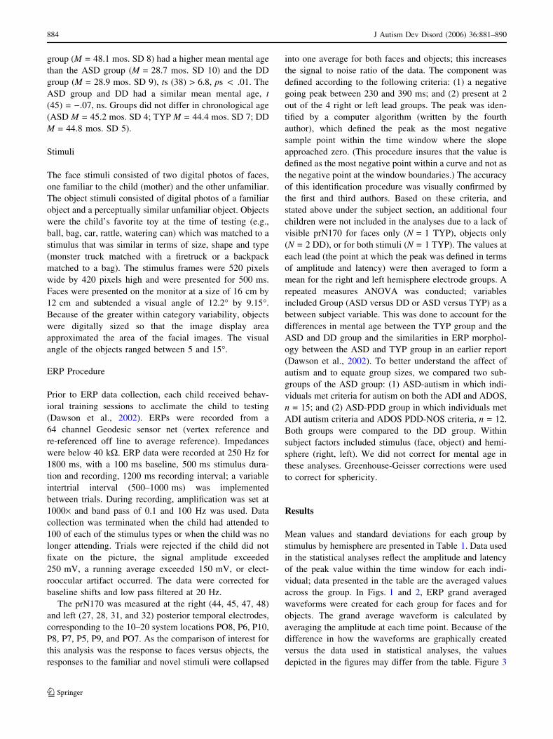
Collapsed across groups, children demonstrated a more
negative response to faces than objects and faster responses
in the right than left hemisphere. When considering the
specific characteristics of the N170, such as faster and more
negative responses in the right hemisphere for faces than
objects, the profile differed by group.
For the children with typical development, the latency of
the response to faces was faster than that to objects, spe-
cifically in the right hemisphere. These findings are similar
to those from a previous study of typically developing
children of this age (Taylor et al., 1999, 2001). In contrast,
the DD group and the ASD group did not show a face
specific hemispheric response. The DD group also failed to
show any differential response to faces and object for
latency and for amplitude. The ASD group demonstrated
the pattern of having a more negative response to faces
than objects; but showed faster responses to objects than
faces.
These ERP findings are suggestive of a number of
conclusions. First, the prN170 may be quantitatively and
qualitatively different in young children. Second, children
with general developmental delay, the mental-age and
chronological-age matched comparison group, did not
show differential responses to faces and objects. Lastly, we
Fig. 2 Mean responses in ms
for the TYP, ASD, and DD
group for face and object
responses at the right and left
posterior temporal electrodes
Fig. 3 Graphical presentation of the latency responses between the
three groups
J Autism Dev Disord (2006) 36:881–890 887
123

suggest that children with autism do have an atypical brain
response to faces and objects, compared to children with
developmental delay and typical development. We will
address each of these statements below.
First, this analysis made use of data collected in the
context of another study designed to address processing of
facial and object familiarity. Children only saw two face
stimuli and two object stimuli, not the multiple exemplars
that have been used in previous N170 studies. The degree
to which this manipulation affected the timing and the
latency of the precursor N170 is unknown. In our report,
the response to stimulus type was a within subject
manipulation and highlights the relative difference in face
and object processing within the individual and the group.
Given the difficulty in assessing children with autism and
developmental delay at this age, this report represents a
novel first attempt at defining an early component that
responds preferentially to faces; further work will be nee-
ded to assess the influence of this manipulation.
Recent reports with adults also conflict in regard to the
response of the N170 to familiarity and repetition. Reports
with adults have suggested the N170 is: (1) affected by
repetition (Caharel et al., 2002; Itier & Taylor, 2004; Jemel
et al., 2003); (2) not affected by stimulus repetition (e.g.,
Begleiter, Porjesz & Wang, 1995; Schweinberger. Pfutze &
Sommer, 1995; Schweinberger, Pickering, Jentzsch, Bur-
ton, & Kaufman 2002); and (3) not affected by familiarity
(e.g., Bentin & Deouell, 2000; Eimer, 2000; Rossion et al.,
1999). However, a later component, the N250 has been
shown to be manipulated by familiarity and repetition
(Schweinberger et al., 2002). In the grand averaged
waveform (Figs. 1, 2), only one negative component
existed within the window of interest. In addition, a visual
inspection of the graphs in Dawson et al. (2002), which did
compare a familiar to novel response, does not suggest a
reliable presence of two negative components (prN170 and
precursor N250) within the waveform prior to the P400. It
is unclear whether the use of a different protocol might
have resulted in different findings.
Second, in post hoc tests, children with developmental
delay did not show differential responses to faces and
objects either in respect to latency or to amplitude. While it
is difficult to draw conclusions from null results, in Daw-
son et al. (2002), the same children with DD did show
differential responses to familiar and novel faces and
objects but did at the late slow wave component, which
occurs between 670 and 1670 ms after stimulus onset.
Nelson (1996) suggests that the slow wave component in
infancy is reflective of stimulus updating and differs
between repeating familiar, repeating novel, and unique
novel stimuli (Nelson & Collins, 1991, 1992). Further,
Snyder, Webb and Nelson (2002) found that the amplitude
of slow wave component was affected by the number of
trials an infant had seen during an experiment. The lack of
evidence of ‘‘early’’ component differentiation for the DD
group in this report and Dawson et al. (2002) suggest
several possibilities. Idiopathic developmental delay with
no known genetic etiology, is a broad diagnostic category
and may result in increased variability. While the mean
results for the ASD-autism and ASD-PDD groups re-
mained similar despite smaller group sizes, these groups
also became more specific in terms of diagnostic defini-
tions. Larger sample sizes may be needed to fully inves-
tigate responses from the DD group and to create
meaningful subgroups.
Lastly, children with autism showed a reversal of the
typical N170 pattern, with significant faster responses to
objects than faces. These results primarily reflect latency
delays in the left hemisphere. This result is similar to that
found in Dawson et al. (2004b), which examined responses
to fear faces. Responses to fear and neutral faces in children
with autism were also significantly slower in the left
hemisphere than the right; responses in the right hemisphere
in children with ASD were similar to those of the typical
children. These two reports utilize the same population of
children, but employed different stimuli sets. For the autism
group, the faster neural response to objects relative to faces
could be interpreted as preferential or altered processing
abilities for nonsocial as compared to social stimuli (Daw-
son, Meltzoff, Osterling, & Rinaldi, 1998; Dawson et al.,
2003). Further replication of these paradigms with another
sample of children will be needed.
Measures of the amplitude of the prN170 also revealed
interesting differences between the groups. Whereas all
three groups showed a similar amplitude to faces, the chil-
dren with ASD showed a more positive response to objects
compared to the children with typical development and
those with developmental delay and showed significant
differences in the processing of faces and objects in regard to
amplitude. The children with autism showed amplitude
responses, in terms of face object differentiation, that are
more similar to what is seen in typical adults. However, the
pattern differs greatly from the two comparison groups,
which did not show amplitude differences to faces and
objects.
Given that children with autism showed relatively typ-
ical patterns of amplitude and latency responses to faces in
the right hemisphere compared to the two control groups
but showed abnormalities in speed of processing of faces in
the left and amplitude responses to objects, we suggest that
these patterns reflect abnormal cortical specialization. It is
unknown whether these atypical responses in children with
autism are the result of an innate disturbance in face pro-
cessing or whether experiential effects related to a lack of
attention to faces or greater attention to objects influence
the development of the neural circuits involved in
888 J Autism Dev Disord (2006) 36:881–890
123
face processing. The first possibility dictates that face
processing abnormalities may represent a faulty ‘‘starter
set’’ in the neural substrates that are to become specialized
for faces (e.g., fusiform gyrus) or in the mechanisms that
are needed to process faces effectively (e.g., configural
processing circuitry) (Kanwisher, 2000). The second
hypothesis is that face processing deficits arise from lack of
experience with faces. Visual experience is necessary for
some aspects of face processing (Geldart, Mondloch,
Mauer, de Schonen, & Brent, 2002; Grelotti, Gauthier, &
Schultz, 2002; de Schonen & Mathivet, 1989). While
children with autism do not experience perceptual depri-
vation, it has been proposed that they may lack the social
motivation to attend to stimuli such as faces (Dawson et al.,
2002, 2005, Grelotti et al., 2002). This attention to faces
may support the development of expertise and the organi-
zation of the requisite neural systems necessary to support
aspects of face processing. Further research is needed to
differentiate these theories.
This report represents a novel first attempt at defining an
early component that responds preferentially to faces in
young children. Children with autism differed from typical
development and developmental delay both in amplitude
and latency responses to faces and objects. Further work
will be needed to assess the influence of the experimental
design and the reliability of these results.
Acknowledgments This research was funded by a program project
grant from the National Institute of Child Health and Human
Development and the National Institute on Deafness and Communi-
cation Disability (PO1HD34565), which is part of the NICHD/NID-
CD Collaborative Program of Excellence in Autism and by a center
grant from the National Institute of Mental Health (U54MH066399),
which is part of the NIH STAART Centers Program. The Murdock
Trust provided funds for purchase of the system for recording elec-
troencephalographic activity. We gratefully acknowledge the contri-
butions of these funding sources, the Clinical and Statistical Cores of
this program project, the parents and their children who participated
in this study and several other people who made significant contri-
butions to this research, including Cathy Brock, Leslie Carver, Jon-
athan Gray, James McPartland, Jeff Munson, Megan Paul, Jessica
Shook, and several undergraduate research assistants.
References
Adrien, J., Faure, M., Perrot, A., Hameury, L., Garreau, B., Bar-
thelemy, C., & Sauvage, D. (1991). Autism and family home
movies: preliminary findings. Journal of Autism and Develop-
mental Disorders, 21(1), 43–49.
Baron-Cohen, S. (1995). Mindblindness, An essay on autism and
theory of mind. Cambridge, MA: Bradford/MIT Press.
Begleiter, H., Porjesz, B., & Wang, W. (1995). Event-related brain
potentials differentiate priming and recognition to familiar and
unfamiliar faces. Electroencephalography and clinical neuro-
physiology, 94(1), 41–49.
Bentin, S., & Deouell, L. Y. (2000). Structural encoding and identi-
fication in face processing, ERP evidence for separate mecha-
nisms. Cognitive Neuropsychology, 17, 35–54.
Bentin, S., Deouell, L. Y., & Soroker, N. (1999). Selective visual
streaming in face recognition: evidence from developmental
prosopagnosia. Neuroreport, 10(4), 823–827.
Bentin S., Allison, T., Puce, A., Perez, E., & McCarthy, G. (1996).
Electrophysiological studies of face perception in humans.
Journal of Cognitive Neuroscience, 8, 551–565.
Boucher, J., & Lewis, V. (1992). Unfamiliar face recognition in rel-
atively able autistic children. Journal of Child Psychology and
Psychiatry, 33(5), 843–859.
Boucher , J., Lewis, V., & Collis, G. (1998). Familiar face and voice
matching and recognition in children with autism. Journal of
Child Psychology and Psychiatry, 39(2), 171–181.
Bushnell, I., Sai, F., & Mullin, J. (1989). Neonatal recognition of the mo-
ther’s face. British Journal of Developmental Psychology, 7, 3–15.
Caharel, S., Poiroux, S., Bernard, C., Thibaut, F., Lalonde, R., &
Rebai, M. (2002). ERPs associated with familiarity and degree of
familiarity during face recognition. International Journal of
Neuroscience, 112(12), 1499–1512.
Carver, L. J., Dawson, G., Panagiotides, H., Meltzoff, A. N., McPart-
land, J., Gray, J., & Munson, J. (2003). Age-related differences in
neural correlates of face recognition during the toddler and pre-
school years. Developmental Psychobiology, 42(2), 148–159.
Carver, L., & Dawson, G. (2002). Developmental and neural bases of
face recognition in autism. Molecular Psychiatry, 7(suppl. 2),
S18–S20.
Coles, M., & Rugg, M. (1995). Event-related brain potentials: An
introduction. In M. Rugg & M. Coles (Eds.), Electrophysiology
of mind: Event related brain potentials and cognition. Oxford:
Oxford University Press.
Dawson, G., Carver, L., Meltzoff, A., Panagiotides, H., McPartland,
J., &Webb, S. (2002). Neural correlates of face and object rec-
ognition in young children with autism spectrum disorder,
developmental delay, and typical development. Child Develop-
ment, 73, 700–717.
Dawson, G., Webb, S., Carver, L., Panagiotides, H., & McPartland, J.
(2004a). Young children with autism show atypical brain re-
sponses to fearful versus neutral facial expressions of emotion.
Developmental Science, 7(3), 340–349.
Dawson, G. Webb, S., & McPartland, J. (2005). Event-Related
Potential studies of face processing: Implications for a theory of
autism.
Dawson, G., Webb, S., Schellenberg, G., Dager, S., Friedman, S.,
Aylward, E., & Richards, T. (2003). Defining the phenotype of
autism: Genetic, brain and behavioral perspectives. Development
and Psychopathology, 14, 581–611.
Dawson, G., Meltzoff, A., Osterling, J., & Rinaldi, J. (1998). Children
with autism fail to orient to social stimuli. Journal of Autism
Developmental Disorders, 28, 479–485.
Dawson, G., Toth, K., Abbott, R., Osterling, J., Munson, J., Estes, A.,
& Liaw J. (2004b). Early social attention impairments in autism:
social orienting, joint attention, and attention to distress.
Developmental Psychology, 40(2), 271–283.
de Haan M., & Nelson, C. A. (1999). Brain activity differentiates face
and object processing in 6-month-old infants. Developmental
Psychology, 35(4), 1113–1121.
de Haan M., & Nelson, C. A. (1997). Recognition of the mother’s
face by six-month-old infants: a neurobehavioral study. Child
Development, 68(2), 187–210.
de Schonen S., & Mathivet, E. (1989). First come, first served: A scenario
about the development of hemispheric specialization in face rec-
ognition during infancy. Current Psychology of Cognition, 9, 3–44.
Eimer, M. (2000). Event related potentials distinguish processing
stages involved in face perception and recognition. Clinical
Neurophysiology, 111, 694–705.
Eimer, M. (1998). Does the face specific component reflect the activity
of a specialized eye processor? NeuroReport, 9, 2945–2948.
J Autism Dev Disord (2006) 36:881–890 889
123

Fagan, J. F. 3rd. (1972). Infants’ recognition memory for faces.
Journal of Experimental Child Psychology, 14(3), 453–476.
Filipek, P. A., Accardo, P. J., Baranek, G. T., Cook, E. H., Dawson, G.,
Cordon, B., et al. (1999). The screening and diagnosis of autism
spectrum disorders. Journal of Autism and Developmental
Disorders, 29, 439–484.
Geldart, S., Mondloch, C., Maurer, D., de Schonen, S., & Brent, H.
(2002). The effect of early visual deprivation on the development
of face processing. Developmental Science, 5, 490–501.
Gepner, B., de Gelder, B., & de Schonen, S. (1996). Face processing in
autistics: a generalized deficit? Child Neuropsychology, 2:1–17.
Goren, C. C., Sarty, M., & Wu, P. Y.(1975). Visual following and
pattern discrimination of face-like stimuli by newborn infants.
Pediatrics, 56(4), 544–549.
Grelotti, D., Gauthier, I., & Schultz, R. (2002). Social interest and the
development of cortical face specialization: What autism teaches
us about face processing. Developmental Psychobiology, 40,
213–225.
Hobson, R., Ouston, J., & Lee A. (1988). What’s in a face? The case
of autism. British Journal of Psychology, 79, 441–453.
Itier, R., & Taylor, M. (2004). Effects of repetition and configural
changes on the development of face recognition processes.
Developmental Science, 7(4), 469–487.
Jemel, B., Schuller, A., Cheref-Khan, Y., Goffaux, V., Crommelinck,
M., & Bruyer R. (2003). Stepwise emergence of the face-sen-
sitive N170 event-related potential component. Neuroreport,
14(16), 2035–2039.
Johnson, M. H., Dziurawiec, S., Ellis, H., & Morton, J. (1991).
Newborns’ preferential tracking of face-like stimuli and its
subsequent decline Cognition, 40(1–2), 1–19.
Joseph, R., & Tanaka, J. (2003). Holistic and part-based face recog-
nition in children with autism. Journal of Child Psychology and
Psychiatry, 44(4), 529–542.
Kanwisher, N. (2000). Domain specificity in face perception. Nature
Neuroscience, 3, 759–763.
Klin, A., Jones, W., Schultz, R., Volkmar, F., & Cohen, D. (2002).
Visual fixation patterns during viewing of naturalistic social
situations as predictors of social competence in individuals with
autism. Archives of General Psychiatry, 59(9), 809–816.
Klin, A., Sparrow, S. S., de Bildt, A., Cicchetti, D. V., Cohen, D. J., &
Volkmar, F. R. (1999). A normed study of face recognition in
autism and related disorders. Journal of Autism and Develop-
mental Disorders, 29(6), 499–508.
Langdell, T. (1978). Recognition of faces: an approach to the study of
autism, Journal of Child Psychology and Psychiatry, 19(3), 255–
268.
Lord, C., Rutter, M., Goode, S., & Hemsbergen, J. (1989). Autism
Diagnostic Observation Schedule: a standardized observation of
communicative and social behavior. Journal of Autism and
Developmental Disorders, 19, 158–212.
Lord, C., Rutter, M., & Le Couteur, A. (1994). Autism Diagnostic
Interview-Revised: A revised version of a diagnostic interview
for caregivers of individuals with possible pervasive develop-
mental disorders. Journal of Autism Developmental Disorders,
24, 659–685.
McPartland, J., Dawson, G., Webb, S.J., Carver, L., & Panagiotides,
H. (2004). Event-related brain potentials reveal anomalies in
temporal processing of faces in autism. Journal of Child Psy-
chology and Psychiatry, 45(7), 1235–1245.
Mullen, E. (1997). Mullen scales of early learning. Los Angeles, CA:
Western Psychological Services.
Nelson, C. (1996). Electrophysiological correlates of early memory
development. In H. W. Reese & M. D. Franzen (Eds.), Thir-
teenth West Virginia University Conference on Life Span
Developmental Psychology: Biological and Neuropsychological
Mechanisms (pp. 95–131). New Jersey: Lawrence Erlbaum.
Nelson, C. A., & De Haan, M. (1996). Neural correlates of infants’
visual responsiveness to facial expressions of emotion. Devel-
opmental Psychobiology, 29(7), 577–595.
Nelson, C. A., & Collins, P. (1991). Event related potential and
looking time analysis of infant’s responses to familiar and novel
events: Implications for visual memory. Developmental Psy-
chology, 27, 50–58.
Nelson, C. A., & Collins, P. (1992). Neural and behavioral correlates
of recognition memory in 4- and 8- month old infants. Brain and
Cognition, 19, 105–121.
Osterling, J., & Dawson, G. (1994). Early recognition of children with
autism: A study of first birthday home videotapes. Journal of
Autism Developmental Disorders, 24, 247–257.
Osterling, J., Dawson, G., & Munson, J. (2002). Early recognition of
1-year-old infants with autism spectrum disorder versus mental
retardation. Development and Psychopathology, 14, 239–251.
Pelphrey, K., Sasson, N., Reznick, J., Paul, G., Goldman, B., & Piven,
J. (2002). Visual scanning of faces in autism. Journal of Autism
Developmental Disorders, 32(4), 249–261.
Rossion, B., Campanella, S., Gomez, C., Delinte, A., Debatisse, D.,
Liard, L., Dubois, S., Bruyer, R., Crommelinck, M., & Guerit, J.
(1999). Task modulation of brain activity related to familiar and
unfamiliar face processing: an ERP study. Clinical Neurophys-
iology, 110(3), 449–462.
Schweinberger, S., Pickering, E., Jentzsch, I., Burton, A., Kaufman, J.
(2002). Event-related brain potential evidence for a response of
inferior temporal cortex to familiar face repetitions. Brain
Research. Cognitive Brain Research, 14(3), 398–409.
Schweinberger, S. R., Pfutze, E. M., & Sommer, W. (1995). Repe-
tition priming and associative priming of face recognition: Evi-
dence from event-related potentials. Journal of Experimental
Psychology: Human Perception and Performance, 21, 722–736.
Snyder, K. A., Webb, S. J., & Nelson, C. A. (2002). Theoretical and
methodological implications of variability in infant brain
response during a recognition memory paradigm. Infant behav-
ior & development, 25, 466–494.
Tantam, D., Monaghan, L., Nicholson, H., & Stirling, J. (1989).
Autistic children’s ability to interpret faces: A research note.
Journal of Child Psychology and Psychiatry, 30(4), 623–630.
Taylor, M., McCarthy, G., Saliba, E., & Degiovanni, E. (1999). ERP
evidence of developmental changes in processing of faces.
Clinical Neurophysiology, 110, 910–915.
Taylor, M., Edmonds, G., McCarthy, G., & Allison T. (2001). Eyes
first! Eye processing develops before face processing in children.
NeuroReport, 12, 1671–1676.
Trepagnier, C., Sebrechts, M., & Peterson, R. (2002). Atypical face
gaze in autism. Cyberpsychological Behavior, 5(3), 213–217.
Valenza, E., Simion, F., Cassia, V. M., & Umilta, C. (1996). Face
preference at birth. Journal of Experimental Psychology. Human
Perception and Performance, 22(4), 892–903.
van der Geest J., Kemner, C., Verbaten, M., & van Engeland, H.
(2002). Gaze behavior of children with pervasive developmental
disorder toward human faces: a fixation time study. Journal of
Child Psychology and Psychiatry, 43(5), 669–678.
890 J Autism Dev Disord (2006) 36:881–890
123


![ADOPTED REGULATION OF THE STATE BOARD OF EDUCATION … · 2010-05-07 · disorder, [aspeger’s] asperger’s disorder, atypical autism, pervasive developmental disorder and other](https://img.dokumen.tips/doc/110x75/5fb827387372d9390e7ce909/adopted-regulation-of-the-state-board-of-education-2010-05-07-disorder-aspegeras.jpg)