Embed Size (px)
Citation preview

1
Telomerase activation in a model of lung adenocarcinoma
Florent Suau 1, Vincent Cottin 1,2,*, Fabienne Archer 1, Séverine Croze 1, Joelle Chastang 1, Geneviève Cordier 1, Francoise Thivolet-Béjui 1,3 Jean-François
Mornex 1,2, Caroline Leroux 1
1 UMR754 Rétrovirus et Pathologie Comparée INRA ; Université Lyon I ; Ecole Nationale Vétérinaire de Lyon ; Ecole Pratique des Hautes Etudes ; IFR128 Biosciences Lyon-Gerland; 2 Service de pneumologie � Centre des maladies orphelines pulmonaires, Hôpital Louis Pradel, Hospices Civils de Lyon ; 3 Service d�anatomie et cytologie pathologiques, Hôpital Louis Pradel, Hospices Civils de Lyon ; Lyon, France.
Running title: Telomerase and lung adenocarcinoma * Correspondence: Vincent Cottin, UMR754, Lyon Gerland, Université Lyon I, 50 avenue Tony Garnier, 69007 Lyon, France ; Tel : +33 (4) 37 28 76 20, Fax : +33 (4) 37 28 76 05 ; E-mail: [email protected].
Word count 4,372
. Published on February 2, 2006 as doi: 10.1183/09031936.06.00125105ERJ Express
Copyright 2006 by the European Respiratory Society.

2
Abstract (198 words)
Ovine pulmonary adenocarcinoma (OPA) is a lung cancer strikingly similar
to the pneumonic-type mixed invasive adenocarcinoma with a predominant
bronchioloalveolar component in humans. We investigated telomerase activity in
OPA and the potential involvement of the kinase Akt in telomerase activation and
regulation of cell proliferation.
Lung tissues were collected from sheep with a histopathological diagnosis
of OPA or controls. Epithelial cell cultures were derived in vitro from lung tissues.
Telomerase activity was evaluated by the Telomeric Repeat Amplification
Protocol method. Phosphorylation of Akt was detected by western-blotting.
Telomerase activity was significantly higher in OPA lung tissues as
compared with control lung tissues. A high telomerase activity was detected in
8/12 (67%) primary cell cultures derived from tumours. A high level of expression
of phosphorylated Akt was found in 10/27 (37%) tumours, with abolition of Akt
activation in response to EGF stimulation demonstrated in primary cell cultures
derived from tumours.
Telomerase activation takes place in OPA tumour cells and may be partly
attributable to Akt activation. Telomerase may inhibit cellular senescence and
contribute to the accumulation of tumour cells in mixed adenocarcinoma with
bronchioloalveolar component. Further work is necessary to identify alternative
signalling pathways of telomerase activation in tumours.
Keywords: bronchioloalveolar carcinoma, lung cancer, Akt, telomerase,
type-II pneumocyte, JSRV

3
Lung cancer is the first cause of cancer mortality in developed countries.
Non-small cell lung cancer represents approximately 80% of lung cancers,
adenocarcinoma being the most frequent cell type, accounting for ~40% of all
cases of lung cancer. Lung adenocarcinoma may present as the pneumonic-type
adenocarcinoma (pADC), that associates typical radiological features with diffuse
or disseminated alveolar condensation with or without air bronchogram evolving
towards pulmonary right-to-left shunting, together with evidence of
adenocarcinoma tumour cells in the lung (1). The histological pattern in pADC
may include bronchioloalveolar carcinoma (BAC) (defined as an adenocarcinoma
with a pure bronchioloalveolar growth pattern with no evidence of stromal,
vascular, or pleural invasion) (2), or more commonly a mixed-type
adenocarcinoma with a predominant bronchioloalveolar component and papillary
or acinar invasive component (1, 3).
Ovine pulmonary adenocarcinoma (OPA) is a naturally occurring lung
cancer that occurs spontaneously in sheep infected by the Jaagsiekte Sheep
RetroVirus (JSRV), and that may be reproduced by the experimental inoculation
of lambs with the virus (4). It grows with a disseminated pattern at the periphery
of the lung. OPA is a mixed type adenocarcinoma containing a significant
proportion of BAC component, together with papillary and acinar growth patterns
(2, 3). OPA also shares striking clinical and radiological homology with human
pADC, including progressive intrapulmonary spread and lack of distant
extrathoracic metastasis (3), and thus represents a unique natural and
reproducible animal model of peripheral lung cancer, especially pADC.
In contrast with previous studies that focused on defining oncogenic
properties of JSRV structural proteins in rodents (5), we aimed at identifying
pathogenic processes taking place in spontaneous tumours in vivo. Mechanisms
potentially involved in tumour formation include extensive cell division as a result
of oncogenic mutations, and inactivation of cellular senescence, tumour
suppressor pathways, or apoptosis mechanisms that may otherwise arrest the

4
proliferation or induce the death of potential cancer cells (6). Cell senescence is a
process mostly described in vitro, whereby primary normal cells grown in culture
do not proliferate indefinitely, but withdraw from the cell cycle (after a period of
rapid proliferation) in response to diverse regulatory mechanism including
dysfunctional telomeres (7). More recently, cellular senescence has been also
demonstrated in vivo in premalignant but not malignant cells, suggesting that it
may be an important anticancer defence (8).
Cellular senescence is mainly regulated by telomerase, a
ribonucleoprotein enzyme able to stabilize telomere length by de novo synthesis
of telomeres and elongation of existing telomeres. Telomerase activation is
considered mandatory for tumour cells to escape cell senescence and gain
increased proliferative capacities (9). The telomerase reverse transcriptase
(TERT) catalytic subunit is the major determinant of telomerase activity in vitro
and in vivo. Activation of TERT has been well established in human cancer cell
lines and tumours including lung cancer (10), while telomerase activity is
repressed in most normal somatic cells. Complex regulation of telomerase
activity may include the phosphatidylinositol 3-kinase (PI3-K) pathway through
phosphorylation of TERT by Akt (11). Involved in regulation of cell survival and
cell cycle progression, Akt is constitutively activated in a variety of human
tumours including lung cancer (12).
In this study, we demonstrate increased telomerase activity in tumours
and in primary cultures of tumour cells derived from OPA, suggesting that
inhibition of cell senescence may be involved in tumorigenesis and accumulation
of tumoral cells within the lung. We next show that the regulatory kinase Akt is
constitutively activated in OPA tumours and disregulated in primary cultures
derived from OPA, suggesting that Akt may be involved in telomerase activation
in a proportion of tumours.
5
Materials and methods
Lung tissues
Lung tissues were collected immediately post-mortem from sheep
presenting loss of weight, dypsnea, profuse lung secretions suggestive of OPA or
without signs of OPA. Tissue sections were sampled, stored at -70°C until use or
fixed in formol for histopathological examination. The tissues were classified as
tumoral and non-tumoral lung (henceforth referred to as �controls�) following the
current 2004 World Health Organization (2).
Culture of alveolar epithelial type II cells
Tumour cells and normal type-II pneumocytes were isolated and
characterized from ovine lungs as described elsewhere (Archer et al, manuscript
in preparation). Briefly tissue samples were digested overnight at 4°C in Eagle�s
minimum essential medium (MEM, Eurobio, France) with 0.025% collagenase I,
10 µg/ml deoxyribonuclease, 1 mg/ml protease XIV (Sigma), 50 µg/ml
streptomycin, 50 U/ml penicillin, 50 µg/ml gentamycine, 2.5 µg/ml amphotericin B
and 100 U/ml nystatin. The cells were then homogenized and filtered through a
40 µm filter to eliminate cell debris. After centrifugation at 430g for 10 min at 4°C,
pulmonary cells were plated onto collagen, laminin and fibronectin coated-plates
in selective medium for epithelial cells (Quantum 286, PAA, Austria) with 5 ng/ml
Hepatocyte growth factor and 10 ng/ml Keratinocyte Growth Factor (Abcys).
Cells derived from OPA tumours (n=12) and non tumoral lungs (n=4) are referred
to as tumoral and control cells respectively.
Detection of JSRV proviral DNA
Total genomic DNA from lung tumours and control lungs was prepared
with the Fastprep device following recommendations of the supplier (BIO 101,
France). A semi-nested PCR protocol was used to detect proviral DNA. Briefly,
reaction mixtures contained 500 ng of total genomic DNA, 50 µl of 1X buffer (1.5
mM MgCl2, 67 mM Tris-HCl pH 8.8, 16 mM (NH4)2SO4, 0.01% Tween 20), 0.2
6
mM of each desoxynucleotide triphosphate, 0.2 mM of each primer and 1.25
units of Taq polymerase (Eurobio, France). The first round of PCR was
performed with the primers JSRV42 (sense) 5�-CTTTGTATTTCCCTGTGTCG-3�
corresponding to nucleotides 7041-60 in the env gene of JSRV genomic
sequence (Genbank accession number AF105220), and JSRV53 (antisense) 5�-
GGATTCTTACACAATCACC-3� corresponding to nucleotides 7381-62 in the LTR
U3 region of JSRV genomic sequence (Genbank accession number AF105220).
The second round of PCR was performed with 1-5 µl of PCR product using
JSRV42 and JSRV52 (antisense) 5�-CACCGGATTCTTATATAATC-3�
corresponding to nucleotides 7366-46 in the LTR U3 region of JSRV genomic
sequence (Genbank accession number AF105220). PCR reactions were
performed as followed: 5 min at 95°C, 35 cycles of 30 s at 95°C, 30 s at 50°C
and 1 min at 72°C with a final elongation of 10 min at 95°C.
Cell proliferation assay
Cell numbers were determined by an MTT (3-(4,5-dimethylthiazol-2-yl)-2-
5-diphenyl tetrazolium bromide) assay. Briefly, 5000 cells per well were plated in
96-well plates, incubated for 96h at 37°C with 5% CO2. MTT was incorporated as
recommended with the �MTT Cell Proliferation Assay kit� (Chemicon, France).
Ten microliters of MTT were added to each well for 4h at 37°C. After
solubilization with 100 µl of isopropanol 1N - HCl 0.04N solution, absorbance was
read at 595 nm with a Wallac Victor II device (Perkin-Elmer, France). Each
measurement was performed in triplicates.
Flow cytometry
Quantitative measurement of cell cycle was performed by flow cytometry
analysis of nuclear DNA contents following propidium iodide staining. Briefly, 106
cells were harvested following dissociation with trypsin, washed twice in
Phosphate Buffered Saline (PBS), and then fixed with 70% ethanol at -20°C.
After one wash, cells were treated with RNAse A (1mg/ml) (Sigma-Aldrich,
France) for 30 min at 4°C, incubated in 20 µg of propidium iodide (Sigma-Aldrich,

7
France) and analysed by flow cytometry. DNA content was analysed on >10,000
events by FACScan flow cytometry (Becton-Dickinson, France) with a 488-nm
argon ion laser.
Measurement of telomerase activity
Frozen tissues or cells were lysed in 250 µl ice-cold 1X Chaps lysis buffer
(Intergen Company, USA) and pulverized with the Fastprep device (BIO 101,
France). The lysates were incubated for 30 min on ice, centrifuged at 18,000 g
for 30 min at 4°C. The supernatants were collected and proteins concentrations
were measured by a modified Lowry protein assay (Pierce, USA). Telomerase
activity was assayed by the TRAP (Telomeric Repeat Amplification Protocol)
method using the TRAP-eze ELISA Telomerase Detection Kit (Intergen
Company, USA) following recommendations of the manufacturer. Briefly, 200 ng
of total proteins were used. Lysis buffer without protein was used as negative
control. Control cell extracts containing telomerase activity and a synthetic
oligonucleotide with 8 telomeric repeats (supplied with the kit) were used as
positive controls. For each cell sample a heat-inactivated (10 min at 85°C)
negative control sample was also prepared. After amplification by PCR, the
TRAP products were resolved on 12% polyacrylamide gels and visualized with
1:10,000 SYBR Gold (Molecular Probes, USA). Amplification efficiency in each
reaction was determined using the provided internal control oligonucleotides
forming a 36-bp band. Telomerase activity was evaluated using a
semiquantitative ELISA method and expressed as the ratio of telomerase activity
in sample over telomerase activity in telomerase-positive control cells. All
experiments were performed at least in duplicate. Measurements were reported
as mean ± standard error of the mean (SEM).
Immunodetection of P-Akt, total Akt and capsid protein
Frozen tissues or cells were lysed in 250 µl lysis buffer (0.5M Tris pH 8.0,
10% glycerol, 150 mM NaCl, 1% Triton X-100, 5 mM EDTA, 1mM Na3VO4, 1 mM
phenylmethylsulfonyl fluoride, 10 µg/ml leupeptine and 10 µg/ml aprotinine),

8
homogenized with the Fastprep system device (BIO 101, France) and incubated
for 30 min on ice. The lysates were centrifuged at 18,000 g for 30 min at 4°C and
50 µg of protein were separated on a 12% SDS-polyacrylamide gel and
transferred onto a 0.2 µM nitrocellulose membrane (Biorad Laboratories,
France). The membranes were pre-incubated with TSBT (25 mM Tris pH 7.6,
0.15 M NaCl, and 0.05% Tween 20) containing 5% nonfat dry milk for 1h at room
temperature. After three washes in TSBT, the membranes were incubated
overnight at 4°C with 1:1,000 rabbit polyclonal antibodies against P-Akt (serine
473) or total Akt (Cell Signalling, USA) diluted in TSBT containing 5% bovine
serum albumine (BSA). For detection of capsid antigenic protein, membranes
were incubated for 1 h at room temperature with 1:10,000 rabbit polyclonal
antibody to JSRV capsid protein (generously provided by Dr J. De Martini, Fort
Collins, Co, USA). Membranes were washed 3 times for 5 minutes at room
temperature with TBST and incubated with horseradish peroxidase-labeled anti-
rabbit IgG antibody (Sigma, France) diluted in TBST containing 5% nonfat dry
milk (1:10,000) for 1 h at room temperature. The immunoreactive bands were
detected with an enhanced chemiluminescence detection kit (Pierce, France),
quantified with the Un-Scan-It software (Silk Scientific Corporation, USA) and
expressed as percentage of sample in comparison to A549 control cells.
Measurements of expression of P-Akt were reported as mean ± SEM. Protein
quantity loaded onto gels was controlled by immunodetection of actin (Sigma,
France) with a mouse monoclonal antibody against ß-actin (Sigma, France).
Statistical analysis
Statistical analyses were performed with the Prism4 software (GraphPad
Software Inc, USA). The values for telomerase activity and expression level of P-
Akt in tumoral and non tumoral samples were compared using the Mann-Whitney
U-test. Tests were two-tailed except mentioned otherwise. Only p-values < 0.05
were considered as significant.

9
Results
Characterisation of tumours
Lung tissue was obtained from OPA tumours and non-tumoral control
lungs. The diagnosis of OPA (invasive mixed adenocarcinoma with a
bronchioloalveolar component, associated with acinar and (or) papillary growth
patterns) was confirmed by histopathological examination in all tumours, whereas
absence of tumour was confirmed in all control lungs. In addition, lung tissues
were assessed for the presence of the JSRV provirus and of the capsid antigenic
protein as described in Methods. JSRV proviral DNA was detected by PCR
analysis in 11/12 (92%) tumour lungs, and none of the 4 control lungs. Similarly,
the JSRV capsid protein was detected by western-blot analysis in 12/12 OPA
lungs, but none of the 4 control lungs.
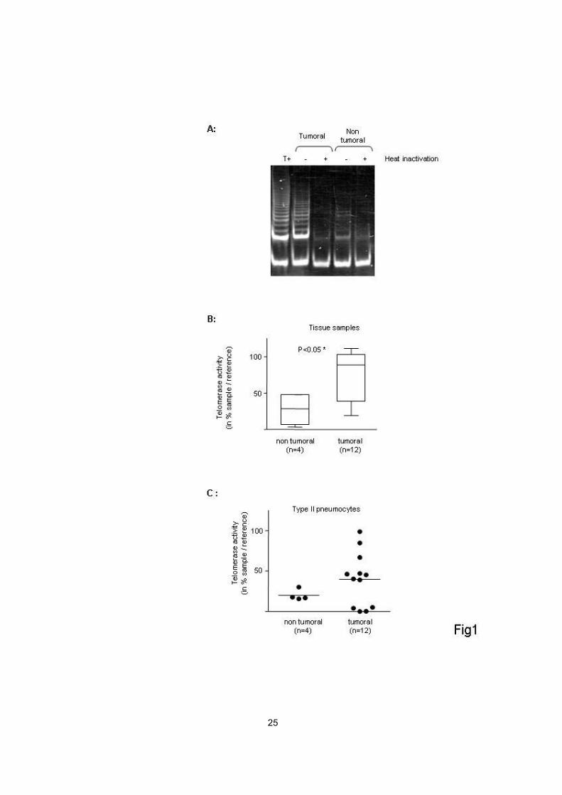
Telomerase activity in tumours
In a first approach to study the involvement of telomerase activation in
OPA, we measured telomerase activity in lung tissues from OPA tumours (n=12)
and non-tumoral control lungs (n=4), using the TRAP assay as described. As
illustrated in figure 1A, PCR products visualised as a DNA ladder on
polyacrylamide gels indicated the presence of telomerase activity in all OPA
samples (n=12), but none of the samples from control lungs. Semi-quantification
using ELISA demonstrated that the telomerase activity was significantly higher in
OPA tissues as compared to control lungs (p=0.03, one-tailed Mann-Whitney
test) (Figure 1B). These results thus demonstrated that a high telomerase activity
was present in tumours (invasive mixed adenocarcinoma with a
bronchioloalveolar component) in which JSRV infection was confirmed.
Characterisation of tumoral cell cultures

10
We next aimed at demonstrating that the telomerase activity observed in
whole tumours was attributable to tumour cells per se (and not to accompanying
non-tumoral cells present in the tumoral lung). Primary cultures were derived
from tumours (n=12) and from control lungs (n=4) as described in Methods.
These cells were characterized by immunocytochemistry using antibodies
against specific markers of type-II pneumocytes (surfactant proteins A and C),
and transmission electron microscopy (Archer et al, manuscript in preparation).
As expected, primary cells derived from tumours and from normal lungs had a
cuboidal morphology typical of epithelial cells. The purity of the cultures was
confirmed by expression of the surfactant proteins A and C in >95% of the cells,
and was maintained over all passages. The JSRV genome was detected by PCR
analysis in cell cultures derived from all OPA lungs, but in none of the control
type-II pneumocytes derived from control lungs. Cultured cells derived from
tumours could be maintained for 7 to 10 passages, as compared to only 2 to 3
passages for control cells.
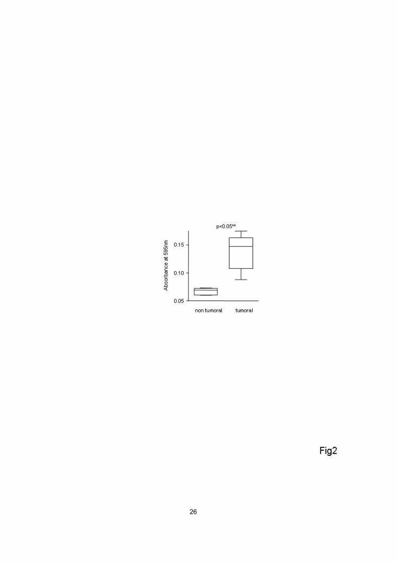
To assess the biological relevance of cell cultures derived from tumours
and control lungs, we next analysed cell proliferation and distribution throughout
the cell cycle of tumour and control cells. Using a MTT assay, we showed that
cells derived from lung tumours had a statistically significant proliferative
advantage as compared to control type-II pneumocytes derived from control
lungs (Figure 2). Similarly, cell cycle analysis using flow cytometry indicated that
the proportion of cells in S-phase was higher in cultures of tumoral cells (17% ±
3.2) than in control type-II pneumocytes (8.5% ± 1.9).
Telomerase activity in tumoral cell cultures
To demonstrate that the telomerase activity observed in whole tumours
was attributable to tumour cells per se, telomerase activity was assessed in
primary cell cultures derived from tumours and controls. Semi-quantitative
measurement of telomerase activity by the TRAP assay followed by ELISA
demonstrated a particularly high level of telomerase activity in 8 / 12 (67%)

11
primary cultures derived from tumours (Figure 1C), as compared with control
type-II pneumocytes, in which a low level of enzymatic activity could be detected.
Surprisingly, no telomerase activity was found in 4 / 12 (33%) cell cultures
derived from tumours (in which telomerase activity had been detected in whole
tumours). Therefore, these results showed that a high level of telomerase activity
was present in a majority of cell cultures derived from lung tumours, and
indicated that the telomerase activity observed in whole tumours was indeed
attributable to tumour cells per se in a majority of tumours.
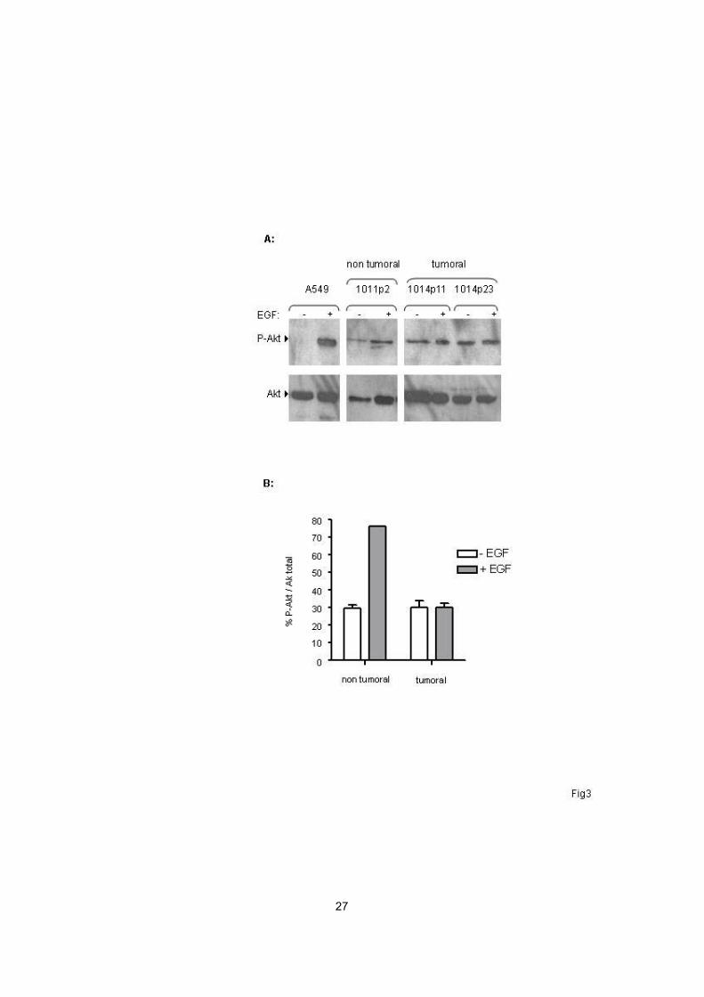
Disregulation of Akt in tumoral cell cultures
Regulation of telomerase activity is complex and may involve activation of
TERT through its phosphorylation by the kinase Akt. In order to determine
whether telomerase activation may be related to the Akt pathway, we next
studied the phosphorylation state of Akt on serine-473 in OPA-derived cell
cultures. Primary cells derived from tumours and control type-II pneumocytes
were cultured in the absence of growth factors during 24 h, and then exposed or
not for 30 min to epidermal growth factor (EGF) (100 ng/ml), a known activator of
Akt in mammalian cells. Moderate expression of phosphorylated Akt was
detected by western blotting at similar levels in unstimulated cells derived from
lung tumours and control type-II pneumocytes (Figure 3), suggesting that culture
conditions (including deprivation of growth factors) might lead to basal
phosphorylation and activation of Akt in unstimulated cells. However, EGF
stimulation in vitro lead to a dramatic increase in Akt activation in control cells,
while cells derived from lung tumours were not responsive to stimulation by EGF
(Figure 3). These results showed that the EGF - Akt pathway was disregulated in
OPA cell cultures, with lack of Akt activation in response to growth factor
stimulation in tumoral cell cultures.
Activation of Akt in tumours
To further characterise the disregulation of Akt in OPA, Akt activation was
next studied in whole tumours (n=27) and normal lung tissues (n=14) using

12
western blotting analysis, and using human A549 epithelial cells as a reference.
As shown in figure 4, significant expression of phosphorylated Akt was
demonstrated in 10 / 27 (37%) OPA tumours, and none of 14 control lungs (table
1), while expression of total Akt and actin was similar between both groups.
Thus, Akt is activated and may thus participate in telomerase activation in a
significant proportion of OPA tumours.
Discussion
In this study, we showed that telomerase is activated in OPA, a mixed-
type adenocarcinoma of the lung with prominent bronchioloalveolar component.
Telomerase activation was demonstrated in tumours as well as epithelial cell
cultures derived from the tumours. We next investigated the activation of the
regulatory kinase Akt as a potential activator of telomerase, and demonstrated
that Akt is indeed activated in a proportion of tumours. Taken together, these
results suggest that inhibition of cell senescence may be involved in
tumorigenesis and accumulation of tumoral cells within the lung in OPA, and that
Akt activation may participate in telomerase activation in a proportion of tumours.
pADC differs from other types of non-small cell lung cancer in several
ways, including higher incidence in women, a lesser role of tobacco smoking,
lack of distant metastatic spread, and higher rate of sensitivity to EGF-tyrosine
kinase inhibitors. The clinical syndrome of pADC remains a rare presentation of
non-small cell lung cancer, hampering research.
Ovine pulmonary adenocarcinoma (OPA) was chosen as a model of
pADC since it shares with the human disease a variety of key features, including
similar clinical and radiological presentation with progressive dyspnea, and
abundant bronchorrhea ; multifocal pulmonary disease with alveolar
consolidation and nodules ; and almost identical pathology (3). As in BAC,
tumour cells in OPA derive from type-II pneumocytes and to a lesser extent Clara

13
cells. The BAC component of both the human and the ovine tumours is
characterised by lepidic spread, where tumoral cells grow following the alveolar
septa; as opposed to the rare form of pure BAC, mixed adenocarcinoma with
bronchioloalveolar component includes evidence of stromal, vascular and (or)
pleural invasion, together with associated acinar and mostly papillary growth
patterns (1, 3). We have evidence that OPA is immunohistochemically similar to
human mixed BAC (13), with expression of cytokeratin-7, nuclear expression of
thyroid transcription factor-1, lack of expression of cytokeratin-20, and by
electronic microscopy studies (data not shown). As opposed to previous works
on OPA where studies were mostly conducted in immortalised cell lines or rodent
fibroblasts tranfected with JSRV gene expression vectors (14-16), in the work
reported herein we were able to derive primary cell cultures from naturally
occurring tumours and control lungs. Genomic proviral DNA of the causative
agent JSRV was detected only in cells derived from tumours. In addition, cells
derived from OPA tumours expressed surfactant pulmonary-associated proteins
A and C (markers of type-II pneumocytes), and thyroid transcription factor-1,
indicating that their specific phenotype was maintained in vitro. Importantly,
concordant results of telomerase activation were obtained in parallel in tumours
and in epithelial cell cultures derived from the tumours, doubtlessly increasing the
biological relevance of this observation.
We demonstrated a high level of telomerase activity in OPA lung tumours
as compared to control non-tumoral lungs. Telomerase activity was detected in
all OPA tumours, and was likely due to telomerase activation within tumour cells.
As cells located within the tumour other than cancer cells per se (17-19) such as
lymphocytes of the bronchial mucosa (20) may also express telomerase activity,
we derived primary cell cultures from the same lung tumours; telomerase activity
was found in two thirds of epithelial cell cultures derived from tumours, thus
indicating that the telomerase activity observed in whole tumours was at least
partly attributable to tumour cells per se. No telomerase activity was found in a
third of cell cultures derived from telomerase-positive tumours; we hypothesize
that activated lymphocytes or other inflammatory cells infiltrating the tumour may
14
have been responsible for telomerase activity in such cases, as previously
reported (17, 18). It remains to be determined whether alternative pathways to
telomerase activation may also contribute to escaping cell senescence in
telomerase-negative tumour-cell cultures (21). Conversely, a low telomerase
activity was found in control lung tissues and cell cultures, potentially resulting
from a subpopulation of type-II pneumocytes with self-renewal capacities that are
assumed to repair lung alveolar epithelium after injury (22 113). Alternatively,
telomerase activity in control lungs may be due to activated lymphocytes within
non-tumoral lungs as a result of a variety of infraclinical infections that are
common in naturally bred animals, although this was not suggested by the
pathological analysis of control lungs in our study.
Telomerase activation in OPA suggests that inhibition of cell senescence
may be involved in tumorigenesis and in the process of accumulation of tumour
cells along the alveolar septa. Telomeres terminate eukaryotic chromosomes and
are involved in chromosome integrity. Continued telomere shortening in normal
somatic cells eventually results in an arrest of cell proliferation, a physiological
process referred to as cell senescence that controls cell lifespan and limits the
number of cell divisions (9). Activation of telomerase activity, mainly dependent
on its catalytic subunit TERT, contributes to telomere length maintenance and
inhibition of cell senescence, and thus to cell immortalization and cancer (9).
Hence, we found maintained telomere length in OPA (Leroux et al, manuscript in
preparation). In addition to its enzymatic activity, the TERT subunit may enhance
genomic stability by direct interaction with telomeres (23), and may participate in
the regulation of p53-induced apoptosis (24). Telomerase activation is an early
event in carcinogenesis, concomitant with P53 overexpression, Rb inactivation,
and decrease in Bcl-2/Bax ratio in high grade preinvasive bronchial lesions
(beginning at the level of moderate dysplasia), suggesting a coupling between
telomerase activation, proliferation, and resistance to apoptosis (20). Although
telomerase activation has been described in a variety of human cancers including
lung cancer (10), it has not been extensively studied in pADC and mixed
adenocarcinoma with BAC features. Some telomerase activity as assessed by a

15
non-quantitative method was detected in 4/10 cases of human BAC presenting
as solitary nodules (25), and in 97% of peripheral and small-sized nonmucinous
BAC (26), but such tumours were unlikely representative of the clinically-defined
pADC (1). Interestingly, telomerase activation alone is not sufficient to transform
human cells in vitro (27). Mechanisms other than cell senescence inhibition (such
as inhibition of apoptosis, or deregulation of cell proliferation) may also take place
in pADC and OPA, as suggested by the increased proliferation of tumour-derived
cell cultures which was observed as compared to control cells.
The complex regulation of telomerase activity involves several pathways
including the phosphorylation and activation of TERT by Akt (11), a kinase
involved in the regulation of processes characteristic of cancer such as cell
proliferation and survival, cell size, response to nutrient availability, angiogenesis
and tissue invasion (28). Overexpression of Akt can transform NIH3T3 cells,
indicating that Akt is a potential oncogene (29). We have studied the potential
role of Akt in telomerase activation, and observed disregulation of the EGF - Akt
pathway in OPA tumors. Hence, Akt activation was present in a significant
proportion of OPA tumours but none of control lungs. Lack of Akt activation in
response to EGF stimulation was further demonstrated in cell cultures derived
from tumours, as compared to control cells (in which EGF stimulation
dramatically induced Akt phosphorylation), demonstrating that the EGF - Akt is
disregulated in cells derived from tumours. The basal level of Akt activation was
comparable in cell cultures derived from tumours and non tumoral lung, a finding
likely related to artefacts of cell culture (deprivation in growth factors or
replacement of medium may have contributed to moderate activation of Akt in
both tumoral and non tumoral cell cultures (28)). Taken together, our results
suggest that Akt activation may participate in telomerase activation and
regulation of cell senescence in OPA, as recently shown in human mixed
adenocarcinoma with BAC features (30). Alternative pathways such as the Ras-
MEK-MAPK pathway (15) are also likely to participate to telomerase regulation in
this tumour, as Akt activation was not found in all telomerase-positive tumours.
16
Several studies have identified the gene encoding the envelope of JSRV,
(the causative agent of OPA) as a potential oncogene, and have shown that its
overexpression was sufficient to transform rodent fibroblasts (31) and epithelial
cell lines in vitro (14). Transfection of JSRV env in mammalian cells induces
constitutive activation of Akt; studies using chemical inhibitors of the PI3K-Akt-
mTOR pathway have further demonstrated the central role of Akt and of the Ras-
MEK-MAPK pathway in JSRV-induced cell transformation (15, 32), although the
precise mechanism of Akt activation in naturally occurring OPA remains
speculative.
Recently, activating mutations of the EGF receptor (EGFR) mutually
exclusive of K-ras mutations have been reported in lung adenocarcinoma
especially with a prominent nonmucinous BAC component, and may be
associated with clinical response to the EGFR tyrosine kinase inhibitors gefitinib
and erlotinib (33-35). Activation of EGFR is associated with activation of
downstream signalling pathways including the PI3K-Akt and the Ras-MEK-ERK
pathways in this tumour (30, 36). In addition, increased Akt phosphorylation (not
inhibited by EGFR tyrosine kinase inhibitors) has been demonstrated in vitro in
specific subpopulations of adenocarcinoma cell lines that have become naturally
resistant to gefitinib, despite the loss of the EGFR gene mutation when compared
with parental cell lines (37). These observations thus indicate that constitutive
activation of the PI3K-Akt-pathway may occur independently of EGFR mutations,
and may be an attractive therapeutic target in lung adenocarcinoma. Studies are
currently undertaken to determine whether disregulation of the EGF � Akt
pathway in cells derived from OPA may be similarly related to mutations of the
EGFR gene.
In conclusion, we have shown that telomerase activation takes place in
OPA tumour cells and may be partly attributable to Akt activation. Telomerase
activation may contribute to the accumulation of tumour cells within the lung
through inhibition of cellular senescence. Future strategies for the treatment of
human pADC with BAC features may be envisioned, through telomerase specific

17
inhibition and (or) modulation of the Akt pathway. OPA provides an attractive
model for the preclinical assessment of the efficacy of innovative approaches to
treat this incurable disease.
Acknowledgements
This work was supported by research grants from the Ligue nationale
contre le cancer (Rhône, Drôme, Loire, and Ardèche departmental committees)
and the Région Rhône-Alpes. F.S. is a recipient of a fellowship from the French
Ministry of Research.

18
References
1. Wislez M, Massiani MA, Milleron B, Souidi A, Carette MF, Antoine M, Cadranel J.
Clinical characteristics of pneumonic-type adenocarcinoma of the lung. Chest
2003;123:1868-77.
2. Travis WD, Brambilla E, Muller-Hemerlink HK, Harris CC. World Health
Organization Classification of Tumours. Pathology and Genetics of Tumours of the Lung,
Pleura, Thymus and Heart. IARC Press, Lyon (France), 2004.
3. Mornex JF, Thivolet F, De las Heras M, Leroux C. Pathology of human
bronchioloalveolar carcinoma and its relationship to the ovine disease. Curr Top
Microbiol Immunol 2003;275:225-48.
4. Palmarini M, Sharp JM, de las Heras M, Fan H. Jaagsiekte sheep retrovirus is
necessary and sufficient to induce a contagious lung cancer in sheep. J Virol
1999;73:6964-72.
5. Wootton SK, Halbert CL, Miller AD. Sheep retrovirus structural protein induces lung
tumours. Nature 2005;434:904-7.
6. Hanahan D, Weinberg RA. The hallmarks of cancer. Cell 2000;100:57-70.
7. Sharpless NE, DePinho RA. Telomeres, stem cells, senescence, and cancer. J Clin
Invest 2004;113:160-8.
8. Collado M, Gil J, Efeyan A, Guerra C, Schuhmacher AJ, Barradas M, Benguria A,
Zaballos A, Flores JM, Barbacid M, et al. Tumour biology: senescence in premalignant
tumours. Nature 2005;436:642.
9. Mathon NF, Lloyd AC. Cell senescence and cancer. Nat Rev Cancer 2001;1:203-13.
10. Lee JC, Jong HS, Yoo CG, Han SK, Shim YS, Kim YW. Telomerase activity in lung
cancer cell lines and tissues. Lung Cancer 1998;21:99-103.

19
11. Kang SS, Kwon T, Kwon DY, Do SI. Akt protein kinase enhances human telomerase
activity through phosphorylation of telomerase reverse transcriptase subunit. J Biol Chem
1999;274:13085-90.
12. Shah A, Swain WA, Richardson D, Edwards J, Stewart DJ, Richardson CM, Swinson
DE, Patel D, Jones JL, O'Byrne KJ. Phospho-akt expression is associated with a
favorable outcome in non-small cell lung cancer. Clin Cancer Res 2005;11:2930-6.
13. Simsir A, Wei XJ, Yee H, Moreira A, Cangiarella J. Differential expression of
cytokeratins 7 and 20 and thyroid transcription factor-1 in bronchioloalveolar carcinoma:
an immunohistochemical study in fine-needle aspiration biopsy specimens. Am J Clin
Pathol 2004;121:350-7.
14. Danilkovitch-Miagkova A, Duh FM, Kuzmin I, Angeloni D, Liu SL, Miller AD,
Lerman MI. Hyaluronidase 2 negatively regulates RON receptor tyrosine kinase and
mediates transformation of epithelial cells by jaagsiekte sheep retrovirus. Proc Natl Acad
Sci U S A 2003;100:4580-5.
15. Maeda N, Fu W, Ortin A, de las Heras M, Fan H. Roles of the Ras-MEK-mitogen-
activated protein kinase and phosphatidylinositol 3-kinase-Akt-mTOR pathways in
Jaagsiekte sheep retrovirus-induced transformation of rodent fibroblast and epithelial cell
lines. J Virol 2005;79:4440-50.
16. Liu SL, Duh FM, Lerman MI, Miller AD. Role of virus receptor Hyal2 in oncogenic
transformation of rodent fibroblasts by sheep betaretrovirus env proteins. J Virol
2003;77:2850-8.
17. Onishi T, Nouso K, Higashi T, Toshikuni N, Nakatsukasa H, Kobayashi Y, Uemura
M, Yumoto E, Fujiwara K, Sato S, et al. Cellular distribution of telomerase reverse
transcriptase in human hepatocellular carcinoma. J Gastroenterol Hepatol 2003;18:1168-
74.
20
18. Fukushima M, Shimomura N, Nakamura K, Kammori M, Koizumi K, Shimizu K,
Takubo K. Demonstration of human telomerase reverse transcriptase by in situ
hybridization in lung carcinoma. Oncol Rep 2004;12:1227-32.
19. Lantuejoul S, Soria JC, Moro-Sibilot D, Morat L, Veyrenc S, Lorimier P, Brichon
PY, Sabatier L, Brambilla C, Brambilla E. Differential expression of telomerase reverse
transcriptase (hTERT) in lung tumours. Br J Cancer 2004;90:1222-9.
20. Lantuejoul S, Soria JC, Morat L, Lorimier P, Moro-Sibilot D, Sabatier L, Brambilla
C, Brambilla E. Telomere shortening and telomerase reverse transcriptase expression in
preinvasive bronchial lesions. Clin Cancer Res 2005;11:2074-82.
21. Bryan TM, Englezou A, Dalla-Pozza L, Dunham MA, Reddel RR. Evidence for an
alternative mechanism for maintaining telomere length in human tumors and tumor-
derived cell lines. Nat Med 1997;3:1271-4.
22. Driscoll B, Buckley S, Bui KC, Anderson KD, Warburton D. Telomerase in alveolar
epithelial development and repair. Am J Physiol Lung Cell Mol Physiol 2000;279:L1191-
8.
23. Sharma GG, Gupta A, Wang H, Scherthan H, Dhar S, Gandhi V, Iliakis G, Shay JW,
Young CS, Pandita TK. hTERT associates with human telomeres and enhances genomic
stability and DNA repair. Oncogene 2003;22:131-46.
24. Rahman R, Latonen L, Wiman KG. hTERT antagonizes p53-induced apoptosis
independently of telomerase activity. Oncogene 2005;24:1320-7.
25. Marchetti A, Bertacca G, Buttitta F, Chella A, Quattrocolo G, Angeletti CA,
Bevilacqua G. Telomerase activity as a prognostic indicator in stage I non-small cell lung
cancer. Clin Cancer Res 1999;5:2077-81.
26. Nakanishi K, Kawai T, Kumaki F, Hirot S, Mukai M, Ikeda E. Expression of human
telomerase RNA component and telomerase reverse transcriptase mRNA in atypical
adenomatous hyperplasia of the lung. Hum Pathol 2002;33:697-702.

21
27. Morales CP, Holt SE, Ouellette M, Kaur KJ, Yan Y, Wilson KS, White MA, Wright
WE, Shay JW. Absence of cancer-associated changes in human fibroblasts immortalized
with telomerase. Nat Genet 1999;21:115-8.
28. Bellacosa A, Kumar CC, Di Cristofano A, Testa JR. Activation of AKT kinases in
cancer: implications for therapeutic targeting. Adv Cancer Res 2005;94:29-86.
29. Cheng JQ, Altomare DA, Klein MA, Lee WC, Kruh GD, Lissy NA, Testa JR.
Transforming activity and mitosis-related expression of the AKT2 oncogene: evidence
suggesting a link between cell cycle regulation and oncogenesis. Oncogene
1997;14:2793-801.
30. Erman M, Grunenwald D, Penault-Llorca F, Grenier J, Besse B, Validire P, Morat L,
Girard P, Le Chevalier T, Sabatier L, et al. Epidermal growth factor receptor, HER-2/neu
and related pathways in lung adenocarcinomas with bronchioloalveolar features. Lung
Cancer 2005;47:315-23.
31. Maeda N, Palmarini M, Murgia C, Fan H. Direct transformation of rodent fibroblasts
by jaagsiekte sheep retrovirus DNA. Proc Natl Acad Sci U S A 2001;98:4449-54.
32. Zavala G, Pretto C, Chow YH, Jones L, Alberti A, Grego E, De las Heras M,
Palmarini M. Relevance of Akt phosphorylation in cell transformation induced by
Jaagsiekte sheep retrovirus. Virology 2003;312:95-105.
33. Lynch TJ, Bell DW, Sordella R, Gurubhagavatula S, Okimoto RA, Brannigan BW,
Harris PL, Haserlat SM, Supko JG, Haluska FG, et al. Activating mutations in the
epidermal growth factor receptor underlying responsiveness of non-small-cell lung cancer
to gefitinib. N Engl J Med 2004;350:2129-39.
34. Paez JG, Janne PA, Lee JC, Tracy S, Greulich H, Gabriel S, Herman P, Kaye FJ,
Lindeman N, Boggon TJ, et al. EGFR mutations in lung cancer: correlation with clinical
response to gefitinib therapy. Science 2004;304:1497-500.
35. Miller VA, Kris MG, Shah N, Patel J, Azzoli C, Gomez J, Krug LM, Pao W, Rizvi N,
Pizzo B, et al. Bronchioloalveolar pathologic subtype and smoking history predict
22
sensitivity to gefitinib in advanced non-small-cell lung cancer. J Clin Oncol
2004;22:1103-9.
36. Yoshida Y, Shibata T, Kokubu A, Tsuta K, Matsuno Y, Kanai Y, Asamura H,
Tsuchiya R, Hirohashi S. Mutations of the epidermal growth factor receptor gene in
atypical adenomatous hyperplasia and bronchioloalveolar carcinoma of the lung. Lung
Cancer 2005.
37. Kokubo Y, Gemma A, Noro R, Seike M, Kataoka K, Matsuda K, Okano T, Minegishi
Y, Yoshimura A, Shibuya M, et al. Reduction of PTEN protein and loss of epidermal
growth factor receptor gene mutation in lung cancer with natural resistance to gefitinib
(IRESSA). Br J Cancer 2005;92:1711-9.
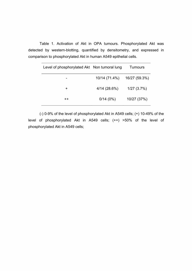
Table 1. Activation of Akt in OPA tumours. Phosphorylated Akt was
detected by western-blotting, quantified by densitometry, and expressed in
comparison to phosphorylated Akt in human A549 epithelial cells.
(-) 0-9% of the level of phosphorylated Akt in A549 cells; (+) 10-49% of the
level of phosphorylated Akt in A549 cells; (++) >50% of the level of
phosphorylated Akt in A549 cells;
Level of phosphorylated Akt Non tumoral lung Tumours
- 10/14 (71.4%) 16/27 (59.3%)
+ 4/14 (28.6%) 1/27 (3.7%)
++ 0/14 (0%) 10/27 (37%)

24
Figure Legends
Figure 1: Activation of telomerase in lung tissues and primary cell cultures
derived from lung tumours and control lungs. Telomerase activity was measured
by a TRAP assay and expressed as the ratio of telomerase activity in sample /
positive control cells (mean ± SEM) in duplicate experiments. (A) Polyacrylamide
gel analysis of PCR products of the TRAP assay in lung tumours and control lung
tissues. Heat-inactivated samples were used as negative controls. T+:
telomerase-positive control cell line. (B) Telomerase activity in OPA and control
lung tissues. (C) Telomerase activity in cell cultures derived from tumours and
control lungs.
Figure 2: Cell proliferation of cultures derived from OPA tumours and
normal lungs (MTT assay).
Figure 3: Disregulation of Akt in cells derived from OPA tumours. (A)
Western blot analysis of phosphorylated-Akt protein (Ser-473) in cell lysates from
cultures derived from OPA lung tumours and normal lungs. Cells were deprived
of serum and growth factors for 24h and exposed or not to stimulation by EGF
(100 ng/ml) for 30 minutes. The amount of total Akt was determined by reprobing
the membranes with an anti-Akt antibody (Cell Signalling). (B) Quantification by
densitometry of phosphorylated and total Akt (mean ± SEM).
Figure 4: Activation of Akt in OPA tumours. (A) Detection by western-blot
of phosphorylated Akt (Ser-473), total Akt and β-actin in lysates of tumoral and
control lung tissues. (B) Quantification by densitometry, expressed as percentage
of phosphorylated Akt in sample compared to EGF-stimulated A549 cells as
reference. Each dot represents the mean of 2 to 3 independent experiments ;
horizontal bars indicate the median value.
25
26
27

28

29